Embed Size (px)
Citation preview
Nanomed J, Vol. 1, No. 2, Winter 2014 107
Received: Jun.3; Accepted: Sep. 21
Vol. 1, No. 2, Winter 2014, page 107-111
Original Research
Online ISSN 2322-5904
http://nmj.mums.ac.ir
Synthesis of new dental nanocomposite with glass nanoparticles
Marzieh Monfared*, Shamseddin Mirdamadi, Alireza Khavandi
School of Metallurgy and Materials Engineering , Iran University of Science and Technology , Tehran, Iran
Abstract
Objective(s): The aim of this study was to synthesis new dental nanocomposites reinforced with
fabricated glass nanoparticles and compare two methods for fabrication and investigate the
effect of this filler on mechanical properties.
Materials and Methods: The glass nanoparticles were produced by wet milling process. The
particle size and shape was achieved using PSA and SEM. Glass nanoparticles surface was
modified with MPTMS silane. The composite was prepared by mixing these silane-treated
nanoparticles with monomers. The resin composition was UDMA /TEGDMA (70/30 weight
ratio). Three composites were developed with 5, 7.5 and 10 wt% glass fillers in each group.
Two preparation methods were used, in dispersion in solvent method (group D) glass
nanoparticles were sonically dispersed in acetone and the solution was added to resin, then
acetone was evaporated. In non-dispersion in solvent method (group N) the glass nanoparticles
were directly added to resin. Mechanical properties were investigated included flexural
strength, flexural modulus and Vickers hardness.
Results: Higher volume of glass nanoparticles improves mechanical properties of composite.
Group D has batter mechanical properties than group N. Flexural strength of composite with
10%w filler of group D was 75Mpa against 59 Mpa of the composite with the same filler
content of group N. The flexural modulus and hardness of group D is more than group N.
Conclusion: It can be concluded that dispersion in solvent method is the best way to fabricate
nanocomposites and glass nanoparticles is a significant filler to improve mechanical properties
of dental nanocomposite.
Keywords: Dental nanocomposites, Dispersion,Glass nanoparticles, Mechanical properties
*Corresponding author: Marzieh Monfared, School of Metallurgy and Materials Engineering , Iran University of
Science and Technology (IUST), Tehran, Iran .
Tel: 09179113964, Email: [email protected]
Dental nanocomposite with glass nanoparticles
Nanomed J, Vol. 1, No. 2, Winter 2014 108
Introduction Dental restorative filling composite resins
have been introduced to dental community in
1960s (1). They have good aesthetics and are
less expensive compared with cast gold and
ceramic materials (2), but still suffer from a
key shortcoming: definition of mechanical
strength (3). Studies have been undertaken to
evaluate and improve restorative composite
resin against wear and fracture (4). A new
approach aiming to achieve improved
performance is related to nanotechnology and
uses inorganic nanofillers such as
nanoparticles to reinforce the polymer matrix.
However, it should be pointed out that the
effect of nanoparticles enhancement is highly
dependent on the size of particles and the level
of their dispersion (2). Nanoparticles can be
produced from the gas or liquid phase, in high
temperature aerosol or plasma reactors or by
the sol–gel route (5). These bottom-up
processes for fabrication of nanoparticles have
one major disadvantage and limiting factor: it
is hardly feasible to synthesize nanoparticles
with complex composition, like glasses,
which contain 5–10 elements (7). A successful
route to achieve nanosized particles of such
materials is high-energy comminution of the
bulk (top-down process). In comminution
processes, stirred media mills are employed
for various industrial applications,
pharmaceuticals, and agrochemicals (5).
Stirred media mills show a higher power
density than dry ball mills, and are generally
more efficient regarding energy requirement
for achievement of nanoscaled powders (7).
It is well known that inorganic nanoparticles
tend to agglomerate and finally result in a poor
dispersion in polymeric matrix. This poor
dispersion makes it difficult to achieve a
stable complex system and the desirable
properties of final products. Therefore, it is
important for nanoparticles reinforced
materials to ensure good dispersion and
stability of particles in the medium (8). Some
efforts, such as the surface modification of
nanoparticles (9) and changing the system pH
and ionic concentration (10), were made to
achieve improved compatibility.
Generally, surface modification is commonly
used to change the surface structure of
particles in order to enhance the compatibility
between the two phases and the dispersion of
particles in organic media (11). The
compatibility and adhesion between the
organic and inorganic phases can be improved
after modification, and the mechanical
strength of composites would likely undergo
significant enhancement by the addition of
nanoparticles in conjunction with coupling
agents (12).
In this work, first the wet comminution of an
amorphous borosilicate glass to achieve
nanoparticles were done and then the two
methods of composite production including
dispersion and non-dispersion in solvent
investigated. The effect of filler content on
mechanical properties of composite was
studied too.
Materials and Methods
Materials UDMA, TEGDMA, CQ, 4-EDMAB and
MPTMS were purchased from Sigma-Aldrich
Co. and used without further purification.
Amorphous borosilicate glass from Schott Co.
with a composition according to table 1 was
used as filler.
Table1. The composition of glass.
Methods Milling process The glass was comminuted in a planetary mill
for 30 hours with the speed of 400rpm.
Hexane was used as the liquid dispersion
medium. The particle size and distribution
were determined by particle size analyzer. Silanization of particles To overcome the agglomeration of particles
and also improve bonding to resin the particles
were silanized with MPTMS. The 20g of glass
particles dispersed in a mixture of 160cc of
ethanol and 5g of MPTMS and then sonicated.
Ethanol was removed in a rotary evaporator
and glass particles washed in a centrifuge by
Components SiO2 B2O3 Na 2O+K2O Al2O3
Weight percent 81 13 4 2
Monfared M, et al
Nanomed J, Vol. 1, No. 2, Winter 2014 109
distilled water. Finally they were dried in the
oven and stored in a desiccator. The particle
size distribution and SEM photos of these
silanized particles were achieved. The particle
size distribution fig. 3 and SEM photos of
these silanized particles were achieved fig. 4.
Fabrication of composites by dispersion
method (Group D) For production composites by dispersion in
solvent method in solvent (group D) modified
glass particles of different fractions (5, 7.5 and
10%w) were dispersed in acetone by
sonication. This solution was added to resin
(70/30 UDMA,TEGDMA in weight ratio) and
sonicated again. The mixture surrounded by
an ice enclosure to inhibit heat build-up. After
the sonication process, the mixture was placed
in the rotary evaporator to remove acetone.
Then CQ and 4EDMAB (0.5/0.5 the weight
ratio) were added in a dark room. Finally the
composite were placed in an ultrasonic bath to
exit air bubbles and better mixing.
Fabrication of composites by non-dispersion
method (Group N) For production composites in non-dispersion
in solvent method (group N). The resin
composition was prepared by
UDMA/TEGDMA, 70/30 in weight ratio,
then various amount of glass particles (5, 7.5,
and 10%w) were added, and mixed with
spatula. Then CQ and 4EDMAB (0.5/0.5 in
weight ratio) were added in a dark room.
Finally the composite were placed in an
ultrasonic bath to exit air bubbles and better
mixing.
Mechanical properties evaluation We define 3 groups of samples with 5, 7.5 and
10wt% fillers in each method denoted D1, D2
and D3 (in dispersion in solvent method) and
N1, N2 and N3 (in non-dispersion in solvent
method) respectively. 3 bar shapes specimens
(25×2×2 mm3) were prepared in a stainless
steel mold to evaluate flexural strength for all
composites. All the samples were light-cured
between two glass slides and stored in distilled
water for 24 hours at 37°C. Then flexural
strength (FS) and flexural elastic modulus (Ef)
of specimens were obtained by three point
bending using universal testing machine with
a span of 20 mm, at 60 N load cell and a
cross-head speed of 0.5 mm/min. calculations
were made using formulas as follows(13):
(1) FS = 3PL/2WT2
(2) Ef = (p/d) (L3/4WT
3)
Where P is the load at fracture, L is the
distance between two supports, W is the width
of the specimen, T is the thickness of the
specimen and d is the deflection at load P. The
fractured samples of flexural test were used
for microhardness test. The microhardness
was evaluated by applying 50 g load for 10 s.
VHN was measured at 8 indentation points per
cross-section and averaged.
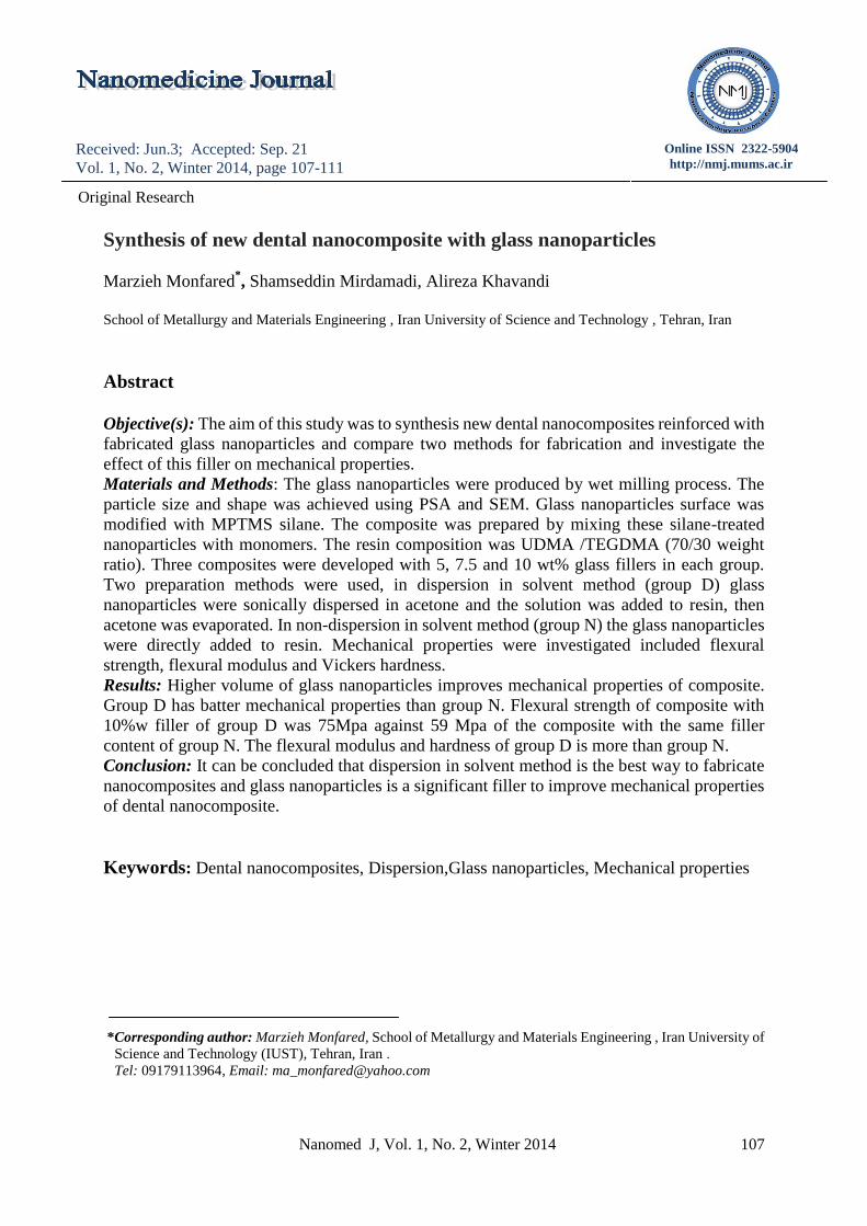
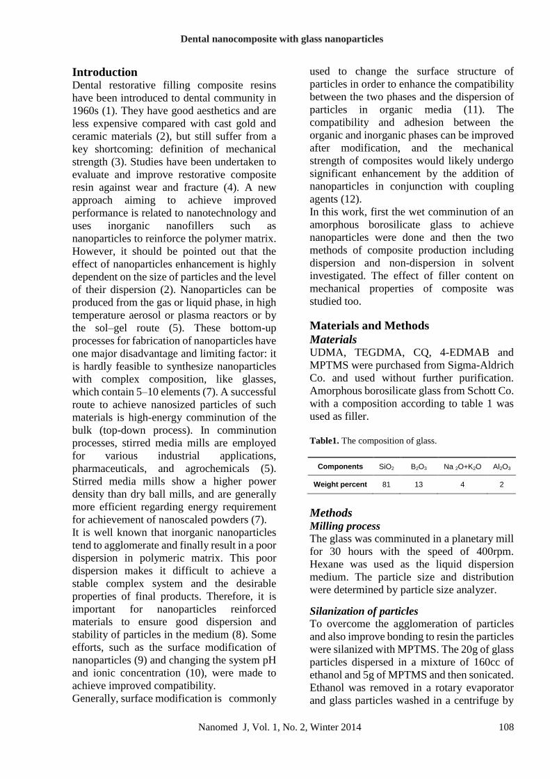
Results and Discussion The size and distribution of glass particles
showed in figure 1. The SEM picture of
fabricated particles showed in figure 2.
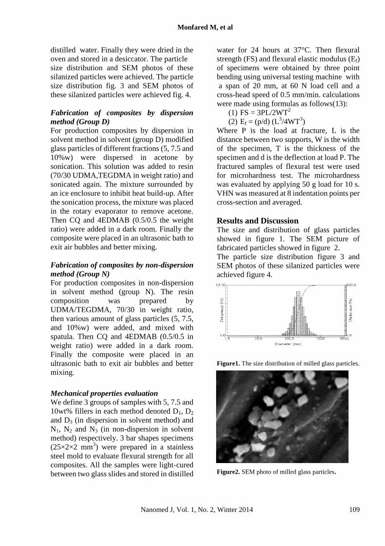
The particle size distribution figure 3 and
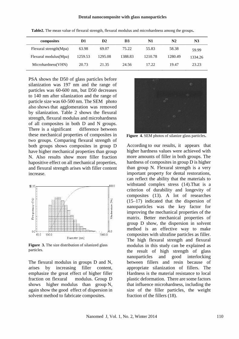
SEM photos of these silanized particles were
achieved figure 4.
Figure1. The size distribution of milled glass particles.
Figure2. SEM photo of milled glass particles.
Dental nanocomposite with glass nanoparticles
Nanomed J, Vol. 1, No. 2, Winter 2014 110
Table2. The mean value of flexural strength, flexural modulus and microhardness among the groups.
PSA shows the D50 of glass particles before
silanization was 197 nm and the range of
particles was 60-600 nm, but D50 decreases
to 140 nm after silanization and the range of
particle size was 60-500 nm. The SEM photo
also shows that agglomeration was removed
by silanization. Table 2 shows the flexural
strength, flexural modulus and microhardness
of all composites in both D and N groups.
There is a significant difference between
these mechanical properties of composites in
two groups. Comparing flexural strength of
both groups shows composites in group D
have higher mechanical properties than group
N. Also results show more filler fraction
hapositive effect on all mechanical properties,
and flexural strength arises with filler content
increase.
Figure 3. The size distribution of silanized glass
particles.
The flexural modulus in groups D and N,
arises by increasing filler content,
emphasize the great effect of higher filler
fraction on flexural modulus. Group D
shows higher modulus than group N,
again show the good effect of dispersion in
solvent method to fabricate composites.
Figure 4. SEM photos of silanize glass particles.
According to our results, it appears that
higher hardness values were achieved with
more amounts of filler in both groups. The
hardness of composites in group D is higher
than group N. Flexural strength is a very
important property for dental restorations,
can reflect the ability that the materials to
withstand complex stress (14).That is a
criterion of durability and longevity of
composites (13). A lot of researches
(15–17) indicated that the dispersion of
nanoparticles was the key factor for
improving the mechanical properties of the
matrix. Better mechanical properties of
group D show, the dispersion in solvent
method is an effective way to make
composites with ultrafine particles as filler.
The high flexural strength and flexural
modulus in this study can be explained as
the result of high strength of glass
nanoparticles and good interlocking
between fillers and resin because of
appropriate silanization of fillers. The
Hardness is the material resistance to local
plastic deformation. There are some factors
that influence microhardness, including the
size of the filler particles, the weight
fraction of the fillers (18).
composites D1 D2 D3 N1 N2 N3
Flexural strength(Mpa) 63.98 69.07 75.22 55.83 58.38 59.99
Flexural modulus(Mpa) 1259.53 1295.08 1388.83 1210.78 1280.49
1334.26
Microhardness(VHN) 20.73 21.35 24.56 17.22 19.47 23.23
Monfared M, et al
Nanomed J, Vol. 1, No. 2, Winter 2014 111
The good hardness of prepared composites
is a result of high hardness of glass
nanoparticles.Higher hardness of
composite in group D is the results of
fabrication method by dispersion, that
distribute ultrafine particles more
effectively and the particles can act as a
good reinforced parameter in smaller size
than agglomerate.
Conclusion Glass nanoparticles were produced by wet
grinding in a planetary mill. Dispersion in
solvent and non-dispersion in solvent
methods were used to fabricate composite.
The results showed, by increasing filler
content flexural strength, flexural modulus
and microhardness arise in both groups.
The mechanical properties of composites in
group D are significantly higher than group
N. It can be concluded that dispersion in
solvent method is the best way to fabricate
nanocomposites and glass nanoparticles is a
significant filler to improve mechanical
properties of dental nanocomposite.
Acknowledgments This work was funded by the Iran
University of Science and Technology,
Tehran, Iran.
References 1. Bowen RL. Properties of a
silica-reinforced polymer for dental
restorations. J Am Dent Assoc. 1963; 66:
57-64.
2. Du M, Zheng Y. Modification of silica
nanoparticles and their application in
UDMA dental polymeric composites.
Polym Composite. 2007; 28: 198-207
3. Garoushi S, Vallittu PK, Watts DC, Lassila
LVJ. Effect of nanofiller fractions and
temperature on polymerization shrinkage
on glass fiber reinforced filling material.
Dent Mater. 2008; 24: 606-610.
4. Garoushi S, Vallittu PK, Lassila LVJ. Short glass fiber reinforced restorative
composite resin with semi-inter penetrating
polymer network matrix. Dent Mater. 2007;
23: 1356-1362.
5. Vitala A, Zürchera S, Dittmanna R,
Trottmannb M, Lienemannb P, Bommera
B, Graulea T, Apel E and Höland W.
Ultrafine comminution of dental glass in a
stirred media mill. Chem Eng Sci. 2008; 63:
484-494.
6. Tooley FV. The Handbook of Glass
Manufacture. New York: Ashlee
Publishing; 1984.
7. Mende S, Stenger F, Peukert W, Schwedes
J. Mechanical production and stabilization
of submicron particles in stirred media
mills. Powder Technol. 2003; 132: 64-73.
8. Verwey EJ, Overbeek JTG. Theory of
Stability of Lyophobic Colloids. New York:
Dover Publications; 2000.
9. Wang H, Zhu M, Li Y, Zhan Q. Mechanical
properties of dental resin composites by
co-filling diatomite and nanosized silica
particles. Mater Sci Eng. 2011; C 31:
600-605.
10. Widegren J, Bergstrom L. Electrostatic
Stabilization of Ultrafine Titania in Ethanol.
J Am Ceram Soc. 2002; 85: 523-528.
11. Adler JJ, Singh PK, Patist A, Rabinovich
YL, Shah DO, Moudgil BM. Correlation of
particulate dispersion stability with the
strength of self-assembled surfactant films.
Langmuir. 2000; 18: 7255-7262.
12. Moszner N, Salz U. New developments of
polymeric dental composites. Prog Polym
Sci. 2001; 26: 535-576.
13. Zandinejad AA, Atai M, Pahlevan A. The
effect of ceramic and porous fillers on the
mechanical properties of
experimental dental composites. Dent
Mater. 2006; 22: 382.


![Nanocomposite [5]](https://img.dokumen.tips/doc/110x75/577c7ecf1a28abe054a26499/nanocomposite-5.jpg)

