Embed Size (px)
Citation preview

Reoperative Pediatric Surgery

Reoperative Pediatric
Surgery
Edited by
Steven Teich, md
Clinical Assistant Professor of SurgeryOhio State University College of MedicineSurgical Director, Neonatal Intensive Care UnitNationwide Children’s HospitalColumbus, Ohio
Donna A. Caniano, md
Professor of Surgery and PediatricsOhio State University College of MedicineSurgeon-in-ChiefNationwide Children’s HospitalColumbus, Ohio

EditorsSteven Teich Donna A. CanianoDepartment of Pediatric Surgery Department of Pediatric SurgeryNationwide Children’s Hospital Nationwide Children’s Hospital700 Children’s Drive – ED323 700 Children’s Drive – ED379Columbus, Ohio 43205 Columbus, Ohio [email protected] [email protected]
ISBN: 978-1-58829-761-7 e-ISBN: 978-1-60327-071-7
Library of Congress Control Number: 2007936724
© 2008 Humana Press, a part of Springer Science+Business Media, LLCAll rights reserved. This work may not be translated or copied in whole or in part without the written permission ofthe publisher (Humana Press, 999 Riverview Drive, Suite 208, Totowa, NJ 07512 USA), except for brief excerpts inconnection with reviews or scholarly analysis. Use in connection with any form of information storage and retrieval,electronic adaptation, computer software, or by similar or dissimilar methodology now known or hereafter developedis forbidden.The use in this publication of trade names, trademarks, service marks, and similar terms, even if they are not identifiedas such, is not to be taken as an expression of opinion as to whether or not they are subject to proprietary rights.While the advice and information in this book are believed to be true and accurate at the date of going to press,neither the authors nor the editors nor the publisher can accept any legal responsibility for any errors or omissions thatmay be made. The publisher makes no warranty, express or implied, with respect to the material contained herein.
Cover illustration: The picture on the front cover depicts the two authors performing surgery together. We wish tothank Dr. Jon Groner for his photographic expertise.
Printed on acid-free paper
9 8 7 6 5 4 3 2 1
springer.com

We are inspired by our patients and their families, who place their trust in our surgicalexpertise and wisdom. During our training in pediatric surgery we were fortunateto have witnessed commitment to long term patient care by our esteemed teachers,Drs. H. William Clatworthy, Jr., E. Thomas Boles, Jr., and Marc I. Rowe.
We dedicate this book to our respective parents, Pauline and Abraham Teich and Maryand James Caniano, who taught us that we could accomplish anything through hardwork and perseverance.
We also dedicate this book to our respective spouses, Esther Chipps and RichardFlores, who are our closest friends, wisest advisors, and sources of daily strength.
Steven Teich, MD
Donna A. Caniano, MD
Preface
“Good judgment comes from experience, andoften experience comes from poor judgment.”
Rita Mae Brown
Reoperative surgery is a challenge that is confronted by every surgeon. Althougha particular operation may be initially performed with technical skill and followedby appropriate postoperative care, functional and/or anatomic problems may requirefurther surgical attention.
The unique circumstances of pediatric patients may predispose them to a greaterlikelihood of requiring reoperation after a major procedure.
• A bowel resection in a neonate may develop a stricture if the anastamosis does not growat the same rate as the adjacent bowel. The reoperative anastamotic technique is critical,as is the decision whether to resect or taper dilated bowel.
• The cancer survival rate has increased dramatically for many pediatric tumors. Thesepatients often require reoperation for treatment of recurrences, as well as for treatmentof complications of chemotherapy, such as second malignancies.
• Pediatric surgical patients often require lifelong follow-up that is obviously much longerthan for adults. This increases the chances of requiring reoperation for many conditions,including gastroesophageal reflux disease and inguinal hernia.
• Even a “simple” gastrostomy may develop complications related to growth. With lineargrowth, the skin of the abdominal wall often migrates towards the chest wall. Therefore,the gastrostomy becomes angulated with leakage of gastric contents onto the abdominalwall, necessitating repositioning of the gastrostomy away from the costal margin.
• Pediatric patients with congenital diseases, such as cystic fibrosis, often require multiplereoperations for complications related to their underlying condition.
It is important to mention that not every pediatric surgery reoperative problem hasa wealth of contemporary literature. Often reoperative surgery requires seldom usedand more complex operative techniques. Frequently, these techniques are too new ortoo specialized to be found in current pediatric surgery textbooks. For this reason, wehave enlisted a group of authors who are recognized experts for their respective topicsto provide the most up-to-date information on reoperations for their pediatric surgicalcolleagues.
The pediatric surgery literature on reoperations is fragmented and sketchy. Theneed for a pediatric surgery textbook that critically analyzes and consolidates all theavailable literature on reoperations is obvious. For this reason, we have compiled adetailed source of information on reoperations for all areas of the body, all parts ofthe gastrointestinal tract, all types of pediatric solid tumors, and many common butperplexing problems that we co-manage with other pediatric specialists.
vii

viii Preface
This book has been a labor of love. Now, we hope that it will become avaluable reference for pediatric surgeons, pediatric anesthesiologists, general surgeonsperforming pediatric surgery, and all pediatric physicians.
We wish to thank our secretaries, Cathy Rings and Teresa Rodich, for their invaluableassistance in the preparation of this book.
Steven Teich, MD
Donna A. Caniano, MD

Contents
Preface . . . . . . . . . . . . . . . . . . . . . . . . . . . . . . . . . . . . . . . . . . . . . . . . . . . . . . . . . . . . . . . . . . . . vii
Contributors . . . . . . . . . . . . . . . . . . . . . . . . . . . . . . . . . . . . . . . . . . . . . . . . . . . . . . . . . . . . . . . xiii
SECTION I: GENERAL CONSIDERATIONS
1. Radiology of the Postoperative Patient . . . . . . . . . . . . . . . . . . . . . . . . . . . . . . . . . . . 1William E. Shiels, II, D. Gregory Bates, and Mark J. Hogan
2. Techniques for Difficult Venous and Arterial Access . . . . . . . . . . . . . . . . . . . . . . . 75Jaimie D. Nathan, John M. Racadio, and Brad W. Warner
SECTION II: HEAD AND NECK
3. Reoperative Head and Neck Surgery . . . . . . . . . . . . . . . . . . . . . . . . . . . . . . . . . . . . . 91Bradley M. Rodgers
4. Reoperative Thyroid and Parathyroid Surgery . . . . . . . . . . . . . . . . . . . . . . . . . . . . . 103Elisabeth Tracy and Michael A. Skinner
SECTION III: CHEST
5. Complications of Pectus Excavatum and Carinatum Repair . . . . . . . . . . . . . . . . . 119Robert E. Kelly, Jr. and Donald Nuss
6. Complications of CDH Repair and Recurrent CDH. . . . . . . . . . . . . . . . . . . . . . . 139Melissa L. Hayward, Dario O. Fauza, and Jay M. Wilson
7. Reoperation for Jeune’s Syndrome . . . . . . . . . . . . . . . . . . . . . . . . . . . . . . . . . . . . . . . 157J. Terrance Davis, Alistair B. M. Phillips, Frederick R. Long, and Robert G. Castile
8. Reoperation for Benign Breast Disorder . . . . . . . . . . . . . . . . . . . . . . . . . . . . . . . . . . 169Denise B. Klinkner and Marjorie J. Arca
SECTION IV: ABDOMINAL WALL
9. Abdominal Wall Disruption . . . . . . . . . . . . . . . . . . . . . . . . . . . . . . . . . . . . . . . . . . . . 175Steven Teich and Donna A. Caniano
ix
x Contents
SECTION V: GASTROINTESTINAL TRACT
10. Reoperation after Esophageal Atresia Repair and Other EsophagealConditions . . . . . . . . . . . . . . . . . . . . . . . . . . . . . . . . . . . . . . . . . . . . . . . . . . . . . . . . . . . . 191Brian D. Kenney
11. Reoperation after Failed Fundoplication and Other Gastric Operations . . . . . . 207Mary Brindle and Jacob C. Langer
12. Reoperation after Duodenal Atresia Repair and Managementof Duodenal Fistulas . . . . . . . . . . . . . . . . . . . . . . . . . . . . . . . . . . . . . . . . . . . . . . . . . . . 219Jonathan I. Groner
13. Reoperation after Surgery of the Small Bowel . . . . . . . . . . . . . . . . . . . . . . . . . . . . . 225Moritz M. Ziegler and John Petty
14. Interventions for Appendiceal Complications . . . . . . . . . . . . . . . . . . . . . . . . . . . . . . 241Renata Fabia and Steven Teich
15. Reoperation for Inflammatory Bowel Disease . . . . . . . . . . . . . . . . . . . . . . . . . . . . . 257Christopher R. Moir
16. Reoperation for Stoma Complications . . . . . . . . . . . . . . . . . . . . . . . . . . . . . . . . . . . . 279Benedict C. Nwomeh
17. Reoperative Surgery for Hirschsprung Disease . . . . . . . . . . . . . . . . . . . . . . . . . . . . . 287Mohamed I. El-sawaf, Arnold G. Coran, and Daniel H. Teitelbaum
18. Reoperative Surgery for Anorectal Malformations . . . . . . . . . . . . . . . . . . . . . . . . . . 311Marc A. Levitt and Alberto Peña
19. Reoperation for Recurrent Anal and Perianal Conditions . . . . . . . . . . . . . . . . . . . 327Shahab Abdessalam and Donna A. Caniano
SECTION VI: SOLID ORGANS
20. Reoperation of the Liver and Biliary System . . . . . . . . . . . . . . . . . . . . . . . . . . . . . . 337Jaimie D. Nathan and Frederick C. Ryckman
21. Reoperation of the Pancreas . . . . . . . . . . . . . . . . . . . . . . . . . . . . . . . . . . . . . . . . . . . . . 367Eunice Y. Huang, Max R. Langham, Jr. and Timothy C. Fabian
SECTION VII: REOPERATIVE ONCOLOGY
22. Rhabdomyosarcoma . . . . . . . . . . . . . . . . . . . . . . . . . . . . . . . . . . . . . . . . . . . . . . . . . . . . 385David Rodeberg and Eugene S. Wiener

Contents xi
23. Neuroblastoma . . . . . . . . . . . . . . . . . . . . . . . . . . . . . . . . . . . . . . . . . . . . . . . . . . . . . . . . 397John B. Hamner, Andrew M. Davidoff, and Stephen J. Shochat
24. Wilms’ Tumor . . . . . . . . . . . . . . . . . . . . . . . . . . . . . . . . . . . . . . . . . . . . . . . . . . . . . . . . 411Sanjeev A. Vasudevan and Jed G. Nuchtern
25. Peripheral Neuroectodermal Tumors . . . . . . . . . . . . . . . . . . . . . . . . . . . . . . . . . . . . . 427Michael P. La Quaglia
26. Osteosarcoma Metastases . . . . . . . . . . . . . . . . . . . . . . . . . . . . . . . . . . . . . . . . . . . . . . . 435Matthew T. Harting, Richard J. Andrassy, and Andrea Hayes-Jordan
27. Neurofibromatosis . . . . . . . . . . . . . . . . . . . . . . . . . . . . . . . . . . . . . . . . . . . . . . . . . . . . . 447Ravi S. Radhakrishnan and Richard J. Andrassy
28. Tumors of the Liver . . . . . . . . . . . . . . . . . . . . . . . . . . . . . . . . . . . . . . . . . . . . . . . . . . . 459Rebecka L. Meyers
29. Sacrococcygeal Teratoma . . . . . . . . . . . . . . . . . . . . . . . . . . . . . . . . . . . . . . . . . . . . . . . 467Antoine De Backer and Frans W. J. Hazebroek
SECTION VIII: REOPERATION OF THE GENITOURINARY TRACT
30. Recurrent Hernia, Hydrocele, and Varicocele . . . . . . . . . . . . . . . . . . . . . . . . . . . . . 475Robert E. Cilley, Brett W. Engbrecht, and Andreas H. Meier
31. Failed Orchidopexy and Complications of Circumcision . . . . . . . . . . . . . . . . . . . 487Seth A. Alpert, Stephen A. Koff, and Venkata R. Jayanthi
32. Complications of Vaginoplasty and Clitoroplasty . . . . . . . . . . . . . . . . . . . . . . . . . . 499Lesley L. Breech
SECTION IX: OTHER REOPERATIVE PROBLEMS FOR PEDIATRICSURGEONS
33. Abdominal Complications of Ventricular-Peritoneal Shunts . . . . . . . . . . . . . . . . . 515Benedict C. Nwomeh and Scott Elton
34. Revisional Bariatric Surgery in Adolescents . . . . . . . . . . . . . . . . . . . . . . . . . . . . . . . . 525Marc Michalsky, Steven Teich, and Bradley J. Needleman
Index . . . . . . . . . . . . . . . . . . . . . . . . . . . . . . . . . . . . . . . . . . . . . . . . . . . . . . . . . . . . . . . . . . . . . 539
Contributors
Shahab Abdessalam, md • Assistant Professor of Surgery, University of NebraskaSchool of Medicine, Attending Pediatric Surgeon, Children’s Hospital of Omaha,Omaha, Nebraska
Seth A. Alpert, md • Clinical Assistant Professor, Department of Urology, OhioState University College of Medicine, Attending Pediatric Urologist, NationwideChildren’s Hospital, Columbus, Ohio
Richard J. Andrassy, md • Professor of Surgical Oncology, University of TexasHouston Medical School, Houston, Texas
Marjorie J. Arca, md • Assistant Professor of Surgery, Medical College ofWisconsin, Attending Pediatric Surgeon, Children’s Hospital of Wisconsin,Milwaukee, Wisconsin
D. Gregory Bates, md • Clinical Assistant Professor of Radiology, Ohio StateUniversity College of Medicine, Section Chief, Fluoroscopy, Gastrointestinal &Genitourinary Radiology, Department of Radiology, Nationwide Children’sHospital, Columbus, Ohio
Lesley L. Breech, md • Associate Professor of Obstetrics and Gynecology,University of Cincinnati College of Medicine, Attending Pediatric Gynecologist,Cincinnati Children’s Hospital Medical Center, Cincinnati, Ohio
Mary Brindle, md • Assistant Professor of Surgery, University of Alberta, StaffSurgeon, Alberta Children’s Hospital, Calgary, Alberta, Canada
Donna A. Caniano, md • H. William Clatworthy Professorship in PediatricSurgery, Professor of Surgery and Pediatrics, Ohio State University College ofMedicine, Surgeon-in-Chief, Nationwide Children’s Hospital, Columbus, Ohio
Robert G. Castile, md, ms • Professor of Pediatrics, Ohio State University Collegeof Medicine, Center for Perinatal Research, Nationwide Children’s Hospital,Columbus, Ohio
Robert E. Cilley, md • Professor of Surgery and Pediatrics, Penn State College ofMedicine, Chief, Division of Pediatric Surgery, Penn State Children’s Hospital,Milton S. Hershey Medical Center, Hershey, Pennsylvania
Arnold G. Coran, md • Professor of Surgery, University of Michigan School ofMedicine, Attending Pediatric Surgeon, C.S. Mott Children’s Hospital, Ann Arbor,Michigan
Andrew M. Davidoff, md • Associate Professor of Surgery and Pediatrics,University of Tennessee Health Science Center, Chief, Division of GeneralPediatric Surgery, St. Jude Children’s Research Hospital, Memphis, Tennessee
J. Terrance Davis, md • Professor Emeritus of Clinical Surgery, Ohio StateUniversity College of Medicine, Interim Medical Director, Nationwide Children’sHospital, Columbus, Ohio
xiii
xiv Contributors
Antoine DeBacker, md, phd • Professor of Pediatric Surgery, Free University ofBrussels, Head of Department of Pediatric Surgery, Academic Hospital of the FreeUniversity of Brussels, Brussels, Belgium
Scott W. Elton, md • Clinical Auxiliary Faculty, Department of Neurosurgery,Ohio State University College of Medicine, Attending Pediatric Neurosurgeon,Nationwide Children’s Hospital, Columbus, Ohio
Moharned El-Sawaf, b.s., University of Michigan, Department of PediatricSurgery, C.S. Mott Children’s Hospital, Ann Arbor, Michigan
Brett W. Engbrecht, md • Assistant Professor of Surgery, Penn State College ofMedicine, Attending Pediatric Surgeon, Penn State Children’s Hospital, Milton S.Hershey Medical Center, Hershey, Pennsylvania.
Renata B. Fabia, md • Clinical Assistant Professor of Surgery, Ohio StateUniversity College of Medicine, Attending Surgeon, Nationwide Children’sHospital, Columbus, Ohio
Timothy C. Fabian, md • Harwell Wilson Alumni Professor and Chairman,Department of Surgery, University of Tennessee Health Science Center, AttendingSurgeon, Regional Medical Center at Memphis, Memphis, Tennessee
Dario O. Fauza, md • Assistant Professor of Surgery, Harvard Medical School,Associate in Surgery, Children’s Hospital Boston, Boston, Massachusetts
Jonathan I. Groner, md • Professor of Clinical Surgery and Pediatrics, Ohio StateUniversity College of Medicine, Trauma Medical Director, Nationwide Children’sHospital
John B. Hamner, md • General Surgery Resident, St. Jude Children’s ResearchHospital, University of Tennessee Health Science Center, Memphis, Tennessee
Matthew T. Harting, md • General Surgery Resident, University of Texas MedicalSchool at Houston, Houston, Texas
Melissa Hawyard, md • Surgical Research Fellow, Children’s Hospital Boston,Boston, Massachusetts
Andrea Hayes-Jordan, md • Assistant Professor of Surgery and Pediatrics,University of Texas Medical School at Houston, Attending Pediatric Surgeon,Texas Health Science Center at Houston and MD Anderson Cancer Center,Houston, Texas
Frans W. J. Hazebroek, md, PhD • Professor Emeritus of Surgery, ErasmusMC-Sophia Children’s Hospital, Rotterdam, the Netherlands
Mark J. Hogan, md • Clinical Associate Professor of Radiology, Ohio StateUniversity College of Medicine, Section Chief of Vascular and InterventionalRadiology, Department of Radiology, Nationwide Children’s Hospital, Columbus,Ohio
Eunice Y. Huang, md • Assistant Professor of Surgery, University of TennesseeHealth Science Center, Attending Pediatric Surgeon, Le Bonheur Children’sMedical Center, Memphis, Tennessee
V. Rama Jayanthi, md • Clinical Assistant Professor, Department of Urology, OhioState University College of Medicine, Attending Pediatric Urologist, NationwideChildren’s Hospital, Columbus, Ohio
Robert E. Kelly, Jr., md • Associate Professor of Clinical Surgery and Pediatrics,Eastern Virginia Medical School, Chief of Department of Surgery, Children’sHospital of The King’s Daughters, Norfolk, Virginia
Contributors xv
Brian D. Kenney, md • Assistant Professor of Clinical Surgery, Ohio StateUniversity College of Medicine, Attending Pediatric Surgeon, NationwideChildren’s Hospital, Columbus, Ohio
Denise B. Klinkner, md • Surgical Research Fellow, Department of Surgery,Division of Pediatric Surgery, Children’s Hospital of Wisconsin, Milwaukee,Wisconsin
Stephen A. Koff, md • Professor, Department of Urology, Ohio State UniversityCollege of Medicine, Chief, Section of Pediatric Urology, Nationwide Children’sHospital, Columbus, Ohio
Jacob C. Langer, md • Professor of Surgery, University of Toronto, Robert M.Filler Chair and Chief, Division of General Surgery, Hospital for Sick Children,Toronto, Ontario, Canada
Max R. Langham, Jr., md • Professor of Surgery and Pediatrics, University ofTennessee Health Science Center, Chief, Division of Pediatric Surgery, Le BonheurChildren’s Medical Center, Memphis, Tennessee
Michael P. LaQuaglia, md • Professor of Surgery, Weill Cornell UniversityMedical School, Chief, Pediatric Surgical Service, Memorial Sloan-KetteringCancer Center, New York, New York
Marc A. Levitt, md • Associate Professor of Surgery, University of CincinnatiCollege of Medicine, Associate Director, Colorectal Center for Children,Cincinnati Children’s Hospital Medical Center, Cincinnati, Ohio
Frederick R. Long, md • Clinical Professor of Radiology, Ohio State UniversityCollege of Medicine, Section Chief, Body CT and MRI Imaging, Department ofRadiology, Nationwide Children’s Hospital, Columbus, Ohio
Andreas H. Meier, md • Assistant Professor of Surgery, Penn State College ofMedicine, Attending Pediatric Surgeon, Penn State Children’s Hospital, Milton S.Hershey Medical Center, Hershey, Pennsylvania
Rebecka L. Meyers, md • Professor of Surgery, University of Utah, Chief, Divisionof Pediatric Surgery, Primary Children’s Medical Center, Salt Lake, Utah
Marc P. Michalsky, md • Assistant Professor of Clinical Surgery, Ohio StateUniversity College of Medicine, Surgical Director, Center for Healthy Weight andNutrition, Nationwide Children’s Hospital, Columbus, Ohio
Christopher R. Moir, md • Associate Professor of Surgery, Mayo Clinic College ofMedicine, Consultant, Division of Pediatric Surgery, Mayo Clinic, Rochester,Minnesota
Jaimie Nathan, md • Senior Clinical Fellow, Division of Pediatric and ThoracicSurgery, Cincinnati Children’s Hospital Medical Center, Cincinnati, Ohio
Bradley J. Needleman, md • Assistant Professor of Surgery, Ohio State UniversityCollege of Medicine, Director of Bariatric Surgery, Center for Minimally InvasiveSurgery, Ohio State University Medical Center, Columbus, Ohio
Jed G. Nuchtern, md • Professor of Surgery and Pediatrics, Baylor College ofMedicine, Attending Surgeon, Texas Children’s Hospital, Houston, Texas
Benedict C. Nwomeh, md • Assistant Professor of Clinical Surgery, Ohio StateUniversity College of Medicine, Attending Pediatric Surgeon, NationwideChildren’s Hospital, Columbus, Ohio
Alberto Pena, md • Professor of Surgery, University of Cincinnati College ofMedicine, Director, Colorectal Center for Children, Cincinnati Children’s HospitalMedical Center, Cincinnati, Ohio

xvi Contributors
Alistair B. M. Phillips, md • Assistant Professor of Surgery, Ohio State UniversityCollege of Medicine, Attending Pediatric Cardiothoracic Surgeon, NationwideChildren’s Hospital, Columbus, Ohio
John M. Racadio, md • Associate Professor of Clinical Radiology and Pediatrics,University of Cincinnati, Division Chief, Interventional Radiology, CincinnatiChildren’s Hospital Medical Center, Cincinnati, Ohio
Ravi S. Radhakrishnan, md • General Surgery Resident, University ofTexas-Houston Medical School, Memorial Hermann Children’s Hospital, Houston,Texas
David A. Rodeberg, md • Assistant Professor of Surgery, University of PittsburghSchool of Medicine, Attending Pediatric Surgeon, Children’s Hospital ofPittsburgh, Pittsburgh, Pennsylvania
Bradley M. Rodgers, md • Maurice L. LeBauer Professor of Surgery, University ofVirginia Health System, Division Head, Division of Pediatric Surgery, Children’sMedical Center, Charlottesville, Virginia
Frederick C. Ryckman, md • Professor of Surgery, University of Cincinnati Collegeof Medicine, Director, Liver Transplant, Surgical Director, Intestinal TransplantSurgery, Cincinnati Children’s Hospital Medical Center, Cincinnati, Ohio
William E. Shiels, II, do • Clinical Professor of Radiology, Pediatrics, andBiomedical Engineering, Ohio State University College of Medicine, Chairman,Department of Radiology, Nationwide Children’s Hospital, Columbus, Ohio
Stephen J. Shochat, md • Professor of Surgery and Pediatrics, University ofTennessee Health Science Center, Surgeon-in-Chief and Chair of Department ofSurgery, St. Jude Children’s Research Hospital, Memphis, Tennessee
Michael A. Skinner, md • Edwin Ide Smith, MD, Professor of Pediatric Surgery,The University of Texas Medical School, Vice Chairman, Department of PediatricSurgery, Children’s Medical Center, Dallas, Texas
Elisabeth Tracy, md • Senior Assistant Resident, Duke University, Department ofSurgery, Durham, North Carolina
Daniel H. Teitelbaum, md • Professor of Surgery, The University of MichiganSchool of Medicine, Attending Pediatric Surgeon, C.S. Mott Children’s Hospital,Ann Arbor, Michigan
Steven Teich, md • Clinical Assistant Professor of Surgery, Ohio State UniversityCollege of Medicine, Attending Pediatric Surgeon, Nationwide Children’s Hospital,Columbus, Ohio
Sanjeev A. Vasudevan, md • General Surgery Resident, Michael E. DeBakeyDepartment of Surgery, Baylor College of Medicine, Houston, Texas
Brad W. Warner, md • Apolline Blair Professor, Washington University School ofMedicine, Chief Division of Pediatric Surgery, Surgeon-in-Chief, St. LouisChildren’s Hospital, St. Louis, Missouri
Eugene S. Wiener, md • Deceased, Medical Director, Children’s Hospital ofPittsburgh, Pittsburgh, Pennsylvania
Jay M. Wilson, md • Associate Professor of Surgery, Harvard Medical School,Director of Surgical Critical Care, Children’s Hospital, Boston, Massachusetts
Moritz M. Ziegler, md • Professor of Surgery, University of Colorado School ofMedicine, Surgeon-in-Chief, The Children’s Hospital, Denver, Colorado

1 Radiology of the PostoperativePatientDiagnosis and Intervention
William E. Shiels, II, DO, D. GregoryBates, MD, and Mark J. Hogan, MD
CONTENTS
Introduction
Abscess of the Chest and Abdomen
Intestinal Fistula
Small Bowel Obstruction (SBO) and Ileus
Esophageal Disorders
Stomach
Hepatobiliary Diagnosis and Intervention
Deep Venous Thrombosis (DVT) and Pulmonary Embolus
Thoracic Management Issues
Displaced Venous Catheter
Venous and Arterial Thrombosis
Reoperative Treatment of Vascular Anomalies
Tumor Ablation and Embolization
Vascular Injury
References
INTRODUCTION
The integrated and effective use of radiological diagnostic modalities andinterventional techniques and therapies provides the surgeon with the opportunityto make accurate diagnoses of reoperative issues and complications and to providetimely intervention. A close functional relationship between surgeons and radiologistsallows the surgeon the full advantage of surgical therapies or radiological interven-tional techniques and therapies, as best suits the individual patient needs. Interventionalradiology offers a multimodality image-guided and minimally invasive managementapproach to a multitude of reoperative issues and complications. Consultation between
From: Reoperative Pediatric SurgeryEdited by: S. Teich and D. A. Caniano © Humana Press, Totowa, NJ
1

2 Shiels II et al.
the primary surgeon and the radiology team provides discussion of techniques, inter-ventional therapeutic options, expected outcomes, and contingency plans. The surgeonmust have a clear understanding of the contrast media used, anatomic approachesof interventional procedures, and associated potential complications, should operativeintervention be required following radiological diagnosis and/or intervention.
ABSCESS OF THE CHEST AND ABDOMEN
Abdominal abscesses are the most common indication for image-guided drainage,and appendicitis is the most common etiology (1–5). Appendicitis is more commonin children than adults, and children are more likely to have perforated appendicitisand abscesses (1–3,5–7). Percutaneous drainage combined with antibiotics may allowfor delayed less-invasive surgery (laparoscopic or small right lower quadrant [RLQ]incision) in children who present with rupture and abscess (7–14). Alternatively, thedrainage of postoperative abscesses can eliminate a second surgery (2–7). Other causesof intraabdominal abscesses are less common, but include infected cerebrospinal fluid(CSF) and pancreatic pseudocysts, necrotizing enterocolitis (NEC), Crohn’s disease,and postoperative abscesses of any cause.
The imaging techniques are dependent on the suspected site of abscess. AlthoughCT scanning is most commonly used for abdominal sepsis (Fig. 1), magnetic resonanceimaging (MRI) or ultrasound may be more useful in the musculoskeletal system orwith superficial lesions (Fig. 2).
After obtaining appropriate history, physical examination, laboratory tests, andabdominal plain radiographs, abdominal computed tomography (CT) scan has becomethe gold standard for the diagnosis of abdominal abscess (1,3,4). If the patient’s
Fig. 1. CT scan of a febrile patient after surgery for ruptured appendicitis shows an abscess (*) nearthe gallbladder fossa.
Chapter 1 / Radiology of the Postoperative Patient 3
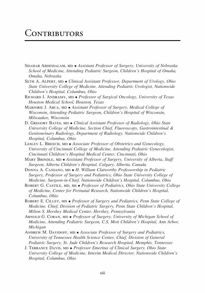
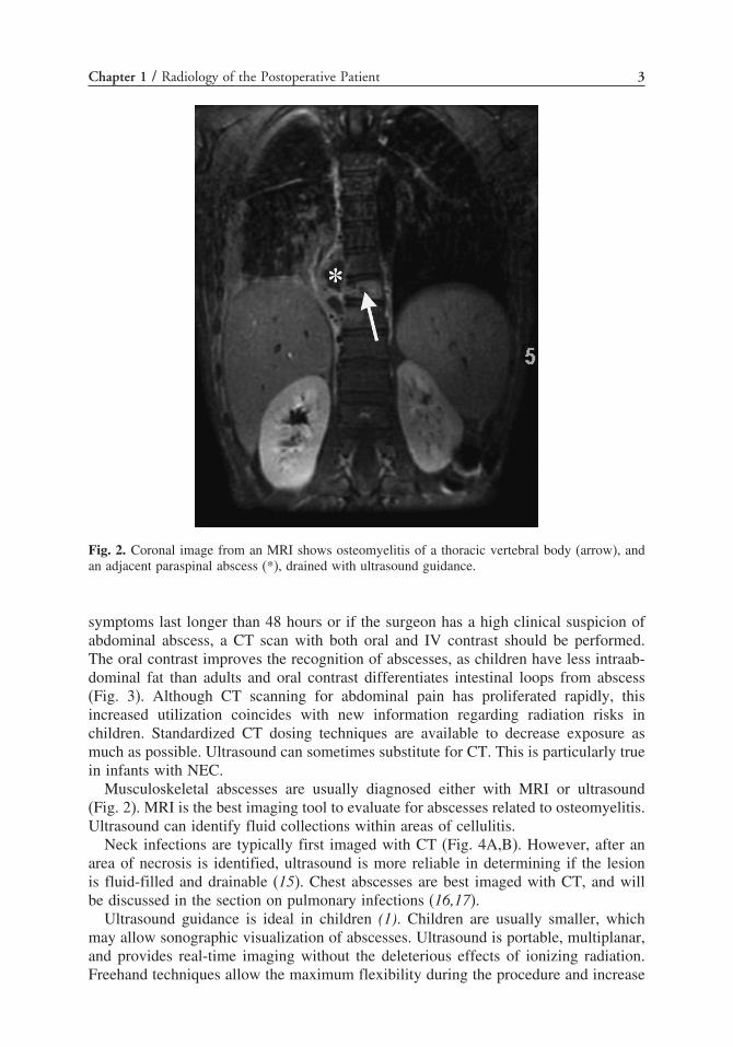
Fig. 2. Coronal image from an MRI shows osteomyelitis of a thoracic vertebral body (arrow), andan adjacent paraspinal abscess (*), drained with ultrasound guidance.
symptoms last longer than 48 hours or if the surgeon has a high clinical suspicion ofabdominal abscess, a CT scan with both oral and IV contrast should be performed.The oral contrast improves the recognition of abscesses, as children have less intraab-dominal fat than adults and oral contrast differentiates intestinal loops from abscess(Fig. 3). Although CT scanning for abdominal pain has proliferated rapidly, thisincreased utilization coincides with new information regarding radiation risks inchildren. Standardized CT dosing techniques are available to decrease exposure asmuch as possible. Ultrasound can sometimes substitute for CT. This is particularly truein infants with NEC.
Musculoskeletal abscesses are usually diagnosed either with MRI or ultrasound(Fig. 2). MRI is the best imaging tool to evaluate for abscesses related to osteomyelitis.Ultrasound can identify fluid collections within areas of cellulitis.
Neck infections are typically first imaged with CT (Fig. 4A,B). However, after anarea of necrosis is identified, ultrasound is more reliable in determining if the lesionis fluid-filled and drainable (15). Chest abscesses are best imaged with CT, and willbe discussed in the section on pulmonary infections (16,17).
Ultrasound guidance is ideal in children (1). Children are usually smaller, whichmay allow sonographic visualization of abscesses. Ultrasound is portable, multiplanar,and provides real-time imaging without the deleterious effects of ionizing radiation.Freehand techniques allow the maximum flexibility during the procedure and increase

4 Shiels II et al.
Fig. 3. Patient is postappendectomy for perforated appendix. Oral contrast on the CT allows fordifferentiation between the bowel loops and the abscess (*).
the access site choices. High-quality ultrasound equipment is required with multipletransducers ranging from high-frequency probes for excellent near-field visualizationto larger lower frequency probes for deeper abscesses. Endocavitary probes are alsoneeded for selective drainage procedures. CT and CT fluoroscopy are often thepreferred guidance modalities in adults. These techniques are also useful in childrenwhen ultrasound cannot visualize the abscess because of overlying gas or bone;however, the radiation exposure must be minimized (18).
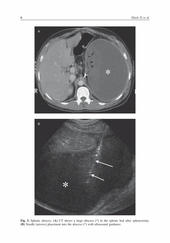
Most abdominal abscesses are accessible from a transabdominal approach withultrasonographic guidance (Fig. 5A,B), although deep pelvic abscesses may requiretransrectal or transgluteal techniques (8,9,12–14,19,20). Transrectal drainage is guidedwith ultrasound, using either a transabdominal transducer and imaging through thebladder, or an endocavitary probe in the rectum (Figs. 6 and 7). Transgluteal drainage isusually performed with CT guidance (14,20), although transgluteal ultrasound guidancehas been described (13). Transgluteal drainage has a reported higher complication ratebecause of vascular injury and is considered more painful; however, even with thesmaller sciatic notch in children, some have excellent success with this technique (14,20). Abscesses in other sites are accessed through the most direct approach, avoidingvascular and other important structures.
Needles, wires, and catheters are similar to those used in adults. Because of sedationconcerns and the lack of cooperation in most children, we choose equipment thatprovides the greatest procedural efficiency. Ultrasound guidance allows the use oflarger needles in smaller patients as the advancement is performed in real time, andthe best access window is chosen. This decreases the number of steps and simplifiesthe procedure. If the access window is small, or if CT guidance is necessary, smalleraccess sets are available. Standard drainage catheters are available from multiplemanufacturers, with most patients receiving 8–12 Fr drains, although that is dependenton the thickness of the fluid. Smaller catheters (5 or 6 Fr) are often helpful in neck or

Chapter 1 / Radiology of the Postoperative Patient 5
A
B
Fig. 4. Patient with fever after tonsillectomy. (A) There is a retropharyngeal abscess (*).(B) Demonstates ultrasound guidance to place a drainage tube (arrows) into the abscess avoidingthe carotid artery (C) and the jugular vein (*).
6 Shiels II et al.
A
B
Fig. 5. Splenic abscess. (A) CT shows a large abscess (*) in the splenic bed after splenectomy.(B) Needle (arrows) placement into the abscess (*) with ultrasound guidance.

Chapter 1 / Radiology of the Postoperative Patient 7
Fig. 6. Pelvic abscess (*) is seen behind the bladder (B). The drain (arrows) is advanced withultrasound guidance into the abscess.
Fig. 7. Transrectal abscess drainage. The ultrasound probe (arrows) will guide placement of a draininto the pelvic abscess (*).

8 Shiels II et al.
superficial abscesses; however, most abdominal abscesses require larger tubes becauseof increased fluid viscosity. For abscesses from NEC, 18–20 G intravenous catheterscan be used for aspiration with ultrasound guidance (Fig. 8). This is often done portablywith the premature infant remaining in an incubator.
The abscess is evacuated as much as possible immediately, and lab samples are sent.Currently, all catheters are placed to bulb suction, as the patients are not compliantwith keeping the bag dependent for gravity drainage. The catheter is secured with anadhesive device unless the patient is too small to allow adequate fixation, in whichcase the catheter should be sutured to the skin. Saline flush with 10 cc is performedevery shift. This amount should be subtracted from the tube output, although this is acommon charting error.
Assessment is performed at least daily. The patient usually becomes afebrile withdrainage less than 10 mL/day within 2 days, and almost always within 4 days; afterwhich time the drain is removed. If tube output continues after 48 hours, we performa tube injection to evaluate for a fistula.
Image-guided drainage procedures in both pre- and postoperative appendicealabscesses are successful in 81–100% of patients (7,8,10–13,20). Complications fromabscess drainage are uncommon, occurring in up to 11% (9–11), with catheter migrationthe most common (8,11). Bloody pus is almost universal, but significant hemorrhage
Fig. 8. An IV cannula (arrow) is advanced into abscess (*) after surgery for NEC.
Chapter 1 / Radiology of the Postoperative Patient 9
is rare. Vascular injury can occur from any approach, but is probably more commonwith the transgluteal technique owing to the proximity of the gluteal vessels (9,13,14).The inferior epigastric artery can be injured during transabdominal drainage, but canusually be identified and avoided with ultrasound during guidance for the procedure.Bowel perforation is another risk, but some authors traverse bowel when necessarywithout significant consequences (7). Inadvertent injury to other organs and the femalereproductive tract are possible but rare.
INTESTINAL FISTULA
Intestinal fistulae can originate from both normal and abnormal bowel. Postoperativefistulae may result from anastomotic leak or disruption of bowel, as well as frominadvertent injury during surgery. Spontaneous fistulae tend to originate from diseasedbowel (i.e., Crohn’s disease, radiation injury, malignancy, and ischemia (21,22)). Thereis approximately 30% incidence of fistula formation in patients with Crohn’s disease(23). Enterocutaneous, enteroenteric or enterocolic, entero- or colovaginal, and entero-or colovesical fistulous communications may be seen, depending on the location andetiology of the underlying disorder. Fistulae from intrinsically normal bowel tendto spontaneously close with conservative management. Those arising from diseasedbowel often require surgical intervention (23). The radiologic evaluation in patientswith fistulizing disease continues to evolve with advances in imaging technology.
Plain film radiographs are commonly utilized as an initial screening evaluation inthe patient with an acute abdomen, but are of limited utility for evaluation of fistulizingdisease. On occasion, extraluminal gas may be seen in fistula or abscess cavity, but isoften difficult to recognize (24). Positive contrast (barium or water-soluble contrast)examinations utilizing the small bowel follow-through (SBFT), small bowel entero-clysis (SBE), or double-contrast barium enema remain valuable tools for evaluatinginternal fistulas between loops of bowel or between bowel and other organs. Fistuloustracts may be outlined by contrast or air, and demonstrate direct intercommunicationbetween structures or end blindly in the soft tissues (24) (Fig. 9). Cutaneous fistulasmay be studied by direct contrast injection following canalization of the cutaneousopening with an angiocatheter, feeding tube or Foley catheter (fistulogram).
Abdominal and pelvic CT combined with oral and intravenous contrast, or CTenteroclysis, are effective in demonstrating some forms of fistulae, predominatelyenteroenteric, enterocolic, enterocutaneous, and enterovesical forms (25). Enhancingextraluminal tracts, when associated with inflammatory disease, may be identifiedcontaining air or fluid. Enterovesical and colovesical fistulas may demonstrate airwithin the bladder and focal bladder wall thickening at the site of fistulous communi-cation. Scanning of the pelvis should be obtained prior to intravenous contrast admin-istration so as not to obscure contrast with in the bladder lumen originating from thefistulous tract (24). Complications related to fistula formation (i.e., abscess formation),are also readily recognized.
Recent advances in MRI techniques, shortened scan times, and improved tissuecontrast have led to increased utility in imaging of the gastrointestinal tract. Contrastedenhanced MRI is highly sensitive for depicting active bowel inflammation, distin-guishing acutely inflamed bowel with luminal narrowing from fibrotic strictures,defining extraintestinal complications, and in colorectal disease. MRI is now theimaging study of choice for evaluating complex perianal fistulae as are commonlypresent in Crohn’s disease. MRI can accurately determine a fistulous tract’s relationship

10 Shiels II et al.
Fig. 9. Enterocolonic fistulae. Anterior radiograph of the abdomen during SBFT. Multiple inflamedsegments of bowel are demonstrated consistent with Crohn’s disease. Contrast containing enterocolicfistulae (arrowheads) are demonstrated with contrast extending into the rectum. Blind-ending sinustracts are noted centrally.
to the sphincters and levator ani, which is crucial for therapy. Fistulae and sinus tractsare hypointense (dark) on T1-weighted sequences and hyperintense (bright) on T2-weighted sequences, depending on the amount of fluid, edema, and inflammation (24).
The interventional radiological management of enteric fistulae centers on percuta-neous drainage of associated abscesses (e.g., intraabdominal or psoas abscesses). In thissetting, the catheter may drain longer than in more common abscess settings; however,when combined with medical therapy and bowel rest, will most often support fistulaclosure.
SMALL BOWEL OBSTRUCTION (SBO) AND ILEUS
Mechanical bowel obstruction results from an anatomic obstruction to flow ofintestinal contests. In adynamic or paralytic ileus, there is reduced or absent peristalsisin all or a portion of the intestinal tract without actual mechanical obstruction. Differen-tiation between obstruction and ileus in the postoperative patient is difficult because theclinical presentation is clouded by incisional pain, narcotics, abdominal distention, andnormal adynamic ileus (26). The radiologic evaluation relies on direct communicationbetween the radiologist and surgeon to avoid unnecessary delays in treatment. Promptand precise imaging diagnosis allows triage of patients into a surgical or non-surgicalmanagement category.
Plain film radiography remains an important and frequently requested initial exami-nation in patients with suspected obstruction. Supine and erect plain films of the abdomenshould be obtained. In patients who are too sick to stand or in the young child, lateraldecubitus views should be obtained (27,28). Additional views include the horizontal beamdecubitus films as well as the prone and crossfire prone views. These views assist in redis-tributing air (negative contrast) within the gastrointestinal tract, allowing more accurate
Chapter 1 / Radiology of the Postoperative Patient 11
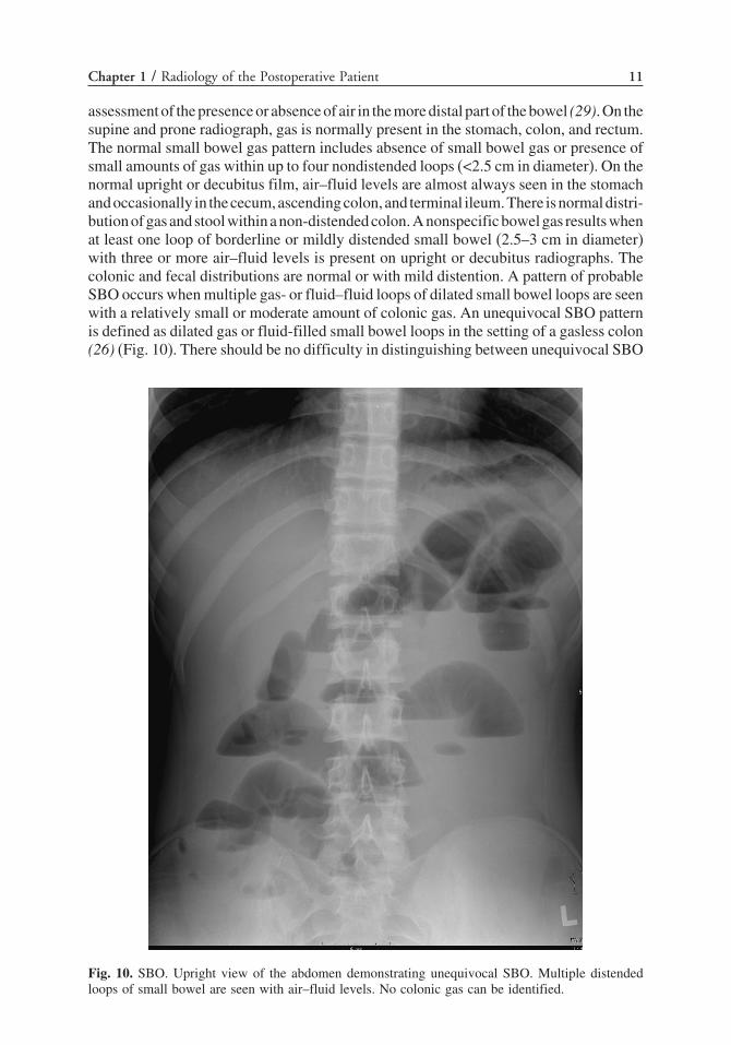
assessment of the presence or absence of air in the more distal part of the bowel (29). On thesupine and prone radiograph, gas is normally present in the stomach, colon, and rectum.The normal small bowel gas pattern includes absence of small bowel gas or presence ofsmall amounts of gas within up to four nondistended loops (<2.5 cm in diameter). On thenormal upright or decubitus film, air–fluid levels are almost always seen in the stomachandoccasionally in thececum,ascendingcolon, and terminal ileum.There isnormaldistri-bution of gas and stool within a non-distended colon. A nonspecific bowel gas results whenat least one loop of borderline or mildly distended small bowel (2.5–3 cm in diameter)with three or more air–fluid levels is present on upright or decubitus radiographs. Thecolonic and fecal distributions are normal or with mild distention. A pattern of probableSBO occurs when multiple gas- or fluid–fluid loops of dilated small bowel loops are seenwith a relatively small or moderate amount of colonic gas. An unequivocal SBO patternis defined as dilated gas or fluid-filled small bowel loops in the setting of a gasless colon(26) (Fig. 10). There should be no difficulty in distinguishing between unequivocal SBO
Fig. 10. SBO. Upright view of the abdomen demonstrating unequivocal SBO. Multiple distendedloops of small bowel are seen with air–fluid levels. No colonic gas can be identified.

12 Shiels II et al.
and the diffuse and proportional dilation of the small bowel and colon characteristic ofparalytic ileus (Fig. 11).
If there is doubt as to the diagnosis after plain films, contrast studies of thebowel help to separate mechanical obstruction from ileus. If colonic ileus or distalcolonic obstruction with an incompetent ileocecal valve is suspected, barium enemais fast and inexpensive. The SBFT examination has been used to triage patients withsuspected SBO into surgical and nonsurgical management categories, but has largelybeen replaced by the widespread use of abdominal CT. The major disadvantages to theSBFT include: inability of patients with suspected SBO to ingest large quantities ofcontrast; difficulty in assessing distensibility and fixation of the small bowel; floccu-lation and dilution of barium in high-grade obstruction with incomplete bowel opaci-fication; and the length of exam—hours or longer before contrast reaches the point ofobstruction (26).
Enteroclysis challenges the distensibility of the bowel wall and exaggerates theeffects of mild or subclinical obstruction (Fig. 12). Intubating the small bowel bypassesthe stomach and allows direct delivery of nondiluted barium or iodinated contrast(CT enteroclysis) directly into the jejunum. Advantages include: controlled infusionof contrast promotes antegrade flow toward site of obstruction despite diminished
Fig. 11. Ileus. Supine radiograph of the abdomen demonstrating diffuse gaseous distention of thesmall and large bowel.

Chapter 1 / Radiology of the Postoperative Patient 13
Fig. 12. Spot radiograph from an enteroclysis demonstrating differential dilatation of proximal versusnon-dilated (arrow) mid-jejunal loops secondary to low-grade adhesive disease.
bowel peristalsis; facilitates detection of fixed and nondistensible bowel segments; highsensitivity (100%) and specificity (88%) for SBO and high accuracy in determining thecause of obstruction (86%); detects multiple levels of obstruction; and most importantly,is highly reliable in diagnosing partial low-grade obstruction or excluding the diagnosiscompared to conventional CT or SBFT (26). SBO is excluded by enteroclysis whencontrast passes unimpeded through normal caliber small bowel loops from duodenumto right colon. Mechanical obstruction is confirmed by the demonstration of a transitionzone from a proximally distended segment to collapsed distal segment beyond theobstruction (26,30).
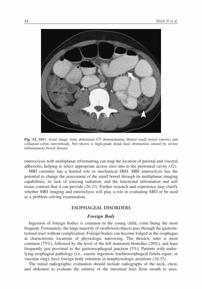
Conventional CT is highly sensitive in distinguishing high-grade small bowelobstruction from ileus (Fig. 13), and should be the initial study of choice in patientswith suspected SBO in the immediate postoperative period or when the clinical presen-tation suggests underlying abscess, closed loop obstruction, or strangulation (26,31).In the absence of clinical suspicion of these conditions, or if the CT findings areequivocal for obstruction, CT enteroclysis may establish the diagnosis by providingvolume-challenged distention of bowel loops. Water-soluble contrast is initially infusedthrough an enteral catheter placed in the proximal small bowel at fluoroscopy, followedimmediately by CT during continued contrast infusion. This technique overcomesthe insensitivity of conventional CT for diagnosing lower grades of obstruction andis equivalent to barium enteroclysis in patients with low-grade partial SBO. CT
14 Shiels II et al.
Fig. 13. SBO. Axial image from abdominal CT demonstrating dilated small bowel (arrow) andcollapsed colon (arrowhead). Not shown is high-grade distal ileal obstruction caused by severeinflammatory bowel disease.
enteroclysis with multiplanar reformatting can map the location of parietal and visceraladhesions, helping to select appropriate access sites into to the peritoneal cavity (32).
MRI currently has a limited role in mechanical SBO. MRI enteroclysis has thepotential to change the assessment of the small bowel through its multiplanar imagingcapabilities, its lack of ionizing radiation, and the functional information and softtissue contrast that it can provide (26,33). Further research and experience may clarifywhether MRI imaging and enteroclysis will play a role in evaluating SBO or be usedas a problem solving examination.
ESOPHAGEAL DISORDERS
Foreign BodyIngestion of foreign bodies is common in the young child, coins being the most
frequent. Fortunately, the large majority of swallowed objects pass through the gastroin-testinal tract without complication. Foreign bodies can become lodged in the esophagusat characteristic locations of physiologic narrowing. The thoracic inlet is mostcommon (75%), followed by the level of the left mainstem bronchus (20%), and leastfrequently just proximal to the gastroesophageal junction (5%). Patients with under-lying esophageal pathology (i.e., caustic ingestion, tracheoesophageal fistula repair, orvascular ring), have foreign body retention in nonphysiologic positions (34,35).
The initial radiographic evaluation should include radiographs of the neck, chest,and abdomen to evaluate the entirety of the intestinal tract from mouth to anus.

Chapter 1 / Radiology of the Postoperative Patient 15
Esophageal foreign bodies are characterized as radiopaque or radiolucent, sharp ordull, and single or multiple (34). The imaging appearance will influence treatmentoptions. Complications of foreign body retention should be sought and include: high-grade esophageal obstruction with air–fluid levels; tracheal narrowing owing to localedema or mass effect; perforation with pneumomediastinum; mediastinal migration ofthe foreign body; and a mediastinal mass secondary to abscess formation.
A contrast esophagram is required for nonradiopaque foreign bodies. The radiolucentforeign body may be outlined by contrast or demonstrated as an irregular contour atthe base of the contrast column in complete obstruction (Fig. 14). Contrast extensionbeyond the confines of the esophageal lumen defines perforation. CT can identify smallforeign bodies not seen on standard radiographs, further characterize the features ofa foreign body, and evaluate the paraesophgeal anatomy for edema, inflammation, orabscess formation. Esophageal foreign bodies associated with prior esophageal surgerymay be retrieved via an endoscope or via transoral interventional radiological snareretrieval with fluoroscopic guidance. Fluoroscopically guided snare removal will eitherbe successful or define foreign bodies that are adherent with granulation tissue thatrequire surgical removal.
Fig. 14. Radiolucent foreign body. Lateral view from an esophagram demonstrating completeesophageal obstruction due to meat impaction (arrow).
16 Shiels II et al.
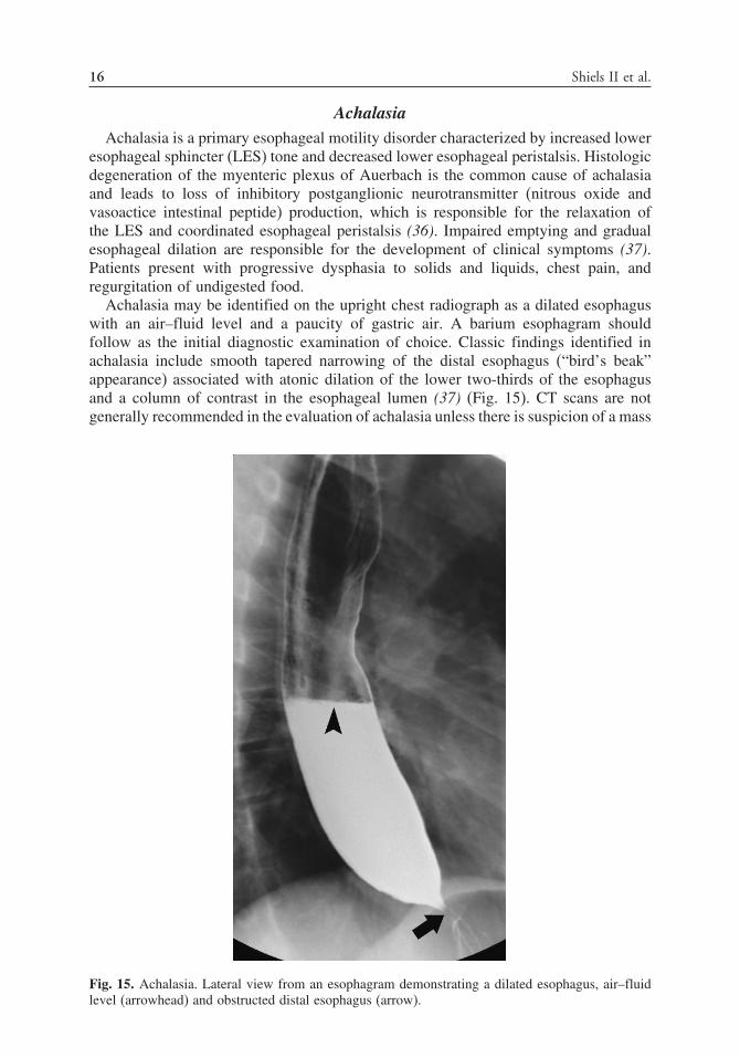
AchalasiaAchalasia is a primary esophageal motility disorder characterized by increased lower
esophageal sphincter (LES) tone and decreased lower esophageal peristalsis. Histologicdegeneration of the myenteric plexus of Auerbach is the common cause of achalasiaand leads to loss of inhibitory postganglionic neurotransmitter (nitrous oxide andvasoactice intestinal peptide) production, which is responsible for the relaxation ofthe LES and coordinated esophageal peristalsis (36). Impaired emptying and gradualesophageal dilation are responsible for the development of clinical symptoms (37).Patients present with progressive dysphasia to solids and liquids, chest pain, andregurgitation of undigested food.
Achalasia may be identified on the upright chest radiograph as a dilated esophaguswith an air–fluid level and a paucity of gastric air. A barium esophagram shouldfollow as the initial diagnostic examination of choice. Classic findings identified inachalasia include smooth tapered narrowing of the distal esophagus (“bird’s beak”appearance) associated with atonic dilation of the lower two-thirds of the esophagusand a column of contrast in the esophageal lumen (37) (Fig. 15). CT scans are notgenerally recommended in the evaluation of achalasia unless there is suspicion of a mass
Fig. 15. Achalasia. Lateral view from an esophagram demonstrating a dilated esophagus, air–fluidlevel (arrowhead) and obstructed distal esophagus (arrow).
Chapter 1 / Radiology of the Postoperative Patient 17
(adenocarcinoma, lymphoma, or others) at the gastroesophageal junction mimickingthe findings of achalasia. Radiological intervention following surgical treatment ofachalasia centers on balloon dilation of postmyotomy strictures. Balloon dilation withfluoroscopic guidance of distal esophageal stricture associated with achalasia is safe,to a diameter of 30 mm.
Esophageal PerforationInstrumentation of the esophagus accounts for approximately two-thirds of all
reported cases of esophageal perforation and can be seen following esophagoscopy,esophageal dilatation (bougienage or pneumatic), sclerotherapy, intraesophageal tubeplacement, and endotracheal intubation. Other causes include spontaneous perforation(Boerhaave’s syndrome), foreign body ingestion, blunt chest trauma, operative injury,tumor, and severe esophagitis (38,39). Rapid development of necrotizing mediastinitis,empyema, sepsis, and multiorgan failure account for the high mortality associated withesophageal perforation. Accurate diagnosis and early treatment are essential to thesuccessful management of patients.
Lateral neck films may detect subcutaneous emphysema before identification onchest radiographs or by physical examination in cervical esophageal perforation (40).When thoracic or abdominal perforation is suspected, posteroanterior and lateral radio-graphs of the chest as well as an upright or decubitus view of the abdomen shouldbe obtained. Chest radiographs detect 90% of esophageal perforations; however, theycan be negative if taken too early. Soft tissue and mediastinal emphysema are seenafter 1 hour, whereas pleural effusions and mediastinal widening take several hours todevelop (39,40). The presence of pleural effusion, pneumomediastinum, subcutaneousemphysema, hydrothorax, hydropneumothorax, or subdiaphragmatic air on radiographsis highly suggestive of esophageal perforation (41).
The contrast esophagram remains the standard for diagnosis of esophageal perfo-ration (Fig. 16). Water-soluble contrast is recommended for initial screening ofsuspected perforation. Contrast extravasation is identified in only 50% of cervical and75–80% of thoracic perforations. If no perforation is initially identified, barium esopha-gography is recommended. Barium improves detection of small primary or unsuspectedsecondary perforations. The detection rate increases to 60% for cervical and 90% forthoracic perforations (42). Contrast studies have an overall 10% false negative rate.
CT identifies esophageal perforations that are difficult to diagnose or when contrastesophagrams cannot be performed. Abnormal findings suggestive of perforation includeextraluminal air in the soft tissues of the mediastinum, esophageal thickening, visiblecommunication of the air-filled esophagus with a contiguous mediastinal or parame-diastinal air–fluid collection, or abscess cavities adjacent to the esophagus in themediastinum or pleural space. Left-side pleural effusions are highly suggestive. Inpatients who fail to improve after initial treatment, CT assists in localizing pleuralfluid or abscess collections amenable to interventional radiological drainage catheterplacement.
Complications of FundoplicationGastroesophageal reflux disease (GERD) is a common condition for which medical
management is effective in the large majority of patients. Those with intractablevomiting, persistent esophagitis, apnea, and pulmonary infections require furthersurgical management (43). Antireflux surgery has been shown to be highly effective

18 Shiels II et al.
Fig. 16. Esophageal perforation. Anterior view from an esophagram demonstrating a large thoracicesophageal perforation (arrowheads) with extravasation into the right thorax (arrows).
in managing complications related to GERD. With advances in minimally invasivetechniques, laparoscopic Nissen fundoplication has become the preferred surgicaltechnique (44). Despite a success rate of more than 90% and low mortality, antirefluxsurgery is not without complications.
The upper gastrointestinal series (UGI), with barium administered orally or througha gastrostomy tube, is the study of choice to evaluate the integrity and the functionof the wrap. Loosening or breakdown of the fundoplication is identified by contrastfilling the folds of the wrap. Small amounts are generally inconsequential, whereaslarger amounts reflect more significant loosening. In complete breakdown of the wrap,there is loss of the normal “pseudotumor” identified at the fundus. Hiatal hernias areseen when the fundoplication and upper stomach herniate through the hiatus. Thewrap may remain intact. Paraesophageal hernias represent extension of a portion ofthe wrap through the hiatus, typically along posterolateral margin of the left side ofthe wrap where there is an absence of sutures. The key differentiating point fromhiatal hernia is that the gastroesophageal junction remains below the diaphragm. Amalpositioned fundoplication results from partial disruption of the sutures, allowingdistention of the fundus above the remaining sutures. This has a similar appearanceto the paraesophageal hernia, but the deformed fundus remains entirely below thediaphragm. Esophageal obstruction can result from an excessively tight wrap leading toesophageal dilation and delayed transit of contrast through the fundoplication (Fig. 17).Partial esophageal obstruction may be found in the immediate postoperative periodsecondary to edema; persistent obstruction, however, requires treatment (43).

Chapter 1 / Radiology of the Postoperative Patient 19
Fig. 17. Delayed esophageal transit. Lateral view from an upright esophagram demonstrating anobstructed esophagus following Nissen fundoplication (arrow). Contrast in gastric fundus showsintact Nissen (arrowhead). Contrast was placed through gastrostomy tube prior to esophagram.
Esophageal StricturesEsophageal strictures in the pediatric age group are most frequently benign. Stric-
tures may be congenital, but are most frequently acquired following surgery (repairof esophageal atresia, interposition surgery, restrictive Nissen fundoplication), causticor foreign body ingestion, achalasia, infection (herpes virus, candidiasis), inflam-mation (epidermolysis bullosa, chronic granulomatous disease), reflux esophagitis, andfollowing sclerotherapy of varices. Anastomotic strictures following esophageal atresiarepair occur in 30–70% of patients and are the most commonly acquired stricture ofchildhood (45).
The radiologic evaluation of esophageal strictures requires an esophagram withwater-soluble or barium contrast (46). The diagnosis of stricture is confirmed when apersistent area of circumferential narrowing is identified within the esophageal lumen.The involved segment shows lack of distention during passage of the contrast bolus. Theproximal esophagus is often dilated with delayed passage of the contrast bolus throughthe stricture. The degree of dilation and contrast delay is determined by the severity ofthe stenosis. Retained secretions, ingested food matter, or foreign bodies may be seenwithin the lumen of the proximal esophagus (Fig. 18). Esophageal strictures followingsurgical intervention are most commonly found with repair of esophageal atresia and areeffectively and safely treated with low-profile angioplasty balloon dilation. Long-term

20 Shiels II et al.
Fig. 18. Esophageal stricture. Anterior view from an esophagram demonstrating a focal esophagealstricture (arrow) with dilated proximal esophagus and retained food within the lumen. Patient hadesophageal atresia repair as an infant.
balloon dilation of esophageal anastomotic strictures is successful in nearly 100% ofpatients, and success is greatest when started within 6 months of surgery, in patientswithout GERD, and when solid food ingestion is permitted to maintain the patentesophageal lumen. The incidence of perforation from balloon dilation of anastomoticstrictures ranges from 0–1.8% (47,48).
Anastomotic Leak and Recurrent FistulaComplications seen following repair of esophageal atresia include anastomotic leak,
recurrent tracheoesophageal fistula, and stricture (discussed previously) formation. Theincidence of anastomotic leak following repair is approximately 20% (49). Causesinclude: the use of silk sutures; excessive anastomotic tension; and excessive distalesophageal mobilization. A greater incidence of anastomotic leak is noted in patientswho undergo colonic interposition, attributable to impaired blood supply in the cephaladend of the graft (49). Clinical suspicion of leak occurs when saliva is seen in thechest tube. An esophagram confirms the clinical suspicion by demonstrating contrastextravasation from the anastomotic site into the mediastinum or pleural space.
Recurrent tracheoesophageal fistulae are seen in approximately 3–12% of repairs.The etiology is related to an anastomotic suture line leak with erosion through theprevious site of repair (49). Diagnosis may be difficult and delayed for years. Acontrast esophagram may directly demonstrate the fistula (Fig. 19); however, anesophagram performed after placement of a nasogastric or orogastric tube may be

Chapter 1 / Radiology of the Postoperative Patient 21
Fig. 19. Recurrent tracheoesophageal fistula. Oblique view from an esophagram demonstrating arecurrent fistula between the esophagus and the trachea (arrow). Patient had prior repair of esophagealatresia with tracheoesophageal fistula. Coughing with feeds was the clinical presentation.
required. Repeated contrast injections during withdrawal of the tube achieve greaterlocal luminal distention and increased likelihood of fistula visualization. Direct canal-ization of the fistula is less often performed, but will directly opacify the fistula orserve as site for injection of methylene blue during bronchoscopy (35).
STOMACH
GastroparesisGastroparesis is characterized by impaired gastric empting in the absence of
a mechanical gastric outlet obstruction. Clinical symptoms include early satiety,postprandial bloating, nausea, vomiting, and abdominal pain. Nutrient composition(volume, osmolarity, osmolaltiy, caloric density, viscosity, Ph, and type of macronu-trients), degree of gastric distention, and intestinal feedback from concentration-dependent chemoreceptors along the proximal intestine all interact to determine gastrictransit (50). Water or human milk empties faster than formula, and liquids empty fasterthan solids. Medium chain triglycerides empty faster than long-chain triglycerides.
Nuclear medicine scintigraphy remains the gold standard method for measuringgastric emptying. Gastric emptying studies are performed by combining a radiopharma-ceutical (technetium sulfur colloid) with food and measuring the residual radioactivitywithin the stomach over time (generally 1, 2, and 4 hours) as a percentage of theinitial gastric activity at ingestion (51). Radiolabeling of both liquids and solids is

22 Shiels II et al.
feasible, although the dynamics of emptying are different. Solid meal scintigraphy ispreferred over liquids for detecting delayed gastric emptying because the rate of solidemptying will be affected earlier by abnormalities in receptive relaxation and tonegenerated by the fundus, as well as by abnormalities in motility and coordination ofantropyloroduodenal smooth muscle (52).
Regardless of the technique used, normal values of gastric emptying vary widely amongdifferent individuals and day-to-day within the same patient who undergoes repeated tests.Lack of standardization of test meals makes published normal values of gastric emptyingdifficult to compare between institutions. In routine practice, a T1/2 (time for 50% gastricempting to occur) or, more commonly, the percentage of residual gastric activity at 1,2, and potentially 4 hours is calculated (Fig. 20). The published range of normal gastricemptying for solid phase is 10–50% retention at 2 hours. For those patients with normalor borderline emptying (45–55% retention) at 2 hours, extending the study out to 4 hourshas been suggested (51). The range of normal gastric retention at 4 hours is narrower(0–10% retention) and a greater number of symptomatic patients may be demonstrated tohave delayed gastric emptying at this time. The key is to establish a standard protocol for
Fig. 20. Gastric emptying scan demonstrating delayed emptying at 2 hours.

Chapter 1 / Radiology of the Postoperative Patient 23
scintigraphy at one’s own institution so that variations in gastric emptying are physiologicand not technically related.
Gastric PerforationThe etiology of the gastric perforation is age dependent. In the newborn infant,
gastric perforation is abrupt in onset and occurs in 96% during the first week oflife. Proposed etiologies of perforation include: hypoxia or stress-induced necrosis;gastric hyperacidity in the first few days of life; gastric ulceration; overdistentionof the stomach caused by distal obstruction or mechanical ventilation; prematurity;indomethacin or dexamethasone therapy; vigorous respiratory resuscitative measures;and iatrogenic (53). Perforation in the older child and adult most commonly arise frompeptic ulceration, although iatrogenic and traumatic injuries, postoperative complica-tions, and necrotic or ulcerated malignancies are also reported (54).
Supine, upright, left lateral decubitus and crossfire supine views of the abdomenshould be obtained, depending on the age and the clinical status of the patient. Thecharacteristic finding of gastric perforation is pneumoperitoneum. Associated absenceof an air–fluid level in the stomach and decreased bowel gas are very suggestive of thediagnosis. Recognition of free air on a supine abdominal radiograph include: increasedlucency over the liver compared to the abdominal wall musculature (Fig. 21), the“football” sign (air outlining the falciform ligament) (Fig. 22); The “inverted-V” sign(air outlining the median umbilical folds); and the “Rigler” sign (air on both sidesof the bowel wall) (Fig. 22). On the upright abdominal view, air collects between
Fig. 21. Pneumoperitoneum. Free air on supine radiograph (arrows) seen as abnormal lucency inthe right upper quadrant.

24 Shiels II et al.
Fig. 22. Pneumoperitoneum. Massive pneumoperitoneum on supine radiograph. Free air outlines thefalciform ligament (arrowhead) and is seen on both sides of the bowel wall (arrow).
the diaphragm and the upper abdominal viscera. On the decubitus view, air collectsbetween the liver and anterior abdominal wall. A triangular air collection is seen onsupine crossfire views when air collects between the anterior abdominal wall andtwo adjacent loops of bowel. Free air may not be seen initially if the perforation issmall, self-sealed, well contained by adjacent organs, retroperitoneal, or the result offluid extravasation rather than air (54). The reported sensitivity in the detection ofextraluminal air by pain film radiography is 50–70%.
When perforation is suspected, but not demonstrated on abdominal radiographs, acontrast examination may be obtained. A water-soluble contrast agent is used initiallyin any suspected case of intestinal perforation. If a perforation is not identified on thewater-soluble contrast exam, barium administration should immediately follow. Identi-fication of any extraluminal contrast confirms the diagnosis of perforation (Fig. 23).Free extravasation of contrast into the peritoneal space, however, is detected in approx-imately 50% of patients with anterior perforation of the stomach, the other half formingwalled-off collections (55). Penetrations of the posterior wall of the stomach mayinvolve the pancreas, lesser omentum, transverse mesocolon, liver spleen, biliary tree,or colon. Less than 50% of these patients will demonstrate contrast extravasation,likely caused by early sealing of the leak.
Abdominal CT is the recommended imaging modality for detecting gastrointestinalperforations that are not identified by either plain films or contrast examinations, and

Chapter 1 / Radiology of the Postoperative Patient 25
Fig. 23. Gastric perforation. Extravasation of contrast (arrow) is demonstrated through posteriorgastric wall perforation.
in atypical clinical presentations. CT is superior to plain films in demonstrating smallcollections of free intraperitoneal air or small collections in the retroperitoneum. CTimages need to be viewed in wide window settings (lung window) that assist indiscriminating low-density air from fat (Fig. 24). In addition, the site and cause of theperforation as well as associated complications of phlegmon, abscess, and peritonitiscan be assessed (54). Administration of oral contrast is recommended, although extralu-minal extravasation is not a frequent CT finding in perforation. Diagnosis of perforationis based on direct findings of extraluminal air or luminal contrast material and directvisualization of a focal ulceration or discontinuity of the stomach wall. An indirectsign of perforation is an inflammatory phlegmon in direct continuity with the wall ofthe stomach.
Gastrostomy Tube DislodgementInsertion of gastrostomy tubes has become commonplace, particularly for care
of the special-needs child. Three methods of gastrostomy placement are availabletoday: surgical gastrostomy, percutaneous endoscopic gastrostomy (PEG), and inter-ventional radiological percutaneous gastrostomy (56). Whether done surgically orpercutaneously, gastrostomy placement requires a tract be created through the anteriorabdominal wall into the stomach for an enteral feeding device. Only the surgicaltechnique involves suturing of the stomach to the anterior abdominal wall. The othertechniques rely on scarring to take place between the stomach and the anteriorabdominal wall.

26 Shiels II et al.
A
B
Fig. 24. Pneumoperitoneum. (A) Axial CT image of the upper abdomen viewed in abdominal windowdemonstrating poor discrimination between free air (left arrow) and fat (right arrow) densities.(B) Same image viewed in lung window shows better discrimination between air (arrow) and fat(arrowhead) density. Small bubble of gas is also noted along falciform ligament (short arrow).

Chapter 1 / Radiology of the Postoperative Patient 27
A serious complication of PEG placement is early dislodgement of catheter prior toformation of a mature tract. This is reported to occur in up to 7.8% of patients under-going the procedure (57). Radiologic management in these patients is limited. If thereis a concern for dislodgement of the catheter into the peritoneal cavity, a water-solublecontrast examination performed through the gastrostomy tube may be obtained toconfirm the intraperitoneal position. Attempts to blindly replace a completely dislodgedcatheter, however, are contraindicated owing to the high risk of false passage creation.Under fluoroscopic guidance, the interventional radiologist may on occasion havesuccess in reestablishing continuity between the gastrostomy and the stomach by usingcatheter-directed guidewire manipulation.
Late dislodgement of gastrostomy tubes poses little risk to the patient with amature tract. Immediate replacement is not necessary, although urgent replacementis suggested because of spontaneous closure of the gastrostomy sites within approxi-mately 6 hours (58). This can be prevented by instructing the parents or caregivers toimmediately replace the gastrostomy tube, or an equally sized Foley catheter, throughthe gastrostomy site and tape it in place. A new gastrostomy tube can be directlyreplaced under fluoroscopic guidance when the tract remains open; however, if thegastrostomy tract has stenosed, fluoroscopicly directed dilation of the tract may beperformed. This allows replacement of a similar size gastrostomy tube.
HEPATOBILIARY DIAGNOSIS AND INTERVENTION
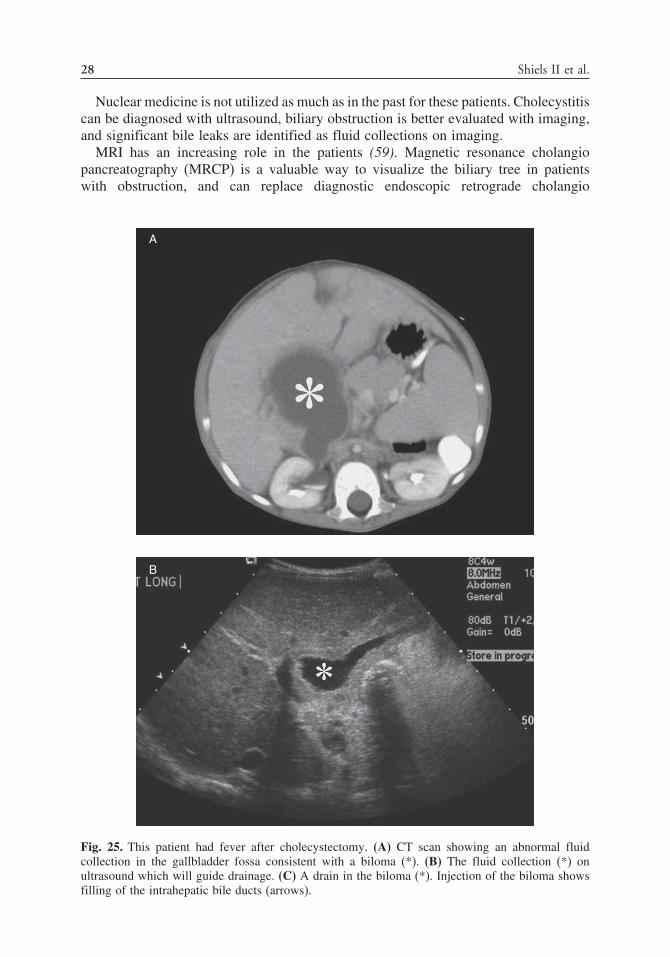
Hepatobiliary disease in children is typically from a benign cause. Postoperatively,the etiology may be caused by obstruction, bile leak, or portal vein thrombosis. Withobstruction, the patients usually present with jaundice, although abdominal pain, fever,vomiting, or abnormal lab values may also be the initial problem (59). Bile duct injury,retained stones, and strictures (particularly with transplantation) (60–62) are the mostcommon causes. Bile leaks after surgery are often accompanied with abdominal painand fever (Fig. 25). Portal vein thrombosis can occur after abdominal sepsis, such asappendicitis. The presentation is usually occult. Septic postoperative or trauma patientsare also at risk for acalculus cholecystitis.
Imaging in these patients should be focused on the specific clinical question. Withpossible obstruction, imaging should confirm or exclude that assumption, and identifythe level of obstruction. If a bile leak is suspected, this needs to be identified, includingthe site of the extravasation.
Ultrasound is an ideal initial scanning technique for these patients (59). Intrahepaticand extrahepatic biliary duct dilation can be readily identified, and retained stonesmay be visible. Abnormal fluid collections are often identified. Doppler analysis canevaluate for the presence and direction of portal venous flow. Cholecystitis can beeasily identified as an enlarged gallbladder with wall thickening, pericholecystic fluid,and a hyperemic wall. The ability of ultrasound to be performed portably is valuablein patients in the intensive care unit (ICU).
CT can add valuable information in patients with postoperative disease (59). Bileduct dilatation can be identified, and the level of obstruction may be better seen thanwith ultrasound. Retained bile stones are rarely seen, as they do not usually containsignificant calcium. Abdominal fluid collections such as a biloma are more readilyidentified. Contrast enhancement of the hepatic and portal vessels allows for evaluationof thrombosis, occlusion, or portal hypertension.
28 Shiels II et al.
Nuclear medicine is not utilized as much as in the past for these patients. Cholecystitiscan be diagnosed with ultrasound, biliary obstruction is better evaluated with imaging,and significant bile leaks are identified as fluid collections on imaging.
MRI has an increasing role in the patients (59). Magnetic resonance cholangiopancreatography (MRCP) is a valuable way to visualize the biliary tree in patientswith obstruction, and can replace diagnostic endoscopic retrograde cholangio
A
B
Fig. 25. This patient had fever after cholecystectomy. (A) CT scan showing an abnormal fluidcollection in the gallbladder fossa consistent with a biloma (*). (B) The fluid collection (*) onultrasound which will guide drainage. (C) A drain in the biloma (*). Injection of the biloma showsfilling of the intrahepatic bile ducts (arrows).

Chapter 1 / Radiology of the Postoperative Patient 29
C
Fig. 25. (Continued).
pancreatography (ERCP) in some instances. Bile duct dilatation and the level ofobstruction are readily identified, and retained stones can be seen.
Obstructive JaundiceIn patients with obstructive jaundice, imaging as described previously will identify
the level and cause of obstruction. ERCP is usually the initial interventional approachin adults; however, in children this may be more difficult. Relief of the obstruction byexternal or internal drainage can be accomplished with interventional radiology (IR)techniques (63). The first decision is the need for solitary or multiple drains. If theobstruction is at the level of the common hepatic duct or more distal, a solitary drain willtreat both the right and left hepatic lobes. Intrahepatic obstruction may require multipledrains to adequately treat the patient. Both right and left lobe approaches are available,each with different advantages and disadvantages. The right lobe approach drains alarger percentage of the liver, but has an increased risk of transpleural traversal (64).This predisposes the patient to pneumothorax, hemothorax, and biliary-pleural fistula;although this usually can be avoided. A left approach has fewer lung complications,but has a smaller liver volume with which to work (Fig. 26).
These patients all need appropriate antibiotic coverage before the procedure. Ultra-sound guidance is the preferred method; although with minimally dilated ducts, fluoro-scopically guided placement of a small-gauge needle via the right mid-axillary lineis also possible. Typically, a small needle (22 G) is used to enter a peripheral bile

30 Shiels II et al.
duct. After insertion of an 0.018-inch wire, a stiff introducer system is used to gainaccess with a 4- to 6-Fr sheath. Through this sheath, diagnostic cholangiography can beperformed followed by catheter and guidewire manipulation through the biliary system.In patients who are critically ill, a drain can be left within the liver. Otherwise, attemptsare made to pass the area of obstruction, and establish internal/external drainage byplacing a drain through the intrahepatic ducts, across the level of obstruction, and intothe duodenum.
A
B
Fig. 26. Biliary stricture. This patient has had a partial hepatic resection for hepatoblastoma. Hehad a choledochojejunostomy, but presents years later with jaundice. (A) Intrahepatic bile ductenlargement (arrows), confirmed on (B) ultrasound (arrow). (C) Small needle access (arrows) intoa peripheral bile duct (arrowhead). Subsequently, a drain was placed. (D) Cholangiogram throughthe drain, showing obstruction (arrow) at the choledochojejunostomy.

Chapter 1 / Radiology of the Postoperative Patient 31
C
D
Fig. 26. (Continued).
After crossing the obstruction initially, or at a later date after the patient’s sepsishas resolved, the reason for the obstruction can be addressed. Strictures can be dilatedwith an angioplasty balloon, and retained stones can be removed via IR fluoroscopi-cally guided techniques, or with a combined IR/surgical fluoroscopic/cholangioscopytechnique (65,66) (Fig. 27). A drainage tube is left in place for several weeks, andonly removed if the patient does well after capping the tube for a week (67). Metallicstents are contraindicated for most benign biliary obstructions.

32 Shiels II et al.
A
B
Fig. 27. Retained biliary stone. This patient had jaundice after cholecystectomy. She has situsinversus. (A) Retained biliary stone (arrowhead) on the MR. (B) Biliary drainage and a cholangiogramwith the retained stone (arrow) at the distal common bile duct. (C) The obstruction has beencrossed with a wire (arrow) for internal drainage. (D) At a later date combined fluoroscopy andcholangioscopy were used to remove the stone. The endoscope (arrowheads) was guided withfluoroscopic guidance to the stone.

Chapter 1 / Radiology of the Postoperative Patient 33
C
D
Fig. 27. (Continued).
34 Shiels II et al.
Biliary LeaksFluid collections identified with imaging in symptomatic patients after hepatobiliary
surgery are often bilomas (62) (Fig. 25). The initial treatment for these collectionsis external drainage of the collection. These techniques are described in the sectionon abdominal abscesses. External drainage is usually the only necessary treatment;however, some collections show continued drainage caused by an active bile leak. When
A
B
Fig. 28. (A) CT scan from a septic postoperative patient shows a distended gallbladder (*). (B)Ultrasound guided needle (arrows) placement into the gallbladder (*). (C) Injection of the drainagetube confirming placement in the gallbladder (*).
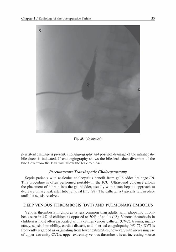
Chapter 1 / Radiology of the Postoperative Patient 35
C
Fig. 28. (Continued).
persistent drainage is present, cholangiography and possible drainage of the intrahepaticbile ducts is indicated. If cholangiography shows the bile leak, then diversion of thebile flow from the leak will allow the leak to close.
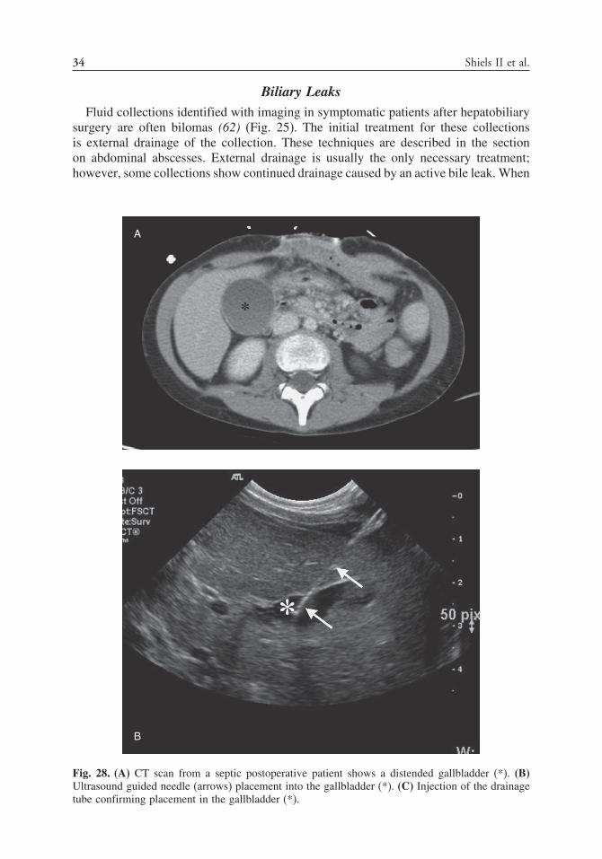
Percutaneous Transhepatic CholecystostomySeptic patients with acalculus cholecystitis benefit from gallbladder drainage (9).
This procedure is often performed portably in the ICU. Ultrasound guidance allowsthe placement of a drain into the gallbladder, usually with a transhepatic approach todecrease biliary leak after tube removal (Fig. 28). The catheter is typically left in placeuntil the sepsis resolves.
DEEP VENOUS THROMBOSIS (DVT) AND PULMONARY EMBOLUS
Venous thrombosis in children is less common than adults, with idiopathic throm-bosis seen in 4% of children as opposed to 30% of adults (68). Venous thrombosis inchildren is most often associated with a central venous catheter (CVC), trauma, malig-nancy, sepsis, immobility, cardiac disease, and inherited coagulopathy (68–72). DVT isfrequently regarded as originating from lower extremities; however, with increasing useof upper extremity CVCs, upper extremity venous thrombosis is an increasing source

36 Shiels II et al.
of asymptomatic and symptomatic thrombosis (71,72). Pulmonary thromboembolism(PTE) is uncommon in children, yet fatal in 30% of untreated cases (74). In pediatrictrauma, older children with high Injury Severity Scores, thoracic injuries, spinalinjuries, and CVCs are at greatest risk of venous thromboembolism. Diagnostic imagingmodalities for thromboembolism are best used in a targeted fashion: venous sonog-raphy for upper and lower extremity thrombosis; multidetector CT angiography fordiagnosis of PTE. CT angiography has largely replaced ventilation/perfusion scanningas a screening study for PTE, and with greater than 90% sensitivity (Fig. 29), haslargely replaced conventional pulmonary arteriography for definitive diagnosis of PTE
A
B
Fig. 29. CT angiogram (CTA) of pulmonary embolus. (A) Coronal reconstruction of CTA demon-strates the large right lower lobe pulmonary artery embolus (arrow) outlined by contrast media. (B)Transaxial CTA image demonstrates bilateral lower lobe pulmonary emboli, with prominent leftlower lobe embolus (arrow) clearly outlined with contrast media.

Chapter 1 / Radiology of the Postoperative Patient 37
Fig. 30. This patient had extensive DVT, proven PTE by CT, and spinal trauma precluding antico-agulation. A retrievable IFC filter (arrow) had been placed. A sheath (arrowhead) has been advancedthrough a right internal jugular approach, and is advanced to subsequently remove the filter.
(75–77). Although a superb diagnostic modality when all factors are favorable, themost common causes for an indeterminate scan are motion artifact and poor contrastenhancement (75). Treatment of DVT is primarily done with anticoagulation therapy,to include prophylaxis with low-molecular-weight heparin. IR adjunctive treatmentincludes the use of intravascular thrombolytic therapy and placement of temporaryor permanent vena caval filters (VCFs) (77–83). Thrombolytic therapy with streptok-inase, urokinase, and tissue plasminogen activator (tPA, discussed in greater detailin a later section) has been used safely and successfully in children to treat PTE(77–79). With improved device technology and the development of retrievable VCFs,the VCF provides a safe and effective treatment option (Fig. 30) for patients witheither DVT or PTE who are not candidates for anticoagulant therapy, patients whohave failed anticoagulant therapy, and those with idiopathic infrarenal inferior venacava (IVC) thrombosis (79–83). The authors are using VCFs more commonly than inprior years, especially the removable VCF, for these indications and in patients whoare at significant risk of PTE from DVT.
THORACIC MANAGEMENT ISSUES
Postoperative PneumothoraxPneumothorax following primary surgical intervention is most frequently effec-
tively treated with thoracostomy tubes placed at the time of surgery. Occassionally,thoracostomy tubes may fail to drain the pneumothorax because of mechanicalocclusion or persistent bronchopleural fistula. In the setting of unresponsive treatmentof pneumothorax, the interventional radiologist is able to accurately place a small-borepigtail (8–14 F) catheter under fluoroscopic guidance. When a persistent air leak ispresent with a bronchopleural fistula, pleural suction of 30–60 cm H2O may be requiredfor evacuation of the pleural air to allow the visceral and parietal pleural surfaces tooppose and seal the pleural leak.
PneumoniaPneumonia is a potential complication of surgery, particularly with prolonged
endotracheal intubation and immobilization. The interpretation of plain radiographswith postoperative atelectasis that persists longer than 2–3 days should include

38 Shiels II et al.
pneumonia in the differential diagnosis to facilitate timely treatment of an earlypneumonia. The lower lobes are the most common location of postoperative pneumonia,although any segment or lobe may become infected. Pneumonia most commonly istreated adequately with antibiotic therapy without complications. The diagnosis ofpneumonia is most commonly made with plain radiographs, with CT scanning reservedfor pneumonia in which complications are suspected. Bacterial pneumonia is mostcommonly complicated with parapneumonic effusion, pulmonary necrosis, pulmonaryabscess, and empyema. Pulmonary necrosis in children, with or without cavitation, ismost often treated without further surgical or radiological intervention. Radiographs3–12 months after detection of necrotizing pneumonia most often demonstrate completehealing of the necrotic segments without other sequelae. A pulmonary abscess thatfails to respond to antibiotic therapy is often amenable to catheter (5–12 F, age andsize dependent) drainage, especially when abuting the pleural surface, with little riskof pneumothorax or other complications.
Pleural Effusion and EmpyemaBacterial pneumonia is common in children, with most cases being caused by
Streptococcus pneumoniae (84–87). Multiple reports have cited the incidence ofassociated pleural effusion to be 36–57%, with 15–20% progressing to empyema,defined as pus within the pleural space (88–92). Empyema is classically divided intothree separate stages (85,87–90,93,94):
1. Early exudative: Lasts from 24–48 hours, and fluid forms because of pleuralinflammation.
2. Fibrinopurulent: Lasts from 7–10 days, and is characterized by thickened exudates,fibrin deposition, white cell infiltration, and formation of loculations.
3. Organized: Starts 2–4 weeks from effusion onset. There is fibroblast formation andcreation of a thick peel.
Grading of pleural disease based on laboratory criteria have also been described,with pH, glucose levels, lactic dehydrogenase (LDH), Gram stain, and cultures allbeing performed (90,91). However, this lab evaluation does not define how the patientshould be treated, with the exception of antibiotic coverage for the appropriate pathogen(95). Ultrasound has also been used to grade empyemas (Fig. 1A and 1B), as Stage I(anechoic fluid), Stage II (loculations), and Stage III (solid peel) (96). This informationis prognostic, concerning the need for additional treatment such as fibrinolytics orsurgery (91,92,96).
It is well established that appropriate treatment of empyema will help control sepsis,restore pulmonary function, and prevent lung entrapment from the fibrous peel (88,91).In prior years, empyemas were treated initially with large-bore chest tubes and/orwith open thoracotomy with decortication. Two more recent techniques for dealingwith emypema were then described: video-assisted thoracoscopic surgery (VATS), andsmall tube placement with fibrinolytic therapy. These techniques are now consideredthe most appropriate treatments for patients with empyema, with thoracotomy reservedfor patients with a large peel. However, which is best is quite controversial (85,88,89,97,99). At institutions where IR performs image-guided small tube placement andfibrinolytic therapy, success rates are 77–98% (90,91,93,95,100,101). It is obvious thatthese small tubes are easier on patients. VATS is also usually successful, and mighthave some potential advantages (87,99,102–105). Some authors think that a patient’s

Chapter 1 / Radiology of the Postoperative Patient 39
symptoms might improve more quickly, and the hospital length of stay may be less(89,94). However, with IR drainage, the temporal improvement in symptoms is similarto VATS, the operating room (OR) risks are obviated, and the patients avoid having alarge chest tube.
A simple and effective treatment protocol has been developed for treatment ofempyemas at the authors’ institution (101). All patients with empyemas consideredfor percutaneous treatment are evaluated by IR following chest CT. Chest CT helpsdefine parenchymal disease, and areas of necrosis are readily identified. This givesprognostic information about the duration of therapy, and possible need for futureinterventions (Fig. 31). Prior to insertion of the chest tube in IR, the empyema is
A
B
Fig. 31. Percutaneous treatment of organized empyema. (A) Chest CT demonstrates left lower lobepneumonia with large left pleural empyema (asterisk). (B) Sonogram of the left pleural empyemademonstrates grade 2 organization with large amount of organization with fibrin strands (arrow)separating purulent locules. (C) Chest radiograph following placement of 12-F pigtail catheter forfibrinolytic therapy and drainage. (D) Chest CT following fibrinolysis demonstrating resolution ofthe empyema with a trace amount of pleural thickening following therapy (arrow).

40 Shiels II et al.
C
D
Fig. 31. (Continued).
assessed with sonography. Patients with a solid peel as identified by ultrasound werereferred for surgery. All other patients had small-bore chest tubes (10–14 F) placed,with fibrinolytics given if the fluid was echogenic or had loculations, or if the patienthad suboptimal response defined as residual fluid, or no symptomatic improvement(Fig. 31). The tube is then attached to a water-seal chest tube system and is placedto 20-cm water suction. The tPA dose is 2 mg mixed in 20 mL of saline. The tPA isinjected into the tube, the tube is clamped for 1 hour, and then the tube is returnedto suction. This is repeated every 12 hours until fluid output deceases to less than25 mL in a 12-hour period. Chest tube output, daily chest radiographs, and patientsymptoms are monitored throughout the protocol. At 48 hours, if the patient has not

Chapter 1 / Radiology of the Postoperative Patient 41
improved, and residual fluid is suspected because the tube output is less than expectedor the chest radiographgot worse, repeat chest CT is performed. If significant pleuraldisease is present, then tube drainage with fibrinolytics is considered a failure, andthe patient if referred for surgical intervention. Otherwise, the tube is removed whenoutput falls below 25 mL in a 12-hour period, and one of the two following criteria aremet: (1) The patient is afebrile and off supplemental oxygen; or (2) Chest radiographhas demonstrated no significant pleural disease.
This protocol of drainage and fibrinolytic therapy was effective in 92% of patients,with the patients’ mean length of hospitalization at 8.4 days and average numberof tPA doses at 5.9. Most initial failures in the evolution of this protocol werepatients where tPA was either started late, or stopped after a few doses. Sincepublication, expanded use of this protocol has demonstrated efficacy in 98% ofpatients.
DISPLACED VENOUS CATHETER
CVCs may be subject to malposition or fracture, resulting in the need for eitherrepositioning, removal, or retrieval of migrated catheter fragments. Subclavian CVCsmay infrequently penetrate either the subclavian vein or superior vena cava (SVC) withresultant pleural effusion from the infusate. This malpositioning is treated with pigtailcatheter thoracentesis as well as removal and replacement of the CVC. Femoral CVCswill infrequently be placed into lumbar veins rather than the IVC, requiring replacementover a guidewire with fluoroscopic guidance, or complete replacement. The mostcommon site of CVC malposition is either an arm percutaneously inserted centralcatheter (PICC) or subclavian CVC in which the tip has flipped into the ipsilateralinternal jugular vein. Flexible PICC lines may be repositioned using a simple techniqueof rapid saline injection, coupled with deep inspiration, both working in concert torelocate the catheter tip in the SVC. When simple maneuvers such as these fail, thetip may be repositioned with fluoroscopic guidance and guidewire manipulation orCVC tip redirection using an angiographic catheter placed through the brachial/basilicor femoral venous routes. In the rare instance of CVC fracture and migration, nitinolretrieval snare techniques may be used, most frequently from a transfemoral accessroute, safely capturing and removing the CVC fragment.
VENOUS AND ARTERIAL THROMBOSIS
Imaging of Suspected Vascular ThrombosisVascular thrombosis is much less common in children that in adults. Thrombosis
is related to Virchow’s Triad, where slow blood flow, an abnormal endothelium,and hypercoagulable states are predisposing factors (108). Although children maydevelop lower extremity DVTs or effort thrombosis (Thoracic outlet syndrome, Paget-Schroetter) as in adults, a larger percent have thrombosis related to a procedure suchas central venous or arterial catheterization (109–114). Vascular catheters injure theendothelium, and alter the blood flow in the vessel. In addition, many patients who needvascular catheters have inflammatory processes that can cause a hypercoagulable state(115,116). Most of these thromboses lead to occult occlusions. No acute symptomsare present, and obstruction is only identified later with inability to recannulate thevessel. However, some of these patients will have extremity swelling or limb ischemia

42 Shiels II et al.
and will present acutely with their thrombosis. Dialysis access is another conditionwhere vascular thrombosis is important. Thrombosis of an arteriovenous fistula or graftprecludes hemodialysis until remedied or a catheter is placed (117,118).
Ultrasound is the best initial imaging technique in patients with suspected throm-bosis (119,120). The vessel is often filled with hyperechoic clot, and has no Dopplerflow (Fig. 32). Secondary findings may be present such as inability to compress thevessel, and the lack of Doppler findings such as respiratory waveform variation andaugmentation. Some vascular territories are outside of the typical ultrasound windows.The subclavian veins are difficult to visualize with ultrasound, and collateral veins inthe area may mimic a patent subclavian vein. The IVC can be obscured with bowelgas. Although less common, arterial thromboses in similar positions may be obscured.
When ultrasound evaluation is inconclusive, or when the area of interest cannot beevaluated, angiography is indicated, and can identify thrombosis or occlusion. Withhemodialysis access, angiography will identify thrombosis, and the underlying vascularproblem (118).
MRI with angiographic techniques can evaluate for vascular patency (120,121). Thistechnique is valuable in patients with suspicion of prior venous occlusion (usuallyowing to multiple prior lines). MRI is most useful in patients for planning for additionalIV access, as ultrasound and angiography are indicated for acute thrombosis (Fig. 33).
CT angiography techniques are a newer way to evaluate for thrombosis. Multide-tector imaging allows for images to be obtained with a more uniform contrast bolus.Volumetric acquisition allows for better image reconstruction and processing (120).
When vascular thrombosis is diagnosed, it is important to determine if the occlusionis acute or chronic. Acute thrombosis can be treated with thrombolysis and treatmentof the underlying problem. Chronic occlusion will not respond to these treatments.If the thrombosis is acute, the benefit of the procedure must be weighed againstthe risks. Contraindications to thrombolysis include brain tumors, recent surgery, orknown stroke (122). Young children undergoing thrombolysis must be admitted to thepediatric ICU. If they cannot hold still during the treatments, they may need paralysisand intubation. For simple asymptomatic venous thrombosis related to a catheter, thisis not typically worth the risks. However, if the limb is swollen, access is critical,or for venous or arterial thrombosis with limb ischemia, the other problems can bemanaged. The next decision is for systemic or local thrombolytic therapy. If the throm-
Fig. 32. Ultrasound without (left) and with (right) compression demostrates a noncompressible rightcommon femoral vein (*) consistent with DVT.

Chapter 1 / Radiology of the Postoperative Patient 43
Fig. 33. This source image from an MR angiogram shows a patent right internal jugular vein (arrow).The left is not visualized.
bosis is diffuse, systemic therapy is indicated (123,124) (Fig. 34). For a focal clot,catheter-directed thrombolysis is best, and an infusion catheter is placed across thethrombosis (Figs. 35 and 36). Dosing of the current preferred thrombolytic, recom-binant tPA is not well established. Pediatric doses have ranged from 0.02 mg/kg/hourto 0.5 mg/kg/hour, but are not standardized for systemic or catheter-directed therapy(114). Adult dosing may be either weight-based or set for arterial (0.5 mg/hour) orvenous (1.0 mg/hour) administration (125). The authors use a weight-based dose of0.05–0.1 mg/kg/hour for catheter-directed therapy with maximum doses of 0.5 mg/hourfor arterial and 1.0 mg/hour for venous administration. The infusion is continued for6–12 hours. Follow-up angiography is then performed, with continuation of the throm-bolysis, treatment of underlying vascular pathology, or discontinuation of treatmentdone depending on the results. All patients are placed on one-half-dose heparin duringthrombolysis to prevent thrombosis around the catheters. Residual stenoses are oftenidentified from a prior central venous line or effort vein thrombosis, and these canbe treated with angioplasty. Stenting of vascular stenoses is controversial in children.Stents will not grow with a pediatric patient, and intimal hypertrophy will limit thelong-term patency in these vessels.
Thrombosed dialysis grafts or fistulas are more of a problem for adult IR physicians,although some pediatric patients have these devices. Thrombolysis, graft studies, andangioplasty can be performed as in adult patients. The dose of tPA to clear a throm-bosed graft is 2–4 mg, which is an acceptable dose in most children old enough for adialysis shunt. National Kidney Foundation guidelines should be followed (118). Portalvenous thrombosis may occur in children with abdominal sepsis or dehydration. Usuallyoccult initially, these patients may present with portal hypertension and “cavernoustransformation” of the portal vein; although these patients may present with nonspe-cific abdominal pain (Fig. 37). This diagnosis can be confirmed with ultrasound,CT, or MRI. The diagnosis of portal hypertension may be difficult if varices orsplenomegaly are absent. Techniques such as wedged hepatic venous pressures willnot be accurate because of the discontinuity of the portal triad with the mesentericvascularity. Transhepatic portal systemic shunts (TIPSS) are typically difficult becauseof the occluded portal vein, and are a temporary solution in a child with a long-termproblem. Surgical shunts are possible, and ultrasound and CT imaging are used toassess patency. IR techniques can be used to identify and treat thrombosis or stenosis

44 Shiels II et al.
A
B
Fig. 34. SVC syndrome. This patient with bilateral central lines had acute head swelling andheadache consistent with SVC syndrome. (A) Occlusion at the level of the subclavian vein (arrows).(B) Restoration of flow after system thrombolytic infusion into the central lines.

Chapter 1 / Radiology of the Postoperative Patient 45
of these shunts (126–128). Arterial or venous thrombosis in transplant patients can betreated similarly (129,130).
IVC filters decrease the risk of pulmonary embolism from lower extremity andpelvic DVTs (131,132). Indications include a patient at risk for PTE who cannot
A
B
Fig. 35. SVC thrombus. (A) Central line injection shows a narrowing (arrow) in the SVC with a jet(*) into the right atrium. (B) MR demonstrates a filling defect (arrows) along the wall, representingclot. (C) Image after thrombolytic infusion shows a patent SVC without clot (*).

46 Shiels II et al.
C
Fig. 35. (Continued).
be anticoagulated, or a patient with recurrent PTE on appropriate anticoagulation.Both permanent and removable filters are available, and the removable filters can beretreived when the patient can be anticoagulated or when there is no more risk of PTE(Fig. 30).
REOPERATIVE TREATMENT OF VASCULAR ANOMALIES
In the reoperative care of patients with vascular anomalies, a clear understanding ofthe treated tumor or malformation is essential for both the surgeon and treating interven-tional radiologist. Vascular anomalies are categorized into two main groups: vasculartumors (infantile hemangioma) and vascular malformations. Vascular malformationsare commonly divided into two major groups: high-flow and low-flow lesions (133).Arteriovenous malformation is a high-vascular-flow lesion; with capillary malforma-tions, venous malformations (VM), and lymphatic malformations (LM) constitutingslow-flow lesions. The most common reoperative care scenarios in this spectrum areresidual VM and LM following primary surgical resection. These two lesions willprovide the focus for the following discussion.
VMs are most often a network of cavernous venous channels with poor intercom-munication and obstructed venous drainage mechanisms. Reoperative VMs are foundin two primary settings: residual foci of disease following attempts at primary surgicalresection, and venous components of a mixed venolymphatic malformation that enlarge

Chapter 1 / Radiology of the Postoperative Patient 47
years after primary resection of the LM in childhood. Radiological diagnosis is bestperformed with contrast-enhanced MRI, demonstrating the full extent of the lesionin a global perspective; sonography is best to plane procedureal mapping and clarifydiscrepant cases (i.e., differential diagnosis between LM and VM).
Given the poorly demarcated and often infiltrative nature of the VM, and lack ofclear feeding arterial and venous structures in VMs, IR techniques are playing anincreasing role in the treatment of VMs. IR techniques provide the surgeon and patientminimally invasive technique options that are highly effective, safe, and avoid majorblood loss and cosmetic deformity. VMs are effectively treated with a combinationof embolization and sclerotherapy for endolesional therapy. Coil embolization is usedin VMs with significant outflow channels in order to provide lesional venous statis,in which sclerosant agents will work to produce endothelial cell death and permanentfibrosis of the vascular spaces. In lesions with a favorable slow-flow state, a varietyof sclerosant agents have proven effective in the majority of VMs. The two mostcommonly used agents are sodium tetradecyl sulfate (STS) and ethanol (133,134).
A
Fig. 36. Neonatal arterial thrombosis. This patient was a premature infant who developed right lowerextremity ischemia after right femoral central line insertion. (A) Abrupt occlusion (arrow) of the rightcommon iliac artery. After receiving catheter directed thrombolytics, the follow-up (B) demonstratesresolved thrombus in the superficial femoral artery (arrows) and the trifurcation (arrowhead).

48 Shiels II et al.
B
Fig. 36. (Continued).
Other sclerosants include sodium morrhuate, ethanolamine, and polydocanol. Ethanolis a superb sclerosant agent; however, ethanol carries a lower maximum dose allowancefor each procedure and the risk of death from acute cardiopulmonary collapse if ethanolis injected into the systemic venous supply at a significant rate and/or dose. STS ismost effective in the 3% solution (134) and as an agitated foam solution rather than asthe simple liquid form. Sclerosant agents are able to be injected via small-gauge needlesystems (22–23 G) (Fig. 38) or via small catheters following coil embolization ofoutflow channels. Preliminary results suggest efficacy (pain reduction 100%; reductionof swelling 63%) of intralesional laser therapy of symptomatic VMs when sclerosanttherapy is not effective or desired (135).
LMs present at all ages from fetus through adulthood. The typical LM is oftena combination of large macrocysts with small foci of accompanying smaller cysts(microcystic disease). Less commonly, LM presents solely as a small number of largemacrocysts. LM is most frequently diagnosed in the head and neck, but can presentin numerous locations to include the orbit, mediastinum, retroperitoneum, abdomen,extremities, scrotum, and penis. A LM that is diagnosed at birth usually presents asa soft, spongy, nontender mass. In older children and adults, a LM may present withrapid development of a soft or firm nontender mass, this occurring as a result of
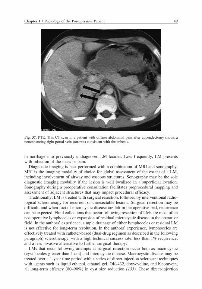
Chapter 1 / Radiology of the Postoperative Patient 49
Fig. 37. PTE. This CT scan in a patient with diffuse abdominal pain after appendectomy shows anonenhancing right portal vein (arrows) consistent with thrombosis.
hemorrhage into previously undiagnosed LM locules. Less frequently, LM presentswith infection of the mass or pain.
Diagnostic imaging is best performed with a combination of MRI and sonography.MRI is the imaging modality of choice for global assessment of the extent of a LM,including involvement of airway and osseous structures. Sonography may be the solediagnostic imaging modality if the lesion is well localized in a superficial location.Sonography during a preoperative consultation facilitates preprocedural mapping andassessment of adjacent structures that may impact procedural efficacy.
Traditionally, LM is treated with surgical resection, followed by interventional radio-logical sclerotherapy for recurrent or unresectable lesions. Surgical resection may bedifficult, and when foci of microcystic disease are left in the operative bed, recurrencecan be expected. Fluid collections that occur following resection of LMs are most oftenpostoperative lymphoceles or expansion of residual microcystic disease in the operativefield. In the authors’ experience, simple drainage of either lymphoceles or residual LMis not effective for long-term resolution. In the authors’ experience, lymphoceles areeffectively treated with catheter-based (dual-drug regimen as described in the followingparagraph) sclerotherapy, with a high technical success rate, less than 1% recurrence,and a less invasive alternative to further surgical therapy.
LMs that recur following attempts at surgical resection occur both as macrocystic(cyst locules greater than 1 cm) and microcystic disease. Macrocystic disease may betreated over a 1-year time period with a series of direct-injection sclerosant techniqueswith agents such as liquid ethanol, ethanol gel, OK-432, doxycycline, and bleomycin,all long-term efficacy (80–90%) in cyst size reduction (133). These direct-injection
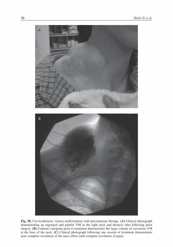
50 Shiels II et al.
A
B
Fig. 38. Cervicothoracic venous malformation with percutaneous therapy. (A) Clinical photographdemonstrating an engorged and painful VM in the right neck and thoracic inlet following priorsurgery. (B) Contrast venogram prior to treatment demonstrates the large volume of cavernous VMat the base of the neck. (C) Clinical photograph following one session of treatment demonstratesnear complete resolution of the mass effect (and complete resolution of pain).
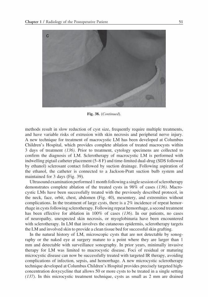
Chapter 1 / Radiology of the Postoperative Patient 51
C
Fig. 38. (Continued).
methods result in slow reduction of cyst size, frequently require multiple treatments,and have variable risks of extrusion with skin necrosis and peripheral nerve injury.A new technique for treatment of macrocystic LM has been developed at ColumbusChildren’s Hospital, which provides complete ablation of treated macrocysts within3 days of treatment (136). Prior to treatment, cytology specimens are collected toconfirm the diagnosis of LM. Sclerotherapy of macrocystic LM is performed withindwelling pigtail catheter placement (5–8 F) and time-limited dual-drug (SDS followedby ethanol) sclerosant contact followed by suction drainage. Following aspiration ofthe ethanol, the catheter is connected to a Jackson-Pratt suction bulb system andmaintained for 3 days (Fig. 39).
Ultrasound examination performed 1 month following a single session of sclerotherapydemonstrates complete ablation of the treated cysts in 98% of cases (136). Macro-cystic LMs have been successfully treated with the previously described protocol, inthe neck, face, orbit, chest, abdomen (Fig. 40), mesentery, and extremities withoutcomplications. In the treatment of large cysts, there is a 2% incidence of repeat hemor-rhage in cysts following sclerotherapy. Following repeat hemorrhage, a second treatmenthas been effective for ablation in 100% of cases (136). In our patients, no casesof neuropathy, unexpected skin necrosis, or myoglobinuria have been encounteredwith sclerotherapy. In LM that involves the cutaneous epidermis, sclerotherapy targetsthe LM and involved skin to provide a clean tissue bed for successful skin grafting.
In the natural history of LM, microscopic cysts that are not detectable by sonog-raphy or the naked eye at surgery mature to a point where they are larger than 1mm and detectable with surveillance sonography. In prior years, minimally invasivetherapy for LM was limited to macrocystic disease. Foci of residual or maturingmicrocystic disease can now be successfully treated with targeted IR therapy, avoidingcomplications of infection, sepsis, and hemorrhage. A new microcystic sclerotherapytechnique developed at Columbus Children’s Hospital provides precisely targeted high-concentration doxycycline that allows 50 or more cysts to be treated in a single setting(137). In this microcystic treatment technique, cysts as small as 2 mm are drained

52 Shiels II et al.
A
B
Fig. 39. Macrocystic LM treated with dual drug time-limited ablation therapy. (A) Neck and chestsonogram demonstrate the macrocystic LM (asterisk). (B) Contrast angiogram during treatmentdemonstrates the vast extent of the macrocystic LM (arrow) in the neck and shoulder. (C) Fluoro-scopic image during treatment demonstrates complete evacuation of intracystic contrast prior todual-drug ablation therapy. Clinically, there was resolution of the mass 3 days following treatment.(D) Neck and shoulder sonogram 6 months following treatment demonstrates complete resolutionof the LM with no evidence of residual or recurrent disease. Normal neck muscle planes are present(arrow) at the site of prior LM, with no evidence of scarring.

Chapter 1 / Radiology of the Postoperative Patient 53
C
D
Fig. 39. (Continued).
and ablated with intracystic doxycycline sclerotherapy. In the authors’ experience,controlled intracystic treatment of microcysts has resulted in 100% cyst ablation andallows for controlled and staged treatment of large areas of microcystic disease.
TUMOR ABLATION AND EMBOLIZATION
Percutaneous image-guided radiofrequency ablation (RFA) of tumors continues togain momentum in adult patients as a viable and effective therapeutic option in thetreatment of solid tumors in a variety of locations, such as the skeleton, liver, spleen,kidney, adrenal gland, lung, and pancreas (138). In children, RFA has been most widelyused in the treatment of osteoid osteoma. The authors use RFA for a growing numberof pediatric indications, to include treatment of osteoid osteoma, recurrent/residualhepatoblastoma, hepatic adenoma (139), palliative treatment of pulmonary metastatic

54 Shiels II et al.
Fig. 40. Recurrent intraabdominal LM treated with dual-drug time-limited ablation therapy. (A)A 6-year-old male with recurrent LM following two surgical resections. Contrast study duringpercutaneous ablation procedure demonstrates large macrocystic LM in the abdomen and pelvis.Imaging follow-up 2 years later demonstrates no recurrence of the macrocystic LM. (B) A 1-year-oldmale with large recurrent intraabdominal and retroperitoneal LM following three surgical attemptsat resection. (C) Contrast study from the percutaneous ablation procedure demonstrates the largestof three macrocystic components of the LM. Follow-up sonography demonstrates no recurrence ofthis macrocystic mass following one treatment. Microcystic disease in the pelvis was treated in asubsequent procedure with no recurrence.
osteogenic sarcoma, metastatic leiomyosarcoma, unresectable leiomyoma, aneurysmalbone cyst, and chondroblastoma. Promising uses of renal RFA include treatment ofmultifocal Wilms tumor, especially in a solitary kidney, and patients with Hippel-Lindau disease; percutaneous RFA is well tolerated, providing an effective nephron-sparing alternative to surgery.
Hepatic and Lung/Tumor AblationThe majority of experience documented in adult hepatic tumor RFA is in treatment
of hepatocellular carcinoma or metastatic disease from organs such as the colon,

Chapter 1 / Radiology of the Postoperative Patient 55
pancreas, lung, and breast (138). Treatment of hepatic tumors (hepatoblastoma) inchildren has been sporadically reported (138). In our clinical practice, focal liver tumorsthat have been treated with RFA include hepatoblastoma, hepatic adenoma (139),and metastatic leimyosarcoma. Patients with hepatoblastoma who present the greatestclinical challenge are those with Beckwith-Wiedemann, with multiple hepatoblastomasdeveloping over time. Indeed, these are ideal candidates for RFA, given the risks ofrepeated surgeries for resection. A percutaneous approach is used in the majority ofhepatic tumor RFAs (Fig. 41). In RFA of pediatric liver tumors, we use a 200-WRF generator with internally cooled electrodes (Radionics; Burlington, MA), either
A
B
Fig. 41. Hepatic adenoma primarily treated with radiofrequency ablation (RFA). (A) Coronal MRIof the right hepatic lobe demonstrate the focal mass, proven to be hepatic adenoma by percutaneouscore needle biopsy (arrow). (B) Sonographic image during RFA demonstrates the RF needle probe(arrowheads) in the adenoma at the beginning of the ablation treatment. (C) CT scan 1 year followingtreatment demonstrates focal scar (arrow) at the site of the RFA with no residual tumor (CT andMRI 2 years later demonstrated further retraction of the hepatic scar and no recurrent tumor).

56 Shiels II et al.
C
Fig. 41. (Continued).
as a single electrode, or a cluster of electrodes for larger lesions. Overlapping RFAsare performed, with each site reaching a target temperature of at least 60 °C over12 minutes.
In the treatment of focal liver tumors, the experience in children is currently small,as pediatric-unique guidelines are developed. At the current time, the authors applyguidelines similar to adult literature (138), such that we can expect complete tumorablation in 90% of patients with tumors smaller than 2.5 cm. Tumors 2.5–3.5 cmin diameter can be expected to be ablated in 70–90% of cases; ablation of tumors3.5–5.0 cm in diameter will be seen in only 50–70% of cases. In large tumors (greaterthan 3.5 cm), and with conditions such as Beckwith-Wiedemann with recurrent hepato-blastoma, palliative RFA is a reasonable goal, if treatment can prolong life expectancywith limited systemic symptoms and few minor complications. The volume of effectivetumor ablation can be enlarged with hypertonic saline tumor pretreatment and withcombination chemoembolization (138).
In the lung, our experience is limited to the treatment of metastatic osteogenicsarcoma patients who are not operative candidates. Internally cooled RF electrodes areused with a 200-W RF generator (Radionics). Large lesions are treated with a three-electrode cluster, each burn reaching a minimum target temperature of 60 °C over 12minutes. We have used both ultrasound and CT guidance for lung RFA cases, withCT proving to be most useful when lung precludes an effective sonographic window.We have provided palliative RF treatment for metastatic lesions as large as 8 cm. Theinsulating effect of lung limits the extension of necrosis into adjacent tissue.
Reoperative Tumor EmbolizationTumor embolization in children is most frequently performed for emergency
treatment of hemorrhage of unresectable tumors and treatment of multifocal recurrent

Chapter 1 / Radiology of the Postoperative Patient 57
tumors, such as renal angiomyolipoma in tuberous sclerosis patients. Uncontrolledhemorrhage in malignancy is rare, yet occurs most commonly with retroperitonealtumors such as extensive renal tumors, large chest wall tumors, and exophytic hepato-blastoma with peritoneal hemorrhage. More frequently used as the first-line therapy,embolization of hepatic hemangiomas is useful as a means of decreasing the vascularshunting that causes heart failure, diffuse intravascular coagulation (DIC), and death.Diagnosis of tumor-related hemorrhage is made primarily with clinical factors, toinclude need for repeated transfusions. CT scan is the most common noninvasivemodality for diagnosis, rarely demonstrating active extravasation of contrast from asite of hemorrhage. Embolization is performed via transarterial catheterization of thenative organ, first diagnosing the focus of hemorrhage with arteriography. Once thesite of origin is identified, microcatheter techniques can isolate the selective arterieswith subsequent embolization with gelfoam slurry or polyvinyl alcohol particles. Ifnecessary, the main artery supplying the organ of origin (e.g., renal artery with primaryrenal malignancy) may be coil embolized as the means of controlling hemorrhage.
A
Fig. 42. This is an angiogram obtained on a trauma patient with right lower extremity ischemia.(A) Contrast injection from above, and occlusion (arrow) of the right common iliac artery. (B) Retro-grade injection from the right common femoral artery, and demonstrates thrombus within the artery(arrows). An attempt was made to cross the occlusion, but was not successful. At surgery, the arterywas completely transected.

58 Shiels II et al.
B
Fig. 42. (Continued).
VASCULAR INJURY
Vascular injury can be either after external trauma or iatrogenic. Trauma victims canhave direct vascular injury such as amputation, avulsion, crush injury, or occlusion orhemorrhage related to fractures. Iatrogenic injury can present after surgery as bleeding(such as after resection or biopsy, or at an anastamotic site), or ischemia. CT is excellentfor excluding vascular trauma (140–142). CT scanning can identify hematomas, andnewer CT angiography techniques can accurately depict vascular injury and occlusion(3). However, CT does not have the dynamic information present with angiography thatcan better identify the occlusion, length of involved segment, amount of reconstitution,and associated findings such as an intimal flap or pseudoaneursym formation (Fig. 42).This information is important for surgical planning, and angiography is still indicatedin certain circumstances when vascular injury is identified by imaging.
Angiography can provide diagnostic information and guide therapeutic interventionsin patients with vascular injury (143–155). If there is hemorrhage in an unstable patient,

Chapter 1 / Radiology of the Postoperative Patient 59
the bleeding site can be identified and embolized using particulate embolic material,coils, or other agents (147–151) (Fig. 43–45). Splenic embolization can be performedfor hemorrhage in unstable patients (148–150). Bleeding from pelvic fractures can becontrolled with internal iliac arterial embolization (151).
Vascular occlusion is readily identified. If there is an obvious intimal flap from adissection, a stent may be indicated, although this is controversial in children (143–146).Endovascular techniques with aortic stent grafting are becoming more common inadolescents and adults with trauma and aortic injury (143,147). There are reportedcases in adults of recannalization of posttraumatic occluded arteries (144–146). In our
A
Fig. 43. This patient presented with hemobilia after surgery for a large posttraumatic hepatichematoma. (A) Angiogram that shows a pseudoaneurysm (arrow), which is better seen after super-selective angiography (B) (arrow). (C) Embolization with coils (arrow), and occlusion of thepseudoaneurysm. The hemobilia resolved.

60 Shiels II et al.
experience, occlusion of a peripheral artery in a child is often caused by completedisruption, although vasospasm stops continued hemorrhage (Fig. 42). Attempts atrecannulation in these patients might lead to greater problems. Arterial occlusivedisease is uncommon in children. However, fibromuscular dysplasia or other arteritides,trauma (external or iatrogenic), venous stenoses from prior central venous access,and anastamotic strictures (usually after transplantation or with dialysis shunts) maypresent with vascular stenosis (152–155) (Figs. 46 and 47]. Angioplasty is indicated inthese patients, with stenting reserved for elastic recoil or dissection after the dilatation(152–155).
B
Fig. 43. (Continued).

Chapter 1 / Radiology of the Postoperative Patient 61
C
Fig. 43. (Continued).
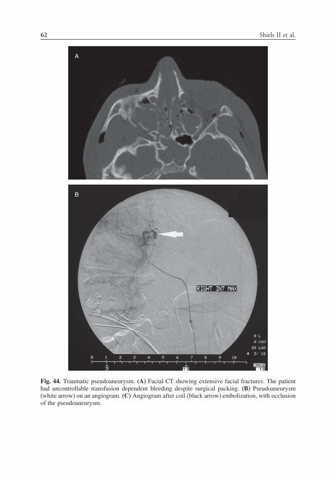
62 Shiels II et al.
A
B
Fig. 44. Traumatic pseudoaneurysm. (A) Facial CT showing extensive facial fractures. The patienthad uncontrollable transfusion dependent bleeding despite surgical packing. (B) Pseudoaneurysm(white arrow) on an angiogram. (C) Angiogram after coil (black arrow) embolization, with occlusionof the pseudoaneurysm.

Chapter 1 / Radiology of the Postoperative Patient 63
C
Fig. 44. (Continued).

64 Shiels II et al.
A
B
Fig. 45. This postsurgical child had significant hematemesis. (A) Angiogram shows a bleedingsite (arrow) with extensive extravasation into a hematoma (*). (B) Resolution of the hemorrhagefollowing embolization with coils (arrow).
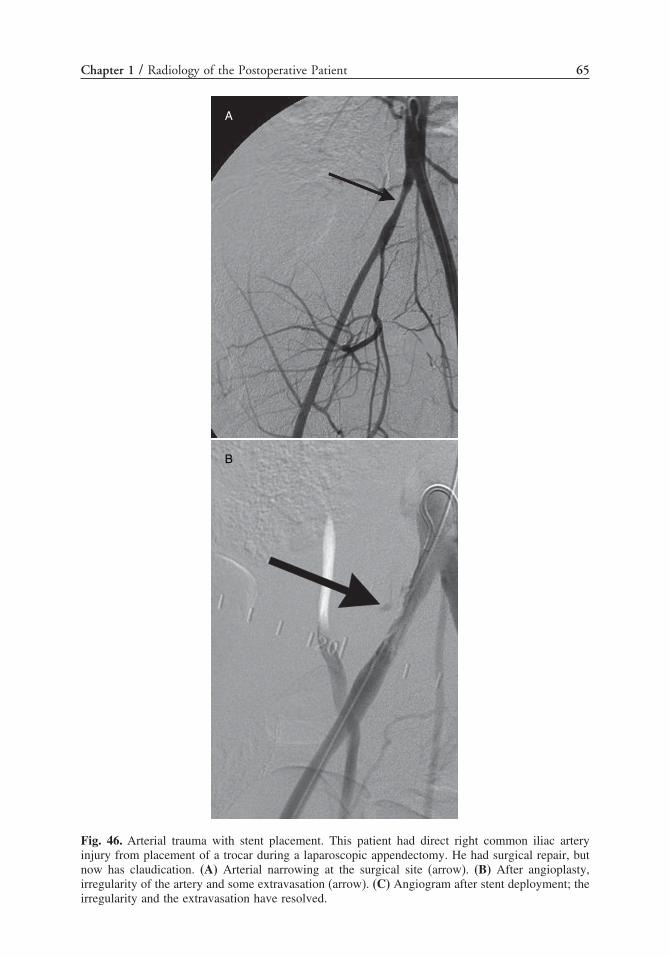
Chapter 1 / Radiology of the Postoperative Patient 65
A
B
Fig. 46. Arterial trauma with stent placement. This patient had direct right common iliac arteryinjury from placement of a trocar during a laparoscopic appendectomy. He had surgical repair, butnow has claudication. (A) Arterial narrowing at the surgical site (arrow). (B) After angioplasty,irregularity of the artery and some extravasation (arrow). (C) Angiogram after stent deployment; theirregularity and the extravasation have resolved.
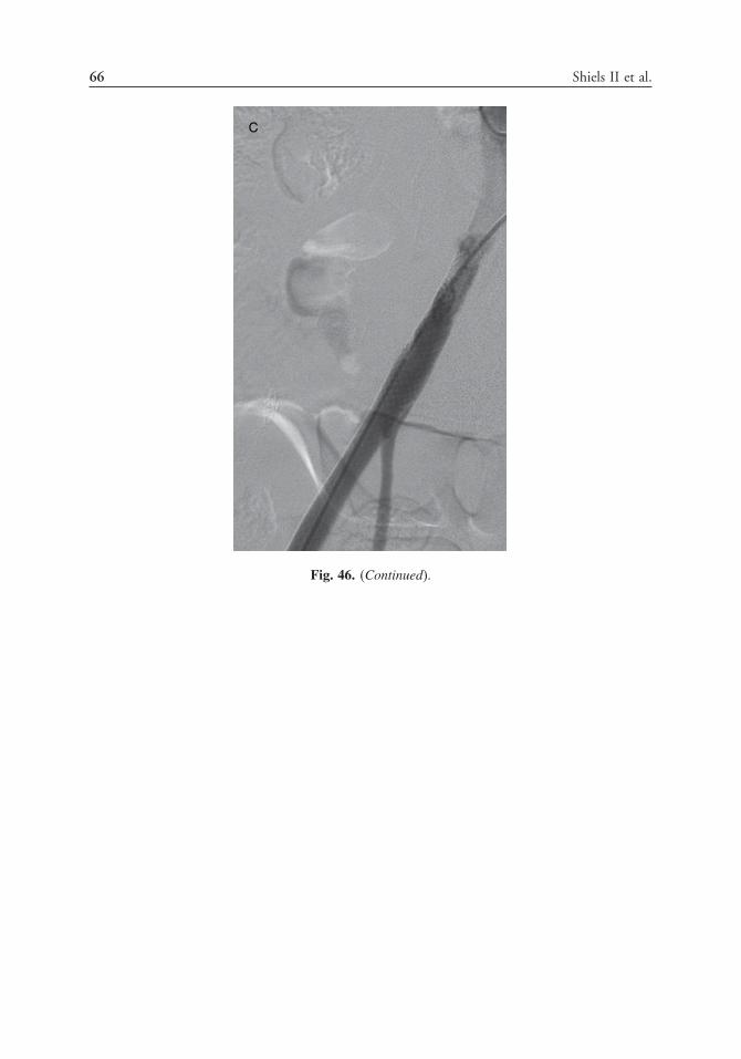
66 Shiels II et al.
C
Fig. 46. (Continued).

Chapter 1 / Radiology of the Postoperative Patient 67
A
B
Fig. 47. Venous stenosis. (A) Trauma from the catheter tip has caused a venous stenosis of the SVC(arrow). (B) Balloon angioplasty was performed, with resolution of the stenosis (C) and good flowthrough the SVC (arrow).

68 Shiels II et al.
C
Fig. 47. (Continued).
REFERENCES
1. Sivit CJ, Applegate KE. Imaging of acute appendicitis in children. Semin Ultrasound CT MR2003;l24:74–82.
2. Curran TJ, Muenchow SK. The treatment of complicated appendicitis in children using peritonealdrainage: results from a public hospital. J Pediatr Surg 1993;28:204–208.
3. Weyant MJ, Eachempati SR, Maluccio MA, et al. The use of computed tomography for the diagnosisof acute appendicitis in children does not influence the overall rate of negative appendectomy orperforation. Surg Infect 2001;2:19–23.
4. Patrick DA, Janik JE, Janik JS, et al. Increased CT scan utilization does not improve the diagnosticaccuracy of appendicitis in children. J Pediatr Surg 2003;38:659–662.
5. Newman, K, Ponsky T, Kittle K, et al. Appendicitis 2000: variability in practice, outcomes, andresource utilization at thirty pediatric hospitals. J Pediatr Surg 2003;38:372–379.
6. Samelson SL, Reyes HM. Management of perforated appendicitis in children—revisited. Arch Surg1987;122:691–696.
7. Lasson A, Lundagards J, Loren I, Nilsson PE. Appendiceal abscesses: primary percutaneous drainageand selective interval appendicectomy. Eur J Surg 2002;168:264–269.
8. Pereira JK, Chait PG, Miller SF. Deep pelvic abscesses in children: transrectal drainage underradiologic guidance. Radiology 1996;198:393–396.
9. Harisinghani MG, Gervais DA, Hahn PF, et al. CT-guided transgluteal drainage of deep pelvicabscesses: indications, technique, procedure-related complications, and clinical outcome. Radio-graphics 2002;22:1353–1367.
10. Towbin R. Interventional procedures in pediatrics. Semin Pediatr Surg 1992;1:296–307.11. Shuler FW, Newman CN, Angood PB, et al. Nonoperative management for intra-abdominal
abscesses. Am Surg 1996;62:218–222.12. Alexander AA, Eschelman DJ, Nazarian LN, Bonn J. Transrectal sonographically guided drainage
of deep pelvic abscesses. AJR Am J Roentgenol 1994;162:1227–1230.

Chapter 1 / Radiology of the Postoperative Patient 69
13. Walser E, Rzaz S, Hernandez A, et al. Sonographically guided transgluteal drainage of pelvicabscesses. AJR Am J Roentgenol 2003;181:498–500.
14. Gervais DA, Hahn PF, O’Neill MJ, et al. CT-guided transgluteal drainage of deep pelvic abscessesin children: selective uses as an alternative to transrectal drainage. AJR Am J Roentgenol2000;175:1393–1396.
15. Poe LB, Petro GR, Matta I. Percutaneous CT-guided aspiration of deep neck abscesses. AJNR AmJ Neuroradiol 1996;17:1359–1363.
16. Hoffer FA, Bloom DA, Colin AA, et al. Lung abscess versus necrotizing pneumonia: implicationsfor interventional therapy. Pediatr Radiol 1999;29:87–91.
17. Ball WS Jr, Bissett GS III, Towbin RB. Percutaneous drainage of chest abscesses in children.Radiology 1989;171:431–434.
18. Linton OW, Mettler FA. National conference on dose reduction in CT with an emphasis on pediatricpatients. AJR Am J Roentgenol 2003;181:321–329.
19. Jamieson DH, Chait PG, Filler R. Interventional drainage of appendiceal abscesses in children. AJRAm J Roentgenol 1997;169:1619–1622.
20. Cahill AM, Kaye RD, Towbin RB, et al. Pelvic abscess drainage in children: the trans-glutealapproach. Pediatr Radiol 2003;33 (Suppl):S81 (abstract).
21. Tassiopoulos AK, Bum G, Halverson JD. Small bowel fistulas. Surg Clin North Am 1996;76:1175–1181.
22. Berry SM, Fischer JE. Enterocutaneous fistulas. Current Prob Surg 1994;31:474–566.23. O’Dwyer ST. Enterocutaneous fistula: conservative and surgical management. In: Allan RN,
Rhodes JM, Hanauer SB, Keighley MRB, Alexander-Williams J, Fazio VF (eds), Inflammatorybowel diseases, third edition. New York: Churchill Livingstone; 1997: 883–893.
24. Carroll K. Crohn’s disease: new imaging techniques. Ballieres Clin Gastroenterol 1998;12(1):35–72.25. Booya F, Fletcher JG, Huprich JE, et al. CT enterography findings in fistulizing Crohn Disease.
Radiologic Society of North America meeting, Chicago, IL, November 2004.26. Maglinte DDT, Kelvin FM, Sandrasegaran K, et al. Radiology of small bowel obstruction: contem-
porary approach and controversies. Abdom Imaging 2005;30:160–178.27. Kelvin FM, Rice RP. Radiologic evaluation of acute abdominal pain arising from the alimentary
tract. Radiol Clin North Am 1978;16(1):25–36.28. Laufer I. The left lateral view in the plain-film assessment of abdominal distention. Radiology
1976;119:265–269.29. Lorenzo RL, Harolds JA. The use of prone films for suspected bowel obstruction in infants and
children. AJR Am J Roentgenol 1977;129:617–622.30. Maglinte DDT, Balthazar EJ, Kelvin FM, et al. The role of radiology in the diagnosis of small
bowel obstruction. AJR Am J Roentgenol 1997;168:1171–1180.31. Sandrasegaran K, Maglinte DDT. Imaging of small bowel-related complications following major
abdominal surgery. Eur J Radiol 2005;53(3):374–386.32. Maglinte DDT, Kelvin FM, Rowe MG, et al. Small-bowel obstruction: optimizing radiologic inves-
tigation and nonsurgical management. Radiology 2001;218:39–4633. Maglinte DDT, Siegleman ES, Kelvin FM. MR enteroclysis; the future of small-bowel imaging
[editorial]. Radiology 2000;215:639–641.34. Kaye RD, Towbin RB. Interventional procedures in the gastrointestinal tract in children. Gastroen-
terol Clin North Am 2002;31;897–923.35. Kottamasu SR, Stringer DA. Pharynx and esophagus. In: Stringer DA (ed), Stringer and Babyn’s
pediatric gastrointestinal imaging and intervention, second edition. Ontario: Decker Inc; 2000:161–236.
36. Goldblum JR, Rice TW, Richter JE. Histopathological features in esophagomyotomy specimensfrom patients with achalasia. Gastronenterology 1996;111:648–654.
37. Woltman TA, Pellegrini CA, Oelschlager BK. Achalasia. Surg Clin N Am 2005;85:483–493.38. Karnak I, Tanyel FC, Buyukpamukcu N, et al. Esophageal perforations encountered during the
dilation of caustic esophageal strictures. J Cardiovasc Surg 1998;39:373–377.39. Brinster CJ, Singhal S, Lee L, et al. Evolving options in the management of esophageal perforation.
Ann Thorac Surg 2004;77:475–483.40. Han SY, McElvain RB, Aldrete JS, et al. Perforation of the esophagus: correlation of site and cause
with plain film findings. AJR Am J Roentgenol 1985;145:537–540.41. Eroglu A, Kurkcuoglu IC, Karaoglanoglu N, et al. Esophageal perforation: the importance of early
diagnosis and primary repair. Dis Esophagus 2004;17:91–94.

70 Shiels II et al.
42. Foley MJ, Ghahremai GG, Rogers LF. Reappraisal of contrast media used to detect upper gastroin-testinal perforations; comparison of ionic water-soluble media with barium sulfate. Radiology1982;144:231–237.
43. Trinh TD, Benson JE. Fluoroscopic diagnosis of complications after Nissen antireflux fundoplicationin children. AJR Am J Roentgenol 1997;169:1023–1028.
44. Connor F. Gastrointestinal complications of fundoplication. Curr Gastroenterol Rep 2005;7:219–226.45. Fasulakis S, Andronikou S. Balloon dilation in children for esophageal strictures other than those
due to primary repair of esophageal atresia, interposition or restrictive fundoplication. Pediatr Radiol2003;33:682–687.
46. Sty JR, Wells RG, Starshak RJ, et al. The gastrointestinal tract. In: Sty JR, (ed), Diagnostic imagingof infants and children, volume 1. Maryland: Aspen; 1992: 139–246.
47. Ball WS Jr, Strife J, Rosekranz J, et al. Esophageal strictures in children: treatment by balloondilatation. Radiology 1984;150:263–264.
48. Bisset GS III, Ball WS Jr. Balloon dilatation of esophageal strictures in the pediatric population.Semin Intervent Radiol 1991;8:204–209.
49. Tsai JY, Berkery L, Wesson DE, et al. Esophageal atresia and tracheoesphageal fistula: surgicalexperience over two decades. Ann Thorac Surg 1997;64:778–783.
50. Lin HC, Prather C, Fisher RS, et al. Measurement of gastrointestinal transit. Dig Dis Sci2005;30:989–1004.
51. Guo JP, Maurer AH, Fisher RS, et al. Extending gastric emptying scintigraphy from two to fourhours detects more patients with gastroparesis. Dig Dis Sci 2001;40:24–29.
52. Singh SJ, Gibbons NJ, Blackshaw PE, et al. Gastric emptying of solids in normal children—apreliminary report. J Pediatr Surg 2006;41:413–417.
53. Jawad AJ, Al-Rabie A, Hadi A, et al. Spontaneous neonatal gastric perforation. Pediatr Surg Int2002;18:396–399.
54. Furukawa A, Sakoda M, Yamasaki M, et al. Gastrointestinal tract perforation: CT diagnosis orpresence, site and cause. Abdom Imaging 2005;30:524–534.
55. Rubesin SE, Levine MS. Radiologic diagnosis of gastrointestinal perforation. Radiol Clin N Am2003;41:1095–1115.
56. Crawley-Coha T. A practical guide for the management of pediatric gastrostomy tubes based on14 years of experience. J Wound Ostomy Continence Nurs 2004;31(4):183–200.
57. Pofahl WE, Ringold F. Management of early dislodgement of percutaneous endoscopic gastrostomytubes. Surg Laparosc Endosc Percutan Tech 1999;9(4):253–256.
58. Kutiyanawaia MA, Everson NW, Hussain A, et al. Gastrostomy complications in infants and children.Ann R Coll Surg Engl 1998;80:240–243.
59. Levy AD. Noninvasive imaging approach to patients with suspected hepatobiliary disease. TechVasc Interv Radiol 2001;4(3):132–140.
60. Chang JM, Lee JM, Suh KS, et al. Biliary complications in living donor liver transplantation: imagingfindings and the roles of interventional procedures. Cardiovasc Intervent Radiol 2005;28:756–767.
61. Schindel D, Dunn S, Casas A, Billmire D, Vinocur C, Weintraub W. Characterization and treatmentof biliary anastomotic stricture after segmental liver transplantation. J Pediatr Surg 2000;35:940–942.
62. Peclet MH, Ryckman FC, Pedersen SH, et al. The spectrum of bile duct complications in pediatricliver transplantation. J Pediatr Surg 1994;29:214–219; discussion 219–220.
63. Venbrux AC, Osterman FA. Percutaneous management of benign biliary strictures. Tech Vasc IntervRadiol 2001;4:141–146.
64. De Meester X, Vanbeckevoort D, Aerts R, Van Steenbergen W. Biliopleural fistula as a latecomplication of percutaneous transhepatic cholangioscopy. Endoscopy 2005;37:183.
65. Venbrux AC, McCormick CD. Percutaneous endoscopy for biliary radiologic interventions. TechVasc Interv Radiol 2001;4:186–192.
66. Kato S, Kamagata S, Asakura T, et al. A newly developed small-caliber videoduodenoscope forendoscopic retrograde cholangiopancreatography in children. J Clin Gastroenterol 2003;37:173–176.
67. Wise JN, Gervais DA, Akman A, Harisinghani M, Hahn PF, Mueller PR. Percutaneous cholecys-tostomy catheter removal and incidence of clinically significant bile leaks: a clinical approach tocatheter management. AJR Am J Roentgenol 2005;184:1647–1651.
68. Andrew M, David M, Adams M, et al. Venous thromboembolic complications (VTE) in children:first analysis of Canadian Registry of VTE. Blood 1994;83:1251–1257.
69. Rask O, Berntorp E, Ljung R. Risk factors for venous thrombosis in Swedish children and adoles-cents. Acta Paediatr 2005;94:717–722.

Chapter 1 / Radiology of the Postoperative Patient 71
70. Nuss R, Hays T, Manco-Johnson M. Childhood thrombosis. Pediatrics 1995;96:291–294.71. Babyn PS, Gahunia HK, Massicotte P. Pulmonary thromboembolism in children. Pediatr Radiol
2005;35:258–274.72. Dollery CM, Sullivan ID, Bauraind O, Bull C, Milla PJ. Thrmobosis and embolism in long-term
central venous access for parenteral nutrition. Lancet 1994;344:1043–1045.73. Hingorani A, Ascher E, Lorenson E, et al. Upper extremity deep venous thrombosis and its impact
on morbidity and mortality rates in a hospital-based population. J Vasc Surg 1997;26:853–860.74. Evans DA, Wilmott RW. Pulmonary embolism in children. Pediatr Clin North Am 1994;41:569–584.75. Jones SE, Wittram C. The indeterminate CT pulmonary angiogram: imaging characteristics and
patient clinical outcome. Radiology 2005;237:329–337.76. Maldjian PD, Chew FS. Imaging of pulmonary embolism: self-assessment module. AJR Am J
Roentgenol 2006;186:S215–S218.77. Cannizzaro V, Berger F, Kretschmar O, Saurenmann R, Knirsch W, Albisetti M. Thrombolysis
of venous and arterial thrombosis by catheter-directed low-dose infusion of tissue plasminogenactivator in children. J Pediatr Hematol Oncol 2005;27:688–691.
78. Manco-Johnson MJ, Nuss R, Hays T, Krupski W, Drose J, Manco-Johnson ML. Combined throm-bolytic and anticoagulant therapy for venous thrombosis in children. J Pediatr 2000;136:425–426.
79. Huang GJ, Wang JN, Tsai YC, Lin CS, Wu JM. Intrapulmonary thrombolytic therapy in a childwith acute pulmonary embolism due to primary antiphospholipid syndrome. Acta Paediatr Taiwan202;43:351–353.
80. Cook A, Shackford S, Osler T, Rogers F, Sartorelli K, Littenberg B. Use of vena cava filters inpediatric trauma patients: data from the National Trauma Data Bank. J Trauma 2005;59:1114–1120.
81. Reed RA, Teitelbaum GP, Stanley P, Mazer MJ, Tonkin IL, Rollins NK. The use of inferior venacava filters in pediatric patients for pulmonary embolus prophylaxis. Cardovasc Intervent Radiol1996;19:401–405.
82. Cahn MD, Rohrer MJ, Martella MB, Cutler BS. Long-term follwo-up of Greenfield inferior venacava filter placement in children. J Vasc Surg 2001;34:820–825.
83. Haider EA, Rosen JC, Torres C, Valenti, DA. Serial repositioning of Gunther tulip retrievableinferior vena cava filter in a pediatric patient. Pediatr Radiol 2005;35:1135–1138.
84. Ostapchuk M, Roberts DM, Haddy R. Community-acquired pneumonia in infants and children. AmFam Physician 2004;70:899–908.
85. Campbell PW III. New developments in pediatric pneumonia and empyema. Curr Opin Pediatr1995;7:278–282.
86. Byington CL, Spencer LY, Johnson TA, et al. An epidemiological investigation of a sustained highrate of pediatric parapneumonic empyema: risk factors and microbiological associations. Clin InfectDis 2002;34:434–440.
87. Gates RL, Caniano DA, Hayes JR, Arca MJ. Does VATS provide optimal treatment of empyemain children? A systematic review. J Pediatr Surg 2004;39:381–386.
88. Russell-Taylor M. Bacterial pneumonias: management and complication. Paediatr Respir Rev2000;1:14–20.
89. Coote N, Kay E. Surgical versus non-surgical management of pleural empyema. Cochrane DatabaseSyst Rev 2005;19:CD001956.
90. Ekingen G, Guvenc B, Sozubir S, Tuzlaci A, Senel U. Fibrinolytic treatment of complicated pediatricthoracic empyemas with intrapleural streptokinase. Eur J Cardiothorac Surg 2004;26:503–507.
91. Ozcelik C, Ulku R, Onat S, Ozcelik Z, Inci I, Satici O. Management of postpneumonic empyemasin children. Eur J Cardiothorac Surg 2004;25:1072–1078.
92. Lewis RA, Feigin RD. Current issues in the diagnosis and management of pediatric empyema.Semin Pediatr Infect Dis 2002;13:280–288.
93. Misthos P, Sepsas E, Konstantinou M, Athanassiadi K, Skottis I, Lioulias A. Early us of intrapleuralfibrinolytics in the management of postpneumonic empyema. A prospective study. Eur J CardiothoracSurg 2005;28:599–603.
94. Chang YT, Dai ZK, Kao EL, Chou SH, Huang MF. Thoracosocopic decortication: first-line therapyfor pediatric empyema. Eur Surg Res 2005;37:18–21.
95. Barnes NP, Hull J, Thomson AH. Medical management of parapneumonic pleural disease. PediatrPulmonol 2005;39:127–134.
96. Ramnath RR, Heller RM, Ben-Ami T, et al. Implications of early sonographic evaluation of parap-neumonic effusions in children with pneumonia. Pediatrics 1998;101:68–71.
72 Shiels II et al.
97. Wong KS, Lin TY, Huang YC, Chang LY, Lai SH. Scoring system for empyema thoracis and helpin management. Indian J Pediatr 2005;72:1025–1028.
98. Skeete DA, Rutherford EJ, Schlidt SA, Abrams JE, Parker LA, Rich PB. Intrapleural tissueplasminogen activator for complicated pleural effusions. J Trauma 2004;57:1178–1183.
99. Liu HP, Hsieh MJ, Lu HI, Lie YH, Wu YC, Lin PJ. Thoracoscopic-assisted management of postp-neumonic empyema in children refractory to medical response. Surg Endosc 2002;16:1612–1614.
100. Feola GP, Shaw CA, Coburn L. Management of complicated parapneumonic effusions in children.Tech Vasc Interv Radiol 2003;6:197–204.
101. Gates RL, Hogan M, Weinstein S, Arca MJ. Drainage, fibrinolytics, or surgery: a comparison oftreatment options in pediatric empyema. J Pediatr Surg 2004;39:1638–1642.
102. Luh SP, Chou MC, Wang LS, Chen JY, Tsai TP. Video-assisted thoracoscopic surgery in thetreatment of complicated parapneumonic effusions or empyemas: outcome of 234 patients. Chest2005;127:1427–1432.
103. Brutsche MH, Tassi GF, Gyorik S, et al. Treatment of sonographically stratified multiloculatedthoracic empyema by medical thoracoscopy. Treatment of sonographically stratified multiloculatedthoracic empyema by medical thoracoscopy. Chest 2005;128:3303–3309.
104. Subramaniam R, Joseph VT, Tan GM, Goh A, Chay OM. Experience with video-assisted thora-coscopic surgery in the management of complicated pneumonia in children. J Pediatr Surg2001;36:316–319.
105. Schultz KD, Fan LL, Pinsky J, et al. The changing face of pleural empyemas in children: epidemi-ology and management. Pediatrics 2004;113:1735–1740.
106. Cleveland RH, Foglia RP. CT in the evaluation of pleural versus pulmonary disease in children.Pediatr Radiol 1998;18:14–19.
107. Moulton JS. Image-guided drainage techniques. Semin Respir Infect 1999;14:59–72.108. Hume M. Venous thrombosis: mechanisms and treatment. Adv Exp Med Biol 1978;102:215–224.109. Stein PD, Kayali F, Olson RE. Incidence of venous thromboembolism in infants and children: data
from the National Hospital Discharge Survey. J Pediatr 2004;145:563–565.110. Levy ML, Granville RC, Hart D, Meltzer H. Deep venous thrombosis in children and adolescents.
J Neurosurg 2004;101:32–37.111. Beck C, Dubois J, Grignon A, Lacroix J, David M. Incidence and risk factors of catheter-related deep
vein thrombosis in a pediatric intensive care unit: a prospective study. J Pediatr 1998;133:237–241.112. DeAngelis GA, McIlhenny J, Willson DF, et al. Prevalence of deep venous thrombosis in the lower
extremities of children in the intensive care unit. Pediatr Radiol 1996;26:821–824.113. Brandao LR, Williams S, Kahr WH, Ryan C, Temple M, Chan AK. Exercise-induced deep vein
thrombosis of the upper extremity. 2. A case series in children. Acta Haematol 2006;115:221–229.114. Manco-Johnson MJ, Grabowski EF, Hellgreen M, et al. Recommendations for tPA thrombolysis
in children. On behalf of the Scientific Subcommittee on Perinatal and Pediatric Thrombosisof the Scientific and Standardization Committee of the International Society of Thrombosis andHaemostasis. Thromb Haemost 2002;88:157–158.
115. Wakefield TW, Henke PK. The role of inflammation in early and late venous thrombosis: are thereclinical implications? Semin Vasc Surg 2005;18:118–129.
116. Fox EA, Kahn SR. The relationship between inflammation and venous thrombosis. A systematicreview of clinical studies. Thromb Haemost 2005;94:362–365.
117. Rotmans JI, Pasterkamp G, Verhagen HJ, Pattynama PM, Blankestijn PJ, Stroes ES. Hemodialysisaccess graft failure: time to revisit an unmet clinical need? J Nephrol 2005;18:9–20.
118. Eknoyan G, Levin NW, Steinberg E. The National Kidney Foundation Dialysis Outcomes QualityInitiative. Curr Opin Nephrol Hypertens 1997;6:520–523.
119. Andrews EJ Jr, Fleischer AC. Sonography for deep venous thrombosis: current and future applica-tions. Ultrasound Q 2005;21:213–225.
120. Katz DS, Hon M. Current DVT imaging. Tech Vasc Interv Radiol 2004;7:55–62.121. Baarslag HJ, Koopman MM, Reekers JA, van Beek EJ. Diagnosis and management of deep vein
thrombosis of the upper extremity: a review. Eur Radiol 2004;14:1263–1274.122. Baker WF Jr. Thrombolytic therapy: current clinical practice. Hematol Oncol Clin North Am
2005;19:147–181, vii.123. Baldwin ZK, Comerota AJ, Schwartz LB. Catheter-directed thrombolysis for deep venous throm-
bosis. Vasc Endovascular Surg 2004;38:1–9.

Chapter 1 / Radiology of the Postoperative Patient 73
124. Sharafuddin MJ, Sun S, Hoballah JJ, Youness FM, Sharp WJ, Roh BS. Endovascular managementof venous thrombotic and occlusive diseases of the lower extremities. J Vasc Interv Radiol2003;14:405–423.
125. Arepally A, Hofmann LV, Kim HS, et al. Weight-based rt-PA thrombolysis protocol for acute nativearterial and bypass graft occlusions. J Vasc Interv Radiol 2002;13:45–50.
126. Bambini DA, Superina R, Almond PS, Whitington PF, Alonso E. Experience with the Rex shunt(mesenterico-left portal bypass) in children with extrahepatic portal hypertension. J Pediatr Surg2000;35:13–18; discussion 18–19.
127. Hidajat N, Stobbe H, Griesshaber V, Felix R, Schroder RJ. Imaging and radiological interventionsof portal vein thrombosis. Acta Radiol 2005;46:336–343.
128. Thornton RH, Kerlan RK, Gordon RL, et al. Percutaneous salvage of a failing rex shunt. J VascInterv Radiol 2005;16:399–402.
129. Sze DY, Esquivel CO. The role of interventional radiology in a pediatric liver transplant program.Pediatr Transplant 2002;6:1–4.
130. Karani JB, Yu DF, Kane PA. Interventional radiology in liver transplantation. Cardiovasc InterventRadiol 2005;28:271–283.
131. Chiou AC, Biggs KL, Matsumura JS. Vena cava filters: why, when, what, how? Perspect Vasc SurgEndovasc Ther 2005;17:329–339.
132. Kinney TB. Update on inferior vena cava filters. J Vasc Interv Radiol 2003;14:425–440.133. Burrows PE, Mason KP. Percutaneous treatment of low flow vascular malformations. J Vasc Interv
Radiol 2004;15:431–445.134. O’Donovan JC, Donaldson JS, Morello FP, Pensler JM, Vogelzang RL, Bauer B. Symptomatic
hemangiomas and venous malformations in infants, children, and young adults: treatment withpercutaneous injection of sodium tetradecyl sulfate. AJR Am J Roentgenol 1997;169:723–729.
135. Sidhu MK, Perkins JA, Shaw DW, Bittles MA, Andrews RT. Ultrasound-guided endovenous diodelaser in the treatment of congenital venous malformations: preliminary experience. J Vasc IntervRadiol 2005;16:879–884.
136. Shiels WE, Murakami, JW, Coley BD, Hogan MJ. Focused sclerotherapy of lymphatic malforma-tions. Pediatr Radiol 2003;33:S75 (abstract).
137. Shiels, WE, Cahill KD, Foster JA. Percutaneous orbital interventional radiology: challenges,techniques, and new care opportunities. Pediatr Radiol 2006;36:S63 (abstract).
138. Shiels WE, Brown SD. Radiofrequency ablation in children. In: van Sonnenberg E (ed), Tumorablation. Berlin: Springer; 2005: 489–497.
139. Rocourt DV, Shiels WE, Hammond S, Besner GE. Contemporary management of benign hepaticadenoma using percutaneous radiofrequency ablation (RFA). J Pediatr Surg 2006;41(6):1149–1152.
140. Strouse PJ, Close BJ, Marshall KW, Cywes R. CT of bowel and mesenteric trauma in children.Radiographics 1999;19:1237–1250.
141. Sievers EM, Murray JA, Chen D, Velmahos GC, Demetriades D, Berne TV. Abdominal computedtomography scan in pediatric blunt abdominal trauma. Am Surg 1999;65:968–971.
142. Stuhlfaut JW, Barest G, Sakai O, Lucey B, Soto JA. Impact of MDCT angiography on the use ofcatheter angiography for the assessment of cervical arterial injury after blunt or penetrating trauma.AJR Am J Roentgenol 2005;185:1063–1068.
143. Milas ZL, Milner R, Chaikoff E, Wulkan M, Ricketts R. Endograft stenting in the adolescentpopulation for traumatic aortic injuries. J Pediatr Surg 2006;41:E27–E30.
144. Kickuth R, Anderson S, Kocovic L, Ludwig K, Siebenrock K, Triller J. Endovascular treatmentof arterial injury as an uncommon complication after orthopedic surgery. J Vasc Interv Radiol2006;17:791–799.
145. Lonn L, Delle M, Karlstrom L, Risberg B. Should blunt arterial trauma to the extremities be treatedwith endovascular techniques? J Trauma 2005;59:1224–1227.
146. McArthur CS, Marin ML. Endovascular therapy for the treatment of arterial trauma. Mt Sinai J Med2004;71:4—11.
147. Malloy PC, Richard HM 3rd. Thoracic angiography and intervention in trauma. Radiol Clin NorthAm 2006;44:239–249.
148. Madoff DC, Denys A, Wallace MJ, et al. Splenic arterial interventions: anatomy, indications,technical considerations, and potential complications. Radiographics 2005;25 (Suppl 1):S191–S211.
149. Yardeni D, Polley TZ Jr, Coran AG. Splenic artery embolization for post-traumatic splenic arterypseudoaneurysm in children. J Trauma 2004;57(2):404–407.
74 Shiels II et al.
150. Cloutier DR, Baird TB, Gormley P, McCarten KM, Bussey JG, Luks FI. Pediatric splenic injurieswith a contrast blush: successful nonoperative management without angiography and embolization.J Pediatr Surg 2004;39(6):969–967.
151. Velmahos GC, Toutouzas KG, Vassiliu P, et al. A prospective study on the safety andefficacy of angiographic embolization for pelvic and visceral injuries. J Trauma 2002;53:303–308;discussion 308.
152. Turmel-Rodrigues L, Pengloan J, Bourquelot P. Interventional radiology in hemodialysis fistulaeand grafts: a multidisciplinary approach. Cardiovasc Intervent Radiol 2002;25:3–16.
153. D’Souza SJ, Tsai WS, Silver MM, et al. Diagnosis and management of stenotic aorto-arteriopathyin childhood. J Pediatr 1998;132:1016–1022.
154. McLaren CA, Roebuck DJ. Interventional radiology for renovascular hypertension in children. TechVasc Interv Radiol 2003;6:150–157.
155. Moray G, Boyvat F, Sevmis S, et al. Vascular complications after liver transplantation in pediatricpatients. Transplant Proc 2005;37:3200–3202.
156. Andrews EJ Jr, Fleischer AC. Sonography for deep venous thrombosis: current and future applica-tions. Ultrasound Q 2005;21:213–225.
157. Katz DS, Hon M. Current DVT imaging. Tech Vasc Interv Radiol 2004;7:55–62.158. Baarslag HJ, Koopman MM, Reekers JA, van Beek EJ. Diagnosis and management of deep vein
thrombosis of the upper extremity: a review. Eur Radiol 2004;14:1263–1274.159. Baker WF Jr. Thrombolytic therapy: current clinical practice. Hematol Oncol Clin North Am
2005;19:147–181.160. Baldwin ZK, Comerota AJ, Schwartz LB. Catheter-directed thrombolysis for deep venous throm-
bosis. Vasc Endovascular Surg 2004;38:1–9.161. Sharafuddin MJ, Sun S, Hoballah JJ, Youness FM, Sharp WJ, Roh BS. Endovascular management
of venous thrombotic and occlusive diseases of the lower extremities. J Vasc Interv Radiol2003;14:405–423.
162. Arepally A, Hofmann LV, Kim HS, et al. Weight-based rt-PA thrombolysis protocol for acute nativearterial and bypass graft occlusions. J Vasc Interv Radiol 2002;13:45–50.
163. Bambini DA, Superina R, Almond PS, Whitington PF, Alonso E. Experience with the Rex shunt(mesenterico-left portal bypass) in children with extrahepatic portal hypertension. J Pediatr Surg2000;35:13–18; discussion 18–19.
164. Hidajat N, Stobbe H, Griesshaber V, Felix R, Schroder RJ. Imaging and radiological interventionsof portal vein thrombosis. Acta Radiol 2005;46:336–343.
165. Thornton RH, Kerlan RK, Gordon RL, et al. Percutaneous salvage of a failing rex shunt. J VascInterv Radiol 2005;16:399–402.
166. Sze DY, Esquivel CO. The role of interventional radiology in a pediatric liver transplant program.Pediatr Transplant 2002;6:1–4.
167. Karani JB, Yu DF, Kane PA. Interventional radiology in liver transplantation. Cardiovasc InterventRadiol 2005;28:271–283.
168. Chiou AC, Biggs KL, Matsumura JS. Vena cava filters: why, when, what, how? Perspect Vasc SurgEndovasc Ther 2005;17:329–339.
169. Kinney TB. Update on inferior vena cava filters. J Vasc Interv Radiol 2003;14:425–440.

2 Techniques for Difficult Venousand Arterial Access
Jaimie D. Nathan, MD, John M. Racadio,MD, and Brad W. Warner, MD
CONTENTS
Introduction
Preoperative Planning
Preoperative Imaging
Interventional Techniques
Surgical Techniques
References
INTRODUCTION
The maintenance of central venous access is critical to the management of pediatricpatients in various clinical scenarios, and the establishment of vascular access isone of the most common procedures performed by pediatric surgeons. This is ofutmost importance particularly in those who are dependent on parenteral nutrition.Although the internal jugular or subclavian vein is an adequate route of access in mostpatients, a common pitfall associated with long-term venous access is the eventualexhaustion of traditional sites of peripheral or central catheterization over time. Thisis caused by multiple complications, including catheter sepsis, venous stenosis and/orthrombosis, or catheter malfunction. It is therefore critical to understand the variousoptions for long-term central venous access in these patients. In this chapter, we providea comprehensive and multidisciplinary approach to the management of reoperativevenous access with particular attention to preoperative planning and imaging, as wellas specific techniques in interventional radiology and surgery.
PREOPERATIVE PLANNING
Most patients who require long-term vascular access have an associated complexmedical and surgical history. In addition to their underlying disease, such patientshave also often had complications related to venous access itself. The developmentof a rational strategy for central venous access is dependent on the recognitionof the disease processes most likely to require long-term vascular access and the
From: Reoperative Pediatric SurgeryEdited by: S. Teich and D. A. Caniano © Humana Press, Totowa, NJ
75

76 Nathan et al.
identification of suitable vein(s) that will provide extended patency. Patients at highrisk of requiring prolonged venous access generally include those with intestinalfailure, owing to either quantitative (short bowel syndrome) or qualitative (intestinaldysmotility or long-segment Hirschsprung’s disease) dysfunction. In other patients, thepresence of malignancy may require prolonged central venous access for the deliveryof parenteral nutrition, chemotherapeutic agents, and/or frequent blood sampling.Therefore, the prevention of central vein occlusion is a critical goal. In these patients,it is important to avoid ligation of major veins during placement of central venouscatheters (CVCs). Minimization of the number of CVCs inserted in these patientsis also very important. Referring physicians should understand the risks associatedwith CVC insertion. Although CVCs greatly facilitate patient care in terms ofavoiding needle sticks for blood draws and limiting peripheral intravenous lines,future standard insertion sites may be compromised by the overliberal utilizationof CVCs.
Central venous catheter replacement in patients requiring prolonged central venousaccess should be performed only when absolutely necessary. For example, cuts orbreaks in the catheter should seldom dictate the need for a catheter replacement, butrather catheter repair kits should be used to preserve CVCs. In addition, catheterpatency can usually be reestablished from occlusion owing to drug, clot, or lipidsolutions. In order to salvage a CVC, bacteremia can often be managed with a courseof antibiotics through the catheter. Finally, it is important to assess the purpose ofcentral venous access. Insertion of a second CVC may be avoided if a schedule isdevised to enable medications and/or blood draws to all be done through an existingcatheter.
The patient population requiring prolonged central venous access is prone to catheter-related complications. Technical misadventures during CVC insertion may be avoidedby eliciting a careful history, which reveals the need for preoperative imaging. Manyfactors contribute to catheter-related complications, including catheter tip location,duration, and prior catheterization (1,2). The number and sites of previous CVCs,complications from previous CVC insertion, and the presence of coexisting pathologyare all key factors that must be considered. In addition, a history of prior head andneck surgery, previous radiation exposure to the mediastinum, or known mediastinal,lower extremity, or abdominal pathology should alert the clinician that standard sitesfor central venous access may be unsuitable.
The use of preoperative imaging prior to CVC insertion may be guided by physicalexamination, as such findings are frequently related to central vein occlusion orstenosis. For the upper extremities and torso, signs associated with occlusion of theveins that drain into the superior vena cava (SVC) may be identified. For example,upper extremity edema, prominent collateral veins, varicosities, and plethora are allcharacteristic of SVC obstruction. Alternatively, if venous return via the inferior venacava (IVC) is impeded, similar findings of edema and venous hypertension will beevident in the lower extremities.
PREOPERATIVE IMAGING
Venous anatomy may be imaged by a variety of modalities, but ultrasound andmagnetic resonance venography are the most commonly utilized techniques. In thefollowing section, we discuss the use of these and other radiologic modalities. Figure 1

Chapter 2 / Techniques for Difficult Venous and Arterial Access 77
Fig. 1. Proposed algorithm for radiological evaluation of high-risk patients for CVC insertion. MRV:magnetic resonance venography.
depicts a proposed algorithm for the radiologic evaluation of high-risk patients forCVC insertion.
UltrasoundUltrasound is a useful noninvasive method to evaluate suspected upper extremity
deep vein thrombosis (DVT) and central venous obstruction.(3–5). It is typicallyperformed with high-resolution ultrasound equipment using 7.5- to 10-MHz trans-ducers. A typical ultrasound examination to evaluate the status of upper extremitygreat veins involves examination of the bilateral internal jugular (IJ), brachiocephalic,subclavian (SC), and axillary veins. Direct visualization of the SVC, brachiocephalic,and medial aspect of the subclavian veins is often limited by the sternum and clavicles.
Accurate diagnosis of upper extremity and central venous obstruction is dependenton three components of the ultrasound examination: (1) real-time gray scale evalu-ation; (2) color Doppler imaging; and (3) Doppler waveform analysis. The lumina ofnormal patent IJ and SC veins are hypoechoic. Grey scale signs of thrombosis includevariable echogenic intraluminal thrombus (Fig. 2), noncompressibility, loss of respi-ratory variability and vessel pulsation, and absence of response to Valsalva or sniffmaneuvers (6). Color Doppler imaging allows rapid localization of vessels, differenti-ation of veins from arteries, and delineation of thrombus. Doppler waveform analysisallows evaluation of reflected right atrial pressures and changes associated with respi-ration. Patency of interrogated veins to the right atrium is implied by normally reflectedtriphasic atrial waveforms with subtle respiratory variation.
With the patient in the supine position, the IJ veins, which are superficial andeasily visible and compressible by the transducer, are followed to their junction with

78 Nathan et al.
Fig. 2. Transverse ultrasound of the IJ vein with delineation of thrombus (arrow). Arrowheaddemonstrates the normal hypoechoic appearance of the patent portion of the lumen. CA: carotidartery.
the brachiocephalic veins. The latter may be visualized from either a suprasternal ormedial infraclavicular acoustic window. The SC veins, which are usually more caudaland superficial to the SC arteries, are imaged to their junction with the IJ veins. It isimportant to differentiate large venous collaterals that may develop in the setting ofprior SC vein thrombosis. Compared to the SC veins, such collaterals, however, aretypically smaller and in atypical locations relative to the SC artery. Increased velocityand turbulence identified within the vessel signify stenosis. Although it may not bepossible to directly image the SVC and brachiocephalic veins, careful examination ofthe SC and IJ veins for dampened atrial pulsations and abnormal response to respiratorymaneuvers may be suggestive of central obstruction. With regard to the detection ofSC vein stenosis by ultrasound, false-positive results are rare. False-negative studiesmay occur in cases of small nonobstructive thrombus or in patients with extensivecollateral networks.
Magnetic Resonance Imaging (MRI) VenographyMRI venography is highly accurate in the delineation of central venous anatomy, and
the correlation of venous obstruction with contrast venography has been excellent (7–9).The multiplanar capabilities of MR venography are particularly useful in assessingthoracic inlet and mediastinal veins, which may be difficult to image entirely in a
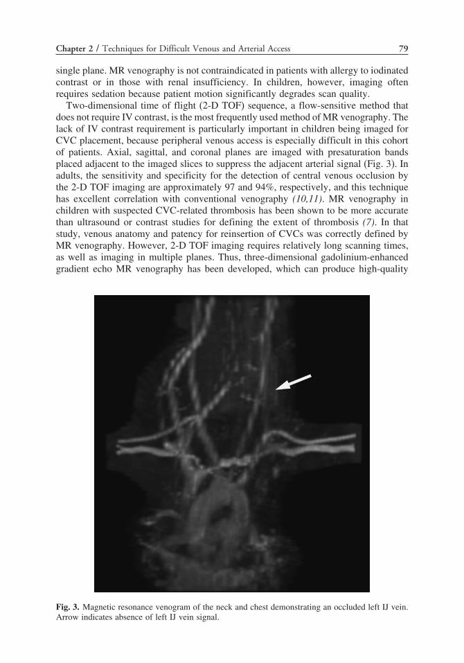
Chapter 2 / Techniques for Difficult Venous and Arterial Access 79
single plane. MR venography is not contraindicated in patients with allergy to iodinatedcontrast or in those with renal insufficiency. In children, however, imaging oftenrequires sedation because patient motion significantly degrades scan quality.
Two-dimensional time of flight (2-D TOF) sequence, a flow-sensitive method thatdoes not require IV contrast, is the most frequently used method of MR venography. Thelack of IV contrast requirement is particularly important in children being imaged forCVC placement, because peripheral venous access is especially difficult in this cohortof patients. Axial, sagittal, and coronal planes are imaged with presaturation bandsplaced adjacent to the imaged slices to suppress the adjacent arterial signal (Fig. 3). Inadults, the sensitivity and specificity for the detection of central venous occlusion bythe 2-D TOF imaging are approximately 97 and 94%, respectively, and this techniquehas excellent correlation with conventional venography (10,11). MR venography inchildren with suspected CVC-related thrombosis has been shown to be more accuratethan ultrasound or contrast studies for defining the extent of thrombosis (7). In thatstudy, venous anatomy and patency for reinsertion of CVCs was correctly defined byMR venography. However, 2-D TOF imaging requires relatively long scanning times,as well as imaging in multiple planes. Thus, three-dimensional gadolinium-enhancedgradient echo MR venography has been developed, which can produce high-quality
Fig. 3. Magnetic resonance venogram of the neck and chest demonstrating an occluded left IJ vein.Arrow indicates absence of left IJ vein signal.

80 Nathan et al.
images that can be acquired within a single breath hold (9,12–14). This modality mayonly be useful in a cooperative pediatric patient or under general anesthesia.
Contrast Computed Tomography (CT)Contrast enhanced CT is a reliable technique for the evaluation of the etiology of
SVC obstruction, and its primary advantage over ultrasound is its ability to imagebeyond bone and air (15). The SVC is opacified with iodinated contrast material, andthe luminal patency and relationship of the SVC to adjacent structures may be studied.Helical CT phlebography can provide multiplanar and three-dimensional reconstructionof the central venous anatomy and mapping of collateral tributaries in cases of SVCobstruction (16). Technical limitations of this technique include streak artifact andintraluminal filling defects caused by contrast dilution by unopacified blood from theIJ and azygous veins and peripheral displacement of intraluminal contrast by laminarflow (17). As a result of these potential flow-related artifacts, the diagnosis of venousobstruction by CT scanning requires two criteria: (1) decreased or absent venous opaci-fication distal to the site of an obstructive intraluminal filling defect or compressivemass; and (2) the presence of collateral pathways (16). As with conventional contrastvenography, potential contraindications to CT scanning include the need for iodinatedcontrast in a patient with a history of renal insufficiency or a history of allergic reaction.
Conventional Contrast VenographyThe gold standard for the evaluation of central venous obstruction is conventional
contrast venography, and it is used generally when other studies are impractical orequivocal, and in order to thoroughly define venous anatomy in complicated clinicalscenarios. The advantage of contrast venography is not only identification of venousstenosis or obstruction, but also real-time evaluation of flow through the central veinsand collateral channels. This information is critical in planning the most appropriateapproach for CVC placement. Suspected SVC syndrome may be evaluated by conven-tional contrast venography (18–20). Venography has been reported to be superior to CTscanning in the opacification of collateral venous tributaries and in the determinationof the degree of venous obstruction (17).
Evaluation of axillary veins and central veins in the chest requires placement ofa large-bore intravenous cannula in the antecubital fossa. Nonionic contrast is hand-injected, and digital acquisition is performed with the arm in a neutral palmar position.The filling of the deep veins of the arm (brachial and axillary veins) may be aidedby placement of a tourniquet. To better visualize the central veins, the contrast-filledarm may be elevated or manually compressed to increase contrast drainage, and theSVC can be evaluated by simultaneous antecubital injections. In patients who cannotreceive iodinated contrast because of renal failure or allergic reaction, gadolinium orcarbon dioxide can be used (21).
INTERVENTIONAL TECHNIQUES
Translumbar Catheterization of the IVCThe use of translumbar percutaneous IVC cannulation for long-term parenteral
nutrition in adults was first described in 1985 (22). This technique is a safe and effectiveoption in both adults and children for the administration of parenteral nutrition, long-term antibiotic therapy, and chemotherapy, as well as for stem cell harvesting prior to

Chapter 2 / Techniques for Difficult Venous and Arterial Access 81
bone marrow transplantation (23–26). In the adult population, infectious and thromboticcomplications have been reported to be less than 5% (23,26,27). Although it has beendescribed, the risk for IVC thrombosis is less in patients with translumbar cathetersthan in the setting of femoral venous catheterization, because of the relatively high IVCflow (25,28–30). In the majority of cases of thrombotic complications, managementconsists of the administration of fibrinolytic agents.
Complications specific to the technique of translumbar catheterization are migrationof the catheter into the subcutaneous tissues, retroperitoneum, or iliac veins (29).Contraindications to translumbar CVCs include coagulopathy (owing to the risk ofinadvertent aortic puncture) and presence of infection on the lower back or rightanterolateral abdomen in the region of the anticipated puncture or tunnel sites. Markedtruncal obesity has also been proposed to be a contraindication, because of an increasedrisk of catheter migration (28,29).
Preprocedure imaging should confirm IVC patency and exclude retroperitonealpathology. With the patient in the prone position, a puncture site is chosen just cephaladto the right iliac crest and approximately 8–10 cm to the right of midline. Underfluoroscopic guidance, a 21-G microaccess needle is advanced into the infrarenal IVCat the L2 to L3 level by directing it cephalad toward the anterolateral margin of thevertebral bodies. Alternatively, a femoral venous catheter placed with the tip in theIVC can confirm position of the IVC and be used as a target for puncture. One grouphas recommended a more cephalad puncture of the IVC, with a skin puncture site justbelow the 12th rib (31). In infants and small children, translumbar ultrasound may beutilized to visualize the IVC, and access can be achieved under real-time ultrasoundguidance. After confirmation of IVC access by aspiration of blood, contrast may beinjected to confirm IVC placement and exclude entry into the renal vein. By Seldingertechnique, a guidewire is then advanced into the IVC, and following placement ofa coaxial dilator, a stiffer guidewire is advanced into the SVC or right atrium. Thetract is then serially dilated, and an appropriately sized peel-away sheath is placed. Asubcutaneous tunnel is extended laterally to the anterior lower abdominal wall, and thecatheter is advanced through the peel-away sheath into the central circulation. Finalcatheter tip position should be as high as possible at the IVC/right atrial junction.Manual compression and supine patient position are sufficient to achieve hemostasisat the time of catheter removal.
Transhepatic Catheterization of the IVCAlthough uncommon, occlusion of both the SVC and the infrarenal IVC presents
a challenge to central venous catheterization. In such cases, the retrohepatic portionof the IVC is typically patent. Percutaneous access of the retrohepatic IVC can beachieved directly via the liver parenchyma or the hepatic veins. The technique oftranshepatic central venous catheterization was first described in adults in 1991 forthe administration of long-term parenteral nutrition (32). Transhepatic catheterizationhas been successful in children for short-term interventional radiology proceduresand cardiac catheterizations (33), emergency short-term dialysis (34), and prolongeddelivery of parenteral nutrition (35,36). Rates of infectious and thrombotic complica-tions are comparable to standard central venous access approaches (35).
Catheter dislodgement with subsequent bleeding from the hepatic parenchymal tractand hepatic vein thrombosis are potential complications unique to transhepatic CVCplacement. Catheter migration and dislodgement may occur because of diaphragmatic
82 Nathan et al.
movement during deep respiration. In infants and small children, severe abdominaldistension and growth spurts may also result in catheter malposition (35). Hepaticvein thrombosis is usually asymptomatic if the other hepatic veins are patent, althoughacute Budd-Chiari syndrome and subsequent death has been described in 1 child(37). IVC thrombosis as a complication of transhepatic catheterization has not beenreported in the pediatric population. Cholangitis, hemobilia, and liver abscess are otherpotential complications of this approach. Uncorrectable coagulopathy, massive ascites,and biliary infection represent contraindications to the placement of a transhepatic CVC.
Patient evaluation prior to transhepatic central venous catheterization usually consistsof contrast-enhanced CT scanning of the abdomen to evaluate the liver, hepatic veins,retrohepatic IVC, subphrenic space, and perihepatic peritoneal cavity. Ultrasound canfurther evaluate the hepatic veins and IVC, and is useful in planning the optimalapproach (38).
With the patient in the supine position, the right hepatic vein is most commonlyaccessed via a lateral approach in the mid axillary line through the 10th or 11thinterspace. Transpleural puncture must be avoided, as it can result in pneumo- orhemothorax. Alternatively, the middle hepatic vein may be accessed via an anteriorsubcostal approach. Direct ultrasound and fluoroscopic guidance is utilized to directa 22-G Chiba needle into the appropriate hepatic vein. Following contrast injectionto confirm needle tip position, a platinum-tipped guidewire is advanced into the rightatrium. A coaxial dilator is advanced into the IVC, and the guidewire is exchanged for astiffer wire with the tip placed ideally in the SVC to avoid dysrhythmias. Alternatively,puncture can be made with a sheathed 19-G needle, through which a guidewire canbe directly passed. Serial dilation over the wire by Seldinger technique is followed byinsertion of a peel-away sheath, and finally the venous access catheter after tunnelingto the anterolateral abdomen. Final catheter tip position should be at the SVC/rightatrial junction to allow for catheter migration, particularly in infants who are growingrapidly (35). In children who are growing rapidly, chest radiographs can be obtainedat 2- to 3-month intervals to assess catheter tip position. If necessary, a new cathetercan be exchanged over a wire and placed via a new tunnel and catheter exit site.
Removal of transhepatic catheters carries a risk of intraperitoneal or subcap-sular hemorrhage along the catheter tract. Track embolization should be performedby deploying coils or Gelfoam pledgets as the catheter is slowly withdrawn fromthe liver (34).
Catheterization of Collateral VeinsPercutaneous placement of CVCs can occasionally be placed via enlarged upper
or lower extremity collateral veins. Only a few case reports have documented thisapproach, and they include CVC placement via intercostal veins (39–42) and viathyrocervical collaterals (43). Contrast venography is very useful in evaluating thesize and course of the collateral veins, as well as the reconstituted central vein. It isimportant to recognize the presence and status of other collateral veins, because severesymptoms may be precipitated by catheter-induced thrombosis of a solitary majordraining vein. Successful cannulation and advancement of CVCs through tortuouscollateral veins usually requires ingenuity and the application of angiographic tools,including hydrophilic selective catheters and torque-control guidewires. Combinedinterventional radiology and surgical approaches have been described, and in manycases, may offer the best chance for successful CVC placement.

Chapter 2 / Techniques for Difficult Venous and Arterial Access 83
Angioplasty or Stent PlacementIn the setting of an indwelling CVC, obstruction of the vein harboring the catheter can
result in acute signs and symptoms, including edema, pain, and venous hypertension.In this scenario, thrombolytic therapy has a fairly high rate of success. Subsequentvenography often demonstrates a residual venous stenosis. Balloon angioplasty ofthe residual stenosis should be considered in order to preserve the central vein forcontinued vascular access (Fig. 4) (44,45). The placement of an endovascular stentis an additional strategy, but this intervention requires further study to determine itsutility in the salvage of central venous access sites. One indication for stenting may be“early recoil” of the venous stenosis to its original size after successful dilation. Morerecently, sharp recanalization of chronically occluded central veins has been describedand may offer another option for management of these challenging patients (46–48).These techniques require advanced endovascular skills and are probably limited tocenters committed to such interventional radiology procedures.
Peripherally Inserted Central Catheters (PICCs)Recently, PICCs have become widely utilized for prolonged administration of
parenteral nutrition, antibiotics, and chemotherapy, as well as for intermittent bloodsampling (2,49–51). It has been reported that with fluoroscopic guidance, PICCplacement is successful in children in more than 92% of cases (52). PICCs have beendemonstrated to be a reliable and safe route of central venous access in neonatesand children, with an infectious complication rate of approximately 2% (53,54). Theoverall complication rate is low and most commonly involves catheter occlusion,displacement, and fracture, as well as phlebitis and venous thrombosis. In a study inchildren, a higher risk of complications has been demonstrated more than 30 days afterPICC placement, and this has called into question whether PICCs should be used forlong-term central venous access (2). A recent review of the use of PICCs for long-termantibiotic and parenteral nutrition administration in adults reported no clear advantageto the use of PICCs compared to traditional CVCs (51). Prospective, randomizedstudies are required to determine whether an advantage of one technique over the otherexists. Contraindications to PICC placement include coagulopathy, lack of peripheral
Fig. 4. Balloon dilation of left SC vein stenosis. (A) Venogram demonstrating near-completeocclusion of left SC vein. (B) Venogram after successful balloon dilation demonstrating patency ofSC and innominate veins without residual stenosis.

84 Nathan et al.
venous access, the presence of venous thrombosis, end-stage renal disease (owing tothe potential future need for the creation of an arteriovenous fistula for hemodialysisaccess), a history of radiation therapy, and prior axillary lymph node dissection.
The most commonly used PICC sizes in children are 2-, 3-, and 4-F catheters. An appro-priate vein is selected for cannulation, and usually superficial veins are used, includingthe cephalic or basilic vein in the upper extremity, the temporal or posterior auricular veinin the scalp, and the saphenous vein in the lower extremity. If necessary, deeper veins,including the axillary, femoral, popliteal, and external jugular veins, may be catheterizedfor PICC placement. The patient is placed in the supine position with the arm abductedat a 90� angle, and the head is turned toward the arm of insertion. The catheter lengthis estimated as the distance from a marked insertion site to the midpoint of the sternum.If the insertion site is in the lower extremity, the catheter should be measured to atleast 2 cm above the umbilicus. Under sterile conditions, the vein is localized by eitherpalpation or direct visualization or is identified by ultrasound guidance. Venipunctureis performed with the appropriately sized needle, a guidewire is advanced through theneedle into the vein, a dilator/peel-away sheath is inserted over the wire by Seldingertechnique, and after removal of the dilator and wire, the PICC is advanced through thepeel-away sheath to the previously measured catheter length. Alternatively, some centersprefer to access the vein initially with a peel-away sheathed needle, remove the needle,and immediately pass the PICC through the sheath. If the PICC is not placed underfluoroscopy, a chest radiograph is performed to confirm the position of the catheter tipin the SVC, and incorrectly positioned catheters are adjusted as necessary. Because ofa high rate of initial incorrectly positioned PICCs in children, it has been advocatedthat pediatric PICC placement be performed in an interventional radiology suite withfluoroscopic assistance to allow real-time adjustment of catheter tip position and toreduce the number of catheter manipulations required (55). Others have proposed that adedicated PICC team with ultrasound capability for bedside PICC placement can reducecost by avoiding the use of expensive interventional radiology facilities (56). The relativecost-effectiveness of these two approaches to PICC placement requires further study.
SURGICAL TECHNIQUES
The SC vein and external or IJ veins are the traditional sites for access to the SVC,and either a percutaneous or surgical cut-down approach may be performed. When theSVC is inaccessible owing to thrombosis of the IJ, SC, or brachiocephalic veins, theIVC should be utilized. Percutaneous and surgical approaches to central venous accessvia the IVC have included cannulation of the saphenous (57,58), femoral (59–62),inferior epigastric (63–66), gonadal (67–69), iliac (70–72), lumbar (73), and hepaticveins (35,36,38).
Originally considered a less favorable approach owing to activity restriction andincreased rates of catheter infection, femoral venous catheters are safe and haveinfection rates equal to that of SVC catheters, particularly when tunneled to the anteriorthigh or abdominal wall. (61,62,74). The approach to placement of a femoral venouscatheter is usually via a percutaneous technique, and this is especially useful in theneonatal population (59,75).
Access to the inferior epigastric vein is via the groin (63,65,66). This approach isindicated in cases where the femoral vein cannot be accessed because of the presenceof soilage or open wounds. In addition, if the femoral vein cannot be percutaneously

Chapter 2 / Techniques for Difficult Venous and Arterial Access 85
cannulated and the saphenous vein has already been ligated, the inferior epigastricapproach provides a good option to cannulation of the IVC. Access to the inferiorepigastric vein is via a transverse inguinal incision and exposure of the internal inguinalring, and the vein courses along the medial margin of the internal ring. The proximalcatheter or reservoir can be tunneled up to the abdominal wall, which may lessen therisk of contamination and infection. In cases in which the femoral veins are inaccessiblebecause of interruption or thrombus, catheterization of the iliac vein may be performedvia a retroperitoneal approach (71). Retroperitoneal exposure of the iliac vein allowscatheter placement by direct venipuncture or via venotomy without the need for veinligation. The CVC is tunneled onto the chest wall for access. Similarly, the gonadalvein can be accessed via an oblique incision in the iliac fossa (67–69). Gonadal venouscatheters are also usually tunneled onto the lower anterior chest wall. If the iliofemoralor lower IVC is inaccessible, percutaneous transhepatic or translumbar techniquesshould be considered as outlined previously.
If access to both the SVC and IVC is impossible because of vessel thrombosis, centralvenous catheterization may be accomplished via alternate SVC collaterals, such as theazygous (76), intercostal (39–42,77), and internal mammary veins (78). An additional,more invasive, option is direct right atrial cannulation (79,80). The traditional approachto azygous, hemiazygous, and intercostal vein catheterization is via thoracotomy, andthe CVC is tunneled to the anterior chest. The advent of video-assisted thoracic surgery(VATS) has significantly improved these approaches, and has provided a minimallyinvasive technique for direct right atrial catheterization (36,80). The thoracoscopicapproach to right atrial catheterization involves opening the pericardium, exposing theatrium, and placing the catheter over a guidewire. The catheter is secured by using aright atrial pursestring suture. Figure 5 depicts a proposed algorithm for CVC insertion
Fig. 5. Proposed algorithm for placement of CVC in patients with chronic occlusion of the SVC.

86 Nathan et al.
Fig. 6. Proposed algorithm for placement of CVC in patients with chronic occlusion of the IVC.
in the setting of chronic SVC occlusion, and Fig. 6 demonstrates an algorithm for CVCplacement in patients with an IVC occlusion.
In conclusion, the establishment of central venous access in patients with a historyof multiple previous CVCs and known venous thrombosis is a challenge. There aremultiple options for CVC insertion in this difficult patient population, and approachesinclude both interventional and surgical techniques. In order to avoid exhaustingcentral venous cannulation approaches, it is critical to limit the placement of CVCsas much as possible and to replace them only when absolutely necessary. In high-riskpatients, thorough preoperative imaging will identify patent vessels, delineate a rationaloperative approach, and avoid technical misadventures during CVC insertion.
REFERENCES
1. Racadio JM, Doellman DA, Johnson ND, Bean JA, Jacobs BR. Pediatric peripherally inserted centralcatheters: complication rates related to catheter tip location. Pediatrics 2001;107(2):E28.
2. Matsuzaki A, Suminoe A, Koga Y, Hatano M, Hattori S, Hara T. Long-term use of periph-erally inserted central venous catheters for cancer chemotherapy in children. Support Care Cancer2006;14(2):153–160.
3. Gaitini D. Current approaches and controversial issues in the diagnosis of deep vein thrombosis viaduplex Doppler ultrasound. J Clin Ultrasound 2006;34(6):289–297.
4. Salcuni M, Fiorentino P, Pedicelli A, DiStasi C. Diagnostic imaging in deep vein thrombosis of thelimbs. Rays 1996;21(3):328–339.
5. Gooding GA, Woodruff WA. Color Doppler imaging in the subclavian-axillary region and upperextremity. Clin Imaging 1994;18(3):165–172.

Chapter 2 / Techniques for Difficult Venous and Arterial Access 87
6. Patel MC, Berman LH, Moss HA, McPherson SJ. Subclavian and internal jugular veins at DopplerUS: abnormal cardiac pulsatility and respiratory phasicity as a predictor of complete central occlusion.Radiology 1999;211(2):579–583.
7. Shankar KR, Abernathy LJ, Das KS, et al. Magnetic resonance venography in assessing venouspatency after multiple venous catheters. J Pediatr Surg 2002;37(2):175–179.
8. Kroencke TJ, Taupitz M, Arnold R, Fritsche L, Hamm B. Three-dimensional gadolinium-enhancedmagnetic resonance venography in suspected thrombo-occlusive disease of the central chest veins.Chest 2001;120(5):1570–1576.
9. Thornton MJ, Ryan R, Varghese JC, Farrell MA, Lucey B, Lee MJ. A three-dimensionalgadolinium-enhanced MR venography technique for imaging central veins. AJR Am J Roentgenol1999;173(4):999–1003.
10. Hartnell GG Hughes LA, Finn JP, Longmaid HE. Magnetic resonance angiography of the centralchest veins. A new gold standard? Chest 1995;107:1053–1057.
11. Rose SC, Gomes AS, Yoon HC. MR angiography for mapping potential central venous access sitesin patients with advanced venous occlusive disease. AJR Am J Roentgenol 1996;166:1181–1187.
12. Li W, David V, Kaplan R, Edelman RR. Three-dimensional low dose gadolinium-enhanced peripheralMR venography. J Magn Reson Imaging 1998;8:630–633.
13. Oxtoby JW, Widjaja E, Gibson KM, Uzoka K. 3D gadolinium-enhanced MRI venography: evaluationof central chest veins and impact on patient management. Clin Radiol 2001;56:887–894.
14. Tanju S, Sancak T, Dusunceli E, Yagmurlu B, Erden I, Sanlidilek U. Direct contrast-enhanced 3D MRvenography evaluation of upper extremity deep venous system. Diagn Interv Radiol 2006;12(2):74–79.
15. Bechtold RE, Wolfman NT, Karstaedt N, Choplin RH. Superior vena caval obstruction: detectionusing CT. Radiology 1985;157:485–487.
16. Qanadli SD, El Hajjam M, Bruckert F, et al. Helical CT phlebography of the superior vena cava:diagnosis and evaluation of venous obstruction. AJR Am J Roentgenol 1999;172:1327–1333.
17. Engel IA, Auh YH, Rubenstein WA, Sniderman K, Whalen JP, Kazam E. CT diagnosis of mediastinaland thoracic inlet venous obstruction. AJR Am J Roentgenol 1983;141:521–526.
18. Lochridge SK, Knibbe WP, Doty DB. Obstruction of the superior vena cava. Surgery 1979;85:14–24.19. Benenati JF, Becker GJ, Mail JT, Holden RW. Digital subtraction venography in central venous
obstruction. AJR Am J Roentgenol 1986;147:685–688.20. Stanford W, Jolles H, Ell S, Chiu LC. Superior vena cava obstruction: a venographic classification.
AJR Am J Roentgenol 1987;148:259–262.21. Hahn ST, Pfammatter T, Cho KJ. Carbon dioxide gas as a venous contrast agent to guide upper-arm
insertion of central venous catheters. Cardiovasc Intervent Radiol 1995;18:146–149.22. Kenney PR, Dorfman GS, Denny DF Jr. Percutaneous inferior vena cava cannulation for long-term
parenteral nutrition. Surgery 1985;97:602–605.23. Bennett JD, Papadouris D, Rankin RN, et al. Percutaneous inferior vena caval approach for long-term
central venous access. J Vasc Interv Radiol 1997;8:851–885.24. Robertson LJ, Jaques PF, Mauro MA, Azizkhan RG, Robards J. Percutaneous inferior vena cava
placement of tunneled silastic catheters for prolonged vascular access in infants. J Pediatr Surg1990;25:596–598.
25. Robards JB, Jaques PF, Mauro MA, Azizkhan RG. Percutaneous translumbar inferior vena cavacentral line placement in a critically ill child. Pediatr Radiol 1989;19:140–141.
26. Elduayen B, Martinez-Cuesta A, Vivas I, Delgado C, Pueyo JC, Bilbao JI. Central venous catheterplacement in the inferior vena cava via the direct translumbar approach. Eur Radiol 2000;10(3):450–454.
27. Lund GB, Trerotola S, Scheel PJ Jr. Percutaneous translumbar inferior vena cava cannulation forhemodialysis. Am J Kidney Dis 1995;25:732–737.
28. Lund GB, Lieberman RP, Haire WD, Martin VA, Kessinger A, Armitage JO. Translumbar inferiorvena cava catheters for long-term venous access. Radiology 1990;174:31–35.
29. Rajan DK, Croteau DL, Sturza SG, Harvill ML, Mehall CJ. Translumbar placement of inferior venacaval catheters: a solution for challenging hemodialysis access. Radiographics 1998;18:1155–1167.
30. Denny DF Jr, Greenwood LH, Morse SS, Lee GK, Baquero J. Inferior vena cava: translumbarcatheterization for central venous access. Radiology 1989;172:1013–1014.
31. Malmgren N, Cwikiel W, Hochbergs P, Sandstrom S, Mikaelsson C, Westbacke G. Percutaneoustranslumbar central venous catheter in infants and small children. Pediatr Radiol 1995;25:28–30.
32. Kaufman JA, Greenfield AJ, Fitzpatrick GF. Transhepatic cannulation of the inferior vena cava.J Vasc Interv Radiol 1991;2:331–334.
88 Nathan et al.
33. McLeod KA, Houston AB, Richens T, Wilson N. Transhepatic approach for cardiac catheterizationin children: initial experience. Heart 1999;82(6):694–696.
34. Bergey EA Kaye RD, Reyes J, Towbin RB. Transhepatic insertion of vascular dialysis catheters inchildren: a safe, life-prolonging procedure. Pediatr Radiol 1999;29:42–45.
35. Azizkhan RG, Taylor LA, Jaques PF, Mauro MA, Lacey SR. Percutaneous translumbar andtranshepatic inferior vena caval catheters for prolonged vascular access in children. J Pediatr Surg1992;27:165–169.
36. Rodrigues AF, Sharif K, Barron DJ, et al. Management of end-stage central venous access in childrenreferred for possible small bowel transplantation. J Pediatr Gastrenterol Nutr 2006;42(4): 427–433.
37. Pieters PC, Dittrich J, Prasad U, Berman W. Acute Budd-Chiari sydnrome caused by percu-taneous placement of a transhepatic inferior vena cava catheter. J Vasc Interv Radiol 1997;8:587–590.
38. Johnston TA, Donnelly LF, Frush DP, O’Laughlin MP. Transhepatic catheterization using ultrasound-guided access. Pediatr Cardiol 2003;24(4):393–396.
39. Tannuri U, Tannuri AC, Maksoud JG. The second and third right posterior intercostal veins: analternate route for central venous access with an implantable port in children. J Pediatr Surg2005;40(11):e27–e30.
40. Kaufman JA, Crenshaw WB, Kuter I, Geller SC. Percutaneous placement of a central venous accessdevice via an intercostal vein. AJR Am J Roentgenol 1995;164:459–460.
41. Andrews JC. Percutaneous placement of a Hickman catheter with use of an intercostal vein for access.J Vasc Interv Radiol 1994;5:859–861.
42. Solomon BA, Solomon J, Shlansky-Goldberg R. Percutaneous placement of an intercostal centralvenous catheter for chronic hyperalimentation guided by transhepatic venography. JPEN J ParenterEnteral Nutr 2001;25:42–44.
43. Funaki B, Zaleski GX, Leef JA, Rosenblum JD. Radiologic placement of long-term hemodialysiscatheters in occluded jugular or subclavian veins or through patent thyrocervical collateral veins. AJRAm J Roentgenol 1998;170:1194–1196.
44. Maskova J, Komarkova J, Kivanek J, Danes J, Slavikova M. Endovascular treatment of central veinstenoses and/or occlusions in hemodialysis patients. Cardiovasc Intervent Radiol 2003;26(1):27–30.
45. Haage P, Gunther RW. Radiological intervention to maintain vascular access. Eur J Vasc EndovascSurg 2006;32(1):84–89.
46. Ferral H, Bjarnason H, Wholey M, Lopera J, Maynar M, Castaneda-Zuniga WR. Recanalization ofoccluded veins to provide access for central catheter placement. J Vasc Interv Radiol 1996;7:681–685.
47. Farrell T, Lang EV, Barnhart W. Sharp recanalization of central venous occlusions. J Vasc IntervRadiol 1999;10:149–154.
48. Honnef D, Wingen M, Gunther RW, Haage P. Sharp central venous recanalization by means of aTIPS needle. Cardiovasc Intervent Radiol 2005;28(5):673–676.
49. Strahilevitz J, Lossos IS, Verstandig A, Sasson T, Kori Y, Gillis S. Vascular access via peripherallyinserted central venous catheters (PICCs): experience in 40 patients with acute myeloid leukemia ata single institute. Leuk Lymphoma 2001;40(3–4):365–371.
50. Knue M, Doellman D, Rabin K, Jacobs BR. The efficacy and safety of blood sampling throughperipherally inserted central catheter devices in children. J Infus Nurs 2005;28(1):30–35.
51. Turcotte S, Dube S, Beauchamp G. Peripherally inserted central venous catheters are not superiorto central venous catheters in the acute care of surgical patients on the ward. World J Surg2006;30(8):1605–1619.
52. Chait PG, Ingram J, Phillips-Gordon C, Farrell H, Kuhn C. Peripherally inserted central catheters inchildren. Radiology 1995;197(3):775–778.
53. Thiagarajan RR, Ramamoorthy C, Gettmann T, Bratton SL. Survey of the use of peripherally insertedcentral venous catheters in children. Pediatrics 1997;99(2):E4.
54. Safdar N, Maki DG. Risk of catheter-related bloodstream infection with peripherally inserted centralvenous catheters used in hospitalized patients. Chest 2005;128(2):489–495.
55. Fricke BL, Racadio JM, Duckworth T, Donnelly LF, Tamer RM, Johnson ND. Placement of periph-erally inserted central catheters without fluoroscopy in children: initial catheter tip position. Radiology2005;234(3):887–892.
56. Robinson MK, Mogensen KM, Grudinskas GF, Kohler S, Jacobs DO. Improved care and reducedcosts for patients requiring peripherally inserted central catheters: the role of bedside ultrasound anda dedicated team. JPEN J Parenter Enteral Nutr 2005;29(5):374–379.
Chapter 2 / Techniques for Difficult Venous and Arterial Access 89
57. Stovroff M, Teague WG. Intravenous access in infants and children. Pediatr Clin North Am1998;45:1373–1393.
58. Gauderer MW. Vascular access techniques and devices in the pediatric patient. Surg Clin North Am1992;72:1267–1284.
59. Wardle SP, Kelsall AW, Yoxall CW, Subhedar NV. Percutaneous femoral arterial and venouscatheterization during neonatal intensive care. Arch Dis Child Fetal Neonatal Ed 2001;85:F119–F122.
60. Serrao PR, Jean-Louis J, Godoy J, Prado A. Inferior vena cava catheterization in the neonate by thepercutaneous femora vein method. J Perinatol 1996;16:129–132.
61. Sovinz P, Urban C, Lackner H, Kerbl R, Schwinger W, Dornbusch H. Tunneled femoral centralvenous catethers in children with cancer. Pediatrics 2001;107:E104.
62. Haas NA. Clinical review: vascular access for fluid infusion in children. Crit Care 2004;8(6):478–484.63. Donahoe PK, Kim SH. The inferior epigastric vein as an alternate site for central venous hyperali-
mentation. J Pediatr Surg 1980;15:737–738.64. Maher JW. A technique for the positioning of permanent central venous catheters in patients with
thrombosis of the superior vena cava. Surg Gynecol Obstet 1983;156:659–660.65. Vyskocil JJ, Kruse JA, Wilson RF. Alternative techniques for gaining venous access. What to do
when peripheral intravenous catheterization is not possible. J Crit Illn 1993;8(3):435–442.66. Krog M, Gerdin B. An alternative placement of implantable central venous access systems. JPEN J
Parenter Enteral Nutr 1989;13(6):666–667.67. Chang MY, Morris JB. Long-term central venous access through the ovarian vein. JPEN J Parenter
Enteral Nutr 1997;21:235–237.68. Shankar KR, Anbu AT, Losty PD. Use of the gonadal vein in children with difficult central venous
access: a novel technique. J Pediatr Surg 2001;36:E3.69. Sugarbaker PH. Central venous access through the right gonadal vein. J Surg Oncol 2002;79(1):71.70. Mathur MN, Storey DW, White GH, Ramsey-Stewart G. Percutaneous insertion of long-term venous
access catheters via the external iliac vein. Aust N Z J Surg 1993;63(11):858–863.71. Ikeda S, Sera Y, Ohshiro H, et al. Transiliac catheterization of the inferior vena cava for long-term
venous access in children. Pediatr Surg Int 1998;14:140–141.72. Tokgoz H, Onaran M, Senocak C, Polat F, Sert S. Temporary vascular access via the external iliac
vein as a salvage procedure: a report of two cases. J Vasc Access 2005;6(4):200–202.73. De Keulenaer BL, Democratis JC, Wilks M, Stephens DP, De Backer AI. Percutaneous translumbar
inferior vena cava venous access in an intensive care patient. Acta Clin Belg 2005;60(6):373–376.74. Deshpande KS, Ulrich HL, Currie BP, et al. The incidence of infectious complications of central
venous catheters at the subclavian, internal jugular, and femoral sites in an intensive care unitpopulation. Crit Care Med 2005;33(1):13–20.
75. Wicks C, Somasundaram S, Bjarnason I, et al. Comparison of enteral feeding and total parenteralnutrition after liver transplantation. Lancet 1994;344:837–840.
76. Malt RA, Kempster M. Direct azygos vein and superior vena cava cannulation for parenteral nutrition.JPEN J Parenter Enteral Nutr 1983;7:580–581.
77. Newman BM, Cooney DR, Karp MP, Jewett TC Jr. The intercostal vein: an alternate route for centralvenous alimentation. J Pediatr Surg 1983;18:732–733.
78. Jaime-Solis E, Anaya-Ortega M, Moctezuma-Espinosa J. The internal mammary vein: an alternateroute for central venous access with an implantable port. J Pediatr Surg 1994;29:1328–1330.
79. Oram-Smith JC, Mullen JL, Harken AH, Fitts WT. Direct right atrial catheterization for total parenteralnutrition. Surgery 1978;83:274–276.
80. Birnbaum PL. Direct right atrial catheter insertion with video-assisted thoracic surgery. Ann ThoracSurg 1996;62:1197.

3 Reoperative Head and Neck Surgery
Bradley M. Rodgers, MD
CONTENTS
Thyroglossal Duct Cysts
Cervical Dermoid Cysts
Branchial Cleft Cysts And Sinuses
Atypical Mycobacterial Cervical Lymphadenitis
Drooling
Tracheostomy
References
Operative procedures performed in the head and neck in children compose a signif-icant element in pediatric surgical practice. There are a broad variety of these proce-dures, from correction of complex congenital anomalies to treatment of a varietyof acquired lesions. The successful conduct of these operations requires a thoroughknowledge of normal head and neck anatomy and precise attention to the technicaldetails of the procedure. Inadequate or incomplete resection of congenital or acquiredlesions can lead to recurrences, requiring secondary operative procedures. Inadvertentinjury to surrounding structures can lead to life-threatening complications or permanentdisability. This chapter will review the more common operative procedures performedin the head and neck in children and discuss their potential complications.
THYROGLOSSAL DUCT CYSTS
Thyroglossal duct remnants comprise one of the most common cystic neck massesrequiring surgery in children. These lesions usually present as midline neck masses inthe first decade of life. Their initial presentation may be as an infected midline cyst.Complications of surgery for thyroglossal duct cysts are uncommon and generally canbe avoided by meticulous attention to operative technique during the initial procedure.Preoperative infection should be treated with intravenous antibiotics and surgerydelayed until signs of infection have completely cleared. Attempts should be made toavoid incision and drainage of infected thyroglossal duct cysts, as this may result inspread of ductal epithelial cells to the surrounding tissue and increase the chance ofrecurrence after excision.
Thyroglossal duct cysts arise from tissue associated with the embryologic descentof the thyroid gland from the foramen cecum to its normal anatomic location at the
From: Reoperative Pediatric SurgeryEdited by: S. Teich and D. A. Caniano © Humana Press, Totowa, NJ
91

92 Rodgers
base of the neck. The thyroid gland has reached its final position in the neck by 7weeks of gestation. The thyroglossal duct tissue should disappear by 8–10 weeks (1).The path of the thyroid’s descent passes through the central portion of the hyoid bone,or immediately adjacent to this bone, anteriorly or posteriorly. Therefore, completeexcision of thyroglossal duct remnants requires excision of the cyst itself as well as themidline tissue between the cyst and the foramen cecum, including the central 1 cm ofthe hyoid bone. Usually there is an evident tract, with or without a visible lumen, butoccasionally this “tract” consists of multiple strands of ductal epithelial cells. Sistrunk,in addition to emphasizing the importance of resection of the central hyoid bone, alsoadvocated resecting a wide core of tissue between the hyoid and the foramen cecum(2). Sade illustrated the frequent occurrence of multiple ducts in this portion of thetract, noting multiple tracts in 11 of 14 specimens (3).
Complications of thyroglossal duct cyst excision are listed in Table 1. The diagnosisof a thyroglossal duct cyst is usually a clinical one, with the finding of a midline masslocated between the hyoid bone and the isthmus of the thyroid gland. Nonetheless,the preoperative diagnosis will be an error in 25–30% of the cases (4). Most of theerroneous diagnoses will prove to be epidermoid cysts, but some will be lymph nodes.Rarely, the midline lesion will be found to be median ectopic thyroid tissue. In theabsence of either preoperative symptoms of hypothyroidism, or the inability to palpatethe thyroid gland in its normal location, preoperative imaging with ultrasound or thyroidscans is unnecessary. Incisional infection should occur in less than 2–3% of patients.Generally, surgery should not be performed in the presence of active infection of thecyst. Patients with a history of prior infection should be treated with perioperativeantibiotics, selected to cover oral flora.
Recurrent thyroglossal duct cysts should occur quite infrequently if a Sistrunkprocedure was done as the original operation. Howard described 1.5% recurrencesafter a Sistrunk, and approximately 88% recurrences without removal of the hyoid(5). It is said that factors predisposing to recurrence include young patient age, directskin involvement by the cyst, history of previous infections, lobulation of the cyst,rupture of the cyst at operation, and failure to adequately remove the central hyoidbone and the tissue between it and the foramen cecum. Most recurrences occur within1 year of surgery, and they usually present as midline neck masses in the region of thehyoid bone. Recurrent cysts require reexcision. This operation should be planned toremove all of the midline neck tissue down to the pretracheal fascia from the isthmusof the thyroid gland to the foramen cecum. The original neck incision should be widelyexcised with the subcutaneous tissue beneath it. Inferiorly, the pyramidal lobe of thethyroid gland should be resected down to its union with the isthmus of the thyroid.The dissection should then proceed cephalad immediately on the pretracheal fascia,including approximately 1–2 cm of the midline strap muscles on either side of themidline. Complete excision of the recurrent cyst with the hyoid bone, including a wide
Table 1Complications of Thyroglossal Duct Cyst Excision
1. Error in diagnosis2. Incisional infection3. Recurrence (requires reoperation)
Chapter 3 / Reoperative Head and Neck Surgery 93
excision of the fibrous tissue in the central portion, should be performed. A broadresection of the midline tissue between the hyoid and the foramen cecum should betaken en bloc. Occasionally, recurrent thyroglossal duct cysts will present lateral tothe midline and may represent missed lobules of the original cyst. In these situations,careful reading of the previous operative note is important to help identify areas ofdifficult dissection. The lateral cyst should be widely excised en bloc with the midlineneck tissue described previously. With such aggressive resection of recurrent cysts,second recurrences should be very uncommon.
CERVICAL DERMOID CYSTS
Dermoid cysts are congenital cysts that include both epidermis and epidermalelements, such as sebaceous glands and hair follicles. They generally occur in the linesof embryonic fusion in the anterior neck and are often closely associated with thehyoid bone (6). These lesions are often misdiagnosed preoperatively as thyroglossalduct cysts, because they occur in the midline and may move with swallowing. Theyoften are adherent to the underside of the normal skin overlying them. They are notassociated with tracks through the hyoid bone, however. Operative excision of theselesions should include any portion of attached normal epithelium in continuity withthe cyst itself. Resection of those lesions attached to the hyoid bone should includeremoval of the central portion of the hyoid bone with the cyst.
Complications of excision of dermoid cysts requiring reoperation are primarilyrelated to recurrence of the cysts (Table 2).
Factors associated with recurrence include intraoperative rupture of the cyst orincomplete excision of the cyst, either by leaving a portion attached to the overlyingskin or to the hyoid bone. Recurrences should be excised with a generous ellipse ofthe skin around the primary incision and the resulting scar tissue from the primaryexcision, as well as the recurrent cyst. If the original cyst was attached to the hyoidbone, the entire hyoid should be resected with the recurrence. Second recurrences ofthese lesions have not been described.
BRANCHIAL CLEFT CYSTS AND SINUSES
Branchial cleft anomalies are some of the most common congenital conditions ofthe head and neck in children. These anomalies can form simple cysts or sinuses withfistulas from the posterior pharynx to the skin of the neck. Their specific diagnosis isusually not complex, but successful surgical therapy rests upon a thorough knowledgeof the embryology and anatomy of the neck and precise surgical technique. Firstbranchial cleft anomalies are associated with the external auditory canal, and maypresent with fistulous openings either anterior or posterior to the ear. The anteriorfistulas lie in close proximity to the facial nerve. Second branchial cleft anomalies,
Table 2Complications of Cervical Dermoid Cyst Excision
1. Error in diagnoses2. Incisional infection3. Recurrence (requires reoperation)

94 Rodgers
Table 3Complications of Brachial Cleft Anomaly Excision
1. Infection2. Nerve or vascular damage3. Recurrence (requires reoperation)
which comprise more than 90% of these conditions, may form fistulas that open alongthe anterior border of the sternocleidomastoid muscle in the lower one-third of theneck or present as a lateral neck mass, usually a little higher than the fistula opening(7). In the Mayo Clinic series, 74% of the second branchial cleft anomalies presentedas cysts (1). The internal opening is in the supratonsillar fossa, and the tract usuallypasses in close proximity to the bifurcation of the carotid artery. Third and fourth cleftanomalies are far less common, and they involve tracks in the superior mediastinum.Branchial anomalies with fistulas are usually diagnosed early in life, because of thesmall amount of mucoid drainage from the external opening. Branchial cysts, on theother hand, may not present until later childhood or adult life, being discovered asmasses in the lateral neck.
The cardinal principal of surgery on branchial cleft anomalies is complete excision ofthe anomaly with excision of the tract onto the pharyngeal musculature, without injuryto surrounding structures. Failure to excise the entire tract may result in recurrences(Table 3). With second cleft fistulas, a cephalad counter-incision may be necessary toachieve complete resection of the tract. Telander described a 6% recurrence rate in 208patients operated on for branchial anomalies at the Mayo clinic (1). Factors associatedwith recurrence included a prior history of infection and a history of previous surgicalintervention on the lesion.
Recurrence of branchial cleft cysts or fistulas requires reoperation. A preoperativecomputed tomography (CT) scan of the head and neck may help in defining the preciseanatomy of the anomaly, often with contrast injection in an external opening, if present.Whether the recurrence presents as a cyst or a true fistula, the presumption must bethat there is a persistent internal opening to the posterior pharynx. This connectionmust be identified, using operating loops, and suture obliterated at the pharyngeal wall.Similarly, recurrences of a first branchial cleft anomaly must be assumed to still havea connection with the external auditory canal, and this must be identified and closed.Recurrences of first cleft anomalies as cysts in the region of the facial nerve implyincomplete resection in this area at the initial operation. At the time of reexploration,the facial nerve should be identified as it exits the skull and all of its branchescarefully identified before excising the recurrent cyst. A superficial parotidectomy maybe necessary in some of these cases to allow complete exposure of the facial nerveduring this dissection.
ATYPICAL MYCOBACTERIAL CERVICAL LYMPHADENITIS
Atypical or nontuberculous mycobacteria are currently the most common causativeagents of mycobacterial lymphadenitis in children in developed countries (8). Inimmuno-competent hosts, this is considered to be a local infectious process, usuallycaused by Mycobacterium avium or Mycobacterium intracellulare. Although truetuberculous lymphadenitis (Mycobacterium tuberculosis) had, at one time, nearly

Chapter 3 / Reoperative Head and Neck Surgery 95
disappeared in the United States, it is currently being seen with increasing frequencyin patients suffering with HIV/AIDS. Nonetheless, the vast majority of mycobacteriallymphadenitis in children in the United States remains caused by atypical organisms.These nodes usually occur in otherwise healthy young children between the ages of 1and 5 years and are most commonly seen in the submandibular triangle�(9,10). Theyare minimally tender and usually present with some discoloration of the overlyingskin. They may present with draining sinuses, particularly if percutaneous aspirationhas been previously employed. Chest radiographs are normal in these patients andstandard purified protein derivative (PPD) skin testing is usually negative or showsonly intermediate reactivity (9).
Unlike tuberculous lymphadenitis, atypical mycobacterial infections do not respondto standard antibiotics, and complete surgical excision is the treatment of choice.Because most of these lymph nodes are in the region of the facial nerve, this nerveand it’s branches are at risk for injury during the initial surgical dissection (Table 4).Full knowledge of the anatomy of the facial nerve and particularly the marginalmandibular branch is required prior to undertaking such a dissection. Care must betaken to specifically identify this nerve and prevent injury to it, even if a portion ofthe surrounding inflammation must be left adjacent to the nerve. In this case, carefulcurettage of this region may allow removal of the majority of the infected lymph nodetissue with preservation of the nerve. Less than complete excision of the infected nodeleaves these children vulnerable to development of persistent infection and a chronicfistula through the incision. The surgical specimen should be submitted for pathologyexamination, with acid fast and fungal stains. Portions of the specimen should becultured.
Persistent drainage through the operative incision following partial excision of anatypical mycobacterial lymphadenitis must be treated surgically. Sensitivity studiesshould be performed on the original organism isolated using isonicotinic hydrazide(INH), Rifampin, and ethambutol, as well as the quinolone antibiotics. Bailey hasdivided atypical tuberculosis organisms into those that are usually responsive to antibi-otics and those that are not (11). If the organisms are found to be sensitive to any ofthese agents, they should be used for perioperative coverage and continued for severalweeks postoperatively.
Reexcisional surgery significantly increases the risk of injury to the marginalmandibular branch of the facial nerve. The original skin incision, with the drainingsinus tract, should be widely excised and a nerve stimulator used to aid in identifyingthe marginal mandibular nerve. This nerve should be first identified well proximal tothe region of inflammation and dissection should proceed carefully along the nerveto elevate the infected tissue from it. As with the primary operative procedure, it ispreferable to leave small fragments of infected lymph tissue on the nerve than to divide
Table 4Complications of Atypical Mycobacterial Cervical
Lymph Node Excision
1. Facial nerve damage2. Recurrence (requires reoperation)3. Chronic sinus (requires reoperation)

96 Rodgers
the nerve during this dissection. It is generally not necessary to do a wide local nodalexcision, although any surrounding lymph nodes adherent to the primary inflammatorymass should be resected with the specimen.
DROOLING
Persistent or uncontrollable drooling is an important clinical problem in between10–40% of children with cerebral palsy. The presence of such drooling does notindicate an increase in basal salivary secretion, but rather an inadequate clearance ofthe saliva from the anterior oral cavity. Persistent drooling is a problem for personalhygiene in these patients and often results in maceration and infection of the skinof the face and neck. In addition, it may lead to social isolation of these children.Approximately 60–70% of resting salivary secretion comes from the submandibularglands. Only 20–30% is produced by the parotid glands, and the remainder comes fromthe sublingual salivary glands (12). Salivary secretion is stimulated by the cholinergicnervous system.
Treatment strategies for children with drooling involve both medical and surgicalapproaches. Medical approaches use anticholinergic medications such as the scopo-lamine patch or glycopyrrolate. The use of these medications for treatment of droolingin patients with neurologic deficits is an off-label use. Because these drugs are admin-istered systemically, they have a very high incidence of complications, many of whichresult in cessation of usage. In most series, between 70–90% of these patients achieveearly relief of their drooling, but 70% of the patients have significant side effects, suchas xerostomia, restlessness, and blurred vision, and choose to terminate therapy (13).Because of the difficulties with systemic drug administration, more recently botulinumtoxin-Type A has been employed for treatment of drooling. Botulinum toxin is injectedinto the submandibular glands bilaterally, under general anesthesia, with ultrasoundcontrol. Approximately 60% of patients respond to Botox injection with reduction indrooling, but this effect only lasts about 3 months. Inadvertent injection of Botox intothe tissues surrounding the submandibular glands and diffusion of the material intothe surrounding tissues has been implicated in the production of dysphagia in approxi-mately 6% of these patients (14). Attempts at retrograde injection of the Botox throughthe parotid and submandibular ducts have resulted in a greater percentage of painfulswelling and dysphagia in these patients and appears to be poorly tolerated (15).
The surgical principles for treatment of drooling involve attempts to diminish thevolume of secretion of saliva or redirect the normal flow of saliva to the posteriorpharynx. Submandibular gland excision has been employed with satisfactory relief ofdrooling in the majority of the patients, but this procedure puts both the marginalmandibular branch of the facial nerve and the lingual nerve at risk (Table 5). Resection
Table 5Complications of Operations for Drooling
1. Failure (requires further therapy)2. Dysphagia3. Loss of taste4. Ranula formation (requires reoperation)5. Facial/lingual nerve damage
Chapter 3 / Reoperative Head and Neck Surgery 97
of the parotid gland is a more difficult technical procedure, placing the proximal facialnerve at risk. Attempts to ligate the parotid and submandibular gland ducts intraorallyhave been successful in a small number of children (16).
The parotid, submandibular, and sublingual glands receive their parasympathetic inter-vention by nerves that can be exposed in the middle ear. The parotid gland is servedby the tympanic nerve, on the medial wall of the middle ear, and the submandibularand sublingual glands are innervated by the chorda tympani nerve, which crosses themidportion of the middle ear. Both of these nerves can be sectioned by reflecting thetympanic membrane under local anesthesia. Relief from drooling is accomplished inapproximately 60% of these patients. Complications of this procedure include inabilityto access the nerves. Section of the chorda tympani nerve results in loss of taste in theanterior two-thirds of the tongue, a complication that is apparently not troublesome inthis patient population. There is at least a theoretic possibility of loss of hearing from anyprocedure performed within the middle ear, but this has not been seen clinically (17).
Recently, a new operation has been evaluated for children with troublesome drooling.In this procedure, the submandibular ducts are transposed to the posterior pharynx, intothe tonsilar fossa (18). This operation is occasionally accomplished with simultaneousexcision of the sublingual glands, which can be performed from within the mouth.Crysdale reported results of submandibular duct transposition in a group of 194 patients,80% of whom were less than 18 years of age. Of these patients, 67% had a goodor excellent outcome, with a poor result in only 11% (19). Ranula formation wasa common complication, and 13% of the patients underwent subsequent sublingualgland excision. The authors have subsequently added sublingual gland resection as aconcomitant procedure with submandibular duct transposition.
By far the most common complication of all of these techniques for treatment ofdrooling in neurologically impaired children has been failure of the techniques toeliminate or improve drooling. This has led to a sequence of procedures in manypatients, such as proceeding to submandibular gland excision after failure of ducttransposition, addition of section of the chorda tympani nerve after duct transpositionand injection of botulinum toxin or institution of drug therapy after failure of anyof these procedures. Reoperative procedures all carry a high risk of morbidity andprobably should be used as a last resort.
TRACHEOSTOMY
The frequency of use of tracheostomy for airway management in infants and childrenhas declined significantly in the past two decades. Modern neonatal respiratory carehas greatly reduced the incidence of acquired subglottic stenosis, as the importance ofusing relatively small endotracheal tubes has been widely accepted. Currently the threemost common indications for tracheostomy in children include: (1) airway obstructionfrom congential or acquired conditions; (2) need for ventilatory support (often inchildren with generalized neuromuscular disorders); and (3) need for pulmonary toiletin patients with a poor or inefficient cough.
The most common complications of tracheostomies are listed in Table 6. Overall,complications arising from tracheostomies in children are estimated to occur in between20–80% of patients (20). The perioperative complications should be avoided by metic-ulous attention to the details of the tracheostomy operative procedure. Late complica-tions are minimized by careful surveilence (21).
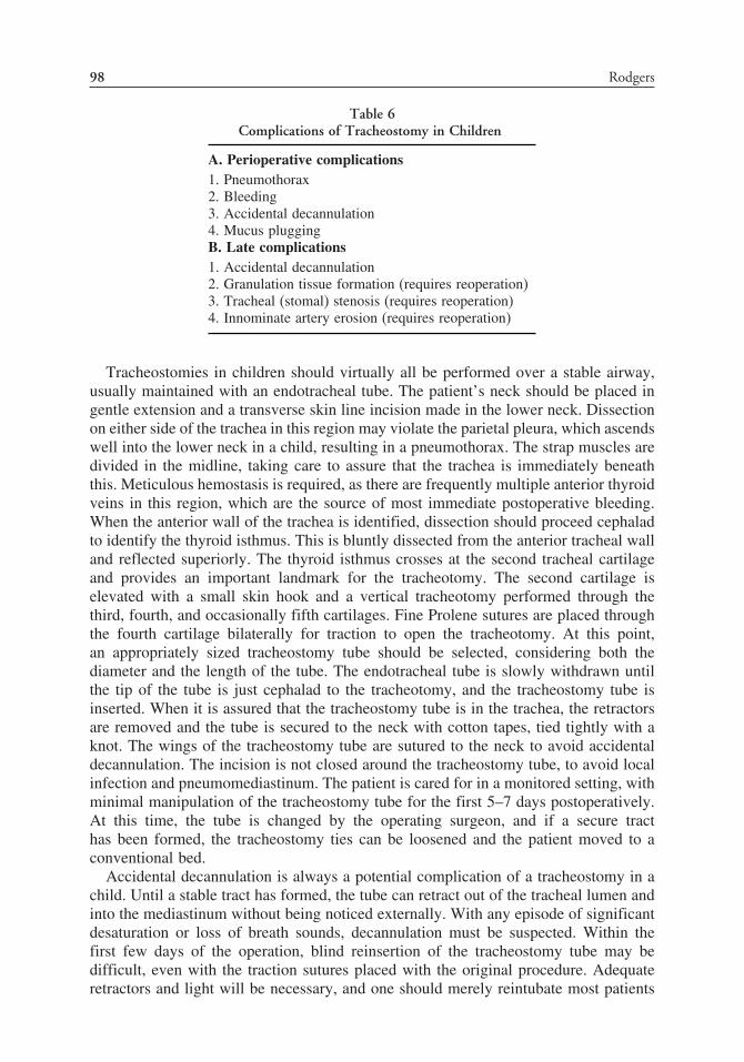
98 Rodgers
Table 6Complications of Tracheostomy in Children
A. Perioperative complications1. Pneumothorax2. Bleeding3. Accidental decannulation4. Mucus pluggingB. Late complications1. Accidental decannulation2. Granulation tissue formation (requires reoperation)3. Tracheal (stomal) stenosis (requires reoperation)4. Innominate artery erosion (requires reoperation)
Tracheostomies in children should virtually all be performed over a stable airway,usually maintained with an endotracheal tube. The patient’s neck should be placed ingentle extension and a transverse skin line incision made in the lower neck. Dissectionon either side of the trachea in this region may violate the parietal pleura, which ascendswell into the lower neck in a child, resulting in a pneumothorax. The strap muscles aredivided in the midline, taking care to assure that the trachea is immediately beneaththis. Meticulous hemostasis is required, as there are frequently multiple anterior thyroidveins in this region, which are the source of most immediate postoperative bleeding.When the anterior wall of the trachea is identified, dissection should proceed cephaladto identify the thyroid isthmus. This is bluntly dissected from the anterior tracheal walland reflected superiorly. The thyroid isthmus crosses at the second tracheal cartilageand provides an important landmark for the tracheotomy. The second cartilage iselevated with a small skin hook and a vertical tracheotomy performed through thethird, fourth, and occasionally fifth cartilages. Fine Prolene sutures are placed throughthe fourth cartilage bilaterally for traction to open the tracheotomy. At this point,an appropriately sized tracheostomy tube should be selected, considering both thediameter and the length of the tube. The endotracheal tube is slowly withdrawn untilthe tip of the tube is just cephalad to the tracheotomy, and the tracheostomy tube isinserted. When it is assured that the tracheostomy tube is in the trachea, the retractorsare removed and the tube is secured to the neck with cotton tapes, tied tightly with aknot. The wings of the tracheostomy tube are sutured to the neck to avoid accidentaldecannulation. The incision is not closed around the tracheostomy tube, to avoid localinfection and pneumomediastinum. The patient is cared for in a monitored setting, withminimal manipulation of the tracheostomy tube for the first 5–7 days postoperatively.At this time, the tube is changed by the operating surgeon, and if a secure tracthas been formed, the tracheostomy ties can be loosened and the patient moved to aconventional bed.
Accidental decannulation is always a potential complication of a tracheostomy in achild. Until a stable tract has formed, the tube can retract out of the tracheal lumen andinto the mediastinum without being noticed externally. With any episode of significantdesaturation or loss of breath sounds, decannulation must be suspected. Within thefirst few days of the operation, blind reinsertion of the tracheostomy tube may bedifficult, even with the traction sutures placed with the original procedure. Adequateretractors and light will be necessary, and one should merely reintubate most patients

Chapter 3 / Reoperative Head and Neck Surgery 99
by mouth before proceeding to try to reinsert the tracheostomy tube. After 5–7 dayspostoperatively, reinsertion of the tube should be straightforward.
Obstruction of the tracheostomy cannula with mucus plugs occurs more commonlyin children than in adults because of the small size of the tracheostomy tubes used andthe lack of an inner cannula. The parents are taught to change the tracheostomy tubeon a weekly basis at home, but more frequent changes may be necessary when thepatients have increased secretions. The use of humidified air may reduce the incidenceof plugging.
Formation of granulation tissues around the tracheostomy tube is a uniform findingin children with tracheostomies. This tissue forms in response to irritation from theforeign body of the tracheostomy tube itself and occasionally can be exuberant enoughto cause a foul-smelling exudate. Exuberant granulations can also interfere with thechanging of the tracheostomy tube and, in this case, they must be excised or cauterized.Granulations also form on the inside of the airway, usually on the proximal border ofthe tracheostomy stoma and at the tip of the tube. Children with tracheostomies shouldundergo bronchoscopy every 6 months and the internal granulation tissue should beremoved, before it forms a fibrous stricture of the airway.
Significant erosions of the trachea are less commonly seen with the flexible silastictracheostomy tubes currently employed, compared to the metallic tubes that wereemployed years ago. Nonetheless, anterior erosion into the innominate artery is stilloccasionally encountered. Tracheoinnominate artery fistulas occur from erosion of theanterior tracheal wall caused by pressure at the tip of the tracheostomy tube or at thelevel of the cuff. Usually there is some indication of a problem, with sentinel bleedingas an anterior ulcer is created (22). Patients with tracheostomies in whom more than10 mL of blood is seen in the tracheal aspirate, in the absence of any obvious cause,such as tracheitis, should undergo fiberoptic bronchoscopy to look for an anterior ulcer(23). If a superficial ulcer is encountered, the length of the tracheostomy tube maybe changed to change the point of impact of the tip of the tube with the trachealmucosa. Actual innominate artery erosion presents with massive bleeding through thetracheostomy tube and usually is fatal, unless the patient is in a hospital environment. Ifin such an environment, the tracheostomy tube should quickly be removed and replacedwith a cuffed endotracheal tube. That tube is positioned so that the cuff tamponadesthe bleeding. If this is unsuccessful in stopping the hemorrhage, an index finger maybe placed through the tracheostomy stoma and the artery compressed against thesternum (Utley maneuver) (24). The patient should then be rapidly transported to theoperating room for a thoracotomy, either by median sternotomy or right anterior thirdinterspace thoracotomy. Proximal control of the innominate artery should be achievedinitially, and then distal control of the right subclavian and right common carotidarteries individually. The innominate vessel should then be dissected from the anteriorwall of the trachea. The balloon on the endotracheal tube can then be deflated. Themargin of the tracheal fistula should be débrided and the fistula closed with interruptedabsorbable sutures, placed around adjacent cartilages. The innominate vessel is ligatedalong with the right carotid and right subclavian vessels. At the completion of thethoracotomy, the endotracheal tube may be removed from the trachea and replaced withan appropriately sized tracheostomy tube. Reconstruction of the innominate artery israrely required in pediatric patients, but, if it is thought to be necessary, the procedureshould be delayed until the mediastinal infection is completely eliminated.

100 Rodgers
Table 7Criteria for Anterior Laryngotracheal Decompression
1. Failure of extubation on two occasions secondary to subglottic pathology2. Weight greater than 1500 g3. Absence of ventilator support for 10 days4. FI02 less than 30%5. Absence of congestive heart failure for 30 days6. Absence of acute respiratory tract infection7. Absence of antihypertensive medication for 10 days
Percutaneous dilatational tracheostomy has become quite popular in adult practicesince its original description in 1985 (25). There have been very few reports of the useof this technique in children, and the manufacturer’s literature specifically indicates thatthe device is not to be used in pediatric patients (26). The most significant complicationsof this procedure encountered in adults have been creation of a tracheoesophagealfistula by inadvertent penetration of the posterior wall of the trachea and anterior wallof the esophagus and tracheal stenosis, usually thought to be secondary to distortion andcollapse of the cartilage on the proximal border of the stoma. Significant stomal stenosishas been described in two children undergoing percutaneous dilatational tracheostomy,one of whom required a local tracheal resection and the other of whom was successfullytreated with laser excision.
Certain forms of acquired and congenital subglottic stenosis in the neonate maybe treated by an anterior laryngotracheal decompression (anterior cricoid split), elimi-nating the need for a tracheostomy. The cricoid split is successful in reliving theairway obstruction in approximately 70% of carefully selected patients (27). Cotton,who initially described the procedure, suggests the criteria for performing anteriorcricoid split listed in Table 7 (28). Failure of the cricoid split to relieve subglotticairway obstruction usually indicates a more extensive subglottic stenosis than initiallyappreciated. Significant circumferential stenosis at the level of the cricoid may notrespond to this simple procedure. Failure may also be secondary to airway pathologyoutside of the subglottic region, usually laryngomalacia. With failure of a cricoid splitthe patient should receive a tracheostomy, inserted below the level of the split.
REFERENCES
1. Telander RL, Deane SA. Thyroglossal and branchial cleft cysts and sinuses. Surg Clin NA1997;57:779–791.
2. Sistrunk WE. The Surgical treatment of cysts of the thyroglossal tract. Ann Surg 1920;71:121–122.3. Sade J, Rosen G. Thyroglossal cysts and tracks: a histological and histochemical study. Ann Otol
Rhinol Laryngol 1968;77:139–145.4. Knight PJ, Hamoudi AB, Vassy IE. The diagnosis and treatment of midline neck masses in children.
Surgery 1983;93:603–611.5. Howard DJ, Lund VJ. Thyroglossal ducts, cysts and sinuses: a recurrent problem. Ann R Coll Surg
Engl 1986;68:137–138.6. Cunningham MJ. The management of congenital neck masses. Ann Otol Rhinol Laryngol 1992;13:
78–92.7. Chandler JR, Mitchell B. Branchial cleft cysts, sinuses, and fistulas. Otol Clin North Am 1981;14:
175–186.8. Muzaffer K, Avni G. Management of mycobacterial cervical lymphadenitis. Wrld J Surg 1997;21:
516–519.

Chapter 3 / Reoperative Head and Neck Surgery 101
9. Rahal A, Abela A, Arcand PH, et al. Nontuberculous mycobacterial adenitis of the head and neck inchildren: experience from a tertiary care pediatric center. Laryngoscope 2001;111:1791–1797.
10. Mushtaq I, Martin HCO. Atypical mycobacterial disease in children: a personal series. Pediatr SurgInt 2002;18:707–711.
11. Bailey WC. Treatment of atypical mycobacterial disease. Chest 1983;84:625–628.12. Blasco A, Allaire J. Drooling in the developmentally disabled: management practices and recommen-
dations. Dev Med Child Neurol 1992;34:849–862.13. Jongerius PH, Rotteveel JJ, vanLimbeek J, et al. Botulinum toxin effect on salivary flow rate in
children with cerebral palsy. Neurology 2004;63:1371–1375.14. Jongerius PH, van den Hoogen FJA, van Limbeck J, et al. Effect of botulinum toxin in the treatment
of drooling: a controlled clinical trial. Pediatrics 2004;114:620–627.15. Winterholler MGM, Erbguth FJ, Wolf S, et al. Botulinum toxin for the treatment of sialorrhoea in
ALS: serious side effects of a transductal approach. J Neurol Neurosurg Psychiatry 2001;70:417–418.16. Klem C, Mair EA. Four-duct ligation:a simple and effective treatment for chronic aspiration from
sialorrhea. Arch Otolaryngol Head Neck Surg 1999;125:796–800.17. Townsend GL, Morimoto AM, Kralemann H. Management of sialorrhea in mentally retarded patients
by transtympanic neurectomy. Mayo Clin Proc 1973;48:776–779.18. O’Dwyer TP, Conlon BJ. The surgical management of drooling—a 15-year follow-up. Clin
Otolaryngol 1997;22:284–287.19. Crysdale WS, Ravek E, McCan C, et al. Management of drooling in individuals with neurodisability:
a surgical experience. Dev Med Child Neurol 2001;43:379–383.20. Rocha, EP, Dias MD, Szajmbok FE, et al. Tracheostomy in children: there is a place for acceptable
risk. J Trauma 2000;49:483–486.21. Rodgers BM, Rooks JJ, Talbert JL. Pediatric tracheostomy: long-term evaluation. J Pediatr Surg
1979;14:258–263.22. Kapural L, Sprung J, Gluncic I, et al. tracheo-innominate artery fistula after tracheostomy. Anesth
Analg 1999;88:777–780.23. Thorp A, Hurt TL, Kim TY, et al. Tracheoinnominate artery fistula: a rare and often fatal complication
of indwelling tracheostomy tubes. Pediatr Emerg Care 2005;21:763–766.24. Utley JR, Singer MM, Roe BB, et al. Definitive management of innominate artery hemorrhage
complicating tracheostomy. JAMA 1972;220:577–579.25. Ciaglia P, Firsching R, Syniec C. Elective percutaneous dilational tracheostomy: a new simple bedside
procedure: preliminary report. Chest 1985;87:715–719.26. Scott CJ, Darowski M, Crabbe DCG. Complications of percutaneous dilatational tracheostomy in
children. Anaesthesia 1998;53:477–480.27. Cotton RT, Myer CM, Bratcher GO, et al. Anterior cricord split, 1977–1987: evolution of a technique.
Arch Otolaryngol Head Neck Surg 1998;114:1300–1302.28. Cotton RT, Myer CM III. Contemporary surgical management of laryngeal stenosis in children. Am
J Otolaryngol 1984;5:360–368.

4 Reoperative Thyroid and ParathyroidSurgery
Elisabeth Tracy, MD
and Michael A. Skinner, MD
CONTENTS
Reoperative Thyroid Surgery
Reoperative Parathyroid Surgery (RPS)
References
REOPERATIVE THYROID SURGERY
Reoperative thyroid surgery is a difficult undertaking, with both diagnostic andtechnical challenges. Ultimately, a better understanding of the pathophysiology ofthyroid disease, advances in preoperative diagnosis and management, and morethorough and evidenced-based resections of malignant disease may eliminate the needfor reoperation. In the interim, it is important to understand thyroid diseases associatedwith reoperation, specific indications for reoperation, options for preoperative work-up,and common complications of reoperation.
Thyroid malignancies account for 3% of all pediatric malignancies, and are the mostfrequent indication for thyroid surgery in children (1,2). Unlike the adult population,thyroid nodules in children are found to be malignant in 20% of cases. Whereasbenign diseases are more prevalent in the pediatric population than malignant disease,surgical resection is rarely used in their treatment. Surgical resection is reserved forvery specific indications in benign pediatric thyroid disease, including recurrent acutesuppurative thyroiditis (for which a thyroid lobectomy is sometimes indicated); Graves’disease after failure of both medical management and abalative therapy; and airwaycompression resulting from an enlarged thyroid in Graves’disease (2).
Malignant thyroid neoplasms are the most common reason for thyroidectomy inchildren. Although thyroid malignancies are less common in children than adults (only10% of all thyroid malignancies occur in patients under age 21), they are one ofthe more common carcinomas in the pediatric age group. The annual incidence ofprimary thyroid malignancies in childhood had steadily increased from the 1960s untilthe mid-1980s, when the link between thyroid radiation and carcinoma led to themore conservative use of radiation therapy in benign disease (2,3). More recently,
From: Reoperative Pediatric SurgeryEdited by: S. Teich and D. A. Caniano © Humana Press, Totowa, NJ
103

104 Tracy and Skinner
however, the increased incidence of thyroid cancer after prior treatment for otherchildhood malignancies (especially leukemia and lymphoma) has become evident.These secondary thyroid malignancies (STMs) result from the carcinogenic effect ofradiation therapy, the sequelae of immune suppression, and the oncogenic potential ofchemotherapy (4). Thyroid malignancies account for 9% of all secondary malignancies.
Most childhood thyroid malignancies (70%) present between the ages of 11–17.The most commonly encountered pediatric thyroid malignancy is papillary thyroidcarcinoma, followed by follicular carcinoma, then medullary thyroid cancer (MTC).Papillary thyroid cancer is the most common malignancy among both primary thyroidmalignancies and STMs. However, primary thyroid malignancies in children tend topresent in a more advanced state than either STMs in children or primary thyroidmalignancies in adults. Papillary thyroid cancer tends to be multicentric, spreadingearly to regional nodes. In most case series, more than 70% of children present withlymph node metastasis at the time of diagnosis (4). Also, larger studies have reportedrates of distant metastasis at diagnosis that range from 19–25% (5). The most commonsite of distant metastasis is the lung. Despite the high incidence of nodal and distantmetastasis, the prognosis of papillary thyroid cancer in children is quite favorable.The overall survival rate in children for both papillary thyroid cancer and the moreaggressive but less common follicular thyroid cancer is 98% (6).
Also uncommon but much more menacing is MTC, a malignancy of parafollicularC cells that is responsible for 2–5% of pediatric thyroid tumors. MTC may arise sponta-neously or as part of a hereditary syndrome including multiple endocrine neoplasia(MEN) type 2A, MEN type 2B, or familial medullary thyroid cancer (FMTC). Notonly is MTC often the first presenting tumor in patients with MEN, but it is alsothe most common cause of death from the disease. Because MTC tends to be lethal,aggressive resection including central compartment lymph node dissection from thehyoid to superior mediastinum and laterally to the carotid sheath is indicated. Fortu-nately, new techniques have lead to the increasingly early identification of MTC inhereditary disorders. Greater attention to MEN disorders in families and screening forreceptor tyrosine kinase (RET) protooncogene mutations has allowed for both the identi-fication of hereditary MTC tumors at a smaller and less advanced stage than theirsporadic counterparts and for the prevention of MTC through prophylactic thyroidectomy.Genetic testing should begin by age 5 for children with a family history of MEN2A and as soon after birth as possible for MEN 2B. In children carrying the RETprotooncogene mutation, prophylactic elective total thyroidectomy has been shown todramatically decrease the development of MTC and, given relatively low associatedmorbity, is universally recommended for patients with MEN 2A and 2B (7). Althoughlarger studies with longer follow-up periods are needed for establishing definitive guide-lines on the surgical treatment of children with MEN 2A and MEN 2B, current datasupport prophylactic total thyroidectomy prior at or before age 5 in MEN 2A and priorto 1 year of age in MEN 2B (6–9). Other thyroid tumors such as lymphomas, sarcomas,and undifferentiated carcinomas have been reported but rarely occur in children.
The optimal initial resection for thyroid malignancies has remained controversial inthe absence of definitive data. Surgical options include total thyroidectomy (completeremoval of the thyroid gland), near total thyroidectomy (leaving less than 3–5g of residual thyroid parenchyma), subtotal thyroidectomy (leaving more than 5 gof residual thyroid parenchyma), and lobectomy plus isthusectomy. An emergingconsensus supports total or near total thyroidectomy as the initial resection of choice

Chapter 4 / Reoperative Thyroid and Parathyroid Surgery 105
for differentiated thyroid malignancies, although some surgeons still perform a morelimited resection. Support for total or near total thyroidectomy includes data showingthe high rate of occurrence of occult, bilobar malignancies in the pediatric population(as many as 80% of affected children) (10). Also, total or near total thyroidectomy aidsin postoperative management by maximizing uptake in any remaining thyroid depositson postoperative imaging for suspected recurrence, and making subsequent radioiodineablative therapy more efficient. Still, some argue for less aggressive surgical resection,citing reports showing no statistically significant difference in outcomes between thetwo approaches (using the need for postoperative radioactive Iodine uptake and the needfor further ablative therapy as outcome measures) (11). Also, the favorable prognosisand long life span in children with advanced disease at presentation and the increasedrisk of recurrent laryngeal nerve injury reported in studies prior to 1990 are used toargue for a more limited resection (12). Despite concerns that more aggressive resec-tions lead to higher rates of complications, especially recurrent laryngeal nerve injury,no significant differences in morbidity occurred in lobectomy versus total/near totalthyroidectomy in adult studies (13,14). Because more definitive data in the pediatricpopulation is needed, the safest approach may be a near total thyroidectomy withpostoperative I-131 remnant ablation.
Another question surrounding initial resection is the extent of lymph node resection.The Society of Surgical Oncology practice guidelines for major cancer centersrecommend total thryoidectomy for thyroid malignancies in adults but does notrecommend routine lymph node dissection unless clinically palpable nodes areevident (8). However, given the different behavior of the disease in children, thereis a growing consensus in the pediatric literature that the standard initial surgicalapproach to papillary thyroid carcinoma should be a near total or total thryoidectomywith resection of involved lymph nodes followed by radioiodine therapy.
Specific Indications for Reoperative Thyroid SurgeryUnfortunately, reoperative surgery is often unavoidable in the management of
pediatric thyroid malignancies. Although scar tissue formation, inflammation, edema,and distortion of landmarks and tissue planes complicate any reoperative neckprocedure, reoperation is indicated for disease recurrence, incomplete excision, andthe discovery of previously unsuspected carcinoma following an initial lobectomy forwhat was thought to be benign disease.
Completion thyroidectomy is necessary in cases of less than near total resection oninitial operation or disease recurrence in remaining thyroid tissue. In any resection lessthan a total thyroidectomy, the possibility of finding unsuspected thyroid carcinoma onpermanent section exists. Despite improvements in fine needle aspiration (FNA) andintraoperative frozen section, nodules initially thought to be benign may not be recog-nized as malignant until examined by permanent pathology. As discussed previously,although total thyroidectomy is advocated by most as standard treatment for well-differentiated thyroid malignancies, some surgeons perform subtotal thyroidectomy asan initial operation, necessitating a completion thyroidectomy for persistent or recurrentdisease.
The more common indication for reoperation, however, is disease recurrence. Thehigh rate of recurrence of thyroid malignancies in children makes a thorough preop-erative evaluation and postoperative follow-up imperative, especially in children withadvanced disease. Papillary thyroid cancer recurrence rates in children ranges from

106 Tracy and Skinner
35–45% (with some reports as high as 47%) (11), with the median time to the devel-opment of a clinically evident recurrence averaging 7 months. Recurrence is morecommon in patients with lymph node involvement at presentation (as high as 69% inone recent study, although other studies have shown a weaker relationship) and patientswith multiple nodules by physical exam on initial presentation. Age, sex, fixation ofnodules, tenderness, family history, and hyperthyroidism have not been shown to beassociated with increased risk of recurrence. And, although recurrence rates in childrenare high for papillary thyroid cancer, recurrence does not seem to confer an increasedrisk of mortality in this age group (11). To lessen the need for reoperation, much of theliterature supports a strategy of careful preoperative inspection for nodal metastasis onpreoperative evaluation prior to initial resection, as well as carrying out a meticulousregional lymph node dissection (11,15).
Exactly what constitutes a meticulous or at least adequate lymph node dissectionis not always clear. In studies examining patterns of nodal metastases, papillarythyroid cancer usually metastasizes to paratracheal and paraesophageal nodes as wellas the ipsilateral cervical and jugular lymph nodes. Recommendations for lymph nodedissection in the initial resection of papillary thyroid cancer include a formal modifiedregional neck dissection to remove pretracheal and paratracheal nodes as well asipsilateral upper, middle, and lower jugular nodes and the recurrent laryngeal nervechain (11). In MTCs, the additional resection of contralateral upper, middle, and lowerjugular nodes and the recurrent laryngeal nerve chain is indicated given the higherincidence of contralateral node involvement.
It is important to note that some “recurrent” nodal metastases are actually present atthe initial operation but missed intraoperatively. In at least one study, fewer than 50% ofpapillary thyroid cancer nodal metastases and fewer than 64% of MTC nodal metastaseswere correctly identified by experienced surgeons intraoperatively (8). Therefore, asystematic and meticulous approach to lymph node dissection may be of benefit inidentifying nodal metastases and may lessen the need for reoperation.
Preoperative AssessmentPreoperative evaluation techniques can be valuable for both the diagnosis and local-
ization of a suspected thyroid malignancy or recurrence. FNA is the clear first stepfor the diagnosis of solid thyroid nodules in adults, but its usefulness in the pediatricpopulation is less clear. Given the high incidence of malignancy in children underage 13 (20%) and an FNA false negative rate of 6%, the current recommendationscall for the surgical removal of all thyroid nodules found in this age group (6). In theadolescent age group, the incidence of malignancy is low enough (11%) and the safetyof nonoperative management of benign lesions well documented enough that FNA isa useful study. Nodules proven benign by FNA can be followed clinically with serialphysical exam and serial ultrasound in this age group. Radioactive thyroid scans withtechnetium-99 or iodine-123 are also useful for identifying hyperfunctioning nodules,which are usually benign.
Advances in preoperative imaging techniques have lead to the detection and local-ization of new nodules and cancer recurrences at an earlier stage. High-resolution ultra-sound, contrast-enhanced magnetic resonance imaging (MRI), and positron emissiontomography (PET) scanning can identify nodules smaller than 1 cm and have givensurgeons both the ability and responsibility to identify and resect nodules that cannotbe readily identified by palpation. Precise preoperative localization of these subclinical
Chapter 4 / Reoperative Thyroid and Parathyroid Surgery 107
nodules is especially important in preparation for reoperation in the central neck nearthe trachea, where adhesions complicate visual identification of small neoplasms (16).Also, preoperative evaluation for metastasis prior to reoperation for suspected orrecurrent malignancy is important. A chest radiograph is a routine part of the work-up prior to initial resection of thyroid cancer, but may miss 30–40% of pulmonarymetastases (5). The whole body radioiodine scan recommended for all children afterinitial resection is also useful as a preoperative study prior to reoperation to detectpulmonary metastases missed by chest radiograph. Alternatively, a chest computedtomography (CT) scan can be obtainied, but care should be taken to avoid the useof iodinated intravenous contrast agents, which complicate subsequent treatment withradioiodine.
Operative ApproachAfter a thorough preoperative evaluation, including review of previous pathology
and operative reports, and discussion of the risks and benefits of reoperation with thepatient and family, care should be taken in planning the reoperative procedure. Thetiming of reoperation is important. If not undertaken during the first week after initialresection, surgery should not be attempted until 6–8 weeks after the initial operation toallow the acute inflammation to subside (17). The operative approach for a completionthyroidectomy or resection of residual thyroid tissue is similar to the initial resection.Care should be taken to maintain hemostasis, to identify and preserve the recurrentlaryngeal nerve and the external branch of the superior laryngeal nerve, and to preservethe parathyroid glands and their blood supply.
After preparation of the patient with the head and neck extended, an incision ismade above the sternal notch transversing the distance between the sternocleido-mastoid muscles. Platysmal flaps are raised. Approaching the thyroid laterally throughthe undisturbed plane on the medial side of the sternocleidomastoid with retractionof the muscle laterally allows exposure of the lateral paratracheal space in a planeminimally disturbed by previous dissection (18). The sternohyoid muscle is reflectedmedially with the carotid sheath reflected laterally. Once the recurrent laryngeal nerveis carefully identified, the strap muscles are divided. Identifying the nerve in a previ-ously undissected area and then following it into the reoperative field is the preferredstrategy, because the loss of normal landmarks and tissue planes in the reoperativeneck may complicate identification. Another approach used in reoperative surgery isthe low anterior approach, which uses the same anterior approach as in the initialresection with lateral retraction of the strap muscles after dividing them at the midline.The recurrent laryngeal nerve is identified low in the neck where previous dissectionis minimal and followed into the field (18).
It is important to note that the nerve is most often injured at the Ligament of Berry(the attachment of the thyroid gland at the trachea). Also, there is a great deal ofvariation in the course of the recurrent laryngeal nerve and thyroid artery and itsbranches. One important variation is the presence of a nonrecurrent laryngeal nerve,which occurs in 1% of patients, most commonly arising directly from the vagus nerveon the right. Use of intraoperative laryngeal nerve monitoring is helpul in tracing thecourse of the nerve, and has been shown to be both safe and beneficial in children (19).Extracapsular dissection of the thyroid with preservation of parathyroid fat minimizesthe risk of permanent hypoparathyroidism. Also, resected thyroid tissue should beinspected after removal for presence of parathyroid tissue. If a parathyroid gland is

108 Tracy and Skinner
inadvertently removed during resection, or removed en bloc with tumor, it should becarefully separated from the specimen and autotransplanted in the sternocleidomastoidmuscle or the forearm (once confirmation that it is uninvolved with the tumor isobtained).
Because patterns of lymph node metastases differ between the initial and reoperativepatient populations in thyroid malignancies, the recommended lymph node dissectionsare a bit more extensive. For reoperative papillary thyroid cancers, removal of pretra-cheal and paratracheal nodes as well as both contralateral and ipsilateral upper, middle,and lower jugular nodes and the recurrent laryngeal nerve chains is advised, becausea contralateral lymph node involvement is more common in these patients (11). Forreoperative MTC, resection includes all central compartment lymph node nodes fromthe hyoid to superior mediastinum and laterally to the ipsilateral and contralateralcarotid sheath, regardless of the size of the tumor (8). In these extensive lymph nodedissections, the removal of multiple lymph nodes in the setting of fibrosis and scarringcomplicates identification of the parathyroid glands and their vascular pedicles. Again,tissue removed should be inspected for the presence of parathyroid glands, which canthen be salvaged and autotransplanted. Most metastatic nodes can be removed throughthe cervical incision. However, thyroid cancer can rarely metastasize to the aorticpulmonary window necessitating a median sternotomy.
ComplicationsIt is little surprise that uniformly higher complication rates are seen in reoper-
ative thyroid surgery than with initial resection (11,15,17,20). The most commoncomplications of thyroid procedures include recurrent laryngeal nerve injuries, transienthypoparathyroidism, and permanent hypoparathyroidism.
With careful identification of the recurrent laryngeal nerve and use of intraoperativeneurological nerve monitoring, rates of transient recurrent laryngeal nerve injury havebeen kept to 2–5% with a permanent vocal cord paralysis rate of 1–5% (15,17). Infact, a few smaller studies report no recurrent laryngeal nerve injuries among theirreoperative patients (15).
Pediatric patients have approximately the same incidence of transient hypocalcemiaas adults (23%) (21). Careful identification and preservation of the parathyroids helpreduce the incidence of this complication. If parathyroids are indevertently or inten-tionally removed with the specimen, one or more glands should be autotransplantedto the sternocleidomastiod muscle or the forearm to reduce the incidence of hypocal-cemia. Although data on reoperative complications in children is limited, the rate ofpermanent hypocalcemia among adults after reoperation is approximately 1% amongexperienced endocrine surgeons.
Postoperative CareCaring for patients with thyroid malignancies or benign thyroid disease does not end
after surgical resection. Follow-up care is imperative postoperatively, especially giventhe high rate of recurrence. Ablative radioiodine therapy is a recommended adjunctpostoperatively and facilitates the use of subsequent thyroglobulin level monitoring forrecurrence. A whole-body radioiodine scan is recommended at 6 weeks postoperatively,and should be followed by a therapeutic dose of radionuclide to ablate residual tissueand treat residual metastatic disease. Special attention should be given to searching for

Chapter 4 / Reoperative Thyroid and Parathyroid Surgery 109
possible pulmonary metastases. In reoperative patients, monitoring for postoperativecomplications is especially important given the difficulty of the procedure. Pediatricsurgeons must also be prepared to coordinate patient care with a pediatric endocrinol-ogist or pediatric oncologist for benign or malignant disease, respectively.
REOPERATIVE PARATHYROID SURGERY (RPS)
As with reoperative thyroid surgery, RPS is a complicated undertaking that shouldbe entered into with care and meticulous planning. One reason for exercising extracaution is the difference in outcomes between RPS and primary resection. Althoughprimary parathyroid operations have a high success rate with low associated morbidity(at least one large study cites cure rates of 99.5% and a permanent morbidity of lessthan 1%) (22), reoperative procedures have a lower cure rate with a higher rate ofassociated morbidity (a 90% cure rate but a rate of at least transient hypocalcemiathat is as high as 35–43%) (22). Understanding the etiologies of recurrent or persistenthyperparathyroidism, the causes of failure after initial resection, and the preparationfor reexploration is crucial for safe and successful reoperation.
Indications for Reoperative SurgeryPrimary hyperparathyroidism usually results from a single adenoma, with initial
surgical resection curative in 95% of these cases. Most parathyroid adenomas inchildren occur in adolescence, with only 12–16% of cases occurring in childrenunder age 10. However, as many as 30–50% of children presenting with primaryhyperparathyroidism have an etiology other than a solitary adenoma (23). They,along with virtually all infants with hyperparathyroidism have parathyroid hyperplasiarelated to an MEN syndrome or isolated familial hyperparathyroidism. The MENsyndromes involving hyperparathyroidism include MEN 1 (parathyroid hyperplasia,pituitary adenoma, pancreatic islet cell neoplasms) and MEN 2A (MTC, pheochromo-cytoma, and hyperparathyroidism). Isolated familial hyperparathyroidism (also callednon-MEN familial hyperparathyroidism) describes a hereditary syndrome of positivefamily history of hyperparathyroidism, adenomatous changes, and absence of otherMEN features. A subset of patients will have primary hyperparathyroidism from fourgland hyperplasia, a double adenoma, or, more rarely, carcinoma. The initial approachto surgical treatment varies based on the etiology of the disease. Carcinomas andadenomas are approached initially with exploration and removal of the abnormal gland.However, if parathyroid hyperplasia consistent with a MEN syndrome or isolatedfamilial hyperparathyroidism is encountered, the preferred treatment is either a subtotalparathyroidectomy or total parathyroidectomy with autotransplant of one gland intothe sternocleidomastoid muscle.
Reoperation is indicated for persistent hyperparathyroidism (biochemical evidenceof hyperparathyroidism immediately postoperatively or in the following 6 months) orrecurrent disease (disease that develops more than 6 months after initial resection).Although only 3.2% of adults experience persistent disease and less than 1% haverecurrent disease after initial resection, the rate of failed primary operation approaches20% in the pediatric population (23,24). Most commonly, persistence or recurrencerelates to failure of the initial exploration. Given the potential for serious compli-cations in RPS, much attention has been given to examining the reasons for failure

110 Tracy and Skinner
of initial exploration and to developing strategies to increase the success of initialoperations (9).
The failures of primary exploration are most often from an abnormal gland residingin a normal anatomic location (accounting for as many as 76% of failed primaryexplorations) (22). The abnormal gland may be adenomatous or simply a hyperplasticgland not recognized during the initial operation. In fact, as many as 37% of patientsundergoing reoperation for persistent primary hyperparathyroidism are found to havehyperplastic glands compared to 15% of patients on initial operation—an indicationthat unrecognized hyperplasia may be a significant cause of disease recurrence andreoperation. Persistent hyperparathyroidism is quite frequent in patients with MEN 1,MEN 2A, or isolated familial hyperparathyroidism (recurrence rates of 20–100% havebeen described depending on the duration of follow-up) (25). These diagnoses shouldbe entertained in cases of persistent hyperparathyroidism in children.
A substantial number of initial parathyroid resection failures may also be attributedto glands that do not reside in normal anatomic locations or supernumerery glands.A fifth parathyroid gland is identified in 15% of reoperative patients. For extraanatomicglands, the most common location is the mediastinum followed by intrathyroid glands,carotid sheath glands, anterior tracheal glands, and retroesophageal glands. To reducethe need for reoperation, careful preoperative localization prior to initial resectionshould be undertaken. An additional rare but significant cause of persistent or recurrenthyperparathyroidism is “parathyromatosis,” or the implantation of adenomatous orhyperplastic parathyroid cells after rupture of parathyroid capsule on initial operation.Because the cells may be disseminated, this complication is notoriously difficult totreat.
Parathyroid carcinoma is extremely rare but has been described in children as youngas 12 years. After initial operation for parathyroid carcinoma, residual disease ormetastasis may manifest as persistent or recurrent primary hyperparathyroidism. Unfor-tunately, the disease recurs in 40–70% of patients, with most recurrences occurring atthe original site of the malignancy. When metastases are present, the most common sitesare regional lymph nodes, bone, lung, and liver. The timing of recurrences is difficultto predict, with some occurring 1 month and some decades after initial resection.Multiple recurrences requiring repeated reoperations are not uncommon (26,27).
Secondary hyperparathyroidism results from multigland hyperplasia caused byend-stage renal disease. As in MEN or isolated familial hyperparathyroidism, theinitial resection involves either a subtotal parathyroidectomy (three-and-a-half glandsremoved) or a total parathyroidectomy with autoimplantation of parathyroid tissue intothe brachialis muscle of the forearm. Unlike primary hyperparathyroidism, preoperativeimaging and localization is of less benefit prior to initial resection. In a comparison ofrecurrence rates for secondary hyperparathyroidism after each of the two procedures, arecent metanalysis found significantly higher recurrence rate (49%) after total parathy-roidectomy with autotransplantation than after subtotal parathyroidectomy (17%) (28).However, other sources document much lower and more equal rates of recurrence(10%) for both procedures, especially in areas with high rates of renal transplantation(28). Failure of the initial operation is attributed most often to autograft hyperplasia(49% of reoperative patients), followed by missed supernumerary glands (20%, themajority of missed glands were intrathymic), remnant hyperplasia (17%) and missedin situ hyperplastic gland (7%) (28).
Chapter 4 / Reoperative Thyroid and Parathyroid Surgery 111
Preoperative EvaluationBecause of high the rate of morbidity, indications for reoperative parathyroid
resection are more conservative than for the initial operation. For persistent or recurrentprimary hyperparathyroidism, the diagnosis of hyperparathyroidism must be reesta-bilished biochemically, with care taken to rule out other potential causes such asfamilial hypocalciuric hypercalcemia. Elevated levels of both serum calcium and intactparathyroid hormone (iPTH) should be documented. A careful history and physicalshould be targeted at excluding other diagnoses including radiation, lithium exposure, orother malignancy (although those most associated with hypercalcemia—lung, ovarian,and pancreatic cancers—are rare in the pediatric population). A review of initialoperative notes and pathology is also prudent prior to reoperation, especially for consid-eration of the particular operative approach. Most commonly, a cervical incision isthe incision of choice, especially because the residual disease in more than 90% ofpatients referred for reoperation is accessible through such an incision. Preoperativelocalization studies (discussed in the next section) are invaluable for operative planning.Another important reoperative consideration is that patients and/or their parents under-stand the risks of reoperative surgery—including the higher incidence of recurrent orsuperior laryngeal nerve injury, hypoparathyroidism, or persistent hyperparathyroidismthan in primary parathyroid resections. Finally, a preoperative laryngoscopy shouldbe part of the preoperative work-up to document any cord paralysis from the initialresection (9,22).
In persistent or recurrent secondary hyperparathyroidism, reoperation is also under-taken with caution. Indications for reoperation include ongoing nephrolithiasis, renalimpairment, worsening osteopenia, serum Ca levels greater than 3mmol/L, and failureof medical management. Less-clear indications include severe aches/pain, fatigue, anddepression. Preoperative evaluation in these patients is determined by the previousoperative procedure. Those who underwent total parathyroidectomy with autotrans-plantation may or may not need a neck exploration, whereas those who underwentsubtotal parathyroidectomy will all need a reoperative neck procedure. Just as inprimary parathyroid disease, a thorough discussion of the risks of reoperation and apreoperative laryngoscopy are advisable prior to reoperation.
Localization StudiesAs mentioned previously, preoperative localization is the standard of care prior to
most primary parathyroid resections (with the exception of resection for secondaryhyperparathyroidism in adults). The unique challenges of RPS make localization studiesan even more crucial part of the preoperative work-up. At a minimum, the fibrosis anddistortion of landmarks and tissue planes from the initial operation complicates accurateidentification of residual parathyroid tissue. This is especially true when the initialoperation involves an extensive bilateral neck exploration, such as in primary resectionfor secondary hyperparathyroidism. The reoperative surgeon may also be faced withuncertainty about number or functional status of parathyroid glands left from previousoperations. Through the use of multiple imaging techniques preoperatively, 80–90%of parathyroid tumors can be localized (24). In fact, reoperative parathyroid resectionis best thought of as an operation designed to identify and remove a preoperativelyidentified abnormality (29). Most recent studies recommend at least two concordant

112 Tracy and Skinner
localization studies prior to reoperation (30). Three studies may be necessary for local-izing a persistent or recurrent parathyroid adenoma if initial study is not categoricallypositive (29).
Whereas selection of preoperative localization studies depends on the nature ofthe initial resection, the almost universally recognized first-line imaging study inparathyroid surgery is sestamibi/technetium 99m scintigraphic scanning. The techniqueis based on the specificity of the radiopharmaceutical agent technetium 99m sestamibifor mitochondrial membranes, which leads to its concentration in metabolically activetissue. The distribution of sestamibi in images of the chest and neck taken at 15 minutesis compared to the distribution in images taken 3 hours later to look for areas ofincreased uptake consistent with a hyperfunctioning gland. Preoperative sestamibiscanning has been shown to have a sensitivity of 82–86%, with a specificity rangingfrom 85–94% and approaching 100% in patients with a solitary adenoma (31). Althoughthe high sensitivity and specificity of preoperative sestamibi scanning have made it afirst-line modality of choice, it is more limited in the setting of multiple hyperplasticglands or small adenomas (especially small adenomas located behind the thyroid gland).Dual-tracer subtraction scintigraphy, a technique involving a second radiotracer thataccumulates in thyroid but not parathyroid tissue, allows for subtraction of the twoimages to more clearly distinguish small amounts of abnormal parathyroid tissue in oraround the thyroid gland. Combining sestamibi scanning with single-photon emissioncomputed tomography (SPECT) is another strategy that allows for better anatomiclocalization of nodules, especially in the anterior-posterior plane. Another limitationof sestamibi scanning is that other metabolically active tissues including glands withdiffuse hyperplasia, thyroid nodules or carcinoma, or surrounding lymph nodes canconfound the scans. However, using simultaneous CT scan or neck ultrasound asa confirmatory study helps overcome these limitations. For the reoperative neck, aminimum of two studies should be used in any case, decreasing the risk of missingadditional adenomas or diffuse hyperplasia.
The other classic first-line study prior to parathyroid resection is high-resolutionultrasound. In reviews of various localization studies, ultrasound has been shownto have a sensitivity and specificity ranging from 75–88 and 65–90%, respectively,on initial operation. The presence of a thyroid nodule has been correlated with ahigher rate of false negative readings on ultrasound, especially in the reoperative neck.Also, visualizing superior parathyroid glands located medially or deep in the neckor retropharyngeal space is difficult. Overall, however, sensitivity and specificity ofhigh-resolution ultrasound in preparation for reoperation is not significantly less thanprior to initial operation (80 and 84%, respectively, for reoperation compared with88 and 90%, respectively, for initial operation in a recent study with experiencedoperators) (32). The strengths of ultrasound include its ability to provide more exactanatomical localization than sestamibi and to examine vascular structures and supplyin addition to adenoma position. Combining ultrasound and sestamibi scanning forpreoperative imaging has been shown to increase the sensitivity of the studies to94–96% (33,34).
Other imagining techniques prior to operation include CT and MRI. Althoughprevious studies had reported lower success rates for these modalities, recent studieshave shown a sensitivity for CT as high as 98%, and as high as 80% for MRI if usedprior to initial resection. However, metallic clip artifacts from clips placed during theinitial operation can lower the sensitivity of CT to 48–56% (33). Small gland size and
Chapter 4 / Reoperative Thyroid and Parathyroid Surgery 113
coexisting thyroid disease limit the accuracy of CT/MRI as well. Few studies havecompared the techniques on the reoperative neck, and neither CT nor MRI shouldreplace sestamibi scanning and ultrasound as a front-line modality. Also, CT andMRI have been shown to offer no additional benefit in the preoperative setting ifsestamibi and ultrasound results are positive. However, these noninvasive techniquesare useful when sestamibi scanning and ultrasound are discordant or inconclusive. CT isespecially useful for ectopic parathyroid tumors, which can be missed by ultrasound,as well as imaging mediastinal tumors or glands within the tracheoesophageal groove.At least one recent study has shown a significant benefit in combining sestamibiscintigraphy with SPECT and CT (giving a sensitivity of 93 versus 31% for sestamibiscintigraphy-SPECT alone) for localizing ectopic adenomas and in reoperative patientswith distorted anatomy from prior operation (35).
More invasive preoperative localization techniques are warranted when noninvasivestudies are ambiguous or discordant. Selective venous sampling (SVS), often regardedas a gold-standard localization procedure, is not routinely used preoperatively unlessnoninvasive studies fail. The technique involves catherization of multiple veins in theneck and mediastinum with samples sent for measurement of iPTH, and usually requiresgeneral anesthesia when performed on children. Gradients in PTH can regionalize andsometimes precisely localize an abnormal gland based on venous drainage patterns.A high vertebral vein gradient predicts a retroesophageal parathyroid gland, whereas ahigh gradient in the internal thoracic veins predicts an ectopic gland in the thymus. Withthe recent availability of rapid PTH assays, interventional radiologists who performselective arteriography can further investigate PTH gradients they find for even moreprecise anatomic localization in the angiography suite. Recent studies have shown SVSeven without arteriography to have a sensitivity as high as 90–94% (36). Given thedifficulty of reoperation, the benefits of SVS outweigh the length of the procedure andrisk of procedure-related thrombosis or hematoma when other studies have failed toadequately or consistently localize an adenoma (37).
Finally, FNA can be used for preoperative localization as a part of ultrasound-guided biopsy or for measurement of iPTH. FNA with ultrasound-guided biopsy hasbeen reported to have a sensitivity rate of 90% and an accuracy of 82% (22). FNAmeasurement of iPTH is not routinely indicated unless preoperative localization isdifficult, but in such cases can be helpful in distinguishing between pathologicalparathyroid tissue versus normal thyroid or parathyroid. Measured iPTH levels greaterthan 1000 pg/mL are predictive of a pathological parathyroid gland with a sensitivityof 87%, whereas levels less than 100 pg/mL are highly predictive of the absence ofparathyroid tissue (as in an intrathyroid aspiration) (30).
For secondary hyperparathyroidism that persists or recurs, the choice of preoper-ative localization techniques depends on the initial operation. For those patients whounderwent a total parathyroidectomy with autotransplantation, the Casanova test is theinitial test of choice. This technique uses peripheral venous sampling of iPTH in theextremity used for prior autotransplant. If the test shows no source of PTH other thanthe graft site, the reexploration can be limited to that area (usually forearm), limitingthe risks of morbidity for patient and, in some cases, limiting the need for generalanesthesia. If the autotransplant is not the only source of PTH, an additional adenomaor engraftment is suspected and further preoperative imaging is needed.
For patients with a previous subtotal parathyroidectomy, sestamibi imaging can helplocalize the remaining hyperfunctioning gland. As with primary hyperparathyroidism,
114 Tracy and Skinner
the combination of sestamibi scanning and ultrasound increases the success of local-izing hyperfunctioning glands prior to reoperation (to as high as 88 from 75%) (38).Performing sestamibi scanning prior to ultrasound guides radiologists to the areas ofhyperfunctioning glands, allowing for more exact localization on ultrasound. Missedglands are often small, superior glands, although ecotopic or supernumerary glandsare possible. CT should be part of the preoperative evaluation if initial studies are notconcordant or there is any ambiguity.
Using intraoperative localization modalities as adjuncts may also increase the successof reoperation. Intraoperative ultrasound (IOUS) and sestamibi scanning with gammaprobe are commonly used techniques in conjuction with intraoperative PTH monitoring.The additional benefit of these techniques compared with thorough preoperative local-ization and intraoperative PTH assay is debated in the literature. IOUS has been shownto detect more abnormal parathyroid glands than preoperative ultrasound and shortenoperative time by as much as 50%, but has not been shown to significantly affectultimate outcomes (39). Operator skill and experience may also limit the usefulness ofIOUS.
Many surgeons have favored intraoperative sestamibi scanning as a technicallyeasier modality than intraoperative ultrasound. The technique requires the injectionof technetium 99m sestamibi injected 2–4 hours prior to making the neck incision.A gamma counter with a 9–14 mm handheld gamma detection probe is used to scanthe neck after division of the strap muscles. The neck is probed in all four quadrants atthe outset, with further exploration directed at those areas of the neck with the highestradioactivity counts. The count directs the surgeon to the hyperfunctioning gland, whichusually has counts elevated as much as 20–50% over background. Once the adenomais removed, radioactivity in each of the four quadrants should equalize. As with IOUS,intraoperative sestamibi scanning is more successful at localizing adenomas than itspreoperative counterpart, especially in the reoperative neck (localizing 91% of hyper-functioning compared with 64% each by preoperative sestamibi and ultrasound) (24).
Finally, intraoperative parathyroid hormone monitoring (IOPTH) is an extremelyinformative technique to confirm quantitatively that all hyperfunctioning parathyroidtissue has been removed. A drop in PTH levels of 50% or greater at 20 minutesfrom parathyroid resection is widely used as confirmation of a successful resection.Some authors have argued for a 70% drop, especially in the reoperative setting toavoid false positive results in patients with multigland disease (22). The use of IOPTHin RPS has been shown to increase the success rate of the procedure from 76 to94% (40). Of note, studies have found that a decrease in PTH of more than 84% isassociated with a high risk of developing hypocalcemia in the postoperative period—making the technique useful for identification of patients who would benefit fromcloser postoperative monitoring (40) (Table 1).
Reoperative ApproachAs described previously for thyroid reoperation, parathyroid reoperation should be
undertaken either during the first week after the failed initial procedure or 3 monthsor more afterwards to avoid the inflammation of the immediate postoperative period.Given to the difficulty of reoperation, the surgeon’s goal is a focused, direct, andlimited approach to resect the abnormality or abnormalities identified on preoperativeimaging.

Chapter 4 / Reoperative Thyroid and Parathyroid Surgery 115
Table 1Parathyroid Localization Techniques
Study Sensitivity Specificity Notes
Sestamibi scintigraphy 82–86% 85–94%High-resolution US 75-88% 65–90%Sestamibi scintigraphy + US 96% 83%CT 80–98% * Sensitivity drops to
48–56% when metallicclip artifact is present;Older studies report CTsensitivity ranges as lowas 13–50%
CT + US 91% *CT + sestamibi scintigraphy 74–90% *CT / SPECT-sestamibiscintigraphy
91–96% *
MRI 50-80% 78%MRI + US 79% 50%MRI + sestamibiscintigraphy
75% 39%
SVS 90–94% 85% More invasive technique(see text)
US-guided FNA with iPTHmeasurement
87% 75%
Data obtained from the following (adult series): Refs. (31–37)CT, computed tomography; US, ultrasound; MRI, magnetic resonance imaging; SVS, selective venous
sampling; FNA, fine needle aspiration; iPTH, intact parathyroid hormone.
Because most missed adenomas will be accessible through a cervical incision (withthe most common site of a missed parathyroid adenoma along the esophagus in theposterior superior mediastinum), a cervical approach is reasonable. After the skinincision, a lateral “back door” approach using the plane lateral to the sternohyoid andsternothyroid muscles and medial to the sternocleidomastoid offers the advantage ofavoiding dissection through previously mobilized strap muscles. The plane is carriedto the level of the carotid sheath. The thyroid and strap muscles are retracted antero-medially. The pharynx and larynx are rotated into view and the recurrent laryngealnerve is identified early in the procedure. Exploration continues above the inferiorthyroid artery to the prevertebral fascia and should include digital exploration of theretropharyngeal space. Meticulous hemostasis is imperative, because blood-stainingmay obscure parathyroid tissue in an already difficult operative environment. Ifthe exploration is unsuccessful despite preoperative localization data, other locationssuch as the thyrothymic ligament, cervical thymus, subcapsular and intraparenchymalthyroid, and carotid sheath should be systematically explored (9,10,41).
When a mediastinal adeonoma is suspected, it is generally accessible through acervical incision. The most common location of a mediastinal adenoma is in theanterior mediastinum associated with thymic tissue. Thymectomy is indicated for a

116 Tracy and Skinner
closely associated adenoma or when the lower parathyroid gland cannot be identifiedin isolated familial hyperparathyroidism or secondary hyperparathyroidism. To aid indissection of the thymus, retraction of the thyroid gland allows the surgeon to followthe inferior thyroid vessels to the anterior mediastinum. After the thymus is identified,it is grasped and pulled into the field, then excised.
If necessary, further mediastinal exploration is possible through a “T” incision madeby carrying a vertical incision down from the cervical incision. A partial mediansternotomy is then made. The thymus is resected and exploration continues to theinnominate veins and along the jugular and subclavian veins. A mediastinal drain shouldbe placed and the sternotomy closed. For very well-localized mediastinal adenomas,video-assisted thoracic surgery has been used with success in adults. Also, a limitedleft thoractomy may be necessary for middle mediastinal adenomas after failed cervicaland anterior mediastinal explorations.
For reoperation after total parathyriodectomy with autotransplantation to an extremity(such as a forearm), reoperation can be safely limited to the extremity if the preoperativeCasanova testing confirms that the extremity was the source of PTH. Reexploration ofthe extremity is a much less involved and less technically difficult procedure.
All reoperative parathyroid resections should use IOPTH monitoring as a confir-matory study. A 50–70% decline in PTH from baseline indicates successful reoperation.If PTH levels do not decline after 20 minutes, continued exploration is necessary.
Results of ReoperationComplications of reoperation include persistant hypercalcemia, which indicates a
failure of reoperation and should prompt further workup for missed or engraftedparathyroid tissue or multigland hyperplasia. The major cause for reoperative failureis multigland disease, which accounts for 73% of such failures (22). Permanenthypoparathyroidism affects almost 13% of reoperative patients. Care should be takenpostoperatively to distinguish between hungry bone syndrome after reoperation forsecondary hyperparathyroidism and true lack of parathyroid function. In hungry bonesyndrome, the serum phosphate level is low (<2.5 mg/dL), whereas in hypoparathy-roidism it is elevated (>4.5 mg/dL). Although care is taken to identify the recurrentlaryngeal nerve, nerve injury still occurs in almost 1% of patients (an improvement from4% two decades ago) (22). Advances in preoperative and intraoperative localization aswell as nerve monitoring have contributed to this improvement.
REFERENCES
1. Harsha WJ, Perkins JA, Lewis CW, Manning SC. Head and neck endocrine surgery in children: 1997and 2000. Arch Otolaryngol Head Neck Surg 2005;131(7):564–570.
2. Skinner MA. Surgical diseases of the thyroid and parathyroid glands. In: Oldham KT, Colombani PM,Foglia RP, Skinner MA, (ed.), Principles and practice of pediatric surgery, volume 1. Philadelphia:Lippincott; 2005.
3. Shamberger R. Thyroid and parathyroid tumors. In: Oneil J, Grosfeld JL, Fonkalsrud EW, Coran AG,Caldamone AA, (eds.), Principles of pediatric surgery, second edition. St. Louis: Mosby; 2003.
4. Gow KW, Lensing S, Hill DA, et al. Thyroid carcinoma presenting in childhood or after treatmentof childhood malignancies: an institutional experience and review of the literature. J Pediatr Surg2003;38(11):1574–1580.
5. Chaukar DA, Rangarajan V, Nair N, et al. Pediatric thyroid cancer. J Surg Oncol 2005;92(2):130–133.6. Safford SD, Skinner MA. Thyroid and parathyroid disease in children. Semin Pediatr Surg
2006;15(2):85–91.

Chapter 4 / Reoperative Thyroid and Parathyroid Surgery 117
7. Skinner MA, Moley JA, Dilley WG, Owzar K, Debenedetti MK, Wells SA Jr. Prophylacticthyroidectomy in multiple endocrine neoplasia type 2A. N Engl J Med 2005;353(11):1105–1113.
8. Machens A, Hinze R, Thomusch O, Dralle H. Pattern of nodal metastasis for primary and reoperativethyroid cancer. World J Surg 2002;26(1):22–28.
9. Cameron J, ed. Current surgical therapy, eighth edition. Philadelphia: Mosby; 2004.10. Zeigler MA, Azizkhan R, Weber TR, Stevens AT, Medascend M. Operative pediatric surgery, first
edition. Columbus: McGraw-Hill Professional; 2003.11. Palmer BA, Zarroug AE, Poley RN, Kollars JP, Moir CR. Papillary thyroid carcinoma in children:
risk factors and complications of disease recurrence. J Pediatr Surg 2005;40(8):1284–1288.12. Weber RS, LiVolsi, V. Pediatric thyroid carcinoma. In: Randolph GW, (ed.), Surgery of the thyroid
and parathyroid glands. Philadelphia: Saunders; 2003: 253–261.13. Sakorafas GH, Peros G. Thyroid nodule: a potentially malignant lesion; optimal management from a
surgical perspective. Cancer Treat Rev 2006;32(3):191–202.14. Farkas EA, King TA, Bolton JS, Fuhrman GM. A comparison of total thyroidectomy and lobectomy
in the treatment of dominant thyroid nodules. Am Surg 2002;68(8):678–682; discussion 682–673.15. Kim MK, Mandel SH, Baloch Z, et al. Morbidity following central compartment reoperation for
recurrent or persistent thyroid cancer. Arch Otolaryngol Head Neck Surg 2004;130(10):1214–1216.16. Desai D, Jeffrey RB, McDougall IR, Weigel RJ. Intraoperative ultrasonography for localization of
recurrent thyroid cancer. Surgery 2001;129(4):498–500.17. Kupferman ME, Mandel SJ, DiDonato L, Wolf P, Weber RS. Safety of completion thyroidectomy
following unilateral lobectomy for well-differentiated thyroid cancer. Laryngoscope 2002;112(7 Pt 1):1209–1212.
18. Pasieka JL. Reoperative thyroid surgery. In: Randolph GW, (ed.), Surgery of the thyroid andparathyroid glands. Philadelphia: Saunders; 2003: 385–391.
19. Brauckhoff M, Gimm O, Thanh PN, et al. First experiences in intraoperative neurostimulation ofthe recurrent laryngeal nerve during thyroid surgery of children and adolescents. J Pediatr Surg2002;37(10):1414–1418.
20. Chao TC, Jeng LB, Lin JD, Chen MF. Reoperative thyroid surgery. World J Surg 1997;21(6):644–647.21. Astl J, Dvorakova M, Vlcek P, Vesely D, Matucha P, Betka J. Thyroid surgery in children and
adolescents. Int J Pediatr Otorhinolaryngol 2004;68(10):1273–1278.22. Thompson GB, Grant CS, Perrier ND, et al. Reoperative parathyroid surgery in the era of sestamibi
scanning and intraoperative parathyroid hormone monitoring. Arch Surg 1999;134(7):699–704;discussion 704–695.
23. Kollars J, Zarroug AE, van Heerden J, et al. Primary hyperparathyroidism in pediatric patients.Pediatrics 2005;115(4):974–980.
24. Rossi HL, Ali A, Prinz RA. Intraoperative sestamibi scanning in reoperative parathyroidectomy.Surgery 2000;128(4):744–750.
25. Kivlen MH, Bartlett DL, Libutti SK, et al. Reoperation for hyperparathyroidism in multiple endocrineneoplasia type 1. Surgery 2001;130(6):991–998.
26. Iacobone M, Lumachi F, Favia G. Up-to-date on parathyroid carcinoma: analysis of an experience of19 cases. J Surg Oncol 2004;88(4):223–228.
27. Witteles RM, Straus FH, Rao Koka ML, Kaplan EL. Parathyroid carcinoma. In: Randolph GW, (ed.),Surgery of the thyroid and parathyroid glands. Philadelpha: Saunders; 2003: 578–589.
28. Richards ML, Wormuth J, Bingener J, Sirinek K. Parathyroidectomy in secondary hyperparathy-roidism: is there an optimal operative management? Surgery 2006;139(2):174–180.
29. Ghaheri BA, Liebler SL, Andersen PE, et al. Perioperative parathyroid hormone levels in thyroidsurgery. Laryngoscope 2006;116(4):518–521.
30. Kiblut NK, Cussac JF, Soudan B, et al. Fine needle aspiration and intraparathyroid intact parathyroidhormone measurement for reoperative parathyroid surgery. World J Surg 2004;28(11):1143–1147.
31. Norman J, Chheda H. Minimally invasive parathyroidectomy facilitated by intraoperative nuclearmapping. Surgery 1997;122(6):998–1003; discussion 1003–1004.
32. Ghaheri BA, Koslin DB, Wood AH, Cohen JI. Preoperative ultrasound is worthwhile for reoperativeparathyroid surgery. Laryngoscope 2004;114(12):2168–2171.
33. Lumachi F, Ermani M, Basso S, Zucchetta P, Borsato N, Favia G. Localization of parathyroid tumoursin the minimally invasive era: which technique should be chosen? Population-based analysis of 253patients undergoing parathyroidectomy and factors affecting parathyroid gland detection. EndocrRelat Cancer 2001;8(1):63–69.
118 Tracy and Skinner
34. De Feo ML, Colagrande S, Biagini C, et al. Parathyroid glands: combination of (99m)Tc MIBI scintig-raphy and US for demonstration of parathyroid glands and nodules. Radiology 2000;214(2):393–402.
35. Krausz Y, Bettman L, Guralnik L, et al. Technetium-99m-MIBI SPECT/CT in primary hyperparathy-roidism. World J Surg 2006;30(1):76–83.
36. Chaffanjon PC, Voirin D, Vasdev A, Chabre O, Kenyon NM, Brichon PY. Selective venous samplingin recurrent and persistent hyperparathyroidism: indication, technique, and results. World J Surg2004;28(10):958–961.
37. Seehofer D, Steinmuller T, Rayes N, et al. Parathyroid hormone venous sampling before reoper-ative surgery in renal hyperparathyroidism: comparison with noninvasive localization procedures andreview of the literature. Arch Surg 2004;139(12):1331–1338.
38. Perie S, Fessi H, Tassart M, et al. Usefulness of combination of high-resolution ultrasonography anddual-phase dual-isotope iodine 123/technetium Tc 99m sestamibi scintigraphy for the preoperativelocalization of hyperplastic parathyroid glands in renal hyperparathyroidism. Am J Kidney Dis2005;45(2):344–352.
39. Norton JA, Shawker TH, Jones BL, et al. Intraoperative ultrasound and reoperative parathyroidsurgery: an initial evaluation. World J Surg 1986;10(4):631–639.
40. Elaraj DM, Remaley AT, Simonds WF, et al. Utility of rapid intraoperative parathyroid hormoneassay to predict severe postoperative hypocalcemia after reoperation for hyperparathyroidism. Surgery2002;132(6):1028–1033; discussion 1033–1024.
41. Gaz RD. Revision parathyroid surgery. In: Randolph GW, (ed.), Surgery of the thyroid and parathyroidgland. Philadelphia: Saunders; 2003:564–570.

5 Complications of Pectus Excavatumand Carinatum Repair
Robert E. Kelly, Jr., MD
and Donald Nuss, MB, Ch.B
CONTENTS
Introduction
Recurrent Pectus Excavatum
Recurrent Pectus Carinatum
Less Frequent Conditions
Acknowledgments
References
INTRODUCTION
TO A FRIEND WHOSE WORK HAS COME TO NOTHING
Now all the truth is out,Be secret and take defeatFrom any brazen throat,For how can you compete,Being honour bred, with oneWho, were it proved he lies,Were neither shamed in his ownNor in his neighbours’ eyes?Bred to a harder thingThan Triumph, turn awayAnd like a laughing stringWhereon mad fingers playAmid a place of stone,Be secret and exult,Because of all things knownThat is the most difficult.
W.B. Yeats (1)
From: Reoperative Pediatric SurgeryEdited by: S. Teich and D. A. Caniano © Humana Press, Totowa, NJ
119
120 Kelly
Unlike Yeats’ friend, the surgeon whose work has met with need for reoperationcannot only console himself with having made a sturdy effort at the first procedure.Reoperative surgery for chest wall problems demands thorough evaluation of thepatient, the previous treatment, and careful planning for revision. In this chapter wewill review recurrent pectus excavatum, carinatum, and some less frequent conditionsaffecting the chest wall.
RECURRENT PECTUS EXCAVATUM
Clinical PresentationReoperative pediatric surgery for pectus excavatum almost always involves recurrent
chest wall depression after previous repair. Occasionally, the condition complicatessurgical treatment for some other condition, such as congenital diaphragmatic herniaor lung resection.
Pectus excavatum can occur or reoccur following any of the operations employedto repair it. Because pectus excavatum is an obvious physical deformity, screening isnot required postoperation.
Frequency of recurrence following the open or Ravitch operation was reviewedand summarized by Ellis and colleagues in a 1997 report (2). Recurrence rates werereported to be 2% by Fonkalsrud, 2.4% by Shamberger, 5% by Haller et al. in acontemporary report, 6% by Sanger et al., 10% by Gilbert, 11.8% by Singh, 16%by Pena, and 20.5% by Willital and Meier if no internal supporting bar was used,but only 8.9% if a bar was used (2). Subsequent to Ellis’s review, Lacquet in 1998reported 16% unsatisfactory results in a large series (3). Ellis and colleagues supportedthe observation of others that recurrence was related to a limited procedure at thefirst operation. When they operated on recurrences after operation by other surgeons,they found deformed cartilages, which appeared to be untouched. However, it is worthemphasizing that recurrence happens for unclear reasons. Recurrence affects patientsof even the most expert surgeons at all centers reporting in the literature.
Two reports cite a higher recurrence in Marfan patients (4,5). Of 28 patientswith Marfan syndrome and pectus excavatum in a 1989 Johns Hopkins report,11 recurred (5). Haller and colleagues recommended delay in repair until skeletalmaturity had been achieved, and internal stabilization with an Adkins strut.
Recurrence rates after the Nuss repair are less well established, because the operationonly came into general use in 1997, less than 10 years ago as of this writing. Numerousobservers have pointed out that until a large cohort of children who have undergone therepair have passed through puberty, reliable recurrence rates will not be available. It isimportant to distinguish in these patients an early bar shift and recurrence after removalof the bars is done 2 years postoperation. In our series of almost 800 patients withpectus excavatum, bar shift has occurred approximately 13% of the time in the first100 patients, while the procedure was in evolution; with current methods of securingthe bar it is approximately 0.5% (1/245 patients). Park recorded bar displacement in2.4% of 335 patients (6). Early bar shift presently is often related to some unexpectedevent such as being tackled by uninformed friends or a violent twisting motion. Laterecurrence has occurred in 0.9% (7/794) of patients treated at Children’s Hospital ofthe King’s Daughters (CHKD)/Eastern Virginia Medical School (EVMS).
Timing of recurrence is affected by the type of repair. There are two operations incommon use: the open or “Ravitch” repair (attributed by Ravitch himself as a variation

Chapter 5 / Complications of Pectus Excavatum and Carinatum Repair 121
of Lincoln Brown’s 1939 method) (7), and the minimally invasive or Nuss operation.After open repair, recurrence is reported most frequently in the first 2 years followingoperation, or else at the age of the pubertal growth spurt (often at age 12 or 13 years) (7).After the Nuss operation, in which stainless steel bars are used to push the sternum tothe normal anatomic position, recurrence in the first 6 months is almost always causedby shift in the position of the support bars. The bars are anchored to the soft tissuewith numerous sutures, and so if they move more than a small distance, the patientusually experiences severe pain. Apart from this circumstance, recurrence almost neveroccurs when the supporting bar is in place. This holds true for the cartilage resection(Ravitch) operation as well if a supporting strut is employed to brace the sternum afterremoving portions of the cartilages connecting it to the ribs. But because the supportbar, when it is used at all, is removed 6 weeks to 6 months following a Ravitch repair,recurrence can occur early after that operation. Early recurrence after Ravitch operationis thought by some surgeons to be caused by inadequate removal of cartilages at theinitial operation. Late recurrence at the pubertal growth spurt is noted after eitherrepair (2).
Symptoms: When the reoccurrence of the depression in the sternum happens later,it is almost always gradual and painless regardless of which operation was used forthe primary repair. The patient or family notes the change in external appearance asthe only symptom. Authorities agree that recurrence is associated often with periodsof rapid growth at adolescence. Gilbert and Zwiren reported 60% of recurrences wereafter 12 years of age (8).
In 1996 the occurrence of asphyxiating thoracic dystrophy (ATD) was reported tofollow too early and too extensive cartilage resection in operations to treat pectusexcavatum (9). In children who underwent operation at less than 5 years of age, andwhose cartilages were aggressively removed, the chest in later years failed to grow,and the thoracic cage was constricted to an inadequate size (Fig. 1). Patients withthis problem often have significant symptoms of exercise intolerance and shortness ofbreath with very mild exertion. Radiographs often show a replacement of the normallycartilaginous connection between the sternum and ribs with dense bone (10). Thisproblem has proven very difficult to treat.Fonkalsrud notes,
When the deformed costal cartilages are removed with preservation of the perichon-drial sheaths, the sheaths are often damaged, and the regenerated cartilage is oftenthin, irregular, and commonly rigid with varying amounts of bone and calcification…If the regenerated cartilage is rigid, the chest essentially becomes a cylinder, withrespiratory motions being largely dependent on diaphragmatic excursions, whichlimits the depth of lung expansion and requires more effort than normal respi-ration (11).
Cartilage resection must not be too extensive, or ATD may result. If it is inadequate,though, recurrence early is a risk. Most authors at present emphasize the need tofree all deformed cartilages and their contralateral partners by a limited resection, butsuggest leaving a few millimeters of cartilage on the rib and sternal ends, hoping thatthis maneuver will encourage regrowth of the cartilage. Haller and associates, whofirst reported the problem after operations done by others, advise against operation inchildren younger than 4 years of age, and against removal of five or more pairs ofcartilages (11,12).

122 Kelly
Diagnostic evaluation depends on the acuity of the recurrence. In the early periodpostoperation, a chest X-ray with a clear lateral view will identify movement of supportbars or Adkins struts. In later recurrence, a computed tomography (CT) scan of thechest will show the area of the sternum involved, and whether the cartilages involvedhave ossified. Ossification of the costal cartilages, seen radiographically as bright whitesignal, and pathologically as cancellous bone if the cartilages are resected, sometimesoccurs following the Ravitch repair (10) (Fig. 2). This is very useful information indeciding on treatment. If a repeat Ravitch repair is to be done, the ossified cartilagesneed to be divided or removed in order to mobilize the sternum. If a Nuss operationis to be used, the patient and family need to be aware of the reduced mobility of thechest wall due to the calcified cartilages.
Pulmonary function studies help to clarify the physiologic effect of the recurrentdepression. Even in its unoperated state, in which the change of shape of the thoracicbellows is not affected by postoperative scarring, the overwhelming majority of patientswith pectus excavatum note exercise intolerance, especially as lack of endurance. MarkRavitch noted 50 years ago that although some patients have no limitation of activity,“It is more common to hear of a boy who can ‘fool around’ shooting basketball butnot play a game, or play a game of tennis but not a set.” (13,14).
“Static” pulmonary function tests including spirometry, which measures the air flowand volume on exhalation, and plethysmography, which measures the volume of thechest cavity taken up with air, show diminutions in unoperated patients with pectusexcavatum. We and others have reported spirometry values which are on average 80%or so of predicted values in such patients (15,16). Remembering that the predictedvalues are mean values for the population, it is not surprising that after operation weand others have found that they are improved by clinically significant amounts (15,16).
We have performed redo pectus operations on 65 patients. In those patients, wefound spirometry values diminished below those of unoperated patients, in the vicinityof 70% of predicted mean values (Table 1) (Previously unpublished data).
Fig. 1. Reconstruction of a preoperative CT scan in a child with ATD 10 years after pectus repairshowing a narrow upper chest cavity secondary to lack of rib growth (36).
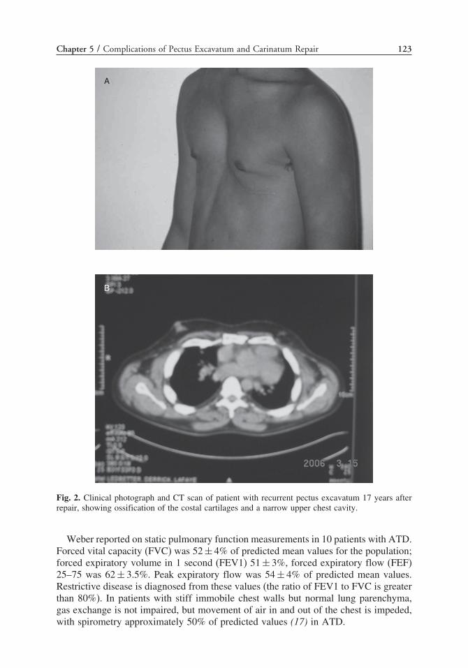
Chapter 5 / Complications of Pectus Excavatum and Carinatum Repair 123
A
B
Fig. 2. Clinical photograph and CT scan of patient with recurrent pectus excavatum 17 years afterrepair, showing ossification of the costal cartilages and a narrow upper chest cavity.
Weber reported on static pulmonary function measurements in 10 patients with ATD.Forced vital capacity (FVC) was 52 ± 4% of predicted mean values for the population;forced expiratory volume in 1 second (FEV1) 51 ± 3%, forced expiratory flow (FEF)25–75 was 62 ± 3.5%. Peak expiratory flow was 54 ± 4% of predicted mean values.Restrictive disease is diagnosed from these values (the ratio of FEV1 to FVC is greaterthan 80%). In patients with stiff immobile chest walls but normal lung parenchyma,gas exchange is not impaired, but movement of air in and out of the chest is impeded,with spirometry approximately 50% of predicted values (17) in ATD.

124 Kelly
Table 1Pulmonary Functions Studies in Patients Presenting for Redo Repair of Pectus Excavatum,
CHKD/EVMS, Norfolk, Virginia
Mean % predicted Standard deviation Number of patients
FVC 71 17 55FEV1 70 18 55FEF 25–25 70 24 53TLC 85 26 20MVV 68 20 16PEF 68 13 14
CHKD, Children’s Hospital of the King’s Daughters; EVMS, Eastern Virginia Medical School; FVC,forced vital capacity; FEV1, forced expiratory volume in 1 second; FEF, forced expiratory flow; TLC,total lung capacity; MVV, mitral valve closing speed; PEF, peak expiratory flow.
Because most patients with pectus excavatum do not complain of shortness of breathat rest, but only with exertion, exercise pulmonary function tests can be very usefulin quantifying the amount of impairment. Malek et al. reported on 21 patients withpectus excavatum and a mean index of 5.1 (normal 2.4; severe pectus is >3.2). Theyfound that maximal oxygen uptake and oxygen pulse were significantly lower thanreference values. VO2 max was 41% of the predicted value! The authors (who arepulmonologists) noted that these findings were not a result of deconditioning; and mostpatients exhibited cardiovascular limitation, not ventilatory limitation (18). Haller andLoughton reported significantly increased duration of exercise after operation (16).
Evaluation should also include echocardiogram. Mitral valve prolapse (MVP) isreported to occur in from 8–45% of patients with pectus excavatum. In approximatelyone-half of those, the MVP resolved after surgical treatment for pectus excavatum(20). Compression of the right ventricle by the chest wall is also frequently seen.Echocardiogram was used by Sigalet to follow stroke volume, which improved from61.6 ± 25 to 77.5 ± 23 mL after primary repair of the chest wall (21). Both of thesepreconditions may be improved by the chest wall operation (16,22,23).
Body image concerns frequently drive the patient or family to seek correction ofthe sternal depression. This is as true with recurrence as with initial operation. A bodyimage survey developed with a psychologist expert on the topic (24) may be used bothto validate the degree of concern about self-image and to document the postoperativeimprovement in activity levels. Improvements in disease-specific quality of life havebeen shown in both children and adults by questioning parents and patients (24,25).
Operative TechniquesHistorically, several techniques have been used for repair of recurrent chest
depression. The external fixation of the repair introduced in the early 1900s is stillused occasionally and needs to be borne in mind as a tool for stabilization (26).
Repair of Recurrent Pectus Excavatum by the Open
or Ravitch Operation
Re-repair by the open operation has been well detailed by several authors. Recentlysome authors have emphasized that only a small portion of the affected cartilages needs

Chapter 5 / Complications of Pectus Excavatum and Carinatum Repair 125
to be resected; earlier others have emphasized that all of the involved cartilages needto be treated to get a good result (2,11).
The technique of open repair for recurrent pectus excavatum is performed undergeneral endotracheal anesthesia (27), with positive pressure a safety feature to assurelung inflation in case the pleura is entered. Thoracic epidural analgesia can be auseful adjunct in the hands of an anesthesiologist skilled in its use. A mixture offentanyl and bupivicaine is very effective and can be continued after the operation.Appropriate anesthetic monitoring equipment includes oxygen pulse oximetry andend-tidal carbon dioxide monitoring to assure adequate gas exchange. Prophylacticantibiotics are appropriate; intravenous Ancef� (cefazolin) is a reasonable choice.
Either a transverse inframammary or a vertical midline incision provides adequateexposure. Generally the incision used for the initial repair is utilized. Skin flaps arecreated. The pectoralis major muscle is detached from its origin on the costal cartilagesbilaterally, and the muscle is mobilized out of the way. Similarly, the rectus abdominismuscles are detached. After inspection of the sternum and cartilages to determine whichcartilages need to be divided, the perichondrial sheath is incised along the long axisof the cartilage. Then the cartilage must be freed from the perichondrial sheath with aFreer elevator or similar instrument. In a redo case, there is often not a well-definedplane (Fig. 3).
Some length of the cartilage must then be removed at each of the involved deformedcartilages. Most authorities are in agreement that only as much cartilage should beremoved as is necessary to allow the ribs and sternum to come into good alignment.Because occurrence of ATD has complicated too early and too extensive operations bythis procedure, removing as little cartilage as practical seems prudent. Some authorshave proposed that only 3–8 mm of cartilage be removed for a primary repair (11); butsometimes more must be removed to allow a twisted sternum with lengthy deformedcartilages to come into good alignment (2). Robicsek notes,
� � �the surgeon should not waste time in performing a meticulous ‘classic’ subperi-chondrial resection of the cartilages and ribs but should just leave enough perichon-drium and periosteum behind to ensure the regeneration of the ribs. For the samereason, a segment of the most lateral portion of the cartilage should be left incontinuity with the ribs (28).
Sternal osteotomy may be needed to allow the sternum to come anteriorly. Fracturingthe anterior table only with an osteotome is usually sufficient to allow twisting andfracturing the posterior table with one’s fingers, allowing the deformed sternum tocome into good position (Fig. 4). The process of sternal mobilization in a reoperativefield may result in entering the pleural cavity, especially on the right. If so, a chest tubeshould be considered. Most surgeons then place a stainless steel support behind thesternum, such as an Adkins strut or Lorenz bar. The support bar is secured to the softtissues with suture or wire (Fig. 5). Occasionally, the support can be placed anterior tothe sternum and held in place with sutures. In larger patients, more than one supportbar may be necessary.
The pectoralis major muscles are sutured together, and the rectus abdominis suturedto the inferior edge of the pectoralis muscles. Closed suction drains may be placedbeneath the muscle layer. Skin closure may be with subcuticular suture.
Support bars may be removed 2–6 months after repair as an outpatient operation.Migration of the supporting strut used for Ravitch repair has been reported, with the bar

126 Kelly
coming to rest in the myocardium, thoracic cavity, or peritoneal cavity, and with injuryto the heart, lungs, or abdominal viscera (29–34). Removal is thus to be encouragedwhen it is not needed.
Operation for treatment of acquired ATD or acquired Jeune’s syndrome has beenpioneered by Dr. Thomas R. Weber. A midline sternotomy is performed, and the pleurais widely opened bilaterally to allow the lung to herniate into the anterior mediastinum.Immediately, this allows pulmonary compliance to improve, with a marked increasein tidal volume with unchanged ventilatory pressures (36).
A subperiosteal lateral osteotomy of all ribs attached to the sternum bilaterallyallowed the sternal halves to be separated by 4–8 cm, increasing the mediastinalherniation of lung. Three straight bony rib segments of appropriate length were resected,generally from the lower ribs, and “wedged” and wired to hold the sternal halves apartpermanently (Fig. 6).
After operation, all patients remained on a ventilator for 3–7 days. Because the heartis no longer covered entirely by bone, patients have been advised to wear a chestprotector for sports after operation. Patients report tremendous subjective improvementin breathing, but pulmonary function studies did not show great improvement. Bodyimage was a concern after operation in approximately 70% of patients (36).
Fig. 3. Division of the right fifth, sixth, and seventh perichondrial bundles and intercostal muscles (2).
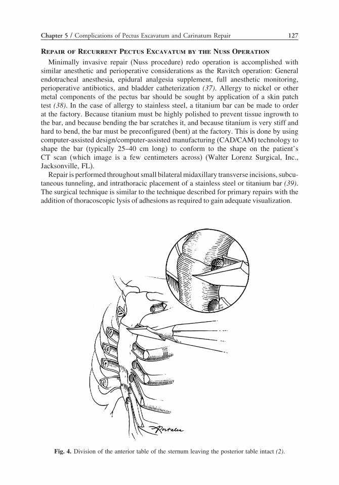
Chapter 5 / Complications of Pectus Excavatum and Carinatum Repair 127
Repair of Recurrent Pectus Excavatum by the Nuss Operation
Minimally invasive repair (Nuss procedure) redo operation is accomplished withsimilar anesthetic and perioperative considerations as the Ravitch operation: Generalendotracheal anesthesia, epidural analgesia supplement, full anesthetic monitoring,perioperative antibiotics, and bladder catheterization (37). Allergy to nickel or othermetal components of the pectus bar should be sought by application of a skin patchtest (38). In the case of allergy to stainless steel, a titanium bar can be made to orderat the factory. Because titanium must be highly polished to prevent tissue ingrowth tothe bar, and because bending the bar scratches it, and because titanium is very stiff andhard to bend, the bar must be preconfigured (bent) at the factory. This is done by usingcomputer-assisted design/computer-assisted manufacturing (CAD/CAM) technology toshape the bar (typically 25–40 cm long) to conform to the shape on the patient’sCT scan (which image is a few centimeters across) (Walter Lorenz Surgical, Inc.,Jacksonville, FL).
Repair is performed throughout small bilateral midaxillary transverse incisions, subcu-taneous tunneling, and intrathoracic placement of a stainless steel or titanium bar (39).The surgical technique is similar to the technique described for primary repairs with theaddition of thoracoscopic lysis of adhesions as required to gain adequate visualization.
Fig. 4. Division of the anterior table of the sternum leaving the posterior table intact (2).

128 Kelly
Fig. 5. Bar secured in place behing the sternum and in front of the ribs (2).
The width of the chest is measured in the operating room prior to surgery, and thecorrect length steel bar is selected and bent to conform to the curvature of the anteriorchest wall. A small transverse incision 2 or 3 cm long is made in each lateral chestwall between the anterior and posterior axillary lines. Subcutaneous tunnels are raised.
A site for entering the chest medial to the pectus ridge is selected (Fig. 7). In a redopatient, it is crucial that the substantial pressure the bar exerts on the chest wall tooppose the pressure of the sternum is borne by the ribs, not the intercostal muscles. Ifthe site of entry into the thorax is too lateral, no rib surface is available for load bearing,and the intercostal muscles will tear because they cannot support that much weight.
A long clamp is used to enter the right hemithorax at this spot under direct visual-ization through a thoracoscope. The long introducer is passed through this tunnel,posterior to the sternum and anterior to the heart, to emerge through the contralateralintercostals space (Fig. 8). An umbilical tape is tied to the end of the introducer, andthe umbilical tape is then pulled back through the tract. Once the umbilical tape isacross the tract, the introducer is removed and the umbilical tape is transferred to thesteel bar. Using the umbilical tape for traction, the steel bar is pulled through the tractwith the convexity facing posteriorly, and then the umbilical tape is removed. Oncethe bar is in position, it is rotated using the bar flipper so that the convexity facesanteriorly, thereby raising the sternum and chest wall into normal position. One end ofthe bar is secured to a lateral stabilizer using steel wire, and each stabilizer is securedto the musculature of the chest wall with absorbable suture. The bar should be securedto the ribs with a heavy absorbable suture such as polydioxanone suture (PDS) under
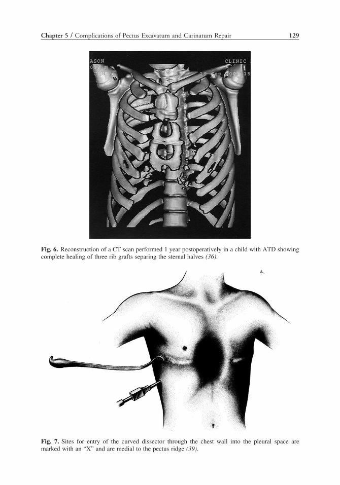
Chapter 5 / Complications of Pectus Excavatum and Carinatum Repair 129
Fig. 6. Reconstruction of a CT scan performed 1 year postoperatively in a child with ATD showingcomplete healing of three rib grafts separing the sternal halves (36).
Fig. 7. Sites for entry of the curved dissector through the chest wall into the pleural space aremarked with an “X” and are medial to the pectus ridge (39).

130 Kelly
Fig. 8. The pectus bar is flipped over, pushing the sternum anteriorly (39).
direct vision with an endo-catch device (Fig. 9). Before closing the incisions, positiveend-expiratory pressure is added to prevent pleural air trapping , then each wound isclosed in layers.
There are several factors to consider for redo operations. In patients with an early recur-rence, for example in the first 2 weeks after operation, the bar has usually flipped from itsinitial and desired position to one in which the sternum is not pushed anteriorly as much
Fig. 9. The bar is secured in place by wrapping heavy absorbable sutures around the bar and theadjacent ribs with thoracoscopic guidance; then the stabilizer and bar are sutured to the muscle ofthe chest wall with absorbable sutures (39).

Chapter 5 / Complications of Pectus Excavatum and Carinatum Repair 131
as desired. When this situation is recognized, it is important to put the bar or bars backwhere they belong. A group in Norway reported erosion of the arch of the aorta froma bar that had flipped to a position putting pressure on the aortic wall (40). Generallyrepositioning can be done through the initial incisions, even when bar displacementoccurs more than 1 month after the operation (6,37,38,41). Thoracoscopy can beconsidered as needed. On-table radiography confirms the correct position of the bars.
It can be difficult at times to tell whether the bar has shifted in the first few daysafter operation. After repair a cross-table lateral taken in the recovery room can helpwith making decisions regarding need to reoperate later.
Usually redo operations are in older patients. Two bars should be considered tosupport the sternum, rather than a single bar. Two bars will not be needed in everyinstance, but one should have a low threshold to use two. The chest is often more rigidin redo ops, and two bars distribute the pressure over a broader area and bear less forceon each bar. This may give a better result.
If two bars will be used, the upper bar is often the better one to put in first. Often,the deepest part of the depression is at the lower sternum or xiphoid process, wherethe lower bar will be placed. Tunneling between the pericardium and sternum may beeasier cephalad to that spot, where the severity of the depression is less.
After passing the first dissector, it should be left in place to support the sternumanteriorly before tunneling with a second dissector at the inferior bar’s site. This greatlyimproves visualization for tunneling at the deeper part of the chest depression.
Sometimes it can be difficult to see the tip of the dissector because of the deep chestdepression. To improve visualization, besides changing from a 0° to 30° scope, it mayhelp things to put the scope in the same opening in the chest as the dissector wentthrough. A group in Japan uses this approach routinely with good results.
A bone hook can also be placed under the xiphoid and used to pull the xiphoid towardthe ceiling of the operating room. A similar incision to that used for a transdiphragmaticpericardial window gives access to the back of the sternum. The operator can sweepthe tissue between the sternum away by blunt dissection and help guide the dissectoracross by palpation through this approach (43). Loss of insufflated gas is to be expectedwhen this approach is used.
After the dissector is passed through successfully, the stiff chest of a redo patientshould be slowly but firmly pulled toward the operating room ceiling by pressure fromthe surgeon and assistant (Fig. 10). If this is not done, the bar may be twisted into aslightly helical shape when it is flipped over by resistance from the chest wall. Wehave had one sternal fracture on a redo patient, and the elevation of the sternum shouldbe done gently but firmly.
A sternal saw should be in the room, and the team prepared to use it. We havehad one patient suffer an arrhythmia leading to full cardiac arrest as the dissector waspassed across the mediastinum. In his very rigid chest, pressure from the dissectormay have produced bradycardia and then arrest. Sternotomy and open defibrillationwere needed for resuscitation. We have had only one such event in almost 800 Nussoperations at our facility.
If the patient has numerous intrapleural adhesions, which can occur in considerableamount even after Ravitch repair, one should have a low threshold to leave a chesttube in place, because small air leaks may result from the dissection.
In reoperative patients, the bars should be especially well secured to the ribs withheavy PDS sutures taken doubled back so two strands go around each rib.

132 Kelly
Fig. 10. Before turning the bar the sternum should be elevated gently but firmly by pulling thedissector anteriorly with the surgeon’s and assistant’s hands close to the site of entry into theskin (39).
Obtaining a chest radiograph on the operating table wise in redo patients, becausethe potential for unrecognized residual air collections (pneumothorax) is greater.
Thoracic epidural catheters are even more useful in redo patients because significantpostop pain is to be anticipated (27).
Ostlie et al. have confirmed our findings that redo correction can be done safelywith minimal blood loss and short operating time in patients who have undergone priorunsatisfactory open repair of pectus excavatum (41).
RECURRENT PECTUS CARINATUM
After open operation:
Pectus carinatum in reoperative surgery occurs after treatment with open or Ravitchrepair. That operation has a low but nonzero recurrence rate. The frequency of recur-rence must be sufficiently low that we are unable to uncover good figures on frequency,timing, or symptoms of recurrence. Lacquet reported in 272 patients with pectus
Fig. 11. Orthotic device used for correction of pectus carinatum by external compression.

Chapter 5 / Complications of Pectus Excavatum and Carinatum Repair 133
carinatum, “Clinical results… were always good for protrusion deformities if enoughdeformed cartilages were resected.” Correia de Matos and colleagues report 24 patientswith pectus carinatum with no recurrences (42). Some authors have suggested thatrecurrence can be minimized by resecting cartilages bilaterally, not just on the side ofthe sternum where they are visibly deformed. Shamberger and Welch reported that of152 patients with pectus carinatum, “three required revision with additional unilaterallower cartilage resection for persistent malformation of the costal arch. All patientsultimately had a satisfactory result” (44).
After treatment with bracing:
Pectus carinatum has been treated with externally applied orthotics. The bracing hasresulted in marked improvement in up to 90% of patients in whom it was applied(45,46) (Fig. 11). Recent improvements in the design of the brace have made it muchsmaller and more comfortable, which has dramatically improved compliance. But useof orthotics to treat recurrent pectus carinatum has not yet been reported (Fig. 12).
LESS FREQUENT CONDITIONS
Congenital Chest Wall ProblemsPectus excavatum has been reported to occur in a fraction of patients who undergo
neonatal repair of sternal cleft primarily. Most of these patients have not requiredintervention (47).
Poland’s syndrome is reported to infrequently recur after surgical repair. Columbanistates that this is likely because of the late age at operation (48) (generally after theteenage growth spurt). Reconstruction often involves repair of the chest wall defectand muscle transfer or free flap for coverage. Problems with nonunion of rib graftsand/or breakdown of the prosthetic repair can lead to lung herniation. These problemsare sufficiently infrequent that treatment must be individualized (48). Remarkably,termination of pregnancy at 22-weeks gestation has recently been reported followingantenatal diagnosis of Poland’s syndrome by ultrasound (49). Fokin and Robicsekreport 20 operative cases without complication. They suggest a staged repair in youngerpatients but a single stage in adults, including muscle flaps and breast implants infemale patients (50).
Repair of pentralogy of Cantrell has infrequently required reoperation, but shouldbe managed by the same principles one uses for other recurrent defects, either thoracicor abdominal wall (48).
Cardiac Surgery and Pectus ExcavatumSeveral reports have described successful combined cardiac operations and open
repair of pectus excavatum (51–54). These have included coronary artery revascular-ization, aortic graft placement, and repair of tetralogy of Fallot (53,55).
Long after open operation to correct pectus excavatum, successful sternotomy hasbeen performed following Ravitch operation for pectus operation (54) and after sternalturnover operation (53).
Combined minimally invasive repair and repair of atrial septal defect and closureof patent ductus arteriosus has been reported recently (56). More importantly,there may be advantage to performing minimally invasive repair in patients withMarfan syndrome before skeletal maturity; then if aortic root dilatation occurs later
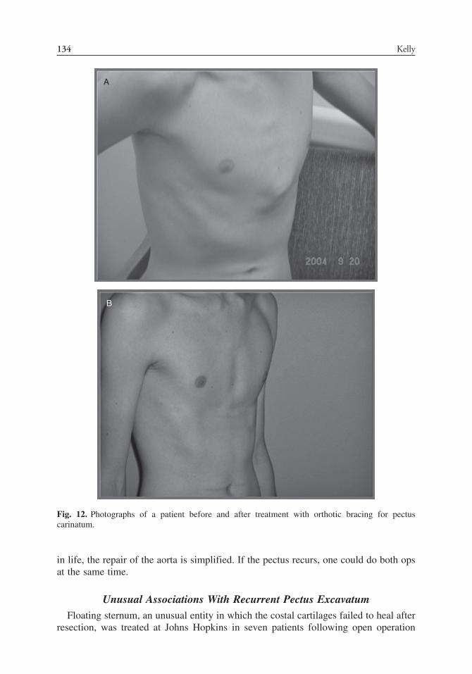
134 Kelly
A
B
Fig. 12. Photographs of a patient before and after treatment with orthotic bracing for pectuscarinatum.
in life, the repair of the aorta is simplified. If the pectus recurs, one could do both opsat the same time.
Unusual Associations With Recurrent Pectus ExcavatumFloating sternum, an unusual entity in which the costal cartilages failed to heal after
resection, was treated at Johns Hopkins in seven patients following open operation
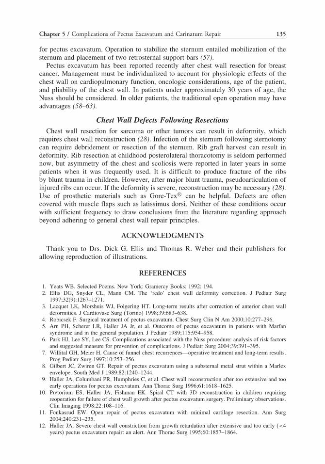
Chapter 5 / Complications of Pectus Excavatum and Carinatum Repair 135
for pectus excavatum. Operation to stabilize the sternum entailed mobilization of thesternum and placement of two retrosternal support bars (57).
Pectus excavatum has been reported recently after chest wall resection for breastcancer. Management must be individualized to account for physiologic effects of thechest wall on cardiopulmonary function, oncologic considerations, age of the patient,and pliability of the chest wall. In patients under approximately 30 years of age, theNuss should be considered. In older patients, the traditional open operation may haveadvantages (58–63).
Chest Wall Defects Following ResectionsChest wall resection for sarcoma or other tumors can result in deformity, which
requires chest wall reconstruction (28). Infection of the sternum following sternotomycan require debridement or resection of the sternum. Rib graft harvest can result indeformity. Rib resection at childhood posterolateral thoracotomy is seldom performednow, but asymmetry of the chest and scoliosis were reported in later years in somepatients when it was frequently used. It is difficult to produce fracture of the ribsby blunt trauma in children. However, after major blunt trauma, pseudoarticulation ofinjured ribs can occur. If the deformity is severe, reconstruction may be necessary (28).Use of prosthetic materials such as Gore-Tex� can be helpful. Defects are oftencovered with muscle flaps such as latissimus dorsi. Neither of these conditions occurwith sufficient frequency to draw conclusions from the literature regarding approachbeyond adhering to general chest wall repair principles.
ACKNOWLEDGMENTS
Thank you to Drs. Dick G. Ellis and Thomas R. Weber and their publishers forallowing reproduction of illustrations.
REFERENCES
1. Yeats WB. Selected Poems. New York: Gramercy Books; 1992: 194.2. Ellis DG, Snyder CL, Mann CM. The ‘redo’ chest wall deformity correction. J Pediatr Surg
1997;32(9):1267–1271.3. Lacquet LK, Morshuis WJ, Folgering HT. Long-term results after correction of anterior chest wall
deformities. J Cardiovasc Surg (Torino) 1998;39:683–638.4. Robicsek F. Surgical treatment of pectus excavatum. Chest Surg Clin N Am 2000;10:277–296.5. Arn PH, Scherer LR, Haller JA Jr, et al. Outcome of pectus excavatum in patients with Marfan
syndrome and in the general population. J Pediatr 1989;115:954–958.6. Park HJ, Lee SY, Lee CS. Complications associated with the Nuss procedure: analysis of risk factors
and suggested measure for prevention of complications. J Pediatr Surg 2004;39:391–395.7. Willital GH, Meier H. Cause of funnel chest recurrences—operative treatment and long-term results.
Prog Pediatr Surg 1997;10:253–256.8. Gilbert JC, Zwiren GT. Repair of pectus excavatum using a substernal metal strut within a Marlex
envelope. South Med J 1989;82:1240–1244.9. Haller JA, Columbani PR, Humphries C, et al. Chest wall reconstruction after too extensive and too
early operations for pectus excavatum. Ann Thorac Surg 1996;61:1618–1625.10. Pretorium ES, Haller JA, Fishman EK. Spiral CT with 3D reconstruction in children requiring
reoperation for failure of chest wall growth after pectus excavatum surgery. Preliminary observations.Clin Imaging 1998;22:108–116.
11. Fonkasrud EW. Open repair of pectus excavatum with minimal cartilage resection. Ann Surg2004;240:231–235.
12. Haller JA. Severe chest wall constriction from growth retardation after extensive and too early (<4years) pectus excavatum repair: an alert. Ann Thorac Surg 1995;60:1857–1864.

136 Kelly
13. Ravitch MM. The chest wall. In: Pediatric surgery, fouth edition . Chicago: Year Book MedicalPublishers; 1986: 568.
14. Ravitch MM. The operative treatment of pectus excavatum. Ann Surg 1949;129:429–444.15. Lawson ML, Mellins RB, Tabangin M, et al. Impact of pectus excavatum on pulmonary function
before and after repair with the Nuss procedure. J Pediatr Surg 2005;40:174–180.16. Haller J, Loughlin G. Cardiorespiratory function is significantly improved following corrective surgery
for severe pectus excavatum. J Cardiovasc Surg 2000;41:125–130.17. Weber TR, Kurkchubasche A. Operative management of asphyxiating thoracic dystrophy after pectus
repair. J. Pediatr Surg 1998;33:262–265.18. Malek MH, Fonkalsrud EW, Cooper CB. Ventilatory and cardiovascular responses to exercise in
patients with pectus excavatum. Chest 2003;124:870–882.19. Raggi P, Callister, et al. Is mitral valve prolapse due to cardiac entrapment in the chest cavity? Clin
Investig 2000;117(3):636–642.20. Shamberger RC, Welch KJ. Surgical repair of pectus excavatum. J Pediatr Surg 1988;23:615–622.21. Sigalet D, Montgomery M, Harder J. Cardiopulmonary effects of closed repair of pectus Excavatum.
J Pediatr Surg 2003;38:380–385.22. Coln E, Carrasco J, Coln D. Demonstrating relief of cardiac compression with the Nuss minimally
invasive repair for pectus excavatum. J Pediatr Surg 2006;41(4):683–686; discussion 683–686.23. Beseir GD, Epstein SE, Stampfer M, et al. Impairment of cardiac function in patients with pectus
excavatum, with improvement after operative correction. N Engl J Med 1972;287:267.24. Lawson M, Cash T, Akers R, et al. A pilot study of the impact of surgical repair on disease-specific
quality of life among patients with pectus excavatum. J Pediatr Surg 2003;38(6):916–918.25. Krasopoulos G, Dusmet M, Ladas G, Goldstraw P. Nuss procedure improves the quality of life in
young male adults with pectus excavatum deformity. Eur J Cardiothorac Surg 2006;29:1–5.26. Ochsner A, DeBakey M. Chone-Chondrosternon Report of a case and review of the literature. J Thorac
Surg 1939;8(5):469–511.27. Futagawa K, Suwa I, Okuda T, et al. Anesthetic management for the minimally invasive Nuss
procedure in 21 patients with pectus excavatum. J Anesth 2006;20:48–50.28. Fokin AA, Robicsek F. Acquired deformities of the anterior chest wall. Thorac Cardiovasc Surg
2006;54:57–61.29. Onursal E, Toker A, Bostanci K, Alpagut U, Tireli E. A complication of pectus excavatum operation:
endomyocardial steel strut. Ann Thorac Surg 1999;68(3):1082–1083.30. Dalrymple-Hay MJ, Calver A, Lea RE, Monro JL. Migration of pectus excavatum correction bar into
the left ventricle. Eur J Cardiothorac Surg 1997;12(3):507–509.31. Pircova A, Sekarski-Hunkeler N, Jeanrenaud X, et al. Cardiac perforation after surgical repair of
pectus excavatum. J Pediatr Surg 1995;30(10):1506–1508.32. Kanegaonkar RG, Dussek JE. Removal of migrating pectus bars by bideo-assisted thoracoscopy. Eur
J Cardiothorac Surg 2001;19(5):713–715.33. Elami A, Lieberman Y. Hemopericardium: a late complication after repair of pectus excavatum.
J Cardiovasc Surg (Torino) 1991;32(4):539–540.34. Paret G, Taustein I, Vardi A, Yellin A, Dekel B. Barzilay A. Laceration of the phrenic artery.
A life-threatening complication after repair of pectus excavatum. J Cardiovasc Surg (Torino)1996;37(2):193–194.
35. Stefani A, Morandi U, Lodi R. Migration of pectus excavatum correction metal support into theabdomen. Eur J Cardiothorac Surg 1998;14(4):434–436.
36. Weber TR. Further experience with the operative management of asphyxiating thoracic dystrophyafter pectus repair. J Pediatr Surg 2005;40:173–175.
37. Croitoru DP, Kelly RE Jr, Goretsky MJ, et al. The minimally invasive Nuss technique for recurrentor failed pectus excavatum repair in 50 patients. J Pediatr Surg 2005;40:181–186.
38. Rushing G, Goretsky MJ, Morales M, et al. When it is not an infection: metal allergy after the Nussprocedure for repair of pectus excavatum. J Pediatr Surg 2007;42(1):93–97.
39. Nuss D, Kelly R. The minimally invasive pectus repair (nuss procedure). In: Holcomb GW III (ed.),Atlas of pediatric laparoscopy and thoracoscopy. Philadelphia: Elsevier; In press.
40. Hoel TN, Rein KA, Svennevig JL. A life-threatening complication of the Nuss procedure for pectusexcavatum. Ann Thorac Surg 2006;81:370–372.
41. Miller KA, Ostlie DJ, Wade K, et al. Minimally invasive bar repair for ‘redo’ correction of pectusexcavatum. J Pediatr Surg 2002;37(7):1090–1092.

Chapter 5 / Complications of Pectus Excavatum and Carinatum Repair 137
42. Correia de Matos A, Bernardo JE, Fernandes LE, et al. Surgery of chest wall deformities. Eur JCardiothorac Surg 1997;12:345–50.
43. Huang PM, Wu ET, Tseng YT, et al. Modified Nuss operation for pectus Excavatum: design fordecreasing cardiopulmonary complications. Thorac Cardiovasc Surg 2006;54:134–137.
44. Shamberger RC, Welch KJ. Surgical correction of pectus carinatum. J Pediatr Surg 1987;22:48–53.45. Frey AS, Garcia VF, Brown RL, et al. Nonoperative management of pectus carinatum. J Pediatr Surg
2006;41:40–45.46. Egan JC, DuBois, JJ, Morphy M, et al. Compressive orthotics in the treatment of asymmetric pectus
carinatum: a preliminary report with an objective radiographic marker. J Pediatr Surg 2000;35:1183–1186.
47. Acastello E, Majluf R, Garrido P, et al. Sternal cleft: a surgical opportunity. J Pediatr Surg2003;38:178–183.
48. Columbami P. Recurrent chest wall anomalies. Semin Pediatr Surg 2003;12:94–99.49. Paladini D, D’Armiento MR, Martinelli P. Prenatal ultrasound diagnosis of Poland syndrome. Obstet
Gynecol 2004;104(5 Pt 2):1156–1159.50. Fokin AA, Robicsek F. Poland’s syndrome revisited. Ann Thorac Surg 2002;74:2218–2225.51. Jones E, Hoffman L, Devereux R, Isom O, Gold J. Staged approach to combined repair of pectus
excavatum and lesions of the heart. Ann Thorac Surg 1994;57(1): 212–214.52. Gittes G, Crisera C, Ginsburg H, Galloway A, Colvin S. Repair of pediatric pectus deformity and
congenital heart disease as a combined procedure. J Pediatr Surg 1999;34(8):1282–1283.53. Sasaki H, Aomi S, Uwabe K, et al. [A re-operative case of bentall operation and aortic arch replacement
using a stent graft for a Marfan syndrome, post sternum turnover and post mitral valve replacement].KyobuGeka 2001;54:871–874.
54. Protopapas AD, Forsyth AT. Coronary revascularisation through re-sternotomy utilizing arterialconduits 47 years after correctionof pectus excavatum: an unusual technical challenge. Heart SurgForum 2001;4:266.
55. Kikuchi S, Ingu A, Ito M. Simultaneous repair of pectus excavatum and tetralogy of fallot: report ofa case. Ann Thorac Cardiovasc Surg 2005;11:320–323.
56. Kim Y, Choe J, Park Y. Minimally invasive concomitant cardiac procedures and repair of pectusexcavatum: case report. Heart Surg Forum 2004;3(7):E216–E217.
57. Prabhakaran K, Paidas CN, Haller JA, et al. Management of a floating sternum after repair of pectusexcavatum. J Pediatr Surg 2001;36:159–164.
58. Mansour KA, Thourani VH, Odessey EA, et al. Thirty-year experience with repair of pectus defor-mities in adults. Ann Thorac Surg 2003;76:391–395.
59. Robicek F, Daugherty HK, Mullen DC, et al. Technical consideration in the surgical management ofpectus excavatum and carinatum. Ann Thorac Surg 1974;18:549–562.
60. Jensen NK, Scmidt WR, Gaamella JJ et al. Pectus excavatum and carinatum: the how, when, andwhy of surgical correction. J Pediatr Surg 1970;5:4–13.
61. Sanger PW, Robicek F, Daugherty HK. The repair of recurrent pectus excavatum. J Thorac CardiovascSurg 1968;56:141–142.
62. Rehbein F, Wernicke HH. The operative treatment of funnel chest. Arch Dis Child 1957;32:5–8.63. Croitoru DP, Kelly RE Jr, Goretsky MJ, et al. Experience and modification update for the minimally
invasive Nuss technique for pectus excavatum repair in 303 patients. J Pediatr Surg 2002;37(3):437–445.

6 Complications of CDH Repairand Recurrent CDH
Melissa L. Hayward, MD, DarioO. Fauza, MD, and Jay M. Wilson, MD
CONTENTS
Overview/Introduction
Reherniation
GER
Gastrostomy Tube
ECMO Recannulation
Miscellaneous
References
OVERVIEW/INTRODUCTION
This chapter is dedicated to discussing those complications and conditions thatrequire further surgical intervention in children who have survived the initial insult ofcongenital diaphragmatic hernia (CDH). The complexity of patients born with CDHcreates a challenge to every surgeon who must bring these infants to the operatingtheater for the initial repair. In this instance, the operation is relatively straightforward,but the physiology is treacherous. Conversely, when situations arise requiring a subse-quent procedure, the physiology is often, but not always, more settled, but the procedureis usually much more difficult. With increased survival has come increased morbidityand, not surprisingly, subsequent operations have become fairly common. In one report,42% of CDH survivors required at least one additional operation. The need for reoper-ation is often predictable, as are the common indications (1,2). First and foremostis a recurrence of the hernia. The next most common indication is either feedingintolerance or gastroesophageal reflux (GER) or both, necessitating placement of agastrostomy tube with or without fundoplication. Then come a variety of situations notunique to CDH, such as development of an incisional hernia, an intestinal obstructionsecondary to adhesions or a persistent hydro- or chylothorax requiring drainage. Thereare several other conditions that are closely associated with CDH such as chest walldeformities, cardiac defects, undescended testicles, scoliosis, and others. However,because these really represent associated primary defects in which the operative fields
From: Reoperative Pediatric SurgeryEdited by: S. Teich and D. A. Caniano © Humana Press, Totowa, NJ
139

140 Hayward et al.
are usually pristine, they will not be discussed fully in this chapter. Finally, there isthe rare neonate that, having been successfully weaned from extracorporeal membraneoxygenation (ECMO), experiences a second insult requiring a second course of ECMO.
The aim of the chapter is to assist in diagnosis and guide the decision pathways whendetermining the need to return to the operating room in this unique patient cohort.
REHERNIATION
IncidenceThe reported incidence of reherniation after a CDH repair has varied from 2–22%,
but has been reported to be as high as 80% in a select group, where a patch repair andthe use of ECMO therapy were concomitantly employed (3–8). Although the reasonfor such a wide discrepancy is unclear, it is almost certainly influenced by the size ofthe initial defect, the method of initial reconstruction, and the length of follow-up. It isnot surprising that small defects, amenable to a relatively tension-free primary repair,are less likely to disrupt than larger defects, which require placement of a prostheticpatch. In a report by Van Meurs et al., 40% of CDH survivors with a patch repaireventually had a recurrence (7). Moss et al reported that within 3 years of initial repair,nearly 50% of patients who underwent a patch repair had developed a reherniation(8). Moss also noted that the majority of reherniations were owing to the patch pullingaway from the posterior thoracic wall (8).
PresentationA diaphragmatic hernia can reoccur at anytime, but the majority present within the
first 24 months of life (6,9–11). Moss and colleagues found a bimodal peak incidenceof recurrence, the first being between 1 and 3 months of age and the second between10 and 36 months (8). Saltzman et al. reported that the age at time of recurrence rangedfrom 2 to 48 months, with the average being approximately 14 months (6). We toohave noted that most reherniations occur prior to 24 months of age, and have tailoredour surveillance program accordingly. We believe that the incidence of patch failure isprobably extremely high, with most, if not all, repairs eventually disrupting. However,in many instances there is sufficient scar tissue formation to either prevent reherniationor to result in only a small degree of reherniation that may not be radiologically evidentor clinically symptomatic. In fact, reherniation is often only discovered on routinechest radiographs, which is why we recommend regular surveillance chest radiographsmonthly for the first 6 months, every 3 months until the age of 24 months, then at30 and 36 months, and every 2–3 years until age 10 in those infants who underwenta patch repair initially (6). The surveillance is initially similar for primary repairs;however, in our experience the incidence of recurrence drops precipitously after 6months, so we do not obtain annual chest x-rays (CXR) for surveillance purposes aloneafter that time.
Patients who present with symptoms related to a reherniation generally fall into twocategories: those with pulmonary symptoms and those with gastrointestinal symptoms(8). Pulmonary symptoms are rare beyond the neonatal period but may present astachypnea, persistent cough, development of aspiration pneumonia, and/or recurrentwheezing requiring increased use of bronchodilators. Respiratory distress in an olderchild is an ominous sign as tachypnea is usually a physiologic response to metabolicacidosis rather than pulmonary compromise. Development of any of these symptoms
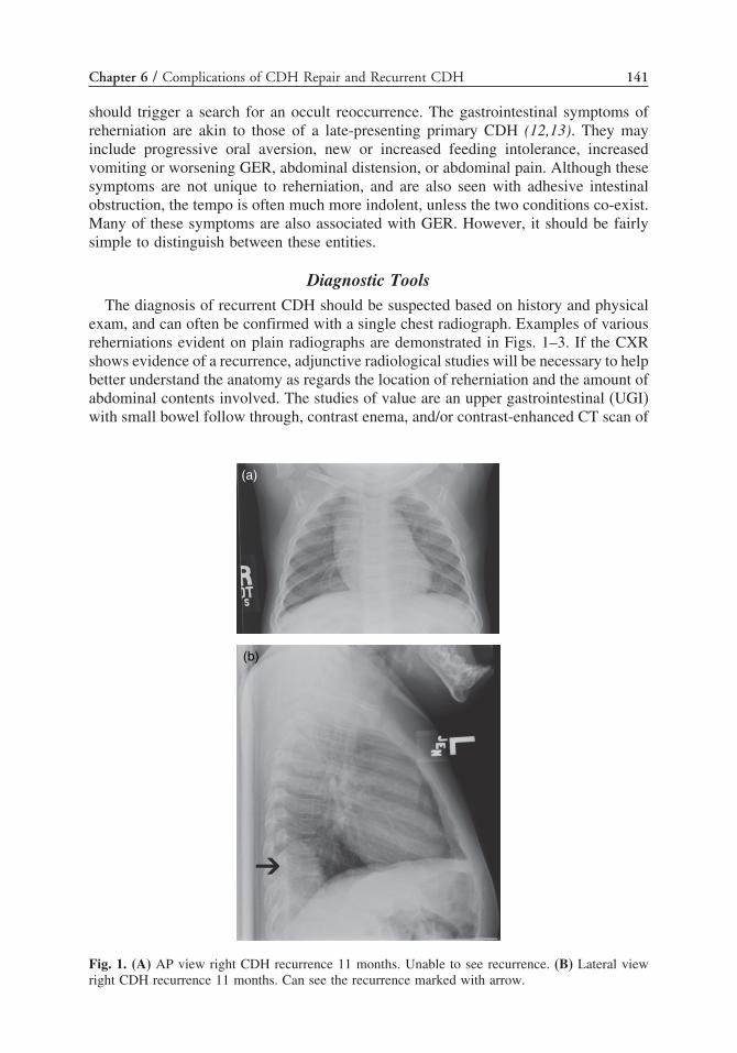
Chapter 6 / Complications of CDH Repair and Recurrent CDH 141
should trigger a search for an occult reoccurrence. The gastrointestinal symptoms ofreherniation are akin to those of a late-presenting primary CDH (12,13). They mayinclude progressive oral aversion, new or increased feeding intolerance, increasedvomiting or worsening GER, abdominal distension, or abdominal pain. Although thesesymptoms are not unique to reherniation, and are also seen with adhesive intestinalobstruction, the tempo is often much more indolent, unless the two conditions co-exist.Many of these symptoms are also associated with GER. However, it should be fairlysimple to distinguish between these entities.
Diagnostic ToolsThe diagnosis of recurrent CDH should be suspected based on history and physical
exam, and can often be confirmed with a single chest radiograph. Examples of variousreherniations evident on plain radiographs are demonstrated in Figs. 1–3. If the CXRshows evidence of a recurrence, adjunctive radiological studies will be necessary to helpbetter understand the anatomy as regards the location of reherniation and the amount ofabdominal contents involved. The studies of value are an upper gastrointestinal (UGI)with small bowel follow through, contrast enema, and/or contrast-enhanced CT scan of
(a)
(b)
Fig. 1. (A) AP view right CDH recurrence 11 months. Unable to see recurrence. (B) Lateral viewright CDH recurrence 11 months. Can see the recurrence marked with arrow.

142 Hayward et al.
(a)
(b)
Fig. 2. (A) Recurrent left CDH with bowel in chest. Note the pH probe. The asterisk marks bowel in thechest—the tell-tale feature of the recurrence. (B) Lateral view recurrent left CDH. Arrow marks bowelin chest. Note again the pH probe and the presence of contrast in the herniated bowel located posteriorly.
(b)(a)
Fig. 3. (A) AP image paraesophageal hernia. Arrow marks recurrence. (B) Lateral view parae-sophageal hernia. Arrow marks recurrence.
the chest and abdomen. In cases where reherniation is suspected on clinical grounds,but the chest film is equivocal, obtaining one of these contrast studies usually makesthe diagnosis clear. With the advent of faster CT scans which have eliminated the needfor sedation, CT has become our preferred diagnostic tool (Figs. 4A and B).
The Surgical ApproachOnce it has been established that a reherniation has occurred, patients can be
divided into symptomatic and asymptomatic groups. We believe that all recurrenceswill eventually necessitate repair. However, few are urgent, and deciding when therepair is to take place should be based on the size of the recurrence and the severity ofthe associated symptoms. The symptomatic patient should be repaired as soon as it is
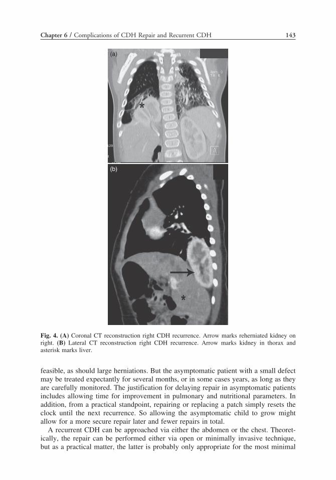
Chapter 6 / Complications of CDH Repair and Recurrent CDH 143
(a)
(b)
Fig. 4. (A) Coronal CT reconstruction right CDH recurrence. Arrow marks reherniated kidney onright. (B) Lateral CT reconstruction right CDH recurrence. Arrow marks kidney in thorax andasterisk marks liver.
feasible, as should large herniations. But the asymptomatic patient with a small defectmay be treated expectantly for several months, or in some cases years, as long as theyare carefully monitored. The justification for delaying repair in asymptomatic patientsincludes allowing time for improvement in pulmonary and nutritional parameters. Inaddition, from a practical standpoint, repairing or replacing a patch simply resets theclock until the next recurrence. So allowing the asymptomatic child to grow mightallow for a more secure repair later and fewer repairs in total.
A recurrent CDH can be approached via either the abdomen or the chest. Theoret-ically, the repair can be performed either via open or minimally invasive technique,but as a practical matter, the latter is probably only appropriate for the most minimal

144 Hayward et al.
of defects. The choice of approach may be influenced by other circumstances such aswhether a fundoplication and/or gastrostomy tube will also be performed at the timeof the repair, or whether an incisional hernia is present.
The transthoracic approach is favored by some, as it is more likely to provide avirgin operative field in patients who initially underwent a transabdominal repair. Butif the child has evidence of pulmonary hypertension or inadequate lung reserve, thetransthoracic approach may not be reasonable. Furthermore, many recurrence sitesare posteromedial, and we have found this difficult to address via the chest. If thepatient also needs a fundoplication, the transthoracic approach may not be ideal. Weusually prefer the transabdominal approach which is often a bit more challengingand time consuming but, allows us better visualization of the defect itself, and theorgans and structures that must be preserved. It also provides ready access to performa fundoplication and/or gastrostomy tube should one be needed. The laparoscopicapproach is likely to be very difficult and in most cases should probably be avoidedunless the initial repair was transthoracic.
Repair TechniquesIn order to plan the most appropriate repair technique, the mode of failure of the
initial repair must be ascertained. In our experience there are four primary modes offailure: (1) failure of a primary repair; (2) failure of the medial aspect of the repair;(3) recurrence at the posterior-lateral margin; and (4) development of a paraesophagealhernia. Anterior and lateral failures are rare, probably because the anterior lip ofdiaphragm seems to be the thickest muscle, and laterally, owing to the absence of anydiaphragm at this location sutures are usually placed around ribs. The location of failure isimportant, as it will dictate subtle but important differences in timing and technique.
In the case of a failed primary repair, the cause of recurrence is often a high-tensioninitial repair in which the sutures simply could not hold the tissue together. In thesecases, another primary repair is usually impossible and a new prosthetic patch shouldbe placed. The new patch may be domed or reefed to allow room for the child togrow. A novel dome technique was described by Loff and coworkers in 2005. Theyutilized a flat polytetrafluoroethylene (PTFE) patch that was folded into a cone shapeand held together by a few simple stitches. The cone was then secured to the rimof the remaining diaphragm with simple stitches, such that the rough portion of thepatch abutted the diaphragmatic rim (14). Other techniques for pleating a patch withabsorbable sutures intended to release in response to tension have also been described(8). We have tried both techniques several times but do not have long enough follow-upto comment on their utility. We tend to favor Gore-Tex� patches (PTFE); however, astudy by Grethel and colleagues reported no difference in outcomes or complicationbetween Surgisis� (intestinal submucosal patch) and Gore-Tex� patch repairs (11).
When the initial patch repair fails, it may do so in the early postoperative period,or weeks, months, or years after the initial repair. An early recurrence usually denotessome technical error and the patch can usually be simply replaced or resutured. Witha late medial recurrence, when scar tissue has had ample time to form, our preferredapproach is to augment the existing patch by placing a new medial portion ratherthan to replace it. In this situation, the spleen, now well adhered to the lateral aspectof the diaphragm and prior patch, does not need to be mobilized, and in fact shouldnot, as long as the posterior-lateral aspect of the repair is intact. To do so is notonly unnecessary; it significantly increases the difficulty of the dissection, and not

Chapter 6 / Complications of CDH Repair and Recurrent CDH 145
uncommonly will result in a splenectomy. When repairing medial defects in a recurrentleft-sided hernia, we augment the patch using a “C” or crescent shaped patch nestledaround the GE junction. Medial defects on the right are difficult because the inferiorvena cava (IVC) comprises most of the medial wall. The reoccurrence is almost alwaysposterior in these situations. In this instance, a secure posterior stitch placed around therib medially, and an anterior medial suture into muscle or around the rib, will usuallyprevent herniation, principally because the liver will protect the weak medial wall. Wecontinue to observe some small right medial recurrences in which only a small portionof liver has herniated, as it is often just as secure as a patch.
When the patch has torn away from the posterior-lateral aspect of the thoracic wall,it is usually because of trusting the integrity of an inadequate rim of diaphragm at theinitial surgery or mistakenly placing posterior sutures into the retroperitoneal fasciaand/or Gerotta’s fascia rather than into diaphragmatic muscle or around a rib. This isprobably the most common mechanism of failure we have seen and can be avoided bybeing sure that the posterior sutures are placed into the thoracic wall. In this scenario, itis common for the kidney to herniate into the chest, thereby creating a “retroperitoneal”herniation and not a true reherniation. In fact, at reoperation it is not unusual to findthe patch repair, itself, intact and the kidney herniated posterior to it. We prefer torepair this mode of recurrence by dissecting free the remnant diaphragm and patchand re-anchoring, augmenting, or replacing the patch with suture around the ribs. If alateral recurrence is present in a right-sided defect, the liver will likely be scarred inplace medially, and, as with the spleen on the left side, one should not need to mobilizethe liver. The patch should be augmented rather than replaced.
The final mode of “recurrence” that we have observed, which always occurs late, isthe paraesophageal hernia. This occurs when the child “outgrows” the original patchand the patch does not tear away from the lateral chest wall. In this situation, the thinrim of medial diaphragm gets pulled laterally by the inflexible patch, resulting in anew paraesophogeal hernia and worsening GER. Immediate repair is seldom necessaryand timing should be guided by symptoms. Again, because of the large amount of scartissue present at the time of repair, we advise augmenting the medial portion, ratherthan replacing the entire patch. We also feel that it is prudent, if not mandatory, toperform a fundoplication procedure at the same time as the process will invariablyre-occur.
A variety of novel approaches to repairing a recurrent hernia both with prostheticpatches and with muscle flaps have been described. One type of “patch” repair utilizesa Bard Mesh plug, similar to those used in inguinal hernia repairs (6). Saltzman andcoworkers described this technique in 2001, employing a transthoracic approach forplacement of the mesh plug in five patients with a posterior-medial recurrence (6). Theaverage follow-up was approximately 14 months (range was 1–33 months), with norecurrences. This approach may be reasonable in small reherniations. Several groupshave described various muscle flaps. Sydorak and colleagues reported the use of areversed latissimus dorsi flap (LDF) in seven patients, three with a recurrence and fourwho were considered to have significant chest wall excursion restrictions secondaryto a patch repair (15). The flap was rotated, with the neurovascular bundle intact, intothe thorax via a thoracotomy. To assist in the function of the LDF, the thoracodorsalnerve was anastomosed to the phrenic nerve (15). In four of the seven children, therewas evidence of physiologic diaphragmatic function (15). Despite attempts to improveupon the repair of a recurrent hernia, placing an additional prosthetic patch remains

146 Hayward et al.
the mainstay of therapy. The “Holy Grail” of CDH repair is a patch that can growand remodel with the child. Recent large animal data suggest that CDH repair withautologous, engineered tendon patches is possible. These grafts can be made availableat birth, as they can be engineered in parallel to gestation, from cells normally presentin, and harvested from, the amniotic fluid (16,17). A proposed protocol for the firstclinical trial of this novel therapeutic concept is currently under review by the Foodand Drug Administration (FDA). However, until pre-engineered bioprosthetic patchesbecome a clinical reality, we will continue to rely on the synthetic devices currentlyavailable.
GER
EtiologyPhysiologic GER is common to many infants and thus can pose a challenge in recog-
nizing the transition to a pathologic process in patients with CDH. In the first severalweeks of life, GER, also called “spitting-up,” is common (18,19). As many as 60–65%of infants with GER are symptom-free and the GER has resolved by 18–24 monthsof age without any medical interventions or therapy (20). When symptoms persist orthe GER is so severe that complications ensure, then it is considered pathologic andthe child is diagnosed as having gastroesophageal reflux disease (GERD). The NorthAmerican Society for Pediatric Gastroenterology and Nutrition estimated that approx-imately 7% of children ages 3–9 years old have GERD, and Nelson et al. reported a5% incidence of daily reflux in 10- to 12-month-old infants (21,22). It wasn’t untilthe 1990’s that the literature began to recognize GERD as an entity associated withincreased morbidity in patients with CDH (23–26). The incidence of GERD in patientswith CDH ranges from 20–72%, depending on the series (23,27).
The current concepts regarding the etiology of GERD revolve around the functionof the lower esophageal sphincter (LES), the competency of which is a coordinationbetween the stomach, esophagus, and diaphragm. Lack of competency at any levelleads to the development of GERD (28,29). In children with CDH, several mechanismshave been proposed to explain the higher rates of GERD observed in this subset ofpatients. Stolar et al. noted ectatic esophagi, with evidence of dysmotility, in most ofthe CDH patients with GERD, suggesting that perturbation of esophageal peristalsismay, in addition to the diaphragm, be an important component in GERD development(26,30). Kieffer et al., who reported a 62% incidence of GERD in their CDH survivors,proposed that fetal swallowing and kinking of the esophagus caused by bowel contentsin the chest may play a role in the development of an abnormal esophagus (27). Ithas also been postulated that operative disruption of the angle of His may also playa role in promoting GERD by creating an obtuse angle as a result of the repair.Similar observations have been made in patients with hiatal hernias who demonstratedan increase incidence of GERD (19). As seen in patients with hiatal hernias and inpatients who have had a tracheo-esophageal fistula repair, the GE junction may also belocated abnormally high, thereby allowing the negative intrathoracic pressure createdwith respiration to promote GERD (19,31). With the altered diaphragmatic structure inCDH, the state of the diaphragmatic crus and the surrounding muscle undoubtedly alsoplay a role in the development of GERD, preventing the diaphragm from maintainingan appropriate LES pressure (27). Finally, delayed gastric emptying, often seen inpatients with CDH, will aggravate any pre-existing GERD (18).
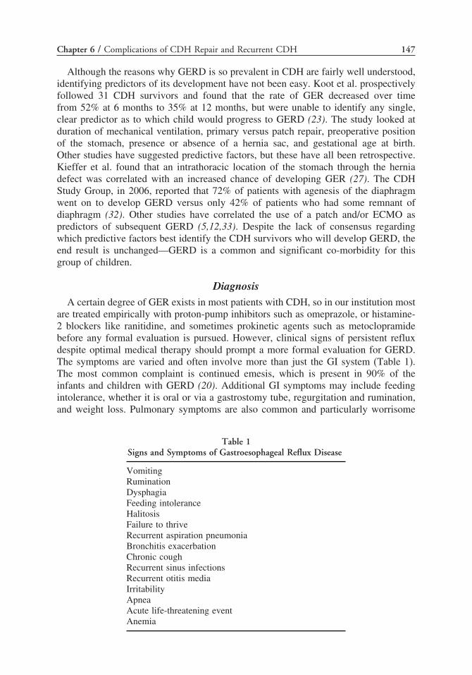
Chapter 6 / Complications of CDH Repair and Recurrent CDH 147
Although the reasons why GERD is so prevalent in CDH are fairly well understood,identifying predictors of its development have not been easy. Koot et al. prospectivelyfollowed 31 CDH survivors and found that the rate of GER decreased over timefrom 52% at 6 months to 35% at 12 months, but were unable to identify any single,clear predictor as to which child would progress to GERD (23). The study looked atduration of mechanical ventilation, primary versus patch repair, preoperative positionof the stomach, presence or absence of a hernia sac, and gestational age at birth.Other studies have suggested predictive factors, but these have all been retrospective.Kieffer et al. found that an intrathoracic location of the stomach through the herniadefect was correlated with an increased chance of developing GER (27). The CDHStudy Group, in 2006, reported that 72% of patients with agenesis of the diaphragmwent on to develop GERD versus only 42% of patients who had some remnant ofdiaphragm (32). Other studies have correlated the use of a patch and/or ECMO aspredictors of subsequent GERD (5,12,33). Despite the lack of consensus regardingwhich predictive factors best identify the CDH survivors who will develop GERD, theend result is unchanged—GERD is a common and significant co-morbidity for thisgroup of children.
DiagnosisA certain degree of GER exists in most patients with CDH, so in our institution most
are treated empirically with proton-pump inhibitors such as omeprazole, or histamine-2 blockers like ranitidine, and sometimes prokinetic agents such as metoclopramidebefore any formal evaluation is pursued. However, clinical signs of persistent refluxdespite optimal medical therapy should prompt a more formal evaluation for GERD.The symptoms are varied and often involve more than just the GI system (Table 1).The most common complaint is continued emesis, which is present in 90% of theinfants and children with GERD (20). Additional GI symptoms may include feedingintolerance, whether it is oral or via a gastrostomy tube, regurgitation and rumination,and weight loss. Pulmonary symptoms are also common and particularly worrisome
Table 1Signs and Symptoms of Gastroesophageal Reflux Disease
VomitingRuminationDysphagiaFeeding intoleranceHalitosisFailure to thriveRecurrent aspiration pneumoniaBronchitis exacerbationChronic coughRecurrent sinus infectionsRecurrent otitis mediaIrritabilityApneaAcute life-threatening eventAnemia

148 Hayward et al.
in patients with CDH who already have lungs compromised by pulmonary hypoplasia(4,10,34). These symptoms may include recurrent bronchitis, lobar pneumonia,worsening bronchopulmonary dysplasia, chronic cough, and persistent wheezingdespite the use of nebulizers, recurrent otitis media, sinusitis, and occasionally apnealeading to sudden infant death or an acute life-threatening event (18,21). In patientswith CDH, it is often difficult to assess whether pulmonary symptoms are caused byGERD or just part of their primary pulmonary disease (35). Further complicating theability to diagnose GERD in patients with CDH is the fact that many patients withCDH display some degree of feeding difficulty. However, continued loss of weight ora drop down of a percentile on the appropriate growth curve are clear signs of GERDthat has failed medical therapy (4,12,21,34).
Once a child is suspected of having GERD, the diagnostic toolbox includes nonin-vasive radiologic studies such as an upper GI series (UGI), to more invasive techniquessuch as endoscopy and esophageal biopsy. However, the major components of theevaluation include an UGI and 24-hour pH monitoring, which is considered the “gold-standard” for evaluating GERD (18,20,36–38). The UGI can provide valuable infor-mation about the anatomy of the esophagus, the height of refluxed contents, and canalso evaluate gastric emptying. As already noted, Stolar et al. found a strong correlationbetween GERD and the presence of an ectatic esophagus, and it is well documentedthat patients with tracheo-esophageal fistula repairs and with hiatal hernias, where theLES is above the diaphragm, are all at high risk of having GERD (19,26,30,31). TheUGI has a positive predictive value for GER of approximately 80–82%, but the sensi-tivity and specificity are inconsistent, from 31–86% and 21–83%, respectively (21). Anuclear medicine study that has been reported to be useful involves feeding the infantradio-labeled juice and looking for tracer in the lung fields and in the mediastinumto quantify the severity of reflux (18,20,21,39). A test that we use frequently is the24-hour pH probe and sleep study, as we believe that it is very effective at recordingreflux events and any associated respiratory compromise such as apnea (20,21,37,38).The study entails the securing of a pH probe at the junction of the middle and distalone-third on the esophagus with the tip of the probe located 2–3 cm above the LES.The number and duration of reflux events (pH < 4.0), are recorded and reconciled withthe patient’s position and presence or absence of symptoms at the time of the event(23,40,41). A similar study to the 24-hour pH probe is a 24-hour impedence probe,which is placed into the stomach and has sensors along its length to assess the pHat various points in the esophagus as well as the stomach. This allows the examinerto assess not only acidic, but also nonacidic reflux events. This is a newer technique,but there is concern as to whether the probe passing into the stomach might affectthe usefulness of the data obtained because it is stenting the LES open, and “standardvalues” are not yet clear (42). Finally, the most invasive test is esophagoscopy, whichallows direct visualization of the esophagus with biopsies. This is generally reservedfor cases in which the results of other tests are inconclusive.
Management of GERD in CDHNonoperative management consists of thickening oral feeds, often with cereal, small
frequent meals, sleeping on a wedge, decreased exposure to secondhand smoke, andpharmacologic therapies such as histamine-2 blockers, proton-pump inhibitors, andprokinetic agents (18,20,34,40). Stolar et al. reported that in his CDH cohort withGERD, all responded to medical therapy and did not require a surgical antireflux
Chapter 6 / Complications of CDH Repair and Recurrent CDH 149
procedure (26). However, this has not been our experience. We observed that approx-imately 15–20% of our patients with CDH will fail nonoperative management forGERD and will require surgical intervention (34). In other studies, it has been reportedthat 9–23% of CDH survivors will need an antireflux procedure (23,27). It is importantto recognize that unlike normal infants with physiologic GER who will improve withtime, infants with CDH and GERD usually worsen. This is especially true of patientswith patch repairs, which predispose to development of paraesophogeal hernias as thechild grows.
Surgical ApproachThe surgical management consists of a fundoplication with or without gastropexy.
In the planning of the operation for the patient with CDH, there are several factors tobe considered. First is to assess if there is evidence of reherniation or development ofa paraesophogeal hernia. Second, it has been our experience that postoperatively mostchildren will not take adequate calories orally and thus a gastrostomy tube is a usefuladjunct as a route to provide adequate caloric intake (12). We almost always place agastrostomy in our patients with CDH during fundoplication. Finally, whereas in mostchildren a laparoscopic approach is favoured, in the majority of patients with CDHthis would be difficult if the CDH repair was done via a transabdominal approach sowe most often will employ an open approach.
Repair TechniqueThere are four major types of fundoplications: Nissen, Thal, Boix-Ochoa, and
Toupet. The Nissen fundoplication is the only one in which the stomach is completelywrapped around the esophagus (360° wrap) (44). The Nissen fundoplication is alsothe most common type of fundoplication used in both adults and children (18). Theother three procedures are all variations of a partial wrap. The Boix-Ochoa and Thalfundoplications are both anterior, 180° wraps with the major difference the fixation ofthe stomach to the left diaphragmatic undersurface with the Boix-Ochoa technique. TheToupet wrap is similar to the Boix-Ochoa technique in the fixation of the stomach tothe diaphragm with three to five single stitches, but the Toupet is a posterior 270° wrap.Although the data is sparse in the pediatric literature, in adults a Nissen fundoplicationis the procedure of choice unless there is evidence of abnormal esophageal motility,and then a partial wrap is the procedure of choice (45). A recent European studyevaluated the efficacy and complication rates between laparoscopic Nissen, Toupet,and Thal fundoplications and found no difference in the rates of dysphagia or othercomplications, suggesting that each technique was equally efficacious in alleviatingGERD (46). In light of the paucity of data, we tend to favor a Nissen fundoplicationor a Toupet wrap if there is a suggestion of poor esophageal motility, with a modifi-cation we have coined “Gulliver’s Travels.” As described by Hendren in 1980, in bothtechniques the crus of the diaphragm, once freely dissected, are brought closer with asimple stitch. The esophagus is then sutured to the diaphragmatic hiatus with a seriesof five (4-0 or 5-0) proline sutures. The wrap is made around an esophageal bougie,the length of which is based on the size of the patient ery. We also modify the suturingof the stomach by using multiple rows of fine interrupted stitches placed from stomachto esophagus instead of the standard three. Finally, we employ multiple sutures to affixthe stomach to the diaphragm or patch. Consequently, the wrap, like “Gulliver,” is

150 Hayward et al.
held together by up to 40 small sutures which individually have very little integritybut collectively are quite strong (47). It is seldom necessary to mobilize the spleento gain access to the medial portion of the diaphragm as long as it was appropriatelypositioned at the initial operation.
This technique has the advantage of distributing the tension across multiple tetherpoints, with no single suture being critical to the integrity of the wrap. It has thedisadvantage of not being practical to perform laparoscopically. In our experience,subsequent failure of the wrap in patients with CDH has been rare utilizing thistechnique.
GASTROSTOMY TUBE
IndicationsNutrition, even in patients without reflux, is a large concern for the CDH survivors.
Several series have reported that between 20 and 56% of all patients with CDH fallbelow the 25th percentile for weight, despite aggressive nutritional support (1,3,12,33).The reason for this is probably multifactoral. Patients with CDH, especially in infancy,have an increased work of breathing and require the use of accessory skeletal andabdominal muscles to maximize ventilation to compensate for the lack of a fullyfunctional diaphragm. This is especially true for those with a patch repair. Many ofthese patients also have prolonged intubations and have some degree of oral aversion,and some have difficulty swallowing. Poor oral intake may also be a sign of severeGERD (4,7,34). It has been our policy to defer placement of a gastrostomy tube aslong as the patient remains on his or her growth curve and as long as they are abovethe fifth percentile. However, in our experience, as least 25% of patients will fail andwill need supplemental nutrition via a gastrostomy tube (12). We have found a strongcorrelation between the need for a gastrostomy and the use of a prosthetic patch (5).The length of intubation and the need for home oxygen also correlate with poor oralintake and feeding intolerance (39).
Surgical ApproachThere are several methods of placing a gastrostomy tube in this population.
Three basic options exist: open placement, laparoscopic placement, and percutaneousendoscopic placement (PEG). Each of these techniques has advantages and disadvan-tages. The majority of children have the CDH repaired via a transabdominal approach,and there are likely to be extensive adhesions. Placing a PEG would carry a significantrisk of injuring colon or small bowel in the patient with CDH, because malrotationand an underdeveloped abdominal cavity predispose the small bowel to occupy everyavailable space, including over the liver and anterior to the stomach. A new techniquecoined “SLiC” was recently reported by Ponsky and colleagues as an alternative forpatients in whom a traditional PEG is not a suitable option. This technique usesendoscopy to insulate the stomach, and a finder-needle is then passed through theanterior abdominal wall into the stomach while being visualized by the endoscopist.After the local anesthetic is injected, a silastic tissue expander prosthesis (STEP) trocaris inserted directly into the stomach while being visualized (48). This avoids pullingthe gastrostomy tube down the esophagus, but offers no particular advantage when theconcern is bowel injury.
Chapter 6 / Complications of CDH Repair and Recurrent CDH 151
A laparoscopic approach, for the same reasons as the PEG, is often not a favorableoption. However, if the patient did not have a transabdominal CDH repair, thenthe laparoscopic approach may be a viable option if an antireflux procedure is alsoto be performed. Because most patients have had a transabdominal repair of theirdiaphragmatic defect, an open gastrostomy tube is still the most common approachfor us. We usually employ the Stamm technique, in which the feeding tube is placedinto the anterior gastric wall near the greater curvature of the stomach and securedby two purse-string sutures. The stomach is then fixed to the anterior abdominal wallsurrounding the tube exit site (49). It has been our experience that most patients will nolonger need the gastrostomy tube by the time they reach school age. Tube removal andclosure of a gastrocutaneous fistula in patients with CDH follow the same algorithmas any other patient.
ECMO RECANNULATION
IndicationsApproximately one-third of our CDH survivors have had ECMO as part of their
treatment course. Meehan and colleagues analysis of the Extracorporeal Life SupportOrganization (ELSO) data through 2002 reported that 1.25% of 16,450 patients in theELSO database required more than one ECMO run. Indications for a second ECMOrun were pulmonary in nature in 82% of the cases. Patients with CDH accounted for62.5% of those cases (50). Indications for a second ECMO run may include sepsis,worsening pulmonary hypertension, and right heart failure. In order to justify a secondECMO run there should be an identifiable, correctable, acute insult that has occurredto an otherwise recovering patient. Failure to maintain adequate gas exchange, aftersuccessful ECMO decannulation (the dwindles) is not a reasonable indication.
TechniqueThe challenges associated with recannulation vary depending on the technique used
(veno-arterial [VA] versus veno-venous [VV]), how the involved vessels were dealtwith during the first decannulation, and how much time has passed since the initialECMO run. In general, it is usually fairly straightforward to recannulate through thepreviously utilized vessel or vessels within the first several days after the initial ECMOrun. Care should be taken to backbleed the vessels to assure patency before placing newcannulas. Embolectomy catheters are useful if thrombus is present, and occasionallyHegar dilators can be helpful if the vessel is scarred or has become stenotic. Thecatheters, dilators, and cannulas must be passed carefully, as arterial dissection is avery real risk.
Although we have been successful in recannulating vessels as late as one monthafter the initial run, it is probably unwise to attempt this beyond two weeks unless thevessels were reconstructed and are known to be patent. Beyond this period of time,the transthoracic approach is preferable, especially in neonates in whom the femoralapproach is impractical.
ComplicationsAnalysis of the ELSO data also shows that the rate of complications increases by
20.6% from the first to second course of ECMO (50). These complications include

152 Hayward et al.
mechanical, (i.e., difficulty with the cannula placement and achieving adequate flowrates), a higher incidence of neurological deficits, and a much higher incidence ofmultisystem organ dysfunction or outright failure. Consequently, very careful consid-eration must be given to the patient’s realistic chances of recovery prior to embarkingon a second run.
MISCELLANEOUS
ChylothoraxChylothorax is reported to occur in up to 10% of patients with CDH and, when
present, is quite difficult to eradicate and significantly complicates postoperativemanagement (51–53). It appears to be more common in patients with a sac thanwithout one and may be caused by disruption of lymphatic channels during sacresection (52,54). The initial treatment of a chylothorax is always nonoperative, whichis successful in nearly all cases (52). The placement of a chest tube is warranted incases were the effusion is causing clinically significant respiratory compromise. Thesecan often be done under ultrasound guidance to achieve optimal placement. In additionto chest tube drainage, enteral feeds are either stopped completely or the formulaswitched to one containing medium chain fatty acids only. Beghetti et al. reported thatdietary management and drainage were effective in 80% of the pediatric patients intheir series (55). Recent studies also favor the addition of Octreotide, a somatostatinanalog, to diminish chylous drainage if the chylothorax is not diminished with dietarychanges alone (56,57). Octreotide may be administered as a continuous intravenousinfusion, or in divided doses via subcutaneous injections. We have found it to be veryuseful in cases where dietary manipulations alone failed to resolve this problem.
If all nonoperative management fails, the last option is to attempt surgical ligationof the thoracic duct. In some cases of congenital chylothorax, pleurodesis is alsoattempted, but because of the pulmonary hypoplasia present in the patient with CDH,this is unlikely to work. Unfortunately there is no clear consensus in the literature asto how long nonoperative management should be employed, or when to abandon it infavor of surgical options. However, the success rate of nonoperative management hasbeen extremely high in our experience.
Small Bowel ObstructionWhen CDH survivors present with abdominal pain, constipation, abdominal
distention, emesis, and/or other signs and symptoms of a bowel obstruction, the firststep is to obtain plain films to look for a recurrence of the CDH. The patient who hasa bowel obstruction, regardless of etiology, should have a nasogastric tube placed fordecompression, intravenous fluids administered, and serum chemistries evaluated forsigns of dehydration and metabolic academia. Lund and colleagues reported that theincidence of bowel obstruction requiring operative intervention in this population wasslightly less than 20% (58). The principle causes of obstruction in their series wereadhesions, midgut volvulus, gastric volvulus, and recurrent CDH. Operative decisionmaking and treatment do not differ from those in any other pediatric patient other thanto determine whether the obstruction is caused by reherniation.

Chapter 6 / Complications of CDH Repair and Recurrent CDH 153
Chest Wall and Spinal DeformitiesThe most common chest wall deformity in this population is pectus excavatum. It
has been our experience that it does not require operative repair in the majority ofcases. There are few cases where the pectus deformity is so severe that there is anelement of respiratory compromise owing to restriction by the deformity (see Fig. 5).Each patient needs to be assessed on an individual basis with pulmonary functiontests, cardiac echocardiograph, evaluation of chest wall mechanics, and anatomicalevaluation utilizing chest CT. The selection of operative approach does not differ from
Fig. 5. Pectus. Lateral view of pectus, with arrow marking inferior edge of sternum.

154 Hayward et al.
any other patient with pectus with the exception that placement of a transthoracic barmay be complicated by pulmonary adhesions.
Scoliosis is the other common skeletal deformity in this population. We recommendassessing the spine at each visit during childhood. A plain chest radiograph can be ofgreat assistance in identifying mild scoliosis. Although common in patients with CDH,very few of our patients have required spinal correction.
REFERENCES
1. Langham MR Jr, Kays DW, Ledbetter DJ, Frentzen B, Sanford LL, Richards DS. Congenital diaphrag-matic hernia. Epidemiology and outcome. Clin Perinatol 1996;23(4):671–688.
2. Torfs CP, Curry CJ, Bateson TF, Honore LH. A population-based study of congenital diaphragmatichernia. Teratology 1992;46(6):555–565.
3. Cortes RA, Keller RL, Townsend T, et al. Survival of severe congenital diaphragmatic hernia hasmorbid consequences. J Pediatr Surg 2005;40(1):36–45; discussion 45–46.
4. Naik S, Greenough A, Zhang YX, Davenport M. Prediction of morbidity during infancy after repairof congenital diaphragmatic hernia. J Pediatr Surg 1996;31(12):1651–1654.
5. West SD, Wilson JM. Follow up of infants with congenital diaphragmatic hernia. Semin Perinatol2005;29(2):129–133.
6. Saltzman DA, Ennis JS, Mehall JR, Jackson RJ, Smith SD, Wagner CW. Recurrent congenitaldiaphragmatic hernia: a novel repair. J Pediatr Surg 2001;36(12):1768–1769.
7. Van Meurs KP, Robbins ST, Reed VL, et al. Congenital diaphragmatic hernia: long-term outcome inneonates treated with extracorporeal membrane oxygenation. J Pediatr 1993;122(6):893–899.
8. Moss RL, Chen CM, Harrison MR. Prosthetic patch durability in congenital diaphragmatic hernia: along-term follow-up study. J Pediatr Surg 2001;36(1):152–154.
9. Ssemakula N, Stewart DL, Goldsmith LJ, Cook LN, Bond SJ. Survival of patients with congenitaldiaphragmatic hernia during the ECMO era: an 11-year experience. J Pediatr Surg 1997;32(12):1683–1689.
10. Crankson SJ, Al Jadaan SA, Namshan MA, Al-Rabeeah AA, Oda O. The immediate and long-termoutcomes of newborns with congenital diaphragmatic hernia. Pediatr Surg Int 2006;22(4):335–340.
11. Grethel EJ, Cortes RA, Wagner AJ, et al. Prosthetic patches for congenital diaphragmatic herniarepair: Surgisis vs Gore-Tex. J Pediatr Surg 2006;41(1):29–33; discussion 29–33.
12. Muratore CS, Utter S, Jaksic T, Lund DP, Wilson JM. Nutritional morbidity in survivors of congenitaldiaphragmatic hernia. J Pediatr Surg 2001;36(8):1171–1176.
13. Elhalaby EA, Abo Sikeena MH. Delayed presentation of congenital diaphragmatic hernia. PediatrSurg Int 2002;18(5–6):480–485.
14. Loff S, Wirth H, Jester I, et al. Implantation of a cone-shaped double-fixed patch increases abdominalspace and prevents recurrence of large defects in congenital diaphragmatic hernia. J Pediatr Surg2005;40(11):1701–1705.
15. Sydorak RM, Hoffman W, Lee H, et al. Reversed latissimus dorsi muscle flap for repair of recurrentcongenital diaphragmatic hernia. J Pediatr Surg 2003;38(3):296–300; discussion 296–300.
16. Fuchs JR, Kaviani A, Oh JT, et al. Diaphragmatic reconstruction with autologous tendon engineeredfrom mesenchymal amniocytes. J Pediatr Surg 2004;39(6):834–838; discussion 834–838.
17. Kunisaki SM, Fuchs JR, Kaviani A, et al. Diaphragmatic repair through fetal tissue engineering:a comparison between mesenchymal amniocyte- and myoblast-based constructs. J Pediatr Surg2006;41(1):34–39; discussion 34–39.
18. Ashcraft KW, Holcomb GW, Murphy JP. Pediatric surgery, fourth edition. Philadelphia:Elsevier/Saunders; 2005.
19. Carre IJ. The natural history of the partial thoracic stomach (hiatus hernia) in children. Arch DisChild 1959;34:344–353.
20. Herbst JJ. Gastroesophageal reflux. J Pediatr 1981;98(6):859–870.21. Rudolph CD, Mazur LJ, Liptak GS, et al. Guidelines for evaluation and treatment of gastroesophageal
reflux in infants and children: recommendations of the North American Society for Pediatric Gastroen-terology and Nutrition. J Pediatr Gastroenterol Nutr 2001;32 (Suppl 2):S1–S31.
22. Nelson SP, Chen EH, Syniar GM, Christoffel KK. Prevalence of symptoms of gastroesophageal refluxduring infancy. A pediatric practice-based survey. Pediatric Practice Research Group. Arch PediatrAdolesc Med 1997;151(6):569–572.
Chapter 6 / Complications of CDH Repair and Recurrent CDH 155
23. Koot VC, Bergmeijer JH, Bos AP, Molenaar JC. Incidence and management of gastroesophagealreflux after repair of congenital diaphragmatic hernia. J Pediatr Surg 1993;28(1):48–52.
24. Nagaya M, Akatsuka H, Kato J. Gastroesophageal reflux occurring after repair of congenital diaphrag-matic hernia. J Pediatr Surg 1994;29(11):1447–1451.
25. Fasching G, Huber A, Uray E, Sorantin E, Lindbichler F, Mayr J. Gastroesophageal refluxand diaphragmatic motility after repair of congenital diaphragmatic hernia. Eur J Pediatr Surg2000;10(6):360–364.
26. Stolar CJ, Levy JP, Dillon PW, Reyes C, Belamarich P, Berdon WE. Anatomic and functionalabnormalities of the esophagus in infants surviving congenital diaphragmatic hernia. Am J Surg1990;159(2):204–207.
27. Kieffer J, Sapin E, Berg A, Beaudoin S, Bargy F, Helardot PG. Gastroesophageal reflux after repairof congenital diaphragmatic hernia. J Pediatr Surg 1995;30(9):1330–1333.
28. Boix-Ochoa J, Canals J. Maturation of the lower esophagus. J Pediatr Surg 1976;11(5):749–756.29. Weihrauch TR. Gastro-oesophageal reflux—pathogenesis and clinical implications. Eur J Pediatr
1985;144(3):215–218.30. Sue K, Yamada T, Hirayama Y, Tanaka N, Hirate T, Araki H. Abnormality of lower esophagus
associated with congenital diaphragmatic hernia. Pediatric Surg Int 1993;8:14–16.31. Jolley SG, Johnson DG, Roberts CC, et al. Patterns of gastroesophageal reflux in children following
repair of esophageal atresia and distal tracheoesophageal fistula. J Pediatr Surg 1980;15(6):857–862.32. Lally KP, Lally PA, Van Meurs KP, et al. Treatment evolution in high-risk congenital diaphragmatic
hernia: ten years’ experience with diaphragmatic agenesis. Ann Surg 2006;244(4):505–513.33. Jaillard SM, Pierrat V, Dubois A, et al. Outcome at 2 years of infants with congenital diaphragmatic
hernia: a population-based study. Ann Thorac Surg 2003;75(1):250–256.34. Nobuhara KK, Lund DP, Mitchell J, Kharasch V, Wilson JM. Long-term outlook for survivors of
congenital diaphragmatic hernia. Clin Perinatol 1996;23(4):873–887.35. Muratore CS, Kharasch V, Lund DP, et al. Pulmonary morbidity in 100 survivors of congenital
diaphragmatic hernia monitored in a multidisciplinary clinic. J Pediatr Surg 2001;36(1):133–140.36. Johnson DG, Jolley SG, Herbst JJ, Cordell LJ. Surgical selection of infants with gastroesophageal
reflux. J Pediatr Surg 1981;16(4 Suppl 1):587–594.37. Jolley SG, Herbst JJ, Johnson DG, Matlak ME, Book LS. Esophageal pH monitoring during
sleep identifies children with respiratory symptoms from gastroesophageal reflux. Gastroenterology1981;80(6):1501–1506.
38. Jolley SG, Johnson DG, Herbst JJ, Pena A, Garnier R. An assessment of gastroesophageal reflux inchildren by extended pH monitoring of the distal esophagus. Surgery 1978;84(1):16–24.
39. Guimber D, Michaud L, Storme L, Deschildre A, Turck D, Gottrand F. Gastrostomy in infants withneonatal pulmonary disease. J Pediatr Gastroenterol Nutr 2003;36(4):459–463.
40. Boyle JT. Gastroesophageal reflux disease in 2006: the imperfect diagnosis. Pediatr Radiol2006;36(Suppl 14):192–195.
41. Vandenplas Y, Loeb H. The interpretation of oesophageal pH monitoring data. Eur J Pediatr1990;149(9):598–602.
42. Lopez-Alonso M, Moya MJ, Cabo JA, et al. Twenty-four-hour esophageal impedance-pH monitoringin healthy preterm neonates: rate and characteristics of acid, weakly acidic, and weakly alkalinegastroesophageal reflux. Pediatrics 2006;118(2):e299–e308.
43. Jolley SG, Herbst JJ, Johnson DG, Matlak ME, Book LS. Surgery in children with gastroesophagealreflux and respiratory symptoms. J Pediatr 1980;96(2):194–198.
44. Nissen R. Gastropexy and “fundoplication” in surgical treatment of hiatal hernia. Am J Dig Dis1961;6:954–961.
45. Townsend CM, Sabiston DC. Sabiston textbook of surgery: the biological basis of modern surgicalpractice, 17th edition. Philadelphia: Elsevier Saunders; 2004.
46. Esposito C, Montupet P, van Der Zee D, et al. Long-term outcome of laparoscopic Nissen, Toupet, andThal antireflux procedures for neurologically normal children with gastroesophageal reflux disease.Surg Endosc 2006;20(6):855–858.
47. Kim SH, Hendren WH, Donahoe PK. Gastroesophageal reflux and hiatus hernia in children:experience with 70 cases. J Pediatr Surg 1980;15(4):443–451.
48. Sabnis A, Liu R, Chand B, Ponsky J. SLiC technique. A novel approach to percutaneous gastrostomy.Surg Endosc 2006;20(2):256–262.
49. Greenfield LJ, Mulholland MW. Greenfield’s surgery : scientific principles and practice, fourthedition. Philadelphia: Lippincott Williams & Wilkins; 2006.

156 Hayward et al.
50. Meehan JJ, Haney BM, Snyder CL, Sharp RJ, Acosta JM, Holcomb GW III. Outcome after recannulationand a second course of extracorporeal membrane oxygenation. J Pediatr Surg 2002;37(6):845–850.
51. Hanekamp MN, Tjin ADGC, van Hoek-Ottenkamp WG, Hazebroek FW, Tibboel D, Postema RR.Does V-A ECMO increase the likelihood of chylothorax after congenital diaphragmatic hernia repair?J Pediatr Surg 2003;38(6):971–974.
52. Kavvadia V, Greenough A, Davenport M, Karani J, Nicolaides KH. Chylothorax after repair ofcongenital diaphragmatic hernia—risk factors and morbidity. J Pediatr Surg 1998;33(3):500–502.
53. Saifuddin A, Arthur RJ. Congenital diaphragmatic hernia—a review of pre- and postoperative chestradiology. Clin Radiol 1993;47(2):104–110.
54. Mercer S. Factors involved in chylothorax following repair of congenital posterolateral diaphragmatichernia. J Pediatr Surg 1986;21(9):809–811.
55. Beghetti M, La Scala G, Belli D, Bugmann P, Kalangos A, Le Coultre C. Etiology and managementof pediatric chylothorax. J Pediatr 2000;136(5):653–658.
56. Goyal A, Smith NP, Jesudason EC, Kerr S, Losty PD. Octreotide for treatment of chylothorax afterrepair of congenital diaphragmatic hernia. J Pediatr Surg 2003;38(8):E19–E20.
57. Lim KA, Kim SH, Huh J, et al. Somatostatin for postoperative chylothorax after surgery for childrenwith congenital heart disease. J Korean Med Sci 2005;20(6):947–951.
58. Lund DP, Mitchell J, Kharasch V, Quigley S, Kuehn M, Wilson JM. Congenital diaphragmatic hernia:the hidden morbidity. J Pediatr Surg 1994;29(2):258–262; discussion 262–254.

7 Reoperation for Jeune’s Syndrome
J. Terrance Davis, MD, Alistair B. M.Phillips, MD, Frederick R. Long, MD,and Robert G. Castile, MD
CONTENTS
Clinical Presentation of Jeune Syndrome
Diagnostic Evaluation
Indications for Operation
Operative Technique
Postoperative Management
Results of Lateral Thoracic Expansion (LTE)
Reoperative or Long-Term Surgical Management
Long-Term Issues
Summary and Speculation
References
CLINICAL PRESENTATION OF JEUNE SYNDROME
Jeune syndrome is an autosomal recessive disorder that is characterized by very smallthoracic volumes, which can lead to progressive respiratory insufficiency and death(1,2). In addition to being anatomically small, the short, broad ribs are horizontallyoriented, resulting in a very stiff chest wall. Jeune syndrome is a form of dwarfism,so the patients are also small overall, but most severely so in the thorax (Fig. 1).There are parenchymal lung changes associated with Jeune syndrome, consisting ofpatchy atelectasis and bronchial crowding, but it is not clear whether these changesare secondary to chronic crowding of normal lung or are a primary characteristic ofthe disease (3). There is an association with renal and hepatic disorders; although, inour experience, most of the time the disease is limited to the skeleton. They appearto be mentally unaffected, and most patients are quite bright. Other asphyxiatingthoracic dystrophies (ATDs) can look similar from a skeletal perspective, but have otherassociations, such as Barne’s syndrome, which is associated with laryngeal stenosis.
Jeune syndrome is actually a spectrum of disease, and over the years we haveencountered all degrees of severity. On the mild side of the spectrum, we have seenadult patients with the stigmata of the disease who are structurally very small, but
From: Reoperative Pediatric SurgeryEdited by: S. Teich and D. A. Caniano © Humana Press, Totowa, NJ
157

158 Davis et al.
Fig. 1. Picture of patient with Jeune syndrome, illustrating small “pinched in” thorax.
are able to carry out the normal activities of daily living and even some mild athleticactivities. At the other end, we have seen infants requiring significant mechanicalventilation from the moment of birth, and who developed progressive respiratoryinsufficiency and died after a brief time. In some instances, significant tracheomalaciaor bronchomalacia can compound the respiratory challenge.
The term “acquired Jeune syndrome” has been encountered from time to time, and isa very different situation with a different etiology. This was described by Haller (4) asthe result of early and aggressive repair of pectus excavatum, such that the rib growthplates are affected and result in a small thorax relative to the rest of the body. However,the ribs are normally oriented with normal obliquity, and the degree of impairment isusually rather mild compared to classic Jeune syndrome.
DIAGNOSTIC EVALUATION
To completely evaluate a patient with Jeune syndrome, a number of studiesare required. In addition to the history and physical examination, a routine chestroentgenogram will provide a baseline for future examinations, and rule out any acuteinfiltrative process that could acutely influence findings in the more sophisticatedstudies.
High-resolution computed tomography (HRCT), accomplished under deep sedationwith volume and respiratory control to obtain motionless images at end-inspirationand end-expiration, can provide valuable information (5). Morphologic analysis willdemonstrate the typical “clover leaf” deformity of the chest characteristic of Jeunesyndrome (Fig. 2).
Measurements of the trachea and bronchi in inspiration and expiration, and theirratios, can provide some objective measurement related to the existence of tracheo-malacia or bronchomalacia. Volumetric measurements of pulmonary parenchyma canalso be made in inspiration and expiration, providing a measure of vital capacity.

Chapter 7 / Reoperation for Jeune’s Syndrome 159
Fig. 2. CT image of the thorax of a patient with Jeune syndrome, illustrating “clover leaf” deformity.
Infant pulmonary function studies using the specialized raised volume rapid thoraciccompression and body plethysmographic techniques described by Castile (6) canprovide a basis for long-term follow-up and evaluation. All studies are expressed inpercentages of expected values. Because of the dwarfism, sitting height is used togenerate expected “normal” values for comparison.
PFT versus CT Volume
y = 0.97 x – 27.2R2
= 0.824
0
100
200
300
400
500
600
700
800
900
1000
0 100 200 300 400 500 600 700 800 900 1000
CT Volume (ml)
PF
T V
olum
e (m
l)
Fig. 3. PFTs, total lung capacity as measured by pulmonary function tests; CT volume, total lungcapacity as measured by CT. All values are in milliliters.

160 Davis et al.
Fig. 4. 3D reconstruction of a CT scan of the boney thorax of a patient with Jeune syndrome.
Comparison of the radiological methods with infant pulmonary function studies’methods of measuring vital capacity shows a remarkably close correlation in our hands(Fig. 3).
3D reconstruction of tomographic images of the pulmonary parenchyma can beuseful to document areas of atelectasis, and to quantitate areas of lung discreetlycompressed by dents of cartilage that is not visualized well by CT. In addition, 3D-CTreconstructions of the boney thorax can provide useful landmarks to the surgeon whenplanning thoracic expansion (Fig. 4).
In follow up, after expansion, the reconstruction nicely demonstrates the resultsalong with new bone formation from liberated periosteum (7) (Fig. 5).
Our evaluation also includes an echocardiogram to rule out pulmonary hypertension;although, perhaps surprisingly, we have yet to find any significant examples in ourseries.
INDICATIONS FOR OPERATION
At our current state of development, we are only recommending surgery for patientswith symptomatic Jeune syndrome. Early in the series, we encountered some patientswith very marginal pulmonary reserves, who spent most of the winter in the hospital.Expansion resulted in significant clinical improvement. More recently the majorityof indications have been infants with Jeune syndrome who have continually required

Chapter 7 / Reoperation for Jeune’s Syndrome 161
Fig. 5. 3D reconstruction of a CT scan of the boney thorax of a patient with Jeune syndrome whohas undergone bilateral expansions. Arrows point to the new bone formation from the liberatedperiosteum.
mechanical ventilation. The settings are frequently fairly minimal, but there has beenno ability to make any progress on weaning prior to expansion. Most have had atracheostomy and a gastrostomy tube placed for management.
OPERATIVE TECHNIQUE
Our preferred surgical approach is lateral thoracic expansion (LTE) (8,9). LTE isperformed first on the side most severely affected. We have done this under the theorythat, in the event of a complication, the better lung would be available to sustainthe child. In reality, we have not experienced respiratory or ventilatory complicationsin our series. We believe the patient should be at least 6–9 months of age to allowsufficient bone size and strength to support an expansion. We position the child in alateral thoracotomy position with the side to be expanded on top.
We expose an area of chest wall that contains a six-rib segment in the posterolateralarea of the thorax. Rib is liberated from the surrounding periosteum, and osteotomiesare performed: the top and bottom ribs are divided in the center of the field, whereasstaggered osteotomies are performed on the middle four, making sure the anteriorosteotomies are far enough behind the costochondral junction to preserve it so that theforward displacement caused by the expansion can be transmitted all the way out tothe sternum (Fig. 6). The residual chest wall, including the periosteum, is then alsodivided in a staggered fashion, with the staggers opposite to those on the boney thorax.Bringing the longer anterior section of one rib to the longer posterior section of anadjacent rib creates the expansion, which is held in position by a titanium mini-plateand screws (Fig. 7). Early in our experience we used 1.5-mm plates, believing theywould be strong enough struts to provide support until new bone formed. However,

162 Davis et al.
Fig. 6. Illustration of LTE with osteotomies performed and underlying chest wall divided.
Fig. 7. Illustration of LTE with expansion completed and plated.
strut fractures occurred with a frequency too great to ignore, probably from repeatedbending and torque at every breath on the relatively rigid metal. Since we beganusing bigger heavier plates (Mandibular Modular Fixation System, 2.4/3.0 lockingreconstruction plate; Synthes Systems, Paoli, PA) 6 years ago, we have not experienceda strut fracture.
Chapter 7 / Reoperation for Jeune’s Syndrome 163
Liberated periosteum is then tacked across the empty space related to the short ribsections. This is tacked to the external surface of the expansion section, to maximizeinternal space. Muscles are reapproximated over the expansion and the incision closed.
POSTOPERATIVE MANAGEMENT
For the first day or two, the patient is heavily sedated and pain managed by narcotics.After day 3, pain does not seem to be much of an issue, so we lighten up on the sedationand go back toward baseline ventilation. Our strategy is to maintain full ventilationfor 4–6 weeks, while the periosteum regenerates new rib. We have demonstrated (7)that the ribs will heal across the plate, despite the absence of surrounding periosteum;and also that the liberated periosteum will generate new rib by 6 weeks. During thistime of new rib formation, we want to maintain it in the most expanded position tomaximize ultimate internal thoracic volume.
Most of our patients come from outside our service area, and are ready to go backto where they came from in approximately 1 week, by which time they are stable andat baseline nutritional and respiratory status. We have arbitrarily waited 4–6 monthsfor complete healing and new rib formation before bringing the patient back to expandthe other side. Once an additional 4–6 weeks has elapsed to allow full healing of thesecond side, a very slow wean can be commenced.
RESULTS OF LATERAL THORACIC EXPANSION (LTE)
Clinical results have been encouraging (9). Updating our last report, we now have aseries of 27 LTE procedures in 18 patients. There has been no early mortality. Therewere two late mortalities early in the series, both related to progressive tracheomalaciaor bronchomalacia. We now screen for those problems, and would avoid operation inthat setting. Another patient died at home from acute airway obstruction after accidentaldecannulation. Autopsy revealed a large, previously unrecognized tracheal granulomaas the most likely source of the mortality. A fourth late mortality was a patient whowas doing very well who was found dead in bed, and we have no confirmed causeof death. Of the last two patients, both had pulmonary function studies suggestingthey were doing very well, and may have ultimately gotten off ventilation over time.Virtually all other patients have had clinical improvement, mostly in the form ofdecreased requirement for mechanical ventilation. In some cases, ventilation is nolonger required, with one patient actually having the tracheostomy removed. Otherscan be managed off of the ventilator for significant parts of the day, and are ventilatedat night, resulting in a significant improvement in lifestyle.
Serial pulmonary function studies repeated 4–6 months following LTE show anincrease in total lung capacity (Fig. 8). Taking into account the overall growth that hasoccurred in the meantime, we estimate that 7–15% increase in capacity is accountedfor by the procedure (10), and the remainder by growth in general. However, overtime, the increase in somatic growth appears to be greater than the increase in totallung capacity, so that, when expressed as “percent of expected total lung capacity,”there appears to be a slight decrease over time (Fig. 9). Although this could be anartifact of applying a normal growth curve to the Jeunes population, our impression inlong-term follow-up is that this reflects a reality: Although the LTE procedure resultsin clinical improvement in the short run, the fundamental problem of Jeune syndrome,

164 Davis et al.
Pre to Post Operative Change in TLC (ml)
100
150
200
250
300
350
400
450
Pre LTE Post LTE
PF
T-T
LC
in m
l
Fig. 8. Effect of LTE on TLC. TLC, total lung capacity in milliliters. PFT-TLC, total lung capacityas measured by pulmonary function tests.
Pre to Post Operative Changes in TLC in % of Predicted Normal
20
25
30
35
40
45
50
55
60
65
Pre Post
PF
T-T
LC
(%
Pre
dic
ted
)
Fig. 9. Change in TLC before and after LTE expressed as percentage of predicted normal. CT-TLC,total lung capacity as measured by CT.
that growth of the thorax does not keep up with that of the body, is not altered. Onlylong-term follow-up will tell us if the benefits of the procedure will last, and, if theydon’t, if reoperation is effective or even possible.
REOPERATIVE OR LONG-TERM SURGICAL MANAGEMENT
Strut FracturesAs mentioned earlier, we noted that fracturing of the titanium struts was a long-term
problem until we changed to the larger struts. Most of the time the fractures occurred

Chapter 7 / Reoperation for Jeune’s Syndrome 165
late, after the ribs had healed and new rib had formed. Therefore, the fractures didnot have any effect on the integrity of the expansion. Reoperation was only doneon a couple of occasions when the fracture occurred in such a way that one of thefragments was pushing into the skin from the chest wall, causing discomfort andprompting removal of the fragment. The lone exception was one case where the strutfracture occurred in the early postoperative period, and resulted in collapse of theexpansion. In that case we reoperated, restoring the expansion and using the largerstruts. That patient went on to do well. After going to the larger struts, and as we wentfor larger expansions, another problem appeared: greenstick fractures behind the strut.These on occasion have been an apparent source of pain and were treated by goingto total ventilatory support for a few weeks until healing occurs. The problem has notoccurred since we have gone to routine elective full ventilation for 4–6 weeks afterthe procedure.
Outgrowth of the ExpansionAs of this writing, we have yet to reexpand a side already expanded. In a few
instances where the patient’s ventilatory requirements seemed to be increasing ratherthan decreasing, we noted they had gained considerable body weight, and total caloriereduction seemed to take care of the problem. With all of that said, the data inFigure 9, as well as our clinical observations, suggest that, given enough time,somatic growth may once again outstrip pulmonary function. Theoretically, it wouldbe possible to reexpand an already operated site, but that will have to be individ-ualized. Finding an area to expand outside of the original expansion may be thelargest challenge. Alternatively, use of external, expandable titanium rods in the cranio-caudal axis as described by Campbell et al. (11,12), may possibly provide additionalcapacity.
LONG-TERM ISSUES
Creation of SynostosisThe LTE procedure in essence creates a synostosis over a significant portion of the
chest wall. Although the space is enlarged, and internal volume is increased, this is atthe expense of chest wall mobility. Ribs are fused and the intercostal muscles renderedimmobile. As previously mentioned, the underlying pathology of Jeune syndrome arethe short, flat, horizontally oriented ribs resulting in no chest wall mobility; conse-quently, they are totally diaphragmatic breathers. Therefore, there is no penalty for aloss of mobility that they never had. This would not be the case in acquired Jeunesyndrome, where rib orientation and chest wall mobility are more normal. In thatsetting the increase in volume from an LTE may be offset by the loss of mobility:hence, our reluctance to offer LTE for those patients.
Tracheal GranulomataIn our recent experience, the incidence of tracheal granulomata is approximately
25%, which is higher than other populations with chronic tracheostomies. As mentionedpreviously, that condition was likely responsible for one of our late mortalities. Asa result, we have recommended periodic screening for these lesions both above

166 Davis et al.
and below the tracheostomy stoma. If found, the granulomata can be removedelectively.
SUMMARY AND SPECULATION
LTE is a relatively safe and effective palliation for symptomatic patients with ATDs.Improvement in respiratory symptoms can be expected in the short and medium term.Some patients may be able to totally overcome ventilator dependence, others maylessen dependence, allowing times of separation from the machine to play or be moremobile. Significant improvement in quality of life can be expected.
However, there are potential long-term problems that require continual follow-up, preferably by an interested and involved local pediatric pulmonologist. Althoughclinical results look good for the first decade, it is possible that, given more time andgrowth, the LTE may not be a permanent solution. Our longitudinal studies confirmthat the underlying pathology of Jeune syndrome has not been affected and that thoracicgrowth will not keep up with somatic growth, and respiratory insufficiency coulddecline with time. At that point, reexpansion, or the use of alternative techniques maybe indicated.
We have yet to recommend the LTE as a prophylactic procedure for patientswith milder forms of Jeune syndrome. These patients have the clinical stigmata ofthe disease, but not to the degree that they have important respiratory insufficiency.Theoretically, expansion in the first 4–6 years of life could result in an increase in thetotal number of alveoli in their lungs, as opposed to expansion later in life. When doneafter 4–6 years of age, one can only expect some recruitment of atelectatic alveolireducing ventilation/perfusion mismatch, but not increasing the actual population ofalveoli. Those extra alveoli might represent a long-term benefit to patients. However,at the present time we do not believe we have sufficient understanding of the naturalcourse of the disease, or the long-term outcome of surgical expansion, to make thatrecommendation.
REFERENCES
1. Jeune M, Carron R, Beraud C, Loaec Y. Polychondrodystrophie avec Blcage Thoracique d’ Evolutionfatale. Pediatrie 1954;9:390–392.
2. Jeune M, Beraud C, Carron R. Dystrophie thoracique asphyxiante de caractere familial. Arch FrPediatr 1955;12:886–891.
3. Williams AJ, Vawter G, Reid LM. Lung structure in asphyxiating thoracic dystrophy. Arch PatholLab Med 1984;108:658–661.
4. Haller JA Jr, Colombani PM, Humphries CT, Azizkhan RG, Loughlin GM. Chest wall constrictionafter too extensive and too early operations for pectus excavatum. Ann Thorac Surg 1996;61:1618–1624.
5. Long F, Castile R, Brody A, et al. Lungs in infants and young children: improved thin-sectioncomputed tomography of the lungs in infants and young children using a non-invasive controlledventilation technique: initial experience. Radiology 1999;212:588–593.
6. Castile R, Filbrun D, Flucke R, Franklin W, McCoy K. Adult-type pulmonary function in infantswithout respiratory disease. Pediatr Pulmonol 2000;30:215–237.
7. Davis JT, Long FR, Adler BH, Castile RG, Weinstein S. Lateral thoracic expansion for Jeunesyndrome: evidence of rib healing and new bone formation. Ann Thorac Surg 2004;77:445–448.
8. Davis JT, Ruberg RL, Leppink DM, McCoy KS, Wright CC. Lateral thoracic expansion for Jeune’sasphyxiating dystrophy: a new approach. Ann Thorac Surg 1995;60:694–696.
9. Davis JT, Heistein JB, Castile RG, et al. Lateral thoracic expansion for Jeune’s syndrome: midtermresults. Ann Thorac Surg 2001;72:872–878.

Chapter 7 / Reoperation for Jeune’s Syndrome 167
10. Davis JT. Chest wall surgery for asphyxiating thoracic dystrophy. Pediatr Pulmonol Suppl2004;26:136–137.
11. Campbell RM, Smith MD, Mayes TC, Mangos JA, Willey-Courand, DB, Surber, J. The treatment ofthoracic insufficiency syndrome associated with scoliosis and fused ribs, Presented at the AmericanAssociation of Orthopaedic Surgery, March 2000, Orlando, FL.
12. Motoyama EK, Deeney VF, Fine GF, Yang CI, Mutich BS, Walczak BS, Moreland MS. Effects onlung function of multiple expansion thoracoplasty in children with thoracic insufficiency syndrome:a longitudinal study. Spine 2006;31:284–290.

8 Reoperation for Benign BreastDisorder
Denise B. Klinkner, MD
and Marjorie J. Arca, MD
CONTENTS
Recurrent Solid Breast Masses
Recurrent Breast Cystic Disease
References
Pediatric breast disease covers a broad spectrum of pathology. Fortunately, benignmasses, such as fibroadenomas, gynecomastia, and macromastia, account for themajority of diseases (1). Thus, operations on the pediatric breast occur infrequently(2). Reoperations may be required for new lesions or recurrence of previous disease.Although reoperative pediatric breast cases are few in number, each must be under-taken carefully, minimizing injury to the breast bud. Damage to the breast bud mayresult in irreversible cosmetic changes and the need for reconstruction.
As in adult patients, pediatric patients may develop solid or cystic disease. Recurrentmasses are typically of the same nature as the original disease. The most commonmalignant mass in the breast of a child or adolescent is a metastatic lesion (3).Rhabdomyosarcoma (4), Hodgkin disease, and non-Hodgkin lymphoma (5) may presentas primary breast lesions, and thus recurrent breast malignancies. Only 74 cases ofbreast carcinoma in the pediatric population have been reported from 1888 to 1977 (6).
RECURRENT SOLID BREAST MASSES
Clinical PresentationSolid breast masses may be noted upon palpation by the patient, the patient’s
caregiver, by the physician, or on follow-up imaging. High-risk patients should becarefully followed long term, because outcomes improve with early detection (7).Risk factors for breast malignancies include positive family history of inherited breastdisease (BRCA1 and BRCA2), history of benign disease associated with prema-lignant changes (i.e., fibrocystic changes with atypia), history of other malignancies,or previous irradiation to the neck and chest areas.
From: Reoperative Pediatric SurgeryEdited by: S. Teich and D. A. Caniano © Humana Press, Totowa, NJ
169

170 Klinker and Arca
The most common recurrent solid masses in the pediatric breast are fibroadenomas,which present as smooth, mobile lesions. These lesions may recur at the site ofthe previously excised lesion, or in another location within the same breast or thecontralateral breast. Up to 15% of patients with fibroadenomas will develop multiplelesions. Rapidly growing juvenile or giant fibroadenomas warrant further intervention,as their large size may cause significant breast distortion.
Patients with resected cystosarcoma phyllodes tumor may develop recurrence in20% of cases (8). These patients will present with a palpable mass at the surgical siteor a physical change of the scar. The recurrence of a phyllodes tumor may suggesta sarcomatous degeneration of the lesion. Aggressive treatment is required, oftenrequiring a mastectomy to provide clear margins in some patients.
Diagnostic EvaluationUltrasound remains the mainstay of diagnostic options in the young breast (9).
Ultrasound is typically painless and allows for definition of borders and/or mass infil-tration. However, ultrasound cannot distinguish between fibroadenoma and phyllodestumor, requiring instead histologic evaluation. In preadolescent children, a biopsyshould be considered with extreme caution because the developing breast bud may beirreparably harmed, even with a needle aspirate. Furthermore, these patients usuallyrequire sedation in order to safely perform interventions at the time of ultrasonography.
In the adolescent, fine needle aspiration (FNA) and core biopsies may beperformed under ultrasound guidance, perhaps eliminating a need to resect (10).FNA has identified metastatic disease to the breast in the pediatric population, withrhabomyosarcoma and acute myeloid leukemia most commonly detected (11). FNAcannot distinguish between fibroadenomas and phyllodes tumors (12). Core biopsy mayprovide additional information in the assessment of the breast mass (13). Mammog-raphy is reserved for those greater than 35 years of age, as the dense breast tissue inyoung children limits visibility (14).
Magnetic resonance imaging (MRI) with gadolinium-based enhancement has drawnrecent interest as a modality to study breast disease. However, findings have beeninconsistent and therefore limiting for the routine use in children and adolescents.For example, age and menstrual cycle may affect the parenchymal contrast mediumenhancement on MRI (15). Because these variables cannot be controlled, reading theimages consistently has been difficult. In women with a high risk of breast cancer,breast MRI does have a high sensitivity (88%) but only moderate specificity (67%) (16).In addition, MRI cannot adequately distinguish between fibroadenomas and phyllodestumors (17). Thus, MRI does not obviate the need for tissue diagnosis. The use of MRIin the pediatric and adolescent population for breast disease has not been validated.
Operative TechniquesExcisional biopsies are usually performed using a circumareolar incision; if the mass
is distant from the areola, an incision directly overlying the mass may be performed. Ifthe location of the recurrent or second mass is amenable to use of the previous incision,the same incision may be utilized. In the event of malignancy, the original incisionsite should be excised with the tissue. Alternatively, confirmed benign fibroadenomasmay be treated with cryotherapy under ultrasound guidance, thus avoiding multipleincisions and possible breast distortion (18).
Chapter 8 / Reoperation for Benign Breast Disorder 171
Masses found with ultrasound examination only or masses in large breasts may belocalized using ultrasound to place wire(s) preoperatively. After the skin incision ismade, the wire is brought into the incision. A wide margin around the mass and thetip of the wire ensures complete excision. Use of a knife or cautery allows safe andcomplete resection of the wire. The ex vivo tissue sample is then sent for mammographyto confirm the removal of the target tissue.
Because up to 25% of phyllodes tumors can be malignant, any recurrent diseasemandates additional resection. The axilla must be carefully examined preoperatively,as lymph node dissection would be necessary only in clinically palpable disease.Breast conservation is recommended in the pediatric population (19). Mastectomy isappropriate in patients whose tumor fails wide local excision. Sarcomas appearingin previously irradiated fields likewise mandate aggressive surgical therapy. Patientstreated with chemotherapy alone (14 patients) had a 0% 5-year survival, as opposedto 39% survival after surgical resection (28 patients) (20).
Although rare, secretory carcinoma of the male breast affects the pediatricpopulation. Treatment has included lumpectomy to mastectomy with lymph nodebiopsy in the male patients. de Bree et al. reported lymph node involvement inthree of 10 patients undergoing lymphadenectomy, suggesting mastectomy withlymphadenectomy as the first choice in treatment (21). In recurrent disease, this wouldbe the operation of choice.
Patients requiring extensive resection may benefit from preoperative assessmentswith plastic or reconstructive surgeons (22). Immediate reconstruction has been deemedpossible in older women with breast cancer (23). Postpubertal young women maybe candidates, whereas adolescents may require multiple staging procedures or useof prosthetic devices until puberty has completed (24). Zion et al. reported a greaterneed for reoperation in patients undergoing reconstruction with implants comparedto patients with mastectomy alone (25). Data comparing mastectomy and implants totissue reconstruction have not been reported.
Long-Term IssuesChildren and adolescents with benign recurrent lesions such as fibroadenomas should
be taught to perform self-breast examinations. In addition, a breast examination shouldbe performed as part of the regular yearly physical examination. If the breast is nodular,making the examination difficult, ultrasonography may be used to follow a massserially. If a single focus of atypical ductal hyperplasia is present and is completelyremoved, judicious observation may be applied. Johnson has reported a single case of a16-year-old patient who presented with multiple foci of atypical ductal hyperplasia, whowas managed with prophylactic mastectomy and immediate prosthetic reconstruction(26). Adolescents who present with breast carcinoma should be screened for the BRCAgenes.
Although uncommon in the pediatric population, younger women tend to have moreaggressive breast cancer. Thus, systemic adjuvant chemotherapy is strongly advisedin all young women with breast carcinoma. There are no concrete recommendationsregarding the use of endocrine methods (i.e., ovarian ablation) in treatment of breastcancer in these women (27). Patients who test positive for BRCA1 or BRCA2 mayopt for bilateral prophylactic mastectomies (28). This may be delayed until age 35 orchildbirth has been completed (29). Young patients must be included in the decision-making process for both genetic testing and operations.
172 Klinker and Arca
RECURRENT BREAST CYSTIC DISEASE
Clinical PresentationFibrocystic breast changes commonly occur in the adolescent population. Physical
examination findings may reveal discrete breast cysts or diffuse small lumpsthroughout. Cystic changes may be simple or complicated by infection or papillo-matosis (30). These fluid-filled masses may coincide with the patient’s menstrual cycle.Eliciting this information from the history may obviate the need for additional operativeinterventions.
Diagnostic EvaluationIn the event of previous benign cyst disease, a FNA may be performed in the clinical
setting. The fluid should be sent for cytopathological examination if bloody. Fibrocysticchanges are classified histologically into three categories: nonproliferative changes,proliferative changes without atypia, and proliferative changes with atypia. Patientswith proliferative changes and/or atypia have a higher risk for future malignancies.If the cyst fails to resolve, ultrasound examination may guide the drainage of thecyst or provide evidence of malignant disease. As with solid masses, ultrasound isthe modality of choice and may guide additional evaluation (30). Cysts with irregularwalls, septations or mixed cystic-solid masses, a single dominant lump that is presentfor several months, or recurrent cyst after FNA likely require excisional biopsy toaccurately assess the pathology (31,32). Surgical evaluation combined with ultrasoundand core biopsy provides a safe means of diagnosis in patients younger than 30 yearsof age (33).
Operative TechniquesBenign cysts may be removed at the patient’s request or in the event of persistence.
Cysts do not require extensive margins. Thus, simple cyst excision is possible. In fullydeveloped females, a periareolar or incision along the lines of Langer will provide acosmetic outcome. If the recurrent cyst is near the previous cyst, the same incisionmay be used.
Long-Term IssuesAlthough no specific data are available in adolescents, the malignancy risks in
adults are well-described. Proliferative fibrocystic disease (which has been defined asmoderate or florid hyperplasia, sclerosing adenosis, or papilloma with a fibrovascularcore) has been associated with a 1.5- to twofold increased risk of developing breastcancer. The most substantial increase in risk of breast cancer is observed in patientswith atypical or lobular hyperplasia; this is associated with a 4.4-fold increase in cancerrisk, which increases to 9-fold with a positive family history (34). Recent data suggestgross cystic disease itself is not a significant risk factor for breast cancer (35).
Screening guidelines for patients with a history of atypia on breast biopsy are stillevolving. In adults, current recommendations are for monthly self-breast examinations,yearly physician examinations, and yearly mammograms. No data indicate that theadditive radiation from mammograms increases the risk of breast cancer. Aside fromannual mammograms, these recommendations should be followed in children andadolescents.
Chapter 8 / Reoperation for Benign Breast Disorder 173
REFERENCES
1. Dehner LP, Hill DA, Deschryver K. Pathology of the breast in children, adolescents, and youngadults. Semin Diagn Pathol 1999;16(3):235–247.
2. West KW, Rescorla FJ, Scherer LR III, et al. Diagnosis and treatment of symptomatic breast massesin the pediatric population. J Pediatr Surg 1995;30(2):182–187.
3. Rogers DA, Lobe TE, Rao BN, et al. Breast malignancy in children. J Pediatr Surg 1994;29(1):48–51.4. Hays DM, Donaldson SS, Shimada H, et al. Primary and metastatic rhabdomyosarcoma in the breast:
neoplasms of adolescent females, a report from the Intergroup Rhabdomyosarcoma Study. MedPediatr Oncol 1997;29(3):181–189.
5. Barista I, Baltali E, Tekuzman G, et al. Primary breast lymphomas: a retrospective analysis of twelvecases. Acta Oncologica 2000;39(2):135–139.
6. Ashikdri H, Jun MY, Farrow JH, et al. Breast carcinoma in children and adolescents. Clin Bull1977;7:55–62.
7. Kirova YM, Vilcoq JR, Asselain B, et al. Radiation-induced sarcomas after radiotherapy for breastcarcinoma: a large single-institution review. Cancer 2005;104:856–863.
8. Parker SJ, Harries SA. Phyllodes tumours. Postgrad Med J 2001;77:428–435.9. Kronemer KA, Rhee K, Siegel MJ, et al. Gray scale sonography of breast masses in adolescent girls.
J Ultrasound Med 2001;20(5):491–496.10. Panikar N, Agarwal S. Sclerosing lobular hyperplasia of the breast: fine needle aspiration cytology—a
case report. Diagn Cytopathol 2004;31:340–341.11. Shukla R, Pooja B, Radhika S, et al. Fine-needle aspiration cytology of extramammary neoplasms
metastatic to the breast. Diagn Cytopathol 2005:32:193–197.12. Gordon PB. Image-directed fine needle aspiration biopsy in nonpalpable breast lesions. Clin Lab Med
2005;25:655–678.13. Jacklin RK, Ridgway PF, Ziprin P, et al. Optimizing preoperative diagnosis in phllodes tumour of
the breast. J Clin Pathol 2006;59:454–459.14. Chateil JF, Arboucalot F, Perel Y, et al. Breast metastases in adolescent girls: US findings. Pediatr
Radiol 1998;28(11):832–835.15. Müller-Schimpfle M, Ohmenhäuser K, Stoll P, et al. Menstrual cycle and age: influence on parenchymal
contrast medium enhancement in MR imaging of the breast. Radiology 1997;203: 145–149.16. Bluemke DA, Gatsonis CA, Chen MH, et al. Magnetic resonance imaging of the breast prior to
biopsy. JAMA 2004;292(22):2735–2742.17. Wurdinger S, Herzog AB, Fischer DR, et al. Differentiation of phyllodes breast tumors from fibroade-
nomas on MRI. Am J Roentgenol 2005;185(5):1317–1321.18. Littrup PJ, Freeman-Gibb L, Andea A, et al. Cryotherapy for breast fibroadenomas. Radiology
2005;234:63–72.19. Agarwal P, Sparnon AL. Benign breast lesions in adolescent girls: an overview with a case report.
Pediatr Surg Int 2005;21:381–382.20. Lagrange J-L, Ramaioli A, Chateau M-C, et al. Sarcoma after radiation therapy: retrospective multi-
institutional study of 80 histologically confirmed cases. Radiology 2000;216:197–205.21. de Bree E, Askoxylakis J, Giannikaki E, et al. Secretory carcinoma of the male breast. Ann Surg
Oncol 2002;9(7):663–667.22. Hasen KV, Few JW, Fine NA. Plastic surgery: a component in the comprehensive care of cancer
patients. Oncology 2002;16(12):1685–1698.23. Mustonen P, Kataja V, Berg M, et al. Recurrences after immediate reconstruction in breast cancer.
Scand J Surg 2005;94(1):21–24.24. Sadove AM, van Aalst JA. Congenital and acquired pediatric breast anomalies: a review of 20 years’
experience. Plast Reconstr Surg 2005;115:1039–1050.25. Zion SM, Slezak JM, Sellers TA, et al. Reoperations after prophylactic mastectomy with or without
implants. Cancer 2003;98:2152–2160.26. Johnson D, Pyke CM, Norris DL, et al. Atypical ductal hyperplasia of the breast in young women:
two case reports. Asian J Surg 2003:26(1):37–39.27. Curigliano G, Rigo R, Colleoni M, et al. Adjuvant therapy for very young women with breast cancer:
response according to biologic and endocrine features. Clin Breast Cancer 2004:5(2):125–130.28. Rebbeck TR, Friebel T, Lynch HT, et al. Bilateral prophylactic mastectomy reduces breast cancer
risk in BRCA1 and BRCA2 mutation carriers: the PROSE study group. J Clin Oncol 2004:22(6):1055–1062.

174 Klinker and Arca
29. Seeber B, Driscoll DA. Hereditary breast and ovarian cancer syndrome: should we test adolescents?J Pediatr Adolesc Gynecol 2004;17:161–167.
30. Bock K, Duda VF, Hadji P, et al. Pathologic breast conditions in childhood and adolescence: evaluationby sonographic diagnosis. J Ultrasound Med 2005;24:1347–1354.
31. Berg WA, Campassi CI, Ioffe OB. Cystic lesions of the breast: sonographic-pathologic correlation.Radiology 2003;227:183–191.
32. Houssami N, Irwig L, Ung O. Review of complex breast cysts: implications for cancer detection andclinical practice. ANZ J Surg 2005;75:1080–1085.
33. Vargas HI, Vargas MP, Eldrageely K, et al. Outcomes of surgical and sonographic assessment ofbreast masses in women younger than 30. Am Surg 2005;71(9):716–719.
34. Dupont WD, Page DL. Risk factors for breast cancer in women with proliferative breast disease. NEngl J Med 1985;312(3):146–151.
35. Chun J, Joseph K-A, El-Tamer M, et al. Cohort study of women at risk for breast cancer and grosscystic disease. Am J Surg 2005;190:583–587.

9 Abdominal Wall Disruption
Steven Teich, MD
and Donna A. Caniano, MD
CONTENTS
Intraoperative Prevention
Fascial Closure
Technique of Closure
Retention Sutures
Presentation and Management of Dehiscence
The Open Abdomen
Management of the Open Abdomen
Vacuum-Assisted Closure (VAC) of Open Wounds
Techniques to Aid Abdominal Reoperation
Intraoperative Aids
Techniques for Secondary Closure of the
Disrupted Abdominal Wall
Abdominal Compartment Syndrome
Treatment of Incisional Hernias
References
Postoperative disruption of the abdominal wall, either by dehiscence or evisceration,is a rare occurrence with an incidence of 0.4–3% (1–6). The low rate of wounddisruption is believed to be the result of advances in postoperative care, use of broad-spectrum antibiotics, and a better understanding of the deleterious effects of systemicillness and malnutrition on wound healing (1–6). The mortality rate in adults who sufferfrom wound disruption approaches 15%, usually owing to their underlying associatedcondition (7,8).
In children, four clinical studies from 1953 to the present have addressed the problemof wound dehiscence and evisceration, all of which are retrospective in nature. Grossand Ferguson demonstrated a decline in the evisceration and mortality rates at theBoston Children’s Hospital from 1931 to 1950. By the last 5 years of their study, theevisceration rate was 0.39% and the mortality rate was 27% (9). In 1972, Campbelland Swenson examined the dehiscence rate of paramedian, midline, and transverseincisions (10). Their results suggested that transverse or grid-iron muscle splittingincisions were preferable to longitudinal (midline or paramedian) incisions (dehiscence
From: Reoperative Pediatric SurgeryEdited by: S. Teich and D. A. Caniano © Humana Press, Totowa, NJ
175
176 Teich and Caniano
rate 1.6% versus 0.1%). These results were confirmed by Waldhausen and Davies, whofound that transverse incisions had a dehiscence rate of 0.25% versus a dehiscence rateof 1.75% in vertical incisions (11). Furthermore, vertical incisions were much morelikely to dehisce in patients under 1 year of age, especially when other risk factors fordisruption were present. These risk factors include preexisting disease, infection, age(very young and very old), immunosuppression, method of closure, nutritional status,and presence of a drain or stoma. They recommended that a transverse incision be theincision of choice for infants less than 1 year of age unless there are no other risk factorspresent or there is a clear exposure advantage for another type of incision. Ventralincisions should be used primarily in older children. Among the adult population, thereis currently little evidence that the orientation of the incision plays a significant role inabdominal wound dehiscence. The nature of the operation itself with its risk of woundinfection, the technique of wound closure, and postoperative factors appear to be morecritical to proper wound healing.
The technique of wound closure for children is also controversial. Kiely and Spitz foundno difference in wound infection rate, dehiscence, and incisional hernia between layeredand mass closure of abdominal wounds in infants and children (12). Both transverse andvertical incisions were included in their study and with both types of closure.
Many predisposing factors that put an adult at risk of wound dehiscence do not playa role in children, such as advanced age and poor vascular supply. However, otherfactors such as preexisting pulmonary disease, malnutrition, and immunosuppressionare probably relevant in pediatric patients. Preoperative pulmonary disease and subse-quent postoperative respiratory complications may play a role in the development ofwound failure in infants and children, but their role in healing of operative incisionshas not been investigated by clinical studies. In adults, pulmonary complications arethe most common event preceding postoperative wound dehiscence (13–15).
The importance of adequate nutrition in wound healing was first reported morethan 65 years ago with several reports documenting the association between hypopro-teinemia and wound disruption (1,16,17). Protein deficiency is rare in the United Statestoday but its prevalence among surgical patients who have serious underlying diseasesremains an ongoing problem. One study in adult patients documented hypoproteinemiain 85% of patients who had wound dehiscence (18,19). Therefore, protein deficiencyshould be corrected prior to reoperative surgery. When enteral nutrition is not possible,total parental nutrition should be instituted until malnutrition and hypoproteinemiahave been corrected prior to elective reoperative surgery.
INTRAOPERATIVE PREVENTION
Perhaps the most important measure for prevention of wound disruption followingreoperative laparotomy is avoidance of bowel injury and subsequent contamination ofthe peritoneal cavity. Several studies have shown that wound infection is present in70–80% of patients who experience wound dehiscence (20–23). Infection interfereswith the normal healing processes. Infected wounds contain less collagen and thecollagen is not as highly cross-linked as in a healthy wound. The detrimental effect ofwound infection on tissue strength is extremely important, because this weakness setsthe stage for postoperative wound herniation and dehiscence.
The use of intraabdominal drains following reoperative laparotomy remains contro-versial. Valid indications for drain placement include: (1) anticipated leakage from an

Chapter 9 / Abdominal Wall Disruption 177
adjacent organ such as the gallbladder bed; (2) an isolated abscess cavity with an attemptto collapse it; and (3) a questionable intestinal anastomosis caused by tension or compro-mised tissue at the suture line (24). Closed suction drainage is preferable to a Penrose drainbecause the open drainage system can serve as an entry route for pathogens.
The route by which drains are brought through the abdominal wall is important tothe risk of subsequent wound infection. After draining an intra-abdominal abscess,the drain should not be brought through the incision, because this technique increasesthe likelihood of a wound infection (7,12,25). The drain should be brought througha separate, more dependent opening in the abdominal wall, as far away from theoperative incision as possible. A stoma should not be brought through the incision,because this location also increases the risk of wound infection and disruption (12).
FASCIAL CLOSURE
Whereas different suture materials for closure of the abdominal fascia have beenstudied extensively in adults, there are no parallel studies in children. The suturematerials that have been developed over the last 20–30 years strive to achieve theprinciples of the “ideal” suture: (1) maintain adequate strength until the tissue iscompletely healed; (2) disappear after healing to minimize patient discomfort andsuture granulomas; (3) have a low risk of promoting wound infection or inflammation;and (4) handle easily and tie securely with minimal difficulty.
Permanent monofilament sutures such as polyprolene and nylon have decreased theincidence of wound dehiscence and incisional hernia dramatically (26–29). Althoughthese sutures have achieved the same extremely low dehiscence rate as stainless steel,both have a propensity for incisional discomfort and suture granuloma formation(30,31). These complications are particularly common in thin patients with minimalsubcutaneous fat, including children and lean adolescents. It is unclear if “long-term”absorbable sutures such as polydioxanone (PDS) have a lower incidence of wounddisruption or simply alter the time curve of wound failure so that an incisional herniais more likely to occur rather than a wound dehiscence (5,31). Furthermore, sinusformation may result from absorbable as well as nonabsorbable sutures. We do notrecommend absorbable sutures for wounds at high risk for dehiscence. Monofilamentpolyprolene or nylon may lead to occasional chronic suture sinuses, but this problemcan be minimized by using a continuous closure and burying the knots at both ends ofthe incision.
TECHNIQUE OF CLOSURE
The technique of abdominal wound closure is the most important factor in theprevention of postoperative wound dehiscence. Several important principles have beenelucidated over the past 30 years. Numerous clinical trials have confirmed that it isunnecessary to close the peritoneum (19,32,33). Sutures that enter the peritoneal cavitycause a foreign body reaction and increase the potential for adhesions and possiblebowel obstruction. Fascial closure should be performed via a preperitoneal techniquewithout penetrating the peritoneum.
Mass closure of the fascia is superior to a layered closure because it imparts greaterwound strength. Several studies have demonstrated a decrease in wound dehiscencewith mass closure (26,34–38). In addition to providing a less secure wound, layered

178 Teich and Caniano
closure is more time-consuming and significantly increases the amount of foreignmaterial within the wound.
Animal studies have confirmed that a continuous fascial closure in comparison tointerrupted closure better distributes wound tension along the length of the incision toaccommodate increased stress at any point (26,39). Fascial tearing caused by woundtension is considered to be the primary cause of wound dehiscence in most patients.
RETENTION SUTURES
Retention sutures are placed to hold the fascial edges together and reduce shearingon the suture line until adequate healing has taken place. Patients who have one ormore risk factors for wound dehiscence may be excellent candidates for retentionsutures. “Conventional” retention sutures may fail because they only indirectly apposethe fascial edges and allow deep separation of the fascial edges while the skin remainsintact (40). Therefore, several important technical points must be followed in placingretention sutures: (1) the sutures should be placed 4–5 cm lateral to the incision, traverseall layers of the abdominal wall except the perineum, and cross the midline beneaththe mass fascial closure just under the posterior sheath superficial to the peritoneum;(2) each suture must enter the skin closer to the incision than to the point where ittraverses the ipsilateral posterior fascia to allow approximation of the fascia withoutcompression and potential necrosis of the overlying skin; and (3) if the fascia is closedwith the abdomen distended, the retention sutures must be checked periodically in thepostoperative period to ensure that tension is maintained (Fig. 1). The retention suturescan be removed 14–21 days after the operation.
PRESENTATION AND MANAGEMENT OF DEHISCENCE
Because the mortality rate after wound dehiscence is approximately 15%, earlyrecognition and treatment are critical (7,8,33,34,41). Wound dehiscence that occursbefore the fifth postoperative day is usually caused by an error in technique. Thetypical wound dehiscence occurs between the seventh and eighth postoperative days.It usually follows progressive abdominal distention secondary to ileus or after severevomiting or coughing. An alternate presentation is the drainage of serosanguinous fluidfrom the wound. When the skin sutures are removed viscera is visible in the wound.
Fig. 1. Placement of retention sutures. (A) Improperly placed retention sutures; (B) Resulting fascialseparation; (C) Properly placed retention sutures.

Chapter 9 / Abdominal Wall Disruption 179
The initial management of dehiscence includes prompt coverage of the exposedintestine with warm saline dressings. The dressings should be kept moist to avoid desic-cation of the bowel and minimize the loss of fluids and heat. In infants the use of a sterilebowel bag is preferable, because wet dressings may lead to hypothermia. The stomachshould be decompressed with a nasogastric tube. Intravenous fluids should be given at arate to compensate for maintenance fluid requirements and fluid losses owing to drainagefrom the open abdomen, and broad-spectrum antibiotics should be instituted.
Once the patient is anesthetized, the entire wound should be reopened and the fascialclosure inspected to determine the cause of the dehiscence. Any devitalized tissueshould be debrided. A formal exploratory laparotomy should then be performed toassess for an intraabdominal abscess or other abnormalities. We recommend irrigationof the peritoneal cavity with warm saline solution. Fascial closure should be performedusing a heavy nonabsorbable suture in a continuous fashion. Retention sutures shouldalso be utilized. Postoperatively, the patient should remain on nasogastric decom-pression and total parental nutrition should be instituted. Vigorous postoperativepulmonary toilet must be achieved in conjunction with adequate pain control.
THE OPEN ABDOMEN
Successful management of the open abdomen remains a formidable challenge forpediatric surgeons. The mortality rate after laparotomy in adult patients with severeintraabdominal injuries and a subsequent open abdomen is 26% (42). Treatment of theopen abdomen is costly and the hospitalization is lengthy. Short-term complicationsinclude evisceration, peritonitis, third-space fluid losses, reperfusion syndrome, andprolonged ileus. Long-term sequelae are substantial and include enterocutaneous fistula,incisional hernia, intraabdominal abscess, unstable wounds prone to breakdown, andcosmetic issues. Banwell and Teot have proposed a classification system for abdominalwounds that stratifies them according to increasingly difficult clinical managementrequirements (43). Table 1 outlines this classification paradigm.
MANAGEMENT OF THE OPEN ABDOMEN
The open abdominal wound may involve the skin only or there may be a concomitantmuscle and fascial defect. In wounds left open for more than 72 hours, there maybe secondary retraction of the fascia, making delayed primary closure more difficult.Temporary abdominal wound closure is desirable when tension-free primary closureof the abdominal wall is impractical or when planned reexploration is necessary.The principles of management of an open abdominal wound include: contain theintraabdominal viscera, protect the viscera from mechanical injury, prevent bowel
Table 1Classification of Open Abdominal Wounds
Classification Wound Type
I Superficial Skin defect only Prosthetic material statusII Deep Exposed bowel and omentum a. AbsentIII Complex Presence of intraabdominal sepsis b. PresentIV Complex Presence of enterocutaneous fistula

180 Teich and Caniano
desiccation, actively remove wound and visceral exudate, minimize abdominal walltissue damage, prevent peritoneal contamination, minimize complications, and expeditehospital discharge.
The secondary goal of open wound management is to facilitate closure of theabdominal wall. This can be achieved by delayed primary closure or by promotinggranulation tissue with subsequent skin graft. Closure of the abdominal wall mayinvolve skin only or skin and fascia. If the fascia cannot be closed primarily, a numberof strategies have been described for adult patients that can be modified to achievetemporary closure in a difficult pediatric abdominal wound. These include the Bogotabag (a modified plastic irrigation bag (44,45)), the Wittmann patch (a Velcro-like deviceto sequentially reduce the abdominal wall defect (46)), and prosthetic mesh (absorbable[e.g., polyglactin] or nonabsorbable [e.g., Gore-Tex, polyprolene, and silastic] (47)).Pediatric surgeons experienced in the use of the pre-made silastic spring-loaded silofor nonreducible gastroschisis can apply this device in the open abdominal wound ofa neonate or small child. Nonabsorbable mesh increases the rate of enterocutaneousfistula formation, whereas absorbable mesh is prone to late incisional hernia. A zipper-type closure has been described to allow planned multiple reoperations (47).
VACUUM-ASSISTED CLOSURE (VAC) OF OPEN WOUNDS
In 1933 Fleischmann et al. first described the concept of controlled subatmosphericpressure in the treatment of open and infected wounds (48). The VAC system consistsof a polyurethane ether sponge with pore sizes of 400–600 �m, which is cut tofit over the open wound. An adhesive dressing is placed over the sponge with adrainage tube connected to an adjustable vacuum pump (49). Subatmospheric pressureof –125 mmHg, or less for neonates and small infants, is then applied to the wound(Fig. 2). Multiple studies have demonstrated an increase in granulation tissue formationand decreased time for healing in VAC-treated wounds (50–52). The negative pressuregenerated by the VAC system has been postulated to accelerate wound healing byreducing edema, improving local blood flow, and removing extracellular fluid to createan optimal environment for wound healing.
Teich et al. reported a series of 51 pediatric patients ranging in age from neonates toteenagers with complex wounds that were successfully treated with the VAC negativepressure system (53). The wounds included pilonidal abscesses, sacral and extremityulcers, traumatic soft tissue wounds, and extensive tissue loss from the abdominal wall,perineum, and axilla. Multiple other series and case reports document the efficacy ofthe VAC system in pediatric patients for treating a wide variety of complex wounds(54). Markley et al. reported a series of six pediatric patients who had the vacuumpacking (Vac-Pac) technique for abdominal wound closure following damage-controllaparotomy (55). Five of the six patients survived and there were no abdominal compli-cations related to the Vac-Pac treatment.
TECHNIQUES TO AID ABDOMINAL REOPERATION
Prior to performing reoperative abdominal surgery several essential decisions must bemade by the pediatric surgeon. A preoperative bowel preparation should be employedwhen there is no bowel obstruction and a lengthy, complex intra-abdominal reoperationis planned. This decreases the contamination risk from planned or unplanned entero-tomies. Preoperative bowel decompression with a nasogastric tube can aid with entering
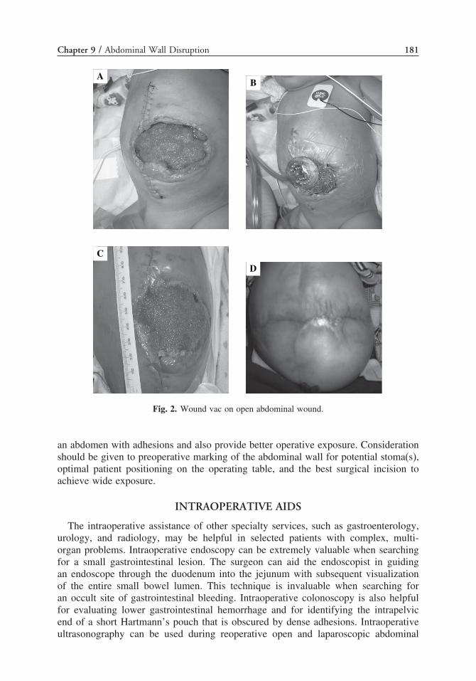
Chapter 9 / Abdominal Wall Disruption 181
C
D
BA
Fig. 2. Wound vac on open abdominal wound.
an abdomen with adhesions and also provide better operative exposure. Considerationshould be given to preoperative marking of the abdominal wall for potential stoma(s),optimal patient positioning on the operating table, and the best surgical incision toachieve wide exposure.
INTRAOPERATIVE AIDS
The intraoperative assistance of other specialty services, such as gastroenterology,urology, and radiology, may be helpful in selected patients with complex, multi-organ problems. Intraoperative endoscopy can be extremely valuable when searchingfor a small gastrointestinal lesion. The surgeon can aid the endoscopist in guidingan endoscope through the duodenum into the jejunum with subsequent visualizationof the entire small bowel lumen. This technique is invaluable when searching foran occult site of gastrointestinal bleeding. Intraoperative colonoscopy is also helpfulfor evaluating lower gastrointestinal hemorrhage and for identifying the intrapelvicend of a short Hartmann’s pouch that is obscured by dense adhesions. Intraoperativeultrasonography can be used during reoperative open and laparoscopic abdominal

182 Teich and Caniano
procedures. It can identify lesions within the liver that cannot be palpated or visualized,and assist in characterizing unanticipated findings such as a mass within the pancreas(56). Dye substances, such as methylene blue or dilute povidone-iodine, may behelpful in testing the integrity of an anastomosis (57). Tumors, inflammatory lesions,and dense inflammation within the pelvis may obscure and involve the ureters. Inthese situations, preoperative cystoscopic insertion of lighted ureteral catheters may behelpful in delineating the course of the ureters (58).
TECHNIQUES FOR SECONDARY CLOSURE OF THE DISRUPTEDABDOMINAL WALL
Management of surgically created abdominal wall defects has changed significantlyover the past 25 to 30 years. Tension-free repair is now considered to be the treatmentof choice; therefore, primary closure should only be utilized to repair small ventralhernias (59,60). Synthetic mesh, readily available in most operating rooms permitsclosure of defects as great as 25 × 33 cm2 in size. The mesh can be implanted byeither open or laparoscopic techniques. However, its use is fraught with many potentialcomplications including rejection, infection, wound seromas, enterocutaneous fistulas,and adhesions. Large sheets of mesh may lead to decreased abdominal wall complianceand increased abdominal wall rigidity.
Component separation (CS) is a technique of abdominal wall closure that hasgained popularity since it was first reported in 1990 (61) (Fig. 3). Several clinicalstudies have demonstrated the utility of CS in the repair of ventral wall defects withacceptable results after abdominal compartment syndrome, in infected wounds, and inintractable defects (62–64). CS is effective because the technique includes mobilizationof vascularized myofascial flaps that are brought together in the midline. Native vascu-larized tissue is extremely important in an infected environment and has a greaterpotential for healing than prosthetic material. In an intractable hernia, CS leads to
A B
C D
Fig. 3. Abdominal wall component separation. (A) Elevation of skin flap off anterior abdominalwall. (B) Incision of external oblique fascia at junction with rectus abdominus. External obliquedissected off internal oblique laterally. (C) Incision of posterior rectus sheath, which is dissected offrectus muscle. (D) Completed flap.

Chapter 9 / Abdominal Wall Disruption 183
the reapproximation of native tissue under minimal tension, which allows for moresecure healing. If a mesh is required, the use of the CS technique allows for a smallersize mesh to be placed and increases the likelihood of healing and a better long-termfunctional result.
ABDOMINAL COMPARTMENT SYNDROME
Abdominal compartment syndrome (ACS) is defined as multi-organ systemdysfunction involving the renal, pulmonary, and cardiovascular systems secondary to anacute, sustained elevation of intra-abdominal pressure (IAP). ACS was first describedby Baggot in 1951, but only in the last 25 years has it become a widely recognizedlife-threatening problem that is potentially treatable by abdominal decompression (65).Pediatric surgeons have long been aware of the deleterious effects of increased IAPbecause of our experience with the treatment of congenital abdominal wall defects.Pediatric surgeons were the first general surgeons to utilize prosthetic materials forthe temporary closure of abdominal wall defects (66). Furthermore, pediatric surgeonsutilized measurements of IAP as a guide to safe silo reduction and closure of theabdominal wall (67–69). Recent clinical reports have documented the occurrence ofACS in children beyond the neonatal period (70–72).
The most common cause of ACS is severe blunt abdominal trauma with secondaryhemorrhage and coagulopathy (73–75). It is more likely to develop with combinedabdominal and pelvic trauma (74). Other causes of ACS in children include tight packingused for tamponade of uncontrolled abdominal hemorrhage, after fluid resuscitation forsevere burns, extraperitoneal injuries leading to the use of high-end expiratory pressureswithhighintrathoracicpressures,andahighInjurySeverityScore (73,76–79).ACSshouldbe suspected in a critically ill patient who develops a tense abdomen over a short period oftime. Typically the child exhibits elevated intra-abdominal and peak airway pressures,decreased cardiac output, reduced renal function, and hypoxia and hypercarbia withprogressively increasing airway pressures. IAP must be measured to confirm thediagnosis. This is most commonly performed by measuring urinary bladder pressure,an indirect measure of IAP. Bladder pressure is determined by instilling 1 mL/kg bodyweight of saline into the Foley catheter and connecting the end of the catheter to apressure transducer or manometer via a three-way stopcock (80). The symphysis pubisis the zero reference point. A urinary bladder pressure greater than 25 mmHg requiresimmediate abdominal decompression, although children may develop ACS with apressure as low as 15 mmHg (73,81–84). The decision to decompress the abdomen in achild is not determined solely by a specific pressure measurement but by concomitantphysiological parameters, such as progressive renal failure or cardiopulmonary insuf-ficiency (73). Abdominal computed tomography (CT) may suggest increased IAPin patients at risk for ACS by narrowing of the inferior vena cava (IVC), roundedappearance of the abdomen on axial CT, direct compression of the kidney parenchyma,bowel wall enhancement and thickening, and bowel dilatation (85–87).
The only treatment for ACS is immediate decompression with the use of prostheticmesh or the placement of a wound vac for negative pressure wound therapy (NPWT).In high-risk surgical patients, a prophylactic wound vac should be placed over theopen abdomen at the end of the surgical procedure (Fig. 4). NPWT applies constantand uniform negative pressure to the abdominal wall. It allows the fluid losses to bequantified and replaced more precisely. Closure rates of 71–92% have been reported

184 Teich and Caniano
Fig. 4. Wound vac on open abdomen.
in multiple series of patients with open abdomens treated with NPWT (88–90). Themean closure time varied from 10 to 21 days, depending on the etiology of the openabdomen.
TREATMENT OF INCISIONAL HERNIAS
Wound dehiscence is the most severe and rare sequelae of failure of healing ofthe abdominal wall after laparotomy. Incisional hernia, however, remains a relativelycommon late complication of faulty healing of the abdominal wall. In multiple seriesof adult patients, the incisional hernia rate varies from 1–14% of all laparotomies(91–93). Recurrence rates after incisional hernia repair vary from 10–50% (94–98).The incidence of incisional hernias and the recurrence rate after incisional herniarepair in the pediatric population is unknown, because no clinical series has ever beenpublished!
An incisional hernia usually presents as an asymptomatic bulge over the incisionor immediately adjacent to it. More than one-half of all incisional hernias in adultsare noticeable within the first two years after the operation, but a high percentagecan occur years later (99–101). Over time incisional hernias enlarge and may becomepainful. Several causative factors are thought to increase the risk for development of anincisional hernia both in adults and children. These factors include a prolonged initialoperation, long or multiple incisions, reoperation, obesity, postoperative hematomas,increased dead space, wound drainage, layered closure, absorbable sutures, inadequatefascial fixation of sutures, and excessive wound tension (102). Systemic considerationsin children include sepsis, malnutrition, malignancy, chemotherapy, liver or kidneydisease, paralytic ileus, and BPD (bronchopulmonary dysplasia). In one series of adultpatients, one-half of all incisional hernias were associated with emergency procedures,infected wounds, or postoperative evisceration (103).
Except in the case of a traumatic incisional hernia or an obvious technical error (e.g.,a broken suture or an unclosed trochar site) most incisional hernias result from defectsin wound healing (104). This basic principle helps to explain the following obser-vations about incisional hernias: (1) a large percentage of incisional hernias presenta considerable time following the previous operation; (2) the cumulative incidencefollows a linear time course; (3) repeating a previously unsuccessful closure techniquehas a high risk of failure; (4) the entire incision should be reinforced regardless of the

Chapter 9 / Abdominal Wall Disruption 185
intraoperative findings; (5) patients with a proven defect in collagen metabolism havea higher incidence of incisional hernias; (6) the use of absorbable materials increasesthe failure rate; and (7) a successful mesh repair requires significant mesh overlap ofthe hernia defect.
Reinforcement of the repaired incisional hernia by mesh allows for the ingrowthof fibrous tissue into the prosthetic mesh with a resulting scar-mesh compound (104).The amount of mesh material influences the intensity of the scar formation but not thequality of the scar. Therefore, mesh fixation by fibrosis will only prevent recurrenceby having a wide overlap underneath healthy tissue. In reported clinical series, thereis a trend toward using larger mesh prostheses (104,105).
The only nonoperative alternative for treatment of an incisional hernia is the useof an abdominal binder, which may be applicable in selected patients who are notimmediate candidates for operative repair. We advocate early repair, because mostincisional hernias tend to increase in size over time and cause parental and patientanxiety. Mesh should be used routinely for any defect over 3 cm in older children andmay be required for smaller defects in infants and young children (100,101).
Table 2Techniques for Incisional Hernia Repair
Type of IncisionalHernia Repair Technique Recurrence Rate
Primary repair only • Fascial defects <5 cm in diameter Up to 50% (97,106–109)
Primary repairwith relaxingincision/componentseparation
See Fig. 1 10% (102,104)(10% significant skin andwound problems)
Primary repair withinlay mesh
• Hernia sac excised• Interrupted or continuous
permanent sutures to fascial edges
10–20% (109)
Primary repair withretro-rectus mesh
• Hernia sac left intact as buffer• Mesh fixated to muscle layer
above
<10% (110,111)(Mesh infection/removal rate5–12%)
Primary repair withintraperitonealunderlay mesh
• Allows for largest underlay ofmesh
• Can be performed open orlaparoscopically
• Fixation to fascial edges or tolateral posterior abdominal wall
<5% (112)
Primary repairwith other meshtechniques
• Onlay mesh only• Sandwich technique with mesh
placed both onlay and eitherretro-rectus or intraperitoneal
• Cuff technique with placement ofmesh around muscle on eachside of defect with closure ofmesh-reinforced edges
Unknown

186 Teich and Caniano
In most cases, incisional hernia repair is an elective operation. Every attempt shouldbe made to minimize the influence of risk factors that lead to recurrence. The skinshould be in good condition with no areas of superficial excoriation. A preoperativebowel preparation should be performed for the repair of large abdominal wall defects tominimize postoperative tension on the abdominal wall closure, and prophylactic antibi-otics should be utilized whenever use of a mesh material is contemplated. Significantoozing may result if there is a need for extensive dissection, warranting preoperativeinvestigation for an unsuspected bleeding abnormality.
Many different techniques for incisional hernia repair have been published in theliterature (see Table 2). They vary from primary closure only to primary closure withmesh placement in various positions. Some techniques have a documented series ofpatients whereas others are only reported in the literature without evidence as to theireffectiveness. With the use of mesh materials, the recurrence rate for incisional herniarepair in adults has decreased to 5–10% or less in various series. Current investigationsinclude the best approach for incisional hernia repair (open versus laparoscopic) and thebest means of fixation (partial versus full-thickness abdominal wall/fascia) to stabilizethe mesh during tissue ingrowth.
REFERENCES
1. Wilson S, Steiner J, Williams R. Reoperation for abdominal wound dehiscence. In: McQuarrie D,Humphrey E, Lee J, (eds.), Reoperative general surgery, second edition. St. Louis: Mosby-YearBook, 1997: 533–553.
2. Greenall M, Evans M, Pollock A. Midline or transverse laparotomy? A random controlled clinicaltrial. Part I: influence on healing. Br J Surg 1980;67:188–190.
3. Ellis H, Coleridge-Smith P, Joyce A. Abdominal incisions—vertical or transverse? Postgrad Med J1984;60:407–410.
4. Irvin T, Koffman C, Duthie H. Layer closure of laparotomy wounds with absorbable and non-absorbable suture materials. Br J Surg 1976;63:793–796.
5. Wissing J, van Vroonhoven T, Schattenkerk M. Fascia closure after midline laparotomy: results ofa randomized trial. Br J Surg 1987;74:P738–P741.
6. Fagniez PL, Hay J, Lachaine F, Thomsen C. Abdominal midline incision closure. Arch Surg1985;120:1351–1353.
7. Guiney EJ, Morris PJ, Donaldson GA. Wound dehiscence: a continuing problem in abdominalsurgery. Arch Surg 1966;92:47–51.
8. Goligher JC, Irvin TT, Johnston D, et al. A controlled trial of three methods of closure of laparotomywounds. Br J Surg 1975;62:823–829.
9. Gross RE, Ferguson CC. Abdominal incisions in infants and children. Ann Surg 1953;137:349.10. Campbell D, Swenson O. Wound dehiscence in infants and children. J Pediatr Surg 1972;7:123–126.11. Waldhausen J, Davies L. Pediatric postoperative abdominal wound dehiscence: transverse versus
vertical incisions. J Am Coll Surg 2000;190:688–691.12. Kiely E, Spitz L. Layered versus mass closure of abdominal wounds in infants and children.
Br J Surg 1985;72:739–740.13. Wolff WI. Disruption of abdominal wounds. Ann Surg 1950;131:534–555.14. Hampton JR. The burst abdomen. Br Med J 1963;2:1032–1035.15. Alexander HC, Pavdden JF. The causes of abdominal wound disruption. Surg Gynecol Obstet
1966;122:1223–1229.16. Thompson WD, Radvin IS, Frank IL. Effect of hypoproteinemia on wound disruption. Arch Surg
1938;36:500–518.17. Elman R. Protein metabolism and the practice of medicine. Med Clin North Am 1943;27:303–313.18. Kraybill WG. Total disruption of surgical wounds of the abdominal wall. Am J Surg 1944;66:220–223.19. Thompson JB, MacLean KF, Coller FA. Role of the transverse abdominal incision and early
ambulation in the reduction of postoperative complications. Arch Surg 1949;59:1267–1277.20. Halasz NA. Dehiscence of laparotomy wounds. Am J Surg 1968;116:210–214.

Chapter 9 / Abdominal Wall Disruption 187
21. Irvin TT, Koffman CG, Durhie HL. Layer closure of laparotomy wounds with absorbable andnonabsorbable suture materials. Br J Surg 1976;63(10):793–796.
22. Singleton AO, Blocker TG. The problem of disruption of abdominal wounds and postoperativehernia. JAMA 1939;112:122–127.
23. Keill RH, Reitzer WF, Nichols WK, et al. Abdominal wound dehiscence. Arch Surg1973;106:573–577.
24. Postlethwait RW. Principles of operative surgery: antisepsis, technique, sutures and drains, In:Sabiston DC (ed.), Textbook of surgery, 12th edition. Philadelphia: WB Saunders;1981:329.
25. Tweedie FJ, Long RC. Abdominal wound disruption. Surg Gynecol Obstet 1954;99:41–47.26. Jenkins TPN. The burst abdominal wound: a mechanical approach. Br J Surg 1976;63:873–886.27. Martyak SN, Curtis LE. Abdominal incision and closure. A systems approach. Am J Surg
1976;131:476–480.28. Hermann RE. Abdominal wound closure using a new polyproplylene monofilament suture. Surg
Gynecol Obstet 1974:138:84–86.29. Knight CD, Griffen FD. Abdominal wound closure with a continuous monofilament prolypropylene
suture. Arch Surg 1983;118:1305–1308.30. Leaper DJ, Pollock AV, Evans M. Abdominal wound closure: a trial of nylon, polyglycolic acid
and steel sutures. Br J Surg 1977;64:603–606.31. Pollock AV, Greenall MJ, Evans M. Single-layer mass closure of major laparotomies by continuous
suturing. J R Soc Med 1979;72:889–893.32. Ellis H, Heddle R. Does the peritoneum need to be closed at laparotomy? Br J Surg 1977;64:733–736.33. McFadden PM, Peacock EE. Preperitoneal abdominal wound repair: incidence of dehiscence. Am J
Surg 1983;145:213–214.34. Jones TE, Newell ET, Brubaker RE. The use of alloy steel wire in the closure of abdominal wounds.
Surg Gynecol Obstet 1941;72:1056–1059.35. Bucknall TE, Cox PJ, Ellis H. Burse abdomen and incisional hernia: a prospective study of 1129
major laparotomies. Br Med J 1982;284:931–933.36. Irvin TT, Stoddard CJ, Greaney MC, et al. Abdominal wound healing: a prospective clinical study.
Br Med J 1977;2:351–352.37. Ellis H, Heddle R. Closure of the abdominal wound. J R Soc Med 1979;72:17–18.38. Kirk RM. Effect of method of opening and closing the abdomen of incidence of wound bursting.
Lancet 1972;2:352–353.39. Poole GV, Meredith JW, Kon ND, et al. Suture technique and wound bursting strength. Am Surg
1984;50:569–572.40. Price PB. Stress, stain, and sutures. Ann Surg 1948;128:408–421.41. Poole GV. Mechanical factors in abdominal wound closure: the prevention of fascial dehiscence.
Surgery 1985;97:631–639.42. Barker D, Kaufman H, Smith L, Ciraulo D, Richart C, Burns R. Vacuum pack technique of temporary
abdominal closure: a 7-year experience with 112 patients. J Trauma 2000;48:201–207.43. Banwell PE, Teot L. Topical negative pressure (TNP): the evolution of a novel wound therapy.
J Wound Care 2003;12:22–28.44. Agarwal T, Refson J, Gould S. The ‘telly-tubby tummy’. A novel technique for laparostomy
management. Ann R Coll Surg Engl 2001;83:440.45. Hirschberg A, Walden R. Damage control for abdominal trauma. Surg Clin North Am 1997;77:
813–810.46. Wittmann DH, Aprahamian C, Bergstein JM, et al. A burr-like device to facilitate temporary
abdominal closure in planned multiple laparotomies. Eur J Surg 1993;159:75–79.47. Garcia-Sabrido JL, Tallado JM, Christou NV, et al. Treatment of severe intra-abdominal sepsis
and/or necrotic foci by an ‘open-abdomen’ approach. Zipper and zipper-mesh techniques. Arch Surg1988;123:152–156.
48. Fleischmann W, Strecker W, Bombelli M, et al. Vacuum sealing as a treatment of soft tissue damagein open fractures [in German]. Unfallchirurg 1993;96:488–492.
49. Venturi M, Attinger C, Mesbahi A, Hess C, Graw K. Mechanisms and clinical applications of thevacuum-assisted closure (VAC) device. Am J Clin Dermatol 2005;6:185–194.
50. Morykwas MJ, Argenta LC, Shelton-Brown EI, et al. Vacuum-assisted closure: a new method forwound control and treatment: animal studies and basic foundation. Ann Plast Surg 1997;38:553–562.
51. Urschel JD, Scott PG, Williams HTG. The effect of mechanical stress on soft and hard tissue repair:a review. Br J Plast Surg 1988;41:182–186.
188 Teich and Caniano
52. Olenius M, Dalsgaard C, Wickman M. Mitotic activity in the expanded human skin. Plast ReconstrSurg 1993;91:213–216.
53. Caniano D, Ruth B, Teich S. Wound management with vacuum-assisted closure: experience in 51pediatric patients. J Pediatr Surg 2005;40:128–132.
54. Arca M, Somers K, Derks T, et al. Use of vacuum-assisted closure system in the management ofcomplex wounds in the neonate. Pediatr Surg Int 2005;21:532–535.
55. Markley M, Mantor P, Letton R, Tuggle D. Pediatric vacuum packing wound closure for damage-control laparotomy. J Pediatr Surg 2002;37:512–514.
56. Rifkin M, Rosato F, Branch H, et al. Intraoperative ultrasound of the liver. An important adjunctivetool for decision making in the operating room. Ann Surg 1987;205:466–472.
57. Trowbridge P. Intraoperative testing for anastomoses of the gastrointestinal tract. Surg Gynec Obstet1986;163:275–276.
58. Ney C, Cruz F, Carvajal S, Miller H, Reyes P. Ureteral involvement secondary to diverticulitis ofthe colon. Surg Gynec Obstet 1986;163:215–218.
59. Read JRC, Yoder G. Recent trends in the management of incisional herniation. Arch Surg1989;124:484–488.
60. Millikan KM, Baptista M, Amin B, et al. Intraperitoneal underlay ventral hernia repair utilizingbilayer ePTFE and polypropylene mesh. Am Surg 2003;69:258–263.
61. Ramirez OM, Ruas E, Dellon L. Component separation method for closure of abdominal walldefects: an anatomic and clinical study. Plast Reconstr Surg 1990;86:519–526.
62. Thomas WO, Parry SW, Rodning CB. Ventral/incisional herniorrhaphy by fascial partition/release.Plast Reconstr Surg 1993;91:1080–1086.
63. DiBello JN, Moore JH. Sliding myofascial flap of the rectus abdominus muscles for the closure ofrecurrent ventral hernias. Plast Reconstr Surg 1996;98:464–469.
64. Fabian TC, Croce MA, Pritchard FE, et al. Planned ventral hernia: staged management for acuteabdominal wall defects. Ann Surg 1994;219:643–653.
65. Baggot MG. Abdominal blow-out: a concept. Anesth Analg 1951;30:295–299.66. Schuster SR. A new method for the staged repair of large omphaloceles. Surg Gynecol Obstet
1967;125:837–850.67. Wesley JR, Drongowski R, Croan AG. Intragastric pressure measurement: a guide for reduction and
closure of the silastic chimney in omphalocele and gastroschisis. J Pediatr Surg 1981;16:264–270.68. Lacey SR, Bruce J, Brooks SP, et al. The relative merits of various methods of indirect measurement
of intra-abdominal pressure as a guide to closure of abdominal wall defects. J Pediatr Surg1987;22:1207–1211.
69. Lacey SR, Carris LA, Beyer J, et al. Bladder pressure monitoring significantly enhances careof infants with abdominal wall defects: a prospective clinical study. J Pediatr Surg 1993;28:1370–1375.
70. Stylianos S, Jacir NN, Hoffman MA, et al. Pediatric blunt liver injury and coagulopathy managedwith packs and a silo: case report. J Trauma 1990;30:1409–1410.
71. Stylianos S. Abdominal packing for severe hemorrhage. J Pediatr Surg 1998;33:339–342.72. Kawar B, Siplovich L. Abdominal compartment syndrome in children: the dilemma of treatment.
Eur J Pediatr Surg 2003;13:330–333.73. Eddy V, Nunn C, Morris JA Jr. Abdominal compartment syndrome. The Nashville experience.
experience. Surg Clin North Am 1997;77:801–812.74. Ertel W, Oberholzer A, Platz A, et al. Incidence and clinical pattern of the abdominal compartment
syndrome after ‘damage-control’ laparotomy in 311 patients with severe abdominal and/or pelvictrauma. Crit Care Med 2000;28:1747–1753.
75. Cheatham ML, White MW, Sagraves SG, et al. Abdominal perfusion pressure: a superior parameterin the assessment of intra-abdominal hypertension. J Trauma 2000;49:621–626.
76. Meldrum DR, Moore FA, Moore EE, et al. Prospective characterization and selective managementof the abdominal compartment syndrome. Am J Surg 1997;174:667–672.
77. Maxwell RA, Fabian TC, Croce MA, et al. Secondary abdominal compartment syndrome: anunderappreciated manifestation of severe hemorrhagic shock. J Trauma 1999;47:995–999.
78. Kopelman T, Harris C, Miller R, et al. Abdominal compartment syndrome in patients with isolatedextraperitoneal injuries. J Trauma 2000;49:744–747.
79. Ivatury RR, Porter JM, Simon RJ, et al. Intra-abdominal hypertension after life-threateningpenetrating abdominal trauma: prophylaxis, incidence, and clinical relevance to gastric mucosal pHand abdominal compartment syndrome. J Trauma 1998;44:1016–1021.
Chapter 9 / Abdominal Wall Disruption 189
80. DeCou J, Abrams R, Miller R, Gauderer M. Abdominal compartment syndrome in children:experience with three cases. J Pediatr Surg 2000; 35:840–842.
81. Ivatury RR, Diebel L, Porter JM, et al. Intra-abdominal hypertension and the abdominal compartmentsyndrome. Surg Clin North Am 1997;77:783–800.
82. Meldrum DR, Moore FA, Moore EE, et al. Prospective characterization and selective managementof the abdominal compartment syndrome. Am J Surg 1997;174:667–673.
83. Saggi BH, Sugerman HJ, Ivatury RR, et al. Abdominal compartment syndrome. J Trauma1998;45:597–609.
84. Kron IL, Harman PK, Nolan SP. The measurement of intra-abdominal pressure as a criterion forabdominal re-exploration. Ann Surg 1984;199:28–30.
85. Epelman M, Soudack M, Engel A, Halberthal M, Beck R. Abdominal compartment syndrome inchildren: CT findings. Pediatr Radiol 2002;32:319–322.
86. Wachsberg RH, Sebastiano LL, Levine CD. Narrowing of the upper abdominal inferior vena cavain patients with elevated intraabdominal pressure. Abdom Imaging 1998;23:99–102.
87. Sivit CJ, Taylor GA, Bulas DI, et al. Post-traumatic shock in children: CT findings associated withhemodynamic instability. Radiology 1992;182:723–726.
88. Mayberry JC, Goldman RK, Mullins RJ, et al. Surveyed opinion of American trauma surgeons onthe prevention of the abdominal compartment syndrome. J Trauma 1999;47:509–514.
89. Hirshberg A, Stein M, Adar R. Reoperation planned and unplanned. Surg Clin North Am1997;77:897–907.
90. Marshall JC, Innes M. Intensive care unit management of intra-abdominal infection. Crit Care Med2003;31:2228–2237.
91. Blomstedt B, Welin-Berger T. Incisional hernias. Acta Chir Scand 1972;138:275–278.92. Bucknall TE, Cox PJ, Ellis H. Burst abdomen and incisional hernia: a prospective study of 1129
major laparotomies. Br Med J 1982;284:931–933.93. Richards PC, Batch CM, Aldrete JS. Abdominal wound closure. A randomized prospective study
of 571 patients comparing continuous vs. interrupted suture techniques. Ann Surg 1983;197:238–243.
94. Mudge M, Hughes LE. Incisional hernia: a 10 year prospective study of incidence and attitudes.Br J Surg 1985;72:70–71.
95. George CD, Ellis H. The results of incisional hernia repair: a twelve year review. Ann R Coll SurgEngl 1986;68:185–187.
96. Langer S, Christiansen J. Long-term results after incisional hernia repair. Acta Chir Scand1985;151:217–219.
97. Anthony T, Bergen PC, Kim LT. Factors affecting recurrence following incisional herniorrhaphy.World J Surg 2000;24:95–101.
98. Leber GE, Garb JL, Alexander AJ, et al. Long-term complications associated with prosthetic repairof incisional hernias. Arch Surg 1998;133:378–382.
99. Bucknall TE, Cox PJ, Ellis H. Burst abdomen and incisional hernia: a prospective study of 1129major laparotomies. BMJ 1982;284:931–933.
100. Lamont PM, Ellis H. Incisional hernia in re-opened abdominal incisions: an overlooked risk factor.Br J Surg 1988;75:374–376.
101. Pollock AV, Evans M. Early prediction of late incisional hernias. Br J Surg 1989;76:953–954.102. Read RC. Recurrent and incisional hernias. In: McQuarrie DB, Humphrey EW, Lee JT (eds.),
Reoperative general surgery. St. Louis: Mosby-Year Book; 1997: 558–567.103. Read RC, Yoder G. Recent trends in the management of incisional herniation. Arch Surg
1989;124:485–488.104. Klinge U, Conze J, Krones C, Schumpelick V. Incisional hernia; open techniques. World J Surg
2005;29:1066–1072.105. Millikan K. Incisional hernia repair. Surg Cl N Am 2003;83:1223–1234.106. Read RC, Yoder G. Recent trends in the management of incisional herniation. Arch Surg
1989;124:485–488.107. Paul A, Korenkov M, Peters S, et al. Unacceptable results of the Mayo procedure for repair of
abdominal incisional hernias. Eur J Surg 1998;164:361–367.108. van der Linden FTPM, wan Vroonhoven TJMV. Long-term results after surgical correction of
incisional hernia. Neth J Surg 1988;40:127–129.109. Luijendijk RW, Hop WC, van den Tol P, et al. A comparison of suture repair with mesh repair for
incisional hernia. N Eng J Med 2000;343:392–398.

190 Teich and Caniano
110. Temudom T, Siadati M, Sarr MG. Repair of complex or recurrent ventral hernia by using tension-free intraparietal prosthetic mesh (Stoppa technique): lessons learned from our initial experience(fifty patients). Surgery 1996;120:738–744.
111. McLanahan D, King LT, Weems C, et al. Retrorectus prosthetic mesh repair of midline abdominalhernia. Am J Surg 1997;173:445–449.
112. Millikan KW, Baptista M, Amin B, et al. Intraperitoneal underlay ventral hernia repair utilizingbilayer ePTFE and polypropylene mesh. Am Surg 2003;69:258–263.
10 Reoperation after Esophageal AtresiaRepair and Other EsophagealConditions
Brian D. Kenney, MD
CONTENTS
Anatomy
General Complications in Esophageal Surgery
Specific Conditions Requiring Surgery
of the Esophagus in Children
References
Reoperative surgery of the esophagus in children is challenging because of theunique position and surgical anatomy of the esophagus, the general complications thatcan occur in esophageal surgery, and finally the specific clinical problems that requiresurgical attention in children.
ANATOMY
The esophagus is a muscular tube that allows food to pass from the pharynx tothe stomach. The esophagus begins in a median position, which shifts slightly tothe right of the spine in the midthoracic portion. The lower esophagus crosses overthe vertebral column so that the esophagogastric junction is on the left. The upperesophagus is in direct contact with the membranous part of the trachea. The azygosvein crosses anterior to the midthoracic esophagus to enter the superior vena cava.The esophagus is divided into three anatomic portions. The cervical esophagus beginsat the level of the cricoid cartilage and includes the upper esophageal sphincter. Thelongest segment of the esophagus is the thoracic esophagus, which extends from thesternal notch to the diaphragmatic hiatus. The third portion is the abdominal esophagus,which is the short segment below the hiatus extending to the gastroesophageal junction.The central portion of the esophagus is contained within loose areolar tissue inthe posterior mediastinum, which allows for esophageal motility to accommodateeffortless swallowing. The fascial compartment surrounding the esophagus extendslongitudinally into the pretracheal space and deep cervical fascia superiorly and intothe retroperitoneum inferiorly. Surgical procedures of the esophagus must respect its
From: Reoperative Pediatric SurgeryEdited by: S. Teich and D. A. Caniano © Humana Press, Totowa, NJ
191
192 Kenney
vascular supply: the cervical esophagus receives its blood supply from the thyrocer-vical arteries; the mid esophagus is supplied directly from the aorta at segmental levels;and the lower esophagus is supplied from the cardiac branches of the left gastricartery (1).
GENERAL COMPLICATIONS IN ESOPHAGEAL SURGERY
Reoperative esophageal surgery is required for complications of surgery and forsymptoms that develop following uncomplicated surgery. In common with surgeriesinvolving other areas of the gastrointestinal (GI) tract, operations on the esophaguscan be complicated by stricture, leakage, or dilation (focal or diffuse). Strictures resultfrom circumferential healing fibrosis and are exacerbated by acid reflux or poor bloodsupply. The result of a stricture is the inability to swallow, the impaction of food, andpotentially aspiration. Esophageal leakage can occur following an anastomosis and ismost hazardous in the thoracic portion of the esophagus. The thoracic portion is alsothe most frequent area of leakage, perhaps owing to the lack of serosa. Leakage canalso occur following instrumentation of the esophagus either by esophagoscopy or withesophageal dilatation. Leakage from the esophagus can lead to severe mediastinitiswith extension of the infection along tissue planes into the neck or retroperitoneum.
SPECIFIC CONDITIONS REQUIRING SURGERYOF THE ESOPHAGUS IN CHILDREN
Tracheoesophageal Fistula and Esophageal AtresiaProblems in the formation of the esophagus leading to esophageal atresia and various
forms of tracheoesophageal fistula mandate surgical repair, usually within the firstfew days of life. The treatment of these problems is challenging because of the ageand size of the patient and because of the relatively high frequency of associatedcongenital anomalies. Upon repair of the tracheoesophageal fistula, patients can requirereoperation for recurrence of the fistula, anastomotic leakage, anastomotic stricture, orsevere gastroesophageal reflux. The contributions of individual factors to the incidenceof these problems is poorly defined but includes: surgeon experience; patient-relatedfactors such as age, birth weight, and type of defect; and technical factors such aschoice of suture material, surgical approach (open versus thoracoscopic), and type ofanastomosis (single-layer versus double-layer).
Both the incidence of these complications and the frequency of reoperations aredifficult to estimate. A review of several large series of patients treated for tracheoe-sophageal fistula, with over 1170 patients combined, shows a marked variability inthe frequency of postoperative problems (2–8). Recurrence of the tracheoesophagealfistula was the least common complication, occurring in 1–10% of patients. Althoughone study showed “no discernible anastomotic leaks” (2), most other series included aleakage rate of 8–19%. The incidence of stricture formation varied most widely of thecomplications, perhaps owing to the intensity of surveillance for this problem, rangingfrom 6–52%. The percentage of patients in these studies that had gastroesophagealreflux severe enough to require fundoplication also varied widely, ranging from 7–44%.The wide range likely reflects controversies about surgical versus medical treatmentof reflux in this situation.

Chapter 10 / Esophageal Surgery 193
Recurrent Tracheoesophageal Fistula
Recurrence of the fistula may be caused by inflammation or contamination at thesurgical site or perhaps by poor wound healing. Most patients present with chroniccough or with recurrent episodes of pneumonia. Although three-dimensional computedtomography (CT) has been described to demonstrate recurrence (9), the diagnosticevaluation usually requires a prone esophagogram with direct instillation of radio-graphic contrast in the esophagus through a nasogastric tube (10). Endoscopy can behelpful for diagnosis, but is less sensitive and more invasive than contrast radiography.The value of endoscopy is found in the intraoperative localization of fistulae to directthe treatment of this complication. Patients with recurrent tracheoesophageal fistulaemust be differentiated from those who have both proximal and distal fistulas in whomthe proximal fistula was missed at the initial surgery.
Reoperation for a recurrent fistula usually includes interposition of adjacent softtissue such as pleura, intercostal muscle, or pericardium between the divided endsof the fistula (11). The repeat thoracotomy is technically challenging for severalreasons: fibrosis and inflammatory tissue obscure tissue planes; the esophagus ormembranous trachea can be injured, as can the vagus or recurrent laryngeal nerves;and recurrence continues to be a risk. Because of these difficulties, other operativeapproaches have been described, including median sternotomy with interposition ofa sternocleidomastoid muscle flap (12) or transtracheal fistula closure via an anteriorcervical approach (13). Another approach to avoid many of these technical diffi-culties is with an endoscopic closure of the recurrent fistula. A variety of agents havebeen reported including: fibrin glue; methyl methacrylate; n-butyl-z-cyanoacrylate andaethoxysclerol; Nd:YAG laser; or insulated wire electrocautery (14–22). No idealagent has been found because all have a significant risk of recurrence (23). Recurrenttracheoesophageal fistula is attended by significant morbidity, and extreme cases ofmultiple recurrences may require esophageal replacement (24).
Anastomotic Leaks After Tracheoesophageal Fistula Repair
The causes of anastomotic leakage after tracheoesophageal fistula repair are notfully known but have been associated with the use of braided silk sutures compared tomonofilament sutures (absorbable or nonabsorbable) (25). Anastomotic leakage usuallybecomes obvious in the first few days after surgery or after commencement of feedings.A drainage tube is routinely left near the anastomosis at the initial surgery to help detectand control any leakage. An esophageal swallow is performed 7–10 days followingsurgery and, if leakage is not detected, feedings are commenced. If no leakage is notedafter 24 hours of feeding, the drainage tube is removed. When present, anastomoticleakage is most often treated nonoperatively with tube thoracostomy. Patients aregiven nothing by mouth, maintained on parenteral nutrition, and given intravenousantibiotics; most patients will respond to these measures (26,27).
Although the majority of postoperative leaks are minor, approximately one-fourthinvolve a major anastomotic disruption. Some major disruptions will be found withthe routine postoperative contrast swallow but most will be evident earlier, manifestedclinically by a persistent pleural fluid collection or pneumothorax (25). Patients witha major dehiscence or those with progressive sepsis will usually require reoperation.Options at reoperation include: primary repair of small areas of dehiscence if the tissuesappear healthy; repair with pleural or intercostal muscle reinforcement; improveddrainage of the leak; or, in the more severe situations, cervical esophagostomy and

194 Kenney
closure of the distal esophagus with plans for subsequent esophageal replacement(25,26). The long-term prognosis for patients with a leak after tracheoesophageal fistularepair includes a higher risk of stricture formation (25).
Anastomotic Stricture After Tracheoesophageal Fistula Repair
Perhaps the most common reason for reoperation following repair of esophagealatresia is anastomotic stricture. Several factors have been associated with an increasedrisk of stricture formation including: patients who have had an anastomotic leak;patients with gastroesophageal reflux; or anastomoses with braided silk sutures ratherthan with polyglycolic acid and polypropylene sutures (28). Strictures usually becomeevident 1–6 months postrepair or sometimes with transition to solid foods. Dysphagiacaused by a postoperative stricture usually has an abrupt onset and patients may evendevelop difficulty with salivary secretions. The diagnosis is confirmed by contrastesophagography (Fig. 1A).
Strictures have traditionally been treated by bougienage or more recently by balloondilatation; greater than 95% will respond to dilatation. (29–31) (Fig. 1B and C). In theminority of patients who require multiple dilatations, investigation for gastroesophagealreflux is imperative. Patients with reflux should receive aggressive medical treatment,often including gastric fundoplication. Strictures that are unresponsive to dilatation andfundoplication will require local resection and reanastomosis or possibly esophagealreplacement (32,33).
Gastroesophageal Reflux After Tracheoesophageal Fistula Repair
Gastroesophageal reflux is common following tracheoesophageal fistula repair andis often difficult to control because tension in the lower esophagus to create the anasto-mosis can distort the gastroesophageal junction, leading to incompetence. Contributingfactors to the severity of reflux in these patients may include dysmotility of the distalesophageal segment and, in some patients, delayed gastric emptying (34,35). Becauseapproximately one-fourth of children with tracheoesophageal fistula repair will suffergastroesophageal reflux disease severe enough to require fundoplication, surgeons mustmaintain close follow-up of these patients.
Many patients are placed on medical therapy for reflux immediately after tracheoe-sophageal fistula repair, and this is continued for at least 6 months. Patients with severereflux, and consequently the majority of patients requiring fundoplication, will oftenexhibit those symptoms in the first 6 months of life. Symptoms may include vomiting,failure to thrive, anastomotic stricture formation, or pulmonary problems such asreactive airway disease, recurrent pneumonia, or evidence of aspiration. Diagnosticevaluation should include an upper GI study to look for anastomotic stricture as wellas to determine if there is any pathology that could exacerbate reflux, such as duodenalstenosis or malrotation. An esophageal probe for 24-hour pH monitoring is helpful indefining the reflux severity.
The operative management of severe gastroesophageal reflux is approached witheither open or laparoscopic surgery. Controversy exists over the use of a complete (e.g.,Nissen) versus a partial (e.g., Toupet, Thal) fundoplication. Because dysmotility of thedistal esophageal segment is almost universal in patients who have had esophagealatresia, partial fundoplication may diminish the risk of postfundoplication dysphagia.All approaches and types of repair appear to have a higher failure rate in this populationof patients than in otherwise normal children treated for severe reflux (36–39).

Chapter 10 / Esophageal Surgery 195
A
B
Fig. 1. (A) Esophageal stricture following thoracoscopic repair of tracheoesophageal fistula.(B) Balloon dilatation of anastomotic stricture. (C) Esophagogram following successful balloondilatation.

196 Kenney
C
Fig. 1. (Continued).
Concern has been developing about the long-term implications of gastroesophagealreflux following tracheoesophageal fistula repair. Perhaps one-third of these patientswill have chronic reflux symptoms and evidence of reflux esophagitis. Some evidencesuggests an increased risk for the development of Barrett’s esophagus (40,41). Onelong-term study of patients with esophageal atresia showed esophageal inflammationin 51% of patients and Barrett’s esophagus in 6% of patients (42). Although furtherstudy is needed to define these risks, and symptomatic improvement has been reportedwith age, patients should continue to be followed for the evaluation and treatment ofreflux symptoms (43).
Recurrent Reflux-Induced Esophageal StrictureReflux-induced esophageal strictures may become symptomatic with food impaction
or may be found simply in the evaluation of patients with gastroesophageal refluxsymptoms. Strictures are documented either by contrast radiography or endoscopy(Fig. 2A). Most of these strictures are amenable to treatment with bougienage orballoon dilatation (44). Fundoplication to control the reflux is important to help preventrecurrence. Most recurrent strictures can be treated successfully with dilatation andfundoplication, but some patients will require additional dilatation (45,46). The recur-rence rate of strictures may be decreased with the topical application of mitomycin C tothe esophageal mucosa by endoscopy (47,48). Additional surgical options for those whodo not respond to dilatation and fundoplication include local stricturoplasty (Fig. 2B)or repair of the esophagus using an intestinal or gastric patch (32,33,49). In thefew patients who do not respond to the above procedures, the ultimate treatment isesophageal replacement with intestinal or gastric interposition (50,51).

Chapter 10 / Esophageal Surgery 197
A
B
Fig. 2. (A) Peptic stricture caused by severe reflux in patient with cerebral palsy. (B) Esophagogramdemonstrates irregular but unimpeded flow following thoracoscopic stricturoplasty of peptic stricturethat had been unresponsive to balloon dilatation and fundoplication.
Barrett’s EsophagusBarrett’s esophagus is a condition in which the esophageal mucosa undergoes
intestinal metaplasia. The condition is sometimes asymptomatic but often occurs in thesetting of gastroesophageal reflux disease. Most patients are adults that present withreflux symptoms and are found to have Barrett’s esophagus after endoscopic biopsy.Adult patients with Barrett’s esophagus have a 50- to 100-fold increased incidence ofesophageal adenocarcinoma, which approximates a 1 in 200 annual risk (52). Compa-rable incidence rates are not well defined in children because of the paucity of reports

198 Kenney
about patients with Barrett’s esophagus in this age group. Patients who have hada tracheoesophageal fistula repair as an infant appear to be at higher risk for thedevelopment of Barrett’s esophagus (40,41).
Patients with Barrett’s esophagus should be offered treatments including surgeryto control symptoms of reflux with the hope that this may also have a favorableeffect on the metaplasia. Unfortunately, the relationship of adequate symptom relief toimprovement of metaplasia or the prevention of adenocarcinoma is uncertain (53–58).Consequently, the long-term management in adults is controversial. The rare cases inchildhood are burdened with the same concerns, and management is further hamperedby even less published data (59,60).
Surveillance biopsies of the distal esophagus are performed to evaluate for dysplasiabecause the diagnosis is made at the histological level and no better screening testis currently available. Evidence of high-grade dysplasia or atypia would suggest theneed for mucosal ablation or esophagectomy. Endoscopic luminal ablation of theesophageal mucosa treats the Barrett’s metaplasia directly with laser or electrocautery.The procedure is effective in removing metaplastic mucosa but associated with a majorcomplication risk of approximately 10%, including stricture and perforation (61,62).Long-term outcomes are not yet available in adults and thus the value of this modalityin pediatric patients is unknown.
Perforation After Esophageal DilatationEsophageal dilatation is useful in the treatment of strictures from a variety of
causes. Although strictures are amenable to treatment with bougienage or balloondilatation, balloon dilatation by the interventional radiologist offers the advantage ofa postdilatation esophagogram. The response to esophageal dilatation is somewhatdependent on the cause of the stricture: postoperative, corrosive, or peptic. Pepticstrictures respond best to this treatment and require fewer repeat dilatations. Thecorrosive strictures are the most difficult to treat and have the highest rate of recurrenceand perforation (63,64).
Fortunately, perforations following dilatation of an esophageal stricture are rare andusually well contained (Fig. 3). Small perforations can be treated with antibiotics,observation, and fasting (65–67). The mortality rate for perforation appears to besignificantly less among children than for adults (68,69). For patients who do notimprove over 24–48 hours, a repeat swallow study is performed. Continued leakagewill require direct repair or diversion.
Achalasia SurgeryThe surgery for achalasia has undergone significant change in the past two decades
with the advent of botulinum toxin, pneumatic dilatation, and minimally invasivetechniques (70). Although botulinum toxin provides relaxation of the lower esophagealsphincter, this is usually temporary and many patients require further therapy. Mostpatients undergo pneumatic dilatation, but one-half of these will have recurrentsymptoms. Esophageal myotomy results in relaxation of the lower esophageal sphincterand can be performed either by a thoracic or abdominal approach (71). Postoperativeesophageal reflux has been minimized by the inclusion of an anterior partial fundopli-cation (72,73). For this reason the laparoscopic approach is favored over thoracoscopyin the treatment of this disease. The thoracoscopic myotomy is reserved for patients

Chapter 10 / Esophageal Surgery 199
Fig. 3. Esophageal leak (contained) following balloon dilatation.
who have had previous extensive upper abdominal surgery. Indications for reoperationsfollowing esophageal myotomy include esophageal leak, the recurrence of achalasia,or the development of esophageal reflux.
A mucosal injury is usually recognizable during the operation and can be repairedprimarily using absorbable sutures. The repair is then reinforced with an anteriorfundoplication. Rarely an esophageal leak will become apparent in the first few daysfollowing surgery. These can be treated with drainage by interventional radiology alongwith the use of antibiotics, proper nutrition, and bowel rest. Diagnostic evaluationwould include an esophageal swallow or upper abdominal CT scan with nonbariumcontrast. Operative repair is required for large, poorly contained leaks.
After myotomy, approximately 10% of patients develop recurrence of symptoms.Recurrence can often be treated by pneumatic dilatation, and those who areunresponsive to dilatation can be treated with laparoscopic or open surgery (74).Most patients requiring reoperative surgery have problems related to the performanceof the fundoplication; others have either a fibrotic stricture formation or an incom-plete myotomy (75). All of the postoperative problems appear to be less commonas surgeons gain greater experience with the procedure. Rarely a dilated esophagusdoes not decrease in size after myotomy and, if symptoms are severe, esophagealreplacement is needed.
Esophageal Replacement SurgeryReplacement of the esophagus is challenging because of its unique anatomy and
function. A variety of structures have been used for replacement, including skin tubesand enteric interpositions (colon, small intestine, stomach, and stomach tubes). Allhave complications that are significant for their frequency and severity (76–83). Graftischemia is perhaps the most dangerous and problematic complication. Ischemia with
200 Kenney
intestinal interposition can be mitigated by the use of microvascular anastomoses withcervical vessels to improve perfusion (84). Gastric or jejunal grafts can be used toreplace failed colonic interposition (85).
One of the most frequent early complications is anastomotic leakage sometimescaused by graft ischemia. An anastomotic leakage can occur in the first few daysfollowing surgery and can be diagnosed clinically by salivary drainage from the wound.Drains are placed at the anastomotic site during surgery to help in the diagnosis andtreatment of this situation. Appropriate drainage may allow healing of a leak withoutfurther operation.
Partial obstruction of a conduit can occur because of an anastomotic stricture ortwisting of the graft (76). Strictures can be treated with balloon dilatation. Twistingof the graft is often a result of redundancy and is especially problematic with coloninterposition. Treatment of redundancy is best approached from an abdominal incisionwhich allows dissection into the mediastinum with care to avoid injury to the mesentery.The conduit can then be straightened, and the lower portion of the conduit resectedand reanastomosed to the stomach.
Intestinal interposition between the pharynx and stomach can lead to reflux withulceration of the graft. This can be treated with vagotomy and pharmacologicalacid suppression. For intestinal interposition, a partial gastric fundoplication can beperformed (86). The reflux is thought to be responsible for the development of Barrett’smetaplasia in the cervical mucosa, and patients should undergo surveillance endoscopy(87,88).
Esophageal VaricesOver the last 30 years, the treatment of esophageal varices has moved from large
surgical operations to create portal systemic shunts to less invasive radiological andendoscopic techniques (89,90). Esophageal varices are caused by portal hypertension,which can be prehepatic, intrahepatic, or posthepatic. In children, the most commoncause of portal hypertension is prehepatic caused by portal vein thrombosis; thesepatients usually have normal liver function. Biliary atresia leading to biliary cirrhosisis the most common cause of intrahepatic obstruction; most of these patients havesignificant liver disease. Posthepatic obstruction (Budd-Chiari syndrome) is causedby hepatic vein thrombosis, is less common, and presents with ascites and hepaticdysfunction.
The clinical presentation of esophageal varices depends on the etiology but is mostoften evident after 2 years of age with symptoms of upper GI bleeding. After cardio-vascular stabilization and reversal of coagulation abnormalities if present, endoscopyis required to establish the diagnosis and initiate treatment. Endoscopic sclerotherapytypically controls the initial bleeding episode, and repeat sclerotherapy is usuallysuccessful at eradication of varices and prevention of recurrent bleeding (91,92).Variceal banding has also been shown to be effective, but is somewhat more difficultbecause of the size of the pediatric esophagus (93–96). Operative management isconsidered for failure to control initial bleeding or to prevent further episodes ofbleeding (90).
Reoperative options for esophageal varices that persist or recur after sclerotherapycan include a transjugular intrahepatic portal venous shunt. These are placed by inter-ventional radiology via the jugular vein and create a vascular shunt from intrahepaticportal veins into the hepatic veins. This strategy is only useful for intrahepatic portal
Chapter 10 / Esophageal Surgery 201
hypertension (89). Operative shunting procedures are most helpful for prehepatic portalvenous obstruction but also can be used in selected patients with intrahepatic portalvenous obstruction who have relatively preserved hepatic function. The variety ofprocedures includes portal caval, mesocaval, splenorenal, and meso- to left portal vein(Rex) shunts (90,97–99). For patients with poor liver function, the best option totreat portal hypertension is liver transplantation (90). Procedures for operating directlyon the esophageal varices, including gastroesophageal devascularization (Sugiura) oresophageal transection, have very high morbidity and recurrence rates and are rarelyused but may be helpful in patients whose portal, mesenteric, or splenic veins are notamenable to shunting (100–102).
REFERENCES
1. Leibermann-Meffert D. In: Pearson FG, Cooper JD, Deslauriers J, et al. (eds.), Esophageal surgery,second edition. Philadelphia: Churchill Livingstone; 2002: 8–31.
2. Foker JE, Linden BC, Boyle EM Jr, Marquardt C. Development of a true primary repair for the fullspectrum of esophageal atresia. Ann Surg 1997;226(4):533–541; discussion 541–543.
3. Tsai JY, Berkery L, Wesson DE, Redo SF, Spigland NA. Esophageal atresia and tracheoesophagealfistula: surgical experience over two decades. Ann Thorac Surg 1997;64(3):778–783; discussion783–784.
4. Touloukian RJ. Reassessment of the end-to-side operation for esophageal atresia with distal tracheoe-sophageal fistula: 22-year experience with 68 cases. J Pediatr Surg 1992;27(5):562–567.
5. Myers NA, Beasley SW, Auldist AW. Secondary esophageal surgery following repair of esophagealatresia with distal tracheoesophageal fistula. J Pediatr Surg 1990;25(7):773–777.
6. Konkin DE, O’Hali WA, Webber EM, Blair GK. Outcomes in esophageal atresia and tracheoe-sophageal fistula. J Pediatr Surg 2003;38(12):1726–1729.
7. Holcomb GW III, Rothenberg SS, Bax KM, et al. Thoracoscopic repair of esophageal atresia andtracheoesophageal fistula: a multi-institutional analysis. Ann Surg 2005;242(3):422–428; discussion428–430.
8. Engum SA, Grosfeld JL, West KW, Rescorla FJ, Scherer LR III. Analysis of morbidity and mortalityin 227 cases of esophageal atresia and/or tracheoesophageal fistula over two decades. Arch Surg1995;130(5):502–508; discussion 508–509.
9. Dogan BE, Fitoz S, Atasoy C, Yagmurlu A, Akyar S. Tracheoesophageal fistula: demonstrationof recurrence by three-dimensional computed tomography. Curr Probl Diagn Radiol 2005;34(4):167–169.
10. Stringer DA, Ein SH. Recurrent tracheo-esophageal fistula: a protocol for investigation. Radiology1984;151(3):637–641.
11. Wheatley MJ, Coran AG. Pericardial flap interposition for the definitive management of recurrenttracheoesophageal fistula. J Pediatr Surg 1992;27(8):1122–1125; discussion 1125–1126.
12. Holland AJ, Ford WD, Guerin RL. Median sternotomy and use of a pedicled sternocleidomastoidmuscle flap in the management of recurrent tracheoesophageal fistula. J Pediatr Surg 1998;33(4):657–659.
13. Martin LW, Cox JA, Cotton R, Oldham KT. Transtracheal repair of recurrent tracheoesophagealfistula. J Pediatr Surg 1986;21(5):402–403.
14. Al-Samarrai AY, Jessen K, Haque K. Endoscopic obliteration of a recurrent tracheoesophagealfistula. J Pediatr Surg 1987;22(11):993.
15. Bhatnagar V, Lal R, Sriniwas M, Agarwala S, Mitra DK. Endoscopic treatment of tracheoesophagealfistula using electrocautery and the Nd:YAG laser. J Pediatr Surg 1999;34(3):464–467.
16. Gutierrez C, Barrios JE, Lluna J, et al. Recurrent tracheoesophageal fistula treated with fibrin glue.J Pediatr Surg 1994;29(12):1567–1569.
17. Hoelzer DJ, Luft JD. Successful long-term endoscopic closure of a recurrent tracheoesophagealfistula with fibrin glue in a child. Int J Pediatr Otorhinolaryngol 1999;48(3):259–263.
18. Lopes MF, Pires J, Nogueria Brandao A, Reis A, Morais Leitao L. Endoscopic obliteration ofa recurrent tracheoesophageal fistula with enbucrilate and polidocanol in a child. Surg Endosc2003;17(4):657.
202 Kenney
19. Tzifa KT, Maxwell EL, Chait P, et al. Endoscopic treatment of congenital H-Type and recurrenttracheoesophageal fistula with electrocautery and histoacryl glue. Int J Pediatr Otorhinolaryngol2006;70(5):925–930.
20. Vandenplas Y, Helven R, Derop H, et al. Endoscopic obliteration of recurrent tracheoesophagealfistula. Dig Dis Sci 1993;38(2):374–377.
21. Willetts IE, Dudley NE, Tam PK. Endoscopic treatment of recurrent tracheo-oesophageal fistulae:long-term results. Pediatr Surg Int 1998;13(4):256–258.
22. Wiseman NE. Endoscopic closure of recurrent tracheoesophageal fistula using Tisseel.J Pediatr Surg 1995;30(8):1236–1237.
23. Montedonico S, Diez-Pardo JA, Lassaletta L, Tovar JA. Tissue adhesives in closing of fistulas aftersurgery of esophageal atresia. Cir Pediatr 1999;12(3):110–112.
24. Ghandour KE, Spitz L, Brereton RJ, Kiely EM. Recurrent tracheo-oesophageal fistula: experiencewith 24 patients. J Paediatr Child Health 1990;26(2):89–91.
25. Chittmittrapap S, Spitz L, Kiely EM, Brereton RJ. Anastomotic leakage following surgery foresophageal atresia. J Pediatr Surg 1992;27(1):29–32.
26. Chavin K, Field G, Chandler J, Tagge E, Othersen HB. Save the child’s esophagus: management ofmajor disruption after repair of esophageal atresia. J Pediatr Surg 1996;31(1):48–51; discussion 52.
27. D’Urzo C, Buonuomo V, Rando G, Pintus C. Major anastomotic dehiscence after repair of esophagealatresia: conservative management or reoperation? Dis Esophagus 2005;18(2):120–123.
28. Chittmittrapap S, Spitz L, Kiely EM, Brereton RJ. Anastomotic stricture following repair ofesophageal atresia. J Pediatr Surg 1990;25(5):508–511.
29. Said M, Mekki M, Golli M, et al. Balloon dilatation of anastomotic strictures secondary to surgicalrepair of oesophageal atresia. Br J Radiol 2003;76(901):26–31.
30. Koivusalo A, Turunen P, Rintala RJ, van der Zee DC, Lindahl H, Bax NM. Is routine dilatationafter repair of esophageal atresia with distal fistula better than dilatation when symptoms arise?Comparison of results of two European pediatric surgical centers. J Pediatr Surg 2004;39(11):1643–1647.
31. Ko HK, Shin JH, Song HY, et al. Balloon dilation of anastomotic strictures secondary to surgicalrepair of esophageal atresia in a pediatric population: long-term results. J Vasc Interv Radiol2006;17(8):1327–1333.
32. Anderson KD, Acosta JM, Meyer MS, Sherman NJ. Application of the principles of myotomy andstrictureplasty for treatment of esophageal strictures. J Pediatr Surg 2002;37(3):403–406.
33. Othersen HB Jr, Parker EF, Chandler J, Smith CD, Tagge EP. Save the child’s esophagus, Part II:colic patch repair. J Pediatr Surg 1997;32(2):328–333.
34. Tomaselli V, Volpi ML, Dell’Agnola CA, Bini M, Rossi A, Indriolo A. Long-term evaluation ofesophageal function in patients treated at birth for esophageal atresia. Pediatr Surg Int 2003;19(1–2):40–43.
35. Romeo C, Bonanno N, Baldari S, et al. Gastric motility disorders in patients operated onfor esophageal atresia and tracheoesophageal fistula: long-term evaluation. J Pediatr Surg2000;35(5):740–744.
36. Bergmeijer JH, Tibboel D, Hazebroek FW. Nissen fundoplication in the management of gastroe-sophageal reflux occurring after repair of esophageal atresia. J Pediatr Surg 2000;35(4):573–576.
37. Esposito C, Langer JC, Schaarschmidt K, et al. Laparoscopic antireflux procedures in themanagement of gastroesophageal reflux following esophageal atresia repair. J Pediatr GastroenterolNutr 2005;40(3):349–351.
38. Snyder CL, Ramachandran V, Kennedy AP, Gittes GK, Ashcraft KW, Holder TM. Efficacy ofpartial wrap fundoplication for gastroesophageal reflux after repair of esophageal atresia. J PediatrSurg 1997;32(7):1089–1091; discussion 1092.
39. Wheatley MJ, Coran AG, Wesley JR. Efficacy of the Nissen fundoplication in the management ofgastroesophageal reflux following esophageal atresia repair. J Pediatr Surg 1993;28(1):53–55.
40. Krug E, Bergmeijer JH, Dees J, de Krijger R, Mooi WJ, Hazebroek FW. Gastroesophageal refluxand Barrett’s esophagus in adults born with esophageal atresia. Am J Gastroenterol 1999;94(10):2825–2828.
41. Deurloo JA, Ekkelkamp S, Taminiau JA, et al. Esophagitis and Barrett esophagus after correctionof esophageal atresia. J Pediatr Surg 2005;40(8):1227–1231.
42. Somppi E, Tammela O, Ruuska T, et al. Outcome of patients operated on for esophageal atresia:30 years’ experience. J Pediatr Surg 1998;33(9):1341–1346.
Chapter 10 / Esophageal Surgery 203
43. Little DC, Rescorla FJ, Grosfeld JL, West KW, Scherer LR, Engum SA. Long-term analysis ofchildren with esophageal atresia and tracheoesophageal fistula. J Pediatr Surg 2003;38(6):852–856.
44. Weintraub JL, Eubig J. Balloon catheter dilatation of benign esophageal strictures in children. J VascInterv Radiol 2006;17(5):831–835.
45. al-Bassam A. Surgical management of severe peptic esophageal stricture in children. Hepatogas-troenterology 2003;50(51):714–717.
46. Briganti V, Oriolo L, Calisti A. Reflux strictures of the oesophagus in children: personal experiencewith preoperative dilatation followed by anterior funduplication. Pediatr Surg Int 2003;19(7):544–547.
47. Olutoye OO, Shulman RJ, Cotton RT. Mitomycin C in the management of pediatric causticesophageal strictures: a case report. J Pediatr Surg 2006;41(5):e1–e3.
48. Uhlen S, Fayoux P, Vachin F, et al. Mitomycin C: an alternative conservative treatment for refractoryesophageal stricture in children? Endoscopy 2006;38(4):404–407.
49. Ramirez Schon G, Suarez Nieves A, Cebollero Marcucci J. Gastric vascular pedicle patch esophago-plasty for stricture. Ann Thorac Surg 1989;48(3):356–358.
50. Moghissi K, Goebells P. Relevance of anatomopathology of high oesophageal strictures to the designof surgical treatment. Eur J Cardiothorac Surg 1990;4(2):91–95; discussion 96.
51. Ohhama Y, Tsunoda A, Nishi T, Yamada R, Yamamoto H. Surgical treatment of reflux stricture ofthe esophagus. J Pediatr Surg 1990;25(7):758–761.
52. American Cancer Society. What are the risk factors for cancer of the esophagus? http://www.cancer.org/docroot/CRI/content/CRI_2_4_2X_What_are_the_risk_factors_for_esophagus_cancer_12.asp.Date accessed: 7/18/07.
53. Bonino JA, Sharma P. Barrett’s esophagus. Curr Opin Gastroenterol 2006;22(4):406–411.54. Bowers SP, Mattar SG, Smith CD, Waring JP, Hunter JG. Clinical and histologic follow-up after
antireflux surgery for Barrett’s esophagus. J Gastrointest Surg 2002;6(4):532–538; discussion 539.55. Gurski RR, Peters JH, Hagen JA, et al. Barrett’s esophagus can and does regress after antireflux
surgery: a study of prevalence and predictive features. J Am Coll Surg 2003;196(5):706–712;discussion 712–713.
56. Hofstetter WL, Peters JH, DeMeester TR, et al. Long-term outcome of antireflux surgery in patientswith Barrett’s esophagus. Ann Surg 2001;234(4):532–538; discussion 538–539.
57. O’Riordan JM, Byrne PJ, Ravi N, Keeling PW, Reynolds JV. Long-term clinical and pathologicresponse of Barrett’s esophagus after antireflux surgery. Am J Surg 2004;188(1):27–33.
58. Spechler SJ, Lee E, Ahnen D, et al. Long-term outcome of medical and surgical therapies forgastroesophageal reflux disease: follow-up of a randomized controlled trial. JAMA 2001;285(18):2331–2338.
59. Beddow EC, Wilcox DT, Drake DP, Pierro A, Kiely EM, Spitz L. Surveillance of Barrett’s esophagusin children. J Pediatr Surg 1999;34(1):88–90; discussion 91.
60. Cheu HW, Grosfeld JL, Heifetz SA, Fitzgerald J, Rescorla F, West K. Persistence of Barrett’sesophagus in children after antireflux surgery: influence on follow-up care. J Pediatr Surg1992;27(2):260–264; discussion 265–266.
61. Bowers SP, Mattar SG, Waring PJ, et al. KTP laser ablation of Barrett’s esophagus after anti-refluxsurgery results in long-term loss of intestinal metaplasia. Potassium-titanyl-phosphate. Surg Endosc2003;17(1):49–54.
62. Conio M, Repici A, Cestari R, et al. Endoscopic mucosal resection for high-grade dysplasiaand intramucosal carcinoma in Barrett’s esophagus: an Italian experience. World J Gastroenterol2005;11(42):6650–6655.
63. Bittencourt PF, Carvalho SD, Ferreira AR, et al. Endoscopic dilatation of esophageal strictures inchildren and adolescents. J Pediatr (Rio J) 2006;82(2):127–131.
64. Karnak I, Tanyel FC, Buyukpamukcu N, Hicsonmez A. Esophageal perforations encountered duringthe dilation of caustic esophageal strictures. J Cardiovasc Surg (Torino) 1998;39(3):373–377.
65. Adams H, Roberts GM, Smith PM. Oesophageal tears during pneumatic balloon dilatation for thetreatment of achalasia. Clin Radiol 1989;40(1):53–57.
66. Kim IO, Yeon KM, Kim WS, Park KW, Kim JH, Han MC. Perforation complicating balloon dilationof esophageal strictures in infants and children. Radiology 1993;189(3):741–744.
67. Lan LC, Wong KK, Lin SC, et al. Endoscopic balloon dilatation of esophageal strictures in infantsand children: 17 years’ experience and a literature review. J Pediatr Surg 2003;38(12):1712–1715.
68. Avanoglu A, Ergun O, Mutaf O. Management of instrumental perforations of the esophagus occurringduring treatment of corrosive strictures. J Pediatr Surg 1998;33(9):1393–1395.

204 Kenney
69. Engum SA, Grosfeld JL, West KW, Rescorla FJ, Scherer LR, Vaughan WG. Improved survival inchildren with esophageal perforation. Arch Surg 1996;131(6):604–610; discussion 611.
70. Pineiro-Carrero VM, Sullivan CA, Rogers PL. Etiology and treatment of achalasia in the pediatricage group. Gastrointest Endosc Clin N Am 2001;11(2):387–408, viii.
71. Mehra M, Bahar RJ, Ament ME, et al. Laparoscopic and thoracoscopic esophagomyotomy forchildren with achalasia. J Pediatr Gastroenterol Nutr 2001;33(4):466–471.
72. Patti MG, Albanese CT, Holcomb GW III, et al. Laparoscopic Heller myotomy and Dor fundopli-cation for esophageal achalasia in children. J Pediatr Surg 2001;36(8):1248–1251.
73. Rothenberg SS, Partrick DA, Bealer JF, Chang JH. Evaluation of minimally invasive approaches toachalasia in children. J Pediatr Surg 2001;36(5):808–810.
74. Karnak I, Senocak ME, Tanyel FC, Buyukpamukcu N. Achalasia in childhood: surgical treatmentand outcome. Eur J Pediatr Surg 2001;11(4):223–229.
75. Gorecki PJ, Hinder RA, Libbey JS, Bammer T, Floch N. Redo laparoscopic surgery for achalasia.Surg Endosc 2002;16(5):772–776.
76. Ahmad SA, Sylvester KG, Hebra A, et al. Esophageal replacement using the colon: is it a goodchoice? J Pediatr Surg 1996;31(8):1026–1030; discussion 1030–1031.
77. Appignani A, Lauro V, Prestipino M, Centonze N, Domini R. Intestinal bypass of the oesophagus:117 patients in 28 years. Pediatr Surg Int 2000;16(5–6):326–328.
78. Ein SH. Gastric tubes in children with caustic esophageal injury: a 32-year review. J Pediatr Surg1998;33(9):1363–1365.
79. Marujo WC, Tannuri U, Maksoud JG. Total gastric transposition: an alternative to esophagealreplacement in children. J Pediatr Surg 1991;26(6):676–681.
80. Pasch AR, Putnam T. Jejunal interposition for recurrent gastroesophageal reflux in children. Am JSurg 1985;150(2):248–251.
81. Raffensperger JG, Luck SR, Reynolds M, Schwartz D. Intestinal bypass of the esophagus. J PediatrSurg 1996;31(1):38–46; discussion 46–47.
82. Spitz L, Kiely E, Pierro A. Gastric transposition in children—a 21-year experience. J Pediatr Surg2004;39(3):276–281; discussion 276–281.
83. Saeki M, Tsuchida Y, Ogata T, Nakano M, Akiyama H. Long-term results of jejunal replacementof the esophagus. J Pediatr Surg 1988;23(5):483–489.
84. Chen HC, Tang YB. Microsurgical reconstruction of the esophagus. Semin Surg Oncol2000;19(3):235–245.
85. Dunn JC, Fonkalsrud EW, Applebaum H, Shaw WW, Atkinson JB. Reoperation after esophagealreplacement in childhood. J Pediatr Surg 1999;34(11):1630–1632.
86. Canty TG S, LoSasso BE. One-stage esophagectomy and in situ colon interposition for esophagealreplacement in children. J Pediatr Surg 1997;32(2):334–336; discussion 337.
87. Lindahl H, Rintala R, Sariola H, Louhimo I. Cervical Barrett’s esophagus: a common complicationof gastric tube reconstruction. J Pediatr Surg 1990;25(4):446–448.
88. Salo JA, Heikkila L, Nemlander A, Lindahl H, Louhimo I, Mattila S. Barrett’s oesophagus andperforation of gastric tube ulceration into the pericardium: a late complication after reconstructionof oesophageal atresia. Ann Chir Gynaecol 1995;84(1):92–94.
89. Krige JE, Beckingham IJ. ABC of diseases of liver, pancreas, and biliary system. Portal hypertension-1: varices. BMJ 2001;322(7282):348–351.
90. Karrer FM, Narkewicz MR. Esophageal varices: current management in children. Semin PediatrSurg 1999;8(4):193–201.
91. Goncalves ME, Cardoso SR, Maksoud JG. Prophylactic sclerotherapy in children withesophageal varices: long-term results of a controlled prospective randomized trial. J Pediatr Surg2000;35(3):401–405.
92. Yachha SK, Sharma BC, Kumar M, Khanduri A. Endoscopic sclerotherapy for esophageal varicesin children with extrahepatic portal venous obstruction: a follow-up study. J Pediatr GastroenterolNutr 1997;24(1):49–52.
93. Celinska-Cedro D, Teisseyre M, Woynarowski M, Socha P, Socha J, Ryzko J. Endoscopic ligationof esophageal varices for prophylaxis of first bleeding in children and adolescents with portalhypertension: preliminary results of a prospective study. J Pediatr Surg 2003;38(7):1008–1011.
94. Price MR, Sartorelli KH, Karrer FM, Narkewicz MR, Sokol RJ, Lilly JR. Management of esophagealvarices in children by endoscopic variceal ligation. J Pediatr Surg 1996;31(8):1056–1059.
95. Shim CS, Cho JY, Park YJ, et al. Mini-detachable snare ligation for the treatment of esophagealvarices. Gastrointest Endosc 1999;50(5):673–676.
Chapter 10 / Esophageal Surgery 205
96. Zargar SA, Javid G, Khan BA, et al. Endoscopic ligation compared with sclerotherapy forbleeding esophageal varices in children with extrahepatic portal venous obstruction. Hepatology2002;36(3):666–672.
97. Bambini DA, Superina R, Almond PS, Whitington PF, Alonso E. Experience with the Rex shunt(mesenterico-left portal bypass) in children with extrahepatic portal hypertension. J Pediatr Surg2000;35(1):13–18; discussion 18–19.
98. Sigalet DL, Mayer S, Blanchard H. Portal venous decompression with H-type mesocaval shunt usingautologous vein graft: a North American experience. J Pediatr Surg 2001;36(1):91–96.
99. Superina R, Bambini DA, Lokar J, Rigsby C, Whitington PF. Correction of extrahepatic portal veinthrombosis by the mesenteric to left portal vein bypass. Ann Surg 2006;243(4):515–521.
100. Uchiyama M, Iwafuchi M, Ohsawa Y, et al. Long-term results after nonshunt operations foresophageal varices in children. J Pediatr Surg 1994;29(11):1429–1433.
101. Qazi SA, Khalid K, Hameed AM, Al-Wahabi K, Galul R, Al-Salamah SM. Transabdominal gastro-esophageal devascularization and esophageal transection for bleeding esophageal varices after failedinjection sclerotherapy: long-term follow-up report. World J Surg 2006;30(7):1329–1337.
102. Belloli G, Campobasso P, Musi L. Sugiura procedure in the surgical treatment of bleeding esophagealvarices in children: long-term results. J Pediatr Surg 1992;27(11):1422–1426.

11 Reoperation after FailedFundoplication and Other GastricOperations
Mary Brindle, MD and Jacob C. Langer, MD
CONTENTS
Fundoplication
Gastrostomy
Pyloromyotomy
References
FUNDOPLICATION
Fundoplication is usually a successful method for dealing with gastroesophagealreflux. The pediatric population requiring fundoplication is heterogeneous, indicationsfor the operation are diverse, techniques of fundoplication vary widely from surgeonto surgeon, and reported success rates are also variable. In the last 15 years, thelaparoscopic approach has become very popular, and this may have also changed theindications for and techniques of fundoplication (1).
The goals of a fundoplication are to reconstruct the normal anatomy of the gastroe-sophageal junction, to recreate the normal physiologic barriers to reflux, and to createa high pressure zone or valve to prevent reflux. Despite the large number of fundopli-cations that are done each year, complications are seen frequently, and many patientsare candidates for reoperation (2,3).
Presentation of the “Failed Fundoplication”A fundoplication can fail in one of two ways: recurrence of reflux, or complica-
tions related to the fundoplication itself. As with children who initially present withreflux, patients who have recurrent reflux can present with either symptoms of reflux(vomiting or pain), or complications of reflux (failure to thrive, esophagitis, or respi-ratory complications).
Complications of fundoplication include bleeding, infection, and injury to otherorgans, as with any operation. Longer-term symptoms relating to the fundoplicationitself may be caused by a wrap that is too tight (gas bloat syndrome, inability to burp,or dysphagia) or by herniation of the wrap into the chest. Creation of a fundoplication
From: Reoperative Pediatric SurgeryEdited by: S. Teich and D. A. Caniano © Humana Press, Totowa, NJ
207

208 Brindle and Langer
can also exacerbate an underlying esophageal motility disorder and worsen more subtlesymptoms. Pain after fundoplication can occasionally be caused by correctable lesionssuch as paraesophageal hernia, which can become incarcerated.
Fundoplications fail for a number of reasons (2,4). Patient comorbidities such asprematurity, neurological impairment, and other congenital anomalies may predisposechildren to wrap failure. Inaccurate diagnosis of gastroesophageal reflux in a childwith central vomiting will doom the operation to failure. Finally, there are a numberof technical pitfalls in performing the operation that may predispose to failure eitherby leaving a wrap that is too loose or too tight. Many of these remain controversial,and include type of fundoplication (particularly partial versus complete wrap), divisionof the short gastric vessels, closure of the hiatus, and use of a concomitant gastricemptying procedure.
Incidence and Predisposing Factors for Fundoplication FailureThe risk of fundoplication failure requiring additional surgical intervention is
dependent on the population of patients being studied, and ranges from 4 to over 35%.The incidence is highest in patients with neurological impairment (5,6), respiratorydisease (7), repaired esophageal atresia (8), and infants who were less than 1 year ofage at the time of surgery (9).
In addition to having a high incidence of gastroesophageal reflux, neurologicallyimpaired children often suffer from oropharyngeal dysfunction and develop frequentrespiratory infections secondary to this form of aspiration. In patients who have ongoingrespiratory issues postoperatively, it is often difficult to determine whether symptomsare related to ongoing reflux or aspiration from above. Gagging and retching also occurfrequently in this patient population, and may be one of the reasons for performing afundoplication in the first place (10). Unfortunately these symptoms commonly persistpostoperatively despite an adequate fundoplication. If a wrap is performed in a patientwhose primary symptoms are actually related to oromotor dysfunction, symptoms mayactually worsen because esophageal clearance is impaired. The incidence of recurrentreflux is also higher in this population, likely because of increased intraabdominalpressure owing to spasticity, and the high incidence of delayed gastric emptying (11).
Children with esophageal atresia also have a higher rate of fundoplication failure,either because of recurrent reflux or, more commonly, dysphagia. This is caused bypoor esophageal motility, which is exacerbated by the relative obstruction provided bythe fundoplication.
A number of papers have documented a high risk of recurrent reflux in childrenundergoing fundoplication during infancy, particularly if the child was premature (12).The reasons for this are not well understood.
Nonoperative Approaches to Failed FundoplicationNot all patients with a failed fundoplication require reoperation. Asymptomatic
children, who are noted to have a “slipped” fundoplication (i.e., migrated into themediastinum) with no demonstrable reflux, do not need intervention. Patients withsymptoms of dysphagia that are found to have some degree of obstruction at the siteof the wrap can be treated with dilation, although this must be done with cautionas it may lead to recurrent reflux or in rare cases to perforation. Mild or moderaterecurrent reflux can sometimes be managed pharmacologically with acid reduction andprokinetic agents (13).

Chapter 11 / Reoperation after Failed Fundoplication 209
Transesophageal endoscopic plication (TEP) is a novel endotherapeutic approachin the management of gastroesophageal reflux disease (14). This procedure involvesthe endoscopic placement of stitches circumferentially at the base of the esophagusto create a barrier to reflux. Its role in treating recurrent reflux postfundoplication isnot established. However, fundoplication after failed TEP has been reported in adultsand appears to result in similar outcomes to that achieved after primary fundoplication(15). Other endoscopic techniques that have been reported involve the submucosalimplantation of polymer, or the formation of scar with the aid of radiofrequency. Thelatter procedure has been successfully used in a small group of children with recurrentreflux following fundoplication (16). Further data are necessary before recommendingany of these techniques routinely in children who have failed fundoplication, althoughthey may ultimately be very useful particularly in those children who are at high riskfor surgery or who have a limited life expectancy.
Another option is the use of distal feeding via a gastrojejunostomy (G-J) tube (17).These tubes are placed through the stomach into the duodenum and jejunum underfluoroscopic guidance. The use of distal feeding tubes has the advantage of avoidingthe major morbidity that accompanies an operation. However, there is a high rate ofminor complications such as dislodgement, breakage, and clogging for patients withG-J tubes, and ongoing reflux is still an issue in these patients. In addition, the needfor continuous rather than bolus feeding can be burdensome for the family.
Preoperative AssessmentPrior to considering surgical management of a failed fundoplication, it is important to
develop an understanding of the reasons for the failure. These may include anatomicalproblems, physiological issues (particularly abnormal esophageal or gastric motility),central neurological problems, or psychological factors. The initial work-up should betargeted at these possibilities, and include a thorough history and physical examination,radiological assessment of the anatomy, evaluation of esophageal motility and gastricemptying, and neurological and psychological consultations if necessary.
An upper gastrointestinal (GI) contrast study can help to determine whether ornot the wrap is intact and whether the wrap has slipped down onto the stomach,migrated into the chest, or formed a paraesophageal hernia. A contrast study may alsoreveal anatomic abnormalities such as malrotation or malformations of the stomach orduodenum that could contribute to the child’s presentation. In patients with a historysuggestive of reflux, but no reflux seen on the contrast study, 24-hour pH monitoringis a more sensitive technique. Upper endoscopy with esophageal biopsy is also usefulin some cases to document the presence of esophagitis and to look for anatomicalabnormalities.
Further investigations may include esophageal manometry if there is some questionas to whether esophageal dysmotility may be contributing to the child’s presentation.In these cases, the wrap itself may significantly worsen symptoms.
Although the need for a gastric emptying scan in patients being evaluated for initialfundoplication is controversial (18), it is probably a good idea in a child with recurrentreflux, because delayed emptying may be part of the reason for the recurrence and agastric emptying procedure may be beneficial as part of the surgical solution (19).
In some cases there is remaining uncertainty whether reflux is contributing to thechild’s symptoms. Discriminating between oropharyngeal aspiration and reflux-relatedaspiration pneumonia can be challenging. The use of bronchoalveolar lavage (BAL)

210 Brindle and Langer
to assess for lipid-laden macrophages is often used to test for reflux-related aspiration(20). Examination of BAL fluid for pepsin is another recently described strategy.
Surgical Approaches
Repeat Fundoplication
Whether the first operation was done laparoscopically or open, repeat fundoplicationis associated with a higher risk of injury to the vagus nerves, esophageal perforation,and wrap failure than the initial fundoplication (21). A previous open fundoplicationis associated with a higher risk of adhesions, particularly between the liver and thestomach. Patients with a hiatal hernia or paraesophageal hernia may have adhesions thatmake it difficult to mobilize the stomach from the chest, and the risk of intraoperativepneumothorax is higher. However, previous open surgery is not a contraindicationto a laparoscopic approach to repeat fundoplication, and it is usually worth trying alaparoscopic approach first (22).
Technical factors are very important in contributing to the success of a fundopli-cation, and careful attention to these factors is especially helpful when planning areoperation (Table 1). Often the details of the operation must be made intraoperativelyonce the adhesions have been taken down and the anatomy can be identified. The goalsof the operation are creation of an acute angle of His, ensuring adequate intraabdominalesophageal length, and creation of a loose but secure wrap. If there is a hiatal hernia,the crus must be closed securely; some surgeons have recommended pledgets for thispurpose. If the hiatal hernia is very large, placement of a mesh patch may be necessary(23). In cases where the wrap has come undone, one must suspect that there has beentension on the wrap, and if the short gastric vessels were not taken the first time theyshould be divided in the reoperation (24). In cases of esophageal shortening, a Collisgastroplasty may be necessary in order to ensure adequate intraabdominal esophageallength. This procedure requires that a stapler be fired down along the lateral aspect ofthe stomach inline with the lateral border of the esophagus. This angle is difficult toachieve laparoscopically. In this situation, the case can either be converted to an openprocedure or a thoracic port can be inserted so that a stapler can be directed downthrough the hiatus from the chest (25). This is a complicated maneuver. An alternativeto this is the wedge gastroplasty, which involves a transverse staple line towards theesophagus from the lateral edge of the fundus (with bougie in place) followed by a
Table 1Causes of Fundoplication Failure and Surgical Strategies for Revision
Problem Cause Strategy for correction
Wrap in chest Short esophagus Collis gastroplastyLarge hiatal hernia Pledget or patch repairParaesophageal hernia Take down and repair
Recurrent reflux Delayed gastric emptying Pyloroplasty or antromyotomyWrap undone Divide short gastric vessels
Convert partial to complete wrapDysphagia Esophageal dysmotility Convert complete to partial wrap
Wrap around stomach Redo wrap around esophagusCrural closure too tight Looser crural repair over bougie
Retching Delayed gastric emptying Pyloroplasty or antromyotomy

Chapter 11 / Reoperation after Failed Fundoplication 211
vertical staple line fired up from below. This alternative has been found to be successfuland saves a thoracic approach or conversion to an open technique (26).
If the previous wrap is intact, it should be carefully examined for length, location(whether around the stomach or esophagus), and whether it is loose or tight. The cruraldefect should also be examined, as an overly tight closure can result in dysphagia. Ifdysphagia has been an issue, passing the bougie into the stomach under direct visionmay be helpful in determining any site of hold-up. Standard use of a bougie to ensurethat the wrap is not closed too tightly and that the closure of the crus does not impingeon the esophagus is recommended, especially in reoperation.
Patients with delayed gastric emptying and either recurrent reflux or incapacitatingretching may be good candidates for a gastric emptying procedure. This can take theform of a pyloroplasty or an antromyotomy (19). If the repeat fundoplication is beingdone for dysphagia in a patient who has esophageal dysmotility (i.e., patients who haveundergone repair of esophageal atresia), consideration should be given to convertingto a partial wrap such as a Toupet or Thal.
Surgical Jejunostomy
Most surgical jejunostomies for feeding are done using a Roux-en-Y configuration(27,28). Some surgeons recommend this approach as the primary method for feedingthe neurologically impaired child who has gastroesophageal reflux (29). Others use itfor those that have failed fundoplication owing to recurrent reflux. The advantage ofthis approach is that it is an easier operation than repeat fundoplication, and it is lesslikely to be associated with repeated tube dislodgement and blockage than placementof a G-J tube (see Nonoperative Approaches to Failed Fundoplication). However, thedisadvantage of surgical jejunostomy is that the patient continues to have symptomsrelated to reflux of stomach contents, and most of these children require long-termcontinuous feeds.
Esophagogastric Dissociation
Esophagogastric dissociation was originally described by Bianchi, and has recentlybeen reported by a number of authors as an effective operation for neurologicallyimpaired patients either as a primary procedure or as an alternative to repeat fundo-plication (30,31). The operation consists of division of the esophagus at the gastroe-sophageal junction and oversewing of the distal esophagus. A 30- to 40-cm roux-limb is brought up in a retrocolic fashion to the esophagus and anastomosed inan end-end fashion. The patient is fed via gastrostomy. Although reflux is essen-tially eliminated by this operation, retching remains an ongoing issue. Because it isa major operation, complications have included subphrenic collections, esophagoje-junal dehiscence, stenosis, bowel obstruction, and herniation of the stomach into thehiatus requiring partial gastric resection. The rate of morbidity appears to be higher inpatients undergoing esophagogastric dissociation as a rescue therapy after a previousfundoplication.
GASTROSTOMY
Gastrostomy tubes provide tremendous value by allowing long-term enteral nutritionto be maintained in infants and children who are unable to take calories and fluids bymouth. Indications for gastrostomy insertion include failure to thrive, risk of oropha-ryngeal aspiration caused by pharyngeal dyscoordination or neurological impairment,
212 Brindle and Langer
or inability to achieve adequate oral intake owing to chronic disease (cystic fibrosis,cardiac disease, inflammatory bowel disease, and others) or oral aversion. Gastrostomytubes can be placed in a number of ways, including percutaneous radiologically guided,percutaneous endoscopic, laparoscopic, and traditional open insertion. All of thesetechniques have advantages and potential complications.
There are a number of situations in which a gastrostomy tube may require reoperativesurgery, including leakage, migration, inappropriate position, and persistent gastro-cutaneous fistula after tube removal (32). It is also important to recognize that theplacement of a G-tube may unmask or exacerbate gastroesophageal reflux (33). Thereare a number of proposed reasons for this, such as alteration of the angle of His andincreasing the volume of feeds that the stomach experiences. This problem is particu-larly common in the neurologically impaired child, and many of these go on to requirefundoplication after insertion of a gastrostomy (34).
Migration of the Gastrostomy TubeGastrostomy tubes tend to migrate cranially on the abdominal wall as the child
grows. This is a well-recognized problem that has not been well documented in theliterature. In many cases, a tube that has been positioned in the left upper quadrantof an infant migrates up over the costal cartilage when the child gets older. In orderto prevent this, gastrostomy tubes that are expected to be long-standing should bepositioned just above the umbilicus on the left side. In cases where the migratedtube becomes painful or functions poorly, it should be replaced with a new, properlypositioned tube.
Leakage Around the TubeLeakage around a gastrostomy tube can be a debilitating problem causing significant
skin break-down, soft tissue infection, and difficulty maintaining enteral nutrition.Most of these problems can be dealt with by an experienced team of nurses and stomaltherapists who can provide parental support and advice. Appropriate barrier creamsapplied around a tube that leaks may prevent wound breakdown and allow for healing.If the tube has a balloon, it should be pulled up to the abdominal wall so that it blocksthe tract. In addition, some balloons can migrate to the pylorus and cause gastricoutlet obstruction, which exacerbates the leakage by increasing intragastric pressure.However, the balloon should not be pulled up too tight, as this may lead to necrosis andbreakdown of the gastric and abdominal wall with worsening of the leakage problem.It is important that lateral tension not be applied to the tube as it passes through theabdominal wall, as this can lead to creation of a larger defect and increased difficultieswith leaking. A wide-based phalange, a low-profile tube, or a button can help with thisproblem. Low-profile tubes and buttons are also less likely to be accidentally pulledout (35).
If these steps don’t work, the tube can be removed and replaced by a smaller gaugetube, which allows the site to close down around the new tube. The old tube can alsobe exchanged for a longer tube that permits postpyloric feeding while the site heals. Onrare occasions, feeds must be held entirely and the gastrostomy tube must be removedcompletely. If a new tube is required, it should be resited and the old site should besurgically closed if it doesn’t close spontaneously.

Chapter 11 / Reoperation after Failed Fundoplication 213
InfectionCellulitis and minor wound infections can occur around a gastrostomy tube. Most
purulent discharge is related to foreign body reaction with the development of granu-lation tissue, rather than true infection. Any complaints of fever, redness, tenderness,or other skin changes should be evaluated. Necrotizing fasciitis has occurred aroundgastrostomy tubes, and this rare but devastating condition requires early diagnosis,broad-spectrum antibiotics, and aggressive abdominal wall debridement (36).
Dislodgement and Malposition of the TubeEarly tube dislodgement often requires operative repair. By 2 weeks, the tract
between the stomach and the skin has usually formed well enough so that another tubecan be introduced back into the stomach without difficulty. Once the tube is out, thetract may close within hours and a Foley catheter or other replacement tube should beinserted as soon as possible. Early dislodgement of a radiologically or endoscopicallyplaced tube is more problematic, because there is nothing tacking the stomach up to theabdominal wall and the stomach is more likely to fall away from the abdominal wall.In these cases, leaking gastric contents may require surgical management. Attempts tothread a catheter into an immature tract should only be undertaken under fluoroscopicguidance. If this is not possible, attempts may be made to gently thread a Foley catheterinto the old G-tube site but it is crucial that these attempts be performed carefully andwithout undue pressure. Tube position should always be confirmed by contrast study.
Gastric perforation and intraabdominal leak after placement of a gastrostomy tuberequire operative management. There is often free air on plain radiographs afterplacement of a gastrostomy tube, but the presence of significant pain and tendernesswith this finding may indicate leakage. Enteric feeds should immediately be held anda contrast study via the gastrostomy tube should be performed.
In some cases during percutaneous gastrostomy placement, the tube may traverse thecolon or small bowel, resulting in a fistula (37). Occasionally this may not be evidentuntil after the first tube change, when the new tube is positioned in the colon or smallbowel. These patients require revision of the gastrostomy with appropriate positioningof the tube in the stomach.
Gastrocutaneous FistulaMost short-term gastrostomy tube sites will close spontaneously. Many surgeons
use acid suppression with a histamine blocker or proton pump inhibitor in order todecrease the volume of gastric secretion and to minimize skin injury from leakinggastric acid. Topical application of silver nitrate to minimize granulation tissue mayalso help accelerate healing of the site. Factors that prevent closure include the presenceof foreign body (retained suture or tube remnant), epithelialization, local infection, orgranulomatous disease (i.e., Crohn’s disease). The technique of tube insertion does notappear to have an effect. Length of time that the tube has been in place appears to bethe most important determinant (38).
For those sites that have not closed by 1 month after tube removal, surgical closureis usually the best alternative. This can be done either as an open procedure or acombined open and laparoscopic approach. In most cases, a small incision around thetract, excising it completely down to gastric wall, and primary closure of the stomachand abdominal wall in layers will solve the problem and leave a small incision.

214 Brindle and Langer
PYLOROMYOTOMY
Pyloric stenosis is one of the most common conditions faced by the pediatric surgeon.Results of surgical correction are generally excellent. The principles of repair asdescribed by Ramstedt involve splitting the hypertrophic pyloric muscle from theantrum to its most distal aspect, where the hypertrophic muscle ends and the duodenumbegins. Traditionally, the operation was performed using an open approach througheither a right upper quandrant incision or, more recently, through a periumbilicalincision. Many surgeons now prefer a laparoscopic approach, although it remainsunclear whether there is any difference in outcome between the open and laparoscopicapproaches (39).
An adequate pyloromyotomy requires that the pyloric muscle be divided completely,allowing the mucosa to pout into the pyloromyotomy and allow the superior and inferiormargins of the pyloric muscle to move freely. Too aggressive a pyloromyotomy willresult in perforation of the mucosa, and an incomplete pyloromyotomy will result inongoing gastric outlet obstruction (40).
PerforationThe incidence of perforation during pyloromyotomy varies in the literature, but in
most series it is rare and usually results from extending the pyloromyotomy too fardistally. Perforation of the mucosa can and should be recognized by careful inspectionof the mucosa after completion of the pyloromyotomy. If the perforation is recog-nized at the time of the primary operation, the surgeon can either repair the mucosalperforation alone or close the pyloromyotomy site completely and perform a secondpyloromyotomy in a new position.
An unrecognized perforation is associated with the greatest morbidity and potentialmortality in patients who have undergone pyloromyotomy. A high index of suspicionmust be maintained in infants who have excessive pain, fever or instability postopera-tively. In a child who is clearly unwell and has peritoneal signs on physical examination,no further investigation is necessary. If there is any question, a contrast study withwater-soluble contrast material should be done. The surgical approach is the same asfor the initial pyloromyotomy and the previous incision should be adequate. As with aperforation discovered at the time of initial pyloromyotomy, the options include simpleclosure of the perforation or complete closure of the pyloromyotomy with a secondpyloromyotomy in a different location. Butressing the repair with omentum may alsobe helpful, although the omentum in an infant is usually quite small and flimsy.
Incomplete PyloromyotomyThe incidence of incomplete myotomy is rare but should be suspected in patients who
have ongoing emesis that is prolonged after surgery, recognizing that most childrencontinue to vomit for 12–48 hours after surgery. Determining when to consider inves-tigating and potentially reoperating on these children is a matter of clinical judgement.Gastroesophageal reflux is a common pediatric condition that must be differentiatedfrom obstructive vomiting. Infants who have ongoing difficulties with spitting uppostoperatively but gain weight and maintain their hydration are unlikely to haverecurrent pyloric stenosis and appropriate parental counseling as well as medicalmanagement for reflux is usually sufficient. For infants who have ongoing pathologicvomiting, a careful review of the child’s history, physical, and investigations may

Chapter 11 / Reoperation after Failed Fundoplication 215
make an alternate diagnosis clear. Radiological studies including ultrasound and upperGI contrast study may be confusing, as they frequently look the same as they didpreoperatively long after a successful operation is completed (41). The duration ofabnormal length of the pylorus after pyloromyotomy has been found to persist for atleast 6 months, although the thickness of the muscle appears to normalize by 3 months.Prior to a second operation, an upper GI contrast study is valuable not so much todemonstrate ongoing pyloric stenosis, but to suggest an alternative diagnosis. It isimportant to recognize that duodenal duplications and webs may present in a fashionsimilar to pyloric stenosis, and may occur rarely in a child who also develops pyloricstenosis.
An incomplete myotomy can be approached through the same incision. Mostsurgeons prefer to do a new myotomy in a different site around the circumference ofthe pylorus, rather than attempt to extend the previous myotomy.
There are rare reports of repeat pyloromyotomy necessary in infants who havealready had successful pyloromyotomy in the first few weeks of life (42). Theseinfants may initially have resolution of symptoms but develop recurrence of projectilevomiting within the weeks following. It is felt that this may be progression of theinitial condition with ongoing muscular hypertrophy.
In rare cases when a patient is not able to tolerate a second anesthetic, nonoper-ative management with atropine can be tried for a child with persistent or recurrentobstruction after a pyloromyotomy. This is also a recognized alternative to pyloromy-otomy in some countries, and has been particularly popular in Japan (43). The majorityof the time, this nonoperative approach would be considered only in patients whowould not tolerate operative intervention, including patients with severe, uncorrectedcardiac anomalies. The atropine is initially administered intravenously, and is graduallytransitioned to the oral route. Even if this technique does not work, most childrenwith pyloric stenosis will resolve over time, as long as hydration and nutrition aremaintained.
REFERENCES
1. Dutta S, Langer JC. Minimal access surgical approaches in infants and children. Adv Surg2004;38:337–361.
2. Langer JC. The failed fundoplication. Sem Pediatr Surg 2003;12:110–117.3. Rothenberg SS. The first decade’s experience with laparoscopic Nissen fundoplication in infants and
children. J Pediatr Surg 2005;40:142–146.4. Caniano DA, Ginn-Pease ME, King DR. The failed antireflux procedure: analysis of risk factors and
morbidity. J Pediatr Surg 1990;25(10):1022–1025; discussion 1025–1026.5. Pearl RH, Robie DK, Ein SH, et al. Complications of gastroesophageal antireflux surgery in neuro-
logically impaired versus neurologically normal children. J Pediatr Surg 1990;25:1169–1173.6. Kawahara H, Nakajima K, Yagi M, Okuyama H, Kubota A, Okada A. Mechanisms responsible for
recurrent gastroesophageal reflux in neurologically impaired children who underwent laparoscopicNissen fundoplication. Surg Endosc 2002;16:767–771.
7. Taylor LA, Weiner T, Lacey SR, Azizkhan RG. Chronic lung disease is the leading risk factorcorrelating with the failure (wrap disruption) of antireflux procedures in children. J Pediatr Surg1994;29(2):161–164; discussion 164–166.
8. Wheatley MJ, Coran AG, Wesley JR. Efficacy of the Nissen fundoplication in the management ofgastroesophageal reflux following esophageal atresia repair. J Pediatr Surg 1993;28:53–55.
9. Kubiak R, Spitz L, Kiely EM, Drake D, Pierro A. Effectiveness of fundoplication in early infancy.J Pediatr Surg 1999;34(2):295–299.
10. Richards CA, Milla PJ, Andrews PLR, Spitz L. Retching and vomiting in neurologically impairedchildren after fundoplication: predictive preoperative factors. J Pediatr Surg 2001;36:1401–1404.

216 Brindle and Langer
11. Alexander F, Wyllie R, Jirousek K, Secic M, Porvasnik S. Delayed gastric emptying affects outcome ofNissen fundoplication in neurologically impaired children. Surgery 1997;122(4):690–697; discussion697–698.
12. Diaz DM, Gibbons TE, Heiss K, Wulkan ML, Ricketts RR, Gold BD. Antireflux surgery outcomesin pediatric gastroesophageal reflux disease. Am J Gastroenterol 2005;100:1844–1852.
13. Dominitz JA, Dire CA, Billingsley KG, Todd-Stenberg JA. Complications and antireflux medicationuse after antireflux surgery. Clin Gastroenterol Hepatol 2006;4:299–305.
14. Mahmood Z, Byrne PJ, McMahon BP, et al. Comparison of transesophageal endoscopic plication(TEP) with laparoscopic Nissen fundoplication (LNF) in the treatment of uncomplicated reflux disease.Am J Gastroenterol 2006;101:431–436.
15. Velanovich V, Ben Menachem T. Laparoscopic Nissen fundoplication after failed endoscopic gastro-plication. J Laparoendosc Adv Surg Tech A 2002;12:305–308.
16. Islam S, Geiger JD, Coran AG, Teitelbaum DH. Use of radiofrequency ablation of the loweresophageal sphincter to treat recurrent gastroesophageal reflux disease. J Pediatr Surg 2004;39(3):282–286; discussion 282–286.
17. Wales PW, Diamond IR, Dutta S, et al. Fundoplication and gastrostomy versus image-guided gastro-jejunal tube for enteral feeding in neurologically impaired children with gastroesophageal reflux. JPediatr Surg 2002;37:407–412.
18. Johnson DG, Reid BS, Meyers RL, et al. Are scintiscans accurate in the selection of reflux patientsfor pyloroplasty? J Pediatr Surg 1998;33:573–579.
19. Fonkalsrud EW, Ashcraft KW, Coran AG, et al. Surgical treatment of gastroesophageal reflux inchildren: a combined hospital study of 7467 patients. Pediatrics 1998;101:419–422.
20. Langer JC, Miller S. Ultrasonography and bronchoalveolar lavage for the diagnosis of children withgastroesophageal reflux. In: Esposito C, Montupet P, Rothenberg SS, (eds.), The gastroesophagealreflux in infants and children. Berlin: Springer-Verlag; 2004: 125–128.
21. Dalla Vecchia LK, Grosfeld JL, West KW, Rescorla FJ, Scherer LR III, Engum SA. Reoperationafter Nissen fundoplication in children with gastroesophageal reflux: experience with 130 patients.Ann Surg 1997;226(3):315–321; discussion 321–323.
22. Granderath FA, Kamolz T, Schweiger UM, et al. Is laparoscopic refundoplication feasible in patientswith failed primary open antireflux surgery? Surg Endosc 2002;16(3):381–385.
23. Simpson B, Ricketts RR, Parker PM. Prosthetic patch stabilization of crural repair in antirefluxsurgery in children. Am Surg 1998;64:67–69.
24. Soper NJ, Dunnegan D. Anatomic fundoplication failure after laparoscopic antireflux surgery. AnnSurg 1999;229(5):669–676; discussion 676–677.
25. Swanstrom LL, Marcus DR, Galloway GQ. Laparoscopic Collis gastroplasty is the treatment ofchoice for the shortened esophagus. Am J Surg 1996;171(5):477–481.
26. Terry ML, Vernon A, Hunter JG. Stapled-wedge Collis gastroplasty for the shortened esophagus. AmJ Surg 2004;188:195–199.
27. DeCou JM, Shorter NA, Karl SR. Feeding Roux-en-Y jejunostomy in the management of severelyneurologically impaired children. J Pediatr Surg 1993;28:1276–1280.
28. Langer JC, Mazziotti MV, Winthrop AL. Roux-en-Y jejunostomy button in infants. Pediatr Surg Int2000;16:40–42.
29. Gilchrist BF, Luks FI, DeLuca FG, Wesselhoeft CW Jr. A modified feeding Roux-en-Y jejunostomyin the neurologically damaged child. J Pediatr Surg 1997;32(4):588–589.
30. Goyal A, Khalil B, Choo K, Mohammed K, Jones M. Esophagogastric dissociation in the neuro-logically impaired: an alternative to fundoplication? J Pediatr Surg 2005;40(6):915–918; discussion918–919.
31. Morabito A, Lall A, Lo Piccolo R, et al. Total esophagogastric dissociation: 10 years’ review.J Pediatr Surg 2006;41(5):919–922.
32. Crosby J, Duerksen D. A retrospective survey of tube-related complications in patients receivinglong-term home enteral nutrition. Dig Dis Sci 2005;50:1712–1717.
33. Mollit D, Golladay E, Seibert J. Symptomatic gastroesophageal reflux following gastrostomy inneurologically impaired patients. Pediatrics 1985;75:1124–1126.
34. Langer JC, Wesson DE, Ein SH, et al. Feeding gastrostomy in neurologically impaired children: isan antireflux procedure necessary? J Pediatr Gastroent Nutr 1988;7:837–841.
35. Almalki T, Langer JC, Lau G, et al. A prospective evaluation of the Button gastrostomy in children.Can J Surg 1991;34:247–250.

Chapter 11 / Reoperation after Failed Fundoplication 217
36. MacLean AA, Miller G, Bamboat ZM, Hiotis K. Abdominal wall necrotizing fasciitis from dislodgedpercutaneous endoscopic gastrostomy tubes: a case series. Am Surg 2004;70:827–831.
37. Gauderer MWL, Olsen MM, Stellato TA, Dokler ML. Feeding gastrostomy button: experience andrecommendations. J Pediatr Surg 1988;23:24–28.
38. Gordon JM, Langer JC. Gastrocutaneous fistula after gastrostomy tube removal in children: incidenceand predictive factors. J Pediatr Surg 1999;34:1345–1346.
39. Hall NJ, Van Der Zee J, Tan HL, Pierro A. Meta-analysis of laparoscopic versus open pyloromyotomy.Ann Surg 2004;240:774–778.
40. Hulka F, Harrison MW, Campbell TJ, Campbell JR. Complications of pyloromyotomy for infantilehypertrophic pyloric stenosis. Am J Surg 1997;173:450–452.
41. Jamroz GA, Blocker SH, McAlister WH. Radiographic findings after incomplete pyloromyotomy.Gastrointest Radiol 1986;11:139–141.
42. Ankermann T, Engler S, Partsch CJ. Repyloromyotomy for recurrent infantile hypertrophic pyloricstenosis after successful first pyloromyotomy. J Pediatr Surg 2002;37:E40.
43. Kawahara H, Takama Y, Yoshida H, et al. Medical treatment of infantile hypertrophic pyloric stenosis:should we always slice the “olive”? J Pediatr Surg 2005;40:1848–1851.

12 Reoperation after Duodenal AtresiaRepair and Management of DuodenalFistulas
Jonathan I. Groner, MD
CONTENTS
Incidence
Early Complications After Repair of Congenital
Duodenal Obstruction
Late Complications After Repair of Congenital
Duodenal Obstruction
Very Late Complications After Repair of
Congenital Duodenal Obstruction
Conclusions
References
Duodenal obstruction, which includes duodenal atresia, stenosis, web, and annularpancreas, is a relatively common cause of neonatal intestinal obstruction, and occurswith a frequency of 1 per 5000 to 10,000 live births. More than 50% of infants withduodenal obstruction will have other congenital anomalies including congenital heartdisease, esophageal atresia, and imperforate anus. Trisomy 21 occurs in approximately30% of newborns with duodenal obstruction and may increase the risk of postoperativefeeding problems.
Advances in anesthesiology and neonatal intensive care have improved the survivalfor newborns with duodenal obstruction, particularly those who are premature. Theoverall survival rate for these infants is now 90%, and normal growth and developmentcan be expected for the majority of these patients. Mortality is generally caused byassociated cardiac anomalies. Nevertheless, with the increased survival of very smalland or complex neonates with duodenal obstruction, the rate of complications andreoperation will necessarily increase.
The incidence of complications leading to reoperation following repair of duodenalobstruction is unknown. However, a large, single-institution study recently reporteda complication rate of 12% and a late mortality rate of 6%, stressing the need forlong-term follow-up in these children. This chapter will focus on the management ofthe complications following surgical repair of duodenal obstruction.
From: Reoperative Pediatric SurgeryEdited by: S. Teich and D. A. Caniano © Humana Press, Totowa, NJ
219
220 Groner
INCIDENCE
The success rate for operative repair of duodenal obstruction is generally consideredto be 95%. Most early deaths are caused by associated cardiac disease. The incidenceof complications following duodenal repair is difficult to ascertain because the fewstudies that are available tend to include not only technical failures (such as stricturesand leaks) but also diagnostic failures, which include patients with multiple obstructivelesions or malrotation missed at initial operation.
EARLY COMPLICATIONS AFTER REPAIR OF CONGENITALDUODENAL OBSTRUCTION
Complications following repair of duodenal obstruction can be arbitrarily dividedinto early (<1 month following repair), late (>1 month to years following repair), and“very late” (years to decades following repair). Early complications include: missedmultiple obstructions, anastamotic failure (leak or stricture), postoperative intestinalobstruction, and missed malrotation. Zhang et al. reported that seven of 12 reoperationsamong 298 patients with congenital duodenal obstruction were for “other duodenalatresias that were not discovered initially” (1). These authors stressed the importanceof examining the distal gastrointestinal tract by injecting saline into the lumen beyondthe repair to detect other obstructions.
Although greatly feared, anastamotic leak is seldom reported, and probably occursonly rarely. Zhang et al. reported only one leak (0.3%), and Escobar et al. reported twoleaks (1.2%) in their respective series. A single case report described an enterocutaneousfistula secondary to an anastamotic leak in a premature infant. It was successfullytreated with a course of octreotide (2). Anastamotic stricture is also either extremelyrare or underreported, as it goes unmentioned in several large series. Spigland andYazbeck reviewed 33 patients with duodenal repairs and placed one patient in thecategory of “ischemic stricture,” but provided no other details except to state that thepatient ultimately died of sepsis (3). Another rare early complication of duodenal repairis inadvertent duodenocolostomy, which leads to intractable diarrhea (4).
The management of infants with both duodenal obstruction and esophageal atresiahas been the subject of debate. In the early 1980s, Spitz et al. reported a 33% survivalfor newborns with this combination of anomalies. They recommended that the tracheoe-sophageal fistula and esophageal atresia be repaired first, along with placement of agastrostomy. The duodenal obstruction “may safely be postponed for a few days” (5).Thus, staged repair of these lesions became accepted as standard pediatric surgicalpractice. However, more recently, Dave and Shi have argued that “primary simul-taneous repair of both anomalies without a gastrostomy is justified.” Nevertheless,analysis of their data revealed that only two of nine patients were treated in thisfashion (6). Furthermore, because of the incidence of additional anomalies (particularlycardiac) in these patients, the prolonged operative time required for simultaneous repairof these lesions must be viewed with caution. In fact, analysis of a larger case seriesby Ein et al. (24 patients) concluded that “staged repair (ideally within 1 week) is asafe and suitable management” (7).
Finally, Bittencourt et al. have argued that prenatal diagnosis of congenital duodenalobstruction not only improves outcome but lowers the early complication rate (8).This conclusion was based on an evaluation of 23 infants in whom 10 had a prenatal

Chapter 12 / Reoperation after Duodenal Atresia Repair 221
diagnosis of congenital duodenal obstruction. Unfortunately, the small sample size,retrospective nature, and the potential selection bias of this study (mothers whounderwent ultrasound evaluations likely had more prenatal care) make these findingspreliminary at best.
LATE COMPLICATIONS AFTER REPAIR OF CONGENITALDUODENAL OBSTRUCTION
Perhaps the largest single-institution experience with duodenal obstruction wasreported by Escobar et al. in 2004. This report describes a late complication rateof 12% among 169 patients with duodenal obstruction. Of these 169 patients, 20required additional abdominal operations, and several patients had more than oneprocedure performed (9). The common late complications of surgical correction ofduodenal obstruction include: gastroesophageal reflux, megaduodenum, gastric ulcer,and motility disorders (10).
One of the most common late complications after duodenal repair is gastroesophagealreflux disease (GERD). It is not clear if GERD is truly a complication of the repair, orpart of the pathophysiology of in-utero duodenal obstruction leading to gastric dilation,poor motility, and GERD at birth. Although the initial management of GERD associatedwith duodenal obstruction is medical, up to 5% of these patients will eventuallyrequire surgical correction with a fundoplication. Grosfeld and Rescorla reported thatsix of 103 patients required fundoplication 8 months to 7 years following duodenalrepair (11).
Megaduodenum, dilation of the proximal duodenum resulting in functionalobstruction, is the lesion most likely to lead to revision of the original surgical repair.The etiology in unclear, and at least some authors specifically state that it occurs inthe absence of stricture. The diagnosis of megaduodenum is made by upper gastroin-testinal contrast study (UGI). Surgical management of megaduodenum is accom-plished by reducing the volume of the proximal duodenum until it has approximatelythe same caliber as the distal (postanastamotic) duodenum. There are a variety ofsurgical techniques that can be used. Revision of the duodenal repair was reportedin 16 patients in Escobar’s series. The operations included: tapering duodenoplasty orduodenal plication (seven patients), conversion of duodenojejunostomy to duodenoduo-denostomy (three patients), redo duodenojejunostomy (three patients), redo duoden-oduodenostomy (two patients), and conversion of gastrojejunostomy to duodenoduo-denostomy (one patient).
Bleeding gastric ulcer is a rare late complication. Grosfeld and Rescorla reportedthree patients (3%) who required suture ligation for gastric hemorrhage related to ulcers(11). Escobar also reported two bleeding duodenal ulcers (1%) requiring operation(9). More recent series do not report this complication, suggesting that, with theadvances in medical management of gastric acid secretion, these cases have all butdisappeared.
The unusual association of duodenal obstruction and subsequent choledochal cysthas been noted by a number of authors. Grosfeld and Rescorla reported a fusiformcholedochal cyst that developed 13 years after duodenojejunostomy. It was repaired bycyst resection, conversion to duoduodenostomy, and hepaticojejunostomy (11). Shihet al. attribute three cases to the world’s medical literature, and added their own case,a 7-year-old girl with a type IV choledochal cyst with anomalous pancreaticobiliary

222 Groner
junction. She was successfully treated with hepaticoduodenostomy (12). Biliary atresiahas also been rarely reported in patients following correction of duodenal obstruction.Spigland reported postoperative jaundice and biliary tract anomalies in four of 33patients (12%) with duodenal obstruction. Two of these cases were confirmed to havebiliary atresia. One was not treated and died, the other underwent a Kasai procedurebut succumbed to sepsis (3).
The incidence of duodenal motility disorders following repair of duodenalobstruction is difficult to assess, because theses cases are difficult to separate frommegaduodenum (see previous discussion). Escobar lists seven (4%) late complica-tions with “megaduodenum, motility disorder, ± duodengastric reflux.” Dalla Vecchia,reviewing 138 patients with duodenal obstruction, similarly reported “late duodenaldysmotility resulting in megaduodenum that required tapering duodenoplasty in five(4%)” (13).
VERY LATE COMPLICATIONS AFTER REPAIR OF CONGENITALDUODENAL OBSTRUCTION
The incidence of “very late” (years to decades) complications of duodenal repairis unknown, as few surgeons have been brave enough to report these cases. Oneexception is Ein et al., who reported three patients with “late nonfunctioning duodenalatresia repair” in 1986, and reported two additional cases in 2000 (14,15). The firstreport described three patients between 6 and 18 months, two of which were treatedwith multiple procedures including duodenal bypass with poor results; the third wastreated with “plication only of the dilated atonic proximal duodenum,” with promptreturn of gastrointestinal function. In the second report, two patients with obstructivesymptoms 5 and 24 years following repair were described. The 5-year-old was “curedimmediately” with plication of the proximal dilated duodenum; the second patientunderwent a successful plication after undergoing other failed procedures.
However, duodenoplasty or duodenal plication is not successful in all cases. Spiglandreported a 17-year-old with “megaduodenum with biliary reflux” who was initiallytreated (at birth) with duodenojejunostomy, who underwent partial excision of amissed membrane, takedown of duodenojejunostomy, and creation of a duodenoduo-denostomy. A more radical technique for megaduodenum repair involves subtotalduodenal resection with the proximal jejunum used as an onlay patch. Endo et al.described this technique in two children (ages 16 months and 8 years) who had massiveduodenal dilation (15 cm), multiple previous operations, and failure to thrive. Bothchildren had excellent outcomes with catch-up growth (16). Compared to duodenalplication or duodenoplasty, the theoretical advantage of this operation is that it replacesthe “diseased” duodenal wall (which may have intrinsic dysmotility) with “healthy”jejunum.
CONCLUSIONS
The vast majority of patients who undergo repair of congenital duodenal obstructionhave excellent outcomes. Postoperative duodenal repair patients with poor functionshould be evaluated not only for reflux (with a pH probe and esophagram) but alsofor recurrence of the duodenal obstruction, which could lead to reflux symptoms ormegaduodenum. Early reoperations may be required if a more distal obstruction or
Chapter 12 / Reoperation after Duodenal Atresia Repair 223
malrotation was overlooked during the initial evaluation. Functional obstruction of theduodenum (megaduodenum) can develop months, years, and sometimes even decadesafter an initially successful repair. The most common approaches to megaduodenumrepair are plication and duodenoplasty. However, more radical approaches, such as theresection with jejunal onlay patch, have been described.
REFERENCES
1. Zhang Q, Chen Y, Hou D, Guo W. Analysis of postoperative reoperation for congenital duodenalobstruction. Asian J Surg 2005;28(1):38–40.
2. Buyukyavuz I, Karnak I, Yigit S, Tanyel FC. Successful treatment of enterocutaneous fistula in apremature newborn by using octreotide. Turk J Pediatr 2004;46(3):289–291.
3. Spigland N, Yazbeck S. Complications associated with surgical treatment of congenital intrinsicduodenal obstruction. J Pediatr Surg 1990;25(11):1127–1130.
4. Ryerson R, McAlister WH. Duodenocolostomy: a surgical complication of duodenal atresia repair.Can Assoc Radiol J 1996;47(4):270–271.
5. Spitz L, Ali M, Brereton RJ. Combined esophageal and duodenal atresia: experience of 18 patients.J Pediatr Surg 1981;16(1):4–7.
6. Dave S, Shi EC. The management of combined oesophageal and duodenal atresia. Pediatr Surg Int2004;20(9):689–691.
7. Ein SH, Palder SB, Filler RM. Babies with esophageal and duodenal atresia: a 30-year review of amultifaceted problem. J Pediatr Surg 2006;41(3):530–532.
8. Bittencourt DG, Barini R, Marba S, Sbragia L. Congenital duodenal obstruction: does prenataldiagnosis improve the outcome? Pediatr Surg Int 2004;20(8):582–585.
9. Escobar MA, Ladd AP, Grosfeld JL, et al. Duodenal atresia and stenosis: long-term follow-up over30 years. J Pediatr Surg 2004;39(6):867–871; discussion 867–871.
10. Murshed R, Nicholls G, Spitz L. Intrinsic duodenal obstruction: trends in management andoutcome over 45 years (1951–1995) with relevance to prenatal counselling. Br J Obstet Gynaecol1999;106(11):1197–1199.
11. Grosfeld JL, Rescorla FJ. Duodenal atresia and stenosis: reassessment of treatment and outcomebased on antenatal diagnosis, pathologic variance, and long-term follow-up. World J Surg 1993;17(3):301–309.
12. Shih HS, Ko SF, Chaung JH. Is there an association between duodenal atresia and choledochal cyst?J Pediatr Gastroenterol Nutr 2005;40(3):378–381.
13. Dalla Vecchia LK, Grosfeld JL, West KW, Rescorla FJ, Scherer LR, Engum SA. Intestinal atresia andstenosis: a 25-year experience with 277 cases. Arch Surg 1998;133(5):490–496; discussion 496–497.
14. Ein SH, Shandling B. The late nonfunctioning duodenal atresia repair. J Pediatr Surg 1986;21(9):798–801.
15. Ein SH, Kim PC, Miller HA. The late nonfunctioning duodenal atresia repair—a second look. JPediatr Surg 2000;35(5):690–691.
16. Endo M, Ukiyama E, Yokoyama J, Kitajima M. Subtotal duodenectomy with jejunal patchfor megaduodenum secondary to congenital duodenal malformation. J Pediatr Surg 1998;33(11):1636–1640.

13 Reoperation after Surgeryof the Small Bowel
Moritz M. Ziegler, MD and John Petty, MD
CONTENTS
Stoma and Feeding Access Devices
Access for Feeding or Medication Administration
Access for Antegrade Irrigation
Access for Proximal Decompression
as well as Distal Irrigation
Decompressing Enterostomies
Compromise of Intestinal Integrity:
Perforations/Leaks/Fistulae
SBS
References
STOMA AND FEEDING ACCESS DEVICES
A series of novel techniques have been developed in pediatric surgery to mitigatestoma as well as feeding access problems for the pediatric patient. The sheer numbersof pediatric patients who might be benefited by such procedures, as well as patientcomplexities and comorbidities that include malnutrition, tortuous habitus, behavioralchallenges, and other congenital and acquired defects, makes the routine managementof stomas as well as feeding access devices challenging and fraught with considerablemorbidity (1). Enterostomies have been described for a series of generic indications:feeding and/or medication access, access for antegrade irrigation, and the conventionaldecompression, diversion, and evacuation (1). This section will deal with those compli-cations that either require reoperation or remedial correction following a complicationof the enterostomy.
ACCESS FOR FEEDING OR MEDICATION ADMINISTRATION
GastrostomyThe issues of gastrostomy placement relate predominantly to the nature of the
indwelling product and the presence or absence of underlying gastroesophageal reflux.The techniques in most common use today for children include the percutaneous
From: Reoperative Pediatric SurgeryEdited by: S. Teich and D. A. Caniano © Humana Press, Totowa, NJ
225

226 Ziegler and Petty
endoscopic gastrostomy (PEG) (2), a laparoscopic assisted placement of a gastrostomy,or a conventional open Stamm gastrostomy, either as a part of another procedure or asan isolated event in selected patients. For the PEG, typically a long and somewhat stifforiginal “kit” tube is replaced to a skin-level device following initial tube placement.The Gauderer “Genie” device can be exchanged to a skin-level button either at thetime of initial placement or within a couple of weeks thereafter; the latter is preferredto allow initial postoperative swelling to subside and the appropriate “fit” of the tubelength before cutting (3). After either a laparoscopic assisted gastrostomy or an openStamm gastrostomy, either an externally extending tube (balloon catheter, flange-tippedcatheter) or a skin-level device (button device) can be placed at the initial accessprocedure. Subsequently, first tube exchange can be deferred for an extended time;and when the exchange is done (except for those that require simple tube balloondeflation), local injectable or topically applied anesthetics are used. Tube extraction isdone by an outward pull against an inward supporting hand to provide resistance ofthe abdominal wall to permit easy tube withdrawal. Replacement with an appropriatelysized diameter (same size or 1-F size diameter up or down) and measured-for-lengthskin-level button with an inflatable balloon (MIC-KEY button device) would be thesafest replacement. Demonstrating an intact balloon preinsertion, the device is passedthrough a well-lubricated tract. After balloon inflation, the intragastric position of thedevice is confirmed by the to-and-fro instillation of saline/aspiration of gastric content.Failure to be secure in this assessment, especially with tube replacement for the firsttime or with delayed tube replacement after a tube has been dislodged for severalhours, should be followed by contrast radiography to assure appropriate position andthe absence of contrast leak, which would be seen if the stomach/parietal peritoneal“seal” had been disrupted and the stomach pushed away from the anterior abdominalwall. In the advent of disruption, operative replacement is emergently indicated withcontrol of the peritoneal contamination (4).
Gastrocutaneous fistulas that persist following gastrostomy tube removal might bemanaged with conservative or operative techniques. Those patients whose tube has beenin for more than 6 months, those who have distal bowel dysmotility or delayed gastricemptying, or those with poor wound healing from a variety of causes are prone topersistence of the stomach-skin tract opening after gastrostomy device removal. Trialsof topical sclerosing agents (e.g., silver nitrate) to cauterize and therefore “coapt” themucosa-lined tract most often will not work. Therefore, operative closure of the tractmay take the form of simple tract excision, which requires temporary tube reinsertionduring the immediate postoperative period (5), to formal takedown of the fistula andgastric wall suture closure.
Tube-site granulomas or local skin irritation are the most common issues associatedwith enterostomy tube placement, but in the era of skin-level device use, whichminimizes tube motion and distraction by attached feeding tubes, this issue is much lessprevalent. Topical sclerosing agents (silver nitrate) to cauterize and destroy granulationtissue are often useful, but equally beneficial are topical applications of corticosteroidointments to minimize the local inflammatory response.
Intractable gastroesophageal reflux after gastrostomy tube placement has a seriesof long-term management options: distal permanent feeding jejunostomy, antirefluxfundoplication, and even gastric disconnect procedures. However, for short-termmanagement, the simplest and most prudent option is initial pharmacologic andnutritional medical management to minimize reflux, maneuvers that would include
Chapter 13 / Reoperation after Surgery of the Small Bowel 227
proton pump inhibitors, gastric motility enhancing agents, and altered feeding regimensin which smaller volume constant infusion feeds are administered. If feasible, a trans-gastric guided transpyloric feeding tube may be inserted under fluoroscopic guidance,with the gastrostomy device being concomitantly replaced to facilitate gastric decom-pression. The same objective, though a less reliable and long-lasting access route, canbe achieved by placement of a nasojejunal tube.
JejunostomyThe major advantage of a jejunal access route is the bypass, by virtue of its postpy-
loric location, of the stomach, a strategy that should minimize the issue of gastroe-sophageal reflux. Nasojejunal tubes are at best a short-term solution associated withpatient discomfort because of tube nasal location, the facilitation of gastroesophagealreflux because of its trans-lower esophageal sphincter placement, and because offrequent occlusion or dislodgement, which requires repeated replacement, often neces-sitating repeat fluoroscopy (6). Therefore, alternative operative techniques includea roux-Y configured access device or a direct jejunostomy feeding tube or buttonplacement. The technical challenges of jejunal feeding tube placement are several.First, when working through a limited incision, the proximal jejunum just beyond theLigament of Treitz must be identified with certainty and chosen for the access site.Second, when creating a roux-Y dissection for a jejunal feeding stoma, bowel ischemiamust be avoided and adequate length of the roux-Y limb must be fashioned to mitigateretrograde regurgitation of feeds, which could initiate reflux Third, for adolescents, itis often preferable to directly place the feeding device into the jejunum as a tube orskin-level “button;” however, if a balloon device is inserted in a younger child, theinflated balloon cannot be so large as to compromise the luminal size of the jejunum.In summary, jejunostomies are technically difficult, are frequently associated withmore local skin irritation and breakdown, and they also are associated with internalherniation and peristomal bowel obstructions.
ACCESS FOR ANTEGRADE IRRIGATION
Appendicostomy, Tube Cecostomy, Tube SigmoidostomyBowel management protocols have now been refined to offer patients suffering
from incontinence or chronic dysmotility syndromes an alternative to a permanentdecompressing enterostomy with an external appliance. Instead, a port for irrigation ofthe colon in an antegrade fashion permits a controlled “on the toilet” evacuation of thecolon, which eliminates the need for a formal stoma and appliance. Appendicostomiesare most often done, or a comparable conduit may be constructed by tubularizing theileum in the absence of an appendix (7,8). Retrograde leakage of irrigant or coloncontent onto the abdominal wall is the most common complication. Operative plicationof the cecal wall around the conduit is the most commonly applied remedial techniquethat minimizes such leakage, whereas creation of an internal intususcepted valve isless desirable and more difficult to “hold.” Application of a prosthetic “button” devicecan also be used, but both skin irritation as well as leakage is not an uncommonconsequence (9).
228 Ziegler and Petty
ACCESS FOR PROXIMAL DECOMPRESSIONAS WELL AS DISTAL IRRIGATION
Mikulicz, Bishop–Koop, and Santulli EnterostomiesInitially described for application to neonates with distal intraluminal intestinal
obstruction such as characterizes meconium ileus, these procedures were applied tominimize the need for subsequent laparotomy and the morbidity of stomal takedown ina patient population that had an extremely high overall morbidity and mortality (10).Today, a functional comparable outcome is most often achieved by the insertion ofan enteral “T-tube,” which serves as a route for postoperative intraluminal irrigationwhile decompressing the proximal bowel (11). Perhaps the major advantage that theseprocedures offer is the simple provision of an access route to the distal jejunoileum,through which solubilizing agents or antegrade enemas might be administered to assessas well as “open” the obstructed distal intestine. Rarely, such patients will avoidformal reoperation and transperitoneal takedown and anastomosis of proximal anddistal intestine at the enterostomy site; however, the procedure can be done at a timewhen the patient is physiologically and nutritionally optimized.
DECOMPRESSING ENTEROSTOMIES
The creation of an optimal stoma for a child requires a series of appropriate decisions,and because small bowel stomas in particular are typically temporary, the planningmust also take into account a stoma closure strategy. The selection of a preferredanterior abdominal wall “stoma site” must assess previous scars and incisions, thebelt line, as well as the stoma appliance position when both sitting and upright, anassessment that should be done jointly by patient (or parent) and an experienced careprovider. Stoma placement must also consider in a right-to-left orientation the use ofthe rectus muscle as the preferred location for bowel passage that most appropriatelywill aid the fixation of the conduit to the abdominal wall. Selection of the correctlevel of the gastrointestinal tract, small or large intestine, the correct proximal-distalorientation, and the appropriate length from the anorectum if an eventual pull-throughis planned are each decisions that are facilitated by careful dissection and explorationat the time of stoma placement. The next critical decision is to assure the vascularityof the exteriorized intestine, a loop stoma being the safest whereas an end stoma mustbe carefully constructed to prevent ischemia. This decision also requires a plan forfixation of the stoma and its mesentery to the abdominal wall, a maneuver designed tominimize the chance of ischemia caused by peristomal twisting or volvulus as well asto minimize the opportunity for stoma prolapse. Finally, stoma construction must alsoconsider stoma maturation. The construction of a “matured intestinal bud” is preferredin older children as well as in a loop diverting stoma. Stoma “maturation” may requirea dissection on the bowel wall for a length of 6–8 cm to achieve an intestinal segmentof sufficient length to facilitate turning back on itself, creating a “bud” for appropriateappliance application. This same strategy, however, is unsafe in the small prematurebaby, especially when stricken with ischemia-producing entities such as necrotizingenterocolitis (NEC). Exteriorizing 2–3 cm of intestine in such infants will result in a“stoma self-maturation,” which will eventually produce an acceptable stoma protrusionsuitable for an appliance.
Chapter 13 / Reoperation after Surgery of the Small Bowel 229
Enterostomy ClosureThe timing and preparation for enterostomy closure varies with the level of diversion,
as well as the initial indication for the stoma. In general, more proximal small bowelstomas should be closed sooner, typically 4–6 weeks after placement, whereas colonicstomas will provide greater flexibility in the timing of closure because they are lesslikely to produce fluid, electrolyte, and nutritional consequences. In all ischemicenteropathies, a distal contrast study as well as intraoperative irrigation of distal bowelshould be routinely done prior to, as well as during, operation to assure luminal patency.For colostomy closures, a mechanical and antibiotic bowel preparation should be doneprior to the procedure, a practice that may be avoided in most small bowel stomaclosures, though a preoperative clear liquid diet the day prior to operation may beprudent. For all stoma takedowns, antibiotic prophylaxis is indicated.
Enterostomy Complications (see Table 1)Prolapse of a small bowel stoma is not uncommon in the pediatric patient, and the
extrusion may range from a minor self-reducing prolapse to a major prolapse with theelongated segment at risk for traumatic or ischemic injury. The prolapsing segmentmay be the proximal functional stoma or the distal nonfunctioning portion of a loopor end enterostomy. Prevention of the latter is typically done by making the stomafascial and cutaneous opening very small. Reduction of the prolapsed segment mayrequire general anesthesia, manual reduction, or even laparotomy and a combinationof a push-pull reduction to salvage the prolapsed intestine, often rendered ischemicbecause of occlusion of the venous return. After reduction, prevention of subsequent
Table 1Common Complications of Enterostomas
ProlapseStrictureRetractionWound separation, dehiscenceWound infection, postoperative sepsisParastomal herniaIntestinal wall separation or perforation with catheter changeExteriorization of wrong intestinal segment or endIntestinal obstruction (adhesion, internal hernia)Intestinal torsion with ischemiaFistula formationPerforation by feeding or irrigating catheterPoor appliance fitting and leakagePsychological traumaSkin excoriation, candidiasis, dermatitisMucosal excoriation and bleedingGranulation tissue of mucosa skin interfaceVariceal bleeding with portal hypertensionElectrolyte imbalanceAcidosis (caused by urine absorption in the distal loop of intestine)Fecal impaction (in the distal loop of intestine)

230 Ziegler and Petty
prolapse, short of stoma revision, can be done by an internal bowel fixation or anexternal circlage pursestring stitch at the stoma-cutaneous junction or by a suspendingU-stitch through the abdominal wall tied over an intraluminal and an external prostheticpledget (12).
Stricture typically follows stoma ischemia or a technical error of too small a fascialopening. Revision of the stoma, if repeated and graded dilatation proves unsuccessful,is typically accomplished by an extraperitoneal operative approach in which both skinand fascia are incised in a radial fashion to enlarge the stoma orifice. Formal laparotomyand stoma revision is only indicated when a parastomal hernia is associated withthe stricture. Retraction with resultant inadequate stoma height is certain to produceappliance-sealing difficulty as well as potential spillover into a distal lumen in thepresence of a loop enterostomy. With a loop stoma, such retraction might be preventedwith either a skin bridge or a supporting rod/rubber ring technique (1). Stoma revisionmay be done either by mobilization and advancing more intestinal length through anincision around the stoma or a separate laparotomy may be necessary.
Stoma takedown and bowel reanastomosis have a substantial complication rate in thepediatric population. Complications include wound infection, wound or anastomoticdehiscence, bowel obstruction, or intestinal fistula formation.
COMPROMISE OF INTESTINAL INTEGRITY:PERFORATIONS/LEAKS/FISTULAE
EtiologiesTraumatic intestinal injury following blunt or penetrating abdominal trauma is
uncommon; however, with a high degree of suspicion (abdominal seat belt sign,lumbar vertebral fracture), occasional application of peritoneal lavage, and appro-priate additional diagnostic testing that may include imaging as well as laparoscopy,a resection of ischemic or perforated bowel can be done and continuity restored witha primary anastomosis (13). Intestinal ischemia that progresses to necrosis in thepediatric patient may occur secondary to adhesive closed-loop obstruction and variouslow-flow states of shock, but the “deadly bilious vomit” most often will herald amid-gut volvulus of malrotation of the colon. Reoperative “second-look laparotomy”may be useful in an effort to define extent of irreversible ischemia, and in doing so,limit the extent of resection and the creation of short bowel syndrome (SBS) (seenext section) (14). Iatrogenic intestinal injury may follow open operative proceduressuch as adhesiolysis, interventional radiologic procedures such as the drainage ofintraabdominal abscesses, endoscopy such as colonoscopy with biopsy, or minimallyinvasive laparoscopic procedures where port placement, scope advancement, electro-cauterization, or other instrument manipulations might cause a bowel perforation (15).Recognized small bowel leakage, in the absence of a more distal intestinal obstruction,is generally managed by either laparotomy or laparoscopy with debridement of thesite of perforation followed by a primary single or two-layer closure. It might alsobe prudent to consider a limited intestinal resection with anastomosis. Both optionsmight be augmented with topical omental patching. Perioperative antibiotic therapy isappropriate. If instead the patient is unstable, if distal obstruction is present, or if thereis a need to “protect” a distal anastomosis, then either the creation of a controlledfistula with enterostomy tube insertion (a T-shaped tube is a favorite choice) (16) orenterotomy exteriorization as a stoma may be preferred.

Chapter 13 / Reoperation after Surgery of the Small Bowel 231
NECThe controversy surrounding the preferred treatment of NEC has in part been
clarified; but the spectrum of intestinal ischemia-focal perforation to NEC-totalis—aswell as the spectrum of the afflicted host-micropremature baby to term-birth infant—make appropriate operative intervention a challenge. This procedure selection mayalso influence outcomes that minimize the complication of SBS, and therefore severaloptions for treatment exist.
Drainage Versus Laparotomy
Traditional operation for NEC involves laparotomy for resection of grosslydiseased bowel, debridement of peritoneal contamination, and fecal diversion throughenterostomy formation. Peritoneal drainage was introduced in the late 1970s for themanagement of NEC in newborns who were felt to be too small or too ill to undergolaparotomy (17). Initially conceived of as a temporizing measure, peritoneal drainagehas increasingly been adopted as a primary surgical modality for many newborns withNEC, especially those with very low birthweight (18–20). Comparisons of peritonealdrainage and laparotomy for NEC have been heavily confounded by a selection biasthat favors drainage in the smallest newborns and laparotomy in the larger newborns(21). Outcomes with either peritoneal drainage or laparotomy may be more reflectiveof comorbid patient factors, rather than a particular surgical approach (22).
Published data regarding SBS in newborns treated with peritoneal drainage versuslaparotomy are difficult to interpret. Nonrandomized retrospective series comparingdrainage and laparotomy suggest that the incidence of SBS following peritonealdrainage is 4–9%, compared to 10–46% following laparotomy (23,24). One multiin-stitutional, prospective study of peritoneal drainage of NEC reported SBS in only onepatient of 44 treated with primary drainage (2%) (18).
Simple peritoneal drainage is typically performed at the patient’s bedside in theneonatal intensive care unit (18,19). Using sedation and local anesthesia, a smallincision is made in the lower quadrant of the patient’s abdomen, in which the majorityof gas or fluid resides (typically the right side). Using careful blunt dissection, theperitoneal space is entered, fluid and gas are evacuated, and a Penrose or small catheterdrain is introduced. The drain is sutured and left in place until the drainage abates,systemic illness resolves, and intestinal motility returns. Data reveal that 24–36% ofpatients initially treated with peritoneal drainage will subsequently require laparotomyfor continued clinical deterioration, with subsequent salvage rates dependent on patientsize and time between drainage and deterioration (18–20). Despite the presence ofa perforation, approximately one-third of patients treated with peritoneal drainagealone will seal the perforation and recover full intestinal continuity without furtheroperation. This remarkable observation, which illustrates the restorative capacity ofthe neonatal gastrointestinal tract, has led many to advocate for simple drainage as thepreferred bowel-preserving operation for NEC. However, in a randomized prospectivemultiinstitutional study focusing on laparotomy versus drainage only, the type ofoperative procedure done for perforative NEC did not influence early disease outcomesin the preterm infant (25).
Patch, Drain, and Wait
An interesting modification of simple peritoneal drainage—the “patch, drain, andwait” strategy—has been championed by Moore and colleagues (26). This approach is
232 Ziegler and Petty
based on the principle of resecting no bowel and performing no enterostomies. Thisminimal resection strategy places bowel preservation as the primary goal. It is based onthe observations of “spontaneous bowel anastomosis” in isolated cases of meconiumperitonitis and drained NEC. In addition, it seeks to mimic the environment of in uterointestinal atresia, where the peritoneum is kept sterile (with antibiotics and drainage)and necrotic areas of bowel are processed by the body with maximal preservation ofmarginal bowel through the formation of hypoxia-induced angiogenesis. Moore hasdemonstrated a 24-fold elevation of vascular endothelial growth factor (VEGF) inthe peritoneal fluid of these neonates, and he suggests that this angiogenic protein-induced “good-angiogenesis” may in part be responsible for bowel perfusion andpreservation (26).
The newborn is taken to the operating room for midline laparotomy. Gross contam-ination is debrided and irrigated. A Stamm gastrostomy is performed for postoperativegastrointestinal decompression. The small bowel and large bowel are inspected forNEC. Focal areas of necrosis or perforation are “patched” using simple imbricationwith interrupted silk sutures or by securing adjacent healthy bowel or omentum tothese areas. Long segments of clustered intestinal perforation are treated with resectionand primary anastomosis. In cases of extensive, widespread intestinal involvement,no “patching” maneuvers are performed. “Draining” the abdomen involves placinga Penrose drain on each side of the abdomen from the hemidiaphragm down to thepelvis, exiting out both lower quadrants. “Waiting” involves intravenous antibiotics,total parenteral nutrition (TPN), decompression through the gastrostomy, and ongoingPenrose drainage. Fecal fistulas may form along the drains, but second laparotomy isstrictly avoided during the first 14 days. If gastrointestinal continuity has not returnedby 2 months, a second operation is undertaken at that time. A single center experienceof 23 NEC patients over 15 years managed with “patch, drain, and wait” revealed nomortality, no morbidity, no stricture, no progressive sepsis, and no SBS (26).
Proximal Diverting Enterostomy
Another operative strategy that involves no bowel resection is the use of a proximalenterostomy (28). Proximal enterostomy without resection employs the principle thatdiversion of enteric contents allows downstream NEC to subside in the absence ofinflammatory substrate. It also has the putative benefit of decompressing distendedbowel where the intraluminal pressure itself could compromise capillary flow, furtheraggravating transmural ischemia because of an impaired microcirculation. By resectingno bowel, this approach permits the patient’s own abdomen to sort out viability,marginality, and necrosis, and thus maximize bowel length preservation.
At initial laparotomy, an enterostomy is fashioned proximal to the intestine involvedwith NEC. Peritoneal drainage may or may not be used as an adjunct. A secondlaparotomy is eventually done after the inflammatory and septic response has subsided,and limited resection with one or more anastomoses restores intestinal continuity. Asingle-institution series reported overall initial operative survival of 74%. Of the 14long-term operative survivors, two (14%) had SBS, but both subsequently outgrewtheir need for TPN (28).
Resection and Primary Anastomosis
After an initial resection for NEC, the creation of stomas requires reoperation forrestoration of intestinal continuity. This “staged approach” to intestinal ischemia and

Chapter 13 / Reoperation after Surgery of the Small Bowel 233
perforation could result in a degree of bowel length loss because of stoma creationfollowed by “takedown.” Therefore, resection followed by primary intestinal anasto-mosis was promoted as an alternate approach if outcomes would only approximatethose achieved with the staged technique. Several groups subsequently trialed andreported favorable patient outcomes with this strategy (28). Thereafter, a subset ofNEC, the focal neonatal intestinal perforation, began to be recognized with greaterfrequency; namely, the premature baby with isolated ileal perforation, often followingindomethacin therapy (29). It was this group of babies that were particularly amenableto the primary resection and anstomosis strategy. The entire concept was thereafterstrengthened by a more recent report of a substantial incidence of both morbidity (68%)and mortality (26%) related to the staged technique of stoma creation and closure inpremature babies (30).
The application of resection and primary anastomosis is designed most appropri-ately for limited intestinal disease as well as in those circumstances where a cleardemarcation exists between viable and compromised intestine and where peritonealcavity contamination is either absent or limited. Ancillary techniques have been appliedthat facilitate the definition of viable intestine (31), applying intraoperative techniquesthat define intestinal perfusion and putative viability. A limited resection is done,followed by a careful end-to-end primary anastomosis using precise technique designedto minimize further intestinal injury. Both anastomotic leak as well as narrowing withdelayed obstruction has been reported as outcomes.
Small Bowel Obstruction (SBO)Adhesive SBO secondary to either congenital or postoperative adhesions can produce
ischemic injury and perforation of the small bowel. Typical early postoperative adhesiveSBO is less likely to result in ischemic change; however, late (weeks to monthsto years) adhesive obstruction always presents with that risk. The postlaparotomy“free-air” is typically gone in 1–2 days after neonatal laparotomy and in 3–5 daysfollowing abdominal surgery in older children (32). A useful adage of “never let thesun set on SBO” would most often avoid ischemic injury. It is also useful to bearin mind that an incidence of postoperative obstruction in children following neonatalsurgery approximates 5–10%, and is most common in children who had abdominalwall gastroschisis defect repair or those following malrotation surgery (33). Clinicalassessments that includes fever, tachycardia, signs of peritoneal irritation, white bloodcell count elevation or left shift, or elevated levels of inflammatory mediators (e.g.,sedimentation rate, C-reactive protein) are useful, as are radiographic findings of apersistently fixed and dilated intestinal loop, evidence of a closed-loop obstruction,or signs of pneumatosis intestinalis. All of these parameters suggest the potential forirreversible ischemic injury and a need to intervene operatively. Abdominal computedtomography (CT) has become a recent useful adjunct in defining SBO, both as atool to define proximal/distal intestinal size disparity as well as an adjunct to definemesenteric vessel orientation when diagnosing rotational anomalies (34). The indexof suspicion and the need to operate early for SBO should be heightened in theface of a competent antireflux fundoplication, an anatomy that functionally creates a“closed-loop” intestinal obstruction.
The conservative management of adhesive SBO in children has been shown to besafe as well as of substantive success if signs of intestinal ischemia are closely followedand used as a reason to operate. As many as 40% of children managed nonoperatively

234 Ziegler and Petty
have been spared a laparotomy (35,36). However, classic teaching has advocated forthe value of early operation for SBO (37); and in the laparoscopic era, early interventionis not only feasible, but it can be done in most patients with a reduced operativemorbidity (38).
Intussusception
Ischemic injury may follow either recurrence or incomplete hydrostatic reductionof an intussusception. In contrast, complete hydrostatic reduction will not typicallyoccur if irreversibly ischemic intestine is left behind. Perforation during attemptedhydrostatic reduction for ileocolic intussusception has several unusual features: air asthe hydrostatic medium is characterized by smaller perforations; less bowel damageand certainly less peritoneal contamination; the perforation site is typically distal to theintussusceptum and would likely be in the distal colon; and laparotomy with resectionand anastomosis (air contrast) or resection and diversion (barium contrast) would bethe preferred management.
Postoperative intussusception is typically a small bowel intussusception. The typicalpresentation is a delayed postoperative SBO 7–10 days after the initial abdominalprocedure. Fortunately, such patients will rarely progress to having intestinal ischemia.However, pneumatic reduction is not a treatment option for the persistent obstruction,and laparotomy or laparoscopy is needed to manually deintussuscept the bowel (39,40).
Malrotation With Volvulus
A Ladd procedure to treat this entity inherently is a frequent cause of subsequentSBO, likely because it includes a substantial amount of dissection and mobilization ofthe duodenojejunum, maneuvers that create adhesions that have the benefit of “fixing”the intraabdominal position of the bowel designed to prevent recurrent volvulus (41).
Interestingly, more recent reports of laparoscopic Ladd procedures seem to likewiseprevent recurrent volvulus while producing a lower frequency of postoperative adhesiveobstruction (42).
Ischemic Enteropathy
This entity rarely occurs after a low perfusion state that might characterize a postop-erative patient undergoing correction, complete or incomplete, of cyanotic congenitalheart disease. However, this entity is difficult to diagnose before irreversible ischemicinjury has occurred. A high index of suspicion is required, clinical or radiographicfindings as noted above for SBO might be present, and a background setting such as aheterotaxia syndrome may herald a risk for this complication (42). In addition, findingscompatible with an abdominal compartment syndrome, suggested by bedside clinicalexam and confirmed by elevated intravesicle or intragastric pressures, would be a signcompatible with an intraabdominal catastrophe.
SBS
Medical ManagementThe preferred medical management of SBS or intestinal insufficiency is prevention.
Therapy is designed to enhance enteral feeding volume and calories, minimize oreven eliminate TPN support, and in doing so, restore normal nutritional physiology,growth and development, and protect the patient from exogenous nutrient toxicity
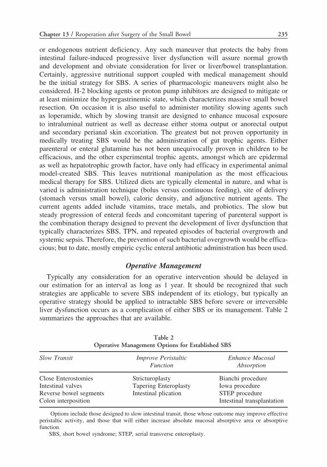
Chapter 13 / Reoperation after Surgery of the Small Bowel 235
or endogenous nutrient deficiency. Any such maneuver that protects the baby fromintestinal failure-induced progressive liver dysfunction will assure normal growthand development and obviate consideration for liver or liver/bowel transplantation.Certainly, aggressive nutritional support coupled with medical management shouldbe the initial strategy for SBS. A series of pharmacologic maneuvers might also beconsidered. H-2 blocking agents or proton pump inhibitors are designed to mitigate orat least minimize the hypergastrinemic state, which characterizes massive small bowelresection. On occasion it is also useful to administer motility slowing agents suchas loperamide, which by slowing transit are designed to enhance mucosal exposureto intraluminal nutrient as well as decrease either stoma output or anorectal outputand secondary perianal skin excoriation. The greatest but not proven opportunity inmedically treating SBS would be the administration of gut trophic agents. Eitherparenteral or enteral glutamine has not been unequivocally proven in children to beefficacious, and the other experimental trophic agents, amongst which are epidermalas well as hepatotrophic growth factor, have only had efficacy in experimental animalmodel-created SBS. This leaves nutritional manipulation as the most efficaciousmedical therapy for SBS. Utilized diets are typically elemental in nature, and what isvaried is administration technique (bolus versus continuous feeding), site of delivery(stomach versus small bowel), caloric density, and adjunctive nutrient agents. Thecurrent agents added include vitamins, trace metals, and probiotics. The slow butsteady progression of enteral feeds and concomitant tapering of parenteral support isthe combination therapy designed to prevent the development of liver dysfunction thattypically characterizes SBS, TPN, and repeated episodes of bacterial overgrowth andsystemic sepsis. Therefore, the prevention of such bacterial overgrowth would be effica-cious; but to date, mostly empiric cyclic enteral antibiotic administration has been used.
Operative ManagementTypically any consideration for an operative intervention should be delayed in
our estimation for an interval as long as 1 year. It should be recognized that suchstrategies are applicable to severe SBS independent of its etiology, but typically anoperative strategy should be applied to intractable SBS before severe or irreversibleliver dysfunction occurs as a complication of either SBS or its management. Table 2summarizes the approaches that are available.
Table 2Operative Management Options for Established SBS
Slow Transit Improve PeristalticFunction
Enhance MucosalAbsorption
Close Enterostomies Stricturoplasty Bianchi procedureIntestinal valves Tapering Enteroplasty Iowa procedureReverse bowel segments Intestinal plication STEP procedureColon interposition Intestinal transplantation
Options include those designed to slow intestinal transit, those whose outcome may improve effectiveperistaltic activity, and those that will either increase absolute mucosal absorptive area or absorptivefunction.
SBS, short bowel syndrome; STEP, serial transverse enteroplasty.
236 Ziegler and Petty
Slowing Intestinal Intraluminal Nutrient Transit
Enterostomy or stomal closure with restoration of oral to anal continuity that includesinterposition of the anal sphincter is perhaps the single most important first step inintestinal rehabilitation where a stoma has been created. Unfortunately, this maneuverhas not been studied in a prospective fashion; however, there are multiple anecdotalexamples that emphasize the importance of this step. This is particularly important ifmost if not all of the colon is intact, and certainly may be particularly important ifdistal ileum and even ileocecal valve can be interposed back into the fecal stream.Though there is little data to suggest that the ileocecal valve impacts patient survival,there is a suggestion that the course of parenteral nutrition support may be prolongedin the absence of this valve (43). However, even in the absence of a large portion ofcolon, there is real value in continuity restoration if the main feature is interposing theanal sphincter mechanism alone.
The technique of stoma takedown and closure follows standard operative techniquestypically using single-layer interrupted suture technique. Timing of such an interventionis difficult to gauge. It is best to preoperatively exclude distal bowel obstruction witha contrast enema, and intraoperatively it is best to also assure a patent downstreamintestine by injection of intraluminal saline through the anastomotic site. Finally,meticulous attention to perianal skin care can prevent breakdown and discomfort relatedto a higher frequency and volume of anticipated stool output.
Intestinal valves have been anecdotally trialed in SBS. Their putative value may beseveral-fold: increase intestinal transit time; minimize or prevent colon to small bowelregurgitation of bacteria and other colonic content; and to produce dilitation of themore proximal bowel, augmenting its potential for an eventual lengthening procedure.These typically have been constructed by intussuscepting proximal into distal smallbowel, typically for a 2-cm segment. There are several adjunctive techniques designedto help hold the valve and prevent its deintussusception: the serosa may be denuded tocreate an inflammatory bond, which may help “secure” the valve; or a prosthetic patchmay be used to encircle the bowel prior to the intussusception. The overlaid bowelintussusceptum is then sutured to this patch to facilitate holding the valve in place.Such valves were first trialed in 1970 (44), but their more recent application has beenas a part of a sequential treatment strategy applied as a bowel-dilating mechanism (45)to prepare the patient’s bowel for an eventual lengthening procedure.
The application of antiperistaltic or reversed small bowel segments to themanagement of SBS is also based on the premise of slowing intestinal transit orprolonging nutrient exposure to the small bowel absorptive surface. The technique ofthis procedure is poorly described: a mid to distal small bowel segment, 3–5 cm inlength is divided from its proximal and distal in situ connection, carrying the dissectioncarefully to the root of the small bowel mesentery to facilitate a gentle torque ortwist of the segment on its mesentery. The “reversed” bowel is then connected by twoend-to-end anastomoses such that the segment is now interposed in an antiperistalticdirection. The danger of this procedure is using a segment length that is too long, whichmight convert the patient from too fast a transit to complete SBO. First described in1959 (46), there have been several small pediatric experiences reported (47,48), thelargest being a five patient series where original bowel length varied from 20–94 cmand the reversed segment was 3 cm in length. Four of five of these patients survivedTPN-free.

Chapter 13 / Reoperation after Surgery of the Small Bowel 237
Small bowel colon interposition was first described in 1971 (49,50). The techniqueis especially applicable to the child with short small bowel who has an intact colon.Whether the colon segment is inserted prejejunal or preileal or whether it is insertedin an isoperistaltic or antiperistaltic direction does not seem to influence its salutarybenefit, which is to slow transit time, enhance vitamin B12, carbohydrate, and fatabsorption and augment weight gain presumably secondary to a more prolongedexposure of nutrients to the absorbing small bowel mucosa. This operation is done byisolating an 8- to 24-cm segment of either the ascending, transverse, or descendingcolon on its respective blood supply and interposing the colon between two ends ofsmall bowel with end-to-end anastomoses. One recognized outcome of this procedurelikely secondary to bacterial overgrowth is the development of D-lactic acidosis, aunique complication that requires aggressive treatment. In a series of patients reportedfrom two institutions where patients had residual small bowel lengths of 15–63 cm,four of seven patients were long-term survivors (51,52).
Improving Peristaltic Function
The singular pathophysiologic mechanism that is most challenging to manage inSBS is bacterial overgrowth and its associated malabsorption and low-grade sepsis.Mechanisms that contribute to this sequence of events include intraluminal nutrients andbacterial stasis secondary to partial obstruction, disparate anastomotic size, or ectaticand dysmotile segments of intestine that preclude apposition of the intestinal wallduring peristalsis. In the face of intestinal narrowing and secondary partial obstruction,stricturoplasty may be used to relieve the obstruction yet preserve intestinal length.Disparate sized bowel secondary to partial obstruction, an eccentric anstomosis, orbowel ectasia can be managed by several techniques. Where small bowel length is notan issue, resection of the dilated segment followed by a new end-to-end anstomosis isthe best solution. An alternate technique would be a tapering enteroplasty, in whichthe dilated segment is narrowed or tapered along its antemesenteric border, mosteffectively done by using an automated stapling device (53). Where length is an issue,either imbrication or plication of the ante-mesenteric border accomplishes the needednarrowing without sacrificing mucosal surface area (54). The main issue with the lattertechnique is that the plicated bowel may eventually become “undone.” In a series of11 children subjected to tapering enteroplasty segments that varied in site and length,nine patients were completely weaned from TPN and the other two had considerablereduction in their parenteral support (55).
Increase Mucosal Surface Area
The tools in use today are the intestinal lengthening procedures that likely constrainabsolute surface area. Instead, these techniques do lengthen bowel, make luminal sizesmaller such that the walls coapt during peristalsis, and the altered intestinal motilitymay enhance the exposure of intraluminal nutrient to functional mucosa.
The Bianchi procedure (also called the Autologous Gastrointestinal Reconstruction[AGIR]) is a technique that divides the intestine in a longitudinal plane separating theleaves of mesentery into two equal sides (56). The two parallel tubes of neointestine,representing half of the original diameter must then be connected in a properistalticfashion by end-to-end anastomoses in a gentle “S” configuration. Kimura described amodification of a bowel lengthening procedure, the Iowa procedure, in which shortenedintestine was sutured to either the undersurface of the liver or the undersurface of the
238 Ziegler and Petty
rectus abdominus muscle, from which a parasitized blood supply was generated (57,58).Once neovascularity was established, the bowel could be divided in a horizontal planeto again double its length. If the Iowa procedure followed the Bianchi procedure, thenthere could be a potential bowel length increase by a factor of four. Georgeson addedto this procedure the sequential lengthening process, including the use of an interposedvalve, to produce bowel dilatation that would facilitate a lengthening procedure (59).The most recent contribution in this field is the Serial Transverse Enteroplasty (STEP)procedure, described by Kim (60). This technically simpler and safer procedure isdone typically by the serial, alternated, and parallel application of a gastrointestinalstapling device between mesenteric vessels spaced to produce a zig-zag intestinalconfiguration in which the neointestine is of common but considerably smaller size.The lengthening procedures are especially useful techniques in the face of focal orelongated intestinal dilatation. Data from the Iowa procedure is represented by isolatedcase reports. The AGIR and STEP procedure has received more emphasis and study.Bianchi has described his personal series of 20 patients, nine of whom survived long-term (61). Lengthening in the nine survivors occurred at a mean age of 23.5 months,and the achieved length increase went from a mean of 62 to 97 cm. Interestingly, inthe nonsurvivors, the absolute length was less (43 to 68 cm), but strikingly, the meanage of the procedure was 2.75 months, suggesting that they represented a populationof babies in trouble with their SBS, likely with developing liver dysfunction. TheSTEP procedure has been shown in both patients as well as in experimental animalsto improve nutritional status, intestinal absorption, and to have a salutary benefit onbacterial overgrowth in SBS (62).
Intestinal transplantation would be the prototype of a SBS treatment to enhancebowel surface area (63). The spectrum of the transplant could be an isolated smallbowel graft from a cadaver or related living donor, a combined cadaveric graft of liverand small bowel used to treat those children who have concomitant liver failure as acomplication of their SBS, or a multivisceral graft to treat the same intestinal failure,liver failure complex. Further discussion of bowel transplantation is beyond the scopeof this chapter.
REFERENCES
1. Gauderer, MW. Stomas of the small and large intestine. In: Grosfeld, JL, O’Neill JA Jr, FonkalsrudEW, Coran AG (eds.), Pediatric surgery. Philadelphia: Mosby Elsevier; 2006: 1479–1492.
2. Gauderer MWL, Ponsky JL. A simplified technique for constructing a tube feeding gastrostomy. SurgGynecol Obstet 1981;152: 83.
3. Gauderer MWL, Abrams RS, Hammond JH. Initial experience with the changeable skin-level port-valve: a new concept for long-term gastrointestinal access. J Pediatr Surg 1998.33:73–75.
4. Gauderer MWL, Stellato TA. Gastrostomies: evolution, techniques, indications, and complications.Curr Probl Surg 1986;23:657–719.
5. Bishop HC. Simplified closure of the long-standing gastrostomy. J Pediatr Surg 1981;16:571–572.6. Raval MV, Phillips JD. Optimal enteral feeding in children with gastric dysfunction: surgical
jejunostomy vs image-guided gastrojejunal tube placement. J Pediatr Surg 2006;41:1679–1682.7. Squire R, Kiely EM, Carr B, et al. The clinical application of the Malone antegrade colonic enema.
J Pediatr Surg 1993;28:1012–1015.8. Levitt MA, Soffer SZ, Pena A. Continent appendicostomy in the bowel management of fecal incon-
tinent children. J Pediatr Surg 1997;32:1630–1633.9. Gauderer MWL, DeCou JM, Boyle JT. Sigmoid irrigation tube for the management of chronic
evacuation disorders. J Pediatr Surg 2002;37:348–351.

Chapter 13 / Reoperation after Surgery of the Small Bowel 239
10. Del Pin CA, Czyrko C, Ziegler MM, et al. Management and survival of meconium ileus. A 30-yearreview. Ann Surg 1992;215:179–185.
11. Mak GZ, Harberg FJ, Hiatt P, et al. T-tube ileostomy for meconium ileus; four decades of experience.J Pediatr Surg 2000;35:349–352.
12. Gauderer MWL, Izant RJ Jr. A technique for temporary control of colostomy prolapse in children. JPediatr Surg 1985;20:653–655.
13. Nance ML, Keller MS, Stafford PW. Predicting hollow visceral injury in the pediatric blunt traumapatient with solid visceral injury. J Pediatr Surg 2000;35:1300–1303.
14. Millar AJ, Rode H, Cywes S. Malrotation and volvulus in infancy and childhood. Semin Pediatr Surg2003;12:229–236.
15. Krabben AA. Morbidity and mortality of inadvertent enterotomy during adhesiotomy. Br J Surg2000;874:467–471.
16. Harberg FJ, Senekjian EK, Pokorny WJ. Treatment of uncomplicated meconium ileus via T-tubeileostomy. J Pediatr Surg 1981;16:61–63.
17. Ein SH, Marshall DG, Girvan D. Peritoneal drainage under local anesthesia for perforations fromnecrotizing enterocolitis. J Pediatr Surg 1997;12:963–967.
18. Demestre X, Ginovart G, Figueras-Aloy J, et al. Peritoneal drainage as primary management innecrotizing enterocolitis: a prospective study. J Pediatr Surg 2002;37:1534–1539.
19. Cass DL, Brandt ML, Patel DL, et al. Peritoneal drainage as definitive treatment for neonates withisolated intestinal perforation. J Pediatr Surg 2000;35:1531–1536.
20. Gollin G, Abarbanell A, Baerg JE. Peritoneal drainage as definitive management of intestinal perfo-ration in extremely low-birth-weight infants. J Pediatr Surg 2003;38:1814–1817.
21. Moss RL, Dimmitt RA, Marion CW, et al. A meta-analysis of peritoneal drainage versus laparotomyfor perforated necrotizing enterocolitis. J Pediatr Surg 2001;36:1210–1213.
22. Ehrlich PF, Sato TT, Short BL, et al. Outcome of perforated necrotizing enterocolitis in the verylow-birth weight neonate may be independent of the type of surgical treatment. Amer Surg 2001;67:752–756.
23. Horwitz JR, Lally KP, Cheu HW, et al. Complications after surgical intervention for necrotizingenterocolitis: a multicenter review. J Pediatr Surg 1995;30:994–999.
24. Sharma R, Tepas JJ, Mollitt DL, et al. Surgical management of bowel perforations and outcome invery low-birth-weight infants (�1,200 g). J Pediatr Surg 2004;39:190–194.
25. Moss RL, Dimmitt RA, Barnhart DC, et al. Laparotomy versus peritoneal drainage for necrotizingenterocolitis and perforation. N Engl J Med 2006;354:2225–2234.
26. Moore TC. Successful use of the “patch, drain, and wait” laparotomy approach to perforated necro-tizing enterocolitis: is hypoxia-triggered “good angiogenesis” involved? Pediatr Surg Int 2000;16:356–363.
27. Luzzatto C, Previtera C, Boscolo R, et al. Necrotizing enterocolitis: late surgical results afterenterostomy without resection. Eur J Pediatr Surg 1996;6:92–94.
28. Pokorny WJ, Garcia-Prats JA, Barry YN. Necrotizing enterocolitis: incidence, operative care, andoutcome. J Pediatr Surg 1986;21:1149–1154.
29. Wolf WM, Snover DC, Leonard AS. Localized intestinal perforation following intravenousindomethacin in premature infants. J Pediatr Surg 1989;24:409–410.
30. O’Connor A, Sawin RS. High morbidity of enterostomy and its closure in premature infants withnecrotizing enterocolitis. Arch Surg 1998;133:875–879.
31. Marfuggi RA, Greenspan M. Reliable introoperative prediction of intestinal viability using afluorescent indicator. Surg Gynecol Obstet 1981;152:33–35.
32. Ein SH, Stephens CA, Reilly BJ. The disappearance of free air after pediatric laparotomy. J PediatrSurg 1985;20:422–444.
33. Wilkins BM, Spitz L. Incidence of postoperative adhesion obstruction following neonatal laparotomy.Br J Surg 1986;73:762–764.
34. Jabra AA, Fishman EK. Small bowel obstruction in the pediatric patient: CT evaluation. AbdomImaging 1997;22:466–470.
35. Akgur FM, Tanyel FC, Buyukpamukcu N, Hicsonmez A. Adhesive small bowel obstruction inchildren: the place and predictors of success for conservative treatment. J Pediatr Surg 1991;26:37–41.
36. Vijay K, Anindya C, Bhanu P, et al. Adhesive small bowel obstruction (ASBO) in children-the roleof conservative management. Med J Malaysia 2005;60:81–84.
37. Janik JS, Ein SH, Filler RM, et al. An assessment of the surgical treatment of adhesive small bowelobstruction in infants and children. J Pediatr Surg 1981;16:225–235.

240 Ziegler and Petty
38. van der Zee DC, Bax NM. Management of adhesive bowel obstruction in children is changed bylaparoscopy. Surg Endosc 1999;13:925–927.
39. West KW, Stephens B, Rescorla FJ, et al. Postoperative intussusception: experience with 36 cases inchildren. Surgery 1988;104:781–787.
40. Linke F, Eble F, Berger S. Postoperative intussusception in childhood. Pediatr Surg Int 1998;14:175–177.
41. Murphy FL, Sparnon AL. Long-term complications following intestinal malrotation and the Ladd’sprocedure: a 15-year review. Pediatr Surg Int 2006;22:326–329.
42. Borenstein SH, Langer JC. Heterotaxia syndromes and their abdominal manifestations. Curr OpinPediatr 2006;18:294–297.
43. Weber TR, Tracy T Jr, Connars RH. Short bowel syndrome in children. Quality of life in an era ofimproved survival. Arch Surg 1991;126:841–846.
44. Waddell WR, Kern FJ, Halgrimson CG, et al. A simple jejunocolic “valve” for relief of rapid transitand the short bowel syndrome. Arch Surg 1970;100:438–443.
45. Georgeson K, Halpin D, Figueroa R, et al. Sequential intestinal lengthening procedures for refractoryshort bowel syndrome. J Pediatr Surg 1994;29:316–321.
46. Hammer JM, Seay PH, Johnston RL, et al. The effect of antiperistaltic bowel segments on intestinalemptying time. Arch Surg 1959;79:537.
47. Wilmore DW, Johnson DJ. Metabolic effects of small bowel reversal in treatment of the short bowelsyndrome. Arch Surg 1968;97:784–791.
48. Warden JM. Personal communication.49. Hutcher NE, Salzberg AM. Pre-ileal transposition of colon to prevent the development of the short
bowel syndrome in puppies with 90% small intestinal resection. Surgery 1971;70:189–197.50. Hutcher NE, Mendez-Picon G, Salzberg A. Prejejunal transposition of colon to prevent the devel-
opment of the short bowel in puppies with 90% small intestine resection. J Pediatr Surg 1973;8:771–777.
51. Garcia VF, Templeton JM, Eichelberger MR, et al. Colon interposition for the short bowel syndrome.J Pediatr Surg 1981;16:994–995.
52. Glick PL, deLorimier AA, Adzick NS, et al. Colon interposition: an adjuvant operation for short gutsyndrome. J Pediatr Surg 1984;19:719–725.
53. Weber TR, Vane DW, Grosfeld JL. Tapering enteroplasty in patients with bowel atresia and shortgut. Arch Surg 1982;117:684–688.
54. deLorimier AA, Harrison MR. Intestinal plication in the treatment of atresia. J Pediatr Surg1983;18:734–737.
55. Falcone RA, Warner BW. Short-bowel syndrome. In: Ziegler MZ, Azizkhan R, Weber TR (eds.),Operative pediatric surgery. New York: McGraw-Hill; 2003: 699–712.
56. Bianchi A. Intestinal loop lengthening-a technique for increasing small intestinal length. J PediatrSurg 1980;15:145–151.
57. Kimura K, Soper RT. Isolated bowel segment (model 1): creation by myoenteropexy. J Pediatr Surg1990;25:512–513.
58. Kimura K, Soper RT. A new bowel elongation technique for the short bowel syndrome using theisolated bowel segment Iowa models. J Pediatr Surg 1993;28:792–794.
59. Collins J III, Vincente Y, Georgeson K, Kelly D. Partial intestinal obstruction induces substantialmucosal proliferation in the pig. J Pediatr Surg 1996;31:415–419.
60. Kim HB, Lee PW, Garza J, et al. Serial transverse enteroplasty for short bowel syndrome: a casereport. J Pediatr Surg 2003;38:881–885.
61. Bianchi A. Experience with longitudinal intestinal lengthening and tailoring. Eur J Pediatr Surg1999;9:256–259.
62. Chang RW, Javid PJ, Oh JT, et al. Serial transverse enteroplasty enhances intestinal function in amodel of short bowel syndrome. Ann Surg 2006;243:223–228.
63. Reyes J. Intestinal transplantation for children with short bowel syndrome. Semin Pediatr Surg2001;10:99–104.
14 Interventions for AppendicealComplications
Renata Fabia, MD and Steven Teich, MD
CONTENTS
Introduction
Risk of Perforation and its Morbidity
Drains, Lavage, and Wound Closure
Role of Antibiotics
Management of the Difficult Appendiceal Stump
Intraoperative Cultures
Intra-abdominal Abscess
Short-Term Postoperative Complications
Long-Term Complication
References
INTRODUCTION
Appendicitis remains the most common cause of acute abdominal surgical disease inchildren (1–3). The treatment of acute appendicitis engenders very little controversy;appendectomy has been the accepted treatment of choice for over 100 years, withexcellent results. However, once the appendix has perforated, the potential morbidityincreases, along with controversy regarding its treatment. Children, especially thoseunder 8 years of age are more likely to present with perforated appendicitis thanadults (4). Despite the commonality of ruptured appendicitis in children, there is notreatment consensus.
This chapter discusses the current diagnostic tools and therapeutic approaches tocomplications of appendicitis in children, as well as controversies in management.
RISK OF PERFORATION AND ITS MORBIDITY
Perforation of the inflamed appendix is rare within the first 24 hours of untreatedsymptoms but climbs to 6 % among patients not yet treated by 36 hours after symptomonset (5). After 36 hours of symptoms, the risk of rupture remains steady at approxi-mately 5% for each ensuing 12-hour period (6). Perforation of an inflamed appendixoccurs in 15–57 % of all patients treated surgically for suspected acute appendicitis
From: Reoperative Pediatric SurgeryEdited by: S. Teich and D. A. Caniano © Humana Press, Totowa, NJ
241
242 Fabia and Teich
(7,8). The highest rates are encountered in children, with some series reporting up to65% perforation rate in their pediatric population, with the perforation rate approaching100% in children under the age of two years (4,9–12). Major complications fromruptured appendicitis occur in 6.4–19.8% of cases (4,13–15).
DRAINS, LAVAGE, AND WOUND CLOSURE
Many surgical techniques have been used in an attempt to minimize complicationsfollowing appendectomy. These included drainage and/or lavage of the peritonealcavity and different methods of skin closure.
Recently, it has been established that drains do not reduce complications after appen-dectomy for any stage of appendicitis (16). Drains can potentially be harmful if theybecome a conduit for invasion of the wound by skin pathogens. In the overwhelmingmajority of cases, a drain is not necessary. However, if the appendiceal stump or cecumis of suboptimal quality, placement of a closed drain may be prudent (17). It shouldbe brought out through a separate small incision in the right lower quadrant and laidalong the cecum into the pelvis to drain those dependent areas. Considering that drainspose a risk and accomplish little, they should rarely be used. When used they shouldbe removed as soon as possible. Prophylactic antibiotics should not be prolonged to“cover” indwelling drains (18).
Peritoneal lavage versus drainage for perforated appendicitis in children has beeninvestigated. Peritoneal lavage appears to be superior to intraperitoneal tube drainagefor the management of perforated appendicitis in children (19,20). The purpose ofirrigation is to remove all debris, purulent fluid collections, and blood from thesurgical field. In early phlegmonous appendicitis without any pus, there is no advantageto irrigation but rather risks spreading contaminated fluid throughout the abdomen.Otherwise, in case of advanced appendicitis, with the appendix removed, a thoroughlavage of the area should be performed. In particular, the pelvis has to be well exposedand any residual contaminated fluid should be aspirated and irrigated by retracting thesigmoid colon and exposing the pouch of Douglas.
A variety of skin closure techniques have been utilized for appendectomies in orderto prevent the most common complication, wound infection. These include: leaving theskin open with packing, delayed primary closure using skin tapes, interrupted approx-imation of the skin with or without various aseptic wound wicks, and subcuticularwound closure with various sutures (21–24). The use of wound wicks is not associatedwith decreased wound infection rates following appendectomy (21). Furthermore, alarge study demonstrated that primary closure of the skin and subcutaneous tissueafter appendectomy for gangrenous or perforated appendicitis, combined with the useof antibiotic therapy in the perioperative period, is not associated with an increasedrisk of wound infection when compared with delayed skin closure (25). Subcuticularclosure is therefore deemed appropriate in view of its greater convenience and safety.In addition, syringe pressure irrigation of the subcutaneous tissue after appendectomywas found to significantly decrease the incidence of postoperative wound infection incomplicated cases (26).
ROLE OF ANTIBIOTICS
Empiric antibiotic therapy has been shown to significantly decrease complicationrates in patients with acute as well as complicated appendicitis (27–29). Currentcontroversies involve choice, duration, and route of antibiotic treatment. It is generally

Chapter 14 / Appendiceal Complications 243
believed that it is critical to administer antibiotics before making the surgical incision(30). However, a recent study suggested that antibiotic prophylaxis is effective in theprevention of postoperative complications in appendicitis patients, whether given pre-,peri-, or postoperatively (31). Children with perforated appendicitis can be managedeffectively with multiple antibiotics or a single broad-spectrum antibiotic, such aspiperacillin/tazobactam (30–34). The appropriate duration of antibiotic use followingappendectomy for advanced appendicitis in children is still being debated. There isan ongoing effort to simplify the antibiotic regimen and reduce length of therapy foradvanced appendicitis (35,36).
MANAGEMENT OF THE DIFFICULT APPENDICEAL STUMP
Open AppendectomyThe appendix can usually be removed by the standard technique of stump ligation
after transection, with or without inversion of the stump into the base of the cecum.The inversion of the stump into the cecum may however be associated with a greaterrisk of bowel obstruction (37). When inflammation or necrosis extends into the cecumand the appendix cannot be ligated at its base, there are several options that can beemployed. These include appendicocecostomy, partial cecal excision with a staplinginstrument, and ileocecectomy or right colon resection (38,39). Appendicocecostomyinvolves placing a catheter into the cecum through the appendiceal orifice at theconvergence of the taeniae, and leaving it in place for 2–3 weeks. After that time thecatheter can be removed; resulting in a controlled fistula that usually closes withinone week if there is no residual disease in the colon (38). If inflammatory or necroticchanges of appendicitis involve the base of appendix, but there is a sufficient amount ofhealthy tissue between the cecal base and the ileocecal valve, a partial cecal resectionwith appendectomy using a stapling instrument can be performed. When faced witha difficult appendiceal stump, these techniques help minimize the risk of appendicealstump breakdown with intraperitoneal soilage or cecal fistula (38).
When a patient presents with perforated appendicitis with a large abscess orphlegmon involving the cecum and adjacent structures, a routine appendectomy ishazardous and may lead to serious complications including fecal fistula, intestinalobstruction, and intraabdominal abscess (39). Ileocecectomy, with limited resection ofthe terminal 5–7 cm of ileum and primary anastamosis, is a safe and effective therapyin selected patients (39). Right hemicolectomy is an alternative procedure that can beutilized in patients with excessive inflammatory involvement in the area.
Laparoscopic AppendectomyVarious techniques of appendiceal stump closure and control of the meso-appendix
have been described (40–42). These include: catgut ligature (40), the Endo-loop tyingtechnique (41), use of a linear stapler (40), use of an Endo-GIA automatic stapler(41), and an electro-coagulator technique (42). The Endo-loop technique is cheaperand requires only a 5-mm port; however, it requires more skill and may initially takemore time. The stapling technique needs less skill, is safe, and initially saves time,but it is more expensive and requires a 12-mm port (41). The length of surgery andpostoperative complication rate (intraoperative fecal soilage, and postoperative ileus)may be reduced by using a stapler versus the ligature technique (40).

244 Fabia and Teich
INTRAOPERATIVE CULTURES
Intraoperative cultures obtained at the initial operation for ruptured appendicitis, oncebelieved to be important for guiding postoperative antibiotic therapy, has been shownto be of no clinical value (43–45). Antibiotic changes based on intraoperative culturesdo not affect the clinical outcome or prevent postoperative complications (43,46,47).Lack of clinical improvement does not indicate a failure of an appropriately selectedinitial antibiotic regimen and does not support simply changing the antibiotics (45). Ithighly suggests the probability of intraabdominal abscess or wound infection, which isresponsive to drainage rather than antibiotic treatment (45). However, all postappen-dectomy abscess cavities should be cultured when they are drained, either surgicallyor percutaneously. In this instance, antibiotic therapy should be individualized basedupon abscess culture results, because the abscess will usually grow only one to threeorganisms (48).
INTRA-ABDOMINAL ABSCESS
An intraabdominal abscess can occur during two phases of appendicitis: either duringinitial patient presentation as an aspect of ruptured appendicitis or postoperatively as acomplication of advanced appendicitis. Between 2–6% of cases of acute appendicitisare complicated by development of a periappendicular mass, ranging from a phlegmonto an abscess (49). The incidence of postappendectomy intra-abdominal abscess isbetween 5–20% (4,13–15,50,51), but is significantly higher in the subset of patientsless than 10 years of age (45).
Diagnostic Evaluation
Initial Presentation
Patients usually present with abdominal discomfort, anorexia, change in bowelhabits, fever, and other nonspecific symptoms. Typically, there is a long durationof symptoms and patients often present after 5–7 days of symptoms. On physicalexamination an abdominal mass may be present but abdominal wall guarding maypreclude palpation unless performed under general anesthesia. Typically, fever andwhite cell count (WBC) are higher than in uncomplicated appendicitis (49). Abdominalcomputed tomography (CT) is useful to determine whether an appendiceal abscess ispresent, to differentiate the abscess from a phlegmon, and to determine whether theabscess is amenable to percutaneous drainage. High-resolution ultrasonography withgraded compression also has very good sensitivity and specificity, but is operator-dependent, and there is a limited field of view (52,53).
Following Appendectomy
Typically, these patients do not improve following appendectomy and developrecurrent fevers, abdominal pain, anorexia, and elevated WBC. These symptoms shouldlead to an evaluation for the presence of a postoperative intra-abdominal abscess (45).
Initial Nonoperative Management—Percutaneous Abscess DrainageThe management of appendiceal mass has been evolving, with significant changes
over the last decade. Initial nonoperative treatment of an intra-abdominal abscess by

Chapter 14 / Appendiceal Complications 245
percutaneous drainage and broad-spectrum intravenous antibiotics has been demon-strated to be safe and effective (49,53–55). The abscess drainage is usually performedby an interventional radiologist either percutaneously or transrectally using ultrasoundor CT guidance (53,56) (see radiology chapter of this book).
This approach avoids extensive surgery with the risk of intra-operative injuries,postoperative development of fistulae or recurrent abscesses, and prolonged postoper-ative recovery that is typically associated with immediate surgical treatment of intra-abdominal abscesses (49,55). Approximately 80-90% of patients have been reported tobenefit from this approach (49,55). The patients who do not improve within 72 hoursafter percutaneous drainage will require operative intervention (54–59). When initialnonsurgical treatment is successful, length of hospitalization, cost, and morbidity aredecreased compared to initial surgical management (57).
Operative ManagementPatients with appendiceal abscess who present with signs of diffuse peritonitis,
multiple abscesses or bowel obstruction, and appear toxic, typically are not candidatesfor percutaneous drainage and require early surgical intervention (60). Advocates ofinitial surgery as the treatment of choice in all cases of appendicitis complicated byintra-abdominal abscess cite low morbidity, no need for interval appendectomy (61,62),and elimination of the risk of recurrent appendicitis (63). However, the complications ofimmediate surgery, when encountered, can be extremely serious. Moreover, injudicioussurgical treatment of patients with periappendiceal masses has occasionally requiredemergency right hemicolectomy (61,64).
Early identification of the factors associated with failure of nonoperative initialmanagement is important. It has been reported that patients requiring early appen-dectomy have a more frequent finding of a small bowel obstruction (SBO) on theirinitial X-rays and a higher percent band count on their initial differential WBC countthan those successfully treated with interval appendectomy (59). An initial band countless than 15% is predictive of an uncomplicated course (59).
Role of Interval AppendectomyInterval appendectomy (performing an elective appendectomy in the “interval”
between bouts of appendicitis) remains controversial (55–62). Interval appendectomyis advocated to avoid a high recurrence rate of appendicitis and to confirm the diagnosis(58). Yet, some authors suggest that interval appendectomy is unnecessary (49,65).They point out that the recurrence rate is low and the removed appendix often showsno evidence of previous inflammation; therefore, the procedure is not justified (58,65).In other reported series, pathologic examination demonstrated that less than 10% of theappendices removed during interval appendectomy demonstrated no abnormalities (61).Furthermore, delayed or interval appendectomy specimens often have a characteristicinflammatory pattern that includes granulomas, xanthogranulomatous inflammation,mural fibrosis/thickening, and transmural chronic granulomatous inflammation (66).It would be desirable to perform interval appendectomy only for those patients inwhom recurrent appendicitis is very likely. However, because a reliable and nonin-vasive test to accurately determine the risk does not exist, the reasonable approachis to advise interval appendectomy for all patients (67). A recent study supports theneed for interval appendectomy by showing pathological findings in the majority of
246 Fabia and Teich
excised appendices, and suggests that laparoscopy is a safe alternative to open surgeryfor interval appendectomy with a low complication rate (68,69).
SHORT-TERM POSTOPERATIVE COMPLICATIONS
Wound InfectionWound infection is the most common source of morbidity following open appen-
dectomy for appendicitis, but there has been a steady decline in its occurrence inrecent years (15,70). It generally occurs 5–7 days after surgery. The incidence ofwound infection is more frequent in patients with advanced appendicitis (9), and it maycontribute to a prolonged hospital stay, increased cost of treatment, a longer period ofdiscomfort and pain, and delayed return to normal activities (71). One advantage ofthe laparoscopic approach over open appendectomy for patients with early appendicealperforation is reduction of the incidence of wound infections (69,72).
IncidenceWith the current practice of antibiotic use, pediatric patients with acute, nonperfo-
rated appendicitis have a wound infection rate of less than 1% (70,73). Recent seriesof complicated appendicitis in children report wound infection rates less than 5%(15,25,70,73), yet some institutions still report infectious complication rates that aresignificantly higher, some over 20% (60,71,74,75). Laparoscopic surgery has beendemonstrated to decrease wound complication rates even for advanced appendicitis toapproximately 1% (69,72), provided operative time is less than one hour, which isaccomplished in the majority of cases (18).
Diagnostic Evaluation and TreatmentPostoperative wound infections present as erythema, tenderness, edema, and
occasionally purulent drainage. The wound is often soft or fluctuant at the site of theinfection. The patient may have a leukocytosis and a low-grade fever. Managementof this complication depends on the depth of the infection. For a superficial woundinfection that involves the skin and subcutaneous tissue, the skin staples or suturesare removed over the area of the infection and a cotton-tipped applicator is passedinto the wound, typically with efflux of purulent material. The wound should begently explored and loculations broken up. Debridement of any nonviable tissue isalso important. An assessment of the integrity of the fascia should be made, becausewound dehiscence may occur. If the fascia is intact, wound packing with wet to drydressing changes or wound vacuum-assisted closure is commonly instituted. However,if the fascia has separated there is obvious concern about an intra-abdominal abscess,which may require either CT-guided drainage or possible surgical intervention (9).Incision and drainage of a superficial wound infection can usually be accomplished inthe outpatient setting under conscious sedation (69).
Antibiotics are generally not required for a wound infection, unless there is evidenceof cellulitis, deep tissue penetration, systemic toxicity, an immune compromised patientor an indwelling prosthetic device (74). Necrotizing fasciitis is an extremely rarecomplication of appendicitis, and there are only a few cases documented in the liter-ature. Once necrotizing fasciitis occurs, the mortality rate increases dramatically, sothat correct diagnosis and prompt debridement are mandatory (76).
Chapter 14 / Appendiceal Complications 247
Pathogenic OrganismsAlthough the most common microorganisms in acute appendicitis are Escherichia
coli and Bacteroides fragilis, the organisms most frequently found in postappendectomywound infection are Bacterioides and Clostridium welchii (15,43). Necrotizing fasciitis,along with myositis of the abdominal wall is a very rare but devastating compli-cation. Polymicrobial infection is responsible for this entity: group A Streptococcus,Staphylococcus aureus, E. coli, and anaerobes like Clostridia and Bacteroides (76).
PreventionThere have been multiple techniques proposed for wound prophylaxis in pediatric
appendicitis (71). These include: draining and/or packing the wound, wound irrigation,antibiotic powder into the wound, and preoperative intravenous antibiotics alone.A clinical study demonstrated that patients that received preoperative (or intraoper-ative) intravenous antibiotic (cefoxitin) plus wound antibiotic powder (cefoxitin) hada lower infection rate than those receiving either topical or intravenous antibioticalone (73). The benefit was significant compared to the group receiving no treatment(73). Antibiotic prophylaxis has been determined to be effective in the preventionof postoperative complications in appendectomy patients (30,31). However, addinga course of outpatient oral antibiotics, after completing a course of IV antibiotics,does not decrease postoperative infectious complications in appendicitis patients (77).Mechanical removal of infectious material, whether by peritoneal lavage or woundpressure irrigation has been shown to decrease the wound infection rate (19,20,26).Diligence with aseptic technique and avoidance of a hematoma at the incision sitecomprise additional factors that aid in wound infection prevention. Most recent pediatricseries dealing with complicated appendicitis use protocols of preoperative antibioticswith aerobic and anaerobic coverage, intraoperative lavage, no peritoneal or wounddrains, and continuation of antibiotics postoperatively (69).
Postappendectomy AbscessLack of clinical improvement after appendectomy with continuing fevers, leukocy-
tosis, ileus, or signs of sepsis indicate a high probability of an intra-abdominal abscess(45). Fevers in combination with abdominal pain are the most common sign associatedwith this finding (45,78). The only independent risk factors for development of postap-pendectomy intra-abdominal abscess are appendiceal perforation and age less than 10years (78). Drain placement during appendectomy was not found to make a signif-icant difference in intra-abdominal abscess formation (78). Studies comparing intra-abdominal abscess rates following laparoscopic and open appendectomy have shownconflicting results. Early reports demonstrated an increased rate of abscess formationafter laparoscopic appendectomy (79,80). However, more recent studies demonstrateno statistically significant difference in the rate of postappendectomy abscess amongchildren following laparoscopic and open procedures (81).
Diagnostic Evaluation and TreatmentThe diagnosis of postappendectomy abscess can be made by clinical findings alone,
such as in the case of spontaneous drainage of a subfascial purulent collection. Itcan also be made by abdominal CT scan or ultrasonography. Abdominal CT diagnosis
248 Fabia and Teich
is made by demonstrating a nonenhancing intra-abdominal fluid collection in patientswith clinical signs of abdominal sepsis (78).
Drainage of the purulent material is usually required in order to treat this compli-cation. Cultures of the drained abscesses should be obtained, as the pathogens areusually different from those found during appendectomy and may be resistant to thecurrent antibiotic regimen (45). Drainage procedures may be performed by a percuta-neous method using CT or ultrasound guidance or by an open surgical approach. Thesuccess rate of both techniques is similar and the hospital stay is the same, regardlessof which drainage method is utilized (78).
Suppurative PylephlebitisPyelephlebitis or portal pyemia, also known as septic thrombophlebitis of the
portal vein, or occasionally the superior mesenteric vein (SMV), was common in thepreantibiotic era. Today, it is encountered extremely rarely in association with intra-abdominal infections in the region drained by the portal venous system, includingdiverticular disease and complicated appendicitis (82). Although extremely rare, it isassociated with a high mortality, even in the modern era (83). It is characterized byjaundice, chills, and high fever. This serious illness frequently leads to multiple liverabscesses and the infecting organism is usually E. coli. Prompt diagnosis and treatmentare necessary to prevent hepatic abscess, septicemia, and complete thrombosis of theportal circulation (82,84).
Diagnostic Evaluation and Operative ManagementDiagnosis of pylephlebitis is accomplished by analysis of symptoms, clinical exami-
nation, and imaging studies including abdominal CT. Surgical therapy consists ofappendectomy but occasionally a right hemicolectomy is required (83,85). Liverabscesses must be drained and removal of the thrombus can be performed by using aFogarty catheter. Postoperative histopathological examination is performed to confirmthe diagnoses of appendicitis and septic thrombophlebitis of the portal vein and/orSMV. The patient is then treated with long-term antibiotics and anticoagulation.Complete recovery with appropriate medical and surgical treatment can be achieved(83,85).
Postoperative SBOIntestinal obstruction is considered early when it develops within three weeks after
the operation. Intestinal dysfunction is the second most common short-term compli-cation after appendectomy. In the first few weeks postoperatively, it is usually causedby a combination of paralytic ileus secondary to peritonitis and mechanical obstructionfrom fibrinous adhesions. A small bowel intussusception may also cause an earlyintestinal obstruction postoperatively, with an incidence of 0.06–0.8% (86). The clinicalpicture is one of a patient who initially manifests a return of gut function and advancesto a diet, but then has loss of bowel function along with abdominal distension and pain.
Diagnostic Evaluation
Plain abdominal films, when performed serially, are sometimes useful in theassessment of the postoperative patient with a suspected SBO. Abdominal CT is thebest initial imaging technique in a patient with a history of a laparotomy who complains
Chapter 14 / Appendiceal Complications 249
of mild intermittent abdominal pain but has few physical findings and nonspecificplain radiographs. CT enteroclysis should be used after the conventional abdominal CTstudy if additional management questions are left unanswered (87). Small bowel intus-susception can be identified by abdominal CT or ultrasound, but is rarely diagnosedbefore surgery (86).
Operative Versus Nonoperative Management
Postsurgical patients presenting early after operation with abdominal distension andno signs of bowel compromise (tachycardia, leukocytosis, localized tenderness, orfever) can be treated nonoperatively for several days, with subsequent abdominalCT if the clinical findings and abdominal plain films do not improve. Most early“obstructions” resolve with bowel rest, nasogastric suctioning, and intravenous fluids.If the patient is unable to eat for more than 5–7 days, parenteral nutrition is indicated(9,86). Any postoperative patient at high risk (i.e., very young age or comorbiditiespresent) that demonstrates a clinical picture of secondary loss of bowel function,distension, and abdominal pain should be presumed to have a mechanical SBO,and early operation should be considered (87,88). Patients with fever, tachycardia,leukocytosis, or other signs of bowel compromise should undergo immediate surgicalexploration.
Stump AppendicitisWhen performing an appendectomy, once the appendix has been completely skele-
tonized, it is amputated at the base. In general, there is no need for inversion of theappendiceal stump, but it is important to divide the appendix at the level of healthytissue, even if necessary at the level of the cecum, to avoid breakdown of the sutureor staple line. It has been shown that stump inversion at the time of open surgeryhas no effect on reducing the rate of intraabdominal abscess formation or lengthof hospital stay compared with simple stump ligation. Conflicting evidence exists,however, whether there is a difference in the rate of wound infection. One randomizedclinical trial found no difference whereas another study found a higher rate of infectionfollowing stump inversion (89,90).
Residual appendiceal tissue left after an initial appendectomy risks the developmentof stump appendicitis, which is a real, yet underreported entity. The latest compre-hensive review of the English-language literature revealed 36 reported cases of stumpappendicitis (91). Typically, patients present with signs and symptoms similar to acuteappendicitis; however, because of prior surgery, the diagnosis is difficult and the rate ofappendiceal stump perforation is extremely high (91). There is no correlation betweensimple ligation or inversion of the stump and stump appendicitis (92).
Stump appendicitis can occur after either laparoscopic or open appendectomy. Rarecases of inflammation of the appendiceal stump after open appendectomy have beenreported (93,94). However, it has been suggested that stump appendicitis may increasein prevalence with more frequent performance of laparoscopic appendectomy (92).A small field of view, lack of three-dimensional perspective, and absence of tactilefeedback are factors that may lead to an increased likelihood of a significant appendicealstump left after this approach (95). Its incidence may be minimized with accuratevisualization of the appendiceal base and creation of an appendiceal stump less than 3mm in length. It has been postulated that if laparoscopic appendectomy is performedproperly, there should not be a sudden increase in the incidence of this entity (92,95).
250 Fabia and Teich
Colonoscopy, ultrasonography, and abdominal CT have been shown to be useful inthe diagnosis of stump appendicitis. Abdominal CT can assist in making an accuratepreoperative diagnosis. It may show a characteristic “arrowhead sign” and a retainedappendicolith, both individual specific CT signs of appendicitis that, when combined,are pathognomonic for stump appendicitis (96). Although rare, stump appendicitisshould be considered in the differential diagnosis of right lower quadrant abdominalpain postappendectomy. Minimizing the length of the appendiceal stump is the onlymeans by which to avoid its occurrence.
LONG-TERM COMPLICATION
SBOThere is a 5% incidence of postoperative adhesive obstruction in children after
laparotomy for any reason (15,97). Of these, 80% have been reported to occur within2 years of the operation (97). Intestinal obstruction can occur after laparotomy forappendicitis; the long-term incidence is likely similar to the risk of patients undergoinglaparotomy for other reasons and its frequency varies from center to other (9,98). Onestudy analyzing the risk of surgically treated SBO after open appendectomy showedthe accumulated risk is 0.41% after 4 weeks, 0.63% after 1 year, and 1.3% after 30years of follow-up; compared with 0.003% at 1 year and 0.21% after 30 years offollow-up among nonoperated controls. The highest risk was found after operationfor other diagnoses, followed by operation for perforated appendicitis, nonspecificabdominal pain, and mesenteric lymphadenitis compared with operation for nonperfo-rated appendicitis. Women had a slightly lower risk than men (99). The incidence inanother two series was low, approximately 1–1.3 %, with most patients presenting inthe first 6 months after operation (9,98). In children that were followed 4–6 years afterappendectomy, 1.3% developed clinical symptoms consistent with mechanical SBOresulting in relaparotomy and confirmation of the diagnosis. Regarding the relationshipbetween severity of the disease and development of mechanical bowel obstruction, theincidence was 1.8% in the group with a normal appendix, 0% after simple appendicitis,0.4% after gangrenous appendicitis, and 3.4% after perforated appendicitis (100). Onthe other hand, the magnitude of readmission for abdominal complaints followingappendectomy was found to be more pronounced, and occurred in 2.94% of patientsduring a 10-year follow-up after appendectomy (101). Nonspecific abdominal pain withno sign of SBO caused almost one-half of the readmissions (45%), mostly in females(76%). Patients with a history of complicated appendicitis or a “normal” appendixwere most frequently readmitted (101). Approximately one-half of the patients withsymptoms of bowel obstruction required surgical intervention (101). The main causeof bowel obstruction after open appendectomy is adhesions. Incisional hernias alsooccur in 0.4% of all appendectomies (101).
Intestinal obstruction can occur following all types of laparoscopic abdominalsurgery; in the case of appendectomy, the prevalence is as high as that seen in openprocedures (102). Specific reasons include retained loose linear cutter staples (103,104),residual appendiceal tissue (105), and even rarely cecal volvulus (106). Retrieving asmany loose staples as possible at the termination of the laparoscopic appendectomy istherefore recommended (104).
Chapter 14 / Appendiceal Complications 251
FertilityAlthough perforation of the appendix is considered a risk factor for female
tubal infertility, the epidemiologic evidence supporting this relationship is incon-sistent. The idea dates back to 1932, when a medical report stated: “It is generallyagreed, that acute appendicitis may bring about sterility in women” (107). Subse-quent studies, however, have generated conflicting results (108–113). It has beencommonly thought that a perforated appendix in females may result in tubal dysfunctioncaused by peritoneal adhesions after inflammation, and a subsequent increased risk forextrauterine pregnancy and infertility. It has been suggested that early diagnosis andtreatment of suspected appendicitis in girls and women of reproductive age may reducethe incidence of tubal infertility resulting from the sequelae of a ruptured appendix(109). The liberal use of laparoscopy in women with suspected appendicitis has alsobeen recommended because of a fear of tubal dysfunction secondary to postsurgicalperitoneal adhesions after conventional open appendectomy (114). However, someauthors believe that it is unlikely that appendicitis with perforation will cause infer-tility unless there is an abscess in the pouch of Douglas (108). In more recent studies,neither history of acute appendicitis before puberty nor perforation of the appendixwas a statistically significant risk factor for tubal infertility (110). Others studies agreethat perforated appendicitis has little, if any role in the etiology of tubal infertility(111,112). This may have important implications for the management of young womenwith suspected appendicitis, as the liberal attitude toward surgical exploration witha subsequently high rate of negative appendectomy is often justified by a perceivedincreased risk of infertility after perforation. Women whose appendix was found to benormal at appendectomy in childhood seem to belong to a subgroup of patients with ahigher fertility rate than the general population (112).
A causal relationship between appendicitis and infertility cannot be supported by thedata currently available. Only a well-designed, prospective clinical study with unbiasedascertainment of exposure and adjustment for confounding variables will provide adefinitive answer (115).
REFERENCES
1. Garcia Pena BM, Mandl KD, Kraus SJ, et al. Ultrasonography and limited computed tomographyin the diagnosis and management of appendicitis in children. JAMA 1999;282:1041–1046.
2. Shelton T, McKinla R, Schwartz RW. Acute appendicitis: current diagnosis and treatment. CurrSurg 2003;60:502–505.
3. Cloud DT. Appendicitis. In: Aschcraft KW, Holder TM (eds.), Pediatric surgery, second edition.Philadelphia: Saunders; 1993: 470–477.
4. Pearl RH, Hale DA, Molloy M, Schutt DC, Jaques DP. Pediatric appendectomy. J Ped Surg1995;30:173–181.
5. Smith S. Appendicitis, appendectomy and the surgeon. Bull Hist Med 1996;70:414.6. Bickell NA, Aufses AH Jr, Rojas M, Bodian C. How time affects the risk of rupture in appendicitis.
J Am Coll Surg 2006;202(3):401–406.7. Coldrey E. Five years of conservative treatment of acute appendicitis. J Int Coll Surg 1959;32:
255–261.8. Andersson R, Hugander A, Thulin A, et al. Indications for operation in suspected appendicitis and
incidence of perforation. Br Med J 1994;308:107–110.9. Lally KP, Cox JR, Andrassy, RJ. Appendicitis. In: Townsend CM (ed.), Sabiston textbook of surgery,
17th edition. Philadelphia: Saunders; 2004: 1381–1395.10. Surama R, Quinn F, Puri P. Appendicitis in preschool children. Pediatr Surg Int 1995;10:68–70.
252 Fabia and Teich
11. Mattei PA, Stevenson RJ, Ziegler MM. Appendicitis. In: Wyllie R, Hyams JS (eds.), Pediatricgastrointestinal disease. Philadelphia: Saunders; 1999: 466–472.
12. Sakellaris G, Tilemis S, Charissis G. Acute appendicitis in preschool-age children. Eur J Pediatr2005;164(2):80–83.
13. Lee SL, Walsh AJ, Ho HS. Computed tomography and ultrasonography do not improve and maydelay the diagnosis and treatment of acute appendicitis. Arch Surg 2001;136:556–562.
14. Meier DE, Guzzetta PC, Barber RG, Hynan LS, Seetharamaiah R. Perforated appendicitis in children:is there a best treatment? J Pediatr Surg. 2003;38(10):1520–1524.
15. Anderson KD, Parry RL. Appendicitis. In: O’Neill J Jr (ed.), Pediatric surgery, fifth edition. St. Louis:Mosby; 1998: 1369–1379.
16. Petrowsky H, Demartines N, Rousson V, Clavien PA. Evidence-based value of prophylactic drainagein gastrointestinal surgery: a systematic review and meta-analyses. Ann Surg. 2004;240:1074–1084;discussion 1084–1085.
17. Katkhouda N, Kaiser AM. Appendectomy. OTGS 2005;7:8–14.18. Barie, PS, Eachempati SR. Surgical site infections. Surg Clin North Am 2005;85:1115–1135.19. Toki A, Horimi T, Tokuoka H, et al. Peritoneal lavage versus drainage for perforated appendicitis
in children. Surg Today 1995;25(3):207–210.20. Curran TJ, Muenchow SK. The treatment of complicated appendicitis in children using peritoneal
drainage: results from a public hospital. J Pediatr Surg 1993;28:204–208.21. McGreal GT, Joy A, Manning B, et al. Antiseptic wick: does it reduce the incidence of wound
infection following appendectomy? World J Surg 2002;26:631–634.22. Tsang TM. Delayed primary wound closure using skin tapes for advanced appendicitis in children.
A prospective, controlled study. Arch Surg 1992;127:451–453.23. Wetter LA. Controlled trial of polyglycolic acid versus catgut and nylon for appendectomy wound
closure. Br J Surg 1991;78:985–987.24. Serour F. Subcuticular skin closure as a standard approach to emergency appendectomy in children:
prospective clinical trial. World J Surg 1996;20:38–42.25. Rucinski J, Fabian T, Panagopoulos G, Schein M, Wise L. Gangrenous and perforated appendicitis:
a meta-analytic study of 2532 patients indicates that the incision should be closed primarily. Surgery2000;127(2):136–141.
26. Cervantes-Sánchez CR, Gutiérrez-Vega R, Vázquez-Carpizo JA, Clark P, Athié-Gutiérrez C. Syringepressure irrigation of subdermic tissue after appendectomy to decrease the incidence of postoperativewound infection. World J Surg 2000;24(1):38–41; discussion 41–42.
27. Bauer T, Vennits B, Holm B, et al. Antibiotic prophylaxis in acute nonperforated appendicitis. TheDanish Multicenter Study Group III. Ann Surg 1989;209:307–311.
28. Husser W. Evidenced-based surgery section January 2003. J Am Coll Surg 2003;196:104.29. Solomkin JS, Mazuski JE, Baron EJ, et al. Guidelines for the selection of anti-infective agents for
complicated intra-abdominal infections. Clin Infect Dis 2003;37:997–1005.30. Ceraldi CM, Waxman K. Antibiotic management of surgically treated appendicitis: a review. Infect
Surg 1992;vol:25–37.31. Andersen BR, Kallehave FL, Andersen HK. Antibiotics versus placebo for prevention of postoper-
ative infection after appendicectomy. Cochrane Database Syst Rev 2003(2):CD001439.32. Long SS. Clinical Approach and Management. Clinical and Surgical Approach. In: Principles
and practice of pediatric infectious diseases, second edition. Philadelphia: Churchill Livingstone,Elsevier; 2003.
33. Kaplan S. Antibiotic usage in appendicitis in children. Pediatr Infect Dis J 1998;17:1047–1048.34. Nadler EP, Reblock KK, Ford HR, Gaines BA. Monotherapy versus multi-drug therapy for the
treatment of perforated appendicitis in children. Surg Infect 2003;4:327–333.35. Snelling CM, Poenaru D, Drover JW. Minimum postoperative antibiotic duration in advanced
appendicitis in children: a review. Pediatr Surg Int 2004;20(11–12):838–845.36. Schmittenbecher P. Minimum postoperative antibiotic duration in advanced appendicitis in children:
A review. J Pediatr Surg 2005;40(7):1216.37. Street D, Bodai BI, Owens LJ, et al. Simple ligation vs stump inversion in appendectomy. Arch
Surg 1988;123:689–690.38. Poole GV. Management of the difficult appendiceal stump: how I do it. Am Surg 1993;59(9):
624–625.39. Thompson JE, Bennion RS, Schmit PJ, Hijama DT. Cecectomy for complicated appendicitis. J Am
Coll Surg 1994;179:135–138.

Chapter 14 / Appendiceal Complications 253
40. Ortega AE, Hunter JG, Peters JH, Swanstrom LL, Schirmer B, The Laparoscopic AppendectomyStudy Group. A prospective, randomized, comparison of laparoscopic appendectomy with openappendectomy. Am J Surgery 1995;169:208–213.
41. Kleiber C, Wagner M, Metzger A. Various stapling techniques in laparoscopic appendectomy: 40consecutive cases. Surg Laparosc Endosc 1994;4:205–209.
42. Mohsen AA. Endocoagulator control of the mesoappendix for laparoscopic appendectomy.J Laparosc Surg 1994;4:435–440.
43. McNamara MJ, Pasquale MD, Evans SR. Acute appendicitis and use of intraoperative culture. SurgGynecol Obstet 1993;177:393–397.
44. Thadepalli H, Mandal AK, Chuah SK, Lou MA. Bacteriology of the appendix and the ileum inhealth and appendicitis. Am Surg 1991;57:317–322.
45. Mosdell DS, Morris DM, Fry DE. Peritoneal cultures and antibiotic therapy in pediatric perforatedappendicitis. Am J Surg 1994;167:313–316.
46. Coldrey E. Five years of conservative treatment of acute appendicitis. J Int Coll Surg 1959;32:255–261.
47. Kokoska ER, Silen ML, Tracy TF Jr, et al. The impact of intraoperative culture on treatment andoutcome in children with perforated appendicitis. Pediatr Surg 1999;34(5):749–753.
48. Nathens AB, Ahrenholz DH, Simmons RL, Rotstein O. Peritonitis and other intra-abdominal infec-tions. In: Howard RJ, Simmons RL (eds.), Surgical infectious diseases. Norwalk: Appleton & Lange;1995.
49. Nitecki S, Assalia A, Schein M. Contemporary management of appendiceal mass. Br J Surg1993;80:18–20.
50. Fishman SJ, Pelosi L, Klavon SL, O’Rourke EJ. Perforated appendicitis: prospective outcomeanalysis for 150 children. J Pediatr Surg 2000;35(6):923–926.
51. McKinlay R, Neeleman S, Klein R, et al. Intraabdominal abscess following open and laparoscopicappendectomy in the pediatric population. Surg Endosc 2003;17(5):730–733.
52. Joseph AE, MacVicar D. Ultrasound in the diagnosis of abdominal abscesses. Clin Radiol1990;42:154–156.
53. Hogan MJ. Appendiceal abscess drainage. Tech Vasc Interv Radiol 2003;6(4):205–214.54. Brown CVR, Abrishami M, Muller M, Velmahos GC. Appendiceal abscess: immediate operation
or percutaneous drainage? Am Surg 2003;69(10):829–832.55. Vargas HI, Averbook A, Stamos MJ. Appendiceal mass: conservative therapy followed by interval
laparoscopic appendectomy. Am Surg 1994;60:753–758.56. Jamieson DH, Chait PG, Filler R. Interventional drainage of appendiceal abscesses in children. AJR
Am J Roentgenol 1997;169(6):1619–1622.57. Bufo AJ, Shah RS, Li MH, et al. Interval appendectomy for perforated appendicitis in children.
J Laparoendosc Adv Surg Tech A 1998;8(4):209–214.58. Vervaal VJ, Wobbes T, Goris RJA. Is there still a place for interval appendectomy? Dig Surg
1993;10:285–288.59. Kogut KA, Blakely ML, Schropp KP, et al. The association of elevated percent bands on admission
with failure and complications of interval appendectomy. J Pediatr Surg 2001;36(1):165–168.60. Weber TR, Keller MA, Bower RJ, et al. Is delayed operative treatment worth the trouble with
perforated appendicitis in children? Am J Surgery 2003;186:685–689.61. Nguyen DB, Silen W, Hodin RA. Interval appendectomy in the laparoscopic era. J Gastrointest Surg
1999;3:189–193.62. Ahmed I, Deakin D, Parsons SL. Appendix mass: do we know how to treat it? Ann R Coll Surg
Engl 2005;87(3):191–195.63. Paull D, Bloom P. Appendiceal abscess. Arch Surg 1982;117:1017–1019.64. Riseman JA, Wichterman K. Evaluation of right hemicolectomy for unexpected cecal mass. Arch
Surg 1989;124(9):1043–1044.65. Kaminski A, Liu IL, Applebaum H, Lee SL, Haigh PI. Routine interval appendectomy is not justified
after initial nonoperative treatment of acute appendicitis. Arch Surg 2005;140(9):897–901.66. Guo G, Greenson JK. Histopathology of interval (delayed) appendectomy specimens: strong
association with granulomatous and xanthogranulomatous appendicitis. Am J Surg Pathol2003;27(8):1147–1151.
67. Gahukamble DB, Khamage AS, Gahukamble LD. Management of appendicular mass in children.Ann Trop Paediatr 1993;13(4):365–367.

254 Fabia and Teich
68. Bass J, Rubin S, Hummadi A. Interval appendectomy: an old new operation. J Laparoendosc AdvSurg Tech A 2006;16(1):67–69.
69. Ostlie DJ, Holcomb GW. Laparoscopic appendectomy. OTGS 2004;6(4):317–329.70. Neilson IR, Laberge JM, Nguyen LT, et al. Appendicitis in children: current therapeutic recommen-
dations. J Pediatr Surg 1990;25(11):1113–1116.71. Ein SH, Sandler A. Wound infection prophylaxis in pediatric acute appendicitis: a 26-year
prospective study. Pediatr Surg 2006;41(3):538–541.72. Aziz O, Athanasiou T, Tekkis PP, et al. Laparoscopic versus open appendectomy in children: a
meta-analysis. Ann Surg 2006;243(1):17–27.73. Emil S, Laberge JM, Mikhail P, et al. Appendicitis in children: a ten-year update of therapeutic
recommendations. J Pediatr Surg 2003;38(2):236–242.74. Helmer KS, Robinson EK, Lally KP, et al. Standardized patient care guidelines reduce infectious
morbidity in appendectomy patients. Am J Surg 2002;183:608–613.75. Hoehne F, Ozaeta M, Sherman B, Miani P, Taylor E. Laparoscopic versus open appendectomy: is
the postoperative infectious complication rate different? Am Surg 2005;71(10):813–815.76. Mukoyama S, Mukai M, Yasuda S, et al. A successfully treated case of severe necrotizing fasciitis
caused by acute appendicitis: a case report. Tokai J Exp Clin Med 2003;28(3):139–143.77. Taylor E, Berjis A, Bosch T, Hoehne F, Ozaeta M. The efficacy of postoperative oral antibiotics in
appendicitis: a randomized prospective double-blinded study. Am Surg 2004;70(10):858–862.78. Schmit P, Hiyama D, Swicker S, et al. Analysis of risk factors of postappendectomy intraabdominal
abscess. J Am Coll Surg 1994;179:721–726.79. Golub R, Siddiqui F, Pohl P. Laparoscopic versus open appendectomy: a metaanalysis. J Am Coll
Surg 1998;186:545–553.80. Kisher SL, Browne A, Dibbins A, Tkacz N, Curci M. Intra-abdominal abscess after laparoscopic
appendectomy for perforated appendicitis. Arch Surg 2001;136:438–441.81. McKinlay R, Neeleman S, Klein R, et al. Intraabdominal abscess following open and laparoscopic
appendectomy in the pediatric population. Surg Endosc 2003;17(5):730–733.82. Saxena R, Adolph M, Ziegler JR, Murphy W, Rutecki GW. Pylephlebitis: a case report and review
of outcome in the antibiotic era. Am J Gastroenterol 1996;91(6):1251–1253.83. Nishimori H, Ezoe E, Ura H, et al. Septic thrombophlebitis of the portal and superior mesenteric
veins as a complication of appendicitis: report of a case. Surg Today 2004;34(2):173–176.84. Pohl JF, Murarka P, Farrell MK, Bezerr J. Insights. Pylephlebitis. J Pediatr 1999;135(4):52985. Chang TN, Tang L, Keller K, Harrison MR, Farmer DL, Albanese CT. Pylephlebitis, portal-
mesenteric thrombosis, and multiple liver abscesses owing to perforated appendicitis. J Pediatr Surg2001;36(9):E19.
86. Ein SH, Ferguson JM. Intussusception-the forgotten postoperative obstruction. Arch Dis Child1982;57:788–790.
87. Maglinte DDT, Heitkamp, DE, Howard TJ, Kelvin FM, Lappas JC. Current concepts in imaging ofsmall bowel obstruction. Radiol Clin North 2003;41(2):263–283.
88. Stewart RM, Page CP, Brender J, Schwesinger W, Eisenhut D. The incidence and risk of earlypostoperative small bowel obstruction. A cohort study. Am J Surg 1987;154:643–647.
89. Engstrom L, Fenyo G. Appendicectomy: assessment of stump invagination versus simple ligation: aprospective, randomized trial. Br J Surg 1985;72:971–972. Reviewed in: Clin Evid 2005;13:466–472.
90. Jacobs PP, Koeyers GF, Bruyninckx CM. [Simple ligation superior to inversion of the appendicealstump; a prospective, randomized study]. Ned Tijdschr Geneeskd 1992;136:1020–1023. Reviewedin: Clin Evid 2005;13:466–472.
91. Liang MK, Lo HG, Marks JL. Stump appendicitis: a comprehensive review of literature. Am Surg2006;72(2):162–166. Review.
92. Mangi AA, Berger DL. Stump appendicitis. Am Surg 2000;66(8):739–741.93. Feigin E, Carmon M, Szold A, Seror D. Acute stump appendicitis. Lancet 1993;341:757.94. Thomas SE, Denning DA, Cummings MH. Delayed pathology of the appendiceal stump: a case
report of stump appendicitis and review. Am Surg 1994;60:842–844.95. Walsh DC, Roediger WE. Stump appendicitis—a potential problem after laparoscopic appendectomy.
Surg Laparosc Endoscopy 1997;7:357–358.96. Rao, PM. Sagarin MJ, McCabe CJ. Stump appendicitis diagnosed preoperatively by computed
tomography. Am J Emerg Med 1998;16:309–311.97. Janik JS, Ein SH, Filler FM, Shandling B, Simpson JS, Stephens CA. An assessment of the surgical
treatment of adhesive small bowel obstruction in infants and children. J Pediatr Surg 1981;16:225–229.

Chapter 14 / Appendiceal Complications 255
98. Khairy GA, Afzal MF, Murshid KR, Guraya S, Ghallab A. Post appendectomy small bowelobstruction. Saudi Med J 2005;26(7):1058–1060.
99. Andersson RE. Small bowel obstruction after appendicectomy. Br J Surg. 2001;88(10):1387–1391.100. Ahlberg G, Bergdahl S, Rutqvist J, Soderquist C, Frenckner B. Mechanical small-bowel obstruction
after conventional appendectomy in children. Eur J Pediatr Surg 1997;7(1):13–15.101. Tingstedt B, Johansson J, Nehez L, Andersson R. Late abdominal complaints after appendectomy—
readmissions during long-term follow-up. Dig Surg 2004;21(1):23–7.102. Duron JJ, Hay JM, Msika S, et al. Prevalence and mechanisms of small intestinal obstruction
following laparoscopic abdominal surgery: a retrospective multicenter study. French Association forSurgical Research. Arch Surg 2000;135(2):208–212.
103. Kuehnel F, Marusch F, Koch A, Gastinger I. Retained loose linear cutter staples after laparo-scopic appendectomy as the cause of mechanical small bowel obstruction. Int J Colorectal Dis2005;22(6):717–718.
104. Nottingham JM. Mechanical small bowel obstruction from a loose linear cutter staple after laparo-scopic appendectomy. Surg Laparosc Endosc Percutan Tech 2002;12(4):289–290.
105. Gordon R, Bamehriz F, Birch DW. Residual appendix producing small-bowel obstruction afterlaparoscopic appendectomy. Can J Surg 2004;47(3):217–218.
106. McIntosh SA, Ravichandran D, Wilmink AB, Baker A, Purushotham AD. Cecal volvulus occurringafter laparoscopic appendectomy. JSLS 2001;5(4):317–318.
107. Bull P. What part is played by acute appendicitis in the causation of sterility in young girls andwomen? Acta Chir Scand 1932;71:155–165.
108. Wiig JN, Janssen CW Jr, Fuglesang P, et al. Infertility as a complication of perforated appendicitis.Late follow-up of a clinical series. Acta Chir Scand 1979;145(6):409–410.
109. Mueller BA, Daling JR, Moore DE, et al. Appendectomy and the risk of tubal infertility. N Engl JMed 1986;315:1506–1508.
110. Urbach DR, Marrett LD, Kung R, Cohen MM. Association of perforation of the appendix withfemale tubal infertility. Am J Epidemiol 2001;153(6):566–571.
111. Puri P, McGuinness EP, Guiney EJ. Fertility following perforated appendicitis in girls. Pediatr Surg1989;24(6):547–549.
112. Andersson R, Lambe M, Bergstrom R. Fertility patterns after appendicectomy: historical cohortstudy. BMJ 1999;318(7189):963–967.
113. Malt RA. The perforated appendix. N Engl J Med 1986;315:1546–1547.114. Spirtos NM, Eisenkop SM, Spirtos TW, Poliakin RI, Hibbard LT. Laparoscopy—a diagnostic aid
in cases of suspected appendicitis. Its use in women of reproductive age. Am J Obstet Gynecol1987;156(1):90–94.
115. Urbach DR, Cohen MM. Is perforation of the appendix a risk factor for tubal infertility and ectopicpregnancy? An appraisal of the evidence. Can J Surg 1999;42(2):101–108.
15 Reoperation for InflammatoryBowel Disease
Christopher R. Moir, MD
CONTENTS
Crohn’s Disease—Recurrent Disease
Ulcerative Colitis
Indeterminate Colitis—Operative
Consideration and Management
References
CROHN’S DISEASE—RECURRENT DISEASE
A child diagnosed with Crohn’s disease has a 78% probability of surgery within20 years (1). The risk is higher for ileocolic disease (92%), whereas approximately two-thirds of patients with small bowel involvement and slightly less than 60% of patientswith colonic disease will come to operation (2). Pediatric surgeons planning the firstoperation do so knowing 50% of their patients will return for another procedure (1,3).This section reviews patterns of Crohn’s disease recurrence and operative strategy.
Strictures
Clinical Presentation
Recurrent Crohn’s disease is found most commonly at the surgical anastomosis (4).The degree of obstruction from recurrent inflammation and scarring establishes theintensity of symptoms. Crampy abdominal pain exacerbated by meals is a commonclinical presentation. Onset of recurrence varies with site and extent of initial resection.Much attention has been given to the adequacy of the first surgery; however, recurrenceat the anastomosis is a feature of the disease process rather than the presence ofmicroscopic residual disease at the margins (5). Grossly negative resection marginsare all that is necessary for a good initial procedure (6). Symptom onset can beexpected to occur earlier for patients with diffuse intestinal disease, perianal Crohn’sdisease, and those who had postoperative surgical complications (7). Although somepatients have unfortunately experienced recurrence of symptoms within months ofsurgery, the average time of symptom recurrence is site-dependent and not expected forseveral years after operation. At least 85% of surgical patients have histologic evidence
From: Reoperative Pediatric SurgeryEdited by: S. Teich and D. A. Caniano © Humana Press, Totowa, NJ
257
258 Moir
of recurrence by 3 years after surgery. Such biopsy evidence does not necessarilymean children will go on to develop endoscopic findings of ulceration and strictures.Similarly, endoscopic recurrence does not correlate with development of symptoms orrisk of subsequent reoperative surgery. The best rule of thumb when discussing diseaserecurrence with children and their families is to assume reoperation rates of just higherthan 50% for ileal colic disease and 45% each for isolated colonic or small bowelCrohn’s disease by 10 years (1,3,8–10).
Perforating Crohn’s disease represents a high-risk group thought to fare poorly afterintestinal resection. This is only true for the mode of recurrence, which tends to favorperforation once again. The absolute risk of recurrence is similar to obstructing Crohn’sdisease (11,12). Older adolescents who smoke represent a high-risk group and shouldbe encouraged to quit.
The degree of obstruction dictates the patient’s symptoms and further treatment.Medical intervention for control of symptoms is based on progression to the moreconcerning findings of partial bowel obstruction, bacterial overgrowth, malabsorption,and pain. Such symptoms can make meal time an unpleasant experience, producing a“feeding aversion” that occurs when the patient knows oral intake will produce furtherpain. Obstructive symptoms are particularly distressing for adolescents and youngadults who often eat at irregular intervals associated with their lifestyle and travel.It is not possible to have a good quality of life when oral intake must be regulatedthroughout the day.
Surgical indications for reoperation are based on the intensity of symptoms andinterference with quality of life (13). The amount of obstruction and the medicaltreatment necessary for its control may lead to determination of treatment failure andsurgical intervention (1,3). Perforating Crohn’s disease represents a fortunately rarebut dramatic subcategory for reoperative indications (11).
Diagnostic Evaluation
Patients with Crohn’s disease and recurrent symptoms can make the diagnosisthemselves. The role of the physician and surgeon is to confirm disease recurrenceand assess severity utilizing disease activity indices, radiologic signs and endoscopicfindings (9,10). Physical examination may identify abdominal distention or a mass,but most often diagnostic imaging is necessary to quantify the extent of disease andits effects. Although recurrence is usually limited to the previous site of surgery, it isessential to identify other areas of disease that may have occurred postoperatively (14).Standard imaging includes contrast studies and computed tomography (CT) enterog-raphy for visualization of luminal disease and quantification of the extraluminal effects(15–17). Despite radiologic advances, endoscopy remains the final arbiter of diseaserecurrence (13,18). Occasionally, magnetic resonance imaging (MRI) is used to identifydisease recurrence, penetration or fistulization to bowel and retroperitoneal structures(19). Similarly, capsule endoscopy improves sensitivity in patients with equivocalcontrast studies. Research has identified patient benefit with earlier detection of diseaseby capsule endoscopy (20–22).
The essential but invasive nature of endoscopy has led to renewed calls for laboratorymarkers of disease. Measurement of C-reactive protein may lead to earlier discoveryof disease and help predict response to relapse therapy. It is more useful for Crohn’sdisease than ulcerative colitis and superior to erythrocyte sedimentation rate (ESR),

Chapter 15 / Inflammatory Bowel Disease 259
albumin, and complete blood count (CBC) (23). New more specific biologic tests willsoon be available (24).
Laboratory investigations are always helpful to establish patient readiness for reoper-ation. Routine precautions include an evaluation of liver and pancreatic function inaddition to routine hematology and electrolyte evaluation.
Operative Techniques
Conduct at the second operation is highly dependent on the first. Incision placement,extent of resection, and type of anastomosis all depend on the first surgeon’s decisions.In general, it is easier to reopen a midline incision than to continue operating througha transverse scar. Pediatric surgeons favor a transverse approach in smaller childrenbecause of the wide access to intraperitoneal structures. As a child grows, the abdomenelongates and narrows, making a midline incision preferable for reoperation. Thisdilemma is particularly acute when considering a child with a transverse scar thatcrosses the midline. A new incision that bisects the old scar may increase the risk ofdelayed wound healing, incisional hernia, and unsatisfactory cosmesis. When possible,it is safe to reoperate through a transverse incision at least one more time. Access to theintraperitoneal structures is usually quite adequate and disease recurrence is generallyclose by. There may be an opportunity to repair any incisional herniation or subclinicaldehiscence of the rectus musculature following the first operation. If necessary foradequate exposure in the older child, a new midline incision is generally well tolerated.
Laparoscopic-assisted surgery obviates issues of incision placement (25,26). Repeatlaparoscopy is also feasible. The degree of recurrence, presence of intraperitonealadhesions, and persistence of the surgeon will determine the success of the secondlaparoscopic dissection. Laparoscopic-assisted procedures allow for extraction of thebowel through an incision for anastomosis or stricturoplasties. In one small series,laparoscopy produced longer recurrence-free intervals (27). Although such data is aninteresting side light to the laparoscopic story, it is also possible that concomitantadvances in medical management have decreased recurrence. Additionally, laparoscopyis associated with a higher use of stapled anastomosis, which also reduces the frequencyof reoperation.
An important principle of Crohn’s disease surgery is to create a widely patentanastomosis. Patients with ileocolic resections are natural candidates for a stapledside-to-side anastomosis that can be twice as large as an end-to-end procedure. Studiesof stapled versus handsewn anastomoses have fairly consistently shown improvedrecurrence-free intervals (28,29). With this information, a similar procedure is nowrecommended for small bowel surgery. Although it is particularly important to create alarge anastomosis at the time of reoperation, the same technique is advocated for initialsurgery. Such widely patent anastomoses may be expected to reflux colonic contentsinto the small bowel. This is of uncertain influence in developing children. Cautionshould be exercised in smaller children where issues of growth and developmentare paramount.
Strictureplasties for active disease as well as burned-out fibrotic strictures remain anexcellent option for patients with multifocal disease (30). Such length-sparing proce-dures are particularly attractive for developing children with extensive disease and skipareas. Limited resection or bypass with strictureplasty has been employed with excellentsuccess (31,32). There are no data as yet to support the relative contraindication ofleaving persistent disease behind in developing children (33,34). Adverse effects on

260 Moir
growth and the risk of malignancy have not been quantified. Fortunately, disease inthe area of strictureplasties has been ameliorated by the surgical technique (35,36).
The indications for operation are no longer restricted to the multiply recurrent patientwith widespread disease. Any focal lesion without diffuse extraintestinal indurationis a candidate for limited intervention. Length of stricture and the noninflammatorynature of the obstruction are the two strongest factors determining operability (30).
The techniques of strictureplasty vary according to the lesion. The standard techniqueknown to all surgeons resembles a Heineke-Mikulicz pyloroplasty (Fig.1). Theprocedure is best suited for short fibrotic strictures less than 10 cm without surroundinginflammation adjacent to pliable bowel (37).
The side-to-side Finney strictureplasty may treat up to 25 cm of diseased bowel (38)(Fig. 2). Other variations include a Jaboulay side-to-side anastomosis that bypasses thedisease. Such procedures mimic old-time surgeries for Crohn’s disease, which haveagain come into fashion (39). Overall, 85% of strictureplasties resemble a standardpyloroplasty; however, a metaanalysis suggests the Finney procedure has a lowerrecurrence rate (40). In general, the reoperative rate for strictureplasty approaches 25and 30% at 5 and 10 years, respectively (41).
The long-term concerns of persistent disease must be balanced by the tendencyfor Crohn’s disease to regress over time. Disease recurrence or persistence does notnecessarily correlate with symptoms, and therefore, such procedures can be advocatedfor children as well as adults. Its effectiveness is well established, but increasedmorbidity and recurrence rates suggest that selective use of the procedure is the bestapproach (30). Isolated short resections remain the best option for most children withCrohn’s disease.
Options for recurrence of colonic disease are more limited. Patients with obstructingdisease at the ileorectal anastomosis may be amenable to local resection or
Fig. 1. The Heineke-Mikulicz strictureplasty. A longitudinal incision is closed transversely.

Chapter 15 / Inflammatory Bowel Disease 261
Fig. 2. The Finney strictureplasty. The bowel is laid side-to-side and anastomosed with handsewnor stapled techniques.
strictureplasty. If this is not possible, the complications of anorectal stricturing, perianaldisease, and fistulization to surrounding organs necessitate diversion. Such diversionmay be permanent or at least long-term until rectal disease activity abates. Simplediversion with a loop ileostomy is a common approach to children, with the expec-tation that reanastomosis is performed within 1 year. Unfortunately, many childrenhave chronic persistent disease that does not respond well to therapy. Although it isdifficult to permanently remove the rectum in a child or young adult, especially whennewer therapies are on the horizon, significant ongoing perianal sepsis mandates sucha procedure (42).
Removal of the residual rectal stump via an abdominoperineal approach is reservedfor patients with the most resistant disease (43). Local wound care following rectalstump removal continues to be problematic (44). With this in mind and the presenceof newer biologic therapy, most surgeons prefer to preserve at least a portion ofthe rectum for later surgical reconstruction. In an attempt to obviate the complica-tions of reoperative surgery, all efforts should be made to avoid dissection below theperitoneal reflection at the time of disconnection of ileorectal anastomoses or removalof fistulizing disease (45).
Construction of ileostomies in patients expected to keep their stomas for many yearsrequires a more conservative surgical approach. An end ileostomy is less likely toprolapse and should be positioned in an area of the body where it is least expectedto interfere with daily function. Loop ileostomies may also be used for long-termdiversion; however, it is preferable to divide the bowel completely, leaving the distallimb close by. Placement of an ileostomy through the rectus sheath is mandatory forlong-term control of prolapse and parastomal herniation.
Nonoperative Management
Although 80% of patients with Crohn’s disease recur following surgery, less than50% come to reoperation (1). Most children are managed nonoperatively with thereintroduction of immunosuppression (46). The degree and severity of symptomsand intensity of therapy will determine the decision to reoperate. Fortunately, useof infliximab (Remicade) combined with immunosuppression has helped control
262 Moir
symptoms without operation (47). Milder recurrence has a site-dependent responseto sulfasalazine or budesonide. Oral steroids are reserved for treatment failures (48).Alterations in dietary management, control of bacterial overgrowth, and nutritionalsupplementation may be necessary for long-term symptom control.
Long-term Issues
Many of the ongoing problems with recurrent Crohn’s disease have been addressedin the previous sections. These include control of symptoms by medication oncerecurrence has occurred and careful operative evaluation when surgery is necessary.Incision placement, reduction of postoperative adhesions, anastomotic technique, andstoma creation are all done with reoperation in mind. Laparoscopy remains possiblewith multiply recurrent disease. When an open procedure is planned, a midlineincision is preferred. Gentle handling of tissues, peritoneal lavage, and placement of ananti-adhesion barrier are effective ways to decrease postoperative scarring. Evidencesuggests a wide stapled anastomosis has the best outcome. Long-term stomas workbest placed through the rectus musculature in a site chosen on consultation with stomatherapy.
Delayed onset of puberty is particularly problematic in children with Crohn’s disease(49). Patients with unremitting chronic disease or those with frequent relapses areat highest risk (50). Causative factors include the effects of circulating inflammatorymediators, medications used, and undernutrition (51). The relative contribution of thesefactors affects surgical planning. For example, strictureplasty may not be the bestoption in a patient with very active disease. On the other hand, poor nutrition frommultiple resections will continue the delay in puberty.
Subfertility was thought to be a complication of underlying disease. Recently, studieshave shown a stronger correlation to patient behavior and surgical sequelae rather thanthe disease process itself (52). Perianal disease, dyspareunia, and the risk of incon-tinence may reduce the desire for intercourse (53). Tubal factors are also extremelyimportant (54). There is evidence that fertility may be drastically reduced by 80–90%after ileoanal procedures (55,56). The most important factors for the risk of tubalinfertility are location of surgery and pelvic sepsis. Patients with polyposis or thoseundergoing appendectomy for uncomplicated appendicitis do not share the same tubalinfertility risks as patients undergoing deep pelvic procedures for Crohn’s disease orulcerative colitis (57,58). Although the ileoanal procedure and ileorectal anastomosisfor Crohn’s disease remain excellent operations, care should be taken to reperitonealizethe pelvis and protect the tubes from damage.
Anal Fistulas
Clinical Presentation
Approximately two-thirds of patients with Crohn’s disease will have perianalsymptoms at some point during their illness (59). Three-quarters of these patients willdevelop disease within 10 years of intestinal symptom onset. In approximately one-quarter of patients, perianal disease occurs prior to onset of small or large bowel disease(60). Symptoms are most pronounced in children whose disease is concentrated in theanorectum. These children form a distinct genetic subtype identified on chromosome5 (61,62).
Curative surgery for perianal disease usually fails or heals poorly. Some patientsgo on to fecal diversion in an effort to speed healing. The pattern of poor response

Chapter 15 / Inflammatory Bowel Disease 263
to surgery alerts the surgical team to the possibility of Crohn’s disease. Fortunately,treatment with infliximab reduces local induration enough to allow for more definitivesurgical treatment (63). Symptoms may be cured with medication alone or allow thepatient to carry on without resorting to an ileostomy (46). It is still dangerous to performlocal procedures for perianal disease, but if the child has an established response totherapy, more curative surgery can be considered.
The most common conditions the surgeon encounters are fistula-in-ano, anal fissures,and hypertrophy of the perianal skin and anoderm (64). Often seen together, theycombine to produce exceptionally painful lesions that drain, bleed, and recurrentlyabscess. The degree of pain can be almost unbearable, particularly when anal sphincterspasm complicates the presentation. Defecation is an excruciating experience. Theintensity of the local symptoms leads to stool holding and “feeding aversion,” wherepatients voluntarily limit oral intake to avoid stooling. Older children are unableto sit in class or participate in sports. School absenteeism is high. This is a veryembarrassing complication of Crohn’s disease that is difficult to discuss with friends orfamily. Ongoing surveillance of symptoms is necessary for the early identification ofsurgical conditions. Perianal sepsis may still progress to severe regional infection thatcould produce progressive gangrene. Such complications are rare, as most emergencyinterventions are done in a timely fashion to drain the sepsis (46,65,66).
Diagnostic Evaluation
The harbinger of most fistulas is an abscess. Nearly 70% of patients with ischiorectalabscesses will go on to develop a fistula (67). In general, the more diseased therectum, the more likely a fistula will develop and respond poorly to attempts at cure.Fortunately, these difficult situations are uncommon in children with Crohn’s disease.Although one-third of patients will develop fistulas, most are intraabdominal. In apopulation-based study of patients with Crohn’s disease, of the 35% found to havefistulas, only 20% had perianal disease (65).
Several types of perianal Crohn’s disease may coexist at one time. Physical exami-nation confirms the findings of fistulization, fissuring, or hypertrophy. Crohn’s fistulastend to be complex (Fig. 3). Examination under anesthesia is a kinder option forthe patient and the examining physician. The opportunity for endoscopy of the distal
Fig. 3. Possible location of Crohn’s disease fistulas and abscesses.

264 Moir
rectum, biopsy, and identification of the internal openings of the fistula are enhancedwith the patient under sedation or anesthesia. The evaluation may also allow forredrainage of small abscesses or surgical debridement of chronic cavities.
Should there be a question of more extensive fistulization, MRI has been helpful.It is a highly sensitive (97%) and specific (100%) tool for detection of fistulas andquantification of surgical success (68). MRI fistulography is superior to endoanalultrasound but is more expensive and lacks the advantage of portability. The ultrasoundprobe may be utilized in the examination room, but the procedure can be quite painful.It is most useful as part of the examination under anesthesia (37,69).
Because perianal fistulas may develop in advance of intestinal disease, a thoroughhistory and physical as well as evaluation of the upper and lower gastrointestinaltracts are suggested for all patients with persistent perianal abscesses and fistulas.Concomitant vaginal disease is often missed by regular evaluation. Examination underanesthesia, particularly in a child, is the most sensitive evaluation. In teenage femaleswith Crohn’s disease, “Bartholin’s cysts” should be diagnosed cautiously. Most vaginalor vulvar masses are the result of perianal fistulization rather than a separate diseaseprocess.
Operative Techniques
Incision and drainage of perianal abscesses may be life-saving and certainly providessymptom relief. Debridement and unroofing of chronic abscess cavities filled withgranulation tissue improves patient well-being and reduces the vexing issue of persistentseropurulent drainage. These two surgical interventions form the mainstay of treatmentfor perianal Crohn’s disease. It is a characteristic of Crohn’s disease that definitivecurative surgical intervention fails (70). Typically, wounds close slowly or not atall while repaired fistulas recrudesce months to years after surgery. For this reason,the operative interventions for Crohn’s may be divided into three categories; thefirst is described previously as emergency surgical treatment for septic conditions.The second represents ongoing control of symptoms through surgical debridement orplacement of a drainage seton suture. These two categories of surgery are sufficientto provide control and healing for most perianal conditions. If not, fecal diversionis reserved for severe unremitting or life-threatening symptoms. The third and moredefinitive category of surgery involves curative repair. Several procedures, includingthe traditional fistulotomy and advancement flaps have been tried with success.
Fistulotomy is the definitive cure for persistent anal fistula. Healing occurs in73–81% of patients with low fistulas laid open in the traditional fashion (64). The riskof incontinence and poor wound healing is high in children with a transsphincteric orcomplex fistula. These children and those with active rectal disease should not undergofistulotomy (71). Alternatives to definitive surgical therapy include infliximab andfecal diversion (46). Studies of patients with complex fistulas, including rectovaginaland pouch fistulas, have shown infliximab to produce healing in at least one-third ofpatients without any further intervention. Other patients had partial healing and controlof symptoms using nonoperative methods (72–74).
In those children who have responded to medical therapy but have persistent inter-sphincteric or complex fistulas, advancement flaps may be considered. Rectal mucosalflaps preserve continence and provide healing in 64–71% of patients with transsphinc-teric abscesses (64,75). The recurrence rate may be up to 50% (76). Often advancementflaps are done under cover of ileostomy.

Chapter 15 / Inflammatory Bowel Disease 265
Rectovaginal fistulas may also be cured with advancement flaps. In one study,healing was initially successful in 54% of patients, and after reoperation, increased to60% during follow-up of 5 years (77). A transvaginal approach is recommended forthose with persistent rectal disease. High success rates of 90% may be achieved with thisapproach in patients with controlled disease (64). Rectal dissection is also successfulwhen the disease has settled down after fecal diversion. Cutaneous advancement flapsfor persistent mucosal disease have also been advocated (78). A 70% healing rate maybe expected under the best of circumstances (79).
The ultimate definitive surgical management is proctocolectomy. Despite removalof all disease, persistent draining sinuses may be expected. It is for this reason suchprocedures are avoided in children, particularly as most families would like to wait foradvances in medical therapy.
Nonoperative Management
Routine administration of infliximab with immunosuppression is the mainstay forperianal Crohn’s disease (46). It is essential that sepsis be controlled prior to initiationof therapy. The pediatric surgeon should be consulted and care for the patient as partof the initial evaluation. After the patient has been established on therapy, local woundcare remains an essential part of treatment and symptom relief. Perianal hygiene withdaily sitz baths, debridement, and application of barrier creams and ointments canbe exceptionally effective. Patients may purchase or create specialized cushions forsitting. Bowel management is also helpful in those rare patients with constipation orvery loose stools that exacerbate the issue. A draining seton suture is effective forprevention of abscess and recurrent pain (64). The goal of therapy is to produce awell-established draining tract without obstruction or sepsis. The seton is thereforeplaced in a noncutting fashion using a soft silicon vessel loop or monofilament suture.With the addition of immune and biologic therapy, seton suture placement providesexcellent control of disease and will lead to healing in most circumstances (74).
Injection of fibrin glue into the remaining fistula tract has also been moderatelysuccessful (80). Once again, it is important to establish that the disease is well controlledand that the internal fistula opening may be sutured closed at the time of fibrin glueinjection. There should be minimal to no granulation tissue left within the tract thatwould prevent adhesion of the glue to the walls. Should a cavity still be present, theseton suture is best left in place for further healing.
Long-term Issues
Active severe perianal disease may progress to anorectal stricturing that respondsto dilations under anesthesia. Again, dilation is preferred as noninvasive managementin combination with infliximab or other immune modulation. Surgical anoplasty iseffective for division of the stricture, but is contraindicated in patients with activemucosal disease (81).
Well-established complex perianal fistulization may never heal. Fistulotomy can becurative but may result in a nonhealing open wound or incontinence. Advancementflaps are preferred over fistulotomy in select cases after investigation with MRI andtreatment with immune modulators (47). Even so, symptom control is the goal oftherapy rather than cure of the disease. Massive hypertrophy of anoderm may beexcised, but again, there is a distinct risk of nonhealing. Chronic anal sphincter spasmsrespond to botulinum toxin injection, which is preferred to sphincterotomy in younger
266 Moir
patients. Botulinum toxin injection requires repetition, and patients usually recovermore slowly than if a surgical sphincterotomy was performed, but there is less riskinvolved.
ULCERATIVE COLITIS
Proctocolectomy cures the intestinal manifestations of ulcerative colitis. Reoperativesurgery is most common for the restorative portion of the procedure; creation of anileal reservoir. This section reviews the complications and repair of ileal pouch analanastomotic (IPAA) surgery.
Failed Procedure
Clinical Presentation
Pouch failure is the most common reason for a poor outcome, and the ileoanalanastomosis is the most common source of the complication (82). The risk of pouch-related failure increases with time, 10% at 10 years is a good estimate (83). Earlypelvic or anastomotic sepsis increases long-term risk of failure by five times (84,85).Patients present with anastomotic separation, dense stricturing, abscess formation,and subsequent fistulization. Local symptoms include anal pain, sphincter spasm, andfluctuant sepsis, leading to fistulization. Secondary symptoms from obstruction of thepouch often overshadow the primary manifestations of the complication. Childrensuffer from urgency, frequent bowel movements, inability to fully evacuate, nighttimeincontinence, and poor sleep. Patients may decrease oral intake to reduce the problemswith pouch function. These secondary symptoms, especially nighttime incontinenceand frequent stooling, are directly correlated to poor quality of life (86,87).
Regional inflammation may also disrupt pelvic floor function, interfering withbladder and urethra control. Inflammation extending through the rectovaginal septummay produce abscesses, fistulization, and dyspareunia. These young patients aremiserable, sleep poorly, and must reorder their day to deal with pouch complications.The intensive medical and surgical management contributes to the unacceptable qualityof life.
Pouchitis without other technical complications may also lead to pull-through failure.This condition alone is responsible for approximately 10% of all pouch failures (88).Patients have reverted to ileostomy to avoid the ongoing symptoms of stool frequency,bleeding, mucus discharge, urgency, nighttime continence, and extraintestinal manifes-tations such as arthralgias and erythema nodosum. Prompt treatment will usuallyameliorate symptoms. Treatment resistance usually leads to a diagnostic work-up forCrohn’s disease (85).
Intraabdominal conditions above the level of the pouch are a rare cause of proce-dural failure. Patients may have kinking at the bowel pouch interface or present withintermittent small bowel obstruction secondary to adhesions. Both of these conditionswill produce abdominal distention, cramping, and poor pouch evacuation. There are notthe usual pouch-associated symptoms such as urgency and incontinence, which helpsthe surgeon differentiate these diagnoses. Patients with familial adenomatous polyposiswho develop desmoid tumors have had pouch-related procedural failure secondaryto obstructing tumor masses (89). Chemotherapy or radiotherapy may induce tumornecrosis. Pouch salvage is dependent on regression of the desmoid tumor (90,91).

Chapter 15 / Inflammatory Bowel Disease 267
Primary inability to complete the IPAA procedure may ultimately lead to surgicalfailure. Certain children have insufficient mesenteric length to create a standard J-pouch. Body habitus and the influence of corticosteroids predispose patients to mesen-teric infiltration with fat and edema. Uncontrolled disease activity temporarily shortensthe mesentery by reducing elasticity while increasing fragility. In these circumstances,intraoperative decision-making will usually prevent procedural failure by delayingreconstruction until all reversible factors have been treated. Failed procedures areconverted to delayed procedures for reoperation several months later.
Diagnostic Evaluation
Local examination and MRI imaging are the two best modalities for investigationof pouch failure (85). For many children and young adolescents, examination underanesthesia provides the best setting to diagnose complications of stricture, abscessformation, fistulization, and anastomotic separation. A concurrent pouch endoscopyhelps exclude severe pouchitis. Care should be taken to biopsy the prepouch ileum aswell as the pouch to rule out Crohn’s disease. Endoscopic endoanal ultrasound hasbeen reported as a helpful adjunct in adult patients with anastomotic or pouch-relatedcomplications. This procedure has not been reported in children. Its effectiveness forlocal evaluation has been shown to be similar to MRI scanning (69).
MRI is the state-of-the-art for regional identification of disease, includingfistulization, cuff abscess, wall thickening, and pelvic sepsis. CT scanning is areasonable substitute and may be more practical for younger children. If an MRI isindicated, the scan is often combined with the examination under anesthesia.
Because Crohn’s disease is such an important factor in pouch failure, a generalintestinal evaluation is indicated. Studies include serology, small bowel contrast follow-through and upper and lower endoscopy. Acute phase reactants are usually of less helpbecause most patients will have an inflammatory response to the complication.
Plain radiographs of the abdomen are always helpful to exclude prepouch smallbowel complications such as intermittent obstruction secondary to adhesions or kinkingat the bowel pouch interface. Contrast through the pouch determines the level anddegree of obstruction.
Physiologic function studies of the pouch and the anal sphincter may identifyfunctional causes of IPAA failure. Occasionally, patients will have a large amount ofretrograde defecation or incomplete evacuation secondary to pelvic floor dysfunction.Positive studies indicate a bowel training program rather than further surgical inter-vention.
The 5-fold increased risk of late pouch failure secondary to early anastomotic sepsismandates careful evaluation of the pouch and the ileoanal anastomosis prior to initialclosure of the ileostomy (82,85,92). A pouchogram is performed as a routine investi-gation. Use of water soluble contrast through a small catheter identifies complicationsat the anastomosis or within the pouch itself. Should a fistula be identified, furthertime is allowed for healing prior to stoma closure. For major separations, reeval-uation under anesthesia will help direct future management including laparotomy.Children and parents are interested in ileostomy closure as soon as possible; however,surgery should not be considered prior to 6 weeks after colectomy. It is safer to wait3–6 months after initial operation and mandatory to delay this long if abnormalitiesare identified on pouchogram. Rarely, leaks are noted in the staple line or at the top
268 Moir
of the pouch (84). Surgical judgment is required to determine whether reoperation orfurther observation under cover of an ileostomy is indicated.
Operative Techniques
A temporary diverting ileostomy is recommended for any major revisional surgeryof the pouch. When perianal sepsis is involved, there is a chance ileostomy prior toreoperation will allow for the process to heal without further surgery. If not, and reoper-ative procedures are necessary, fecal diversion will reduce the amount of indurationand provide a more elastic compliant field for operation. Ileostomy is not required forlocal revisions of more minor anastomotic problems without sepsis.
Reoperative surgery for ulcerative colitis is so strongly influenced by the presenceof sepsis that the procedures may be categorized by its presence or absence (82,84,93).Acute and chronic sepsis produces pelvic abscess cavities and perineal fistulas to theskin, vagina, or surrounding viscera. Anastomotic separation and dense stricturingcoexist with these complications.
The risk of early sepsis after initial ileoanal procedures decreases with increasedsurgeon experience (84,85). Identification of the complication prior to ileostomyclosure allows for antibiotic management and percutaneous drainage of abscesses.Local transanal drainage of sepsis or curettage of cavities speeds healing. Occasionally,multiple local procedures are necessary prior to stoma closure. Once eradicated, long-term pouch success rates are similar to those without sepsis (92%) (94). Patientswith more severe infection requiring abdominal reoperation for control have a moredismal outlook. More than one-half lose their pouch, and less than one-third havegood long-term function. Similarly, 30% of patients with extensive separation of theanastomosis have long-term failure despite reoperative attempts (85). Fortunately, mostof the disruptions are localized and minor revisions may be considered.
Such revisions may also be considered without fecal diversion. The pouch isdissected free from the cuff for 2–3 cm and advanced over the separation. A two-layerhand-sewn anastomosis is done with absorbable sutures. Success depends on the degreeof sepsis and the tension on the suture line. There is a strong preference for pediatricsurgeons to revise the pouch using a transanal approach. Salvage is possible in 70–94%of patients when the complication is localized and sepsis controlled (92,93,95–97).Temporary diversion is advocated for more extensive disruptions or ongoing sepsisnot amenable to local treatment.
Redo pull-through procedures may be attempted 6–12 months after divertingileostomy. The procedure is best approached through a combined transanal andabdominal route. Although pediatric surgeons are highly adept at performing musclecuff dissections from below, the pouch remains tethered to the pelvic floor and isnot amenable to full pull-through without mobilization of the proximal bowel andmesentery. It is imperative that a tension-free anastomosis be performed at the anus.The procedure is gratifyingly successful with full mobilization of structures. Generally,there has been interval lengthening of the mesentery, which will allow for a straight-forward handsewn anastomosis at the dentate line. Redissection along the muscle cuffis problematic at the site of prior sepsis, but otherwise goes well. A plane may bevisualized between the serosa of the ileum and the rectal wall and is followed down tothe anastomosis without difficulty. Local resection of the muscle cuff is often necessaryat the site of previous fistulization or abscess. Conversion to a Swenson-style pull-through is not recommended. It is generally difficult to perform a stapled anastomosis

Chapter 15 / Inflammatory Bowel Disease 269
on a redo procedure, but with the added length from the abdominal portion, handsewnresuturing at the dentate line is not a problem. Four stay sutures from pouch to musclecuff help anchor the redo pull-through in position for the final anastomosis betweenfull-thickness pouch and dentate line.
The ileostomy remains for at least 6–12 weeks and is closed after a repeatpouchogram confirms full healing. The patient is examined under anesthesia at the timeof ileostomy closure to ensure that the anastomosis is intact and the inevitable stric-turing is easily dilatable. Successful salvage in combined pediatric and adult reviewsapproaches 90%, but is more realistically estimated at 50–60% (82,92,96). Furthermore,the accumulative failure rate increases with time of follow-up and presence of Crohn’sdisease (84). Patients at highest risk have undergone transabdominal revisional opera-tions or have anastomotic separation or vaginal fistulas (85).
Repair of a pouch vaginal fistula depends on the presumed etiology. Patients withtechnical complications related to dissection of the muscle cuff or stapler injury tothe posterior vaginal wall may expect excellent results from salvage surgery. Fistulasresulting from anastomotic break down and pelvic sepsis carry a poorer prognosis, butunless Crohn’s disease is implicated local repair is successful (98).
Fistulotomy, seton sutures or direct repair are unsatisfactory approaches to pouchfistulas. Transvaginal and endoanal advancement procedures are successful in 50–78%of cases (98,99). Fistula repair proceeds much like an anterior sagittal approach toimperforate anus, with disconnection of the fistula and closure of the vaginal defect. Theileoanal anastomosis is advanced over the fistula and sewn to the dentate line, wheredistal advancement is possible. Otherwise, direct suture closure of the anastomoticdefect is successful.
Transabdominal repair is necessary for higher fistulas or when there is a longercuff of retained rectum. Extensive proximal mobilization of the pouch is necessaryfor distal advancement to the dentate line and reduced tension on the anastomosis.Transabdominal salvage rates approach 80% (85).
Apart from sepsis, ulcerative colitis procedures fail for other mechanical reasons.Several options are available for those patients with inadequate mesenteric length forprimary pull-through. A straight ileoanal anastomosis has good long-term functionalresults. It is technically the best option for patients with significant complications fromprior surgery or inadequate mesenteric length. Storage capacity is initially limited butwill improve over the ensuing years to provide good quality of life and return to normalactivities (100).
Creation of an S-pouch is an alternative to a J-pouch in selected patients. Priorknowledge of an S-pouch creation and specific determination of the length of theoutflow limb are essential for good function. Most difficulties with S-pouches occursecondary to partial obstruction to emptying (101).
Delayed primary creation of a J-pouch is the best option for patients found tohave technical issues with the primary procedure. High-risk patients include those onmaximal steroid therapy, fulminant colitis, and major alterations in body habitus. Suchchildren and their families may be informed of the possibility of delayed repair prior tosurgery. An end ileostomy is created, preserving the ileocolic arcade. It is essential toperform a “just adequate” Brooke ileostomy to preserve as much length as possible forthe future J-pouch. Once the patient recovers from surgery and is weaned off steroids,body habitus and local mesenteric issues improve such that a J-pouch can be created
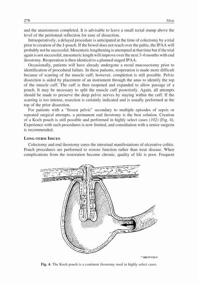
270 Moir
and the anastomosis completed. It is advisable to leave a small rectal stump above thelevel of the peritoneal reflection for ease of dissection.
Intraoperatively, a delayed procedure is anticipated at the time of colectomy by a trialprior to creation of the J-pouch. If the bowel does not reach over the pubis, the IPAA willprobably not be successful. Mesenteric lengthening is attempted at that time but if the trialagain is not successful, mesenteric length will improve over the next 3–6 months with endileostomy. Reoperation is then identical to a planned staged IPAA.
Occasionally, patients will have already undergone a rectal mucosectomy prior toidentification of procedural failure. In these patients, reoperation is made more difficultbecause of scarring of the muscle cuff; however, completion is still possible. Pelvicdissection is aided by placement of an instrument through the anus to identify the topof the muscle cuff. The cuff is then reopened and expanded to allow passage of apouch. It may be necessary to split the muscle cuff posteriorly. Again, all attemptsshould be made to preserve the deep pelvic nerves by staying within the cuff. If thescarring is too intense, resection is certainly indicated and is usually performed at thetop of the prior dissection.
For patients with a “frozen pelvis” secondary to multiple episodes of sepsis orrepeated surgical attempts, a permanent end ileostomy is the best solution. Creationof a Koch pouch is still possible and performed in highly select cases (102) (Fig. 4).Experience with such procedures is now limited, and consultation with a senior surgeonis recommended.
Long-term Issues
Colectomy and end ileostomy cures the intestinal manifestations of ulcerative colitis.Pouch procedures are performed to restore function rather than treat disease. Whencomplications from the restoration become chronic, quality of life is poor. Frequent
Fig. 4. The Koch pouch is a continent ileostomy used in highly select cases.

Chapter 15 / Inflammatory Bowel Disease 271
defecation and loss of nighttime control seriously impair the abilities of the adolescentor young adult to attend school or embark on a career (86). Multiple procedures andreturn appointments hamper patient mobility, such that consideration of college andfirst jobs become problematic. These disadvantages eventually outweigh the stigmataof an ileostomy. When surveyed, patients with permanent ileostomies still prefer arestorative pull-through but have quality of life indicators similar to those withoutileostomy (103). It is helpful to set goals for therapy and establish a time line forreevaluation. Although difficult to consider, ileostomy remains an excellent long-termsolution for patients with colitis after colectomy.
Pregnancy and vaginal delivery after a pull-through procedure are possible andrecommended for those women without serious pouch- or anastomotic-related compli-cations. An updated review shows no difference in birth weight, pregnancy, or deliverycomplications or vaginal delivery rates before or after IPAA procedures. There wasa higher rate of planned Cesarean sections and daytime stool frequency at long-term follow-up; however, vaginal delivery and successful outcomes can be routinelyanticipated after surgery for ulcerative colitis. Pouch function and complications ofpregnancy were unaffected by surgery (104).
Pouchitis
Clinical Presentation
Pouchitis is a nonspecific mucosal inflammation of the ileal reservoir. It occursin 26–47% of children; the incidence increases with length of follow-up (86,96,97).Children with previously excellent function suddenly experience a three- to fourfoldincrease in stool frequency. Nighttime incontinence recrudesces and most complainof urgency, mucousy discharge, and occasional bleeding. Extraintestinal symptoms,particularly arthralgias and erythema nodosum, may occur. The symptoms of urgency,frequency, and nighttime stooling are the primary indicators of disease that drivediagnostic investigation and treatment (84,105,106). The complication of pouchitis isnot life-threatening nor is it usually life-limiting for most children. The symptoms,however, represent the perception of return to former disease. Patients feel ill, areunable to go to school, and cannot socialize.
Diagnostic Evaluation
Pouchitis is a clinical diagnosis based on symptoms. Endoscopic and histologicfindings do not necessarily correlate with the severity of the clinical problem(106–108). The creation of an ileal reservoir is for restoration of bowel function. Anymalfunction of the pouch defeats that goal and is labeled pouchitis when no othersurgical complications are present.
The diagnostic evaluation has two purposes; to identify and treat surgical complica-tions discussed in the previous section, and when possible, discover causative factorsfor pouchitis. The latter include anal stricture, retained rectum, small volume pouch,medication-induced irritability, and Crohn’s disease (105). Such conditions require adifferent approach to therapy than idiopathic pouchitis (97,109,110). The surgical teamis in a unique position to help direct investigation. Their direct knowledge of the patientand the procedure helps prioritize the possible conditions listed previously.
In the simplest of cases, when the likelihood of complications is small, the diagnosisis confirmed by treatment response. Children with sudden onset of classic pouchitis
272 Moir
symptoms without risk factors usually respond to oral antibiotics. A prompt returnto baseline pouch function within 12–24 hours makes the diagnosis. This pragmaticapproach may include some cases of viral or food-induced illness, but when the goal israpid return to normal, stool function and quality of life over diagnosis is acceptable.
Children with risk factors or recurrent symptoms require work up. Examinationunder anesthesia and pouch endoscopy are mainstream investigations (105,109,110).A thorough examination excludes surgical complications and allows for anastomoticdilation. Endoscopy evaluates mucosal disease, notes any progression into the prepouchileum, and obtains biopsy for histologic confirmation and to exclude Crohn’s disease.Typically, diffuse mucosal aphthous ulceration and contact bleeding are identified.The changes revert to normal and the small bowel directly above the pouch. Furtherinvestigations depend on findings at examination under anesthesia and prior knowledgeof the procedure. A pouchogram, MRI, or CT scan may identify septic complicationsor mechanical issues of the pouch and its connections.
Medical Versus Operative Management
Pouch dysfunction secondary to complications of stricture, retained rectal cuffitis,Crohn’s disease, and local sepsis are treated surgically or with other specific medicalintervention (109). For the remaining patients with idiopathic pouchitis, there are nosurgical options. Treatment of pouchitis with the oral antibiotics metronidazole andCiprofloxacillin is the most effective strategy. Children promptly respond to initialdoses similar to those for treatment of serious infection. Gradual reduction of therapyis possible within days of treatment. Once recurrent, most families learn to manage thesymptoms with these antibiotics on a routine basis.
Chronic pouchitis is controlled with daily low-dose metronidazole orCiprofloxacillin, which may be alternated on a weekly or monthly schedule. Amoxi-cillin and vancomycin have also been successful. Many other therapies have been triedfor pouchitis; the most recent has been probiotics (111). Like multiple other agents, itseffectiveness remains in question.
Investigation of lifestyle and over-the-counter medication usage are important in thetreatment of pouchitis. Smoking, caffeine use, stress, and nonsteroidal antiinflammatorydrugs (NSAIDs) all contribute to pouch irritability.
Long-term Issues
Approximately 10% of ileoanal procedures fail secondary to severe mucosalpouchitis that is resistant to medical therapy (84). These patients require pouch excision.Of those with chronic pouchitis controlled by medication, most have serious questionsabout the long-term use of antibiotics. These effects have not been quantified. Dysplasiain chronically inflamed pouches has also been raised as a concern for children facingmany years of disease. A relatively short-term follow-up study did not identify abnor-malities (112). Finally, in children who are expected to keep their pouch for manydecades, aging and decline in function remains a concern. A recent study of 15-yearfollow-up has shown durable pouch function without decline in continence or qualityof life (113).

Chapter 15 / Inflammatory Bowel Disease 273
INDETERMINATE COLITIS—OPERATIVECONSIDERATION AND MANAGEMENT
Indeterminate colitis is a distinct clinical pathologic entity diagnosed in 5–15%of patients requiring surgery for inflammatory bowel disease. With follow-up, thepercentage of patients who continue with this diagnosis decreases, suggesting differen-tiation into ulcerative colitis or Crohn’s categories (114). The major surgical questionis the greater morbidity following an IPAA procedure. Adult patients with indeter-minate colitis do indeed have a higher rate of serious complications (20%) (115)than those with ulcerative colitis (3.5–10%) (84), but still less than patients withCrohn’s disease (35%) (85). The long-term success of IPAA for indeterminate colitis is73–85%, compared to 89% for ulcerative colitis. Such excellent results make a goodcase for IPAA in patients with indeterminate colitis (116).
Two precautionary statements are necessary; first, indeterminate colitis patientswith features favoring Crohn’s disease may benefit from delayed IPAA 6–12 monthsfollowing colectomy. Second, what can be said for children where no data are available?The answer will depend on age of the child, severity of disease, and the imperativeto operate. The younger the child, the more common the presentation of pancolitis inCrohn’s disease. Therefore, surgeons who are asked to see children under the age of8 years for colectomy should proceed cautiously to full reconstruction. In these youngpatients with severe disease, even though it is technically easier to perform a colectomywith J-pouch reconstruction, it is best to delay reconstructive surgery until pathologyconfirmation.
The real dilemma occurs in the younger child who is otherwise well but has steroid-resistant disease. These children are usually on low-dose oral corticosteroids and areexcellent candidates for immediate reconstruction. Many have decreased quality oflives and are unwilling to consider delayed reconstruction and long-term ileostomy.The options are to go ahead with a J-pouch, knowing a higher percentage may haveCrohn’s disease, versus a three-stage procedure. Data from adult studies suggest aprimary IPAA may proceed with excellent results; however, in patients with featuresfavoring Crohn’s disease, the better alternative may be to proceed to ileostomy, whichcan be converted an ileal J-pouch after eliminating the diagnostic possibility of Crohn’sdisease during follow-up.
REFERENCES
1. Mekhjian HS, Switz DM, Watts HD, et al. National cooperative Crohn’s disease study: factorsdetermining recurrence of Crohn’s disease after surgery. Gastroenterology 1979;77:907–913.
2. Farmer RG, Whelan G, Fazio VW. Long-term follow-up of patients with Crohn’s disease.Relationship between clinical pattern and prognosis. Gastroenterology 1985;88:1818–1825.
3. Michelassi F, Balestracci T, Chappell R, et al. Primary and recurrent Crohn’s disease. Experiencewith 1379 patients. Ann Surg 1991;214:230–240.
4. Penner RM, Maden KL, Fedorak RN. Postoperative Crohn’s disease. Inflam Bowel Dis 2005;11:765–777.
5. Griffiths AM, Wesson DE, Shandling B, et al. Factors influencing postoperative recurrence ofCrohn’s disease in childhood. Gut 1991;32:491–495.
6. Fazio VW, Marchetti F, Church M, et al. Effect of resection margins on the recurrence of Crohn’sdisease in the small bowel. A randomized controlled trial. Ann Surg 1996;224:563–571.
7. Greenway SE, Buckmire MA, Marroquin C, et al. Clinical subtypes of Crohn’s disease accordingto surgical outcome. J Gastrointest Surg 1999;3:145–151.
274 Moir
8. Chardavoyne R, Flint GW, Pollack S, et al. Factors affecting recurrence following resection forCrohn’s disease. Dis Colon Rectum 1986;29:495–502.
9. Baldaffano RN, Han PD, Jeshion WC, et al. Pediatric Crohn’s disease: risk factors for postoperativerecurrence. Am J Gastroenterol 2001;96:2169–2176.
10. Besnard M, Jaby O, Mougenot JF, et al. postoperative outcome of Crohn’s disease in 30 children.Gut 1998;43:634–638.
11. Aeberhard P, Berchtold W, Riedtmann HJ, et al. Surgical recurrence of perforating and non-perforating Crohn’s disease. A study of 101 surgically treated patients. Dis Colon Rectum1996;39:80–87.
12. Yamamoto T, Allen RN, Keighley MR. Perforating ileocecal Crohn’s disease does not carry a highrisk of recurrence but usually represents as perforating disease. Dis Colon Rectum 1999;42:519–524.
13. Griffiths AM, Otley AR, Hyams J, et al. A review of activity indices and end points for clinicaltrials in children with Crohn’s disease. Inflam Bowel Dis 2005;11:185–196.
14. McLeod RF. Surgery for inflammatory bowel diseases. Digest Dis 2003;21(2):168–179.15. Rutgeerts P, Geboes K, van Trappen G, et al. Predictability of the postoperative course of Crohn’s
disease. Gastroenterology 1990;99:956–963.16. Olaison G, Smedh K, Sjodahl R. Natural course of Crohn’s disease after ileocolic resection:
endoscopically visualized ileal ulcers preceding symptoms. Gut 1992;33:331–335.17. Horton KM, Fishman EK. The current status of multi-detector row CT and three-dimensional
imaging of the small bowel. Radiol Clin North Am 2003;41:199–212.18. Markowitz J, Markowitz JE, Bosvaros A, et al. Workshop report: prevention of postoperative
recurrence in Crohn’s disease. J Pediatr Gastroenterol Nutr 2005;41:145–151.19. Darbari A, Sena L, Argani P, et al. Gadolinium-enhanced magnetic resonance imaging: a useful
radiologic tool in diagnosing pediatric IBD. Inflamm Bowel Dis 2004;10:67–72.20. Legnani P, Cornbluth A. Video capsule endoscopy in inflammatory bowel disease 2005. Curr Opin
Gastroenterol 2005;21:438–442.21. Appleyard M, Fireman S, Glukhovsky A, et al. A randomized trial comparing wireless capsule
endoscopy with push enteroscopy for the detection of small bowel lesions. Gastroenterology2000;119:431–438.
22. Liangpunfakul S, Chadalawada V, Rex DK, et al. Wireless capsule endoscopy detects smallbowel ulcers in patients with normal results from state-of-the-art enteroclysis. Am J Gastroenterol2003;98:1295–1298.
23. Vermeire S, VanAssche G, Rutgeerts P. C-reactive protein as a marker for inflammatory boweldisease. Inflamm Bowel Dis 2004;10:661–665.
24. Vermeire S, VanAssche G, Rutgeerts P. Laboratory markers in IBD: useful, magic or unnecessarytoys? Gut 2006;55:426–431.
25. Tucker JG, Ambrose WL, Orangio GR, et al. Laparoscopically-assisted bowel surgery: analysis of114 cases. Surg Endosc 1995;9:297–300.
26. Liu CD, Rolandelli R, Ashley SW, et al. Laparoscopic surgery for inflammatory bowel disease. AmSurg 1995;61:1054–1056.
27. Bergamaschi R, Pessaux P, Arnaud JP. Comparison of conventional and laparoscopic ileocolicresection for Crohn’s disease. Dis Colon Rectum 2003;43:1129–1133.
28. Munoz-Juarez M, Yamamoto T, Wolff BG, et al. Wide lumen stapled anastomosis vs. conventionalend-to-end anastomosis in the treatment of Crohn’s disease. Dis Colon Rectum 2001;45:20–26.
29. Tersigni R, Alessandroni L, Barreca M, et al. Doe stapled functional end-to-end anastomosisaffect recurrence of Crohn’s disease after ileocolonic resection? Hepatogastroenterology 2003;50:1422–1425.
30. Roy P, Kumar D. Strictureplasty. Br J Surg 2004;91:1428–1437.31. Cuffari C, Dubinsky M, Darbari A, et al. Crohn’s jejunal ileitis: the pediatrician’s prospective on
diagnosis and management. Inflamm Bowel Dis 2005;11:696–704.32. Ozuner G, Fazio VW, Lavery IC, et al. Reoperative rates for Crohn’s disease following stricture-
plasty. Dis Colon Rectum 1996;39:1199–1203.33. Marchetti F, Fazio VW, Ozuner G. Adenocarcinoma arising from a strictureplasty site in Crohn’s
disease. Report of a case. Dis Colon Rectum 1996;39:1315–1321.34. Jaskowiak NT, Michelassi F. Adenocarcinoma at a strictureplasty site in Crohn’s disease. Report of
a case. Dis Colon Rectum 2001;44:284–287.35. Michelassi F, Hurst RD, Melis M, et al. Side-to-side isoperistaltic strictureplasty in extensive Crohn’s
disease: a prospective longitudinal study. Ann Surg 2000;232:401–408.

Chapter 15 / Inflammatory Bowel Disease 275
36. Stebbing JF, Jewell DP, Kettlewell MG, et al. Recurrence and reoperation after strictureplasty forobstructive Crohn’s’ disease: long-term results. Br J Surg 1995;82:1471–1474.
37. Spencer MT, Nelson H, Wolff BG, et al. Strictureplasty for obstructive Crohn’s disease: the Mayoexperience. Mayo Clin Proc 1994;69:33–36.
38. Keighley MR. Stapled strictureplasty for Crohn’s disease. Dis Colon Rectum 1991;34:945–947.39. Fazio VW, Auffes AH. Evolution of surgery for Crohn’s disease: a century of progress. Dis Colon
Rectum 1999;42:979–988.40. Tichansky D, Kagir B, Yoo E, et al. Strictureplasty for Crohn’s disease: meta-analysis. Dis Colon
Rectum 2000;43:911–919.41. Yamamoto T, Keighley MRB. Follow-up of more than 10 years after strictureplasty for jejunoileal
Crohn’s disease: long-term results of predictive factors for outcome. Colorect Dis 1999;1:101–106.42. Guillem JG, Roberts TL, Murray JJ, et al. Factors predictive of persistent or recurrent Crohn’s
disease in excluded rectal segments. Dis Colon Rectum 1992;35:768–772.43. Yamamoto T, Bain IM, Allen RN, et al. Persistent perineal sinus after proctocolectomy for Crohn’s
disease. Dis Colon Rectum 1999;42:96–101.44. Rius J, Nessim A, Noguerass JJ, et al. Gracilis transposition in complicated perianal fistula and
unhealed perineal wounds in Crohn’s disease. Eur J Surg 2000;166:218–222.45. Sher ME, Bauer JJ, Gorphine S, et al. Hartmann’s procedure for severe anorectal Crohn’s disease.
Dis Colon Rectum 1992;535:975–980.46. Isaacs KL, Lewis JD, Sandborn WJ, et al. State-of-the-art colon IBD therapy and clinical trials in
IBD. Inflamm Bowel Dis 2005;11:S3–S12.47. Sandborn WJ, Fazio VW, Feagan BG, et al. AGA technical review on perianal Crohn’s disease.
Gastroenterology 2003;125:1508–1530.48. Sandborn WJ, Feagan BG. Review article. Mild to moderate Crohn’s disease: defining the basis for
a new treatment algorithm. Aliment Pharmacol Ther 2003;18:263–277.49. Stephens M, Batres LA, Ng D, et al. Growth failure in a child with inflammatory bowel disease.
Semin Gastrointest Dis 2001;12:253–262.50. Ferguson A, Sedgewick DM. Juvenile onset inflammatory bowel disease: height and body mass
index in adult life. Br Med J 1994;308:1259–1263.51. Ballinger AB, Savage MO, Sanderson IR. Delayed puberty associated with inflammatory bowel
disease. Pediatr Res 2003;53:205–210.52. Bradley RJ, Rosen MP. Subfertility in gastrointestinal disease: unexplained is often undiagnosed.
Obstet Gynecol Survey 2004;59:108–117.53. Weber AM, Zigler Z, Belinson JL, et al. Gynecologic history of women with inflammatory bowel
disease. Obstet Gynecol 1995;86:843–847.54. Arkuran C, McComb P. Crohn’s disease and tubal infertility: the effect of adhesion formation. Clin
Exp Obstet Gynecol 2000;27:12–13.55. Ording OK, Juul S, Berndtsson I, et al. Ulcerative colitis: female fecundity before diagnosis, during
disease and after surgery compared with the population sample. Gastroenterology 2002;122:15–19.56. Johnson T, Richard C, Rabid A, et al. Female infertility after ileal pouch anal anastomosis for
ulcerative colitis. Dis Colon Rectum 2004;47:1119–1126.57. Olsen KO, Jul S, Bulow S, et al. Female fecundity before and after operation for familial adenomatous
polyposis. Br J Surg 2003;90:227–231.58. Andersson R, Lambe M, Bergstrom R. Fertility patterns after appendicectomy: historical cohort
study. Br Med J 1999;318:963–967.59. McClane SJ, Rombeau JL. Anorectal Crohn’s disease. Surg Clin North Am 2001;81:169–183.60. Williams DR, Coller JA, Corman ML, et al. Anal complications in Crohn’s disease. Dis Colon
Rectum 1981;24:22–24.61. Armuzzi A, Ahmad T, Ling KL, et al. Genotype-phenotype analysis of the Crohn’s disease suscep-
tibility hap low type on chromosome 5q31. Gut 2003;52:1133–1139.62. Newman B, Siminovitch KA. Recent advances in the genetics of inflammatory bowel disease. Curr
Opin Gastroenterol 2005;21:401–407.63. Bresent D, Rutgeerts P, Targan S, et al. Infliximab for the treatment of fistulas in patients with
Crohn’s disease. New Engl J Med 1999;340:1398–1405.64. Singh B, Mortensen NJ, Jewell DP. Perianal Crohn’s disease. Br J Surg 2004;91:801–814.65. Schwartz D, Loftus E, Tremaine W, et al. The natural history of fistulizing Crohn’s disease: a
population-based study. Gastroenterology 2002;122;875–880.
276 Moir
66. Levi C, Tremaine WJ. Management of internal fistulas in Crohn’s disease. Inflamm Bowel Dis2002;8:106–111.
67. Makowiec F, Jehle EC, Becker HD, et al. Perianal abscess in Crohn’s disease. Dis Colon Rectum1997;40:443–450.
68. Beckingham IJ, Spencer JA, Ward J, et al. Prospective evaluation of dynamic contrast-enhancedmagnetic resonance imaging in the evaluation of fistula-in-ano. Br J Surg 1996;83:1396–1398.
69. Orsoni P, Barthet M, Portier F, et al. Prospective comparison of endosonography, magnetic resonanceimaging and surgical findings in anorectal fistula and abscess complicating Crohn’s disease. Br JSurg 1999;86:360–364.
70. Schwartz DA, Pemberton JH, Sandborn WJ. Diagnosis and treatment of perianal fistulas and Crohn’sdisease. Ann Intern Med 2001;135:906–918.
71. Nordgren F, Fasth S, Hulten L. Anal fistulas in Crohn’s disease: incidence and outcome of surgicaltreatment. Int J Colorectal Dis 1992;7:214–218.
72. Sands BE, Anderson SH, Bernstein CN, et al. Infliximab maintenance therapy for fistulizing Crohn’sdisease. N Engl J Med 2004;350:876–885.
73. Ricart E, Panaccione R, Loftus E, et al. Infliximab for Crohn’s disease and clinical practice at theMayo Clinic: the first 100 patients. Am J Gastroenterol 2001;96:722–729.
74. Topstad DR, Panaccione R, Heine JA, et al. Combined seton placement, Infliximab infusion andmaintenance immunosuppressive improve healing rate in fistulizing anorectal Crohn’s disease: asingle center experience. Dis Colon Rectum 2003;46:577–583.
75. Sonoda T, Ho T, Piedmont MR, et al. Outcomes of primary repair of anorectal and rectovaginalfistulas using the endorectal advancement flap. Dis Colon Rectum 2002;45:1622–1628.
76. Mizrhi N, Wexner SD, Zmora O, et al. Endorectal advancement flap: are there predictors of failure?Dis Colon Rectum 2002;45:1616–1621.
77. Hull TL, Fazio VW. Surgical approaches to low anovaginal fistula in Crohn’s disease. Am J Surg1997;173:95–98.
78. Robertson WG, Mangione JS. Cutaneous advancement flap closure: alternative measure for treatmentof complicated anal fistulas. Dis Colon Rectum 1998;41:884–886.
79. Nelson RL, Cintron J, Abcarian H. Dermal island flap anoplasty for transsphincteric fistula-in-ano:assessment of treatment failures. Dis Colon Rectum 2000;43:681–684.
80. Lindsey I, Smilgin MM, Cunningham C, et al. A randomized controlled trial of fibrin glue vs.conventional treatment for anal fistula. Dis Colon Rectum 2002;45:1608–1615.
81. Arroyo A, Perez F, Serrano P, et al. Surgical vs. clinical (botulinum toxin) sphincterotomy forchronic anal fissure: long-term results of a prospective randomized clinical and manometric study.Am J Surg 2005;189:429–434.
82. Fazio VW, Tekkis PP, Remzi F, et al. Quantification of risk for pouch failure after ileal pouch analanastomosis surgery. Ann Surg 2003;238:605–614.
83. Meagher AP, Farouk R, Dozois RR, et al. Ileal J-pouch anal anastomosis for chronic ulcerativecolitis: complications and long-term outcome in 1310 patients. Br J Surg 1998;85:800–803.
84. Prudhomme M, Dehni N, Dozois RR. Causes and outcomes of pouch excision after restorativeproctocolectomy. Br J Surg 2006;93:82–86.
85. Tulchinsky H, Cohen CRG, Nicholls RJ. Salvage surgery after restorative proctocolectomy. Br JSurg 2003;90:909–921.
86. Stavlo PL, Libsch KD, Rodeberg DA, et al. Pediatric ileal pouch-anal anastomosis: functionaloutcomes and quality of life. J Pediatr Surg 2003;38:935–939.
87. Hueting WE, Buskens E, Vandertweel I, et al. Results and complications after ileal pouch analanastomosis: a meta-analysis of 43 observational studies comprising 9317 patients. Dig Surg2005;22:69–79.
88. Fazio VW, Ziv Y, Church JM, et al. Ileal pouch anal anastomosis complications and function in1005 patients. Ann Surg 1995;222:120–127.
89. Soravia C, Berk T, McCleod RS, et al. Desmoid disease in patients with familial adenomatouspolyposis. Dis Colon Rectum 2002;43:363–369.
90. Sagar PM, Moslen G, Dozois RR. Management of desmoid tumors in patients after ileal pouch analanastomosis for familial polyposis. Dis Colon Rectum 1998;41:1350–1356.
91. Poritz LS, Blackstein N, Berk T, et al. Extended follow-up of patients treated with chemotherapyfor intra-abdominal desmoid tumors. Dis Colon Rectum 2001;44:1268–1273.
92. Tekkis PP, Heriot AG, Smith JJ. Long-term results of abdominal salvage surgery followingrestorative proctocolectomy. Br J Surg 2006;93:231–237.
Chapter 15 / Inflammatory Bowel Disease 277
93. Farouk R, Dozois RR, Pemberton JH, et al. Incidence and subsequent impact of pelvic abscess afterileal pouch anal anastomosis for chronic ulcerative colitis. Dis Colon Rectum 1998;41:1239–1243.
94. Scott NA, Dozois RR, Beart RW, et al. Postoperative intra-abdominal and pelvic sepsis complicatingileal pouch anal anastomosis. Int J Colorectal Dis 1988;3:149–152.
95. Heuschen UA, Allemeyer EH, Hinz U, et al. Outcome after septic complications in J-pouch proce-dures. Br J Surg 2002;89:194–200.
96. Fonkalsrud EW. Surgery for pediatric ulcerative colitis. Curr Opin Pediatr 1995;7:323–327.97. Alexander F, Sarigol F, Difiore J, et al. Fate of the pouch in 151 pediatric patients after ileal pouch
anal anastomosis. J Pediatr Surg 2003;38:78–82.98. Richard G, Nicholls RJ. Management and outcome of pouch vaginal fistulas following restorative
proctocolectomy. Dis Colon Rectum 2005;48:451–458.99. Burke D, Laarhoven CJ, Herbst F. Transvaginal repair of pouch vaginal fistula. Br J Surg
2001;88:241–245.100. Coran AG, Rizofecchi M. Surgery for ulcerative colitis in children. Pediatr Surg Int 2004;20:
565–566.101. Fonkalsrud EW, Phillips JD. Reconstruction of malfunctioning ileoanal pouch procedures as an
alternative to permanent ileostomy. Am J Surg 1990;160:245–251.102. O’Bischere A, Wilkinson K, Rumbles S, et al. Functional outcome after restorative proctocolectomy
for ulcerative colitis decreases an otherwise enhanced quality of life. Br J Surg 2000;87:802–807.103. Camilleri-Brennan J, Monroe A, Steel RJ. Does an ileoanal pouch offer a better quality of life than
a permanent ileostomy for patients with ulcerative colitis? J Gastrointest Surg 2003;7:814–819.104. Hahnloser D, Pemberton JH, Wolff BG, et al. Pregnancy and delivery before and after IPAA
for inflammatory bowel disease: immediate and long-term consequences and outcome. Dis ColonRectum 2004;47:1127–1135.
105. Shen B, Fazio VW, Remzi FH. Risk factors for diseases of ileal pouch anal anastomosis afterrestorative proctocolectomy for ulcerative colitis. Clin Gastroenterol Hepatol 2006;4:81–89.
106. Sandborn WJ, Pardi DS. Clinical management of pouchitis. Gastroenterology 2004;127:1809–1814.107. Heuschen UA, Autschbach F, Allemeyer EH, et al. Long-term follow-up after ileoanal pouch
procedure: algorithm for diagnosis, classification and management of pouchitis. Dis Colon Rectum2001;44:487–699.
108. Hurst RD, Molinari M, Chung TP, et al. Prospective study: the incidence, timing and treatmentof pouchitis in 104 consecutive patients after restorative proctocolectomy. Arch Surg 1996;131:497–500.
109. Shen B, Fazio VW, Remzi FH. Clinical approaches to disease of ileal pouch anal anastomosis. AmJ Gastroenterol 2005;100:2796–2807.
110. Bambrick ML, Bast J, Lashner B. Comprehensive evaluation of inflammatory and non-inflammatorysequelae of ileal pouch anal anastomosis. Am J Gastroenterol 2005;100:93–101.
111. Gionchetti P, Rizzello F, Venturi A, et al. Oral bacteriotherapy as maintenance treatment in patientswith chronic pouchitis: a double-blind placebo control trial. Gastroenterology 2000;119:305–309.
112. Sarigol S, Wyllie R, Gramlich T, et al. Incidence of dysplasia in pelvic pouches in pediatric patientsafter ileal pouch anal anastomosis for ulcerative colitis. J Pediatr Gastroenterol Nutr 1999;28:429–434.
113. Hahnloser D, Pemberton JH, Wolff BG, et al. The effect of aging on function and quality of life inileal pouch patients: a single cohort experience of 409 patients with chronic ulcerative colitis. AnnSurg 2004;240:615–623.
114. Guindi M, Riddell RH. Indeterminate colitis. J Clin Pathol 2004;57:1233–1244.115. Yu CS, Pemberton JH, Larson D. Ileal pouch anal anastomosis in patients with indeterminate colitis:
long-term results. Dis Colon Rectum 2000;43:1487–1496.116. Wolff BG. Is ileoanal the proper operation for indeterminate colitis: the case for. Inflamm Bowel
Dis 2002;8:362–365.

16 Reoperation for Stoma Complications
Benedict C. Nwomeh, MD
CONTENTS
Overview of Complications
Specific Complications
References
OVERVIEW OF COMPLICATIONS
Major complications could occur in up to 75% of children following colostomy orileostomy, with an overall revision rate of approximately 15% (Table 1). Because ofthe higher rate of complications associated with loop ostomies, most pediatric surgeonsprefer divided stomas. A good ostomy is best obtained by careful preoperative planning,meticulous surgical technique, and detailed attention to skin care. Skin complica-tions such as dermatitis, granuloma, and ulceration are the most frequent problemswith ostomies. Stomas made in the small intestine (enterostomas) are associated withmore complications than colostomies. In addition, transverse colostomies cause moreproblems than sigmoid colostomies. The expertise of an enterostomal therapist shouldbe sought in the care of ostomies, and in the prevention and management of skinproblems. With temporary stomas, the occurrence of complications should promptconsideration for closure of the ostomy, rather than revision. The following discussionis limited to those complications that often require reoperative surgery. Other compli-cations usually requiring the care of a good enterostomal therapist are listed in Table 2.
SPECIFIC COMPLICATIONS
Ischemia/NecrosisSignificant ischemia or frank necrosis is apparent early in the postoperative period.
The cause is either arterial insufficiency from excessive stripping of the mesentery, orvenous congestion caused by tension on the bowel, or a tight fascial opening. Obesepatients are particularly at risk of developing stomal ischemia. Some degree of edemaand venous congestion normally occurs with new ostomies, especially if previouslydistended bowel is used, but usually resolves within 48 hours. If mucosal necrosis islimited to the portion superficial to the fascia, an expectant approach may be employedbut the stoma should be monitored closely for progressive necrosis or subsequentdevelopment of stenosis, stricture, or retraction.
From: Reoperative Pediatric SurgeryEdited by: S. Teich and D. A. Caniano © Humana Press, Totowa, NJ
279

280 Nwomeh
Table 1Stoma Complications in Children
Complication Incidence (%)
Ischemia/Necrosis <1Stenosis/stricture 3–6Retraction 2–4Prolapse 12–24Parastomal hernia 1Skin excoriation 20–30Bleeding 1–10Obstruction 1–6
Operative Technique
Sufficient mobilization of bowel and mesentery will be required to allow resectionof the ischemic portion and creation of a new stoma with well-perfused bowel withouttension. It is usually necessary to reopen the main incision to facilitate this revision.Mesenteric stripping of the stoma should be limited to the 1–2 cm necessary to evertthe bowel for maturation.
Stenosis/StrictureStenosis or stricture of the stoma may occur at the skin or fascial level and is
clinically apparent as reduced stoma output or frank bowel obstruction. Potential causesinclude a tight skin or fascial opening, ischemia at the mucocutaneous junction, andprolonged serositis with subsequent fibrosis when the stoma has not been matured.Gentle probing with a soft catheter may help to localize the narrowing. In mild cases,gentle dilatation using serial rubber catheters or the Hegar dilator may resolve theissue, but this procedure caries the risk of bowel perforation. Recurrent Crohn’s diseasecausing a stricture is best detected using contrast studies and endoscopy.
Table 2Management of Other Complications of Stomas
Complication Cause Treatment
Wound infection Intraabdominal infectionStoma located in main incision
AntibioticsDrainage
Skin excoriation Poorly fitting applianceContact dermatitisRetraction, prolapse
Enterostomal careStoma revisionStoma relocation
Bleeding TraumaMucosal lesion (e.g., polyps)
Enterostomal careEndoscopy through stoma
Fistula Full thickness placement of suturePressure necrosis from applianceRecurrent Crohn’s disease
Enterostomal careStoma revision

Chapter 16 / Stoma 281
Operative Technique
For skin-level stenosis, the scar ring should be removed by circumferential excision.The fascial opening can be enlarged if necessary. The bowel is then mobilized suffi-ciently to allow revision of the stoma at the same site. For longer or more proximalstrictures, and in recurrent Crohn’s disease, a laparotomy may be required for moreextensive mobilization of bowel and resection of the affected segment.
RetractionIt is important to recognize the cause of stomal retraction. The most common problem
is undue tension on the stoma caused by inadequate mobilization of the proximal boweland its mesentery. Anchoring the bowel to the fascia with sutures does not mitigate thetension in the bowel and cannot be relied upon to prevent retraction of the soma. Secondly,retraction may follow stomal necrosis. Thirdly, retraction may be caused by prolongedserositis with subsequent shortening of bowel when the stoma has not been matured.Finally, recurrent Crohn’s disease may manifest as stomal retraction and should be investi-gatedwithcontrast studiesandendoscopy.Theretractedstomacauses leakageof intestinalcontents under the stoma pouch, creating difficulty with maintaining the appliance.
Operative Technique
Revision of the stoma is usually required. If the retracted stoma is sufficientlymobile to allow the bowel to be everted, the bowel walls can be fixed together byinserting several interrupted absorbable sutures with full thickness bites. Alternatively,several firings of a noncutting linear stapler can be used to fix the bowel (Fig. 1).
Fig. 1. The stapling technique for fixation of a retracted stoma. The stoma is retracted to its fullextent by placing three pairs of Babcock’s forceps (not shown). A noncutting linear stapler (suchas the GIA stapler without the blade) is placed with the blade toward the mucocutaneous junctionbetween the Babcock forceps. Care is taken to avoid the mesentery before firing the stapler. Threeparallel rows of staples fix the two walls of the ileum together.

282 Nwomeh
Adhesion between the serosal surfaces of the everted stoma will usually have occurredbefore the sutures are absorbed or the staples separate. More commonly, the retractedstoma is fixed in position and the goal is to mobilize sufficient length of boweland mesentery so that maturation of the stoma can be accomplished without tension.Additional mobilization of bowel often requires a laparotomy. Limited bowel resectionis performed to remove ischemic or stenotic segment, or area of recurrent Crohn’sdisease. The new stoma should be matured at the same site except when retraction hasresulted in significant skin excoriation or abdominal wall sepsis; in these cases, a newostomy site should be established.
Prolapse/IntussusceptionA prolapsed ostomy alarms and frightens both child and parents. Prolapse usually
occurs when a skin opening is made to accommodate dilated bowel, which uponshrinking leaves a loose stoma. Inadequate fixation of the mesentery to the parietalperitoneum may also lead to prolapse. Prolapse may manifest as protrusion of theterminal portion of the bowel, or as intussusception of the intraabdominal portionthrough the opening. Loop stomas are more likely to prolapse than end stomas, and thedistal segment of the loop ostomy is most frequently affected. A coexistent parastomalhernia may predispose to prolapse. Prolapsed stomas interfere with the applicationof the stoma appliance, causing leakage. Trauma to the exposed bowel, caused bydesiccation or by an ill-fitting appliance, may cause mucosal ulceration and bleeding.Spontaneous or manual reduction is usually possible in the early stage, but surgicalintervention is eventually required for persistent prolapse, intestinal obstruction, orstrangulated bowel.
Operative Technique
For temporary relief, a simple purse-string suture with a nonabsorbable monofilamentmaterial may be used, similar to the Thiersch technique for rectal prolapse (Fig. 2).This is ideally suited for prolapse of a temporary ostomy because the fixed ring wouldproduce stenosis as the child grows. A simple technique described by Gauderer usesa “U” stitch from the lumen of the reduced bowel through the abdominal wall witha double-armed needle (Fig. 3). Before tying the suture ends, each needle is passedthrough a pledget, thus creating an internal and external bolster that attaches the bowelto the body wall and prevents the suture from cutting through. Amputation of theprolapse is a tempting option that alleviates the problem, but early recurrence is likely.A more definitive revision including resection of prolapsed bowel and fixation of themesentery may bring permanent relief to troublesome ostomy prolapse. This procedureusually requires reopening of the main abdominal incision. In the case of a prolapsedloop ostomy, the loop may be divided, with the closed distal end returned into theabdomen. This converts the loop to an end ostomy, which is less likely to prolapse.Repair of a coexisting parastomal hernia should be considered in order to reduce therisk of further recurrence. When appropriate, closure of the ostomy is the best option.
Parastomal HerniaParastomal hernia appears to occur less frequently in children than adult patients,
with incidence less than 1%. The most likely cause is the creation of a fascial aperturerelatively larger than the bowel used for the ostomy. Although parastomal hernia

Chapter 16 / Stoma 283
Fig. 2. A purse-string suture technique for correcting stoma prolapse. (A) A 1-cm skin incision ismade at the medial angle of the stoma down to the subcutaneous tissue. (B) A finger is insertedinto the stoma as a guide and a 1-0 monofilament nonabsorbable suture with a round, cutting needleis passed around the colostomy staying within the subcutaneous layer. The needle is placed as faras can comfortably go, usually approximately one-quarter of the circumference, then brought outthrough the skin. (C) The needle is passed again through the same skin exit site toward the lateralcorner. (D) One or two more passes of the needle are made as it is marched circumferentially aroundthe stoma, until it is brought out through the medial incision. With a finger remaining in the lumen,the suture is tied, causing puckering of the stoma without completely occluding the lumen.
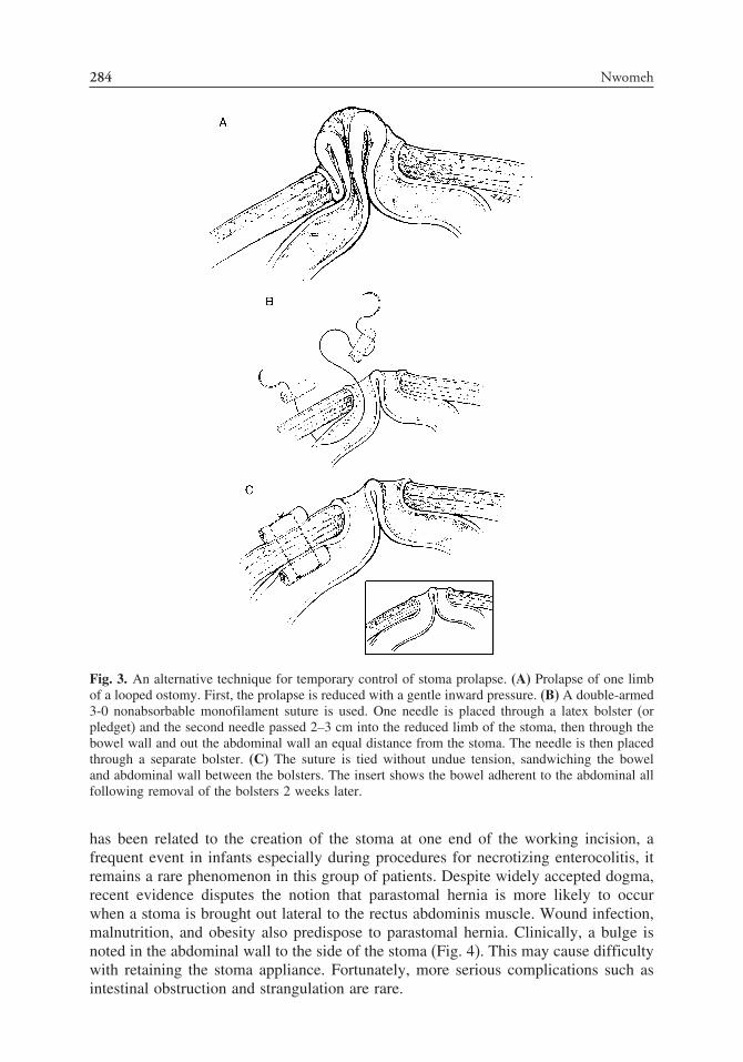
284 Nwomeh
Fig. 3. An alternative technique for temporary control of stoma prolapse. (A) Prolapse of one limbof a looped ostomy. First, the prolapse is reduced with a gentle inward pressure. (B) A double-armed3-0 nonabsorbable monofilament suture is used. One needle is placed through a latex bolster (orpledget) and the second needle passed 2–3 cm into the reduced limb of the stoma, then through thebowel wall and out the abdominal wall an equal distance from the stoma. The needle is then placedthrough a separate bolster. (C) The suture is tied without undue tension, sandwiching the boweland abdominal wall between the bolsters. The insert shows the bowel adherent to the abdominal allfollowing removal of the bolsters 2 weeks later.
has been related to the creation of the stoma at one end of the working incision, afrequent event in infants especially during procedures for necrotizing enterocolitis, itremains a rare phenomenon in this group of patients. Despite widely accepted dogma,recent evidence disputes the notion that parastomal hernia is more likely to occurwhen a stoma is brought out lateral to the rectus abdominis muscle. Wound infection,malnutrition, and obesity also predispose to parastomal hernia. Clinically, a bulge isnoted in the abdominal wall to the side of the stoma (Fig. 4). This may cause difficultywith retaining the stoma appliance. Fortunately, more serious complications such asintestinal obstruction and strangulation are rare.
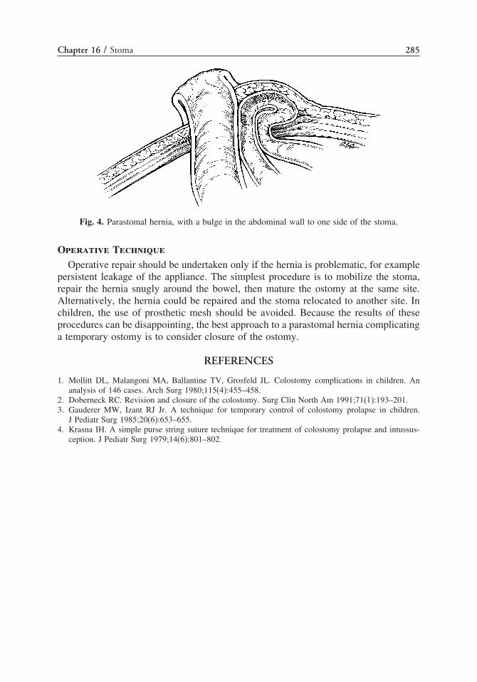
Chapter 16 / Stoma 285
Fig. 4. Parastomal hernia, with a bulge in the abdominal wall to one side of the stoma.
Operative Technique
Operative repair should be undertaken only if the hernia is problematic, for examplepersistent leakage of the appliance. The simplest procedure is to mobilize the stoma,repair the hernia snugly around the bowel, then mature the ostomy at the same site.Alternatively, the hernia could be repaired and the stoma relocated to another site. Inchildren, the use of prosthetic mesh should be avoided. Because the results of theseprocedures can be disappointing, the best approach to a parastomal hernia complicatinga temporary ostomy is to consider closure of the ostomy.
REFERENCES
1. Mollitt DL, Malangoni MA, Ballantine TV, Grosfeld JL. Colostomy complications in children. Ananalysis of 146 cases. Arch Surg 1980;115(4):455–458.
2. Doberneck RC. Revision and closure of the colostomy. Surg Clin North Am 1991;71(1):193–201.3. Gauderer MW, Izant RJ Jr. A technique for temporary control of colostomy prolapse in children.
J Pediatr Surg 1985;20(6):653–655.4. Krasna IH. A simple purse string suture technique for treatment of colostomy prolapse and intussus-
ception. J Pediatr Surg 1979;14(6):801–802.

17 Reoperative Surgeryfor Hirschsprung Disease
Mohamed I. El-sawaf, MD, ArnoldG. Coran, MD, and DanielH. Teitelbaum, MD
CONTENTS
Introduction
Manifestations of Long-Term Complications
After a Pull-Through
When to Start Work-Up for Complications
Work-Up of the Complicated Postoperative Patient
Reoperative Surgery for HD
Conclusions
References
INTRODUCTION
Hirschsprung disease (HD) is the most common cause of neonatal intestinalobstruction. Since Swenson in 1948 described his successful approach, several modifi-cations and new operations had been described in an effort to improve the long-termoutcome and quality of life (QOL) for patients with HD. By far the most commonlyused approaches nowadays are the transanal approach whether laparoscopically assistedor not, modified Soave’s pull-through, and Duhamel’s retrorectal pull-through.
Each of these procedures has its own advantages and drawbacks. All of themultimately have good results concerning the long-term outcome. But at this moment,none of these procedures are perfect and many children suffer from long-term compli-cations. Interestingly, these complications may occur after what may be called a“successful pull-through” without any obvious cause or explanation.
MANIFESTATIONS OF LONG-TERM COMPLICATIONSAFTER A PULL-THROUGH
The most serious long-term complications that strongly impact the QOL of patientswith HD after the definitive treatment falls into three groups; soiling/incontinence,
From: Reoperative Pediatric SurgeryEdited by: S. Teich and D. A. Caniano © Humana Press, Totowa, NJ
287

288 El-sawaf et al.
persistent problems with the passage of stool (e.g., constipation), and recurrent entero-colitis. Not uncommonly, an individual child may have a combination of problems.
Soiling/IncontinenceSoiling/incontinence have been reported in variable percentages and degrees. Rates
range from none in several series to as high as 74% in one series (1–3). This problemmay be caused by scaring or iatrogenic damage of the anal sphincters. Soiling may alsobe caused by a loss of rectal sensation (which is critical for differentiating betweengas, liquid, and solid stool) owing to destruction of the sensory rectal mucosa from avery low anastomosis. Finally, the most common cause of soiling after a pull-throughis chronic constipation with overflow of stool. Incontinence should theoretically bepreventable by using adequate surgical techniques; however, the problem persists. Thework up for many of these problems is discussed later in this section. The managementof incontinence ranges from a simple bowl management program to the placement ofa permanent stoma. The use of an antigrade colonic enema may be quite useful inthis group of patients. More recently, the use of an artificial sphincter or sphincterreconstruction has been employed. These therapeutic options are not discussed in thischapter.
Persistent Stooling ProblemsPersistent stooling problems are a very annoying group of complications. They
include recurrent distension, bloating, and constipation. Constipation is probably themost common complaint after a pull-through procedure. Typically, symptoms do notdevelop until a few weeks to many months after the pull-through (4). However, inthe past decade, a number of investigators have recognized that obstructive symptomsare a significant issue, occurring in 11–42% of children after what appears to be atechnically excellent operation (5–9). Rescorla et al. found that 88% of patients haddifficulty stooling with a follow-up less than 5 years. Although some patients withincontinence were in this group, symptoms improved with greater follow up, with100% having satisfactory stooling with a 15-year or more follow-up (10). This suggeststhat many have persistent problems with their internal anal sphincter after removal ofall aganglionic tissue, and suggests that we are still missing a complete understandingof the pathophysiology of HD.
Stooling problems may arise from a number of etiologies, and include stricture orretained spur after Duhamel’s procedure, retained or acquired aganglionosis, hypogan-glionosis or performance of the pull-through at the level of the transition zone,associated motility disorders either focal or general, internal anal sphincter achalasia(IASA), and finally what is called “functional megacolon,” or a chronically dilateddistal rectum, most likely caused by many years of persistent difficulty in passingbowel movements. In the remaining group of children, there is no identifiable causefor their symptoms, and some of these children fail to respond to surgical or medicalrelaxation of the sphincter. Most of these children suffer from stool-holding behaviorand are best treated by using a bowel management regimen consisting of laxatives,enemas, and behavior modification including support for the child and family (11,12).The approach to address the cause varies and one should be aware of all of thesevarious possibilities to be able to successfully manage a complicated HD case.

Chapter 17 / Hirschsprung Disease 289
HD-Associated Enterocolitis (HAEC)HAEC remains the most common cause of death in HD. It can occur at any time,
including both before as well as after the pull-through procedure. The incidence ofenterocolitis in HD varies widely among published series depending on the definitionand severity of the enterocolitis. A large multicenter study series showed an incidenceranging from 22–42% (13). The incidence of preoperative HAEC occurs in approxi-mately 15–20% (14–16). Postoperative enterocolitis may affect one-third of patients,with a mortality rate varying from 0–30% (17). Many patients have more than oneepisode of HAEC (18). Relapses of enterocolitis can occur despite a defunctioningcolostomy or a successful definitive procedure (14). Therefore, it is of utmost impor-tance that the treatment of enterocolitis is started early and pursued vigorously toprevent the condition from becoming chronic (19). It is also interesting to know thatthe frequency of bouts of enterocolitis tends to decrease with time (20).
The exact etiology and pathogenesis is still obscure (21–23). In a multicenter reviewof primary endorectal pull-throughs, logistic regression analysis of risk factors for thedevelopment of enterocolitis showed that stricture development was a significant riskfactor (13). Other risk factors include increased length of the aganglionic segment,female sex, Trisomy 21, and the presence of other associated congenital anomalies(24). Currently, no clinical factor or test is available to predict the development ofthis disorder. Additionally, there is no single strategy for prophylaxis from this majorcomplication.
WHEN TO START WORK-UP FOR COMPLICATIONS
In general, complicated HD cases should be assessed thoroughly by an experiencedpersonnel, and an algorithm should be tailored for each case according to the type andnature of the complication.
A significant number of children who undergo a successful pull-through developpersistent problems or complications. But frequently they outgrow most of thesecomplications by the age of 5 years for unexplained reasons without the need forfurther operation.
Unfortunately, this is not always the case; some children may develop treatmentresistant symptoms and require further intervention. For example, a child with persistentsevere constipation leading to progressive abdominal distension may develop failureto thrive in spite of several trials of laxatives, enemas, and dietary control. A secondexample would be an infant with recurrent severe bouts of enterocolitis requiringmultiple admissions irrespective of chronic and aggressive medications and irrigations.Another example would be a patient found to have a stricture or retained spur afterDuhamel’s procedure during rectal examination. All of these cases deserve further andthrough evaluation and assessment.
WORK-UP OF THE COMPLICATED POSTOPERATIVE PATIENT
Thorough History-Taking and Meticulous Clinical ExaminationA detailed history should include the neonatal period, associated anomalies, type of
presentation, time and method of diagnosis, preoperative course, type of procedure,detailed postoperative course stating frequency of stooling and number and severity ofenterocolitic episodes, and methods of HAEC management.
290 El-sawaf et al.
General examination should be thoroughly done to assess developmental milestones,growth curve, and to allow recognition of associated syndromes and anomaliesthat may impact the outcome. Abdominal inspection and palpation is necessary toget an appreciation about the magnitude of distension, and to feel any impactedstool.
Rectal examination is essential and can yield very useful information. Patency ofthe anal canal is readily assessed with palpation of any stricture or narrowing of theanastmosis line, and occasionally retained spur after a Duhamel procedure (althoughsuch a spur is often beyond digital palpation). The anastomotic level is palpated, givingan idea about the previous procedure if it is technically done in a correct manner ornot. The tone of the anal sphincter is assessed as normal, increased (suggestive ofIASA), or low (pointing to incontinence). The rectal vault is also evaluated as eitherfull of stool (supporting encopresis) or empty. A finding of encopresis by history oran abnormally large rectal vault may indicate a stool-holding behavior, and the childmay benefit from behavioral/training strategies. A gush of gas and liquid stools withremoval of the examining finger typically indicates persistent obstructive symptoms orongoing HAEC.
Contrast EnemaA radiographic contrast enema can be very useful. It usually can differentiate between
a number of problems, including: anastomotic stenosis or stricture, a twist of the pull-through segment, as well as a chronically dilated rectosigmoid colon. Occasionally, theenema can detect such irregularities of the mucosal lining, suggesting chronic inflam-mation after repeated episodes of HAEC. Such information is useful in understandingthe etiology of symptoms as well as deciding the optimum operative approach thateffectively addresses these problems.
Anorectal ManometryUse of manometry is controversial in the postoperative evaluation of patients with
HD. Anorectal manometry does have several uses. Manometry can be very helpful inthe diagnostic work-up of IASA, which may coexist with HD (25). Manometry mayalso help identify patients with marked increases or decreases in the basal resting analtone. One problem is that most children, even after a successful pull-through, do notregain the rectoanal inhibitory reflex. This is well illustrated in a review of childrenwith stooling difficulties after pull-throughs. Moore et al. reported their experience withthe use of manometry. In their series of 16 patients with stooling problems, manometricassessment was not significantly different from 28 patients who had normal stooling(26). They noted that of these 44 patients, a normal inhibitory relaxation was seen inonly six children, of which three had IASA and three had normal stooling function.Additionally, only two of eight children with a high resting anorectal tone had stoolingproblems. Another problem is the variability in the normal values for basal restingsphincter tone in published series. This may also be compounded by the numerousartifacts associated with movements or crying. To improve cooperation, it is oftenhelpful to perform manometry with mild sedation or anesthesia in these young infants.The rectoanal inhibitory reflex can be elicited even when manometry is performedusing general anesthesia (27). One problem in which anorectal manometry will shedconsiderable information is in the patient with constant soiling. Clearly, there are two
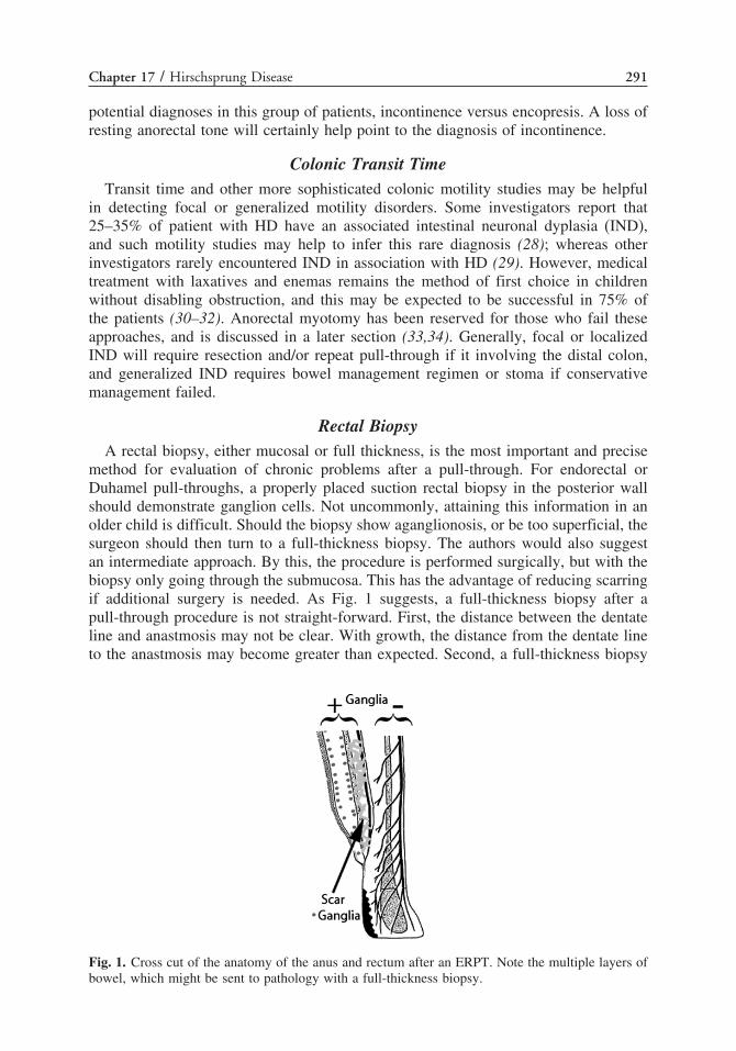
Chapter 17 / Hirschsprung Disease 291
potential diagnoses in this group of patients, incontinence versus encopresis. A loss ofresting anorectal tone will certainly help point to the diagnosis of incontinence.
Colonic Transit TimeTransit time and other more sophisticated colonic motility studies may be helpful
in detecting focal or generalized motility disorders. Some investigators report that25–35% of patient with HD have an associated intestinal neuronal dyplasia (IND),and such motility studies may help to infer this rare diagnosis (28); whereas otherinvestigators rarely encountered IND in association with HD (29). However, medicaltreatment with laxatives and enemas remains the method of first choice in childrenwithout disabling obstruction, and this may be expected to be successful in 75% ofthe patients (30–32). Anorectal myotomy has been reserved for those who fail theseapproaches, and is discussed in a later section (33,34). Generally, focal or localizedIND will require resection and/or repeat pull-through if it involving the distal colon,and generalized IND requires bowel management regimen or stoma if conservativemanagement failed.
Rectal BiopsyA rectal biopsy, either mucosal or full thickness, is the most important and precise
method for evaluation of chronic problems after a pull-through. For endorectal orDuhamel pull-throughs, a properly placed suction rectal biopsy in the posterior wallshould demonstrate ganglion cells. Not uncommonly, attaining this information in anolder child is difficult. Should the biopsy show aganglionosis, or be too superficial, thesurgeon should then turn to a full-thickness biopsy. The authors would also suggestan intermediate approach. By this, the procedure is performed surgically, but with thebiopsy only going through the submucosa. This has the advantage of reducing scarringif additional surgery is needed. As Fig. 1 suggests, a full-thickness biopsy after apull-through procedure is not straight-forward. First, the distance between the dentateline and anastmosis may not be clear. With growth, the distance from the dentate lineto the anastmosis may become greater than expected. Second, a full-thickness biopsy
Fig. 1. Cross cut of the anatomy of the anus and rectum after an ERPT. Note the multiple layers ofbowel, which might be sent to pathology with a full-thickness biopsy.
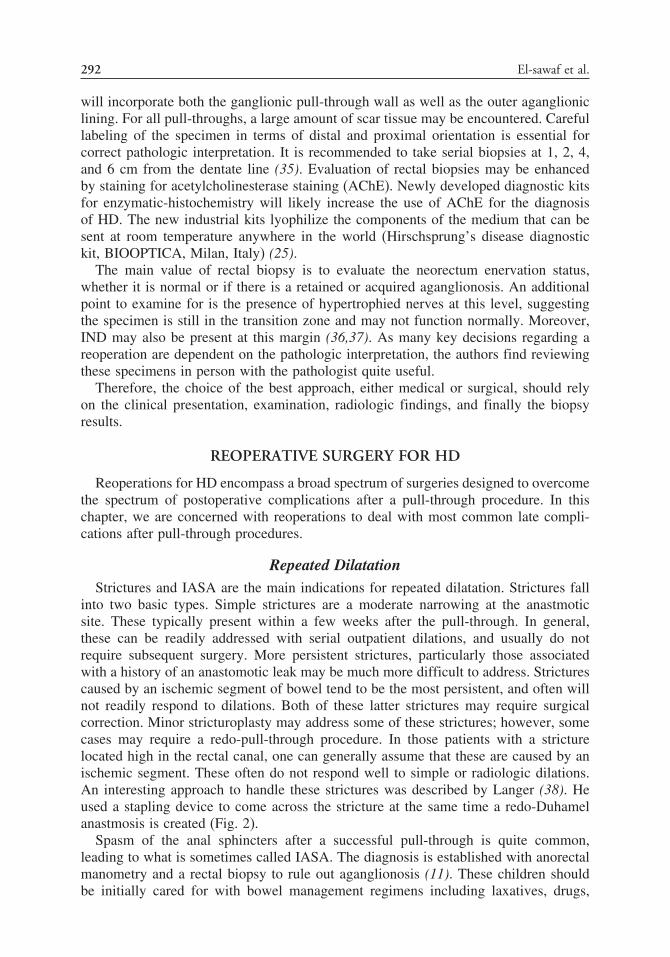
292 El-sawaf et al.
will incorporate both the ganglionic pull-through wall as well as the outer aganglioniclining. For all pull-throughs, a large amount of scar tissue may be encountered. Carefullabeling of the specimen in terms of distal and proximal orientation is essential forcorrect pathologic interpretation. It is recommended to take serial biopsies at 1, 2, 4,and 6 cm from the dentate line (35). Evaluation of rectal biopsies may be enhancedby staining for acetylcholinesterase staining (AChE). Newly developed diagnostic kitsfor enzymatic-histochemistry will likely increase the use of AChE for the diagnosisof HD. The new industrial kits lyophilize the components of the medium that can besent at room temperature anywhere in the world (Hirschsprung’s disease diagnostickit, BIOOPTICA, Milan, Italy) (25).
The main value of rectal biopsy is to evaluate the neorectum enervation status,whether it is normal or if there is a retained or acquired aganglionosis. An additionalpoint to examine for is the presence of hypertrophied nerves at this level, suggestingthe specimen is still in the transition zone and may not function normally. Moreover,IND may also be present at this margin (36,37). As many key decisions regarding areoperation are dependent on the pathologic interpretation, the authors find reviewingthese specimens in person with the pathologist quite useful.
Therefore, the choice of the best approach, either medical or surgical, should relyon the clinical presentation, examination, radiologic findings, and finally the biopsyresults.
REOPERATIVE SURGERY FOR HD
Reoperations for HD encompass a broad spectrum of surgeries designed to overcomethe spectrum of postoperative complications after a pull-through procedure. In thischapter, we are concerned with reoperations to deal with most common late compli-cations after pull-through procedures.
Repeated DilatationStrictures and IASA are the main indications for repeated dilatation. Strictures fall
into two basic types. Simple strictures are a moderate narrowing at the anastmoticsite. These typically present within a few weeks after the pull-through. In general,these can be readily addressed with serial outpatient dilations, and usually do notrequire subsequent surgery. More persistent strictures, particularly those associatedwith a history of an anastomotic leak may be much more difficult to address. Stricturescaused by an ischemic segment of bowel tend to be the most persistent, and often willnot readily respond to dilations. Both of these latter strictures may require surgicalcorrection. Minor stricturoplasty may address some of these strictures; however, somecases may require a redo-pull-through procedure. In those patients with a stricturelocated high in the rectal canal, one can generally assume that these are caused by anischemic segment. These often do not respond well to simple or radiologic dilations.An interesting approach to handle these strictures was described by Langer (38). Heused a stapling device to come across the stricture at the same time a redo-Duhamelanastmosis is created (Fig. 2).
Spasm of the anal sphincters after a successful pull-through is quite common,leading to what is sometimes called IASA. The diagnosis is established with anorectalmanometry and a rectal biopsy to rule out aganglionosis (11). These children shouldbe initially cared for with bowel management regimens including laxatives, drugs,

Chapter 17 / Hirschsprung Disease 293
Fig. 2. Technique for using the Duhamel procedure to deal with a high rectal stricture (right panel).Reproduced with permission from (38).
and bio-feedback training. If this is unsuccessful, some advocate anal sphincterectomy(39,40). Others have had success with temporary sphincter-relaxing measures suchas botulinum toxin (41,42) or nitroglycerine paste (43–45). The latter choices havesome appeal because of the likelihood that the symptoms will improve significantlyover time in most of these children (41). Finally, as a last solution some childrenmay eventually need repeat pull-through for persistently severe stooling problems orintractable HAEC.
Myectomy
Indications
There are basically three indications for a myectomy. The first is the child withpersistent enterocolitis who fails to respond to medical treatment. In these cases astricture has been ruled out, although, the myectomy will often be able to addressstrictures at the level of the previous anastmosis. Prior to the performance of themyectomy, a biopsy should be done to rule out an extensive aganglionic segment. Thesecond indication for a myectomy is the patient with severe constipative symptomswho has normal ganglion cells on repeat biopsy. Typically, these patients will have adilated rectum and sigmoid colon. Caution should be taken in these children, as in someolder patients this rectal dilation may not be corrected, even with a correctly performedmyectomy. Such patients will require a redo-pull-through with resection of this dilatedbowel segment. The third group of patients who may benefit from a myectomy after aprevious pull-through are those children found to have a retained aganglionic bowel.This is the most problematic group, because if the length of aganglionosis is too longit will not be completely addressed by a simple myectomy. In general, if the length ofaganglionosis is more than 5 cm, a redo-pull-through should be performed. Althoughone may be able to perform a myectomy longer than this, the bowel immediatelyproximal to this segment (i.e., transition zone) may not function perfectly well. It hasbeen our experience that a redo-pull-through will work better in these patients (52).
Langer has advanced the use of botulinum toxin (Botox) injections into the internalanal sphincter in patients with either severe constipation or recurrent enterocolitis (46).Results have shown that this technique may produce good results in some patientswith difficulty stooling or persistent enterocolitis. Ideally, a trial of Botox should beattempted prior to the use of a myectomy. The typical dose is 2–5 U/kg or up to 50 Uper treatment in a toddler-sized child. This approach may help the surgeon appreciate
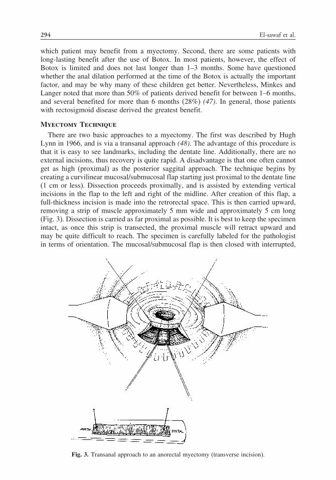
294 El-sawaf et al.
which patient may benefit from a myectomy. Second, there are some patients withlong-lasting benefit after the use of Botox. In most patients, however, the effect ofBotox is limited and does not last longer than 1–3 months. Some have questionedwhether the anal dilation performed at the time of the Botox is actually the importantfactor, and may be why many of these children get better. Nevertheless, Minkes andLanger noted that more than 50% of patients derived benefit for between 1–6 months,and several benefited for more than 6 months (28%) (47). In general, those patientswith rectosigmoid disease derived the greatest benefit.
Myectomy Technique
There are two basic approaches to a myectomy. The first was described by HughLynn in 1966, and is via a transanal approach (48). The advantage of this procedure isthat it is easy to see landmarks, including the dentate line. Additionally, there are noexternal incisions, thus recovery is quite rapid. A disadvantage is that one often cannotget as high (proximal) as the posterior saggital approach. The technique begins bycreating a curvilinear mucosal/submucosal flap starting just proximal to the dentate line(1 cm or less). Dissection proceeds proximally, and is assisted by extending verticalincisions in the flap to the left and right of the midline. After creation of this flap, afull-thickness incision is made into the retrorectal space. This is then carried upward,removing a strip of muscle approximately 5 mm wide and approximately 5 cm long(Fig. 3). Dissection is carried as far proximal as possible. It is best to keep the specimenintact, as once this strip is transected, the proximal muscle will retract upward andmay be quite difficult to reach. The specimen is carefully labeled for the pathologistin terms of orientation. The mucosal/submucosal flap is then closed with interrupted,
Fig. 3. Transanal approach to an anorectal myectomy (transverse incision).

Chapter 17 / Hirschsprung Disease 295
absorbable sutures. One can easily palpate a “keyhole”-like defect once this flap isclosed. Sometimes in postpull-through cases, it is difficult to create this mucosal flap.In this case, one can make a longitudinal elliptical mucosal incision with applicationof traction sutures as shown in Fig. 4A. One then performs the myectomy similar tothe previously mentioned approach. The mucosal defect is then closed longitudinallywithout incorporating the muscularis propria (Fig. 4B). This dissection can be bloody,and one can often accomplish a fair amount of it with cautery.
The second approach is via a posterior saggital approach. This technique was firstdescribed by Thomas et al. in 1970 to primarily treat HD patients with a shortsegment of aganglionosis (49). The technique requires the child to be placed in a pronejackknife position, similar to Peña’s posterior sagittal approach. The dissection beginswith an incision approximately 1–2 cm behind the anal opening and extends to thecoccyx. Dissection then proceeds in the midline, dividing sphincter muscles evenly.The surgeon’s finger, or a Hegar dilator, may be placed in the anal canal to insure thatthe rectal canal is not entered. One then incises a strip of muscularis propria, startingat the level of the internal anal sphincter and extending the dissection as far proximalas possible (Fig. 5). The strip is sent to pathology with correct orientation, and theexternal sphincters and levator complex are reapproximated in the midline.
A
Fig. 4. Transanal approach to an anorectal myectomy (longitudinal incision).

296 El-sawaf et al.
B
Fig. 4. (Continued).
A
Fig. 5. Posterior saggital approach to an anorectal myectomy.
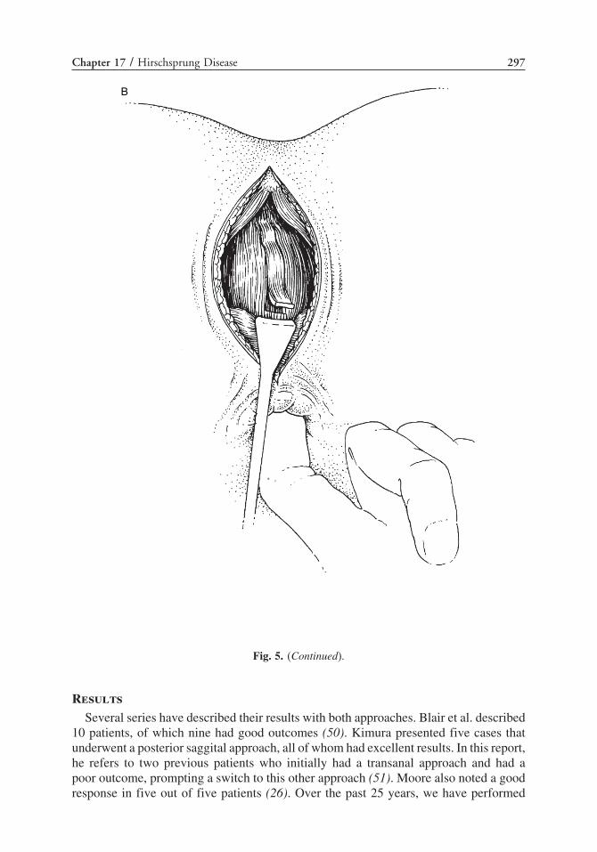
Chapter 17 / Hirschsprung Disease 297
B
Fig. 5. (Continued).
Results
Several series have described their results with both approaches. Blair et al. described10 patients, of which nine had good outcomes (50). Kimura presented five cases thatunderwent a posterior saggital approach, all of whom had excellent results. In this report,he refers to two previous patients who initially had a transanal approach and had apoor outcome, prompting a switch to this other approach (51). Moore also noted a goodresponse in five out of five patients (26). Over the past 25 years, we have performed

298 El-sawaf et al.
39 myectomies in 34 patients with difficulty stooling or persistent enterocolitis, and 29of these 37 myectomies were reported before (52). Complete follow-up was obtainedin 32 patients (36 myectomies). Of those with pure constipative symptoms (12 with13 myectomies), seven had aganglionosis postpull-through. Of those with recurrentHAEC (20 cases with 23 myectomies), only three had aganglionosis postpull-through.Myectomy was performed at a mean of 3.1 years postpull-through. Average follow-upwas 8.6 years. Of those with chronic constipation, six of 12 (50%) had good resultsafter myectomy with one patient requiring two myectomies. The remainder requireda redo-pull-through or colostomy. Interestingly, the majority of patients with retainedaganglionosis and chronic constipation did not respond to myectomy six of seven (85.7%).Of those with recurrent HAEC, 16 of 20 (80%) became free of symptoms or achieved amarked decrease in enterocolitic episodes. Four patients (seven myectomies) had a failedmyectomy. In three of these cases, a repeat myectomy was performed, again withoutimprovement in symptoms and a repeat pull-through is done. In the fourth patient, arepeat pull-through is now being attempted 1 year after the myectomy. In all patientswho responded successfully to the myectomy, no incontinence has been noted.
Repeat Pull-Through ProceduresThe decision to perform a repeat pull-through represents a real challenge for pediatric
surgeons. It carries risks far greater than the original surgery. It should only be done bya highly experienced surgeon after following an appropriate algorithm of management.However, one should never hesitate to redo the pull-through once it is indicated aftercareful planning by detailed preoperative biopsies, contrast enemas, and possibly analmanometry, as detailed previously (Fig. 6).
Weak/absent
Persistent stooling problems
Full
EmptyRectal Ex. Barium Enema
AR tone
++ Tone
Normal
Incontinence Rectal Bx
Ganglionic Aganglionic
IASA Short Long
Bowel management
ImprovedNo
Try Botox Improved
Repeat pullthrough
No
No
Stricture
?? Transitional
No abnormality
Serial dilatations
Motility
Rectal biopsy
Abnormal
Resistance/recurrence
Aganglionic
Ganglionic Normal
Focal Generalized
DistalProximal
No
Bowel management
ResectImproved
Malone ACE
Rectal
AganglionicGanglionic
Short
Long
Colonic
Repeated enemas
Improved
No
Stoma & biopsy
Ganglionic
Aganglionic
Short
Stricturoplasty
Sphincterotomy
Permanent stoma
NoImproved
Twist? Endoscopy
ACE
Fig. 6. Algorithm tailored for different types of long-term complications after pull-through. Botox,botulinum toxin; IASA, internal anal sphincter achaliasia; ACE, antegrade continent enema; AR,anorectal.

Chapter 17 / Hirschsprung Disease 299
Indications
Patients who require a repeat pull-through include those with a retained or acquiredaganglionic segment exceeding 5 cm, a transitional zone pull-through with dilatedneorectum and sigmoid, associated focal IND, persistent long and/or high stricture aftertrial of repeated dilatations, a twisted pull-through, those who have failed to respond toa myectomy and have persistent obstructive symptoms, and children with a megacolonor rectum that is nonresponsive to bowel management regimens and myectomy. Thislatter group is typically seen in those who have had persistent obstructive symptomsfor many years.
Techniques
The approach to performing a repeat pull-through is variable depending on whatprocedure(s) have been previously performed and when. In general, an open laparotomyis probably the best approach. Laparoscopic biopsies may be initially performed togain a better idea of the abnormal pathology, but this may actually be obtained duringa separate procedure. A lower midline incision is ideal, although if the child has had aprevious left hockey-stick type incision, this can be extended, and is usually adequate.A transverse lower abdominal incision is a more cosmetically accepted incision andusually gives an adequate exposure. A thorough lysis of adhesions should be performed.As establishing the correct level for the pull-through segment is of high priority, apathologist comfortable with reading frozen sections for the presence of ganglion cellsand hypertrophied nerves must be available. Second, a complete mobilization of thedistal colon is performed. A critical challenge at this point is to ascertain the vascularityof the distal (remaining) bowel. There is the potential for a devascularization of thedistal pull-through segment if a marginal artery is transected. This may happen whenthe time period between the original pull-through and redo-pull-through is relativelyclose, thus preventing an adequate secondary revascularization.
One variant of a repeat pull-through is when the surgeon is called very shortly afterthe pull-through and told that the pull-through was performed at an aganglionic level(i.e., misreading of the frozen sections). In this case, it is advisable to proceed with animmediate repeat pull-through within 1 week of the first pull-through. If this approachis taken, few adhesions will be present, and one can typically revise the pull-throughthrough the same transanal approach. One should not be dissuaded from proceeding toredo the surgery based on an initial “positive response,” as most infants will start tostool spontaneously; however, obstructive symptoms will eventually return.
For those patients presenting on a more chronic basis, the decision tree in determiningthe approach to redo-pull-throughs is shown in Fig. 7. The decision as to which pull-through should be performed is one of judgment, and may not be readily apparent untilthe actual operation is performed. In general, we have always attempted to performan endorectal pull-through (ERPT) in those patients who have had either a primarySwenson or ERPT. When this fails, because of an inability to adequately get intoa submuscular plan, a Duhamel may be utilized (see example in Fig. 8). For thosepatients who underwent a primary Duhamel, it is occasionally possible to perform anERPT, as the aganglionic and ganglionic walls tend to fuse over time. If this is notpossible, a Swenson pull-through should be performed. A Swenson may be particularlychallenging if one attempts to evert out the rectum, as the last 3 to 4 cm of rectumare often densely adherent within the pelvis. It may be necessary to perform the

300 El-sawaf et al.
NoDuhamel
InitialInitialoperationoperationDuhamelDuhamel SwensonSwenson
TransTrans--abdabd..approachapproach
TransTrans--abdabd..approachapproach
ERPTERPT
Complete Complete abdabd..dissectiondissection
PerinealPerineal anastanast..
Yes
SwensonSwenson ERPTERPTIf possIf poss..
NoDuhamel
InitialInitialoperationoperationDuhamelDuhamel SwensonSwenson
TransTrans--abdabd..approachapproach
TransTrans--abdabd..approachapproach
ERPTERPT
Complete Complete abdabd..dissectiondissection
PerinealPerineal anastanast..
Yes
SwensonSwenson ERPTERPTIf possIf poss..
Fig. 7. Surgical approach to the redo-pull-through.
Fig. 8. Technique for a redo-Duhamel procedure. Reproduced with permission from (38).
anastomosis within the anal canal without eversion. An alternative approach is givenat the end of the chapter.
At the end of the procedure, one must be sure there are no technical problems withthe pull-through including a twisting, kinking, or stretching of the blood supply, ormore commonly a kink of the bowel in the pelvis. This may be particularly troublesomeas one tends to use more proximal segments of bowel, often proximal to the middlecolic vessels. Figure 9A–C show two approaches for pulling through the right colon.
Another technical challenge that may be encountered is difficulty in performing aDuhamel procedure owing to an excess of tissue layers, which must be stapled together.Unlike a primary Duhamel, the surgical stapler is joining another full thickness ofcolon. In the case of an ERPT as the original procedure, this would include a newsegment of ganglionic bowel, the original aganglionic muscularis propria, and theprimary pull-through segment, as well as various amounts of scar tissue. One shouldalways use extra long (4.5-mm) staples. We have encountered two cases where thestaple line has fallen apart immediately after firing the stapler. In these cases, one mustbe prepared to perform a hand-sewn anastomosis. Alternatively, one may divert back

Chapter 17 / Hirschsprung Disease 301
A
B
Fig. 9. (Continued).

302 El-sawaf et al.
C
Fig. 9. Two approaches are given for performing a safe pull-through for patients who need the rightcolon pulled through. (A) The first approach is a rotation of the bowel in a clockwise direction. (B)This first approach results in the ileum coming into the cecum on the patient’s right side, and mayrequire a release of the child’s ligament of Treitz. (C) An alternative approach may be to rotate thecolon anteriorly.
to an original Duhamel procedure, whereby the anastmosis is created by the placementof two extra-long Kocher clamps placed in an inverted ‘V’ configuration, and kept inplace until the crushed bowel forms an anastmosis; generally 7–10 days.
A decision to perform a protective proximal ostomy should be reserved until the endof the redo-pull-through. If the anastomosis is performed with a high degree of tension,or any question remains about the overall success, a proximal colostomy should beplaced. Again, great care needs to be made when creating this diverting colostomy,as the distal blood supply in a redo-pull-through could potentially be compromised.The marginal artery could be lost either during the creation of the stoma, or when theostomy is taken down. In such cases it may actually be safer to perform a temporaryloop ileostomy.

Chapter 17 / Hirschsprung Disease 303
Results
Patients requiring a redo-pull-through procedure clearly represent a very diversepopulation, and are the most challenging group of patients with HD that a pediatricsurgeon may encounter. Thus, any estimate of outcome results an individual patientwill obtain is difficult. Overall, outcomes reported in the literature are reasonably good;however, it is imperative that parents be aware of the difficulty that their child faces,and realistic expectations should be given to the parents prior to the operation. Belowis a brief review of the published literature on this subject as well as our own resultsin patients undergoing a redo-pull-through.
Wilcox and Kiely in 1998 (53) described 20 cases. The indications for reoper-ation were anastmotic stenosis (eight), signs of obstruction or constipation (eight),anocutaneous fistula (one), incontinence of feces (six), and enterocolitis (one); somepatients had more than one indication. They opted for a modified three-stage Duhamelprocedure in all but one of their patients and reported a 70% improvement in symptomsand fecal control. Sarioglu et al. in 1998 (54) reported their experience with fivereoperations for similar indications. Prior to the reoperation, their patients had severestrictures in three patients with a total closure of the anastmotic site in one. Onechild had a rectourethral fistula, and one had an anastmotic disruption. The choice ofredo-pull-through was a Duhamel in three patients and a Swenson in the other twopatients. Average follow-up was 14 years, during which the patients did well; however,objective data regarding continence was not available. Weber et al. in 1999 (55)presented 38 patients undergoing operative procedures after a primary pull-through.Their data included many patients who required only myectomy (24), septum division(four), colostomy then closure for leak (four), or permanent colostomy (four); andin only five cases was a formal redo-pull-through performed. Indications includedretained aganglionic segment (three), enterocolitis after failure of myectomy (one), andnecrosis of pull-through (one). Revisions included a Duhamel in three patients and aSoave in two patients. Because of the diverse operations in this report it is difficultto determine exact outcomes of the redo-pull-through patients; however, excellentresults were noted in 90% of all patients, though continence in the revisions was notquantified. Langer 1999 (38) reported his series of nine repeat pull-through proce-dures over a 4-year period for indications that included resistant stricture (three),acquired aganglionosis (five), and aganglionosis plus IND (one). Their choice ofreoperation was a Duhamel in all but one child, who underwent a Swenson. Overallresults were good in three patients, one had intermittent soiling and the remainingpatients had persistent obstructive symptoms, which are being treated in a nonoperativefashion.
From 1974 to the present, over 386 patients with HD have been referred to oursurgical group at Mott Children’s Hospital. Within this group there are 37 redo-pull-throughs, 19 of which have been previously reported (56). The primary pull-through procedures for these redo cases are ERPT procedure (23 patients), Swenson’sprocedure (six patients), Duhamel’s procedure (six patients), Rehbein’s procedure(one case), and transanal ERPT in one cases. The types of cases and indications fora redo-pull-through are quite varied, and most of the children had more than onereason. By far the most common indication was a retained (or acquired) aganglionicsegment (16 out of 37 cases). A group of 13 patients required repeat pull-throughsecondary to a mechanical problem with their primary pull-through including severeand long strictures caused by to ischemia. Another 13 patients had evidence of

304 El-sawaf et al.
residual segments of dilated colon leading to functional failure of their initial operation.Of this latter group there were eight patients with documented aganglionic bowelpresent at the second pull-through, despite findings of normal ganglion cells duringthe first biopsy in six of these, and a misreading of the pathology in two patients.The remaining five patients had normal ganglion cells on repeat biopsy. However,despite the normal pathology, all had severe difficulty passing stool, and nonehad evidence of strictures. Each of these latter patients had an overlying distendedrectosigmoid colon and presumed internal sphincter achalasia. Of these only onepatient had pathologic evidence of IND. Other indications for repeat pull-throughincluded retained septum (two cases) and fecoloma (two cases) after Duhamel’sprocedure, 360 � twist of the pull-through colon causing obstruction (two cases),retraction of the neorectum (one case), leak (two cases), recurrent abscess and persistentrectocutaneous fistulae (two cases), and transitional zone pull-through (one case)established by documentation of presence of ganglion cells and hypertrophy of thenerve fibers. The presenting symptoms included chronic constipation, repeated andtreatment-resistant HAEC episodes, and sometimes the local cause like leak, abscess, orfistulae.
Distinct from other reports, we found that a redo-ERPT could be effectivelyperformed in several of these patients, and yielded excellent results. An ERPT wasperformed in 21 patients in whom an adequate endorectal dissection could be developedbetween the native rectal cuff and the previously pulled-through colon. In one ofthese, the redo-pull-through was performed via a transanal approach. Additional redoprocedures included a Duhamel in nine patients and a Swenson in four patients. Ourgroup is also reporting a new technique for the performance of a repeat pull-throughusing a stapled anastomosis in three cases (see next section). One technical challengeis that many redo-pull-throughs require the colon proximal to the middle colic vesselto be pulled through. Twisting of the colon can be encountered more frequently withthis type of pull-through. This technical point was initially described by Duhamel; andFig. 9 illustrates two potential approaches that can be used to perform a pull-throughwith the right colon.
Follow-up ranges from 6 months to 32 years (mean 14.2 years). There were nodeaths in the series and one patient required a third pull-through. An assessment ofcontinence was done in all patients who are not neurologically impaired and wereover age 3 years of age. Of these patients all are continent except four children,yielding a continence rate of 89.2%. Two of these patients have minor soiling at nightand during activities only, and the other two have daily leakage of stool. One childwho is mildly mentally retarded has intermittent accidents. Additionally, one patientdeveloped recurrent and antibiotic-resistant perirectal abscesses, which finally led tochronic sacral osteomyelitis. A permanent stoma was the only solution to resolve thisissue. Another patient had leakage from the coloanal anastmosis after closure of thecolostomy, and so required a diverting ileostomy. He is now stable and awaiting closureof his ostomy. One patient suffered from constipation and needed Botox injections intothe anal sphincters, and subsequently underwent a sphincterotomy after the repeat pull-through. Two patients required repeated dilatations for strictures that developed afterthe redo procedure; both of these children responded well to the frequent dilatationswithout further need for another procedure. One final patient developed a cuff abscessthat responded to medical treatment and ultrasound-guided drainage. Stool frequencyper day range from 1 to 10 (mean 3.2).

Chapter 17 / Hirschsprung Disease 305
Repeat Pull-Through with Stapled Anastmosis
We have recently performed a new approach to a repeat pull-through using a stapledanastomosis. The patient is positioned in the lithotomy position. The abdomen is enteredvia a low transverse incision or left Paramedian incision. A thorough lysis of adhesionsand mobilization of the colon is performed, and a series of seromuscular biopsies aresent for frozen section to assess for the correct level of normal ganglionated bowel.The colon proximal to this level is then divided with a gastrointestinal anastomosis(GIA) stapler. One of the greatest difficulties with a repeat pull-through is the last 2–3cm of rectum. This area is typical quite scarred, and any type of mobilization outsideof the rectum is difficult. Therefore, a dissection of the rectum is performed analogousto a Swenson procedure. Dissection is carried down into the pelvis to within 1–1.5 cmabove the dentate line. Great care is taken to avoid injury of the pelvic structures bymeticulous dissection, perfect hemostasis, and by sticking to the colonic wall. Once thesigmoid colon is adequately mobilized, attention is drawn to the neorectum. A Kellyclamp is then introduced from the anus to the neorectum, which had been mobilizedcircumferentially, and the colonic stump is everted out of the anus. A TA stapler(or in some cases an EndoGIA stapler) is used to resect the bulk of the neorectumoutside the anus in its everted state as close as possible to the dentate line (Fig. 10).The neo-rectum is then reinserted into the pelvis. The end result is approximatelya 2.5-cm length of residual rectum. The staple line of the proximal ganglionatedcolon is then removed, and a purse-string suture is used to secure the anvil of theend-to-end anastomosis (EEA) stapler in place. A 21-mm (or larger) EEA stapler is
Fig. 10. Approach for performing a repeat pull-through analogous to a Swenson approach, but usinga stapled anastomosis. (A) Mobilization is done internally until the surgeon encounters significantlyadherent bowel in the most distal rectum. (B) The rectum is then everted out of the peritonealcavity, without a complete dissection of the final 3–4 cm, which remain adherent. (C) Because themost distal few centimeters of the rectum are tightly adherent and do not allow eversion, a stapledanastomosis is performed using an EEA stapling device within the pelvis. (D) Final appearance ofthe pull-through.

306 El-sawaf et al.
C
D
Fig. 10. (Continued).
then inserted into the rectal remnant. An EEA-stapled anastmosis is then performed(Fig. 10C and D). The anastmosis is checked for a leak by verifying complete donutsthat contained both mucosa and wall on both sides. In addition, air is injected withhigh pressure into the rectum, air bubbles should not be noted when the pelvis is filledwith saline. The anastomosis should not be under tension. The pelvis is then irrigatedwith copious amounts of saline and the abdomen is closed in a regular fashion. Aprotective colostomy or very often an ileostomy should be strongly considered after arepeat pull-through.
Results with the new techniqueWe have performed this EEA-stapled procedure for three patients. The indications
were persistent constipation (two) and intractable HAEC (three). Two of the threepatients had a retained aganglionic segment, one of them had a severe stricture that wasnonresponsive to serial dilatation and sphinectrotomy. The third case had a transitional

Chapter 17 / Hirschsprung Disease 307
zone pull-through with presence of ganglia and hypertrophied nerve fibers in thebiopsy. One patient underwent two previous unsuccessful pull-throughs. The ages ofthe patients were 4 years (two) and 8 years (one). The original operation was Soave’s(one), Swenson’s (one), and Duhamel’s (one). Follow-up period has ranged from 6–9 months. Two out of the three patients are doing excellent with 2–5 daily bowelmovements. These two are without soiling and are completely continent. The third,an 8-year-old boy, suffered from anastmotic leak and was diverted by ileostomy. Heis now waiting for a reclosure of his colostomy. In general, we are optimistic withthe new approach and predict that it will be of great value in redo-pull-through caseswhere a more simplified endorectal dissection cannot be performed.
Permanent StomaThere will be a group of patients that may fail a repeat pull-through, or the child’s
parents and physician may decide that they may not benefit from a repeat pull-through.In these cases, one should consider a permanent stoma. Assistance from an enteralstomal therapist may help ensure that this stoma is placed in the most satisfactorylocation for the patient’s entire life.
CONCLUSIONS
Reoperations for HD clearly require a great deal of planning and a thoughtfulwork-up. In cases where a reoperation is needed, however, reasonably good resultsmay be found in the majority of cases. An ERPT is a good procedure to select fora repeat pull-through. However, the pediatric surgeon performing such a proceduremust be experienced with a number of different approaches, as not uncommonly othertechniques must be applied.
REFERENCES
1. Heij HA, de Vries X, Bremer I, Ekkelkamp S, Vos A. Long-term anorectal function after Duhameloperation for Hirschsprung’s disease. J Pediatr Surg 1995;30(3):430–432.
2. Lu WT, Chen CC. Factors affecting postoperative fecal soiling in Hirschsprung’s disease. J FormosMed Assoc 1998;97(3):170–173.
3. Teitelbaum D, Coran, A. Long-term results and quality of life after treatment of Hirschsprung’sdisease and allied disorders. In: Holschneider A, Puri P, (ed.), Hirschsprung’s disease and allieddisorders. Amsterdam: Harwood Academic Publishers; 2000: 457–466.
4. Teitelbaum DH, Coran AG. Reoperative surgery for Hirschsprung’s disease. Semin Pediatr Surg2003;12(2):124–131.
5. Yanchar NL, Soucy P. Long-term outcome after Hirschsprung’s disease: patients’ perspectives. JPediatr Surg 1999;34(7):1152–1160.
6. Marty TL, Seo T, Matlak ME, Sullivan JJ, Black RE, Johnson DG. Gastrointestinal function aftersurgical correction of Hirschsprung’s disease: long-term follow-up in 135 patients. J Pediatr Surg1995;30(5):655–658.
7. Moore SW, Albertyn R, Cywes S. Clinical outcome and long-term quality of life after surgicalcorrection of Hirschsprung’s disease. J Pediatr Surg 1996;31(11):1496–1502.
8. Tariq GM, Brereton RJ, Wright VM. Complications of endorectal pull-through for Hirschsprung’sdisease. J Pediatr Surg 1991;26(10):1202–1206.
9. Teitelbaum DH, Drongowski RA, Chamberlain JN, Coran AG. Long-term stooling patterns ininfants undergoing primary endorectal pull-through for Hirschsprung’s disease. J Pediatr Surg1997;32(7):1049–1052; discussion 1052–1053.
10. Rescorla FJ, Morrison AM, Engles D, West KW, Grosfeld JL. Hirschsprung’s disease. Evaluation ofmortality and long-term function in 260 cases. Arch Surg 1992;127(8):934–941; discussion 941–942.

308 El-sawaf et al.
11. Dasgupta R, Langer JC. Hirschsprung disease. Curr Probl Surg 2004;41(12):942–988.12. Langer JC. Persistent obstructive symptoms after surgery for Hirschsprung’s disease: development of
a diagnostic and therapeutic algorithm. J Pediatr Surg 2004;39(10):1458–1462.13. Teitelbaum DH, Cilley RE, Sherman NJ, et al. A decade of experience with the primary pull-
through for hirschsprung disease in the newborn period: a multicenter analysis of outcomes. AnnSurg 2000;232(3):372–380.
14. Sherman JO, Snyder ME, Weitzman JJ, et al. A 40-year multinational retrospective study of 880Swenson procedures. J Pediatr Surg 1989;24(8):833–838.
15. Teitelbaum DH, Coran AG. Enterocolitis. Semin Pediatr Surg 1998;7(3):162–169.16. Elhalaby EA, Hashish A, Elbarbary MM, et al. Transanal one-stage endorectal pull-through for
Hirschsprung’s disease: a multicenter study. J Pediatr Surg 2004;39(3):345–351; discussion 351.17. Marty TL, Matlak ME, Hendrickson M, Black RE, Johnson DG. Unexpected death from enterocolitis
after surgery for Hirschsprung’s disease. Pediatrics 1995;96(1 Pt 1):118–121.18. Elhalaby EA, Coran AG, Blane CE, Hirschl RB, Teitelbaum DH. Enterocolitis associated with
Hirschsprung’s disease: a clinical-radiological characterization based on 168 patients. J Pediatr Surg1995;30(1):76–83.
19. Swenson O. Hirschsprung’s disease—a complicated therapeutic problem: some thoughts and solutionsbased on data and personal experience over 56 years. J Pediatr Surg 2004;39(10):1449–1453;discussion 1454–1457.
20. Fortuna RS, Weber TR, Tracy TF Jr, Silen ML, Cradock TV. Critical analysis of the operativetreatment of Hirschsprung’s disease. Arch Surg 1996;131(5):520–524; discussion 524–525.
21. Vieten D, Spicer R. Enterocolitis complicating Hirschsprung’s disease. Semin Pediatr Surg2004;13(4):263–272.
22. Teitelbaum DH, Caniano DA, Qualman SJ. The pathophysiology of Hirschsprung’s-associated entero-colitis: importance of histologic correlates. J Pediatr Surg 1989;24(12):1271–1277.
23. Mattar AF, Coran AG, Teitelbaum DH. MUC-2 mucin production in Hirschsprung’s disease: possibleassociation with enterocolitis development. J Pediatr Surg 2003;38(3):417–421; discussion 421.
24. Caniano DA, Teitelbaum DH, Qualman SJ. Management of Hirschsprung’s disease in children withtrisomy 21. Am J Surg 1990;159(4):402–404.
25. Martuccielloa G, Pini Prato A, Puri P, et al. Controversies concerning diagnostic guidelines foranomalies of the enteric nervous system: a report from the fourth International Symposium onHirschsprung’s disease and related neurocristopathies. J Pediatr Surg 2005;40:1527–1531.
26. Moore SW, Millar AJ, Cywes S. Long-term clinical, manometric, and histological evaluationof obstructive symptoms in the postoperative Hirschsprung’s patient. J Pediatr Surg 1994;29(1):106–111.
27. Pfefferkorn MD, Croffie JM, Corkins MR, Gupta SK, Fitzgerald JF. Impact of sedation and anesthesiaon the rectoanal inhibitory reflex in children. J Pediatr Gastroenterol Nutr 2004;38(3):324–327.
28. Meier-Ruge WA, Ammann K, Bruder E, et al. Updated results on intestinal neuronal dysplasia (INDB). Eur J Pediatr Surg 2004;14(6):384–391.
29. Smith B. The neuropathology of pseudo-obstruction of the intestine. Scand J Gastroenterol Suppl1982;71:103–109.
30. Gillick J, Tazawa H, Puri P. Intestinal neuronal dysplasia: results of treatment in 33 patients. J PediatrSurg 2001;36(5):777–779.
31. Holschneider AM, Meier-Ruge W, Ure BM. Hirschsprung’s disease and allied disorders—a review.Eur J Pediatr Surg 1994;4(5):260–266.
32. Ure BM, Holschneider AM, Meier-Ruge W. Neuronal intestinal malformations: a retro- andprospective study on 203 patients. Eur J Pediatr Surg 1994;4(5):279–286.
33. Fadda B, Maier WA, Meier-Ruge W, Scharli A, Daum R. [Neuronal intestinal dysplasia. Critical10-years’ analysis of clinical and biopsy diagnosis]. Z Kinderchir 1983;38(5):305–311.
34. Meier-Ruge WA, Bronnimann PB, Gambazzi F, Schmid PC, Schmidt CP, Stoss F. Histopatho-logical criteria for intestinal neuronal dysplasia of the submucosal plexus (type B). Virchows Arch1995;426(6):549–556.
35. Meier-Ruge WA, Bruder E, Holschneider AM, et al. Diagnosis and therapy of ultrashortHirschsprung’s disease. Eur J Pediatr Surg 2004;14(6):392–397.
36. Kobayashi H, Yamataka A, Lane GJ, Miyano T. Inflammatory changes secondary to postoperativecomplications of Hirschsprung’s disease as a cause of histopathologic changes typical of intestinalneuronal dysplasia. J Pediatr Surg 2004;39(2):152–156; discussion 156.

Chapter 17 / Hirschsprung Disease 309
37. Kobayashi H, Hirakawa H, Surana R, O’Briain D, Puri P. Intestinal neuronal dysplasia is a possiblecause of persistent bowel symptoms after pull-through operation for Hirschsprung’s disease. J PediatrSurg 1995;30(2):253–257.
38. Langer J. Repeat pull-through surgery for complicated Hirschsprung’s disease: indications, techniques,and results. J Pediatr Surg 1999;34:1136–1141.
39. De Caluwe D, Yoneda A, Akl U, Puri P. Internal anal sphincter achalasia: outcome after internalsphincter myectomy. J Pediatr Surg 2001;36(5):736–738.
40. Heikkinen M, Lindahl H, Rintala RJ. Long-term outcome after internal sphincter myectomy forinternal sphincter achalasia. Pediatr Surg Int 2005;21(2):84–87.
41. Ciamarra P, Nurko S, Barksdale E, Fishman S, Di Lorenzo C. Internal anal sphincter achalasia inchildren: clinical characteristics and treatment with Clostridium botulinum toxin. J Pediatr Gastroen-terol Nutr 2003;37(3):315–319.
42. Messineo A, Codrich D, Monai M, Martellossi S, Ventura A. The treatment of internal anal sphincterachalasia with botulinum toxin. Pediatr Surg Int 2001;17(7):521–523.
43. Millar AJ, Steinberg RM, Raad J, Rode H. Anal achalasia after pull-through operations forHirschsprung’s disease—preliminary experience with topical nitric oxide. Eur J Pediatr Surg2002;12(3):207–211.
44. Lindsey I, Jones OM, Cunningham C, George BD, Mortensen NJ. Botulinum toxin as second-line therapy for chronic anal fissure failing 0.2 percent glyceryl trinitrate. Dis Colon Rectum2003;46(3):361–366.
45. Demirbag S, Tander B, Atabek C, Surer I, Ozturk H, Cetinkursun S. Long-term results of topicalglyceryl trinitrate ointment in children with anal fissure. Ann Trop Paediatr 2005;25(2):135–137.
46. Langer J, Birnbaum E. Preliminary experience with intrasphincteric botulinum toxin for persistentconstipation after pull-through for Hirschsprung’s disease. J Pediatr Surg 1997;32(7):1059–1061.
47. Langer JC, Minkes RK. A prospective study of botulinum toxin for internal anal sphincter hyper-tonicity in children with Hirschsprung’s disease. J Pediatr Surg 2000;35(12):1733–1736.
48. Lynn HB, van Heerden JA. Rectal myectomy in Hirschsprung disease: a decade of experience. ArchSurg 1975;110(8):991–994.
49. Thomas C Jr. Posterior sphincterotomy in Hirschsprung’s disease. Surg Gynecol Obstet1967;124(2):365–366.
50. Blair GK, Murphy JJ, Fraser GC. Internal sphincterotomy in post-pull-through Hirschsprung’s disease.J Pediatr Surg 1996;31(6):843–845.
51. Kimura K, Inomata Y, Soper RT. Posterior sagittal rectal myectomy for persistent rectal achalasiaafter the Soave procedure for Hirschsprung’s disease. J Pediatr Surg 1993;28(9):1200–1201.
52. Wildhaber BE, Pakarinen M, Rintala RJ, Coran AG, Teitelbaum DH. Posterior myotomy/myectomyfor persistent stooling problems in Hirschsprung’s disease. J Pediatr Surg 2004;39(6):920–926;discussion 926.
53. Wilcox DT, Kiely EM. Repeat pull-through for Hirschsprung’s disease. J Pediatr Surg1998;33(10):1507–1509.
54. Sarioglu A, Tanyel FC, Buyukpamukcu N, Hicsonmez A. Redo operations of Hirschsprung’s disease.Int Surg 1998;83(4):333–335.
55. Weber TR, Fortuna RS, Silen ML, Dillon PA. Reoperation for Hirschsprung’s disease. J Pediatr Surg1999;34(1):153–156; discussion 156–157.
56. van Leeuwen K, Teitelbaum D, Elhalaby E, Coran A. The long-term follow-up of redo-pullthroughprocedures for Hirschsprung’s disease. J Pediatr Surg 2000;35(6):829–834.

18 Reoperative Surgery for AnorectalMalformations
Marc A. Levitt, MD and Alberto Peña, MD
CONTENTS
Preoperative Evaluation And Management
Definitive Repair
Other Urologic Injuries
Postoperative Management
Sequelae From Constipation
Conclusion
References
Despite significant technical advances in the surgical repair of anorectal malformations,patients sometimes require reoperations when there has been an inadequate preoperativeevaluation, in order to repair technical problems that occurred during the definitiverepair, and when pitfalls in the postoperative management, both in the short andlong term, are encountered. Complications are not isolated to patients with complexmalformations, but include the entire spectrum of malformations, and in fact are morecommon in the but relatively benign malformations.
PREOPERATIVE EVALUATION AND MANAGEMENT
The preoperative evaluation may begin with a prenatal diagnosis, which is an areaof rapidly improving technology. Accurate prenatal diagnosis may impact perinataldecisions and location for a baby’s delivery. This is particularly important for babieswith cloacal malformations and cloacal exstrophy. Most often, an anorectal malfor-mation is not diagnosed prenatally. Once the baby is born, a protocol of evaluation isvital, including both clinical and radiologic assessment (1).
The correct newborn operation, either a newborn pull-through or a colostomy, mustbe chosen. Future reoperations caused by decisions made in the first 24 hours of life arecommon. In patients with cloacal malformations, there are unique problems that canoccur with improper newborn treatment. In such patients, the clinician must recognizeand manage hydrocolpos, perform a colostomy with avoidance of key pitfalls, andmake a correct clinical diagnosis (2).
From: Reoperative Pediatric SurgeryEdited by: S. Teich and D. A. Caniano © Humana Press, Totowa, NJ
311
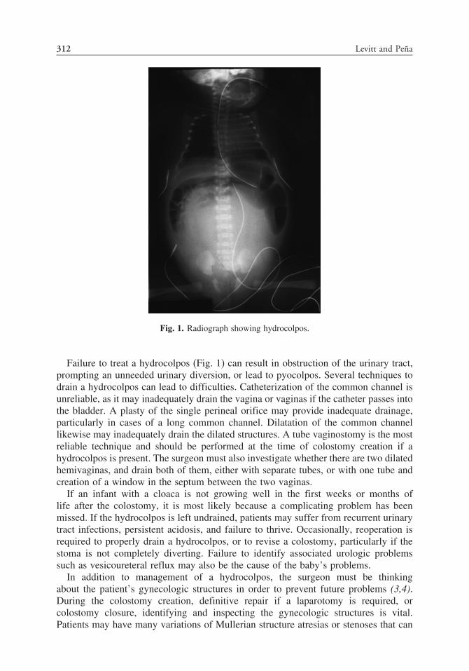
312 Levitt and Peña
Fig. 1. Radiograph showing hydrocolpos.
Failure to treat a hydrocolpos (Fig. 1) can result in obstruction of the urinary tract,prompting an unneeded urinary diversion, or lead to pyocolpos. Several techniques todrain a hydrocolpos can lead to difficulties. Catheterization of the common channel isunreliable, as it may inadequately drain the vagina or vaginas if the catheter passes intothe bladder. A plasty of the single perineal orifice may provide inadequate drainage,particularly in cases of a long common channel. Dilatation of the common channellikewise may inadequately drain the dilated structures. A tube vaginostomy is the mostreliable technique and should be performed at the time of colostomy creation if ahydrocolpos is present. The surgeon must also investigate whether there are two dilatedhemivaginas, and drain both of them, either with separate tubes, or with one tube andcreation of a window in the septum between the two vaginas.
If an infant with a cloaca is not growing well in the first weeks or months oflife after the colostomy, it is most likely because a complicating problem has beenmissed. If the hydrocolpos is left undrained, patients may suffer from recurrent urinarytract infections, persistent acidosis, and failure to thrive. Occasionally, reoperation isrequired to properly drain a hydrocolpos, or to revise a colostomy, particularly if thestoma is not completely diverting. Failure to identify associated urologic problemssuch as vesicoureteral reflux may also be the cause of the baby’s problems.
In addition to management of a hydrocolpos, the surgeon must be thinkingabout the patient’s gynecologic structures in order to prevent future problems (3,4).During the colostomy creation, definitive repair if a laparotomy is required, orcolostomy closure, identifying and inspecting the gynecologic structures is vital.Patients may have many variations of Mullerian structure atresias or stenoses that can

Chapter 18 / Anorectal Malformations 313
lead to future obstruction of menstrual flow, amenorrhea, and may affect their abilityto conceive and carry a baby to term.
The diagnosis of most anorectal malformations is a clinical one. Once the perineumis inspected and the malformation identified, the correct newborn surgical interventioncan be planned. For patients with evidence of rectoperineal fistula, a newborn ordelayed pull-through can be performed without a colostomy. For infants with evidenceof a rectourethral fistula (the most common anomaly in males) a colostomy is therecommended first step, prior to a future definitive reconstruction.
A single perineal orifice defines a cloaca, and if misdiagnosed, may lead to anunnecessary endocrinologic evaluation for “intersex.” The hypertrophied clitoris, whichis sometime visible in cloacas, can be confounding to a clinician not familiar with theexternal perineal appearance of a cloacal malformation. If the surgeon fails to makethe correct diagnosis of a cloaca, they may be prompted to mobilize the rectum, andleave the urogenital sinus intact (Fig. 2). In such a case, a reoperation is required usinga posterior sagittal approach, with rectal mobilization, and correction of the persistenturogenital sinus.
The colostomy is an essential component of the newborn management of the majorityof patients with anorectal malformations, and is a procedure with many preventablecomplications (5). A colostomy placed too distally in the sigmoid can interfere with thefuture pull-through (Fig. 3). Colostomy prolapse is a significant problem, and occurswhen the colostomy is created in a mobile portion of the colon (Fig. 4), or if a looptype of colostomy is used. Colostomy prolapse is particularly problematic for patientswith anorectal malformation who cannot afford to lose colon, as their capacity to formsolid stool impacts their potential for fecal continence. If they are incontinent, theyare more manageable with constipation, than with a tendency toward loose stools.Loop colostomies can lead to urinary tract infections from incomplete diversion of thefecal stream (Fig. 5) and fecal impaction in the distal colon. A Hartman procedure is
Fig. 2. Pull-through only of rectum in a patient with a cloacal malformation, leaving the urogenitalsinus untouched.

314 Levitt and Peña
Fig. 3. Colostomy created too distal in the sigmoid, which will interfere with the pull-through.
problematic in patients with anorectal malformations, as there is no access for contraststudies to define the distal rectal anatomy, and these types of colostomies can lead tothe development of mucoceles. Many of these colostomy problems require reoperations(i.e., a colostomy revision).
In a patient with an anorectal malformation, it is recommended that the stomas beseparated enough to allow the stoma bag to cover only the proximal stoma, isolatingthe mucous fistula to prevent contamination. The stoma should not be separated morethan necessary, so that a big laparotomy can be avoided at the time of colostomyclosure. The opening of a sigmoidostomy in the right upper quadrant can occur if
A
Fig. 4. (Continued).

Chapter 18 / Anorectal Malformations 315
B
C
Fig. 4. Colostomy prolapse can occur when the colostomy is placed in a mobile portion of the colon.(A) Right tranverse; (B) left transverse; (C) descending.
the surgeon tries to find the transverse colon via a right upper quadrant incision andinstead mistakes a dilated rectosigmoid for the transverse colon.
Patients with right transverse colostomies, especially in cases with a long timeinterval between the opening of the colostomy and the main repair, develop a charac-teristic microcolon and a more distal megarectosigmoid (Fig. 6). With tranversecolostomies, it is also almost impossible to irrigate and clean the distal colon adequately.Also, the distal colostogram is difficult to do, as significantly high hydrostatic pressureis needed to show the precise anatomy. Finally, a longer colonic segment can allowfor more resorption of urine and lead to acidosis.
Stricture of the colostomy is caused by a technical problem in which the bowelsuffered from ischemia from an inadequate manipulation of the colon’s blood supply.Dehiscences, infections, and sepsis after colostomy closure are also related to technicalproblems.

316 Levitt and Peña
A
B
Fig. 5. (A) Diagram of loop colostomy. (B) Picture of loop colostomy.

Chapter 18 / Anorectal Malformations 317
Fig. 6. Transverse colostomy with distal microcolon and megarectosigmoid.
To avoid complications, the preferred colostomy seems to be one with separatedstomas, with the proximal end located just after the sigmoid comes off its left retroperi-toneal attachment (5). The stomas should be separated enough to allow for theplacement of a colostomy bag on the proximal stoma (Fig. 7). The proximal stomamust be located away from both the umbilicus and from the iliac crest, thus surroundedby a good portion of normal skin, so that the stoma bag can be easily adapted. Themucous fistula should be located medially and lower, and should be fashioned tiny andflat to avoid prolapse, because it is only used for irrigations and diagnostic tests.
A
Fig. 7. (Continued).

318 Levitt and Peña
B
Fig. 7. (A) Diagram of ideal colostomy. (B) Picture of ideal colostomy.
The purpose of the colostomy, in addition to diverting the fecal stream, is to perform adistal colostogram to define the colorectal anatomy. Understanding the precise locationof the distal rectum, and its relationship to the urinary tract, is vital to avoidingcomplications during the definitive repair (6).
DEFINITIVE REPAIR
The definitive repair has numerous complications associated with it, many of whichare preventable with correct preoperative planning and proper technique. Reoperativesurgery is necessary for several of these complications, to alleviate pain, discomfort,and other sequelae.
It is clear that a patient’s best chance for a good functional result is when theproper operation is performed during the first definitive procedure and complicationsare avoided (7). This is especially true in those patients born with a good prognosisdefect. It is unfortunate when such patients end up with fecal or urinary incontinenceresulting from avoidable complications of the surgical repair.
Rectal ProblemsPatients can experience dehiscence, retraction, infection, and/or acquired atresia of
the rectum related to technical problems during the pull-through. These are usually theresult of excessive tension or inadequate blood supply. In addition, anal strictures mayresult when the prescribed protocol of dilatations is not followed.
Reoperation for these problems can be performed posterior sagittally. In casesof retraction, dehiscence, and acquired atresia, the rectum is usually locatedsomewhere high in the pelvis surrounded by a significant amount of fibrous tissue.Multiple fine silk sutures are placed in the rectal wall in order to exert uniform tractionand facilitate a circumferential dissection of the rectum, trying to stay as close aspossible to the rectal wall without injuring it. Bands and extrinsic vessels surroundingthe rectum are divided and cauterized circumferentially until enough rectal length isgained to place the rectum within the limits of the sphincter mechanism. Short ring-like
Chapter 18 / Anorectal Malformations 319
rectal strictures can be treated with a Heineke-Mikulicz type of plasty. Strictures thatare longer than 1 cm must be resected, with the rectum mobilized until the fibroticportion can be removed, and a fresh nonscarred portion of rectum pulled down, creatinga new anus.
Retraction, dehiscence, and acquired rectal atresias, are most likely caused by inade-quate mobilization of the rectum. During a primary procedure, the rectum, when seenposterior sagittally, is covered by a very characteristic white fascia that contains vesselsto the rectum. The surgeon must dissect this fascia off the rectum, remaining as closeas possible to the rectal wall. Uniform traction provided by multiple fine silk suturesis imperative to facilitate this dissection. The intramural blood supply of the rectum isexcellent, and the rectum can be dissected to gain significant length provided the rectalwall is not injured. The most likely cause for difficulty in dissection of the rectumis working outside of this fascia. Alternatively, dissection too close to the rectumcan injure the rectal wall, interfere with the intramural blood supply, and provokeischemia. The result of all this is an incomplete mobilization, rectal ischemia, and arectum to skin anastomosis performed under tension, which may explain many of thesecomplications.
Strictures are also most likely caused by ischemia of the distal rectum. When therectum is correctly mobilized and the blood supply kept intact, it is extremely unlikelyto have an anal stricture. Patients who fail to follow a protocol of dilatations candevelop a thin fibrotic ring in the area of the anoplasty, which is easily treated eitherwith an anoplasty or dilatations. A long narrow stricture is most likely caused by rectalischemia.
Some surgeons do not have their patients follow a protocol of anal dilatations. Inorder to avoid painful maneuvers to the patient, they follow a specific plan consistingof taking the patient to the operating room every week and under anesthesia performingdilatations. Those forceful dilatations can actually provoke lacerations in the anal verge,which then heal with scarring, only to be reopened during the next forceful dilatation,leading ultimately to an intractable ring of fibrosis.
Rectourinary and Rectovaginal FistulaePatients may have various types of complications involving rectogenitourinary tract
fistulae. Fistulae can be persistent when the original rectourethral fistula remainsuntouched during the main repair, even when the rectum was repaired. Recurrentfistulae may occur if the surgeon repaired the fistula but it reopened. Acquiredrectourethral fistulae are those that are created during the repair of a benign malfor-mation (6).
Acquired rectovaginal fistula can occur during an attempted failed repair of arectovestibular fistula. Prior to the introduction of the total urogenital mobilization,urethrovaginal fistula was the most common and feared complication in cloacal malfor-mations (8). In certain circumstances, the vagina can be rotated to try to prevent thiscomplication. But even with that maneuver, these fistulas can occur.
Persistent rectourethral fistulae can occurr in patients who were born with arectourethral bulbar fistula and underwent a repair that did not address the fistula.Surgeons following the old diagnostic approach may have performed an invertogramand found the bubble of rectal air close to the skin. This may have led to an approachthrough the perineum, with identification of the rectum and subsequent pull-through

320 Levitt and Peña
and anoplasty. Because the surgeon was completely unaware of a low rectourethralbulbar fistula, it was not repaired.
Recurrent rectourethral fistulae may result if the fistula is closed, but the rectum isnot mobilized adequately, leaving the anterior wall under tension. A dehiscence of theanterior rectal wall may explain the recurrence of the fistula. Also, leaving sutures inthe rectum adjacent to the sutures in the urethra may lead to formation of a recurrentfistula. Thus, any rectal tapering should always be performed on the posterior wall.Consequently, an injured rectum that requires a repair to the anterior wall may createa situation for the development of a recurrent fistula. The same explanation may applyto rectovaginal fistulae.
Acquired rectourethral fistulae can occur in male patients who were born withrectoperineal fistulae if they undergo their first operation without a Foley catheter inthe urethra. During the mobilization of the anterior wall of the rectum, an unrecognizedurethral injury can occur and, if the rectum is not adequately mobilized to leave normalrectal wall in front of the urethral injury, an acquired rectourethral fistula will form.
Fistulous complications can be approached posterior sagittally. The posterior rectalwall should be opened and the fistulae identified and closed. The rectum then needs to beseparated from the urinary tract or the vagina, and mobilized to ensure that a completelynormal anterior rectal wall is left in front of the urethral or vaginal suture line.
Persistent Urogenital SinusA persistent urogenital sinus occurs in patients born with a cloacal malformation
who underwent an operation in which the rectal component of the malformation wasrepaired, but the urogenital sinus was ignored (Fig. 2). Their reoperation can also beapproached posterior sagittally. The rectum must be completely dissected and reflectedout of the way. This allows exposure of the urogenital sinus, which can be repairedusing the same technique that is employed during the treatment of a cloaca (9). Manysuch patients with this problem had an original diagnosis of a “rectovaginal fistula”(10) and the true malformation, a cloaca, was not fully recognized.
Acquired Vaginal AtresiaComplete fibrosis of the vagina caused by excessive dissection can occur when a high
vagina is mobilized. This is particularly problematic when the vagina is separated fromthe urethra during the repair of a cloacal malformation. The dissection of the vaginafrom the urinary tract is not an easy maneuver, the vagina may become devascularizedand as a consequence, patients can develop vaginal ischemia and subsequent atresia.This is why the development of the total urogenital mobilization has dramaticallyreduced this complication (8).
Acquired Urethral AtresiaAcquired urethral atresia in patients with cloaca can occur from devascularization
of the pulled through or reconstructed neourethra. In male patients, it can occur whenthe urethra is accidentally transected during an attempt to repair an anorectal malfor-mation. This complication most frequently occurs in males in whom the surgeonapproaches the reconstruction posterior sagitally without a preoperative high-pressuredistal colostogram (6). To repair this problem, the rectum must be mobilized in orderto expose the urethral area. Both urethral ends need to be identified, dissected, andmobilized to perform a tension-free end-to-end anastomosis.

Chapter 18 / Anorectal Malformations 321
Fig. 8. Posterior urethral diverticulum, representing the undissected former distal rectum.
Posterior Urethral DiverticulumThis complication can occur when a retained portion of the rectum is left attached
to the posterior urethra (Fig. 8) and is seen in patients born with a rectourethral bulbarfistula repaired transabdominally. It is easy to understand that the surgeon was unableto reach the fistula site through the abdomen. Consequently, the rectum was amputated,leaving a piece of rectum attached to the urethra. This potential complication is alsopossible with a laparoscopic approach, particularly if the rectum reaches well belowthe peritoneal reflection.
These patients are initially asymptomatic, but after several years develop symptomssuch as passage of mucous through the urethra, dribbling, orchioepididimitis, urinarytract infections, and urinary incontinence. In addition, we have seen one such patientwho, after thirty years, developed an adenocarcinoma in the piece of rectum leftattached to the urethra.
A pelvic magnetic resonance imaging (MRI) can diagnose this condition, withvisualization of a urine-filled diverticulum behind and connected to the urethra. Aposterior sagittal approach can be used to fix this problem, with mobilization ofthe rectum to expose the posterior aspect of the urinary tract, identification of thediverticulum, and dissection of it down to the urethra. The diverticulum can be separatedfrom the urethra in the same manner as in a primary repair of anorectal anomalies,with the urethra closed and the diverticulum resected.
OTHER UROLOGIC INJURIES
Significant urologic injuries such as transection of the bladder neck, transection ofthe urethra, injury to vas deferens, seminal vesicles, prostate, and ectopic ureters haveoccurred, usually when the posterior sagittal approach was performed without a goodprevious distal colostogram, and thus the precise anatomy was not known prior to theposterior sagittal dissection (6). These injuries often require reoperation.

322 Levitt and Peña
Neurogenic BladderNeurogenic bladder in male patients with anorectal malformations is extremely
unusual. In our series, we have only seen it in patients with a very abnormal sacrumor an associated spinal anomaly (11). If it does occur, it may represent a denervationof the bladder and bladder neck during the repair.
Patients with cloacal malformations are very different with regard to bladderfunction. They often have a deficient emptying mechanism and do not have the typical“christmas tree” type of image of a neurogenic bladder seen in patients with spinabifida, but rather have a flaccid, smooth, large bladder that does not empty completely.Fortunately, most patients with cloacas have a very good bladder neck. The combi-nation of a good bladder neck with a floppy, flaccid bladder make these patients idealcandidates for intermittent catheterization, which keeps them completely dry (9).
Two exceptions to this rule in cloacas exist. One is represented by patients thathave a very long common channel, in which the hemivaginas as well as the rectumare attached to the bladder neck. After they are separated, the patients are left withno bladder neck or a very damaged bladder neck. The second group is representedby a small number of patients with cloaca who are born with separated pubic bones,who could be described as having a “covered exstrophy” (12). These patients wereborn with no bladder neck, and they eventually require a continent diversion type ofoperation.
Complications of the Laparoscopic Approach to Anorectal MalformationsLong-term results and complications have not yet been described for the laparoscopic
approach to imperforate anus, which is a relatively new approach, developed to avoid alaparotomy and to minimize the posterior sagittal incision (13). We have reoperated forseveral complications from this approach performed at other institutions, and postulatethe potential for others.
Avoidance of the perineal exposure afforded by the posterior sagittal approach canlead to inadvertent injuries, such as injury to the bladder neck, urethra, or to anectopic ureter particularly during passage of the trocar through the perineum. Preciseunderstanding of the anatomic relationships of the pelvis provided by the laparoscopicview is vital to avoid these problems. As with the former transabdominal approach (14),there is potential for leaving behind the distal rectal cuff and thus a posterior urethraldiverticulum, particularly for malformations below the peritoneal reflection such as arectobulbar fistula. Finally, to avoid rectal prolapse a pelvic hitch is employed (13). Ifthis step is omitted, or done incorrectly, the incidence of prolapse may increase. Withthe avoidance of the posterior sagittal incision, the described laparoscopic operationomits several key steps of the posterior sagittal anorectoplasty (PSARP) that areimportant to avoid prolapse (15), particularly tacking of the posterior rectal wall to themuscle complex.
Rectal ProlapseRectal mucosal prolapse occurs following PSARP, with an incidence of 3% (15).
It is more common in patients with higher malformations and with poor sacra andpelvic musculature. Significant prolapse may lead to ulceration, bleeding, and mucousproduction. It can interfere with anal canal sensation and thus impact a patient’sfunctional prognosis.

Chapter 18 / Anorectal Malformations 323
Correction of prolapse can be performed transanally, with mobilization of redundantfull-thickness rectum, and redo-anoplasty. This is ideally done prior to colostomyclosure. Sometimes, however, prolapse only develops after colostomy closure and inthe presence of constipation.
POSTOPERATIVE MANAGEMENT
The long-term concerns following surgery for anorectal malformations mainlyinvolve a child’s potential for bowel control. Most children have voluntary bowelmovements, but a small number suffer from fecal incontinence despite a technicallycorrect operation (11). Fecal incontinence may occur as a result of a technicallyincorrect operation. Such a patient must be identified, and may be a candidate for areoperation particularly for a patient who be expected to have good bowel control.Constipation is a common problem following operations for anorectal malformations,and may lead to significant sequelae if not proactively managed.
Fecal IncontinenceReoperation to improve a patient’s functional prognosis is indicated in several
circumstances. Early on reoperative surgery was performed on every patient weevaluated who underwent a repair at another institution and subsequently developedfecal incontinence. During those years, we hoped that the new posterior sagittalapproach would give these patients an opportunity to recover bowel control. When theresults were evaluated (16,17), only 30% of those patients experienced a significantimprovement. Therefore, the indications for reoperative surgery were modified.
Presently, reoperation for fecal incontinence is recommended only for patients withvery special criteria. It is for patients born with a malformation associated with a goodprognosis, with a mislocated rectum. The rectosigmoid should be present, the sacrumnormal, and the sphincter mechanism intact. In such cases, restoration of near normalanatomy may improve the functional outcome.
For these reoperations, the rectum is approached posteriorly. Multiple silk stitchesare placed at the mucocutaneous margin in order to apply uniform traction to facilitatethe dissection and mobilization of the rectum. A full rectal dissection and mobilizationis performed, staying as close as possible to the bowel wall but avoiding injury toit. The limits of the sphincters, including the parasagittal fibers, muscle complex, andlevator muscle are determined by electrical stimulation, and the rectum is repositionedwithin it.
In many cases, the patient is found to have had colon, rather than rectum, pulled downto the anus. The rectum may have been resected. This is identified by the presence ofa mesentery attached to the bowel. In such cases, the mesenteric fat should be trimmedfrom the last few centimeters of the rectum to allow for direct contact between thesphincter mechanism and the colonic wall. An anoplasty, performed within the limitsof the sphincter mechanism, completes the reconstruction.
The number of patients that require a reoperation for fecal incontinence has decreasedsignificantly over the years. This is likely due to the increased use of the posteriorsagittal approach, which provides superior exposure and prevents the mislocation ofthe rectum seen with other techniques.
In the past, many patients underwent abdominal perineal pull-throughs withendorectal dissections of the rectosigmoid (14). This procedure essentially resulted in
324 Levitt and Peña
loss of the rectosigmoid. These patients do not suffer from constipation. Instead theysuffer from increased colonic motility and a tendency to diarrhea. Revisional surgeryis not offered to them, because they never regain bowel control, owing mainly tothe loss of the rectal reservoir. They are provided with bowel management, with thegoal of slowing down the colon medically, and empyting it daily with an enema (18).Fortunately, endorectal pull-throughs for anorectal malformations are rarely done andit is rather unusual to see these patients.
Patients born with a poor prognosis defect and fecal incontinence are also consideredinappropriate candidates for reoperation. These patients typically have an abnormalsacrum, flat perineum, and poor sphincters. There is usually evidence that they wereborn with a high rectoprostatic or rectobladder neck fistula, or a cloaca with a commonchannel longer than 3 cm. Their sacral ratio is almost always low, often less than 0.4.Reoperations on these patients, even if they have a completely mislocated rectum, arenot helpful because they do not improve after reoperation. Instead they are offered abowel management program with a daily enema, (18) in order to prevent soiling andto keep them completely clean.
When revisional surgery for fecal incontinence is offered, the likelihood of thepatient regaining bowel control is reviewed with the family. Even with those patientswho are expected to improve, the bowel management program is implemented priorto surgery. If it turns out that the patient does not improve enough after reoperation toavoid enemas, the already proven bowel management is reinstituted.
SEQUELAE FROM CONSTIPATION
Constipation is the most common functional disorder observed in patients whoundergo PSARP (11). Interestingly the incidence of constipation is inversely relatedto the height of the anorectal malformation. This means that patients with the bestprognosis for bowel control have the highest incidence of constipation. Patients withvery poor prognosis, such as bladder neck fistula, have a rather low incidence ofconstipation.
Constipation seems to be related to the degree of preoperative megarectum.Colostomies that do not allow cleaning and irrigation of the distal colon lead to moremegarectum. Transverse colostomies lead to a micro left colon with dilatation of therectosigmoid. Loop colostomies allow for passage of stool and distal fecal impaction.It is clear that keeping the distal rectosigmoid empty and not distended from thetime the colostomy is established, and proceeding with pull-through and subsequentcolostomy closure as early as possible within several months, reduces the developmentof megarectosigmoid and results in better ultimate bowel function.
A significant number of patients suffer from sequelae from the mismanagement oftheir constipation. Many patients are described as having “fecal incontinence” whenthey actually have untreated severe constipation and chronic impaction. This conditionis actually overflow pseudoincontinence. These patients have several common features.All were born with a malformation with good functional prognosis, and all underwenta technically correct, successful operation. Postoperatively, they have severe consti-pation which is not managed aggressively. They therefore developed megasigmoidand chronic fecal impaction, which initiates a vicious cycle. Adequate treatment oftheir constipation, with or without a sigmoid resection (19), can render them fecallycontinent.

Chapter 18 / Anorectal Malformations 325
All patients in this pseudoincontinent group should undergo a “laxative test” todetermine if they are fecally continent. First, large volume enemas are administereduntil the patient’s colon is clean (disimpacted) as documented on a plain radiograph.Daily laxatives are then administered, increasing the amount each day until the amountnecessary to produce colonic evacuation is determined. If the patient demonstrates thecapacity to feel the stool in the rectum, reaches the bathroom, has voluntary bowelmovements and remains clean every day, the patient is continent. Such a patient canthen be offered the option of continuing treatment with large quantities of laxative orundergoing a sigmoid resection (19) in order to make the constipation more manageableand thereby decreasing the laxative requirement.
This group of patients is extremely important to recognize. Some of these patientsmay be wrongly diagnosed as suffering from true fecal incontinence and some haveeven undergone reoperations such as gracilis muscle transfers or artificial sphincters,which can actually make the patient worse. This problem should be suspected when onesees a patient who was born with a benign malformation, who underwent a technicallycorrect operation, but was not treated correctly for constipation.
CONCLUSION
Unfortunately, despite great advances in pediatric surgical care, there remains asignificant number of patients who undergo attempted anorectal repairs with signif-icant complications requiring reoperations. Many of these problems are preventable.For good results, one must have a thorough understanding of the spectrum of anorectalmalformations, employ proper newborn evaluation and management, use meticuloustechnique, and employ rigorous careful postoperative regimens. These basic funda-mentals need to be emphasized in the training of pediatric surgeons, so as to improvethe outlook for children born with anorectal malformations.
REFERENCES
1. Shaul DB, Harrison EA. classification of anorectal malformations—initial approach, diagnostic tests,and colostomy. Semin Pediatr Surg 1997;6:187–195.
2. Levitt MA, Peña A. Pitfalls in the management of newborn cloacas. Pediatr Surg Int 2005;21:264–269.3. Levitt MA, Stein DM, Peña A. Gynecological concerns in the treatment of teenagers with cloaca.
J Pediatr Surg 1998;33:188–193.4. Warne SA, Wilcox DT, Creighton S, Ransley PG. Long-term gynecologic outcome of patients with
persistent cloaca. J Urol 2003;170:1493–1496.5. Peña A, Krieger M, Levitt MA. Colostomy in anorectal malformations—a procedure with significant
and preventable complications. J Pediatr Surg 2006;41:748–756.6. Hong AR, Rosen N, Acuña MF, Peña A, Chaves L, Rodriguez G. Urological injuries associated with
the repair of anorectal malformations in male patients. J Pediatr Surg 2002;37:339–344.7. Peña A, Hong AR, Midulla P, Levitt M. Reoperative surgery for anorectal anomalies. Semin Pediatr
Surg 2003;12:118–123.8. Peña, A. Total urogenital mobilization—an easier way to repair cloacas. J Pediatr Surg 1997;32:
263–268.9. Peña A, Levitt MA, Hong AR, Midulla P. Surgical management of cloacal malformations: a review
of 339 patients. J Pediatr Surg 2004;39:470–479.10. Rosen NG, Hong AR, Soffer SZ, Rodriguez G, Peña A. Recto-vaginal fistula: a common diagnostic
error with significant consequences in female patients with anorectal malformations. J Pediatr Surg2002;37:961–965.
11. Peña A, Levitt MA. Imperforate anus and cloacal malformations. In: Ashcraft KW, Holder TM,Holcomb W, (eds.), Pediatric surgery, fourth edition. Philadelphia: Saunders; 2005: 496–517.

326 Levitt and Peña
12. Soffer SZ, Rosen NG, Hong AR, Alexianu M, Peña A. Cloacal exstrophy: a unified managementplan. J Pediatr Surg 2000;35:932–937.
13. Georgeson KE, Inge TH, Albanese CT. Laparoscopically assisted anorectal pull-through for highimperforate anus—a new technique. J Pediatr Surg 2000;35:927–930, discussion 930–931.
14. Kiessewetter WB. Imperforate anus. II - The rationale and technique of the sacroabdominoperinealoperation. J Pediatr Surg 1967;2:106–110.
15. Belizon A, Levitt M, Shoshany G, Rodriguez G, Peña A. Rectal prolapse following posterior sagittalanorectoplasty for anorectal malformations. J Pediatr Surg 2005;40:192–196, discussion 196.
16. Peña A, deVries PA. Posterior sagittal anorectoplasty: important technical considerations and newapplications. J Pediatr Surg 1982;17:796–811.
17. Peña A. Advances in the management of fecal incontinence secondary to anorectal malformations.In: Nyhus L (ed.), Surgery annual. Connecticut: Appleton & Lange; 1990: 143–167.
18. Peña A, Guardino K, Tovilla JM, Levitt MA, Rodriguez G, Torres R. Bowel management for fecalincontinence in patients with anorectal malformations. J Pediatr Surg 1998;33:133–137.
19. Peña A, El-Behery M. Megasigmoid—a source of pseudo-incontinence in children with repairedanorectal malformations. J Pediatr Surg 1993;28:199–203.

19 Reoperation for Recurrent Analand Perianal Conditions
Shahab Abdessalam, MD
and Donna A. Caniano, MD
CONTENTS
Introduction
Recurrent Rectal Prolapse
Recurrent Perirectal Abscess/Fistula-in-Ano
Persistent Hemorrhoids
Recurrent/Persistent Anal Warts
References
INTRODUCTION
Disorders of the anus and rectum present frequently in infants and children, so it isimportant for the pediatric surgeon to identify conditions that require operative inter-vention. Fortunately, most minor anorectal disorders respond successfully to initialmanagement and rarely recur. The following diseases will be addressed in this chapter,in relation to recurrence and reoperative surgery: rectal prolapse, fistula-in-ano, hemor-rhoids, and anal warts. Management of perianal disease secondary to Crohn’s disease andcongenital anomalies of the anus and rectum are presented in other sections of this book.
RECURRENT RECTAL PROLAPSE
Rectal prolapse is defined as full-thickness protrusion of the rectal wall out of theanal canal. The typical presentation of rectal prolapse is a 1- to 3-year-old child whodevelops a bulging mass exiting the anus after defecation (1). The mass will oftendisappear spontaneously, is usually painless, and may have minor surface bleeding.Rarely, an older child may present with an incarcerated rectal prolapse associatedwith intense pain and bleeding. The etiology of childhood rectal prolapse is thoughtto include chronic constipation, lack of fixation of the rectum, prolonged periods ofstraining during “potty training,” and lack of angulation of the pelvis and rectum inthe first several years of life (1–3).
Evaluation of a child with rectal prolapse includes a thorough history and physicalexamination. Stooling patterns, history of constipation and/or diarrhea, evaluation of
From: Reoperative Pediatric SurgeryEdited by: S. Teich and D. A. Caniano © Humana Press, Totowa, NJ
327

328 Abdessalam and Caniano
muscular or neurological impairment, and dietary habits are all key elements. Thephysical examination includes growth and development parameters, which may beabnormal in children with previously undiagnosed cystic fibrosis. The physical stigmataof a connective tissue disorder, such as Ehlers-Danlos syndrome, should be assessedduring the examination. Visual inspection of the perineum evaluates anal positionand external masses, whereas the digital rectal examination assesses anal tone andthe presence of internal masses. We have observed that referring physicians oftenconfuse rectal prolapse with external hemorrhoids. In rectal prolapse there are circularconcentric folds, whereas in hemorrhoids, the folds are radial, as shown in Fig. 1. If noobvious pathology is visualized on initial examination, it may be helpful to have thechild blow up a balloon or to reexamine the child after allowing time to sit on the toiletand attempt defecation. One can also ask the parents to provide a digital photographof the rectal prolapse when it occurs in the home setting.
A child with documented rectal prolapse should undergo a sweat chloride test,because previously unsuspected cystic fibrosis is found in 6–20% of these patients (3,4).In selected patients, thyroid function studies should be performed to excludehypothyroidism, and genetic testing should be considered if the physical examinationis suggestive of a connective tissue disorder. A contrast enema is helpful in eliminatingan anatomic cause for the prolapse.
Because the majority of children with rectal prolapse will have chronic constipation,treatment includes dietary adjustment, high-fiber foods, and a laxative bowel regimen.If the prolapse fails to respond to a carefully followed bowel regimen for at least6 months, or if the patient has an underlying connective tissue disorder, operativetreatment should be considered. The operations designed for the treatment of rectalprolapse are classified into two main approaches, perineal and abdominal (1,3,5–7). Ourpreferred initial management is placement of a Thiersch wire using an 0-prolene sutureas the “wire,” and tightening the suture over a 10-F Hegar dilator (Fig. 2–4). If prolapserecurs after placement of the initial Thiersch wire, we favor a second wire, especially
Fig. 1. Rectal prolapse.
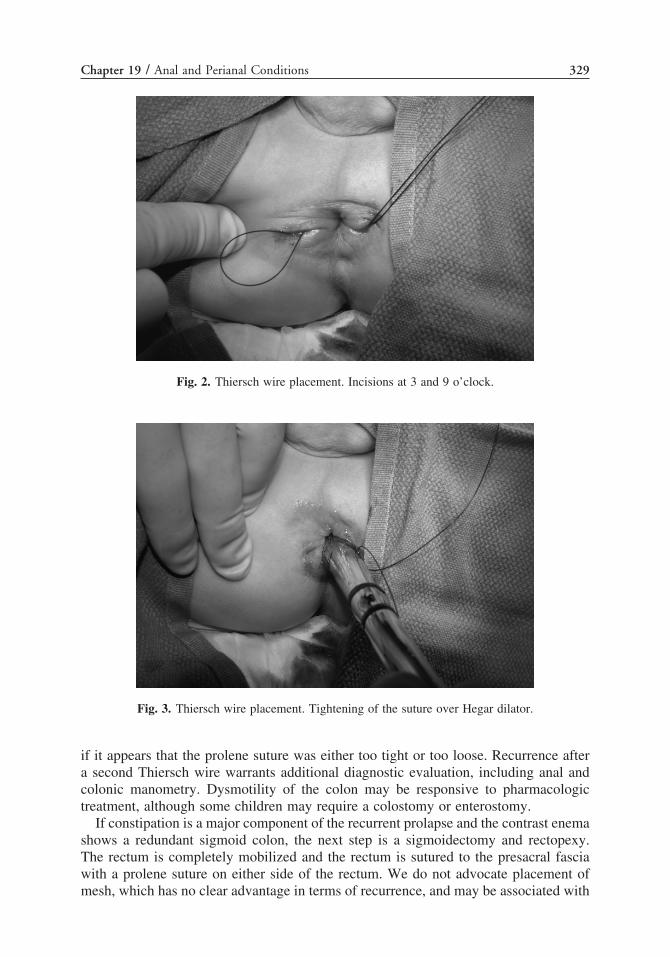
Chapter 19 / Anal and Perianal Conditions 329
Fig. 2. Thiersch wire placement. Incisions at 3 and 9 o’clock.
Fig. 3. Thiersch wire placement. Tightening of the suture over Hegar dilator.
if it appears that the prolene suture was either too tight or too loose. Recurrence aftera second Thiersch wire warrants additional diagnostic evaluation, including anal andcolonic manometry. Dysmotility of the colon may be responsive to pharmacologictreatment, although some children may require a colostomy or enterostomy.
If constipation is a major component of the recurrent prolapse and the contrast enemashows a redundant sigmoid colon, the next step is a sigmoidectomy and rectopexy.The rectum is completely mobilized and the rectum is sutured to the presacral fasciawith a prolene suture on either side of the rectum. We do not advocate placement ofmesh, which has no clear advantage in terms of recurrence, and may be associated with

330 Abdessalam and Caniano
Fig. 4. Thiersch wire placement. Final appearance.
long-term complications, such as obstruction. If constipation is not a major component,we perform a laparoscopic rectopexy, with a suture securing either side of the fullymobilized rectum to the presacral fascia. Recurrence after rectopexy alone would befollowed by a sigmoidectomy and rectopexy. Our recommended treatment algorithmis shown in Fig. 5.
InitialPresentation
Thiersch Wire
Trial ofConservativeManagement
Manometry
Abnormalor Neurological
Impairment
Ostomy
Normal With
Constipation
SigmoidectomyWith Rectopexy
Normal Without
ConstipationLaparoscopic
Rectopexy
3rd Recurrence
SigmoidectomyWith Rectopexy
Recurrence
Repeat Tiersch Wire
Recurrence
Fig. 5. Treatment algorithm for rectal prolapse.

Chapter 19 / Anal and Perianal Conditions 331
The treatment of rectal prolapse has not been studied prospectively in clinical trialsin either adult or pediatric patients, and a Cochrane review could not delineate a clearadvantage of one operative technique (6). The overall recurrence rate for all operationsis 10–40%, regardless of the type of operation (1,3,5,7).
RECURRENT PERIRECTAL ABSCESS/FISTULA-IN-ANO
Peri-rectal abscesses develop from an occluded duct of an anal gland with subsequentbacterial overgrowth (8). The typical patient presents in infancy with a tender, erythe-matous swelling of the soft tissues surrounding the anus. On physical examination thearea of involvement is firm, fluctuant, and tender to palpation. Occasionally, there isspontaneous drainage of purulent material. Traditional treatment for recurrent perirectalabscesses has been incision and drainage with packing or removal of a small ellipse ofskin (9). We do not recommend probing of the abscess cavity at the time of drainage,because it could promote development of a fistula (9,10). If obvious fluctuance isnot appreciated, an ultrasound may be helpful in identifying a fluid collection. In theabsence of a clearly fluctuant mass, the infant should be treated with antibiotics tocover Gram-positive and Gram-negative bacteria and symptomatic pain control.
Recurrent perirectal abscesses and fistulae have been reported to occur in 20–50%of patients (9–11). For infants less than 1 year of age, we recommend incision of therecurrent abscess; and if a fistula develops, a period of observation for 3–6 months.Spontaneous closure of perirectal fistulae in infancy has been noted in more than 40%of cases, justifying a period of observation in most patients (11,12). If the fistulapersists after this time period, the next step should be fistulotomy, because most of thefistulas will be superficial to the sphincter muscles (10). Recurrence after fistulotomyis in the range of 2–9% (13,14). If the fistula tract involves the sphincter muscle, or ifthe fistula recurs after a fistulotomy, we proceed with injection of fibrin glue. Althoughthe use of fibrin glue in children has not been reported, extrapolation from adult datasuggests that success can be achieved in 14–78% of cases (9,15–19). The instillationof fibrin glue is straightforward, can be repeated on multiple occasions, and carriesno risk of incontinence. For those fistulas that persist after fibrin glue instillation, thenext step is a mucosal advancement flap (9). Long-term success for this procedureis between 60–90%, although the risk for incontinence approaches 15% (20–22). Aninteresting new technique for complex fistulas is placement of an anal fistula plugmade from Surgisis�. In a clinical trial by Johnson et al., the anal fistula plug had an87% success rate, in comparison to a 40% success rate with fibrin glue for complexfistulas (23). These results are preliminary and there are no studies of this techniquein pediatric patients.
PERSISTENT HEMORRHOIDS
Although a common disorder afflicting approximately 5% of the American adultpopulation, hemorrhoids are a relatively rare disorder in children, and thus recurrentdisease is fortunately even rarer (24). The anal cushions, which contain the under-lying arterial-venous plexus leading to hemorrhoids, are present in infancy. Althoughthe exact etiology of hemorrhoids is unknown, excessive straining while defecating,spending long amounts of time on the toilet, and chronic constipation are believed tocontribute to alterations in the arterial-venous plexus (25,26).

332 Abdessalam and Caniano
In order to better understand the treatment of hemorrhoids, they are classified asinternal when they are located above the dentate line, and external when they arebelow the dentate line. Internal hemorrhoids are further subcategorized into four typesdepending upon the amount of mucosa that is protruding (27). First-degree hemorrhoidsare those that cause symptoms but do not exit the anal canal, second-degree protrudefrom the anal canal but reduce on their own, third-degree require manual reduction,and fourth-degree are not able to be manually reduced (usually involving an internaland external component). The typical clinical presentation is a child whose parents areconcerned that they have either noticed a bulging mass protruding from the rectumafter a bowel movement or the finding of blood in the toilet after stooling. If thehemorrhoid is thrombosed, the major symptom is pain associated with a perianal mass.
When a patient presents with symptoms consistent with hemorrhoids, a dietaryand stooling history should be obtained, followed by physical examination. The anusshould be inspected with the child relaxed and with the child actively straining. Thisis best accomplished by having the child sit on the toilet for 5–10 minutes, whichhelps the pelvic floor and anal muscle complex to relax fully. If no abnormalities arevisualized and the symptoms persist after management of any stooling problems, suchas constipation, anoscopy should be performed, either under anesthesia for youngerchildren or in the office setting for older adolescents. Anoscopy should reliably confirmthe presence of hemorrhoids and differentiate the disorder from rectal prolapse, analfissures, and skin tags, all of which can be confused with hemorrhoids. If the anoscopicevaluation raises concern for varicosities rather than hemorrhoids, abdominal duplexultrasonography should be performed to assess for portal hypertension.
Treatment for primary or recurrent hemorrhoidal disease depends on the severityof symptoms. For all patients, initial therapy is diet modification to increase waterand fiber intake, lessening the straining with defecation, and decreasing the amount oftime on the toilet (28,29). Corticosteroid creams can be used on a short-term basis toalleviate symptoms related to puritis.
Optimal treatment for an acutely thrombosed hemorrhoid is excision under generalanesthesia. In a recent review by Greenspon et al. (30), the time to symptom resolutionwas quicker (3.9 days versus 24 days) and recurrence was lower (6.3 versus 25.4%)in patients who had immediate operative treatment in comparison to patients in thenonoperative group.
For internal hemorrhoids, treatment is based upon the grade of involvement.Ablative therapy is recommended for patients with persistent internal hemorrhoidsand grades 1–3. The three most common ablative methods are sclerotherapy, bipolardiathermy, and rubber-band ligation. Sclerotherapy is a submucosal injection of 3–5 mLof 5% phenol in oil, 5% quinine and urea, or hypertonic saline into the base of thehemorrhoid (26). Bipolar diathermy is the application of heat directly to the hemor-rhoidal complex (26,31). Rubber-band ligation involves placing a tight-fitting rubberband around the bulk of the hemorrhoidal tissue, which results in necrosis (26,32–34).Initial success for all three ablative treatments is 70–90% (26,31–34). We have utilizedrubber-band ligation in adolescent patients with excellent short- and long-term results.However, recurrence after all ablative modalities ranges from 20–40%, and the patientsand parents must be informed that repeated treatment may be required. If the hemor-rhoids are too extensive for ablative therapy, or there are concomitant anal condi-tions (fissure or fistula), a formal hemorrhoidectomy should be considered (26,31). Inpatients who have had hemorrhoids treated previously, care must be taken to perform

Chapter 19 / Anal and Perianal Conditions 333
the dissection in the submucosal plane in order to avoid injury to the sphincter musclecomplex. Open hemorrhoidectomy is associated with substantial postoperative pain,resulting in prolonged absence from daily activities such as school and sports (35).Recently, the circular stapler has been applied to the excisional treatment of internalhemorrhoids in adult patients, resulting in shorter hospitalization, decreased utilizationof analgesics, and earlier return to daily activities (24,26,36,37). In these series, therecurrence rates with the circular stapler are higher than for open hemorrhoidectomy.
RECURRENT/PERSISTENT ANAL WARTS
Anal warts, also known as condylomata acuminata, are caused by one of at least 30subtypes of human pappilomavirus (38). Although most anal warts are related to sexualtransmission, it is also possible for an infant to acquire the disease during vaginaldelivery from an infected mother (38,39). The lesions rarely produce any symptomsin the patient and the diagnosis can usually be made by physical examination alone.If there is any question about the diagnosis, if the patient is immunocompromised, orthe lesions are not responding to therapy, then a biopsy is recommended to make thedefinitive diagnosis (40).
Untreated anal warts can regress spontaneously, remain stagnant for many years, orprogress and grow very rapidly. Treatment is based on the number, location, and sizeof the lesions, as well as patient and family preference.
There are two basic types of treatment, chemical and ablative. Chemical optionsinclude podophyllin (15–20% solution) and trichloroacetic acid (39,40). Podophyllinrequires multiple applications and has a success rate of up to 50%, but with thelimitation that it cannot be used within the anal canal. Trichloroacetic acid, our first-linetherapy, also requires multiple applications and has a success rate up to 75%. It has theadvantage of being able to be used in the anal canal. Topical 5-FU, previously utilizedfor maintenance therapy after operative removal, is no longer recommended (41).
Fig. 6. Recurrent anal warts. Black arrow shows previously treated lesions and white arrow showsnew lesions.
334 Abdessalam and Caniano
Intralesional injection therapies with fluorouracil or interferon are other options usuallyreserved for refractory cases, and have reported success rates around 70% (41).
Ablative therapies include surgical excision, cryotherapy, cautery, and lasertherapy (39,40). These all have excellent success rates approaching 90%, but havelimitations of potential scarring, and in children require general anesthesia for eachapplication. Our preferred approach for recurrent and refractory disease is operativeresection for larger lesions (>1 centimeter) and laser therapy for smaller lesions. Fig. 6demonstrates a patient with recurrent disease with scars from previous cautery ablation.Laser therapy appears to be better tolerated with respect to postoperative pain. One mustalways keep in mind that these lesions have premalignant potential. We recommendbiopsy and excision of all anal warts, particularly if they are unresponsive to chemicalablation.
REFERENCES
1. Shah A, Parikh D, Jawaheer G, Gornall P. Persistent rectal prolapse in children: sclerotherapy andsurgical management. Pediatr Surg Int 2005;21:270–273.
2. Galladay E. Outpatient adolescent surgical problems. Adolesc Med 2004;15:503–520.3. Antao B, Bradley V, Roberts J, Shawis R. Management of rectal prolapse in children. Dis Colon
Rectum 2005;48:1620–1625.4. Zempsky W, Rosenstein B. The cause of rectal prolapse in children. Am J Dis Child 1988;142:
338–339.5. Steele S, Goetz L, Minami S, et al. Management of recurrent rectal prolapse: surgical approach
influences outcome. Dis Colon Rectum 2006;49:440–445.6. Brazzelli M, Bachoo P, Grant A. Surgery for complete rectal prolapse in adults. Cochrane Database
Syst Rev 2000;(2):CD001757.7. Raftopoulos Y, Senagore AJ, Di Giuro G, et al. Recurrence rates after abdominal surgery for complete
rectal prolapse: a multicenter pooled analysis of 643 individual patient data. Dis Colon Rectum2005;48:1200–1206.
8. Parks A. Pathogenesis and treatment of fistula-in-ano. BMJ 1961;1:463–469.9. Whiteford M, Kilkenny J, Hyman N, et al. Practice parameters for the treatment of perianal abscess
and fistula-in-ano (revised). Dis Colon Rectum 2005;48:1337–1342.10. Macdonald A, Wison-Storey D, Munro F. Treatment of perianal abscess and fistula-in-ano in children.
Br J Surg 2003;90:220–221.11. Serour F. Perianal abscess and fistula-in-ano in infants: a different entity? Dis Colon Rectum
2005;48:359–364.12. Wantanabe Y. Conservative management of fistula-in-ano in infants. Pediatr Surg Int 1998;13:
274–276.13. Vasilevsky C, Gordon P. The incidence of recurrent abscesses or fistula-in-ano following anorectal
suppuration. Dis Colon Rectum 1984;27:126–130.14. van Tets W, Kuijpers H. Continence disorders after anal fistulotomy. Dis Colon Rectum
1994;37:1194–1197.15. Gisbertz S, Sosef M, Festen S, Gerhards M. Treatment of fistulas-in-ano with fibrin glue. Dig Surg
2005;22:91–94.16. Loungnarath R, Dietz D, Mutch M, et al. Fibrin glue treatment of complex anal fistulas has low
success rate. Dis Colon Rectum 2004;47:432–436.17. Singer M, Cintron J, Nelson R, et al. Treatment of fistulas-in-ano with fibrin sealant in combination
with intra-adhesive antibiotics and/or surgical closure of the internal fistula opening. Dis ColonRectum 2005;48:799–808.
18. Tinay O, El-Bakry A. Treatment of chronic fistula-in-ano using commercial fibrin glue. Saudi Med J2003;24:1116–1117.
19. Maralcan G, Gaskonus I, Abasti N, Gokalp A. The use of fibrin glue in the treatment of fistula-in-ano:a prospective study. Surg Today 2006;36:166–170.
20. Aguilar P, Plascencia G, Hardy T, et al. Mucosal advancement in the treatment of anal fistula. DisColon Rectum 1985;28:496–498.

Chapter 19 / Anal and Perianal Conditions 335
21. Sonoda T, Hull T, Piedmonte M, Fazio V. Outcomes of primary repair of anorectal and rectovaginalfistulas using the endorectal advancement flap. Dis Colon Rectum 2002;45:1622–1628.
22. Ozuner G, Hull T, Cartmill J, Fazio V. Long-term analysis of the use of transanal rectal advancementflaps for complicated anorectal/vaginal fistulas. Dis Colon Rectum 1996;39:10–14.
23. Johnson E, Gaw J, Armstrong D. Efficacy of anal fistula plug vs. fibrin glue in closure of anorectalfistulas. Dis Colon Rectum 2006;49:371–376.
24. Cataldo P, Ellis N, Gregorcyk S, et al. Practice parameters for the management of hemorrhoids(revised). Dis Colon Rectum 2005;48:189–194.
25. Thompson W. The nature of hemorrhoids. Br J Surg 1975;62:542–552.26. Madoff R, Flesman J. American Gastroenterological Association technical review on the diagnosis
and treatment of hemorrhoids. Gastroenterology 2004;126:1463–1473.27. Banov L, Knoepp L, Erdman L, Alia R. Management of hemorrhoidal disease. J S C Med Assoc
1985;81:398–401.28. American Gastroenterological Association. Medical position statement: diagnosis and treatment of
hemorrhoids. Gastroenterology 2004;126:1461–1462.29. Alonso-Coello P, Guyatt G, Heels-Ansdell D, et al. Laxatives for the treatment of hemorrhoids.
Cochrane Database Syst Rev 2005;(4):CD004649.30. Greenspon J, Williams S, Young H, Orkin B. Thrombosed external hemorrhoids: outcome after
conservative or surgical management. Dis Colon Rectum 2004;47:1493–1498.31. Kraemer M, Parulava T, Roblick M, et al. Prospective, randomized study: Proximate� PPH stapler
vs LigasureTM for hemorrhoidal surgery. Dis Colon Rectum 2005;48:1517–1522.32. Shanmugam V, Thaha M, Rabindranath K, et al. Rubber band ligation versus excisional hemor-
rhoidectomy for hemorrhoids. Cochrane Database Syst Rev 2005;(3):CD005034.33. Peng B, Jayne D, Ho Y. Randomized trial of rubber band ligation vs. stapled hemorrhoidectomy for
prolapsed piles. Dis Colon Rectum 2003;46:291–297.34. Iyers V, Shrier I, Gordon P. Long-term outcome of rubber band ligation for symptomatic primary
and recurrent internal hemorrhoids. Dis Colon Rectum 2004;47:1364–1370.35. Guenin M, Rosenthal R, Kern B, et al. Ferguson hemorrhoidectomy: long-term results and patient
satisfaction after Ferguson’s hemorrhoidectomy. Dis Colon Rectum 2005;48:1523–1527.36. Nisar P, Acheson A, Neal K, Scholefield J. Stapled hemorrhoidopexy compared with conven-
tional hemorrhoidectomy: systematic review of randomized, controlled trials. Dis Colon Rectum2004;47:1837–1845.
37. Bikhchandani J, Agarwal P, Kant R, Malik V. Randomized controlled trial to compare the early andmid-term results of stapled versus open hemorrhoidectomy. Am J Surg 2005;189:56–60.
38. Hull T. Examination and diseases of the anorectum. In: Feldman (ed.), Sleisenger & Fordtran’sgastrointestinal and liver disease, seventh edition, volume two. Philadelphia: Saunders; 2002:2287–2288.
39. Myhre A, Dalen A, Berntzen K, Bratlid D. Anogenital human papillomavirus in non-abused preschoolchildren. Acta Paediatr 2003;92:1445–1452.
40. Kodner C, Nasraty S. Management of genital warts. Am Fam Physician 2004;70:2335–2342.41. National guideline for the management of anogenital warts. Clinical Effectiveness Group (Association
for Genitourinary Medicine and the Medical Society for the Study of Venereal Diseases). Sex TransmInfect 1999;75(Suppl 1):S71–S75.
20 Reoperation of the Liverand Biliary System
Jaimie D. Nathan, MD
and Frederick C. Ryckman, MD
CONTENTS
Introduction
Choledochal Cysts
Biliary Atresia
CBD Injury
Retained Common Duct Stones
Portal Hypertension
References
INTRODUCTION
Reoperative surgery for hepatobiliary disease and its common consequence, portalhypertension, like so many subdisciplines in pediatric surgery, is a study of compli-cation management. Appropriate initial evaluation and operative planning, coupled withdefined steps in primary surgical management, will allow many potential problems tobe avoided. In this chapter, we attempt to outline not only our approach to the commonproblems requiring reoperation in hepatobiliary surgery, but also highlight the primaryoperative steps we feel are important to achieve initial success, avoiding the need forreoperative intervention.
CHOLEDOCHAL CYSTS
Choledochal cysts present a unique form of surgically correctable biliary obstruction.The incidence of one in 13,000 in the United States contrasts sharply with an occurrencerate of one in 1000 admissions to the Kobi Children’s Hospital in Japan (1). No clearexplanation for this significant difference in incidence in the Orient has emerged. Theetiology is unknown, but choledochal cysts are thought to be congenital. The patho-logic features frequently include anomalous junction of the pancreatic and commonbile ducts (CBDs) (pancreatico-biliary malunion), intrahepatic bile duct dilation and incases diagnosed later, hepatic fibrosis. It is this pancreatico-biliary malunion, allowing
From: Reoperative Pediatric SurgeryEdited by: S. Teich and D. A. Caniano © Humana Press, Totowa, NJ
337

338 Nathan and Ryckman
the reflux of pancreatic enzymes into the bile duct, which can lead to mucosal inflam-mation, distal edema and obstruction, and mucosal dysplasia.
ClassificationThe spectrum of disease that is seen within this unique group of abnormalities was
initially analyzed and classified by Alonso Lej and subsequently modified by Todaniand his colleagues (2–4). Their classification system divides the spectrum of anomaliesinto five types. Type I cysts exhibit a segmental or diffuse fusiform dilation of theextrahepatic biliary duct and represent the most common variant. Type II cysts form adiverticulum off of the extrahepatic bile duct, and type III cysts form a choledochoceleof the distal CBD, most often within the duodenal wall. More diffuse and variable formsof bile duct dilations have led to the establishment of types IV and V, which includefusiform dilation of the extrahepatic bile duct (type I anatomy) occurring in conjunctionwith either intrahepatic bile duct cysts (type IVa), extra hepatic choledochocele (typeIVb), or the presence of single to multiple cystic dilations of the intrahepatic bile duct(type 5) (Fig. 1A). Type I cysts make up the vast majority of cases (>93%), type IImake up 6%, and 2% are type III. Type IV and V cysts are very uncommon in allseries (5). Miyamo has further modified this classification to subdivide the cysts basedon the presence of pancreatico-biliary malunion (Fig. 1B) (6,7).
Clinical PresentationClassification of choledochal cysts can also be based on their clinical presen-
tation rather than their precise ductal anatomy. Patients are generally divided into twocategories, infantile and non-infantile. In the infantile form, distal ductal atresia orsevere stenosis is frequently seen, caused by either a primary anatomic abnormalityor a secondary inflammatory stenosis. In these patients, symptoms occur early in lifeand are part of the spectrum of prolonged conjugated hyperbilirubinemia in infancy.This presentation must be distinguished from biliary atresia, as up to 80% will havejaundice and acholic stools as well as hepatomegaly. A palpable abdominal mass ispresent in 60% (8). They can be distinguished from biliary atresia by the presenceof distinct intrahepatic biliary ducts. It is very uncommon for these infants to presentwith the classic triad seen in older children. In the past, their clinical course was oftencomplicated by progressive biliary cirrhosis and portal hypertension. At present, withthe almost universal use of prenatal ultrasound, progressive liver damage is thankfullyuncommon and most reach operative intervention in infancy (9,10). The noninfantilepresentation is more often manifested by the classic clinical triad of abdominal pain,right upper quadrant mass, and variable jaundice with occasional vomiting. Inter-mittent jaundice and recurrent cholangitis are manifestations of intermittent to completeobstruction (11). Repeated episodes of reflux of pancreatic juice into the extrahepaticbile ducts has been postulated as the etiology inducing progressive injury to the bileduct wall with subsequent ductal dilatation. This progressive ductal injury is associatedwith a slowly progressive clinical course and incomplete or intermittent obstruction,making cirrhosis and portal hypertension more uncommon complications. Biliary stasiscan lead to the formation of both biliary sludge and stones, and pancreatitis is notuncommon.

Chapter 20 / Reoperation of the Liver and Biliary System 339
(a)
(b)
Fig. 1. (A) The Todani modification of the Alonzo-Lej classification of cystic malformations of thebiliary ductal system (5). (B) Classification of choledochal cysts with pancreaticobiliary malunion(PBMU). (A) Cystic dilation of the extrahepatic duct; (B) Fusiform dilatation of the extrahepatic bileduct; (C) Forme fruste choledochal cyst without PBMU; (D) Cystic diverticulum of the commonbile duct; (E) Choledochocele; (F) Intrahepatic bile duct dilatation alone (Caroli’s Disease) (7).

340 Nathan and Ryckman
Operative ManagementThe primary treatment of the common type I choledochal cyst consists of cyst
excision followed by choledocho-jejunostomy with Roux en-Y reconstruction. Type IIcysts, the diverticular variety, can usually be completely excised with primary closureof the bile duct. Type III cysts, or choledochoceles, are best treated by unroofing thecyst. No true excision is necessary as long as appropriate drainage is achieved. In typeIV cysts, the CBD abnormalities are treated as type I anomalies. Additional proceduresmay be necessary in order to provide adequate decompression of the intrahepaticportions of the ductal malformation. It is important in the treatment of all type I cysts toexcise or eliminate the entire cystic mucosa wall prior to reconstruction, as malignanttransformation of the choledochal cyst lining has been reported (12).
The most problematic complications of choledochal cyst excision occur at thetime of primary operative intervention. In cases identified in infancy, inflammationsurrounding the cyst and the other critical portal structures (hepatic artery and portalvein) is less common. Complete excision of the cyst and its entire wall can usually becarefully accomplished without incurring significant risk of damage to the portal veinin particular. In patients who present later in life having undergone multiple bouts ofcholangitis, the connection between the posterior wall of the choledochal cyst and theanterior surface of the portal vein can be both intimate and ill-defined. Under thesecircumstances, attempts to completely excise the posterior wall of the cyst as it isjuxtaposed to the portal vein can lead to critical injury of the portal venous structures.In these cases, complete excision of the cyst mucosa, leaving the posterior fibrouswall of the cyst in continuity overlying the portal vein, is a wiser surgical option(13) (Fig. 2). Damage to the portal vein incurred while undertaking dissection of thisdifficult inflammatory plane can result in portal vein stenosis and subsequent portalhypertension.
Indications for ReoperationStenosis at the hepatico-jejunostomy is the most common anticipated complication
of biliary reconstruction. This is directly related to the level of the anastomosis withinthe biliary tree (common hepatic duct versus right/left hepatic duct), the size of thereconstructed bile duct, and the degree of inflammation within the wall (14). In mostcases, a short section of the proper hepatic duct at the superior extent of the type Icyst can be identified for anastomosis. Rarely is it necessary to excise the cyst to thelevel of the right and left hepatic ducts (15). The primary anastomosis reconstructsthe end of the divided bile duct to the side of a 35–40 cm isoperistaltic retrocolicRoux-en-Y. Care must be taken to place the anastomosis as close as is possible tothe end of the Roux-en-Y, so that there is no blind ending sac at the end of theRoux, which can dilate with time and lead to blind limb syndrome or cholangitis. Theanastomosis can be completed using either a single layer of dissolving monofilamentsutures, 5.0 or 6.0 in most cases and 6.0 in all infants. At times, the bile duct wall isthick enough that several supporting sutures incorporating the bile duct wall and theseromuscular intestine can be placed to take tension off of the primary anastomosis.The size of the anastomosis is usually sufficient, making internal or externalized bileduct stents unnecessary. In cases where individual reconstruction of the right and lefthepatic duct is necessary, a short multifenestrated silastic internal stent can be placedand secured using a dissolving monofilament suture, allowing the stent to pass into the

Chapter 20 / Reoperation of the Liver and Biliary System 341
Portal Vein Cyst
Mucosal
A
B
Dissection
Cyst Wall over
Portal Vein
FCR
Fig. 2. Choledochal cyst excision using mucosectomy of the posterior wall to prevent hilar structuraldamage. (A) Operative photograph; (B) schematic diagram.
intestinal tract in several months. Recurrent cholangitis and biliary anastomotic stenosisrelated to reflux into the Roux-en-Y can be decreased by constructing the Roux-Jejunalanastomosis to the side of the functional jejunal segment, and narrowing the anglebetween the inflow jejunum and the Roux-en-Y with several sutures to approximatethe limbs forming a functional “Y” rather then the “T”-shaped anastomosis that oftenoccurs with the conventional techniques (Fig. 3).
In an effort to identify potential stenosis of the reconstructed bile duct priorto the onset of cholangitis, routine surveillance in the early postoperative periodusing ultrasound is recommended. Initial ultrasound visualization of the hilar anasto-mosis 3 to 4 weeks following cyst excision is sufficient to establish a new baselineanatomic assessment after postoperative edema has resolved. Follow-up examinationsat 3 months, 6 months, and 1 year to ensure there is no progressive dilatation of theintrahepatic bile ducts. If anastomotic narrowing is suggested by ultrasound exami-nation or the development of clinical signs including recurrent cholangitis or inter-mittent jaundice, initial treatment could be instituted by a percutaneous transhepatic

342 Nathan and Ryckman
Fig. 3. Schematic diagram of Roux-en-Y anastomosis to prevent reflux of enteric contents into thebiliary drainage limb. Authors’ technique.
balloon dilatation of the anastomotic site. In cases where serial balloon dilatation wasunsuccessful, surgical exploration and reanastomosis is indicated.
Development of a remnant cyst within the head of the pancreas can also requirereoperation owing to the formation of stones, or the development of recurrentpancreatitis. This is also managed best by prevention at the time of the initial operation.The distal portion of the cyst is always best-excised after the cyst has been openedand divided, allowing excellent visualization of the lower cyst wall and identificationof the distal CBD orifice. In the saccular cysts, the distal ductal remnant is often verysmall in diameter and a small portion of the cyst is present within the pancreatic tissue,making recurrence unlikely. In the fusiform cysts, the distal CBD must be carefullydissected to the lowest margin of the cyst. In cases where there is dilated duct upto the pancreatic duct junction, careful division just superior to the pancreatic duct isnecessary. Residual ductal remnants can dilate with time and develop a recurrent cyst(Fig. 4). Re-resection of this remnant cyst is complicated by residual inflammationand scarring from the primary operation, making identification of this critical ductanatomy more difficult. Mucosectomy and closure of the distal ductal remnant can besuccessfully used in difficult cases.
The incidence of biliary carcinoma in association with choledochal cysts is 20 timesgreater than that of the general population (12). Although malignant bile duct tumorshave been reported in relatively young children, the typical onset of disease is duringadulthood (76%). The typical malignancy is an adenocarcinoma of the bile duct andthe youngest reported patient to date has been 17 years of age. The risk increaseswith increasing age in patients who have not been treated with complete duct mucosalexcision. The etiology of a malignancy is postulated to be from chronic irritation and

Chapter 20 / Reoperation of the Liver and Biliary System 343
A.
B.
C.
Fig. 4. Recurrent cyst formed from distal remnant of fusiform choledochal cyst. (A) 2.5-year-oldat presentation with ruptured choledochal cyst and bile peritonitis. (B) CT Scan of remnant cyst inhead of pancreas 4 weeks after primary CDC resection. (C) Operative photograph of remnant cystresection secondary to enlargement and pancreatitis.
inflammation. Reflux of pancreatic enzymes into the area of the cyst with injury tothe cyst mucosa and disruption of the cyst wall, and attendant stasis and superimposedinfection, may set the stage for emergence of malignant cells within this milieu.Dysplasia of the bile duct mucosa without carcinoma is frequently seen. This hypothesisis supported by the emergence of intestinal metaplasia within the choledochal cyst wallwith advancing age (16).
BILIARY ATRESIA
Biliary atresia is a progressive obstructive cholestasis of unknown etiology leadingin most cases to progressive biliary cirrhosis and portal hypertension. In the late1950s, the Japanese surgeon, Morio Kasai, undertook radical excision of the atretic

344 Nathan and Ryckman
extrahepatic biliary remnants in an effort to expose patent microscopic biliary channelsand achieve effective bile drainage. This operation is the foundation for the initialoperative treatment of biliary atresia today. Many children have achieved long-termsurvival and function following this procedure, but the progressive nature of biliaryatresia and the rapid development of portal hypertension and liver failure makes biliaryatresia the most common indication for liver transplantation in the United States.
The clinical forms of biliary atresia can be divided into two subtypes: a postnatal formthat progresses after birth and a fetal/embryonic form that is present at birth and is oftenassociated with other congenital anomalies (17). The postnatal presentation accountsfor 75–80% of cases of biliary atresia. These infants present with jaundice, whichappears after “physiologic jaundice” should have resolved, and increases in severitywithin weeks. Hepatic histology is significant for the presence of bile duct remnantswith acute and chronic inflammation within the portal triads. This inflammation appearsto be progressive. Associated anomalies are uncommon. The fetal/embryonic subtypeoccurs in 15–20% of infants with biliary atresia, and is characterized by jaundicethat is recognized and progresses during the first few days of life. In most cases,no jaundice-free interval can be identified. The probable in-utero onset of this issuggested by the absence of patency in the biliary structures, and a frequent absence ofbile duct remnants at early laparotomy. Associated congenital anomalies are commonand follow one of three patterns: (1) Most common group (60%)—cardiovascular(atrioventricular canal defects, septal defects, valvular abnormalities, tetralogy of Fallot,patent ductus arteriosus), genitourinary (kidney or collecting system), and gastroin-testinal (Meckel’s diverticulum, midgut anomalies); (2) laterality sequence anomalies(30%)—polysplenia, asplenia, situs inversus, intestinal malrotation, cardiovascularanomalies; and (3) isolated intestinal malrotation (10%). Most studies have demon-strated a poorer prognosis with the fetal/embryonic subtype, although this experienceis not universal (17).
To be successful, the Kasai procedure should be done before irreversible sclerosisof the intrahepatic bile ducts has occurred. Prompt evaluation is necessary for anyinfant with jaundice older than 14 days to determine if conjugated hyperbilirubinemiais present. If infectious, metabolic, and endocrine disorders are excluded, then hepaticbiopsy most accurately identifies infants with suspected biliary atresia from those withalternative diagnoses. In these selected infants, exploratory laparotomy and intraoper-ative examination/cholangiogram should be done expeditiously by a surgeon who hasexperience undertaking the Kasai procedure.
Preliminary laboratory studies to identify biliary atresia are rarely definitive.Abdominal ultrasonography excludes choledocholithiasis, perforation of the bile ducts,and choledochal cyst. In patients with suspected biliary atresia, the gallbladder is eithernot visualized or atretic and the fibrous biliary ductal remnant often forms a “trian-gular cord” appearance. Cholescintigraphy is rarely helpful even with phenobarbitalinduction.
Age at operation has been recognized as a critical determinant of both early biledrainage and long-term survival. Infants undergoing operation prior to 60 days of lifehave high likelihood of successful bile drainage (60–80%), and 70–80% continue toenjoy a successful result at 10 years. This contrasts to infants younger than 90 daysof age at operation, where successful bile drainage is achieved in 40%, with 10–15% enjoying more than 10-year survival without liver transplantation. Histologicexamination of the liver by percutaneous liver biopsy is the most helpful step in
Chapter 20 / Reoperation of the Liver and Biliary System 345
evaluating children with suspected biliary atresia, and selecting appropriate candidatesfor laparotomy. The degree of portal fibrosis is especially important in infants whopresent at a later age for evaluation. The combination of late presentation (>4 monthsof age) and severe fibrosis makes successful bile drainage unlikely; in these casesprimary liver transplantation should be considered to be the preferred option.
We believe all patients younger than 4 months of age at referral should undergo aninitial Kasai portoenterostomy. In individuals referred late (>4 months) who presentwith signs of progressive fibrosis on biopsy and clinical portal hypertension, primaryliver transplantation is indicated. There are individuals who are referred late withminimal liver injury who are still good primary Kasai candidates, but these should beselected on an individualized basis. Primary surgical intervention should occur at acenter with experience in the Kasai procedure, and complex hepatobiliary disease. Boththe presence of an experienced surgical team, and an institution skilled in complexliver disease have been shown to improve overall primary operative success (18,19).
Indications for Early ReoperationReoperation is reserved for infants who have had active bile drainage following
their initial operation, leading to an anicteric state, followed by abrupt cessation ofbile excretion, and who have favorable hepatic histology and biliary ductal remnanthistopathology from their primary procedure. Favorable histology would include theabsence of progression of intrahepatic fibrosis/cirrhosis and the absence of hepatocytedegeneration (20). The disappearance of jaundice is best represented by a sustainedtotal bilirubin of less than 2 mg/dL during the postoperative period (21). Althoughcessation of bile flow most often occurs within the first several postoperative months,other factors such as age, liver enzymes (aspartate amino transferase [AST], alaninetransferase [ALT]), and coagulation status did not predict success. Reconstitution ofbile flow is successful in more than one-half of patients using these selective criteria.Infants with inadequate or no initial bile flow are poor reoperative candidates, andshould not be reexplored. Successful reoperation in this group with poor initial bile flowapproaches only 10%. Multiple reoperative attempts to reconstruct patients with poordrainage only increase the degree of intraperitoneal scarring and vascularized adhesionssurrounding the liver. Multiple studies have documented the increased complexity andrisk of liver transplantation following repetitive unsuccessful reoperative attempts afterthe initial Kasai procedure (22). This risk is manifest by increased bleeding from portalhypertensive collaterals within dense adhesions and an increased risk of intestinal injuryat the time of liver mobilization for transplantation. Although these factors have notincreased posttransplant mortality in experienced hands, they substantially increasedthe postoperative morbidity and risk of bleeding, intestinal perforation, and infectiouscomplications in this already difficult patient group.
Reoperation may also be necessary secondary to obstruction or complications withinthe draining Roux-en-Y limb. A period of enthusiasm existed for constructing “antire-fluxing” valves within the draining Roux-en-Y limb. Subsequent studies have shownthis modification did not significantly decrease the incidence or severity of cholangitis(23–25). At times, these intussuscepting valves can lead to mural fibrosis or luminalstenosis, creating intestinal stasis and paradoxically increasing the risk of cholan-gitis. This can also occur if stenosis develops at the Roux-en-Y–jejunal anastomoticsite. Because this obstruction exists within the defunctionalized Roux-en-Y intestine,conventional contrast studies of the gastrointestinal tract are most often nondiagnostic.

346 Nathan and Ryckman
In patients with sufficient hepatic function, a technetium-99 HIDA scan will showsufficient drainage from the liver but delayed flow and obstruction within the Roux-en-Y. This unique form of intestinal obstruction warrants immediate operative inter-vention; stenotic antirefluxing valves should be resected with primary reconstructionof the intestine.
The unique anatomic circumstance of a patent distal CBD and cystic duct inassociation with proximal hepatic duct atresia has often been treated by porto-cholecystostomy (“gallbladder Kasai”). This reconstruction, using the gallbladder andits patent outflow to drain the biliary fibrous remnant, has the advantage of maintainingnormal distal ductal anatomy and the ampulla of Vater, thereby decreasing the riskof ascending cholangitis (26,27). In cases where the resistance to outflow within thepatent CBD remnant precludes sufficient drainage, persistent jaundiced can occur inspite of an adequate and potentially functional hilar dissection (28). Identification ofthis patient is critical, as early conversion from porto-cholecystostomy to conventionalRoux-en-Y drainage is often successful. Appropriate initial operative assessment isthe key first step to avoiding this complication. The presence of mere patency withinthe distal CBD and cystic duct structures is not sufficient to assure success. Ductsof sufficient diameter and low resistance must be present, and are best selected byintraoperative cholangiography and assessment of resistance to flow at the time theduct is flushed in the operating room. In cases deemed to be adequate for this recon-struction, we also leave a 5-F feeding tube within the porto-cholecystostomy lumen,which is externalized to allow both early low-pressure decompression of the recon-struction as well as assessment of biliary drainage (29). This indwelling tube can alsobe used to carry out a postoperative cholangiogram several days following the initialreconstruction to confirm patency and document flow into the duodenum prior to itsdiscontinuation. Excessive drainage of bilious material from this decompression tubein the early postoperative period in conjunction with poor flow into the duodenumsuggests the need for early conversion to Roux-en-Y porto-enterostomy.
Similar difficulties resulting from higher resistance to drainage from the hilardissection site have been encountered with the appendico-duodenostomy (30,31).Although the appendix has been successfully used for years for urinary drainage,its success for primary reconstruction in patients with biliary atresia or choledochalcysts is less predictable. When undertaking primary reconstruction of the bile ductwith the appendix, the cecal patch is placed at the porta hepatis fibrous remnant site,making the resulting reconstruction antiperistaltic to normal biliary flow. In addition tobeing prone to postoperative ischemic stenosis, manifestations of cholestasis such asincreased gamma–glutamyl transpeptidase level (GGT) and histologic findings of liverfibrosis can occur. When used for patients with primary biliary atresia, clearance ofjaundice was significantly impaired (28%) (32). Although this reconstruction has beensuccessful in individual cases, any patient having undergone appendico-duodenostomyshould be carefully observed for acholic cholestasis or persistent jaundice and beconsidered as a candidate for Roux-en-Y reconstruction if these findings occur in theframework of adequate long-term liver functional status.
A final indication for reoperation exists in patients who were initially reconstructedusing any of the multiple stoma drainage options at the time of their initial Kasaiporto-enterostomy. These procedures were also advocated as a method to prevent orameliorate the effects of ascending cholangitis. However, with the development ofportal hypertension, these stomas become a significant site of portosystemic collaterals,

Chapter 20 / Reoperation of the Liver and Biliary System 347
Fig. 5. Portosystemic varices surrounding a stoma within the Roux-en-Y following portoenterostomy.
leading to varices in the stoma and its underlying bowel, and a site of significanthemorrhage (Fig. 5). At the time of initial operation, we use an undivided 35- to40-cm isoperistaltic retrocolic Roux-en-Y (original Kasai procedure) without stomadiversion. When patients who have undergone initial diversion are encountered, wechoose to close all stomas as soon as possible after the first 3 postoperative months,favoring anatomic reconstruction of the Roux-en-Y (33). Early closure prevents thecomplications of bleeding, and allows intestinal and peritoneal healing prior to anyneed for transplantation.
Indications for Late ReoperationIndications for late reoperation are extremely limited. Recurrent cholangitis occurring
in a patient with stable and suitable liver function, in the absence of portal hypertension,requires careful evaluation and consideration. Imaging of the liver and biliary struc-tures using high-resolution magnetic resonance cholangiopancreatography (MRCP) andcomputed tomography (CT) may identify individual patients with partially obstructedbiliary ducts who may benefit from modifications of their biliary hilar drainage (Fig. 6).In our experience, careful identification of biliary draining radicals after the anteriorwall of the Roux-en-Y is detached from the liver will allow ductal dilation or ducto-plasty, similar to sphincteroplasty, and temporary internal ductal stenting. Candidateselection for this procedure should be extremely limited, as most individuals withrecurrent cholangitis also have progressive liver disease and portal hypertension, beingbetter served through transplantation. Patients with intrahepatic biliary cysts on “lakes”following Kasai portoenterostomy frequently present with recurring cholangitis. Mostrepresent progressive intrahepatic disease rather than specific ductal obstruction andare best treated by transplantation
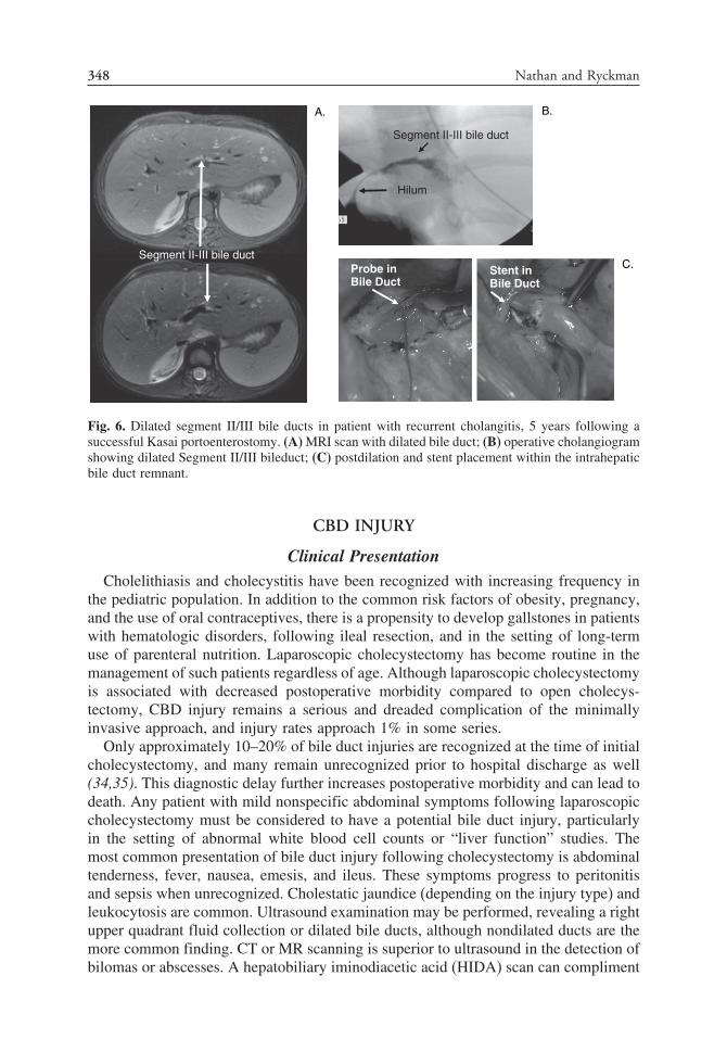
348 Nathan and Ryckman
B. A.
Stent inBile Duct
C. Probe inBile Duct
Hilum
Segment II-III bile duct
Segment II-III bile duct
Fig. 6. Dilated segment II/III bile ducts in patient with recurrent cholangitis, 5 years following asuccessful Kasai portoenterostomy. (A) MRI scan with dilated bile duct; (B) operative cholangiogramshowing dilated Segment II/III bileduct; (C) postdilation and stent placement within the intrahepaticbile duct remnant.
CBD INJURY
Clinical PresentationCholelithiasis and cholecystitis have been recognized with increasing frequency in
the pediatric population. In addition to the common risk factors of obesity, pregnancy,and the use of oral contraceptives, there is a propensity to develop gallstones in patientswith hematologic disorders, following ileal resection, and in the setting of long-termuse of parenteral nutrition. Laparoscopic cholecystectomy has become routine in themanagement of such patients regardless of age. Although laparoscopic cholecystectomyis associated with decreased postoperative morbidity compared to open cholecys-tectomy, CBD injury remains a serious and dreaded complication of the minimallyinvasive approach, and injury rates approach 1% in some series.
Only approximately 10–20% of bile duct injuries are recognized at the time of initialcholecystectomy, and many remain unrecognized prior to hospital discharge as well(34,35). This diagnostic delay further increases postoperative morbidity and can lead todeath. Any patient with mild nonspecific abdominal symptoms following laparoscopiccholecystectomy must be considered to have a potential bile duct injury, particularlyin the setting of abnormal white blood cell counts or “liver function” studies. Themost common presentation of bile duct injury following cholecystectomy is abdominaltenderness, fever, nausea, emesis, and ileus. These symptoms progress to peritonitisand sepsis when unrecognized. Cholestatic jaundice (depending on the injury type) andleukocytosis are common. Ultrasound examination may be performed, revealing a rightupper quadrant fluid collection or dilated bile ducts, although nondilated ducts are themore common finding. CT or MR scanning is superior to ultrasound in the detection ofbilomas or abscesses. A hepatobiliary iminodiacetic acid (HIDA) scan can compliment

Chapter 20 / Reoperation of the Liver and Biliary System 349
ultrasound or CT scanning by providing evidence for a complete biliary obstructionor bile leak. Percutaneous drainage of a right upper quadrant collection that yieldsbilious fluid will confirm an injury. Endoscopic retrograde cholangiopancreatography(ERCP) can localize the site and extent of bile duct injury, and is also useful as atherapeutic modality for nasobiliary stent placement and sphincterotomy in the case ofductal stricture or leak. If ERCP fails to visualize the upper biliary ductal system, suchas in the case of a complete obstruction, percutaneous transhepatic cholangiography(PTC) is utilized to visualize the upper tracts.
Clinical ClassificationThe spectrum of bile duct injuries following laparoscopic cholecystectomy includes
biliary leaks, strictures, ligation, and transection. Several classification systems havebeen proposed to describe this wide array of bile duct injuries. Originating in thetime of open cholecystectomy, the Corlette-Bismuth classification system is based onthe proximal biliary stump length, but does not allow for description of the type orextent of injury (36). More recently devised, the Strasberg classification system offersa more descriptive and detailed schema: Type A, bile leak from cystic duct or liverbed; Type B, partial occlusion of the biliary tree (often an aberrant right hepatic duct);Type C, bile leak from a duct not communicating with the CBD; Type D, lateral injuryto the biliary tree without loss of continuity; and type E, circumferential injury to thebiliary tree with loss of continuity (37) (Fig. 7).
Operative ManagementIf a bile duct injury is detected at the time of laparoscopic cholecystectomy, and the
surgeon is not comfortable with biliary reconstruction, dissection should be minimized,drains placed to control leak, and the patient transferred to an institution with suffi-cient experience in reconstructive hepatobiliary surgery. If biliary reconstruction isconsidered at the time of initial cholecystectomy, the procedure should be convertedto an open operation in order to provide optimal exposure of the biliary system fordetermination of injury extent and severity. Intraoperative cholangiography is indicatedto accurately identify the biliary anatomy. Small lateral ductal injuries can often berepaired primarily over a T-tube. Complete transection of a major bile duct should berepaired with a Roux-en-Y hepatico-jejunostomy, or in circumstances in which thereis little associated damage or tissue loss, may be suitable for a primary end-to-endanastomosis over a T-tube. CBD injuries that involve resection of part of the ductare not amenable to primary repair, and in most such cases, adequate drainage withreferral for elective reconstruction is advised.
More than 75% of patients with bile duct injuries related to laparoscopic chole-cystectomy will present with a delay, ranging from days to months (34,38). Oncebile duct injury is identified, the initial management includes control of sepsis withantibiotics, percutaneous drainage if a biloma or leak is present, and decompression ofthe biliary system by ERCP or by percutaneous transhepatic biliary drainage catheterplacement. Following stabilization and treatment of sepsis, classification of the injuryand identification of the biliary anatomy should be performed prior to reexplorationfor reconstruction. MRCP has emerged as a biliary imaging modality that rivals theanatomic detail provided by the more invasive ERCP or PTC (39).
The timing of hepatobiliary reconstruction after delayed presentation of bile ductinjury is an important contributor to surgical outcome. In patients identified within
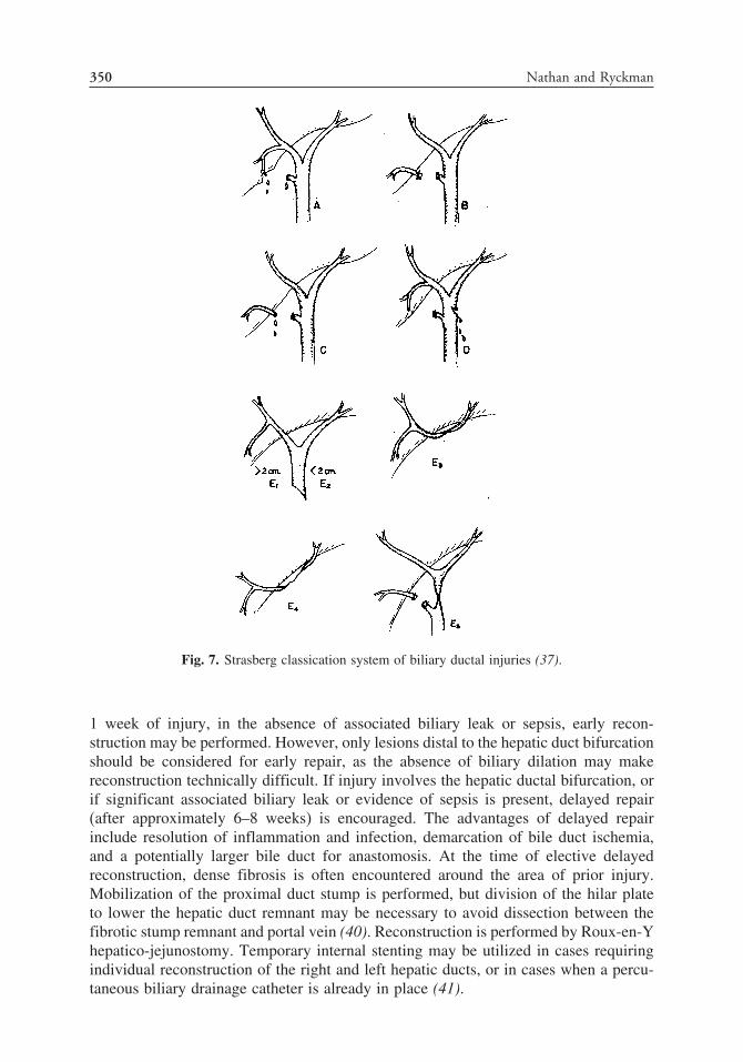
350 Nathan and Ryckman
Fig. 7. Strasberg classication system of biliary ductal injuries (37).
1 week of injury, in the absence of associated biliary leak or sepsis, early recon-struction may be performed. However, only lesions distal to the hepatic duct bifurcationshould be considered for early repair, as the absence of biliary dilation may makereconstruction technically difficult. If injury involves the hepatic ductal bifurcation, orif significant associated biliary leak or evidence of sepsis is present, delayed repair(after approximately 6–8 weeks) is encouraged. The advantages of delayed repairinclude resolution of inflammation and infection, demarcation of bile duct ischemia,and a potentially larger bile duct for anastomosis. At the time of elective delayedreconstruction, dense fibrosis is often encountered around the area of prior injury.Mobilization of the proximal duct stump is performed, but division of the hilar plateto lower the hepatic duct remnant may be necessary to avoid dissection between thefibrotic stump remnant and portal vein (40). Reconstruction is performed by Roux-en-Yhepatico-jejunostomy. Temporary internal stenting may be utilized in cases requiringindividual reconstruction of the right and left hepatic ducts, or in cases when a percu-taneous biliary drainage catheter is already in place (41).
Chapter 20 / Reoperation of the Liver and Biliary System 351
Long-Term IssuesFactors that predict a successful outcome following operative repair of bile duct
injuries include a tension-free repair, anastomosis involving a viable and well-vascularized duct, and preservation of the hepatic duct bifurcation. The occurrence oflate postoperative strictures months to years following biliary reconstruction is relatedto several factors: local ischemia, thermal injury caused by electrocautery use near thebile duct, and inflammation and fibrosis owing to bile leak (42). In addition, approxi-mately 50% of patients who undergo end-to-end biliary anastomosis over a T-tube willdevelop a late stricture. Anastomotic stenosis or stricture can be asymptomatic, or itmay present with jaundice, cholangitis, and portal hypertension. In the most advancedcases, the liver dysfunction progresses to secondary biliary cirrhosis and eventual end-stage liver disease. The mainstay of management of late strictures following biliaryreconstruction is percutaneous biliary dilation. In one recent study, approximately 60%of patients with biliary-enteric anastomotic stenosis after reconstruction for bile ductinjury were successfully managed by dilation and did not require subsequent inter-vention over a mean follow-up period of 76 months (43). Up to 5% of patients mayrequire eventual liver transplantation after failed operative revision or balloon dilationof the hepaticojejunostomy.
RETAINED COMMON DUCT STONES
Clinical PresentationCholedocholithiasis is uncommon in the pediatric population, with an incidence
of approximately 6–13% in children with cholelithiasis (44–46). Congenital CBDanomalies, such as choledochal cysts, pancreatico-biliary maljunction, and preduodenalposition of the CBD, are often found in association with choledocholithiasis in thispopulation of patients. In adults undergoing CBD exploration for choledocholithiasisat the time of cholecystectomy, the incidence of retained CBD stones postopera-tively ranges from 8% following laparoscopic cholecystectomy to 14% following opencholecystectomy (47,48). The overall incidence of retained CBD stones followinglaparoscopic cholecystectomy in adults is approximately 0.7%. The incidence ofretained CBD stones in children remains unknown at this time. Furthermore, thereis little consensus with regard to the management of this problem in the pediatricpopulation, and suggested guidelines have been applied based on the experiencein adults.
Choledocholithiasis can present with a variety of symptoms, and diagnosis on thebasis solely of history and physical examination is often unreliable. Symptoms ofintermittent right upper quadrant pain, often postprandial, with associated nausea andemesis, and a history of fatty food intolerance, are suggestive of biliary tract pathology,but are nonspecific. The spectrum of clinical presentations of choledocholithiasisincludes obstructive jaundice, cholangitis, and acute pancreatitis, and in advanceddisease, CBD stones may present with sepsis. The classic triad of right upper quadrantpain, jaundice, and fever and chills, known as Charcot’s triad, is highly suggestive ofcholangitis, and choledocholithiasis is a common predisposing cause. When cholangitisbecomes severe, patients may also present with hypotension and mental status changes,known as Reynold’s pentad.

352 Nathan and Ryckman
Diagnostic EvaluationIn patients who present following cholecystectomy with symptoms suggestive of
retained CBD stones, a complete blood count, “liver function” studies, amylase, andlipase should be evaluated. Elevations in alkaline phosphatase, GGT, and conjugatedbilirubin are highly suggestive of a retained common duct stone. It should be noted,however, that the negative predictive value of normal laboratory studies in choledo-cholithiasis is low, and a high index of suspicion must be maintained based on clinicalpresentation (49). The white blood cell count and serum amylase level may be elevatedin choledocholithiasis, even in the absence of cholangitis and pancreatitis.
In patients in whom a retained common duct stone is suspected following priorcholecystectomy, transabdominal ultrasound may be performed as an initial radiologictool. In the appropriate clinical setting, the presence of a dilated biliary system onultrasound is highly suggestive of a retained common duct stone, and in some cases, aCBD stone may be identified (Fig. 8). A CBD diameter of 10 mm or greater on ultra-sound examination has been found to be highly prognostic of choledocholithiasis (50).The accuracy of transabdominal ultrasound in the diagnosis of retained common ductstones is closely related to the experience of the examiner (51).
Magnetic resonance cholangiography (MRC) and helical computed tomographycholangiography (HCT-C) are more sensitive for the diagnosis of CBD stones. MRCis noninvasive and not operator-dependent, and its sensitivity and specificity in thediagnosis of common duct stones range from 88–98% and 84–100%, respectively(52–55). Helical CT scanning is another noninvasive option; however, risks of this
Fig. 8. Transabdominal ultrasound demonstrating gallstone (arrow) in the CBD.

Chapter 20 / Reoperation of the Liver and Biliary System 353
modality include radiation exposure and allergic reaction to intravenous contrastmaterial. Its sensitivity (85–88%) and specificity (88–96%) are similar to MRC, andthis modality may be considered in the diagnosis of choledocholithiasis in centerswithout MR capability (54,56,57).
ERCP remains the gold standard in the diagnosis of choledocholithiasis, and isalso an important therapeutic modality for the management of retained CBD stones(Fig. 9). Both the sensitivity and specificity of ERCP in the detection of choledo-cholithiasis range from 95–100% in the adult population (58,59). A disadvantage ofERCP is its invasiveness and risk of complications, including bleeding, perforation,pancreatitis, infection, and adverse events related to sedation and anesthesia. Thesefactors have led to the recommendation that MRC be used when there is a low index ofsuspicion for choledocholithiasis, and ERCP be used in patients with a high likelihoodof having common duct stones (60). Despite the low incidence of biliary disease inthe pediatric population and limitations related to equipment size, ERCP success ratesand complication rates are comparable in children compared to adults (61,62).
ManagementThe initial step in the management of patients with retained CBD stones following
cholecystectomy is the initiation of antibiotic therapy if cholangitis is present. Antibioticcoverage should consist of an agent that is active against enteric microorganisms, as
Fig. 9. Endoscopic retrograde cholangiogram demonstrating choledocholithiasis and CBD dilation.
354 Nathan and Ryckman
the bacteria that are typically identified include Escherichia coli, Klebsiella species,Clostridium species, Streptococcus faecalis, and Enterobacter species.
In patients who present post-cholecystectomy with retained common duct stones,decompression of the biliary tree is critical, and must be performed urgently in thesetting of cholangitis, pancreatitis, or sepsis. The condition of the patient will oftendictate the best approach to acute ductal decompression. In patients who are clinicallystable without pancreatitis, ERCP can be considered in experienced hands. Patientssuffering from clinical instability from sepsis are best addressed by PTC and externalstent placement. This allows access to the obstructed biliary tree and decompression,allowing the treatment of sepsis and pancreatitis prior to instrumenting the ampulla ofvater and injecting the pancreaticobiliary ductal system.
ERCP with endoscopic sphincterotomy and stone extraction is commonly performedfor the acute treatment of the obstructed duct in stable patients. Several techniqueshave evolved for stone extraction, including balloons, baskets, and lithotripsy devices.Using conventional endoscopic techniques, the success rate for the complete clearanceof the CBD is approximately 90% (63). The largest diameter stone that can be retrievedis dependent on the size of the distal CBD and the sphincterotomy (64). Large stonesrequire fragmentation by lithotripsy techniques (mechanical, laser, or electrohydraulic)prior to extraction (65,66).
If retrieval of a retained common duct stone fails by endoscopic techniques, a secondoption is to return to the operating room for a laparoscopic or open CBD exploration(CBDE). CBDE may be performed via a transcystic approach, in which Fogarty ballooncatheters and Dormia wire baskets may be advanced into the distal common duct viathe cystic duct stump. Intraoperative fluoroscopy is often used as an adjunct, and ifthese maneuvers fail, then they may be repeated under direct vision with passage of acholedochoscope also placed via a transcystic approach. The ducts may be forcefullyirrigated with saline to promote passage of small stones and debris, and large stones(>1 cm) may require fragmentation by lithotripsy techniques. Common hepatic orintrahepatic duct stones are difficult to retrieve via the transcystic approach because ofthe acute angle between the cystic duct and common hepatic duct. Transcystic CBDEis successful in stone retrieval in more than 80% of patients (67–69).
CBDE may also be performed via a choledochotomy, and this approach is often usedin the setting of large stones, a CBD that is greater than 10 mm in diameter, or a cysticduct that cannot be dilated large enough to allow passage of the choledochoscope.This approach allows more effective stone clearance of the biliary tree proximal to theinsertion of the cystic duct, and probably is a better option for the more difficult cases ofretained stones, such as a stone impacted at the ampulla. A longitudinal choledochotomyis made as small as possible, the choledochoscope is inserted, and similar techniquesdescribed for transcystic CBDE are utilized to remove stones (70) (Fig. 10). Antegradesphincterotomy may be performed if necessary through the choledochoscope (71,72).The technique of closure of the choledochotomy is critical in order to avoid late biliarystricture. Closure of the CBD is performed using interrupted absorbable 5.0 or 6.0monofilament suture, and the use of a T-tube remains controversial (73,74).
Overall, the success rate of laparoscopic CBD clearance ranges from 73–100%, andthe literature suggests that there is no significant difference in success rate between thetranscystic versus choledochotomy approach (75,76). Conversion to an open CBDE isrequired in approximately 5% of cases. A cystic duct tube or a T-tube choledochotomyclosure may be performed in cases of incomplete clearance of the bile duct, and
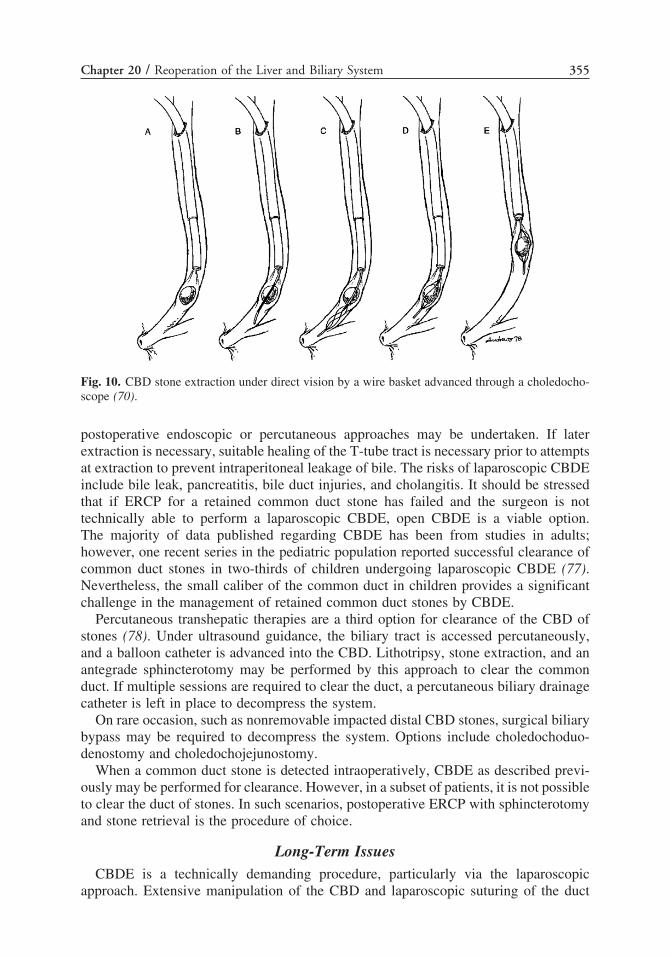
Chapter 20 / Reoperation of the Liver and Biliary System 355
Fig. 10. CBD stone extraction under direct vision by a wire basket advanced through a choledocho-scope (70).
postoperative endoscopic or percutaneous approaches may be undertaken. If laterextraction is necessary, suitable healing of the T-tube tract is necessary prior to attemptsat extraction to prevent intraperitoneal leakage of bile. The risks of laparoscopic CBDEinclude bile leak, pancreatitis, bile duct injuries, and cholangitis. It should be stressedthat if ERCP for a retained common duct stone has failed and the surgeon is nottechnically able to perform a laparoscopic CBDE, open CBDE is a viable option.The majority of data published regarding CBDE has been from studies in adults;however, one recent series in the pediatric population reported successful clearance ofcommon duct stones in two-thirds of children undergoing laparoscopic CBDE (77).Nevertheless, the small caliber of the common duct in children provides a significantchallenge in the management of retained common duct stones by CBDE.
Percutaneous transhepatic therapies are a third option for clearance of the CBD ofstones (78). Under ultrasound guidance, the biliary tract is accessed percutaneously,and a balloon catheter is advanced into the CBD. Lithotripsy, stone extraction, and anantegrade sphincterotomy may be performed by this approach to clear the commonduct. If multiple sessions are required to clear the duct, a percutaneous biliary drainagecatheter is left in place to decompress the system.
On rare occasion, such as nonremovable impacted distal CBD stones, surgical biliarybypass may be required to decompress the system. Options include choledochoduo-denostomy and choledochojejunostomy.
When a common duct stone is detected intraoperatively, CBDE as described previ-ously may be performed for clearance. However, in a subset of patients, it is not possibleto clear the duct of stones. In such scenarios, postoperative ERCP with sphincterotomyand stone retrieval is the procedure of choice.
Long-Term IssuesCBDE is a technically demanding procedure, particularly via the laparoscopic
approach. Extensive manipulation of the CBD and laparoscopic suturing of the duct

356 Nathan and Ryckman
may result in late complications, such as CBD stricture. Factors that may contributeto late stricture formation following CBDE include devascularization of the ductduring dissection and the creation of a choledochotomy in a common duct that has adiameter of less than 7 mm. Studies have also indicated that late strictures may occurfollowing endoscopic sphincterotomy for the management of choledocholithiasis, andthe very long-term outcome of patients who have undergone sphincterotomy is not yetknown (79). Strictures following CBDE or endoscopic sphincterotomy are typicallyaddressed by endoscopic balloon dilation and biliary stent placement.
Other groups have reported a small incidence of late recurrent CBD stones followinglaparoscopic CBDE (80–82). However, on the basis of high initial success ratesin complete duct clearance and the low rate of morbidity following laparoscopicCBDE (ranging from 3–7%), these authors have strongly advocated this “single-stage”approach to managing choledocholithiasis detected intraoperatively at the time ofcholecystectomy.
Although these evolving techniques have shown good success in experienced hands,the potential for injury and a poor outcome is high when these advanced laparoscopicskills are not well developed. It is incumbent on the operating surgeon to honestlyassess their personal experience and address these difficult cases in the fashion thatallows for the greatest chance for an initial excellent outcome, regardless of the natureof the surgical approach and exposure.
PORTAL HYPERTENSION
Portosystemic ShuntsNumerous surgical procedures have been devised to divert portal blood into the low-
pressure systemic venous circulation, thereby decreasing the portal venous pressure.Enthusiasm for the use of portosystemic shunting in children was limited by earlyreports suggesting that children less than 8 years of age, and those with vessels forthe shunt anastomosis less than 8 to 10 mm, would be unsuitable candidates becauseof the high risk of shunt thrombosis (83). In addition, Voorhees et al. suggesteda high incidence of neuropsychiatric disturbances following nonselective shunts inchildren (84). Recent experience in centers skilled in pediatric vascular reconstructionhas established that a high rate of success can be achieved with minimal complica-tions even in small pediatric patients. To achieve these goals, we have found thatthe following principles should be followed: (1) anastomosis should be constructedusing fine (6.0–7.0) monofilament sutures with provision for growth; (2) sufficientmobilization of vessels is necessary to prevent kinking or twisting of the shunt afterthe viscera are returned to their normal location; (3) predivision marking of the vesselwall along the long axis will prevent unappreciated rotation of the vessel prior tothe anastomosis, (4) postreconstructive venography should be performed in order toensure division of all collaterals and adequate shunt flow; (5) selective postoperativeanticoagulation should be performed and antiplatelet drugs administered; and (6) thesurgical team should be skilled at pediatric portal vascular reconstruction.
Primary Surgical OptionsIn general, portosystemic shunts can be classified into three groups: non-selective
and selective shunts, and direct reconstructions of the portal circulation using vasculargrafts (shunts).
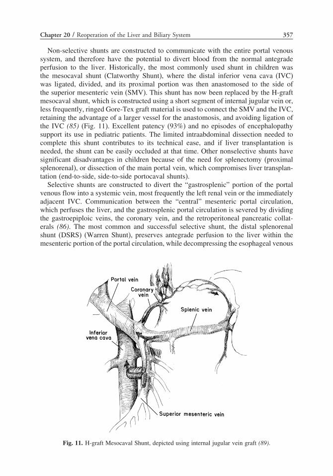
Chapter 20 / Reoperation of the Liver and Biliary System 357
Non-selective shunts are constructed to communicate with the entire portal venoussystem, and therefore have the potential to divert blood from the normal antegradeperfusion to the liver. Historically, the most commonly used shunt in children wasthe mesocaval shunt (Clatworthy Shunt), where the distal inferior vena cava (IVC)was ligated, divided, and its proximal portion was then anastomosed to the side ofthe superior mesenteric vein (SMV). This shunt has now been replaced by the H-graftmesocaval shunt, which is constructed using a short segment of internal jugular vein or,less frequently, ringed Gore-Tex graft material is used to connect the SMV and the IVC,retaining the advantage of a larger vessel for the anastomosis, and avoiding ligation ofthe IVC (85) (Fig. 11). Excellent patency (93%) and no episodes of encephalopathysupport its use in pediatric patients. The limited intraabdominal dissection needed tocomplete this shunt contributes to its technical ease, and if liver transplantation isneeded, the shunt can be easily occluded at that time. Other nonselective shunts havesignificant disadvantages in children because of the need for splenectomy (proximalsplenorenal), or dissection of the main portal vein, which compromises liver transplan-tation (end-to-side, side-to-side portocaval shunts).
Selective shunts are constructed to divert the “gastrosplenic” portion of the portalvenous flow into a systemic vein, most frequently the left renal vein or the immediatelyadjacent IVC. Communication between the “central” mesenteric portal circulation,which perfuses the liver, and the gastrosplenic portal circulation is severed by dividingthe gastroepiploic veins, the coronary vein, and the retroperitoneal pancreatic collat-erals (86). The most common and successful selective shunt, the distal splenorenalshunt (DSRS) (Warren Shunt), preserves antegrade perfusion to the liver within themesenteric portion of the portal circulation, while decompressing the esophageal venous
Fig. 11. H-graft Mesocaval Shunt, depicted using internal jugular vein graft (89).

358 Nathan and Ryckman
Fig. 12. Distal splenorenal (Warren) shunt (89).
plexus through the short gastric veins and splenic vein (Fig. 12). We use this shunt asour primary option in children where direct reconstruction is not possible. When theadrenal vein is appropriately located and dilated, it serves as an alternative anastomoticsite to access the left renal vein (87,88). When performed in centers experienced incomplex vascular reconstruction of the portal system, as is necessary in pediatric livertransplantation, shunt patency has ranged from 83–100% (86,89–91).
Direct reconstruction of the portal circulation in children with extrahepatic portalvein obstruction into the left branch of the portal vein represents the ideal solution(92–95). This mesenterico-portal shunt (REX Shunt) reestablishes normal portal inflowinto the intrahepatic portal vein, using either an interposition jugular venous graft orthe dilated coronary vein (Fig. 13). Candidates for this procedure should fulfill threeconditions: the liver parenchyma must be normal, they must not have a hypercoagulablestate, and the umbilical portion of the left hepatic vein must be accessible and patent.Patients with diffuse intrahepatic portal vein thrombosis are not good candidates for thisreconstruction. Doppler ultrasound and direct portography would suggest, however,that at least two-thirds of children with extrahepatic portal venous obstruction (EPVO)have sufficient left portal vein patency to undergo this procedure. Experience withthis shunt is less extensive. Initial patency seems excellent; however, later revisionsecondary to stenosis or occlusion is somewhat more common (92–98). Although thisis a more complex technical challenge, in these selected patients, this option should be

Chapter 20 / Reoperation of the Liver and Biliary System 359
Fig. 13. REX mesentericoportal venous reconstruction (89).
used in preference to other true “shunts” that divert portal blood rather the reconstructthe normal portal flow patterns.
Indication for ReoperationThe most common technical problem encountered following portacaval shunt
construction is shunt thrombosis. When the reconstructive techniques outlined previ-ously are carefully followed, this is fortunately an uncommon event occurring inless than 5% of all children undergoing primary portacaval shunting. Because of thetechnical complexity of the Rex shunt, this appears to be slightly more commonin these patients.
Appreciation of postoperative shunt thrombosis can occur either through routineDoppler ultrasound surveillance or less commonly postoperative bleeding. It is ourpractice to do routine immediate postoperative ultrasound with Doppler vascular studiesto assure adequate blood flow following abdominal closure and periodic surveillanceduring the first postoperative week.

360 Nathan and Ryckman
When approaching a patient with documented thrombosis, a thorough reevaluationof the most likely responsible factors should be undertaken. The first of these factorsis vascular inflow. Because portal flow is substantial, it is very uncommon to have alimitation of portal inflow as a primary etiology of shunt thrombosis. Unique circum-stances such as patients with diffuse multifocal mesentery venous thrombosis maylimit portal inflow but are rarely the primary factor inducing thrombosis. Appro-priate assessment of inflow at the time of primary shunt placement should avoid thisrare circumstance. However, disturbance of shunt in flow rate because of twisting orkinking within the shunt itself is a significant contributor to early shunt thrombosis.This technical abnormality is best prevented by careful orientation of the vessels priorto undertaking and the initial anastomosis. It is our practice to mark the longitudinalsurface of any vein or graft that is used so that appropriate orientation can be maintainedat the time of the anastomosis. In circumstances where kinking or a rotational twistof the vessel has occurred, immediate thrombectomy and revision of the anastomosisto remove this obstruction is necessary. It is important to carefully survey the shuntfollowing the initial anastomosis to assure that subtle rotational abnormalities have notoccurred. In circumstances where an extremely difficult anastomosis is anticipated,such as the left portal vein anastomosis with the Rex shunt, we find that constructionof the distal anastomosis first allows more successful orientation of the interpositiongraft prior to completing the less complex proximal portal anastomosis at the levelof the mesentery. In cases where a difficult distal anastomosis would be difficult torevise, the free portion of the mobilized splenic or mesenteric vein can be divided,rotated, and reanastomosed at a safer location within the shunt. Although this is rarelynecessary, rotation and reconstruction of the shunt in a safer middle position of thevessel is preferential to attempting to revise or redo a delicate and technically complexdistal anastomosis.
The second technical factor related to shunt thrombosis occurs at the level of theanastomosis. Anastomotic stenosis or poor endothelial approximation contribute tothrombosis in portacaval shunts as in all vascular reconstructive procedures. Again,prevention at the time of the primary operation plays a key rule. The thin vesselwall and extreme flexibility of the portal venous system make it uniquely proneto anastomotic stenosis unless specific technical points are followed. In addition, aprovision for growth should be incorporated into each of these anastomoses in children.Meticulous attention to these technical details will substantially decrease the need foranastomotic revision. We follow several principles in constructing or critical venousanastomoses: (1) we use monofilament 6.0 or 7.0 sutures for all portal anastomoses;(2) if we use a running suture for the difficult to access posterior anastomotic sutureline, we apply lateral tension on both lateral ends of the anastomosis at the time thesutures is tied to prevent “telescoping” of the suture line, which creates posterior wallshortening and stenosis; (3) in all patients where growth is anticipated, the anterioranastomotic suture line is constructed using interrupted sutures. In cases where theanastomotic contour is unclear following reconstruction, an intraoperative venogrammay be helpful.
The final factor that may compromise shunt flow and hence precipitate thrombosisis shunt outflow. The patients with highest risk for elevated post shunt venous pressureare those undergoing Rex reconstruction. In these cases, initial capacitance of theportal veins within the liver is somewhat unpredictable and often limited. Studies havedocumented rapid and substantial improvements in the outflow portal venous size and

Chapter 20 / Reoperation of the Liver and Biliary System 361
capacity following the Rex shunt, but this initial limitation of outflow undoubtedlycontributes to the risk of early thrombosis in this operation. Portal venography of theintrahepatic portal vein may be helpful at the time of initial operation in assessing thiscapacity; however, it does not appear to be predictive of success or risk of thrombosis.In patients undergoing distal splenorenal or H-graft mesocaval shunts, outflow pressureis determined by central venous pressure within the IVC. Unique circumstances causingIVC pressure to be elevated can compromise outflow. These include patients witha cirrhotic compression of the retrohepatic IVC, a condition most commonly seenwith Budd-Chiari syndrome. In this unique circumstance, caval-atrial bypass may benecessary as a second component to portacaval shunting. Children with elevated rightheart pressures secondary to congenital heart disease can present a similar challenge.In these patients, control of the central venous pressure is best undertaken throughimproved medical management of their cardiac disease.
When portal reconstruction is undertaken respecting the previously describedprinciples, postoperative anticoagulation is rarely necessary. Our present protocolincludes the use of postoperative antiplatelet treatment with low-dose aspirin for 30 to90 days. Direct anticoagulation using intermittent subcutaneous or intravenous heparinis restricted to patients with preexisting hypercoagulable states until their baselineanticoagulation can be reestablished. In addition, in patients where shunt reconstructionwas necessary because of initial thrombosis, intravenous anticoagulation for severalpostoperative days may be undertaken at the surgeon’s discretion. All patients withprimary portal/mesenteric venous thrombosis and those with postoperative thrombosisshould undergo a complete evaluation of their coagulation system to identify hyperco-agulability and institute appropriate treatment prior to shunting.
Transjugular Intrahepatic Portosystemic Shunt (TIPS)The introduction of TIPS has added another therapeutic option for the physician
confronted with complex portal hypertension. This procedure uses interventionalradiographic techniques to place an intrahepatic expandable metallic shunt betweena portal vein branch and the hepatic vein, forming a central nonselective portocavalshunt. The procedure is undertaken using access via the right internal jugular vein.The hepatic veins are identified and a fluoroscopic/ultrasonographic-guided puncturefrom the hepatic vein into an intrahepatic portion of the portal vein is undertaken.This tract is then dilated and an expandable mesh stent is placed, forming a commu-nication between the intrahepatic portal vein and the hepatic vein branch (Fig. 14).
Fig. 14. TIPS (transjugular intrahepatic portosystemic shunt) (89).

362 Nathan and Ryckman
Technical difficulties in establishing a safe but large enough tract for sufficient shuntflow limit the usefulness of this procedure in infants. In children with biliary atresia,the close proximity of the biliary Roux-en-Y conduit to the portal vein and the oftendiminutive size of the portal vein increase the risk of stent perforation, malposition,or perforation (89). This procedure has great benefit in the control of refractory portalhypertensive bleeding unresponsive to the pharmacologic methods, and in patientsneeding temporary portal decompression prior to liver transplantation (99). The abilityto embolize bleeding varices from the coronary vein at the time of TIPS placementassists in achieving primary control of bleeding sites.
The two principal long-term complications of TIPS are encephalopathy and shuntocclusion. Being a central nonselective shunt, this procedure can precipitate hepaticencephalopathy, especially when used in patients with severe intrinsic liver disease.The overall risk of encephalopathy ranges from 5–35% in adult patients, a rate similarto that seen with side-to-side surgical shunts. Most episodes of encephalopathy canbe controlled with dietary protein restriction and lactulose administration. Postproce-dural encephalopathy seems to be less common in children, although limited clinicalexperience and the difficulties in diagnosing subtle encephalopathy in children makesthis observation tentative. Selection of a shunt size to allow sufficient portal decom-pression without shunting excessive amounts of blood from the liver is also a theoreticalsolution.
Stenosis of the shunt or shunt thrombosis remains a major complication followingTIPS. Shunt stenosis occurs in 25–75% of cases, with shunt patency decreasing withthe length of time that the shunt is in place. Intimal hyperplasia or incorrect shuntplacement most commonly causes the stenosis. Regular monitoring for shunt patencyand periodic shunt dilation or restenting is necessary (100). Shorter stent lengths andsmaller diameter stents have been constructed for pediatric applications; however,the risks of hepatic perforation and stent malposition are greater in small patients.The complications of shunt stenosis are equally problematic, and patient growth overtime may cause the initial shunt to be too short, requiring revision or restenting tomaintain access to both the portal and hepatic venous circulation. These limitations andrisks make TIPS a reasonable and suitable treatment for acute unresponsive varicealhemorrhage in children with established intrinsic liver disease, often while awaitingliver transplantation. It is particularly helpful when used as a bridge to achieve stabilityby controlling refractory hemorrhage in patients awaiting liver transplantation. Long-term decompression is better achieved through surgical shunts at the present time, andTIPS is not indicated in patients with EPVO.
REFERENCES
1. Hays DM, Goodman GN, Snyder WH Jr, Woolley MM. Congenital cystic dilatation of the commonbile duct. Arch Surg 1969;98(4):457–461.
2. Alonso-Lej F, Rever WB Jr, Pessagno DJ. Congenital choledochal cyst, with a report of 2, and ananalysis of 94, cases. Int Abstr Surg 1959;108(1):1–30.
3. Todani T, Watanabe Y, Narusue M, et al. Congenital bile duct cysts: classification, operativeprocedures, and review of thirty-seven cases including cancer arising from choledochal cyst. Am JSurg 1977;134(2):263–269.
4. Todani T, Narusue M, Watanabe Y, et al. Management of congenital choledochal cyst with intra-hepatic involvement. Ann Surg 1978;187(3):272–280.
5. Karrer F, Pence J. Biliary atresia and choledochal cyst. In: Ziegler M, Azizkhan R, Weber T, (eds.),Operative pediatric surgery. New York: McGraw-Hill; 2003: 775–788.

Chapter 20 / Reoperation of the Liver and Biliary System 363
6. Miyano T, Yamataka A. Choledochal cysts. Curr Opin Pediatr 1997;9(3):283–288.7. Miyano T. Biliary tract disorders and portal hypertension. In: Ashcraft K, Holcomb G, Murphy J,
(eds.), Pediatric surgery. Philadelphia: Elsevier Sanders; 2005: 586–608.8. Kobayashi A, Ohbe Y. Choledochal cyst in infancy and childhood. Analysis of 16 cases. Arch Dis
Child 1977;52(2):121–128.9. Dewbury KC, Aluwihare AP, Birch SJ, Freeman NV. Prenatal ultrasound demonstration of a
choledochal cyst. Br J Radiol 1980;53(633):906–907.10. Howell CG, Templeton JM, Weiner S, et al. Antenatal diagnosis and early surgery for choledochal
cyst. J Pediatr Surg 1983;18(4):387–393.11. Oldham KT, Hart MJ, White TT. Choledochal cysts presenting in late childhood and adulthood. Am
J Surg 1981;141(5):568–571.12. Todani T, Watanabe Y, Toki A, Urushihara N. Carcinoma related to choledochal cysts with internal
drainage operations. Surg Gynecol Obstet 1987;164(1):61–64.13. Lilly JR. The surgical treatment of choledochal cyst. Surg Gynecol Obstet 1979;149(1):36–42.14. Todani T, Watanabe Y, Urushihara N, et al. Biliary complications after excisional procedure for
choledochal cyst. J Pediatr Surg 1995;30(3):478–481.15. Miyano T, Yamataka A, Kato Y, et al. Hepaticoenterostomy after excision of choledochal cyst in
children: a 30-year experience with 180 cases. J Pediatr Surg 1996;31(10):1417–1421.16. Komi N, Tamura T, Miyoshi Y, et al. Histochemical and immunohistochemical studies on devel-
opment of biliary carcinoma in forty-seven patients with choledochal cyst–special reference tointestinal metaplasia in the biliary duct. Jpn J Surg 1985;15(4):273–278.
17. Balistreri WF, Grand R, Hoofnagle JH, et al. Biliary atresia: current concepts and research directions.Summary of a symposium. Hepatology 1996;23(6):1682–1692.
18. McKiernan PJ, Baker AJ, Kelly DA. The frequency and outcome of biliary atresia in the UK andIreland. Lancet 2000;355(9197):25–29.
19. Serinet MO, Broue P, Jacquemin E, et al. Management of patients with biliary atresia in France:results of a decentralized policy 1986–2002. Hepatology 2006;44(1):75–84.
20. Hasegawa T, Kimura T, Sasaki T, et al. Indication for redo hepatic portoenterostomy for insufficientbile drainage in biliary atresia: re-evaluation in the era of liver transplantation. Pediatr Surg Int2003;19(4):256–259.
21. Shneider BL, Brown MB, Haber B, et al. A multicenter study of the outcome of biliary atresia inthe United States, 1997 to 2000. J Pediatr 2006;148(4):467–474.
22. Sugawara Y, Makuuchi M, Kaneko J, et al. Impact of previous multiple portoenterostomies on livingdonor liver transplantation for biliary atresia. Hepatogastroenterology 2004;51(55):192–194.
23. Sartorelli KH, Holland RM, Allshouse MJ, et al. The intussusception antireflux valve is ineffectivein preventing cholangitis in biliary atresia. J Pediatr Surg 1996;31(3):403–406.
24. Reynolds M, Luck SR, Raffensperger JG. The valved conduit prevents ascending cholangitis: afollow-up. J Pediatr Surg 1985;20(6):696–702.
25. Kaufman BH, Luck SR, Raffensperger JG. The evolution of a valved hepatoduodenal intestinalconduit. J Pediatr Surg 1981;16(3):279–283.
26. Kasai M, Suzuki H, Ohashi E, et al. Technique and results of operative management of biliaryatresia. World J Surg 1978;2(5):571–579.
27. Lilly JR. Hepatic portocholecystostomy for biliary atresia. J Pediatr Surg 1979;14(3):301–304.28. Matsuo S, Suita S, Kubota M, et al. Hazards of hepatic portocholecystostomy in biliary atresia. Eur
J Pediatr Surg 2001;11(1):19–23.29. Lilly JR, Stellin G. Catheter decompression of hepatic portocholecystostomy. J Pediatr Surg
1982;17(6):904–905.30. Crombleholme TM, Harrison MR, Langer JC, Longaker MT. Biliary appendico-duodenostomy: a
nonrefluxing conduit for biliary reconstruction. J Pediatr Surg 1989;24(7):665–667.31. Wei MF, Qi BQ, Xia GL, et al. Use of the appendix to replace the choledochus. Pediatr Surg Int
1998;13(7):494–496.32. Delarue A, Chappuis JP, Esposito C, et al. Is the appendix graft suitable for routine biliary surgery
in children? J Pediatr Surg 2000;35(9):1312–1316.33. Meister RK, Esquivel CO, Cox KL, et al. The influence of portoenterostomy with stoma on morbidity
in pediatric patients with biliary atresia undergoing orthotopic liver transplantation. J Pediatr Surg1993;28(3):387–390.
34. Way LW, Stewart L, Gantert W, et al. Causes and prevention of laparoscopic bile duct injuries:analysis of 252 cases from a human factors and cognitive psychology perspective. Ann Surg2003;237(4):460–469.

364 Nathan and Ryckman
35. Russell JC, Walsh SJ, Mattie AS, Lynch JT. Bile duct injuries, 1989–1993. A statewide experience.Connecticut Laparoscopic Cholecystectomy Registry. Arch Surg 1996;131(4):382–388.
36. Bismuth H. Post operative strictures of the bile ducts. In: Blumgart L (ed.), The biliary tract. NewYork: Churchill-Livingston; 1982: 209–218.
37. Strasberg SM, Hertl M, Soper NJ. An analysis of the problem of biliary injury during laparoscopiccholecystectomy. J Am Coll Surg 1995;180(1):101–125.
38. Adams DB, Borowicz MR, Wootton FT III, Cunningham JT. Bile duct complications after laparo-scopic cholecystectomy. Surg Endosc 1993;7(2):79–83.
39. Khalid TR, Casillas VJ, Montalvo BM, et al. Using MR cholangiopancreatography to evaluateiatrogenic bile duct injury. AJR Am J Roentgenol 2001;177(6):1347–1352.
40. Rossi RL, Tsao JI. Biliary reconstruction. Surg Clin North Am 1994;74(4):825–841; discussion843–844.
41. Mercado MA, Chan C, Orozco H, et al. Long-term evaluation of biliary reconstruction afterpartial resection of segments IV and V in iatrogenic injuries. J Gastrointest Surg 2006;10(1):77–82.
42. Shamiyeh A, Wayand W. Laparoscopic cholecystectomy: early and late complications and theirtreatment. Langenbecks Arch Surg 2004;389(3):164–171.
43. Misra S, Melton GB, Geschwind JF, et al. Percutaneous management of bile duct strictures andinjuries associated with laparoscopic cholecystectomy: a decade of experience. J Am Coll Surg2004;198(2):218–226.
44. Pokorny WJ, Saleem M, O’Gorman RB, et al. Cholelithiasis and cholecystitis in childhood. Am JSurg 1984;148(6):742–744.
45. Man DW, Spitz L. Choledocholithiasis in infancy. J Pediatr Surg 1985;20(1):65–68.46. Vrochides DV, Sorrells DL Jr, Kurkchubasche AG, et al. Is there a role for routine preoperative
endoscopic retrograde cholangiopancreatography for suspected choledocholithiasis in children? ArchSurg 2005;140(4):359–361.
47. Rhodes M, Nathanson L, O’Rourke N, Fielding G. Laparoscopic exploration of the common bileduct: lessons learned from 129 consecutive cases. Br J Surg 1995;82(5):666–668.
48. Hall RC, Sakiyalak P, Kim SK, et al. Failure of operative cholangiography to prevent retainedcommon duct stones. Am J Surg 1973;125(1):51–63.
49. Goldman DE, Gholson CF. Choledocholithiasis in patients with normal serum liver enzymes. DigDis Sci 1995;40(5):1065–1068.
50. Sgourakis G, Dedemadi G, Stamatelopoulos A, et al. Predictors of common bile duct lithiasis inlaparoscopic era. World J Gastroenterol 2005;11(21):3267–3272.
51. Rickes S, Treiber G, Monkemuller K, et al. Impact of the operator’s experience on value ofhigh-resolution transabdominal ultrasound in the diagnosis of choledocholithiasis: a prospectivecomparison using endoscopic retrograde cholangiography as the gold standard. Scand J Gastroenterol2006;41(7):838–843.
52. Regan F, Fradin J, Khazan R, et al. Choledocholithiasis: evaluation with MR cholangiography. AJRAm J Roentgenol 1996;167(6):1441–1445.
53. Reinhold C, Taourel P, Bret PM, et al. Choledocholithiasis: evaluation of MR cholangiography fordiagnosis. Radiology 1998;209(2):435–442.
54. Kondo S, Isayama H, Akahane M, et al. Detection of common bile duct stones: comparisonbetween endoscopic ultrasonography, magnetic resonance cholangiography, and helical-computed-tomographic cholangiography. Eur J Radiol 2005;54(2):271–275.
55. Shanmugam V, Beattie GC, Yule SR, et al. Is magnetic resonance cholangiopancreatography thenew gold standard in biliary imaging? Br J Radiol 2005;78(934):888–893.
56. Polkowski M, Palucki J, Regula J, et al. Helical computed tomographic cholangiography versusendosonography for suspected bile duct stones: a prospective blinded study in non-jaundiced patients.Gut 1999;45(5):744–749.
57. Okada M, Fukada J, Toya K, et al. The value of drip infusion cholangiography using multidetector-row helical CT in patients with choledocholithiasis. Eur Radiol 2005;15(10):2140–2145.
58. Cotton PB. Endoscopic retrograde cholangiopancreatography and laparoscopic cholecystectomy. AmJ Surg 1993;165(4):474–478.
59. Erickson RA, Carlson B. The role of endoscopic retrograde cholangiopancreatography in patientswith laparoscopic cholecystectomies. Gastroenterology 1995;109(1):252–263.
60. Onken JE, Brazer SR, Eisen GM, et al. Predicting the presence of choledocholithiasis in patientswith symptomatic cholelithiasis. Am J Gastroenterol 1996;91(4):762–767.

Chapter 20 / Reoperation of the Liver and Biliary System 365
61. Varadarajulu S, Wilcox CM, Hawes RH, Cotton PB. Technical outcomes and complications ofERCP in children. Gastrointest Endosc 2004;60(3):367–371.
62. Rocca R, Castellino F, Daperno M, et al. Therapeutic ERCP in paediatric patients. Dig Liver Dis2005;37(5):357–362.
63. Sauerbruch T. Endoscopic management of bile duct stones. J Gastroenterol Hepatol 1992;7(3):328–334.
64. Schutz SM, Baillie J. Endoscopic extraction of bile duct stones: management related to stone size.Gastrointest Endosc 1994;40(3):398–400.
65. Leung JW, Chung SC, Mok SD, Li AK. Endoscopic removal of large common bile duct stones inrecurrent pyogenic cholangitis. Gastrointest Endosc 1988;34(3):238–241.
66. Lo Menzo E, Schnall R, Von Rueden D. Lithotripsy in the laparoscopic era. JSLS 2005;9(3):358–361.
67. Hunter JG. Laparoscopic transcystic common bile duct exploration. Am J Surg 1992;163(1):53–56;discussion 57–58.
68. Giurgiu DI, Margulies DR, Carroll BJ, et al. Laparoscopic common bile duct exploration: long-termoutcome. Arch Surg 1999;134(8):839–843; discussion 843–844.
69. Hemli JM, Arnot RS, Ashworth JJ, et al. Feasibility of laparoscopic common bile duct explorationin a rural centre. ANZ J Surg 2004;74(11):979–982.
70. Vecchio R, MacFayden B, Miller T. Laparoscopic cholecystectomy and common bile duct explo-ration. In: Moody F, (ed.), Atlas of ambulatory surgery. Philadelphia: Saunders; 1999: 133–149.
71. DePaula AL, Hashiba K, Bafutto M, et al. Laparoscopic antegrade sphincterotomy. Semin LaparoscSurg 1997;4(1):42–44.
72. DePaula AL, Hashiba K, Bafutto M, et al. Laparoscopic antegrade sphincterotomy. Surg LaparoscEndosc 1993;3(3):157–160.
73. Franklin ME Jr, Pharand D, Rosenthal D. Laparoscopic common bile duct exploration. Surg LaparoscEndosc 1994;4(2):119–124.
74. Ha JP, Tang CN, Siu WT, et al. Primary closure versus T-tube drainage after laparoscopic choledo-chotomy for common bile duct stones. Hepatogastroenterology 2004;51(60):1605–1608.
75. Vecchio R, MacFadyen BV. Laparoscopic common bile duct exploration. Langenbecks Arch Surg2002;387(1):45–54.
76. Petelin JB. Laparoscopic common bile duct exploration. Surg Endosc 2003;17(11):1705–1715.77. Bonnard A, Seguier-Lipszyc E, Liguory C, et al. Laparoscopic approach as primary treatment of
common bile duct stones in children. J Pediatr Surg 2005;40(9):1459–1463.78. Nagashima I, Takada T, Shiratori M, et al. Percutaneous transhepatic papillary balloon dilation as a
therapeutic option for choledocholithiasis. J Hepatobiliary Pancreat Surg 2004;11(4):252–254.79. Pozsar J, Sahin P, Laszlo F, Topa L. Endoscopic treatment of sphincterotomy-associated distal
common bile duct strictures by using sequential insertion of multiple plastic stents. GastrointestEndosc 2005;62(1):85–91.
80. Waage A, Stromberg C, Leijonmarck CE, Arvidsson D. Long-term results from laparoscopiccommon bile duct exploration. Surg Endosc 2003;17(8):1181–1185.
81. Paganini AM, Lezoche E. Follow-up of 161 unselected consecutive patients treated laparoscopicallyfor common bile duct stones. Surg Endosc 1998;12(1):23–29.
82. Lezoche E, Paganini AM. Technical considerations and laparoscopic bile duct exploration: transcysticand choledochotomy. Semin Laparosc Surg 2000;7(4):262–278.
83. Orloff MJ, Orloff MS, Girard B, Orloff SL. Bleeding esophagogastric varices from extrahepaticportal hypertension: 40 years’ experience with portal-systemic shunt. J Am Coll Surg 2002;194(6):717–728; discussion 728–730.
84. Voorhees AB Jr, Chaitman E, Schneider S, et al. Portal-systemic encephalopathy in the noncirrhoticpatient. Effect of portal-systemic shunting. Arch Surg 1973;107(5):659–663.
85. Gauthier F, De Dreuzy O, Valayer J, Montupet P. H-type shunt with an autologous venous graft fortreatment of portal hypertension in children. J Pediatr Surg 1989;24(10):1041–1043.
86. Hasegawa T, Tamada H, Fukui Y, et al. Distal splenorenal shunt with splenopancreatic disconnectionfor portal hypertension in biliary atresia. Pediatr Surg Int 1999;15(2):92–96.
87. Kulkarni VM, Nagral SS, Mathur SK. Use of adrenal vein conduit for splenorenal shunts: a casereport. Hepatogastroenterology 1999;46(27):2033–2034.
88. Mazariegos GV, Reyes J. A technique for distal splenoadrenal shunting in pediatric portal hyper-tension. J Am Coll Surg 1998;187(6):634–636.
366 Nathan and Ryckman
89. Ryckman FC, Alonso MH. Causes and management of portal hypertension in the pediatricpopulation. In: Sanyal AJ (ed.), Clinics in liver disease, volume five. Philadelphia: Sanders; 2001:789–818.
90. Miura H, Kondo S, Shimada T, et al. Long-term effects of distal splenorenal shunt with splenopan-creatic and gastric disconnection on hypersplenism due to liver cirrhosis. Hepatogastroenterology1999;46(29):2995–2998.
91. Evans S, Stovroff M, Heiss K, Ricketts R. Selective distal splenorenal shunts for intractable varicealbleeding in pediatric portal hypertension. J Pediatr Surg 1995;30(8):1115–1118.
92. de Ville de Goyet J, Alberti D, Falchetti D, et al. Treatment of extrahepatic portal hypertensionin children by mesenteric-to-left portal vein bypass: a new physiological procedure. Eur J Surg1999;165(8):777–781.
93. de Ville de Goyet J, Clapuyt P, Otte JB. Extrahilar mesenterico-left portal shunt to relieve extrahepaticportal hypertension after partial liver transplant. Transplantation 1992;53(1):231–232.
94. Fuchs J, Warmann S, Kardorff R, et al. Mesenterico-left portal vein bypass in children withcongenital extrahepatic portal vein thrombosis: a unique curative approach. J Pediatr GastroenterolNutr 2003;36(2):213–216.
95. Bambini DA, Superina R, Almond PS, et al. Experience with the Rex shunt (mesenterico-leftportal bypass) in children with extrahepatic portal hypertension. J Pediatr Surg 2000;35(1):13–18;discussion 18–19.
96. Ates O, Hakguder G, Olguner M, Akgur FM. Extrahepatic portal hypertension treated by anasto-mosing inferior mesenteric vein to left portal vein at Rex recessus. J Pediatr Surg 2003;38(10):E10–E11.
97. Dasgupta R, Roberts E, Superina RA, Kim PC. Effectiveness of Rex shunt in the treatment of portalhypertension. J Pediatr Surg 2006;41(1):108–112; discussion 108–112.
98. Zhang H, Zhang N, Li M, et al. Surgical treatment of portal vein cavernous transformation. WorldJ Surg 2004;28(7):708–711.
99. Johnson SP, Leyendecker JR, Joseph FB, et al. Transjugular portosystemic shunts in pediatricpatients awaiting liver transplantation. Transplantation 1996;62(8):1178–1181.
100. Heyman MB, LaBerge JM. Role of transjugular intrahepatic portosystemic shunt in the treatmentof portal hypertension in pediatric patients. J Pediatr Gastroenterol Nutr 1999;29(3):240–249.
21 Reoperation of the Pancreas
Eunice Y. Huang, MD, MaxR. Langham, Jr., MD, and TimothyC. Fabian, MD
CONTENTS
Introduction
Persistent Hypoglycemia After Pancreatectomy
for PHHI
Traumatic Pancreatic Pseudocyst
Acute Recurrent Pancreatitis and Chronic Pancreatitis
Pancreatic Fistula
Conclusion
References
INTRODUCTION
Pancreatic pathology is rare in children. The most common neonatal diagnosisthat may require surgical intervention is persistent hyperinsulinemic hypoglycemia ofinfancy (PHHI). Fortunately, this disease is relatively uncommon, with an incidence ofone in 50,000 newborns per year (1). In addition, many patients with this diagnosis canbe adequately treated with aggressive medical therapy. Pancreatic neoplasms occur inchildren but are also quite rare. At St. Jude Children’s Research Hospital, 890 newlydiagnosed patients with solid organ tumor were seen over the last 5 years. Only twoof these patients had pancreatic tumors. Only one had a malignant tumor. Traumaticinjury is the most common reason surgical intervention is required on the pancreas of achild. Accidental pancreatic injury is rare in infants, so child abuse must be consideredin young children who present with injury in this location. Severe pancreatic trauma ismost common in males over the age of 6 years old. These patients are often cared forby adult trauma surgeons. Epidemiology and personal experience, therefore, suggestthat most pediatric surgeons will only operate on the pancreas sporadically throughouttheir careers. One result is that literature concerning management of many of thecomplications associated with pancreatic diagnoses and pancreatic surgeries in childrenis quite limited, and many recommendations for management of pancreatic problemshave been developed from more extensive experience with adult patients, who sufferfrom different disease processes.
From: Reoperative Pediatric SurgeryEdited by: S. Teich and D. A. Caniano © Humana Press, Totowa, NJ
367
368 Huang et al.
Reviewing the combined experience at our institution over the course of the last5 years, we have performed over 17,000 pediatric surgical procedures. Only sevenof those procedures have been pancreatic operations. Five of the seven proceduresperformed were for traumatic injuries.
In this chapter, we will discuss management options for the following pancreaticcomplications in children: persistent hypoglycemia after pancreatectomy for PHHI,traumatic pancreatic pseudocyst, recurrent pancreatitis, and pancreatic fistula.
PERSISTENT HYPOGLYCEMIA AFTER PANCREATECTOMYFOR PHHI
Neonatal hypoglycemia is a frequently observed problem that requires quickdiagnosis and effective treatment in order to prevent seizures and neurological sequalae.With the exception of prolonged fasting, hyperinsulinism is the most common causeof hypoglycemia in infancy. Transient hyperinsulinism can be caused by maternalfactors, such as maternal diabetes, intravenous glucose administration during laborand delivery, and maternal use of medications (oral hypoglycemics, terbutaline,propranolol). Perinatal stress from factors such as birth asphyxia, maternal toxemia,preeclampsia, or prematurity can also cause prolonged hyperinsulinism. Persistenthyperinsulinism is caused by congenital endocrine or metabolic disorders, of whichPHHI is one etiology. Other etiologies include hypopituitarism, adrenal insufficiency,and Beckwith-Wiedemann syndrome. PHHI represents a group of clinically, genet-ically, and morphologically heterogeneous disorders characterized by dysregulatedinsulin secretion, resulting in severe and persistent hypoglycemia (2). It is an uncommondiagnosis with an incidence of one in 30,000 to 50,000 live births (1).
Five proteins have been shown to cause PHHI when mutations occur: the sulfony-lurea receptor 1 (SUR-1), Kir6.2 (potassium inward rectifying channel), glutamatedehydrogenase, glucokinase, and mitochondrial enzyme short-chain-3-hydroxyacyl-CoA dehydrogenase (2). SUR-1 and Kir6.2 combine to form the �-cell adenosinetriphosphate (ATP)-sensitive potassium channel (KATP channel). The KATP channel isinhibited by sulfonylurea drugs (used therapeutically to stimulate insulin secretion intype 2 diabetes) and activated by diazoxide (the main medical treatment for PHHI).Loss of function mutations in the KATP channel secondary to mutations in SUR-1or Kir6.2 result in KATP channel hyperinsulinism—the most common and severeform of PHHI. Most of these mutations are autosomal recessive in nature, althoughautosomal dominant forms have been reported. These patients usually present in infancywith symptoms secondary to hypoglycemia, such as seizures, lethargy, and loss ofconsciousness. Patients are usually macrosomic at birth. Typically, affected patientsdo not respond to medical treatment and require pancreatic resection. There are twodistinct histological forms of KATP hyperinsulinism—focal and diffuse. Focal diseaseaccounts for 30–60% of cases of hyperinsulinism that progress to surgery, and identi-fication and excision of focal lesions is curative in these patients (3). Patients withdiffuse PHHI require 95% pancreatectomy to control their hyperinsulinism.
The second most common form of PHHI, also known as hyperinsulinism and hyper-ammonemia syndrome, is caused by a gain-of-function mutation in mitochondrialglutamate dehydrogenase (GDH). The mutation reduces the sensitivity of the enzymeto inhibition by guanosine triphosphate (GTP), resulting in excessive flux through GDHand excessive insulin release. In the liver, increased GDH activity leads to excessive

Chapter 21 / Reoperation of the Pancreas 369
ammonia production and impaired urea synthesis. These patients present with recurrentepisodes of hypoglycemia and asymptomatic hyperammonemia. A less frequently seenform of PHHI is caused by an activating mutation in �-cell glucokinase, which resultsin increased affinity of glucokinase for glucose, closure of KATP channels and inappro-priate insulin secretion. Mutations in both GDH and glucokinase have an autosomaldominant inheritance pattern and represent a milder form of PHHI (2,3). Affectedpatients usually are not macrosomic at birth. They usually present at 6–9 months ofage and generally respond to medical therapy.
Recently, a mutation in the gene encoding the mitochondrial enzyme short-chain3-hydroxyacyl-CoA dehydrogenase has been found to be associated with congenitalhyperinsulinism. This is an autosomal recessive disorder, can present early in theneonatal period or late with mild hypoglycemia, and generally responds to medicaltherapy with diazoxide (2).
The diagnosis of hyperinsulinism is made when the circulating insulin concentrationexceeds 5 μU/mL while the glucose concentration in the same sample is less than 45mg/dL (3). Genetic testing can be performed to identify four of the five genes known tobe associated with PHHI through commercial labs (2). The initial goals of managementfor these patients are: (1) to prevent brain damage from recurrent hypoglycemia; (2) toestablish a specific diagnosis and therapy; and (3) to encourage normal feeding behaviorwhile assuring safe fasting tolerance. The first step in management is correction ofhypoglycemia with the goal of maintaining plasma glucose levels above 70 mg/dL.This process often requires placement of central venous access and continous infusionof glucose solution. Once PHHI is diagnosed, medical treatment is instituted withdiazoxide (starting with 5–15 mg/kg/day (2) to a maximum of 15–20 mg/kg/day (4))with concomitant use of a diuretic (i.e., chlorothiazide or furosemide) to preventfluid retention. The second line of medical therapy for patients minimally responsiveto diazoxide is octreotide (5–20 μg/kg/day (2), 25–100 μg/kg/day (4)). Glucagon (1mg/day) has also been used as an adjunct to help maintain euglycemia (2). Althoughnot routinely used, there have been reports of successful treatment of PHHI withnifidepine (0.3–0.8 mg/kg/day dosed orally four times a day) (4,5).
Failure of medical therapy, commonly referenced but often undefined, can rangefrom recurrent episodes of hypoglycemia and seizures during treatment to an inabilityto safely control glucose levels in an outpatient setting. It is the definitive indicationfor pancreatic resection. The extent of pancreatic resection for PHHI refractory tomedical treatment differs depending on whether the patient has focal or diffuse PHHI.Unfortunately, this diagnosis is difficult to make preoperatively. Traditionally, theliterature has recommended performing preoperative pancreatic venous sampling orintraarterial calcium stimulation to help define and locate focal disease. However, boththese procedures have only modest success and are technically difficult and highlyinvasive. More recently, positron emission tomography (PET) scans with flurorine-18 L-3,4-dihydroxyphenylalanine (18F-fluoro-L-DOPA) (with concurrent magnetic resonanceimaging [MRI] for anatomical correlation) have been shown to accurately discriminatebetween focal and diffuse PHHI (6). Although this is a relatively new procedure, itshows promise for future use in the preoperative diagnosis of focal versus diffusePHHI.
Differentiation of focal versus diffuse PHHI can also be made at surgery by intra-operative biopsies. Suchi et al. (2004) published a series of 52 patients who underwentsurgery for persistent hyperinsulinism (7). At surgery, the pancreas was inspected for

370 Huang et al.
a potential focus of adenomatosis, then biopsies (<1 cm diameter) were obtained fromthe pancreatic head, body, and tail. After hematoxylin and eosin (H&E), the frozensections were evaluated for presence of islet cell nuclear enlargement (defined as anucleus that has an area three times larger than the surrounding nuclei) and focaladenomatosis (defined as a confluent proliferation of endocrine cells that occupies atleast 40% of lobules). When islets with unequivocally large nuclei were identified inmultiple biopsies, diffuse PHHI was diagnosed, and a near total pancreatectomy wasperformed. When normal pancreas was seen, the search for a focal lesion continued.Using this method, 17 of 18 diffuse hyperinsulinism cases were correctly diagnosedby frozen section, and 26 of 30 focal hyperinsulinism cases were correctly diagnosedby frozen section.
When a focal pancreatic lesion is found and fully resected, this procedure is curative.However, some focal lesions may be small and difficult to identify. In such cases, someauthors have recommended performing an approximately 75% pancreatectomy, namelyresection of the body and tail of the pancreas to the right of the superior mesentericartery (1). The specimen is submitted for meticulous histopathological examinationin search of a focal lesion, which may be of microscopic dimensions. Unfortunately,focal lesions have been found in all locations in the pancreas. Adzick et al. (2004)reported their experience with partial pancreatectomies for focal PHHI (8). Of the36 patients reported, 19 patients had focal lesions in the pancreatic head, 15 werein the neck, body, or tail of the pancreas, and four had extensive involvement ofthe pancreas. Although most patients underwent distal pancreatectomies, five patientsrequired complete resections of the head of the pancreas with preservation of the bodyand tail and Roux-En-Y pancreaticojejunostomy.
If a patient fails medical treatment and is diagnosed with diffuse PHHI, a near-total pancreatectomy is performed. A 95% pancreatectomy, the procedure of choice,involves resection of the neck, body, and tail of the pancreas as well as the uncinateprocess posterior to the superior mesenterical vein (SMV), and excision of most of thepancreatic head, leaving only a sliver of pancreas on the medial wall of the second partof the duodenum and around the common bile duct. This procedure requires a highlevel of technical sophistication and carries a high risk of future diabetes mellitus. Ofall patients undergoing 95% pancreatectomy, 50–75% will develop insulin-requiringdiabetes mellitus (3,9). At the same time, approximately one-third of patients whoundergo this procedure will suffer from persistent hypoglycemia (3). Therefore, carefulpostoperative care and close long-term follow-up of these patients are imperative.
Two recent publications have reported single institution, long-term follow-up ofpatients who have undergone subtotal or near-total pancreatectomies for persistenthyperinsulinemic hypoglycemia of infancy. Cherian and Abduljabbar (2005) publishedtheir data from Saudi Arabia, where the incidence of PHHI is higher secondary toa higher consanguinity rate (10). They followed 10 infants (four boys and six girls)diagnosed over a 20-year period who underwent 95% pancreatectomy for PHHI. Allpatients failed initial medical therapy. After surgical resection, two patients sufferedtransient episodes (lasting a few weeks to 6 months) of hypoglycemia that responded todiazoxide treatment, and all patients had pathologic diagnosis consistent with diffusehyperinsulinemia. Unfortunately, all of their patients eventually developed diabetesmellitus. Three patients developed persistent hyperglycemia immediately after surgery,and seven patients developed diabetes between 7–11 years after surgery.

Chapter 21 / Reoperation of the Pancreas 371
Ismail and Werther (2005) published their institutional outcome in Australia over acourse of 16 years (11). They followed 14 patients with PHHI. A group of 10 patientsresponded to medical treatment with diazoxide. Of these patients, eight were still onmedication at the time of the last follow-up (range of 6 months to 16 years). Themost common side effect of diazoxide treatment was hirsutism. Four patients failedmedical therapy and underwent surgery. One patient underwent an 80% pancreatectomyfor focal adenomatosis. Three patients underwent 95% pancreatectomies for diffusehyperinsulinemia. One of these patients developed hyperglycemia immediately aftersurgery, which persisted. None of the other three surgical patients had developeddiabetes mellitus or recurrent hypoglycemia at a follow-up of 5–8 years.
Outcomes and rates of morbidity in patients who undergo subtotal or near-totalpancreatectomies for PHHI can differ significantly from institution to institution.Although reasons for these differences in outcomes are unknown, one possible reasonmay be that differing degrees of pancreatic resections are performed from patient topatient. This may be owing to the fact that different surgeons define subtotal andnear-total pancreatectomies differently. It may also be owing to the fact that pancreatictissue distribution may differ from patient to patient, even when anatomic relationshipsare used in defining resection margins. Reyes et al. (1993) illustrated this problem in astudy of 13 pediatric patients who underwent pancreatic autopsies within 24 hours ofdeath (12). He showed that pancreatic tissue from the tail to the right of the superiormesenteric vessels (what is traditionally considered a 75% pancreatectomy) can weighbetween 39–72% of the total pancreatic weight (average = 54%). Pancreatic tissue tothe left of the pancreaticoduodenal vessels can weigh between 44–96% of the totalpancreatic weight (average = 69%). Just the pancreatic head tissue in the duodenalsweep and the uncinate process can weigh between 4–57% of the total pancreaticweight (average = 32%). Therefore, in order to perform an adequate subtotal (60–90%)or near-total (95%) pancreatectomy, the entire gland including the uncinate processmust be visualized at the time of the resection.
Recurrent hyperinsulinism after surgical resection is attributed to residual, nonex-cised pathologic pancreatic tissue. Some patients who develop recurrent hyperin-sulinism after surgery can be adequately treated with long-term medical therapywith diazoxide. However, some patients may require additional surgery in order tocontrol recurrent hypoglycemia (8,13). In these patients, reexploration with resectionof remaining pancreatic tissue is necessary. Martinez-Ibanez et al. (2002) published aseries of 12 patients who suffered recurrent PHHI after subtotal pancreatectomy fordiffuse PHHI (13). Two patients required total pancreatectomies in order to controltheir disease. Both patients subsequently developed diabetes; one died and one wasalive at follow-up. Often, recurrence results from initial difficulty in fully excisingdiseased tissue from the pancreas around the common bile duct or the pancreaticduct (8). Therefore, this area must be carefully inspected during second surgery. Thereis minimal information in the literature addressing usefulness of preoperative imagingfor patients who require reexploration. It is possible that with increased use of PETscans in the future, this imaging modality will be helpful in locating residual pathologictissue prior to surgical reexploration.
Although surgical management of PHHI can often be successful, the morbidityassociated with this procedure is significant. Therefore, medical treatment is stillconsidered the first line of treatment for this disease with surgical resection serving asan adjunct to failed medical therapy.

372 Huang et al.
TRAUMATIC PANCREATIC PSEUDOCYST
Pancreatic pseudocysts in children are the results of blunt abdominal trauma ingreater than 60% of cases (9). The pancreas is the fourth most commonly injuredintraabdominal solid organ in children with blunt trauma (9). The mechanism of injuryusually involves anterior compressive forces applied to the pancreas, such as from abicycle handle bar, off-road vehicles, or contact sport. As stated before, child abusemust be suspected in young children with upper abdominal trauma. Pseudocysts formas a result of egress of pancreatic enzymes after injury, which cause autodigestion ofsurrounding tissues, ultimately resulting in the formation of a cavity of enzymatic fluidlined by fibroblasts and inflammatory cells.
Traumatic pseudocysts occur most commonly after a grade III injury to the pancreas(distal transection or parenchymal injury involving the pancreatic duct) (14). Contro-versy exists on management of this type of injury in children. Some surgeons advocatea nonoperative approach initially, arguing that not all pancreatic ductal injuries developinto pseudocysts and those that do may be amenable to percutaneous or endoscopicdrainage (15,16). Wales et al. (2001) published a retrospective study of nine patientswith computed tomography (CT)-documented traumatic pancreatic transections whowere treated without surgery (16). Only four of the nine patients developed pseudo-cysts. Although CT scans may not produce a 100% accurate diagnosis of pancreatictransection, this data does raise a question as to whether all pancreatic ductal transec-tions require surgical intervention. Some surgeons favor early distal pancreatectomy,arguing that early surgery allows for timely definitive treatment of ductal injury,shortening hospital stay and length of abdominal symptoms. Meier et al. (2001) reporteda series of nine patients who underwent initial operation for pancreatic transection (17).The mean hospital stay for these patients was 11 days with a mean total perenteralnutrition (TPN) use of 8.3 days. They compared this to two patients who were initiallyobserved. Both patients eventually developed pseudocysts and required drainage proce-dures. They also required 20 and 45 days of TPN, respectively. Nadler et al. (1999)reviewed 51 patients treated for blunt pancreatic injuries at their institution (18).They showed that patients who underwent laparotomy within 48 hours of injury forpancreatic transection had a significantly shorter length of hospital stay than thosewho underwent laparotomy more than 48 hours after injury. At our institution, wehave treated five patients with traumatic pancreatic transections over the last 5 years.Four patients underwent immediate spleen-sparing distal pancreatectomies, with amean hospital stay of 8 days. One patient was initially managed nonoperatively. Hedeveloped a pseudocyst, which was treated with a cystojejunostomy. He ultimatelyrequired a second surgery, a distal pancreatectomy, for treatment of recurrent pancre-atitis. The question as to whether nonoperative management should play a role in theinitial management of grade III pancreatic injuries remains to be answered. If oneprefers early surgical intervention, it should be prompt (within 24–48 hours of injury).If the patient is stable and observation is selected, further evaluation with noninvasiveor minimally invasive techniques, such as magnetic resonance cholangiopancreatog-raphy (MRCP) and endoscopic retrograde cholangiopancreatography (ERCP), shouldbe considered as needed. At this point, initial management should be individualizeddepending on site of injury, timing of referral, associated injuries, and institutionalexpertise.

Chapter 21 / Reoperation of the Pancreas 373
Once a pseudocyst forms, there are multiple management options. Some pseudocystsresolve spontaneously. In adult studies, patients who were unlikely to experiencespontaneous resolution of pseudocysts included patients with chronic pancreatitis,patients with pancreatic ductal abnormalities, and patients with cysts lasting greaterthan 6 weeks (19). Size did not predict likelihood of subsequent cyst resolution (19).Persistent pancreatic pseudocysts in children are thought to be less common than inadults because of a lower incidence of chronic pathologies causing pancreatic ductaland parenchymal abnormalities (20).
Early treatment with somatostatin analogs may have some therapeutic effect oncyst resolution in children, as illustrated by a few case reports. Mulligan et al. (1995)reported successful resolution of traumatic pseudocysts in two children after 28 and35 days of subcutaneous octreotide treatment (21). Bosman-Vermeeren et al. (1996)reported one case of a patient who developed a pancreatic pseudocyst after exploratorylaparotomy for traumatic pancreatic rupture (22). The pseudocyst resolved with somato-statin treatment, thereby avoiding the need for a second surgical intervention. However,there are no prospective studies on this subject. Therefore, at this point, somatostatinanalog is not part of standard of treatment for pancreatic pseudocysts in children.
Chronic pseudocysts are defined as those persisting 4–6 weeks. In individual series,the incidence of chronic pseudocyst formation after trauma in children ranges from0–69% (23,24). Persistent pseudocysts, especially those that cause gastrointestinalsymptoms, require drainage in order to prevent possible complications associated withbleeding, rupture, or infection (Fig. 1).
The standard treatment for drainage of chronic pancreatic pseudocysts is a cysto-gastrostomy or a Roux-en-Y cystojejunostomy. Hemorrhage is the most seriouscomplication associated with this procedure and is seen more commonly with
Fig. 1. CT scan of a patient with a pancreatic pseudocyst 8 weeks after blunt abdominal trauma.
374 Huang et al.
cystogastrostomy than cystojejunostomy (20). The most common site of bleeding aftera cystogastrostomy is at the anastomotic site along the submucosa of the gastricwall (20). Precise hemostatis with ligation of vessels in the gastric wall at the time ofanastomosis can decrease the risk of this complication. If bleeding occurs, the mosteffective treatment is reexploration and ligation of the bleeding vessel. If the bleedingrate is relatively slow and the patient is hemodynamically stable, gastroscopy and/orangiography can be useful adjuncts for localization and possible therapeutic treatmentsof the bleeding site. No data is available on pseudocyst recurrence rates after surgicaldrainage procedures in the pediatric patients. In the adult literature, recurrence rates of3–13% have been reported (25–27).
Pancreatic resections have also played a role in the treatment of pancreaticpseudocysts in the adult population, although little is known about its efficacy inchildren in comparison to drainage procedures. Schlosser et al. (2005) compared169 adult patients who underwent a duodenum-preserving pancreatic head resectionfor pancreatic pseudocyst to 37 patients who underwent pseudocystojejunostomy forthe same pathology (25). They found that pain relief was better after resection incomparison to the drainage procedure (94 versus 75% of patients), pseudocyst recur-rence was lower after resection (13 versus 1%), but endocrine function was worseafter resection (65 versus 33% diabetics). There have not been similar reports in theliterature comparing resection versus drainage procedures in children.
Although internal surgical drainage procedures have been advocated as a safe,time-tested, and effective treatment for chronic pseudocysts, there has recently beenincreasing utilization of and an accepted role for external cyst drainage. In patients whohave evidence of cyst infection, external drainage has traditionally been advocated toprevent further spread of infection (9). External drainage is also advocated when thecyst requires drainage sooner than 4–6 weeks after cyst formation, because the lackof a mature pseudocyst wall makes formation of a reliable anastomosis difficult (20).Risks associated with external drainage of pseudocysts include infecting the residualpseudocyst, hemorrhage associated with the drain, and development of a persistentpancreatic fistula. Pseudocyst infection from a drain usually implies inadequate cystdrainage (20). Ultrasound, CT, or fistulograms may be necessary to identify the etiologyof the inadequate drainage, and drain adjustment and replacement may be required inorder to correct this problem.
Even though risk of fistula formation after external drainage of pseudocysts alsoexists in children, there is no published data on the incidence of this complication. Mostavailable case reports and published individual center reviews have shown a relativelyhigh success rate of external drainage of pseudocysts in children. Burnweit er al. (1990)published a review of their experience with pancreatic trauma (23). Pancreatic pseudo-cysts developed in 45% of their patients who suffered blunt pancreatic trauma. Of these13 children, six patients experienced resolution of the pseudocysts with bowel rest andTPN. Two patients underwent standard operative procedures (one for cystogastrostomyand one for distal pancreatectomy) because the surgeon felt that spontaneous resolutionwas highly unlikely based on the size of the pseudocysts. Five patients underwentpercutaneous drainage of the pancreatic pseudocysts either under CT or ultrasoundguidance. All patients were able to eat after the procedure, and the drains wereremoved 6–15 days after the procedure once drain output was minimal and pseudocystdecompression was confirmed by ultrasonography. No recurrence was documented atfollow-up, which ranged from 8 months to 5 years. Lucaya et al. (1998) reported use of

Chapter 21 / Reoperation of the Pancreas 375
external drainage and somatostatin treatment for a traumatic pancreatic pseudocyst ina 13-year-old girl (24). ERCP showed a distal pancreatic ductal disruption. The drainwas removed after 45 days, and the patient did not suffer a recurrence. In the study byWales et al. referenced earlier, three of the four patients who developed pseudocystsrequired intervention (16). All three patients underwent percutaneous catheter drainagewith a length of 14–60 days. No recurrences were reported. Conversely, Rescola et al.(1990) published a series of four cases of pancreatic pseudocysts in which percutaneouscatheter drainage was used (28). In three of the four cases, percutaneous drainage failedto resolve the problem and distal pancreatectomies were required.
An alternative treatment that has gained wider use in the pediatric population recentlywith increasing expertise in endoscopic procedures is the use of endoscopic cystdecompression. Patty et al. (2001) reported three cases of pseudocysts in pediatricpatients (two traumatic, one secondary to pancreatitis) that were drained by endoscop-ically placed cystogastrostomy stents (29). The stents were removed at 12 weeks,7 weeks, and 4 months, respectively. There were no recurrences at the 2-year follow-up.Al-Shanafey et al. (2004) reported three cases of pancreatic pseudocysts (secondary tomedical pancreatitis), all successfully treated with endoscopically placed transpapillarydrainage or endoscopic cystoenterostomy (30).
There is no good data in either the pediatric or adult literature on incidence of fistulaformation after external drainage procedures. However, a recent report published inAnnals of Surgery by Nealon and Walser (2005) evaluated a group of adult patientswho failed endoscopic or percutaneous drainage of pancreatic pseudocysts, defined aspersistent fistula drainage (31). They evaluated 79 patients who were managed by theirinstitution’s surgical service after failed minimally invasive procedures. Of these 79patients, 91% had at least one episode of sepsis with 46% requiring intensive care unit(ICU) stays, and 12% had episodes of hemorrhage. Of the 79 patients, 66 ultimatelyrequired surgical procedures (59 drainage procedures, seven distal pancreatectomies).Out of 66 patients, 27 required surgery urgently mostly because of sepsis. A majorityof these failed patients (80%) had evidence of main pancreatic ductal disruption orstricture along with a pseudocyst on ERCP evaluation. Their conclusion was twofold:Patients who fail endoscopic or percutaneous drainage of pancreatic pseudocyst cansuffer severe complications from this procedure; and secondly, failure of these proce-dures may be associated with severity of main pancreatic ductal disruption. Therefore,preprocedural evaluation of pancreatic ductal anatomy with ERCP in patients withpersistent and symptomatic pseudocysts may help direct choice of surgical versusendoscopic intervention.
It is interesting that there are successful reports of external drainage of pancreaticpseudocysts in pediatric patients with documented pancreatic ductal disruption(16,24,32). The presumption is that the pancreatic duct recannulates during the healingprocess once the pseudocyst is drained. These minimally invasive procedures may bemore successful in children than in adults, because most children with pancreatic injurydo not suffer from generalized pancreatic parenchymal and ductal pathology, whichmay make chronic fistulization and recurrent inflammation an additional problem thatmust be addressed. Nevertheless, even though there are several reports of successfuluse of endoscopic methods of pseudocyst decompression in children, additional inves-tigation is still needed in order to answer the question of what anatomic criteria willpredict success of these procedures in children in comparison to open surgical drainageor resection.

376 Huang et al.
ACUTE RECURRENT PANCREATITIS AND CHRONIC PANCREATITIS
Although acute pancreatitis results from a single insult to the pancreas causinginflammation and injury and is often associated with trauma when seen in children,acute recurrent pancreatitis and chronic pancreatitis represent a stepwise or gradualdestruction of pancreatic parenchyma, involving pancreatic exocrine acinar cells andductular structures, as well as endocrine islet cells. This destruction results in atrophyand fibrosis and ultimately leads to pancreatic insufficiency and diabetes mellitus.The etiology of acute recurrent pancreatitis may include the following: unrecognizedpseudocyst, biliary diseases such as cholelithiasis, sphincter of Oddi dysfunction, chole-dochal cyst, abnormalities of pancreatic drainage such as pancreatic divisum or ductalstrictures, hypertriglyceridemia, or genetic predisposition as in certain abnormalitiesof the trypsinogen gene and cystic fibrosis tranmembrane regulator gene (33). If theinflammation of the pancreas persists without full remission then chronic pancreatitisis the appropriate term.
Chronic pancreatitis in children is most commonly associated with systemic diseaseprocesses involving genetic abnormalities. In pediatric patients, cystic fibrosis is themost common cause of chronic pancreatitis. Depending on the type of mutation seen inthe cystic fibrosis transmembrane conductance regulator, patients present with eitherpancreatic insufficiency or chronic pancreatitis. Patients with one severe and one mildvariable mutation have a 40-fold increase in the risk of developing chronic pancre-atitis over the general population (34). Hereditary pancreatitis is another cause ofpediatric chronic pancreatitis. Hereditary pancreatitis is defined as recurrent inflam-mation of the pancreas that occurs in families for two or more generations withoutother known predisposing factors (35). Trypsinogen mutations have been associatedwith the majority of hereditary pancreatitis kindreds. These patients usually presentwith recurrent bouts of acute pancreatitis in childhood with a median age of onset ofapproximately 10 years of age (34). Patients with this diagnosis have a 50- to 60-foldincrease in their risk of developing pancreatic cancer (33).
Initial management of pancreatitis in all patients is principally medical and supportivein nature, including aggressive hydration, management of metabolic complicationsand pain, and minimization of pancreatic stimulation by fasting with institution ofnutritional support by parenteral or jejunal enteral feedings. Somatostatin analogs havebeen used by some centers as a part of initial therapy, but its efficacy is anecdotaland unproven (33,36,37). Paran et al. (1995) reported a prospective study of patientstreated with octreotide for severe pancreatitis (37). There was a trend towards decreasedrates of complications in patients treated with octreotide, although not all parametersreached statistical significance. Antibiotic therapy has also been shown in some studiesto decrease rates of infectious complications and to confer survival benefit whenused in patients with severe pancreatitis, although conflicting data still exists on thissubject (38). Early surgical intervention should be avoided if possible, as early surgeryhas been associated with increased mortality (39). Early surgical debridement of thepancreas should be reserved only for patient with evidence of infected pancreatiticnecrosis confirmed by percutaneous aspiration of peripancreatic fluid (40). If pancreaticinsufficiency develops over time as patients recover, pancreatic enzymes are added tothe medical regimen.
Patients with pancreatitis secondary to pancreatic ductal or sphincter abnormalitiesmay benefit from ERCP, pancreatic ductal stone removal, and/or sphincterotomy. There

Chapter 21 / Reoperation of the Pancreas 377
are no randomized trials of this therapy in children; however, there are several casereports of its efficacy. Kozarek et al. (1993) reported five patients with chronic recurrentpancreatitis who underwent ERCP and papillotomy, with concurrent stone extractionin three patients (41). Four of the five patients experienced marked improvement intheir chronic abdominal pain. Two patients were symptom-free at 9 and 11 months, onepatient had recurrent pain at 6 months that responded to repeat ERCP and stone removal,and one patient underwent elective surgery for cyst drainage. Varadarajulu and Wilcox(2006) reported a prospective evaluation of six patients with recurrent pancreatitisor persistent right upper quadrant pain diagnosed with sphincter of Oddi dysfunctionby manometry (42). These patients underwent either pancreatic sphincterotomy orpancreatic and biliary sphincterotomy based on location of their pathology. Four of thesix patients were asymptomatic at a follow-up of 329–1445 days. One patient sufferedrecurrent symptoms at 525 days, and one patient had only partial symptom relief at305 days.
Open surgical procedures for pancreatic ductal drainage and pancreatic resection aregenerally reserved for patients with long-term complications of chronic pancreatitis,such as pseudocyst formation, ductal strictures, intractable pain, and pancreatic failure(Fig. 2). The choice of surgical procedure and the timing of the procedure are dependenton multiple factors including the quality of the pancreatic parenchyma and pancreaticduct, the location of fibrosis and strictures, the presence of exocrine or endocrineinsufficiency, and the severity of patient’s abdominal pain. Surgical options includeminimal procedures such as open sphincteroplasty to improve proximal ductal drainage,to lateral pancreaticoenterostomy with or without pancreatic resection to drain moreextensive disease, to distal pancreatic resection to control pain from small ductaldisease. Although no extensive studies have been performed to evaluate the efficacyof these procedures in children, there are several informative retrospective reportsdocumenting single institution experiences with these procedures.
Neblett and O’Neill (2000) reported their experience over a 20-year period with10 pediatric patients with recurrent pancreatitis secondary to pancreatic divisum (43).The average age of first symptoms in these patients was 6 years old. All patients werediagnosed by ERCP at an average age of 11.8 years old. All patients had evidenceof obstruction at the level of the minor ampulla or at the junction of the two ducts.Eight patients had complete pancreatic divisum, and two had incomplete variants. Nineof the 10 patients underwent initial transduodenal sphincteroplasty of the accessoryduct. One patient underwent primary lateral pancreaticojejunostomy (LPJ) because of astricture at the junction of the main and accessory pancreatic ducts. Of the nine patientswho underwent sphincteroplasty, two patients reported excellent results (no ongoingepisodes of pain or pancreatitis), one reported improved results (occasional attacks ofpain requiring hospitalization every 2–3 years), one patient was lost to follow-up, andfive patients failed the procedure. Of these five patients, one refused further surgeryand four underwent lateral pancreaticojejunostomy with improved results. The meanduration of follow-up was 7.3 years. None of these patients ever developed exocrine orendocrine insufficiency and none require chronic analgesics. This study showed thatalthough some patients may benefit from sphincteroplasty for treatment of proximalductal obstruction from pancreatic divisum, many patients will require more extensivedrainage procedures over time.
The pathogenesis of pain in chronic pancreatitis is incompletely understood and isprobably multifactorial in nature. One prominent theory is that pancreatic pain is caused
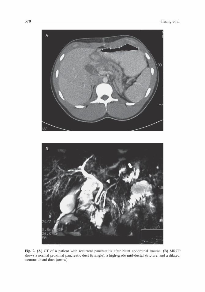
378 Huang et al.
A
B
Fig. 2. (A) CT of a patient with recurrent pancreatitis after blunt abdominal trauma. (B) MRCPshows a normal proximal pancreatic duct (triangle), a high-grade mid-ductal stricture, and a dilated,tortuous distal duct (arrow).

Chapter 21 / Reoperation of the Pancreas 379
by increased intraductal and interstitial pressures caused by fibrosis and obstruction.This is reflected in the fact that in patients with main pancreatic ductal dilation and/orpancreatic ductal stricture(s), ductal drainage procedures have been shown to providesome relief of chronic pancreatitic pain. An additional goal of surgery in children is tofacilitate enzymatic drainage, thereby preserving pancreatic function and contributingto normal growth and development (44). Although many variations of pancreatic ductaldrainage procedures are used, lateral pancreaticojejunostomy, or the modified Puestowprocedure, is one of the most commonly used procedures and has been shown to resultin immediate pain relief in 80% of adult patients (45). There are a few retrospectiveoutcome reports of this procedure in pediatric patients.
DuBay et al. (2000) reported their experience over a 25-year period treating pediatricpatients with hereditary pancreatitis with the modified Puestow procedure (44). Therewere 12 patients in their retrospective study. The mean age at first hospitalizationwas 5.5 years old. The mean duration of symptoms before surgery was 3.9 years. Thepresenting symptom was abdominal pain in 10 patients and failure to thrive in two. Ninepatients also had a greater than 10% weight loss, and eight had a history of pseudocystformation. All patients had dilated main pancreatic ducts diagnosed by CT scan (n = 7),ERCP (n = 4), or at surgery (n = 1). The pancreatic ducts were identified at surgery byvarious techniques: a 1- to 2-cm distal pancreatectomy (n = 6), via a communicatingpseudocyst (n = 3), a blind incision (n = 2), or intraoperative needle localization (n = 1).Distal pancreatectomy for ductal identification prolonged operative time and increasedblood loss in comparison to the other techniques. There were no operative mortalities,but there were four postoperative complications: superior mesenteric artery syndrome,postoperative bleeding requiring reexploration, subphrenic abscess requiring operativedrainage, and Roux stasis until the tenth postoperative day. Postoperative follow-upshowed that hospitalizations for pancreatitis were significantly reduced in all patientsover both a 1- and 3-year period. All patients experienced an increase in their percentilebody weight postoperatively. No patients required insulin pre- or postoperatively. Fivepatients were noted to have steatorrhea preoperatively, which resolved in four patientand was controlled in the fifth patient with enzyme supplements. The median follow-upwas 15 years. At follow-up, seven patients ranked their surgical outcome as excellent,five as good, and one as poor. Eight of the 12 patients had had symptoms of pancreatitis,six patients had needed hospitalizations, four patients had been placed on pancreaticsupplements, and none of the patients had had diabetes mellitus. Their conclusionwas that in children with complicated hereditary pancreatitis, the modified Puestowprocedure improved the quality of life by decreasing hospitalizations, increasing idealbody weight, and improving pancreatic function.
Chiu et al. (2006) reported their experience over a 6-year period with four pediatricpatients who underwent LPJ for chronic pancreatitis (46). There were no mortalities buta significant number of complications including: bowel obstruction caused by internalherniation 1 month after surgery, incisional hernia requiring operative repair, andsevere respiratory complications requiring a prolonged stay in the ICU. The follow-upranged from 2–6 years. There were no recurrences of pancreatitis within this periodand all of the patients were able to eat a normal diet. No patients required pancreaticenzyme supplementation, and only the patient who required ICU stay needed insulinpostoperatively.
Although the modified Puestow procedure has been shown to significantly decreasesymptoms associated with chronic pancreatitis in many children, there have been

380 Huang et al.
noted surgical failures and recurrences. Surgical failures in these patients have beenattributed to inadequate decompression of the head of the pancreas. Therefore, Rollinsand Meyers (2004) reported their experience with a change in their institution fromperforming the LPJ to the Frey procedure (addition of anterior resection of the headof the pancreas to the LPJ) (47). They reported a 7-year retrospective study thatincluded 11 pediatric patients (age range from 8–18 years old) followed over a periodranging from 1.3–8.8 years. All patients underwent preoperative evaluation with ERCP.A group of 10 patients had disease involving the head of the gland, and five patientshad disease involving the entire gland. Three patients underwent LPJ and eight patientsunderwent the Frey procedure. One of the LPJ patients underwent conversion to a Freyprocedure after a pseudocyst developed in the head of the pancreas 6 months afterthe initial procedure. There were no mortalities and four postoperative complications:central line infection, upper gastrointestinal bleeding secondary to gastritis, pleuraleffusion requiring thoracentesis, and renal insufficiency. Of the three LPJ patients, onepatient’s outcome was considered good (occasional pain not requiring hospitalization)and two patients’ outcomes were considered poor (pain causing limitations in activity,regular narcotic use, or repeated hospitalizations). Of the eight Frey procedure patients,six patients’ outcomes were considered excellent (patient reported no pain and hadreturned to normal activity), one patient’s outcome was considered good, and onepatient’s outcome was considered improved (pain caused occasional absence fromschool or work or required occasional use of pain medication). The authors concludedthat the Frey procedure addressed some of the limitations of the LPJ procedure and iscurrently their preferred procedure for surgical management of chronic pancreatitis inchildren.
In patients with chronic pancreatitis and no evidence of large duct disease,pancreatic pain is thought to be neuropathic in origin, secondary to chronic inflam-mation of the pancreatic perineural sheath caused by chronic or recurrent enzymeand cytokine release. Although this is a rare pathology to find in children, data onadult patients suggest that distal pancreatectomy, resection of the diseased pancreaticparenchyma harboring the involved inflamed nerves, can be an effective option.Sakarafas et al. (2001) reported a retrospective study of 38 adult patients diagnosed overa 20-year period with obstructive chronic pancreatitis who underwent distal pancrea-tectomies (48). The diagnoses were made by ERCP and CT scans and by pathologicconfirmation at surgery. Patients were selected who had isolated, dominant majorpancreatic ductal stricture or cutoff and changes of chronic pancreatitis in the glanddistal to the stricture. In these patients, the head of the pancreas typically appearedrelatively normal on imaging tests and lacked parenchymal calcification. The meantime from onset of symptoms to operation was 4 years. All patients underwent a distalpancreatectomy. In five patients, more than an 80% resection was required to reachthe stricture. There were no postoperative deaths. Significant morbidity occurred infive patients: hemorrhage in three patients (all requiring reoperation) and two bowelobstructions (one patient requiring reoperation). Follow-up was complete in 31 patients.Mean follow-up was 6.7 years. A group of 15 (49%) patients had complete painrelief, 10 (32%) patients had partial pain relief, and six (19%) patients had inade-quate pain relief. Preoperatively, only nine patients were able to work or functionnormally; postoperatively, 23 patients were able to do so. Diabetes mellitus developedin 14 (45%) patients, and exocrine insufficiency developed in 15 (47%) patients. The
Chapter 21 / Reoperation of the Pancreas 381
authors’ conclusion was that distal pancreatectomy achieved pain relief in a significantpercentage of patients with low morbidity and no mortality.
Management of complications of chronic pancreatitis can be challenging. Manysurgical options are available with varying degrees of success in relieving symptoms.Choice of surgical procedures depends on location of pancreatic injury and involvementof the main pancreatic duct. Reoperation for persistent or recurrent symptoms usuallyinvolves further resection of diseased pancreas.
PANCREATIC FISTULA
A pancreatic fistula is an abnormal passage of pancreatic juice through a pancreaticductal disruption that exits the pancreatic parenchyma. This fistula can reside totallywithin the capsule of the pancreas and can be minimal and self-healing, or it can breachthe capsule of the pancreas resulting in pancreatic juice entering the retroperitoneal orperitoneal cavities (49). The most important aspects of early management of pancreaticfistulas are similar to management of acute pancreatitis: intensive resuscitation withintravenous fluids and maintenance of adequate nutrition by parenteral hyperalimen-tation. Broad-spectrum antibiotics are usually added to this regimen (49). Most low-output fistulas will close spontaneously. Somatostatin analogs can decrease amountof fistula output and accelerate fistula closure; however, they will not induce closureof fistulas that would not otherwise close (9). Endoscopic placement of transpap-illary endoprotheses can decompress the pancreatic ductal system, relieve downstreamobstruction, and aid in fistula closure.
The most common cause of pancreatic fistula in children is as a postoperativecomplication after pancreatic surgery (9). In adult studies, most pancreatic fistulasresulting from pancreatic resections will close with medical management (50,51).Proactive closure of the pancreatic duct at initial surgery has been shown to decreasethe rate of postoperative fistula formation (52). If pancreatic fistulas persist over time, aRoux-en-Y drainage procedure may be necessary once the fistulous tract has matured.
CONCLUSION
Management of pancreatic diseases and injury in children continues to be achallenging proposition. Complications associated with pancreatic diseases can causesignificant morbidity. Although experiences with adult patients have provided usefulguidelines in the care of pediatric patients, care of the pediatric patient with pancreaticsurgical complications must be individualized to the patient and the presenting clinicalsituation.
REFERENCES
1. Lindley KJ, Spitz L. Surgery of persistent hyperinsulinaemic hypoglycaemia. Semin Neonatol2003;8:259–265.
2. De León DD, Stanley CA. Mechanisms of disease: advances in diagnosis and treatment of hyperin-sulinism in neonates. Nat Clin Pract Endocrinol Metab 2007;3(1):57–68.
3. Sperling MA, Menon RK. Pediatric endocrinology. Endocrinol Metab Clin 1999;28(4):695–708.4. Kane C, Lindley KJ, Johnson PRV, et al. Therapy for persistent hyperinsulinemic hypoglycemia of
infancy: understanding the responsiveness of � cells to diazoxide and somatostatin. J Clin Invest1997;100(7):1888–1893.

382 Huang et al.
5. Bas F, Darendeliler F, Demirkol D, et al. Successful therapy with calcium channel blocker (nifedipine)in persistent neonatal hyperinsulinemic hypoglycemia of infancy. J Pediatr Endocrinol Metab1999;12(6):873–878.
6. Otonkoski T, Näntö-Salonen K, Seppänen M, et al. Noninvasive diagnosis of focal hyperinsulinismof infancy with [18F]-DOPA positron emission tomography. Diabetes 2006;55:13–18.
7. Suchi M, Thornton PS, Adzick NS, et al. Congenital hyperinsulinism: intraoperative biopsy interpre-tation can direct the extent of pancreatectomy. Am J Surg Pathol 2004;28(10):1326–1335.
8. Adzick NS, Thornton PS, Stanley CA, et al. A multidisciplinary approach to the focal form ofcongenital hyperinsulinism leads to successful treatment by partial pancreatectomy. J Pediatr Surg2004;39(3):270–275.
9. Mehta, SS, Gittes GK. Pancreas. In: Oldham KT, Colombani PM, Foglia RP, et al. (eds), Principlesand practice of pediatric surgery. Philadelphia: Lippincott Williams & Wilkins; 2005: 685–695.
10. Cherian MP, Abduljabbar MA. Persistent hyperinsulinemic hypoglycemia of infancy (PHHI): long-term outcome following 95% pancreatectomy. J Pediatr Endocrinol Metab 2005;18:1441–1448.
11. Ismail D, Werther G. Persistent hyperinsulinaemic hypoglycaemia of infancy: 15 years’ experience atthe Royal Children’s Hospital (RCH), Melbourne. J Pediatr Endocrinol Metab 2005;18:1103–1109.
12. Reyes GA, Fowler CL, Pokorny WJ. Pancreatic anatomy in children: emphasis on its importance topancreatectomy. J Pediatr Surg 1993;28(5):712–715.
13. Martinez-Ibanez V, Gussinyer M, Toran N, et al. Pancreatectomy extension in persistent hyperinsuli-naemic hypoglycemia: a new strategy. Eur J Pediatr Surg 2002;12:262–266.
14. Stringer MD. Pancreatitis and pancreatic trauma. Semin Pediatr Surg 2005;14:239–246.15. Keller MS, Stafford PW, Vane DW. Conservative management of pancreatic trauma in children. J
Trauma 1997;42(6):1097–1100.16. Wales PW, Shuckett B, Kim PCW. Long-term outcome after nonoperative management of complete
traumatic pancreatic transaction in children. J Pediatr Surg 2001;36(5):823–827.17. Meier DE, Coln CD, Hicks BA, et al. Early operation in children with pancreas transaction. J Pediatr
Surg 2001;36(2):341–344.18. Nadler EP, Gardner M, Schall LC, et al. Management of blunt pancreatic injury in children. J Trauma
1999;47(6):1098–1103.19. Goodley CD, Rattner DW. Pancreatic pseudocyst. In: Cameron JL (ed), Current surgical therapy,
sixth edition. St. Louis: Mosby; 1998: 507–509.20. Filler RM. Pancreas. In: Welch KJ (ed), Complications of pediatric surgery: prevention and
management. Philadelphia: Saunders; 1982: 256–263.21. Mulligan C, Howell C, Hatley R, et al. Conservative management of pediatric pancreatic pseudocyst
using octreotide acetate. Am Surg 1995;61:206–209.22. Bosman-Vermeeren JM, Veereman-Wauters G, Broos P, et al. Somatostatin in the treatment of a
pancreatic pseudocyst in a child. J Pediatr Gastroenterol Nutr 1996;23(4):422–425.23. Burnweit C, Wesson D, Stringer D, et al. Percutaneous drainage of traumatic pancreatic pseudocysts
in children. J Trauma 1990;30(10):1273–1277.24. Lucaya J, Vázquez E, Caballero F, et al. Non-operative management of traumatic pancreatic pseudo-
cysts associated with pancreatic duct laceration in children. Pediatr Radiol 1998;28:5–8.25. Schlosser W, Siech M, Beger HG. Pseudocyst treatment in chronic pancreatitis – surgical treatment
of the underlying disease increases the long-term success. Dig Surg 2005;22(5):340–345.26. Soliani P, Franzini C, Ziegler S, et al. Pancreatic pseudocysts following acute pancreatitis: risk factors
influencing therapeutic outcomes. J Pancreas 2004;5(5):338–347.27. Usatoff V, Brancatisano R, Williamson RCN. Operative treatment of pseudocyst in patients with
chronic pancreatitis. Br J Surg 2000;87(11):1494–1499.28. Rescorla FJ, Cory D, Vane DW, et al. Failure of percutaneous drainage in children with traumatic
pancreatic pseudocysts. J Pediatr Surg 1990;25(10):1038–1042.29. Patty I, Kalaoui M, Al-Shamali M, et al. Endoscopic drainage for pancreatic pseudocyst in children.
J Pediatr Surg 2001;36(3):503–505.30. Al-Shanafey S, Shun A, Williams S. Endoscopic drainage of pancreatic pseudocysts in children. J
Pediatr Surg 2004;39(7):1062–1065.31. Nealon WH, Walser E. Surgical management of complications associated with percutaneous and/or
endoscopic management of pseudocyst of the pancreas. Ann Surg 2005;241(6):948–960.32. Canty TG, Weinman D. Management of major pancreatic duct injuries in children. J Trauma
2001;50(6):1001–1007.33. Jackson WD. Pancreatitis: etiology, diagnosis, and management. Curr Opin Pediatr 2001;13:447–451.

Chapter 21 / Reoperation of the Pancreas 383
34. Lowe ME. Pancreatitis in childhood. Curr Gastroenterol Rep 2004;6:240–246.35. Lerner A, Branski D, Lebenthal E. Pancreatic diseases in children. Pediatr Clin North Am
1996;43(1):125–156.36. Nwariaku FE, Terracina A, Mileski WJ, et al. Is Octreotide beneficial following pancreatic injury?
Am J Surg 1995;170:582–585.37. Paran H, Neufeld D, Mayo A, et al. Preliminary report of a prospective randomized study of Octreotide
in the treatment of severe acute pancreatitis. J Am Coll Surg 1995;181:121–124.38. Zhou Y-M, Xue Z-L, Li Y-M, et al. Antibiotic prophylaxis in patients with severe acute pancreatitis.
Hepatobiliary Pancreat Dis Int 2005;4(1):23–27.39. Mier J, Luque-de León E, Castillo A, et al. Early versus late necrosectomy in severe necrotizing
pancreatitis. Am J Surg 1997;173:71–75.40. Baron TH, Morgan DE. Acute necrotizing pancreatitis. N Engl J Med 1999;340(18):1412–1417.41. Kozarek RA, Christie D, Barclay G. Endoscopic therapy of pancreatitis in the pediatric population.
Gastrointest Endosc 1993;39:665–669.42. Varadarajulu S, Wilcox CM. Endoscopic management of Sphincter of Oddi dysfunction in children.
J Pediatr Gastroenterol Nutr 2006;42:526–530.43. Neblett WW III, O’Neill JA Jr. Surgical management of recurrent pancreatitis in children with
pancreas divisum. Ann Surg 2000;231(6):899–908.44. DuBay D, Sandler A, Kimura K, et al. The modified Puestow procedure for complicated hereditary
pancreatitis in children. J Pediatr Surg 2000;35:343–348.45. Murayama KM, Joehl RJ. Chronic pancreatitis. In: Cameron JL (ed), Current surgical therapy, sixth
edition. St. Louis: Mosby; 1998: 514–519.46. Chiu B, Lopoo J, Superina RA. Longitudinal pancreaticojejunostomy and selective biliary diversion
for chronic pancreatitis in children. J Pediatr Surg 2006;41:946–949.47. Rollins MD, Meyers RL. Frey procedure for surgical management of chronic pancreatitis in children.
J Pediatr Surg 2004;39:817–820.48. Sakorafas GH, Sarr MG, Rowland CM, et al. Postobstructive chronic pancreatitis. Arch Surg
2001;136:643–648.49. Traverso LW, Newman RM, Kozarek RA. Pancreatic ductal disruptions leading to pancreatic fistula,
pancreatic ascites or pleural effusion. In: Cameron JL (ed), Current surgical therapy, sixth edition.St. Louis: Mosby; 1998: 510–514.
50. Bassi C, Butturini G, Molinari E, et al. Pancreatic fistula rate after pancreatic resection. Dig Surg2004;21:54–59.
51. Kazanjian KK, Hines OJ, Eibl G, et al. Management of pancreatic fistulas after pancreaticoduo-denectomy. Arch Surg 2005;140:849–855.
52. Bilimoria MM, Cormier JN, Mun Y, et al. Pancreatic leak after left pancreatectomy is reducedfollowing main pancreatic duct ligation. Br J Surg 2003;90:190–196.

22 Rhabdomyosarcoma
David Rodeberg, MD
and Eugene S. Wiener, MD
CONTENTS
Background
Surgical Guidelines
Diagnostic Evaluation
Pretreatment Reexcision (PRE)
Reoperation of Primary Tumor
Metastatic RMS
Adjuvant Therapy
Second Malignancies and Late Complications
References
BACKGROUND
Rhabdomyosarcoma (RMS) is one of the more common solid tumors in children,with approximately 250 new cases diagnosed each year (1).The treatment of RMShas improved significantly during the last 30 years, with the overall long-termsurvival rates improving from 25% in 1970 to more than 70% with current multi-modality treatment (2–4).The roles of pediatric surgeons in the treatment of RMS havechanged significantly through the years, as other adjuvant therapies have become moreefficacious.
RMS arises from primitive mesenchymal cells that are present throughout the body,even in areas that are usually not associated with striated muscle (5). However, alltumors show some degree of striated muscle differentiation. RMS may invade localstructures and frequently metastasize early through lymphatics or hematogenous spread.Tumors are usually firm, nodular, and variable in size and consistency; however, theyare not encapsulated and frequently invade surrounding soft-tissue structures. Thereare essentially four types of RMS: embryonal, alveolar, pleomorphic, and undiffer-entiated (3). Embryonal is the most common and is usually found in children lessthan 8 years old. Overall, patients with embryonal RMS have good prognoses, witha 5-year survival rate of 60%. For spindle cell and botryoid subvariants, the survivalrate increases to 95%. Alveolar RMS is found in older children and is associated with
From: Reoperative Pediatric SurgeryEdited by: S. Teich and D. A. Caniano © Humana Press, Totowa, NJ
385

386 Rodeberg and Wiener
tumors in the extremities and trunk. Patients with alveolar RMS have worse prognosesthan those with embryonal, with an average 5-year survival rate of 54%.
SURGICAL GUIDELINES
The basic principle of wide and complete resection of the primary tumor with asurrounding “envelope” of normal tissue should be followed at the initial, or subse-quent, operations whenever possible (6). A somewhat arbitrary margin of 0.5 cmcircumferentially, or an uninvolved fascia margin, is adequate. This size of margingenerally is more easily obtained in the extremities or trunk than in head and necktumors. Adequate margins of uninvolved tissue are required unless excision involvessacrifice of normal tissue that cannot be resected, would result in an unacceptable lossof function/cosmesis, or is not technically feasible. Therefore, the resection of vitalstructures is probably not warranted for most tumors. Also, when the RMS arises froma somatic muscle, excision of the entire muscle of origin or the entire compartmentusually is not necessary (7). The surgeon should mark all margins and orient thespecimen at the operative field, so that margin evaluation is precise. Narrow marginsare unavoidable in some sites. In those situations, the surgeon should take severalseparate biopsies of the “normal” tissue around the margins of resection and theseshould be marked and submitted separately for pathologic review.
Any suspected microscopic or gross residual tumor should be marked in the tumorbed with small titanium clips to aid radiotherapy simulation and subsequent reexcision.A clear margin and no residual disease (Group I) is superior to microscopic margins(Group II) or gross residual disease (Group III) in all outcomes analyses (3,8–10).
Exceptions to this general operative approach would include primaries in the orbit,head and neck, biliary, and geritourinary (GU) sites (11–17). For patients with primarydisease at these sites, the primary treatment modality employed is chemotherapy andradiotherapy. This approach is appropriate given its usual embyonal histology, goodresponse to treatment, and the poor likelihood of achieving complete operative resectionwithout significant morbidity.
Clinical or radiographic evaluation of regional lymph nodes should be done duringdiagnostic work-up and is an important component of pretreatment staging. Clinicallypositive nodes should always be confirmed pathologically. For multiple clinicallypositive nodes, radical debulking may be useful, with radiotherapy, to obtain regionalcontrol (18,19). During biopsy of the regional lymph nodes, a “distant” node shouldbe harvested for pathologic study. Involvement of these distant nodes is analogous tometastatic disease. There is no benefit from extensive nodal dissection if distant nodeor metastatic disease has already occurred.
Pathologic evaluation of clinically uninvolved nodes is site-specific; it is requiredin extremity sites and for children older than 10 years with paratesticular tumors (20).Aggressive regional lymph node sampling is the most appropriate method of surgicalevaluation because resection is diagnostic but not therapeutic. For this reason, prophy-lactic radical node dissection, as used for some other malignancies, is not necessary inchildhood RMS except in children older than 10 years of age with paratesticular RMS,who should undergo a retroperitoneal nerve-sparing lymph node resection.
Sentinel node mapping using blue dye, radioactive-labeled colloid, or both ishelpful in determining regional node status (21). Preliminary data from the IntergroupRhabdomyosarcoma Group (IRSG) suggest that sentinel node biopsy may be effective.

Chapter 22 / Rhabdomyosarcoma 387
DIAGNOSTIC EVALUATION
Usually in this patient population, before enrollment, patients will have had anincisional biopsy to provide diagnosis or document metastatic disease histologically. Aspart of the staging process, patients undergo radiographic assessments with computedtomography (CT) of the chest, CT or ultrasound of the liver, magnetic resonanceimaging (MRI) or CT of the head, bone scintigraphy, and bone marrow aspiration andbiopsy. Additional staging studies such as abdominal CT scans and cerebral spinalfluid (CSF) cytology are individualized, depending on clinical findings and primarytumor location (Table 1).
PRETREATMENT REEXCISION (PRE)
PRE is a wide, nonmutilating procedure carried out in patients with soft-tissue sarcomas(STS) when microscopic residuals are left after initial excision or when there is insuffi-cient data on the completeness of resection. PRE means wide reexcision of the previousoperative site, including an adequate “envelope” of normal tissue, with careful markingand examination of all margins.
Frequently, the initial surgical procedure (a biopsy or excision as used for benigntumor) may have been performed prior to establishing the diagnosis and/or theinvolvement of the oncology team and may result in a situation in which there is: (1)gross residual tumor; (2) microscopically involved margins; or (3) uncertainty as tomargins or residual disease. This applies even if the microscopic margin is thought tobe clear, or if the operation and pathologic study were not done as a “cancer” operation.
Table 1Components of Diagnostic Evaluation for Newly Diagnosed RMS
Hx/PE (Ht/Wt), Clinical assessmentCBC/Diff/PltPT, PTT, fibrinogenUrinalysis, creatinine, SGPT, alkaline phosphate, Ca/Phos/AlbElectrolytes (Na, K, Cl, CO2)Creatinine clearance or GFRBilateral BM aspirate/biopsyCT chestEKG or MUGA for cardiac monitoringMRI or CT of primary tumor³CT or US liver (for abdominal/pelvic tumors)MRI or CT headCT, US, MRI retroperitoneum for lower extremity, GU, pelvic and
abdominal tumors onlyLumbar puncture (for metastatic parameningeal tumors and if multiple
intracranial metastases at initial diagnosis)Bone scanLymph node biopsy for extremity and paratesticular tumors
CBC, complete blood count; PT, parathyroid; PTT, partial thromboplastin time;SGPT, serum glutamate pyruvate transaminase; BM, bone marrow; CT, computedtomography; EKG, electrocardiograph; MUGA, multigated angiogram; MRI, magneticresonance imaging; US, ultrasound; GU, geritourinary.
388 Rodeberg and Wiener
Under these circumstances, the concept of PRE is advisable and should be appliedwherever feasible, unless the resulting disability would be considered unacceptable(i.e., resulting in loss of function or an unacceptable cosmetic result). A lymph nodeevaluation is indicated using the same criteria as outlined previously in the generalsurgical guidelines. PRE is particularly applicable to extremity and trunk lesions,although it should be applied wherever possible.
Prior to PRE imaging, a CT or MRI should be performed to visualize any residualtumor but, more importantly, to identify adjacent structures and determine if the residualarea can be resected without causing significant morbidity. PRE, as its name suggests,should be done prior to the initiation of chemotherapy or radiotherapy. Clinical groupassignment is determined on the basis of pathology from the definitive operation priorto the start of multimodal therapy. The prior procedure(s) will be considered a biopsy.
The initial report of PRE was published in 1989 by Hays et al. (8), looking at41 children who had undergone PRE for extremity/trunk group II disease. Thesepatients were converted to group I and subsequently had a 91% survival. This wassignificantly better than the survival for patients initially classified as group I patients(74%) and group II patients that had not undergone PRE (74%). Approximately 36%of pathologic specimens did not contain viable tumor. Additionally, 29% of PREpathologic specimens had positive margins and required a third procedure. The localtumor recurrence rate (30%) was similar between all three arms of the study. This wasnot a prospective study and so does suffer from some selection bias. In addition, it didnot look at group III patients. And last, pathologic error in initial margin classificationis proposed for why the group II PRE patients did better than the group I patients whenthey should have been equivalent.
A recent study by Cecchetto et al. (22) evaluated the role of PRE in patients withSTS enrolled in two consecutive Italian studies between January 1988 and September1999. Of the 126 patients with grade IIa tumors (microscopic margins positive butno nodal disease), 53 patients (42%) underwent PRE. In this cohort, there were 23patients with RMS and 30 patients with non-RMS STS. The median interval betweenprimary surgery and PRE was 36 days. Of the 53 patients undergoing PRE, only eight(15%) continued to have positive margins postoperatively. Viable tumor was identifiedin 21 pathologic specimens from the 45 patients in whom the tumor was completelyexcised by PRE. Currently, 39/45 patients are in their first complete remission with amedian follow-up of 53 months. Interestingly, the presence of residual viable diseasein the PRE specimen did not correlate with outcome. PRE was effective especiallyin extremity, trunk, and paratesticular sites; however, its role was uncertain in largesarcomas over 5 cm in size. The authors reported that patients with complete resectionhad an 89% 5-year over-all survival. Unfortunately, they did not compare this resultwith a similar cohort that did not receive PRE to quantify the benefit achieved withPRE. It is also unfortunate that they did not look at group III patients (macroscopicresidual), who make up the largest percentage of patients that may have benefitedfrom PRE.
REOPERATION OF PRIMARY TUMOR
These resections occur after initiation of treatment with chemotherapy and mayoccur at any point in relation to radiotherapy. They are usually performed for groupIII patients who have macroscopic residual disease but no evidence of metastatic

Chapter 22 / Rhabdomyosarcoma 389
extension. The most common scenario is for patients that have initially had a biopsy(group III) for large unresectable disease who have undergone adjuvant therapy andnow the mass is decreased in size allowing it to be resected safely. Reoperation ofthe primary tumor in this setting can be broken down into three categories: delayedprimary excision, second look operation (SLO), and end of therapy resection.
Resection During TherapyResection during therapy consists of two scenarios; delayed primary excision is the
attempted complete resection occurring after induction chemotherapy (usually after twoto four cycles of chemotherapy) and SLO is performed at any point during therapy andmay consist of attempted resection or biopsy. Delayed primary excisions are performedif chemotherapy has decreased the tumor size sufficiently to perform a safe resectionwithout significant morbidity. The purpose of delayed excision is to improve localcontrol with the objective to completely excise the tumor. SLOs, after chemotherapyand radiotherapy, may be considered for patients who are deemed to have partialresponses, and selected nonresponders. The purpose of SLO, in patients with controlledmetastatic disease, is to confirm clinical response at the primary site, to evaluate patho-logic response to therapy, and to remove residual local tumor whenever and whereverit is feasible in order to achieve local control (23,24). Similar to delayed primaryexcision, SLO occurs most commonly in group III patients. Optimally, completeresection is the objective of SLO if this is anatomically and functionally possible. SLOis not recommended immediately after irradiation, because within this timeframe localchanges following radiotherapy could cause false positive interpretations of imaging.In addition, time must be allowed after the completion of radiotherapy for the tumor toachieve its final response status. Both delayed primary excision and SLO are performedusing the standard surgical guidelines previously described, including wide reexcisionof the previous operative site, an adequate “envelope” of normal tissue, and exami-nation of all margins. A lymph node resection/sampling is indicated when appropriate.These operations are most efficacious for lesions of the trunk and extremities, tumorssmaller than 5 cm, children over 10 years of age, lower stage disease, and childrenwith negative nodes.
When these procedures should be performed in relation to other therapy is aquestion that should be determined in discussion with the child’s oncologist andradiotherapist. There are no strong data from which to draw any firm conclusionsconcerning timing of operative procedures in relation to chemotherapy and radio-therapy, yet consensus would suggest several guidelines that can be followed. As ageneral rule surgical resection may be performed once platelet counts have recoveredform chemotherapy. The next cycle of chemotherapy may proceed 1–2 weeks postop-eratively for trunk operations and 2–3 weeks for extremities, thus allowing anotherweek after chemotherapy for white blood cell (WBC) counts to reach a nadir. SimilarlyX-ray therapy (XRT) should be delayed 2–4 weeks after resection. However, resectionshould be delayed 2–6 weeks after the completion of XRT so that response to XRTmay be adequately quantified. Each of these guidelines should help to minimize themorbidity of the procedure that can be associated with the adjuvant therapy. Forboth delayed primary excision and SLO, either CT or MRI is most commonly usedto evaluate patients for extent of residual local and metastatic disease prior to theprocedure.

390 Rodeberg and Wiener
The importance of SLO to outcome is debatable. In support of SLO, results fromIntergroup Rhabdomyosarcoma Study (IRS) III showed that 75% of patients classifiedas having partial responses by imaging were found to have complete responses duringSLO or were converted to complete responses by excision of residual tumors (24).Converting them to complete response improved their survival. The SLO were mosteffective in extremities and trunks compared with head and neck lesions. Flaps and/orgrafts were occasionally required for reconstruction, because prior radiation can affectwound closure and healing. For intracavitary sites, such as abdomen or thorax, it hasbeen suggested that a complete second-look evaluation using open, laparoscopic, orthoracoscopic evaluation can be performed, but evaluation must be complete whatevermethod is used. If residual tumor persists after SLO, subsequent procedures should beperformed after further therapy, to resect if feasible, or to confirm clinical responsestatus.
However, in a more recent review of IRS-IV, SLO showed that only 20% ofgroup III RMS patients with primary sites of extremity/trunk, retroperitoneal/pelvic,bladder/prostate, or head/neck/orbit had SLO during protocol therapy (Raney et al.,manuscript in preparation). Those patients that had SLO were more likely to havetrunk primaries, with tumors that were larger and a higher stage compared to thosewho did not undergo SLO. Also, patients who had SLO were less likely to receiveappropriate XRT. Given these caveats, there were no differences in failure-free survivaland overall survival between patients that had SLO and those that did not, even whenadjusted for the known prognostic factors of age, histology, and stage. Some of thiscould be explained by the observation that only one-half of the pathologic specimenscontained viable tumor, and that of those with viable tumor only one-half achievedcomplete resection with negative margins with SLO. Those patients that did not achievecomplete resection were more likely to be infants, with nontrunk disease, at a higherstage with invasive tumor and positive nodes. If SLO was able to achieve completeresection with negative margins, then failure-free survival and overall survival wereboth significantly improved. Not too surprisingly, those patients with viable tumor hada significantly worse outcome.
Similarly, IRS-V encouraged SLO after 12 weeks of chemotherapy. IRS-V alsopermitted a radiation dose reduction from the standard 50.4 to 41.4 or 36 Gy, followingSLO with gross total excision and either microscopic positive or negative margins,respectively. However, most eligible patients did not have SLO because of the antici-pated morbidity of surgical resection at the most common anatomic sites of RMS, suchas parameningeal or bladder/prostate. Outcome data from this study is still pending.
In conclusion, the results from these studies indicate that SLO is indicated, and canimprove survival, for patients in whom a complete resection can be anticipated (usuallypatients with extremity or trunk lesions less than 5 cm in diameter in children over 10years old with lower-stage node-negative disease) and who have viable tumor (usuallypatients with partial response or no response to chemotherapy). If complete resectionis not achieved, then survival is not different compared to biopsy or observation only.
Resection at the End of TherapyAt the end of therapy, some patients with RMS have residual masses despite receiving
all planned therapy. A recent review of IRS IV patients with group III (gross residualdisease) RMS found the presence of a residual mass at the end of therapy did notcorrelate with outcome (25). This study looked at 419 group III patients who completed

Chapter 22 / Rhabdomyosarcoma 391
all protocol therapy without developing progressive disease. At the end of therapy, 341(81%) of children achieved complete resolution of the mass and 78 (19%) had a residualmass. The 5-year failure-free survival was similar for the two groups (80 and 77%,respectively) even after adjustment for age, nodal status, primary site, and histology. Inaddition, those patients that underwent operative resection at the end of therapy for aresidual mass were likely to have significant morbidity associated with their procedure(26). A total of 63% of patients with a residual mass at the end of therapy underwentan attempted resection. Of these patients, 45% had excision of a vital structure duringthe procedure with a resultant loss of function in one-half. Unfortunately only 18%of patients had viable tumor detected in the pathologic specimen, and only two ofthese nine patients had negative margins. In addition, there was no improvement inoutcome for patients that had the tumor resected versus biopsy alone. Therefore, giventhe lack of improvement in outcome and the associated morbidity, aggressive surgicalor alternative therapy is probably not warranted for RMS with a residual mass at theend of planned therapy. In summary, the indications for operative intervention forresidual masses at the end of therapy should probably be limited to excision of thosetumors that would be easily resectable with minimal morbidity or else biopsy alone todocument residual viable disease.
Resection for Recurrent DiseaseRecurrent RMS can occur in as many as 35% of patients with trunk and extremity
primary tumors, with 50–95% of these patients succumbing to progressive disease(3,27,28). In a study by Pappo et al. (29), the median time from tumor recurrenceto death was only 0.8 years, with a 17% 5-year survival rate. In this study the bestpredictor for survival in patients with recurrence was histology with approximately a30% survival for favorable histology (embryonal and botryoid) compared to 5% forunfavorable (alveolar and undifferentiated).
Recurrent RMS can be locally extensive and is usually treated with surgical resectionand adjuvant therapy. The evidence for the efficacy of resection in these patientshas been extrapolated from primary tumors, because there has not been good datalooking specifically at recurrent disease. Recently, Hayes-Jordan et al. (30) looked at 32patients with recurrent RMS. Of these, 13 patients had biopsy only with a subsequentsurvival rate of 8%. Another 19 patients had resection with a 37% survival rate. But,these operations were associated with a 15% major complication rate and an overallcomplication rate of 35%. The survival data does suffer from a couple of flaws. First,it was a retrospective review and therefore contains a large selection bias. Second,those patients that underwent operative resection had less extensive disease and fewerpatients with metastatic disease, compared to those who only had biopsy, and this mayexplain the survival results rather than any true benefit from resection. Given theseconfounding factors, it is difficult to draw any firm conclusions. And given the poorprognosis for these patients, it may be reasonable to perform a resection using the samecriteria previously established if the tumor can be safely and completely resected.
METASTATIC RMS
Patients with metastatic RMS account for 16% of all cases (3,31,32). Unfortunately,for patients who present with metastatic disease, there is only a 20–30% chance ofsurvival (27,29).

392 Rodeberg and Wiener
Clinical Presentation and Risk FactorsMetastatic disease is the single most important predictor of clinical outcome in
patients with RMS. Metastatic disease is most commonly aymptomatic and is frequentlydetected during staging or follow-up radiographic imaging. For those patients withsymptoms, the symptoms are usually related to mass effect and functional compromiseof adjacent structures.
The risk factors for metastatic disease and prognosis for these patients have been welldescribed. In the study by Breneman et al. (33), IRS-IV patients with metastatic disease,compared to patients without, were more likely to be older, have higher incidence ofalveolar histology, more commonly have invasive tumors, have larger tumors, have ahigher incidence of regional lymph node involvement, and have a greater proportionof extremity and trunk/retroperitoneal primary sites. A total of 59% of patients hadmetastases confined to single anatomic site. The most common site of metastasis wasthe lung, followed by the bone marrow, bone, and distant lymph nodes.
IRS-IV patients with only pulmonary metastatic disease were evaluated andcompared to patients with metastatic disease at other sites by Rodeberg et al. (34).Of patients with RMS and metastatic disease, 16% had isolated lung metastases.The majority of patients had bilateral pulmonary metastatic disease. Compared topatients with other sites of metastatic disease, patients with lung-only metastases had agreater proportion of embryonal histology, negative nodal involvement, parameningealprimaries, and a smaller proportion of extremity primaries.
Diagnostic EvaluationThe diagnostic evaluation for metastatic disease is similar to that for the primary
lesion, including laboratory studies followed by evaluation for other metastatic diseaseand lumbar tap if there is a suspicion for central nervous system (CNS) involvement,bone marrow aspirate, bone scintography, and CT evaluation of the liver, chest, andhead. In addition, for those patients with metastatic disease biopsy, proof of tumor isalso recommended unless the lesions are so obviously metastatic disease that pathologicconfirmation is not needed.
Operative TherapyPrimary resection of metastasis is rarely indicated unless performed for pathologic
confirmation of metastatic disease or with the intent to remove all metastatic disease inthe setting of controlled primary disease. A delayed resection of persistent metastaticdisease following radiation and chemotherapy should also only be performed if totalremoval could be performed without significant loss of function, disfigurement, anddelay in initiating systemic treatment. There is really minimal evidence to supportresection of metastatic disease (35). However if there is proven residual disease 3–6 months after radiation therapy, and if there was no residual unresectable diseaseelsewhere, then resection may be feasible.
Positive nodes outside of the regional basin (i.e., scalene node for upper extremitysites, inguinal nodes for paratesticular or vaginal/uterine sites, and paraaortic nodesfor all sites other than immediately adjacent retroperitoneal or paratesticular sites),should be classified as distant metastatic spread (Clinical Group IV, Stage 4). Nodesthat are positive in the regional nodal basin are not considered metastatic disease, butinstead are treated as regional disease. This distinction underscores the importance ofnodal sampling in the distal nodal basin if regional nodes are positive, to determine

Chapter 22 / Rhabdomyosarcoma 393
if disease spread is regional or metastatic. This is especially important for RMS thatcommonly spreads to the lymph nodes such as extremity and paratesticular. Frequently,the nodes are classified positive based on imaging studies. If there is uncertainty as toinvolvement of these or other similar distant nodes, and the patient is classified as Stage4 on the basis of these nodes, then biopsy by open or controlled needle techniquesshould be performed to confirm metastatic disease.
In some rare instances when there are two sites of disease, it can be difficult tobe certain which of the two sites is primary. In this setting both should be resectedwith a curative intent. If there is question as to whether an apparent metastasis isRMS versus other benign disease, involvement should be confirmed by biopsy. Biopsyshould be performed using either open, minimally invasive techniques or controlledneedle biopsy as appropriate.
OutcomesIn IRS-IV the estimated 3-year overall and failure-free survival for all patients
with metastatic disease was 39 and 25%, respectively (33). By univariate analysis,overall survival was significantly worse for alveolar histology and increasing numberof metastatic sites. By multivariate analysis, the presence of two or fewer metastaticsites was the only significant predictor of outcome. The combination of embryonalhistology with two or fewer metastatic sites identified a subgroup with a significantlyimproved survival. These results are similar to others (36,37). Similarly, the report byCarli et al. (38) identified primary tumor in parameningeal, extremity, or “other” sites;age younger than 1 year and older than 10 years; bone or bone marrow metastases;multiple metastases; and multiple sites of metastases as unfavorable prognostic factorsfor both overall and failure-free survival. Two subgroups were identified. Those withfewer than two unfavorable factors had 5-year overall survival of 47%, but for thosewith two or more unfavorable factors survival was only 9%.
Overall survival at 4 years for lung-only metastases was not significantly differentfrom other single-site metastasis (42 versus 34%) (34). Survival was not improved forunilateral disease or fewer than five metastatic lesions. Factors associated with dimin-ished overall survival included unfavorable histology and age greater than 10 years.Overall, outcome is comparable, although slightly better, for patients with isolated lungmetastatic disease compared with other single-site metastatic disease.
ADJUVANT THERAPY
During IRS studies, different chemotherapeutic regimens have been evaluatedas phase II window studies to determine new combinations that may improveoutcome compared to standard vincristine, adriamycin, and cyclophosphamide (VAC)chemotherapy. The window trials have identified the combinations of ifosfamide anddoxorubicin, with a 63% response rate, and topotecan and cyclosphosphamide, witha 47% response rate (39,40). Using this information, the new proposed regimen forhigh-risk RMS patients will include ifosfamide-etoposide and vincristine-doxorubicin-cyclophosphamide.
Other strategies that have been attempted to improve outcome in these childreninclude administration of high-dose chemotherapy (HDCT) followed by peripheralblood stem cell or autologous bone marrow support. In general these interventions havefailed to significantly improve outcomes. The role of high-dose chemotherapy aloneor combined with stem cell rescue (HSCR) in the treatment of relapse or metastatic
394 Rodeberg and Wiener
disease appears to be minimal, with survival rates, at best, of 12% at 2 years (41).These results are similar to the 10% 5-year survival rate reported by Pappo et al. (29)for patients who experienced progression of disease or relapse after treatment in IRSIII, IV-pilot, and IV studies. Similar results were found in the European trial by Carliet al. (38), who reported that there was no survival difference between HDCT andstandard chemotherapy.
Radiotherapy is specified for primary disease sites and any sites of metastases in aneffort to improve disease control. Patients receive 50.4 Gy at 1.8 Gy/day to the primarytumor. Patients with metastases in soft tissue or bone also receive radiotherapy dosesof 50.4 Gy. Patients who had diffuse pleural or peritoneal metastases received 14.4 Gy(whole lung) or 18.0 Gy (whole abdomen) to those cavities. Modification of radiationdose is made to keep adjacent critical structures within tolerance. In addition, there isevidence that radiotherapy may improve recurrence for both primary disease as wellas metastatic disease (34,42).
SECOND MALIGNANCIES AND LATE COMPLICATIONS
Most late complications are related to chemotherapy and radiotherapy and maybe site-specific or may be generalized. Most commonly the systems affected includevisual, endocrine, cardiopulmonary, and neurologic. An excellent review of the gener-alized complications was performed by Punyko et al. using the Childhood CancerSurvivor Study Group (43). The relative risk for developing complications was greatestwithin the first 5 years after the completion of therapy. However, many seriouscomplications occured beyond 5 years including impairments of the visual, endocrine,cardiopulmonary, and neurologic systems. Examples of site-specific complicationsinclude patients with head and neck RMS enrolled on IRS II and III who were reviewedby Raney et al. (44). Over 77% of patients had at least one problem, and the mostcommon included decreased growth and height, hypoplasia or asymmetry of tissues,impaired vision and hearing, as well as learning difficulties. Late effects are evenmore common for pelvis RMS (45). In this study, 92% of patients experienced somelate complication and more than one-half required operative intervention to abate thecomplications. The main risk factor for complications appeared to be treatment withradiotherapy.
As expected, there is a risk of developing a second malignancy as a result ofthe treatment used to treat the initial malignancy. This was most recently reviewedby Cohen et al. (46) looking at 1499 RMS survivors. Patients were 7.7 times morelikely to develop a second malignancy based on general population cancer rates.Second malignancy was much more common for patients treated with radiotherapyand chemotherapy compared to patients only treated by surgery. The types of cancermost likely to occur were acute myelohenous leukemia, cutaneous melanoma, breastcancer, and sarcomas of the bone and soft tissues.
REFERENCES
1. Gurney JG, Wall DA, Jukich PJ, Davis FG. The contribution of nonmalignant tumors to CNS tumorincidence rates among children in the United States. Cancer Causes Control 1999;10(2):101–105.
2. Raney RB Jr, Tefft M, Maurer HM, et al. Disease patterns and survival rate in children withmetastatic soft-tissue sarcoma. A report from the Intergroup Rhabdomyosarcoma Study (IRS)-I.Cancer 1988;62(7):1257–1266.

Chapter 22 / Rhabdomyosarcoma 395
3. Crist W, Gehan EA, Ragab AH, et al. The Third Intergroup Rhabdomyosarcoma Study. J Clin Oncol1995;13(3):610–630.
4. Arndt CA, Crist WM. Common musculoskeletal tumors of childhood and adolescence. N Engl J Med1999;341(5):342–352.
5. Newton WA Jr, Meadows AT, Shimada H, Bunin GR, Vawter GF. Bone sarcomas as second malignantneoplasms following childhood cancer. Cancer 1991;67(1):193–201.
6. Rodeberg D. Surgical principles for children/adolescents with newly diagnosed rhabdomyosarcoma:a report from the Soft Tissue Sarcoma Committee of the Children’s Oncology Group. Sarcoma2002;6(4):111–122.
7. Tabrizi P, Letts M. Childhood rhabdomyosarcoma of the trunk and extremities. Am J Orthop1999;28(8):440–446.
8. Hays DM, Lawrence W, Wharam M, et al. Primary reexcision for patients with ‘microscopic residual’tumor following initial excision of sarcomas of trunk and extremity sites. J Pediatr Surg 1989;24(1):5–10.
9. Hays DM, Raney RB, Crist WM, et al. Secondary surgical procedures to evaluate primary tumor statusin patients with chemotherapy-responsive stage III and IV sarcomas: a report from the IntergroupRhabdomyosarcoma Study. J Pediatr Surg 1990;25(10):1100–1105.
10. Andrassy RJ. Rhabdomyosarcoma. Semin Pediatr Surg 1997;6(1):17–23.11. Baker KS, Anderson JR, Link MP, et al. Benefit of intensified therapy for patients with local or
regional embryonal rhabdomyosarcoma: results from the Intergroup Rhabdomyosarcoma Study IV.J Clin Oncol 2000;18(12):2427–2434.
12. Shields JA, Shields CL. Rhabdomyosarcoma of the orbit. Int Ophthalmol Clin 1993;33(3):203–210.13. Andrassy RJ, Wiener ES, Raney RB, et al. Progress in the surgical management of vaginal
rhabdomyosarcoma: a 25-year review from the Intergroup Rhabdomyosarcoma Study Group. J PediatrSurg 1999;34(5):731–734; discussion 734–735.
14. Spunt SL, Lobe TE, Pappo AS, et al., Aggressive surgery is unwarranted for biliary tractrhabdomyosarcoma. J Pediatr Surg 2000;35(2):309–316.
15. Heyn R, Newton WA, Raney RB, et al. Preservation of the bladder in patients with rhabdomyosarcoma.J Clin Oncol 1997;15(1):69–75.
16. Arndt C, Rodeberg D, Breitfeld PP, Raney RB, Ullrich F, Donaldson S. Does bladder preservation(as a surgical principle) lead to retaining bladder function in bladder/prostate rhabdomyosarcoma?Results from intergroup rhabdomyosarcoma study IV. J Urol 2004;171(6 Pt 1):2396–2403.
17. Arndt CA, Donaldson SS, Anderson JR, et al. What constitutes optimal therapy for patients withrhabdomyosarcoma of the female genital tract? Cancer 2001;91(12):2454–2468.
18. Neville HL, Andrassy RJ, Lobe TE, et al. Preoperative staging, prognostic factors, and outcome forextremity rhabdomyosarcoma: a preliminary report from the Intergroup Rhabdomyosarcoma StudyIV (1991–1997). J Pediatr Surg 2000;35(2):317–321.
19. Lawrence W Jr, Hays DM, Heyn R, et al. Lymphatic metastases with childhood rhabdomyosarcoma.A report from the Intergroup Rhabdomyosarcoma Study. Cancer 1987;60(4):910–915.
20. Wiener ES, Anderson JR, Ojimba JI, et al. Controversies in the management of paratesticularrhabdomyosarcoma: is staging retroperitoneal lymph node dissection necessary for adolescents withresected paratesticular rhabdomyosarcoma? Semin Pediatr Surg 2001;10(3):146–152.
21. Neville HL, Andrassy RJ, Lally KP, Corpron C, Ross MI. Lymphatic mapping with sentinel nodebiopsy in pediatric patients. J Pediatr Surg 2000;35(6):961–964.
22. Cecchetto G, Guglielmi M, Inserra A, et al. Primary re-excision: the Italian experience in patientswith localized soft-tissue sarcomas. Pediatr Surg Int 2001;17(7):532–534.
23. Ortega JA, Wharam M, Gehan EA, et al. Clinical features and results of therapy for children withparaspinal soft tissue sarcoma: a report of the Intergroup Rhabdomyosarcoma Study. J Clin Oncol1991;9(5):796–801.
24. Wiener E. Pediatric Rhabdomysarcoma. In: Postgraduate general surgery, volume three. Austin:R.G. Landes Company; 1991.
25. Hawkins D, Stoner J, Kao S, Rodeberg D, Hayes-Jordan A, Qualman S, Wolden S, Meyer W.Children’s Oncology Group. Prognostic significance of residual mass at the end of therapy in GroupIII rhabdomyosarcoma (RMS): Intergroup Rhabdomyosarcoma Study (IRS) IV experience. AbstractPresented at American Society of Clinical Oncology; 2005.
26. David A Rodeberg, Andrea Hayes-Jordan, Julie Stoner, Chuck Paidas, Ken Brown, Lor Randall,Eugene Wiener, Doug Hawkins. Soft Tissue Sarcoma Committee, Children’s Oncology Group.Resection of Residual Masses at the End of Therapy for Rhabdomyosarcoma (RMS). AbstractPresented at American Pediatric Surgical Association; 2006.

396 Rodeberg and Wiener
27. Raney RB Jr, Crist WM, Maurer HM, Foulkes MA. Prognosis of children with soft tissue sarcomawho relapse after achieving a complete response. A report from the Intergroup RhabdomyosarcomaStudy I. Cancer 1983;52(1):44–50.
28. Klingebiel T, Pertl U, Hess CF, et al. Treatment of children with relapsed soft tissue sarcoma: reportof the German CESS/CWS REZ 91 trial. Med Pediatr Oncol 1998;30(5):269–275.
29. Pappo AS, Anderson JR, Crist WM, et al. Survival after relapse in children and adolescents withrhabdomyosarcoma: a report from the Intergroup Rhabdomyosarcoma Study Group. J Clin Oncol1999;17(11):3487–3493.
30. Hayes-Jordan A, Doherty DK, West SD, et al. Outcome after surgical resection of recurrentrhabdomyosarcoma. J Pediatr Surg 2006;41(4):633–638; discussion 633–638.
31. Maurer HM, Beltangady M, Gehan EA, et al. The Intergroup Rhabdomyosarcoma Study-I. A finalreport. Cancer 1988;61(2):209–220.
32. Maurer HM, Gehan EA, Beltangady M, et al. The Intergroup Rhabdomyosarcoma Study-II. Cancer1993;71(5):1904–1922.
33. Breneman JC, Lyden E, Pappo AS, et al. Prognostic factors and clinical outcomes in children andadolescents with metastatic rhabdomyosarcoma—a report from the Intergroup RhabdomyosarcomaStudy IV. J Clin Oncol 2003;21(1):78–84.
34. Rodeberg D, Arndt C, Breneman J, et al. Characteristics and outcomes of rhabdomyosarcoma patientswith isolated lung metastases from IRS-IV. J Pediatr Surg 2005;40(1):256–262; discussion 262.
35. Temeck BK, Wexler LH, Steinberg SM, McClure LL, Horowitz M, Pass HI. Metastasectomy forsarcomatous pediatric histologies: results and prognostic factors. Ann Thorac Surg 1995;59(6):1385–1389; discussion 1390.
36. Williams BA, Williams KM, Doyle J, et al. Metastatic rhabdomyosarcoma: a retrospective reviewof patients treated at the hospital for sick children between 1989 and 1999. J Pediatr Hematol Oncol2004;26(4):243–247.
37. Koscielniak E, Rodary C, Flamant F, et al. Metastatic rhabdomyosarcoma and histologicallysimilar tumors in childhood: a retrospective European multi-center analysis. Med Pediatr Oncol1992;20(3):209–214.
38. Carli M, Colombatti R, Oberlin O, et al. European intergroup studies (MMT4–89 and MMT4–91)on childhood metastatic rhabdomyosarcoma: final results and analysis of prognostic factors. J ClinOncol 2004;22(23):4787–4794.
39. Sandler E, Lyden E, Ruymann F, et al. Efficacy of ifosfamide and doxorubicin given as a phaseII “window” in children with newly diagnosed metastatic rhabdomyosarcoma: a report from theIntergroup Rhabdomyosarcoma Study Group. Med Pediatr Oncol 2001;37(5):442–448.
40. Walterhouse DO, Lyden ER, Breitfeld PP, Qualman SJ, Wharam MD, Meyer WH. Efficacy oftopotecan and cyclophosphamide given in a phase II window trial in children with newly diagnosedmetastatic rhabdomyosarcoma: a Children’s Oncology Group study. J Clin Oncol 2004;22(8):1398–1403.
41. Weigel BJ, Breitfeld PP, Hawkins D, Crist WM, Baker KS. Role of high-dose chemotherapy withhematopoietic stem cell rescue in the treatment of metastatic or recurrent rhabdomyosarcoma. J PediatrHematol Oncol 2001;23(5):272–276.
42. Wolden SL, Anderson JR, Crist WM, et al. Indications for radiotherapy and chemotherapy aftercomplete resection in rhabdomyosarcoma: a report from the Intergroup Rhabdomyosarcoma StudiesI to III. J Clin Oncol 1999;17(11):3468–3475.
43. Punyko JA, Mertens AC, Gurney JG, et al. Long-term medical effects of childhood and adolescentrhabdomyosarcoma: a report from the childhood cancer survivor study. Pediatr Blood Cancer2005;44(7):643–653.
44. Raney RB, Asmar L, Vassilopoulou-Sellin R, et al. Late complications of therapy in 213 childrenwith localized, nonorbital soft-tissue sarcoma of the head and neck: A descriptive report from theIntergroup Rhabdomyosarcoma Studies (IRS)-II and - III. IRS Group of the Children’s Cancer Groupand the Pediatric Oncology Group. Med Pediatr Oncol 1999;33(4):362–371.
45. Spunt SL, Sweeney TA, Hudson MM, Billups CA, Krasin MJ, Hester AL. Late effects of pelvicrhabdomyosarcoma and its treatment in female survivors. J Clin Oncol 2005;23(28):7143–7151.
46. Cohen RJ, Curtis RE, Inskip PD, Fraumeni JF Jr. The risk of developing second cancers amongsurvivors of childhood soft tissue sarcoma. Cancer 2005;103(11):2391–2396.

23 Neuroblastoma
John B. Hamner, MD,Andrew M. Davidoff, MD,and Stephen J. Shochat, MD
CONTENTS
Prognostic Variables
Surgical Management
Reoperative Surgery
Summary
References
Neuroblastoma is an embryonal tumor of the sympathetic nervous system, arisingduring fetal or early postnatal life from sympathetic cells derived from the neuralcrest. It is the most common solid extracranial malignancy of childhood and the mostcommon malignant tumor in infants (1). The overall incidence of neuroblastoma is onecase per 100,000 children in the United States, or aproximately 700 newly diagnosedpatients per year. Neuroblastoma represents 7–10% of all malignancies diagnosed inpediatric patients younger than 15 years of age, but is responsible for a disproportionatepercentage of pediatric cancer deaths, approximately 15% (2). However, neurob-lastoma is an extremely heterogeneous disease (3); tumors can spontaneously regressor mature, even without therapy, or display a very aggressive, malignant phenotypethat is poorly responsive to current intensive, multimodal therapy. A number of thefactors responsible for this heterogeneity have been identified recently, and increasingevidence indicates that the biologic and molecular features of neuroblastoma are highlypredictive of clinical behavior. The assessment of biological risk can, therefore, beused for treatment assignment and stratification, whereby those at high risk for diseaserelapse are given intensive multimodal therapy in an attempt to effect a cure, whereasthose at low risk for relapse can have treatment intensity diminished in an attempt toavoid therapy-associated toxicity, while still achieving a very high rate of cure. Theimportance of appreciating the predictive value of these biologic factors is true notonly for the oncologist when considering appropriate chemotherapy, but also for thesurgeon when considering the nature and extent of an operative procedure for a childwith neuroblastoma.
From: Reoperative Pediatric SurgeryEdited by: S. Teich and D. A. Caniano © Humana Press, Totowa, NJ
397

398 Hamner et al.
PROGNOSTIC VARIABLES
The most important clinical variables for children with neuroblastoma appear to beage (4) and stage (5) at diagnosis. The most powerful biologic factors at this timeappear to be MYCN status (6,7), ploidy (8,9) (for infants only), and histopathologicclassification (10). These variables currently define the Children’s Oncology Group(COG) risk stratification and therapeutic approach.
StageCriteria for staging neuroblastoma are included in the International Neuroblastoma
Staging System (INSS) (Table 1) (11). The INSS is primarily a surgical staging systemthat includes factors such as the extent of surgical resection, the status of ipsilateral andcontralateral lymph nodes, and invasion of the primary tumor across the midline for thedetermination of the stage of localized tumors. Because the INSS system depends tosome extent on the skill and aggressiveness of the surgeon, an alternate staging systemfor patients with localized disease is being evaluated. This system, the InternationalNeuroblastoma Risk Group (INRG) Staging System, is based solely on the radiographicappearance of the primary tumor and a determination of whether certain anatomic“risk” factors (primarily invasion of major vascular structures) are present (12). Lymphnode involvement is not a factor in this potential new staging system.
AgePatient age at the time of diagnosis is the only other clinical variable, in addition
to stage, with independent prognostic value. For all stages of disease beyond stage
Table 1International Neuroblastoma Staging System
1 Localized tumor with complete gross excision, with or without microscopic residualdisease; representative ipsilateral lymph nodes negative for tumor microscopically (nodesattached to and removed with primary tumor may be positive).
2A Localized tumor with incomplete gross excision; representative ipsilateral nonadherentlymph nodes negative for tumor microscopically.
2B Localized tumor with or without complete gross excision, with ipsilateral nonadherentlymph nodes positive for tumor. Enlarged contralateral lymph nodes must be negativemicroscopically.
3 Unresectable unilateral tumor infiltrating across the midlinea, with or without regionallymph node involvement; or localized unilateral tumor with contralateral regional lymphnode involvement; or midline tumor with bilateral extension by infiltration (unresectable)or by lymph node involvement.
4 Any primary tumor with dissemination to distant lymph nodes, bone marrow, bone,liver, skin, and/or other organs (except as defined for stage 4S)
4S Localized primary tumor (as defined for stage 1, 2A, or 2B), with dissemination limitedto skin, liver, and/or bone marrowb (limited to infants <1 year of age).
a The midline is defined as the vertebral column. Tumors originating on one side and crossing themidline must infiltrate to or beyond the opposite side of the vertebral column.
b Marrow involvement in stage 4S should be minimal, that is, less than 10% of total nucleated cellsidentified as malignant on bone marrow biopsy or on marrow aspirate. More extensive marrow involvementwould be considered to be stage 4. The MIBG scan (if performed) should be negative in the marrow.

Chapter 23 / Neuroblastoma 399
1 localized tumors, patients less than 1 year of age have significantly better disease-free survival rates than older children with equivalent stages of disease (4). Outcomesfor children ages 12–18 months with variable biologic features are currently beingreviewed to determine whether there are subsets of patients in this age groupwho also tend to have a more favorable outcome, similar to that seen in infants lessthan 1 year of age.
Amplification of the MYCN protooncogeneOverall, approximately 25% of primary neuroblastomas in children have MYCN
amplification; MYCN amplification is present in 40% of patients with advanced diseaseand 5–10% of patients with low-stage disease (6). The copy number, which can rangefrom 5- to 500-fold amplification, is usually consistent among primary and metastaticsites, and at different times during tumor evolution and treatment (13). Amplificationof MYCN is associated with advanced stages of disease, rapid tumor progression, andpoor outcome; therefore, it is a powerful prognostic indicator of biologically aggressivetumor behavior (6,7).
HistopathologyShimada and colleagues (14) developed an age-linked classification system of
neuroblastic tumors based on tumor morphology. Neuroblastomas were divided intotwo prognostic subgroups, favorable histology and unfavorable histology, based onpatient age and histologic characteristics, including the degree of neuroblast differ-entiation, the nuclear morphology of neuroblastic cells (mitosis-karyorrhexis index[MKI]), and the presence or absence of surrounding Schwannian stroma (15). Thisclassification system has become widely accepted and has proven to be useful as anindependent predictor of disease outcome (10). Therefore, histopathology is includedin the current multifactorial risk stratification for patients with neuroblastoma. Thisdetermination is particularly important in patients who are older than 1 year and havelocalized disease. Because the histopathologic pattern within a tumor can be hetero-geneous, Shimada has recommended analyzing representative sections from at least1 cm3 of viable, nonnecrotic tissue to determine histopathologic classification. Thisrecommendation has significant implications for the surgeon considering whether toperform an “open” or percutaneous core needle biopsy on a tumor suspected of beinga localized neuroblastoma in a child older than 1 year.
Risk StratificationThe diagnosis of neuroblastoma is generally made by histopathologic evaluation of
primary or metastatic tumor tissue, or by the demonstration of tumor cells in the bonemarrow combined with elevated levels of urinary catecholamines. On the basis of thepreviously mentioned clinical and biological prognostic variables, infants and childrenwith neuroblastoma are categorized into three risk groups predictive of relapse; low,intermediate, and high risk (Table 2). The probability of prolonged disease-free survivalfor patients in each group is 95–100%, 85–90%, and less than 30%, respectively.
Low-risk Disease
The treatment for patients with low-risk disease is generally surgical resection alone.This is true even in the presence of microscopic residual disease (stage 1), gross

400 Hamner et al.
Table 2Neuroblastoma Protocol Assignment Schema
INSS Stage Age (days) MYCN status Shimada histology Ploidy Risk Group
1 ANY ANY ANY ANY LOW2A/B <365 ANY ANY ANY LOW
≥365 Nonamplified ANY – LOW≥365 Amplified FAV – LOW≥365 Amplified UNFAV – HIGH
3 <365 Nonamplified ANY ANY INTERMEDIATE<365 Amplified ANY ANY HIGH≥365 Nonamplified FAV – INTERMEDIATE≥365 Nonamplified UNFAV – HIGH≥365 Amplified ANY – HIGH
4 <365 Nonamplified ANY ANY INTERMEDIATE<365 Amplified ANY ANY HIGH≥365 ANY ANY – HIGH
4S <365 Nonamplified FAV DI >1 LOW<365 Nonamplified FAV DI = 1 INTERMEDIATE<365 Nonamplified UNFAV ANY INTERMEDIATE<365 Amplified ANY ANY HIGH
residual disease (stage 2A), or ipsilateral, nonadherent lymph node involvement (stage2B) if the tumor has favorable biologic characteristics. Infants with stage 4S diseasewho are not experiencing substantial symptoms may undergo an initial biopsy andobservation only, if the tumor has favorable biologic factors.
Intermediate-risk Disease
Intermediate-risk neuroblastoma is further classified into favorable and unfavorablebiology on the basis of MYCN status, Shimada histology, and DNA ploidy. Treatmentfor patients with intermediate-risk neuroblastoma includes surgery and the use offour of the most active agents in neuroblastoma: cyclophosphamide, doxorubicin,carboplatin, and etoposide, given for either four cycles (favorable biology) or eightcycles (unfavorable biology).
High-risk Disease
The general approach to treating patients with high-risk disease has includedintensive induction chemotherapy, surgery, myeloablative consolidation therapy withstem cell rescue, radiation therapy, and targeted therapy for minimal residual disease.
SURGICAL MANAGEMENT
Localized TumorsComplete resection of a tumor is definitive therapy with generally excellent
outcome for patients with most localized neuroblastomas (16). Biopsy of ispilateraland contralateral lymph nodes is also currently required for accurate staging, riskassessment, and therapy assignment, although this may ultimately become an unnec-essary part of staging neuroblastoma regardless of primary tumor site (see prior

Chapter 23 / Neuroblastoma 401
discussion). It is important for surgeons to appreciate that regardless of the presenceof microscopic or even gross residual disease and/or positive ipsilateral lymph nodes,no further therapy is given to patients with low-risk disease. This is true even if only50% of the primary tumor is resected. Therefore, heroic efforts at gross total resection,including sacrifice of vital organs, is not warranted for patients with localized, low-riskdisease (Fig. 1).
In most instances, performing a biopsy before resection is unnecessary in patientswith localized tumors that are likely to be neuroblastoma, appear to be easily resected(based on diagnostic imaging), and have had a negative metastatic work-up. Resection,when performed as the initial intervention, especially for those whose primary site isthoracic, may obviate the need for chemotherapy, as many of these patients will havelow-risk disease and an excellent prognosis, even if gross residual tumor remains. Ifthere is any question of resectability, however, preresection biopsy should be performedto confirm the diagnosis of neuroblastoma and to assess tumor biology, as this mayinfluence the surgical plan. For example, patients determined to have intermediate- orhigh-risk disease, based on unfavorable biologic features of their localized tumors, willreceive chemotherapy regardless of the extent of surgical resection; the administrationof this therapy in a neoadjuvant setting may make resection of the primary tumoreasier.
Although MYCN assessment can be performed on a minimal amount of tumortissue, the COG currently requires 1 cm3 of tissue to accurately assess the tumor’shistopathology, which may be necessary for appropriate risk classification. Therefore,
Fig. 1. An abdominal computed tomography (CT) scan of a 15-month-old child with a localizedleft upper quadrant neuroblastoma. The degree of tumor involvement with the superior mesentericartery and left renal vessels is uncertain by imaging studies, but this patient warrants an explorationto determine resectability of the tumor because, if the tumor biology is favorable, no other therapy inaddition to surgical resection will be required, even if a gross total resection cannot be accomplished.
402 Hamner et al.
an “open” tumor biopsy (or potentially a laparoscopic biopsy) is recommended, asopposed to a percutaneous core biopsy, which generally provides smaller samples.Situations in which an open biopsy for accurate determination of tumor biology inpatients with localized disease is crucial are: (1) If the patient is older than 1 year andhas a nonmetastatic tumor that does not invade across the midline, then considerationmight be given to resection of the primary tumor even if the tumor may involve adjacentorgans or blood vessels. A gross total resection of all visible and palpable diseasewould be the goal of surgery. If, however, at exploration, the surgeon determined thatthis could not safely be achieved without risk to vascular structures or contiguousorgans, but knew that the tumor either was not MYCN amplified or not of unfavorableShimada histology, a safe resection of at least 50% of the tumor would be sufficient.This would still allow the patient to be classified as low-risk, stage 2A/2B and avoidadjuvant therapy, while still having an excellent prognosis. (2) All patients older than 1year with localized, unresectable, nonmetastatic disease that crosses the midline (INSSstage 3) receive neoadjuvant chemotherapy. In these patients, Shimada histology maydetermine whether their disease is classified as intermediate- or high-risk for relapse,and, thus, the intensity of chemotherapy. Therefore, these patients require an openbiopsy. Infants with unresectable stage 3 disease, however, need only a percutaneousbiopsy for diagnosis and biology assessment, because only the MYCN status of thetumor determines the risk classification for these patients.
Locoregional Disease in Patients With Metastatic DiseaseThe role of surgery is less clear in the curative treatment of patients with high-risk
neuroblastoma, given the very poor overall survival rate, but there does appear tobe an association between gross total resection of the locoregional disease and localcontrol. Unfortunately, more than one-half of patients with neuroblastoma present withadvanced local or metastatic disease, which requires intensive multimodal therapy inaddition to surgery. It is crucial that surgeons consider the heterogeneous nature ofneuroblastoma, and the molecular and biologic characteristics associated with good orbad prognoses when determining the role of surgery for any specific case.
Initial Procedures
More than 80% of patients who are older than 1 year of age and have metastaticneuroblastoma, will have tumor in the bone marrow (17). The presence of disseminatedneuroblastoma is often suggested by a patient’s ill appearance, anemia, “raccoon eyes,”bone pain, or a limp, and can be confirmed by demonstrating neuroblasts in thebone marrow. In addition to demonstrating bone marrow involvement with tumor, theCOG requires the documentation of elevated levels of catecholamines in the urineto confirm the diagnosis of neuroblastoma. Therefore, most of these patients do notrequire a surgical biopsy because they will all be classified as high-risk regardlessof Shimada histology (or MYCN status). Under the proposed new risk stratificationschema, however, Shimada histopathology may be important in determining the riskfor patients aged 12–18 months with metastatic disease. Therefore, an open biopsymay be required for these situations. Finally, although some infants with metastaticdisease are not at high risk for relapse, the only consideration in determining their riskclassification is the MYCN status and ploidy, which can be performed on samples ofthe involved bone marrow or percutaneous core biopsies.

Chapter 23 / Neuroblastoma 403
Although accurate diagnosis and risk assessment for children with neuroblastoma canoften be accomplished without an open biopsy, generally little tumor tissue is obtainedby a bone marrow biopsy or percutaneous core biopsy. These small samples, therefore,provide little additional tumor tissue for scientific studies. This is a very importantconsideration. The recent advances in understanding the biology of neuroblastomahave significantly impacted the current approach to patient treatment. Risk assessment,treatment assignment, and targeted therapy have all been made using patient-derivedtumor specimens, banked only after tumor tissue necessary for diagnostic studies weretaken and it is expected that analyzing additional genetic and biologic factors in thefuture will result in further refinement of the current COG risk group schema andwill thereby impact future risk-based approaches to therapy. Because of this and therecognition of the critical importance for advancing the understanding of the biologyof neuroblastoma and improving the outcome for patients with this disease, the currenthigh-risk COG neuroblastoma clinical trial recommends that, if feasible and safe,according to the surgeon’s judgment, and after appropriate informed consent, an openbiopsy be performed in which at least 1 cm3 of tumor tissue be obtained. Becausethere is concordance between primary and metastatic sites with regard to MYCNamplification, the most accessible site should be selected for biopsy, should the surgeonelect to perform one (13).
Delayed Surgical Intervention of Locoregional Disease
The role of surgery in the management of children with high-risk neuroblastomais controversial. Several reports have suggested that patients with INSS stage 3 or4 disease who undergo gross total resection of their primary tumor and locore-gional disease experience improved local tumor control and increased overall survival;however, other reports have not confirmed these observations. LaQuaglia et al. reportedthat the outcome for patients with stage 4 disease who were older than 1 year at the timeof diagnosis was improved with gross total resection of the primary tumor, though theinfluence of resection was not independent of chemotherapy intensity. More recently,the same group has provided further data suggesting that local control and overallsurvival are correlated with gross total resection of the primary tumor in high-riskneuroblastoma and should be the goal in current clinical trials (18). Similarly, Haaseet al. supported the use of aggressive surgery for patients with metastatic disease in anattempt to improve outcome (19). Grosfeld et al. found improved survival in patientswith metastatic disease who underwent complete resection of the primary tumor duringdelayed second-look procedures (20), and Adkins, describing the CCG experience,found a slight improvement in survival following gross total resection, as reported bythe operating surgeon (21).
In contrast, several studies, including a large review by Kiely et al. (22) and recentreports from Castel et al., have found that the extent of surgical resection does not affectsurvival (23). von Schweinitz et al. found that the extent of surgical resection affectedonly certain subgroups of patients with high-risk disease (24). In addition, substantialcomplication rates have also been reported after aggressive attempts at removing allgross tumor from the retroperitoneum (25). Shorter et al. also noted that the outcomefor patients with stage 4 neuroblastoma depended more on the biologic characteristicsof the tumor than on the extent of surgical resection (26). This conclusion is furtherhighlighted by the results of the CCG 3881 study, which showed that, at least ininfants with stage 4 disease and single-copy MYCN tumors, survival was excellent,

404 Hamner et al.
regardless of whether gross total resection (3-year event-free survival [EFS], 91%) orincomplete resection (3-year EFS, 94%) was performed. Recent data suggest that thisfavorable outcome for patients with stage 4, single-copy MYCN disease likely includespatients up to 18 months of age if the tumor’s Shimada histology is also favorable (27).Clearly, the excellent prognosis for these patients should not be jeopardized by overlyaggressive surgery. However, survival was poor if the tumor was MYCN amplified,again, regardless of the extent of surgery; 3-year EFS after total resection was 14%,and 3-year EFS after incomplete resection was 10% (P = 0.18) (28). Perhaps, infantswith stage 4 disease and MYCN-amplified tumors would benefit from more aggressivesurgery despite the attendant risks, given that current adjuvant therapy is unlikely tocure them.
The role of cytoreduction by surgical resection is unclear; however, resection ofas much gross tumor as possible in patients who receive autologous or allogeneicbone marrow transplant (BMT) in combination with high-dose chemotherapy and totalbody irradiation (TBI) may be of some benefit. Despite the uncertainty of the roleof surgery, the COG high-risk protocol currently recommends attempting gross totalresection of the primary tumor and locoregional disease in patients with high-riskneuroblastoma. Most children undergo delayed surgery after the completion of the fifthcycle of induction chemotherapy, even though tumor volume reduction plateaus afterthe second or third cycle of chemotherapy (29).Other groups are performing surgery assoon as locoregional disease appears, radiographically, to be resectable (30). Althoughinitial surgical resection is often not appropriate for patients with neuroblastoma, theprinciple of resection at the earliest feasible time should be considered. Since noprospective, randomized studies have been performed, the influence of the extent ofsurgical resection is unknown. However, in considering aggressive surgical resection,the risks involved, including vascular injury and significant bleeding, kidney or bowelinfarction, infection, delay in chemotherapy, and long-term complications (e.g., renalatrophy and diarrhea), need to be weighed carefully against the uncertain benefits ofextensive surgery. Certainly removal of kidneys or other organs is to be avoided, asthis may hinder or delay the ability to give potentially effective chemotherapy.
Operative Principles
For abdominal/retroperitoneal tumors, a variety of different types of incisions canbe used, including upper transverse, midline, and thoracoabdominal approaches. Thetumor and adjacent lymphadenopathy should be carefully exposed to determine therelation between the tumor and normal organs and vessels. For a detailed descriptionof extensive surgical resection of neuroblastoma, see reference 22. Briefly, the majorpoints espoused by Kiely include: (1) Approach the operation as a vascular-typeoperation in which identification and skeletonization of the major intraabdominalvessels is critical. If encasement of major vessels such as the aorta, vena cava, or theirbranches is found, tumor dissection must be performed to free the vessels completely.Generally tumor can be separated from the vessel by dissecting in a subadventitialplane. (2) The tumor should be removed piecemeal. In particular, undue torque onthe renal artery in an effort to clear tumor from behind the renal hilum in one piecemay result in injury to the intima of the artery with vessel spasm and/or thrombosis,leading to renal ischemia. (3) Dissection commences distal to the lower edge of thetumor, generally along the common or external iliac artery, and proceeds proximallyto encounter the tumor along the aorta identifying the major arterial branches (and

Chapter 23 / Neuroblastoma 405
Fig. 2. An intraoperative photograph of a patient undergoing resection of a left adrenal neuroblastomaand adjacent lymphadenopathy. (A) Exposure of the tumor (T) adjacent to the kidney (K) after medialreflection of the stomach (St), spleen (Sp) and pancreas, and inferior reflection of the colon (C).(B) The operative bed after gross total resection of the primary tumor and adjacent lymphadenpathy.With retraction of the left renal vein (RV), the aorta (A) with its superior mesenteric artery (whitearrow) and left renal artery (black arrow) can be seen.
left renal vein). With deliberate dissection of the tumor from the mesenteric and renalvessels, injury to the liver, bowel, spleen, and kidneys can be avoided, although thisfrequently results in piecemeal division and excision of the tumor (Fig. 2). (4) Right-sided tumors are managed similarly and are generally less complicated unless intimatelyinvolved with the structures of the porta hepatis. Pelvic tumors may involve the sacralplexus in addition to the iliac vessels. Thoracic tumors are usually more easily clearedof the vascular structures but often extend into the intervertebral foramina. Extractionof tumor from this location is of uncertain benefit and can be associated with significantcomplications (see following section).
Use of the Cavitron ultrasonic aspirator in selected patients may allow for bettertumor dissection from the major vessels, with less blood loss and fewer complications(31).Use of the argon beam coagulator and lasers may also help to achieve completeor near-complete resection and, potentially, to reduce operative complications.
Intraspinal Extension of NeuroblastomaIn a subset of patients with paraspinal neuroblastoma, tumor growth may extend
into the spinal canal (“dumbbell” tumors) (Fig. 3). If neurologic symptoms result,urgent treatment is required to prevent permanent injury caused by compression ofthe spinal cord. Each of the three main therapeutic modalities (surgery, radiationtherapy, and chemotherapy) has been used in the past. The Pediatric Oncology Group(POG) report by Katzenstein et al. showed similar rates of neurologic recovery inpatients treated with surgery or chemotherapy, but significant orthopedic sequelae wereseen more commonly in patients treated with surgery (32). Therefore, managementgenerally consists of urgent administration of chemotherapy for patients with stableneurologic symptoms. Emergent surgical intervention is currently reserved for patients
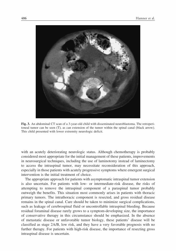
406 Hamner et al.
Fig. 3. An abdominal CT scan of a 3-year-old child with disseminated neuroblastoma. The retroperi-toneal tumor can be seen (T), as can extension of the tumor within the spinal canal (black arrow).This child presented with lower extremity neurologic deficit.
with an acutely deteriorating neurologic status. Although chemotherapy is probablyconsidered most appropriate for the initial management of these patients, improvementsin neurosurgical techniques, including the use of laminotomy instead of laminectomyto access the intraspinal tumor, may necessitate reconsideration of this approach,especially in those patients with acutely progressive symptoms where emergent surgicalintervention is the initial treatment of choice.
The appropriate approach for patients with asymptomatic intraspinal tumor extensionis also uncertain. For patients with low- or intermediate-risk disease, the risks ofattempting to remove the intraspinal component of a paraspinal tumor probablyoutweigh the benefits. This situation most commonly arises in patients with thoracicprimary tumors. The intrathoracic component is resected, and gross residual diseaseremains in the spinal canal. Care should be taken to minimize surgical complications,such as leakage of cerebrospinal fluid or uncontrollable intraspinal bleeding. Becauseresidual foraminal disease rarely grows to a symptom-developing size, the importanceof conservative therapy in this circumstance should be emphasized. In the absenceof metastatic disease or unfavorable tumor biology, these patients’ disease will beclassified as stage 2A/B, low risk, and they have a very favorable prognosis with nofurther therapy. For patients with high-risk disease, the importance of resecting grossintraspinal disease is uncertain.

Chapter 23 / Neuroblastoma 407
NeonatalSmall, localized neuroblastomas in infants younger than 3 months of age tend
to regress spontaneously; therefore, the COG has recently opened a protocol(ANBLOOP2) of expectant observation of patients with these lesions to furtherdefine their natural history. This study is designed to prove the hypothesis that closebiochemical and sonographic observation can be used for safe clinical management ofinfants with these tumors; surgical resection is reserved for those rare cases in whichthere is evidence of continued growth.
Stage 4S NeuroblastomaIn 1971, D’Angio, Evans, and Koop reported a number of patients with a “special”
variant of metastatic neuroblastoma, termed IVS (now referred to as 4S) (33). Thesepatients were infants who typically had a single, small primary tumor; however,these infants often had extensive metastatic disease in the liver, resulting in signif-icant hepatomegaly, skin nodules (“blueberry muffin” lesions), and small amountsof disease in the bone marrow (<10% of the mononuclear cells). Patients with 4Sneuroblastoma were quite remarkable, because the large amount of disease generallyunderwent spontaneous regression, even without treatment, and the infants ultimatelyhad no evidence of disease (Fig. 4).
Only supportive therapy has been recommended for this stage of neuroblastomabecause of the high incidence of spontaneous regression and the resultant goodprognosis (34). Most of these patients have tumor with favorable biology (single-copyMYCN, favorable Shimada histology, and DNA index > 1); therefore, they are assignedto the low-risk classification and receive no therapy. However, despite the generallybenign course of their malignancy, these infants can die of complications caused by
Fig. 4. An abdominal magnetic resonance imaging of a 2-month-old child with stage 4S neurob-lastoma demonstrating extensive, multifocal liver involvement.

408 Hamner et al.
the initial bulk of their disease. Limited chemotherapy, local irradiation, or minimalresection can be used to treat infants with life-threatening symptoms of hepatomegaly.Operative placement of a Silastic pouch as a temporary abdominal wall may be a choicefor those with significant liver enlargement that causes either respiratory compromisesecondary to diaphragmatic elevation or obstruction of the inferior vena cava (35).This procedure may help to avoid life-threatening events until shrinkage of the liver isachieved by either spontaneous regression or therapy.
Recurrent DiseasePatients with recurrent high-risk neuroblastoma have a nearly uniformly dismal
outcome. Nevertheless, surgery may have a limited role in the management of patientswith relapsed neuroblastoma where the recurrence has been documented to be localizedand refractory to available chemotherapeutic agents.
REOPERATIVE SURGERY
Reoperative surgery for children with neuroblastoma, caused by either diseaseprogression or recurrence, is rare, other than for delayed resection of the primarylocoregional disease after an initial biopsy and neoadjuvant chemotherapy. The reasonsfor this are twofold. In children with low- or intermediate-risk disease, the likelihoodof disease recurrence or progression is extremely low; whereas the appropriatenessof surgical resection of recurrent, high-risk disease is very limited, as previouslymentioned, given the extremely poor prognosis for these patients and the high proba-bility of widely disseminated disease in this setting. On rare occasion, staged attemptsat achieving gross total resection of locoregional high-risk disease prior to under-going high-dose consolidative chemotherapy with autologous stem cell rescue may beundertaken, although the value of this approach is unproven.
Reoperative surgery may also be undertaken for reasons relating to complications ofthe surgery. Postoperative bowel obstruction requiring surgical intervention can occurbecause of adhesions or intussusception (Fig. 5). Occasionally, reoperation for surgical
Fig. 5. Midjejunal intussusception in a patient reexplored for a bowel obstruction 6 days after anadrenalectomy for a localized, stage 1 neuroblastoma. At the right is the dilated, fluid-filled proximalsmall bowel. The black arrow shows the collapsed, more distal bowel. The white arrow shows theintussuscipiens containing the intussusceptum.

Chapter 23 / Neuroblastoma 409
closure of a lymphatic channel will be required for persistent chyle leak in the abdomenor chest that is refractory to or persistent despite conservative measures.
SUMMARY
The treatment of neuroblastoma requires a multidisciplinary approach. Althoughsurgical resection may be the only therapy required for patients at low-risk of diseaserecurrence, the surgeon provides but one element of the modern multimodal treatmentof children with high-risk disease. However, it is essential that the surgeon be awareof the heterogeneous nature of neuroblastoma and the factors that have, thus far, beenidentified in predicting the risk for relapse of children with this disease. An appreciationfor the different clinical scenarios and how they impact on the need for tissue toperform necessary studies and the role of surgery with respect to timing and extent oftumor resection is essential for the optimal care of these children.
REFERENCES
1. Brodeur GM, Castleberry RP. Neuroblastoma. In: Pizzo PA, Poplack DG, (eds), Principles andpractice of pediatric oncology, second edition. Philadelphia: J. B. Lippincott; 1993: 739–767.
2. Young JL Jr, Ries LG, Silverberg E, et al. Cancer incidence, survival, and mortality for childrenyounger than age 15 years. Cancer 1986;58:598–602.
3. Brodeur GM, Nakagawara A. Molecular basis of clinical heterogeneity in neuroblastoma. Am JPediatr Hematol Oncol 1992;14:111–116.
4. Breslow N, McCann B. Statistical estimation of prognosis for children with neuroblastoma. CancerRes 1971;31:2098–2103.
5. Evans AE, D’Angio GJ, Propert K, et al. Prognostic factor in neuroblastoma. Cancer 1987;59:1853–1859.
6. Brodeur GM, Seeger RC, Schwab M, et al. Amplification of N-myc in untreated human neuroblas-tomas correlates with advanced disease stage. Science 1984;224:1121–1124.
7. Seeger RC, Brodeur GM, Sather H, et al. Association of multiple copies of the N-myc oncogene withrapid progression of neuroblastomas. N Engl J Med 1985;313:1111–1116.
8. Look AT, Hayes FA, Shuster JJ, et al. Clinical relevance of tumor cell ploidy and N-myc gene amplifi-cation in childhood neuroblastoma: a Pediatric Oncology Group study. J Clin Oncol 1991;9:581–591.
9. Look AT, Hayes FA, Nitschke R, et al. Cellular DNA content as a predictor of response tochemotherapy in infants with unresectable neuroblastoma. N Engl J Med 1984;311:231–235.
10. Shimada H, Umehara S, Monobe Y, et al. International neuroblastoma pathology classification forprognostic evaluation of patients with peripheral neuroblastic tumors: a report from the Children’sCancer Group. Cancer 2001;92:2451–2461.
11. Brodeur GM, Pritchard J, Berthold F, et al. Revisions of the international criteria for neuroblastomadiagnosis, staging, and response to treatment. J Clin Oncol 1993;11:1466–1477.
12. Cecchetto G, Mosseri V, DeBernardi B, et al. Surgical risk factors in primary surgery for localizedneuroblastoma: the LNESG1 study of the European International Society of Pediatric OncologyNeuroblastoma Group. J Clin Oncol 2005;23:8483–8489.
13. Brodeur GM, Hayes FA, Green AA, et al. Consistent N-myc copy number in simultaneous orconsecutive neuroblastoma samples from sixty individual patients. Cancer Res 1987;47:4248–4253.
14. Shimada H, Chatten J, Newton WA Jr, et al. Histopathologic prognostic factors in neuroblastic tumors:definition of subtypes of ganglioneuroblastoma and an age-linked classification of neuroblastomas.J Natl Cancer Inst 1984;73:405–416.
15. Chatten J, Shimada H, Sather HN, et al. Prognostic value of histopathology in advanced neurob-lastoma: a report from the Childrens Cancer Study Group. Hum Pathol 1988;19:1187–1198.
16. Nitschke R, Smith EI, Shochat S, et al. Localized neuroblastoma treated by surgery: a PediatricOncology Group Study. J Clin Oncol 1988;6:1271–1279.
17. DuBois SG, Kalika Y, Lukens JN, et al. Metastatic sites in stage IV and IVS neuroblastoma correlatewith age, tumor biology, and survival. J Pediatr Hematol Oncol 1999;21:181–189.
18. LaQuaglia M, Kushner BH, Gholizadeh M, et al. The impact of gross total resection on local controland survival in high-risk neuroblastoma. J Pediatr Surg 2004;39:412–417.

410 Hamner et al.
19. Haase GM, O’Leary MC, Ramsay NK, et al. Aggressive surgery combined with intensivechemotherapy improves survival in poor-risk neuroblastoma. J Pediatr Surg 1991;26:1119–1123.
20. Grosfeld JL, Baehner RL. Neuroblastoma: an analysis of 160 cases. World J Surg 1980;4:29–38.21. Adkins, ES, Sawin R, Gerbing RB, et al. Efficacy of complete resection for high-rick neuroblastoma:
a Children’s Cancer Group Study. J Pediatr Surg 2004;39:6:931–936.22. Kiely EM. The surgical challenge of neuroblastoma. J Pediatr Surg 1994;29:128–133.23. Castel V, Tovar JA, Costa E, et al. The role of surgery in stage IV neuroblastoma. J Pediatr Surg
2002;37:1574–1578.24. von Schweinitz D, Hero B, Berthold F. The impact of surgical radicality on outcome in childhood
neuroblastoma. Eur J Pediatr Surg 2002;12:402–409.25. Azizkhan RG, Shaw A, Chandler JG. Surgical complications of neuroblastoma resection. Surgery
1985;97:514–517.26. Shorter NA, Davidoff AM, Evans AE, et al. The role of surgery in the management of stage IV
neuroblastoma: a single institution study. Med Pediatr Oncol 1995;24:287–291.27. Schmidt ML, Lal A, Seeger RC, et al. Favorable prognosis for patients ages 12–18 months with
stage 4 MYCN-nonamplified neuroblastoma. J Clin Oncol 2005;23:6474–6480.28. Schmidt ML, Lukens JN, Seeger RC, et al. Biologic factors determine prognosis in infants with stage
IV neuroblastoma: a prospective Children’s Cancer Group study. J Clin Oncol 2000;18:1260–1268.29. Medary I, Aronson D, Cheung NK, et al. Kinetics of primary tumor regression with chemotherapy:
implications for the timing of surgery. Ann Surg Oncol 1996;3:521–525.30. Davidoff AM, Corey BL, Hoffer FA, Santana VM, Furman WL, Shochat SJ. Radiographic assessment
of resectability of locoregional disease in children with high-risk neuroblastoma during neoadjuvantchemotherapy. Pediatr Blood Cancer 2005;44(2):158–162.
31. Loo R, Applebaum H, Takasugi J, et al. Resection of advanced stage neuroblastoma with the cavitronultrasonic surgical aspirator. J Pediatr Surg 1988;23:1135–1138.
32. Katzenstein HM, Kent PM, London WB, et al. Treatment and outcome of 83 children with intraspinalneuroblastoma: the Pediatric Oncology Group experience. J Clin Oncol 2001;19:1047–1055.
33. D’Angio GJ, Evans AE, Koop CE. Special pattern of widespread neuroblastoma with a favourableprognosis. Lancet 1971;1:1046–1049.
34. Nickerson HJ, Matthay KK, Seeger RC, et al. Favorable biology and outcome of stage IV-S neurob-lastoma with supportive care or minimal therapy: a Children’s Cancer Group study. J Clin Oncol2000;18:477–486.
35. Schnaufer L, Koop CE. Silastic abdominal patch for temporary hepatomegaly in stage IV-S neurob-lastoma. J Pediatr Surg 1975;10:73–75.

24 Wilms’ Tumor
Sanjeev A. Vasudevan, MD
and Jed G. Nuchtern, MD
CONTENTS
Unresectable Wilms’ Tumor
Wilms’ Tumor With Major Venous Extension
Bilateral Wilms’ Tumor
Metastatic Disease
Recurrent Wilms’
References
Wilms’ tumor, also known as nephroblastoma, is the predominant renal cancer ofchildhood, affecting approximately 500 new patients in North America per year (1).Nephroblastoma was first recognized and described by Max Wilms in 1899 (2). Thetumor arises from embryonic kidney precursor cells that obtain malignant qualitiesbecause of various degrees of genetic instability. Even though the majority of diagnosedWilms’ tumor are unilateral and arise from sporadic mutations, approximately 10% ofpatients with Wilms’ tumor have bilateral or multifocal tumors and other associatedmalformations and/or clinical syndromes. Inherited alterations of the 11p13 locusincluding the WT1 gene are responsible for syndromes including WAGR (Wilms’tumor, aniridia, genitourinary abnormalities, and mental retardation), Denys-Drash, andFrasier (3). Additionally, alterations within the 11p15 locus are also associated withWilms’, in particular, Beckwith-Wiedemann syndrome.
Wilms’ tumors are typically responsive to chemotherapy; therefore, a strategyconsisting of nephrectomy and adjuvant chemotherapy is the basis of most validatedtreatment protocols. Additional interventions, such as preoperative chemotherapy forunresectable disease and radiation therapy for primary and metastatic disease, have alsobeen employed and shown to improve outcomes in particular clinical circumstances.The surgical standard of care is total nephrectomy of the affected kidney with lymphnode sampling. Partial nephrectomy can be used in a small subset of patients but isusually not considered in unilateral disease because of the low occurrence of renalfailure after unilateral nephectomy.
In oncology, reoperative surgery has several meanings depending on the clinicalcontext. The need for additional surgery owing to disease recurrence or metastatic
From: Reoperative Pediatric SurgeryEdited by: S. Teich and D. A. Caniano © Humana Press, Totowa, NJ
411

412 Vasudevan and Nuchtern
disease would be considered reoperative surgery. Because some of the primary diffi-culties of reoperative surgery are inflammatory changes and adhesions caused by thefirst surgery, a tumor resection after neoadjuvant chemotherapy or radiation therapymust fall under this category because of the multitude of challenges encountered ina posttreatment resection. In this chapter, we will focus on the surgical managementof inoperable tumors treated with neoadjuvant therapy, tumor with major venousextension, bilateral Wilms’ (synchronous or metachronous), metastatic disease, anddisease recurrence.
UNRESECTABLE WILMS’ TUMOR
Clinical PresentationWilms’ tumor that is deemed unresectable has a clinical presentation similar to
operable primary nephroblastoma. On average, patients are approximately 3 years ofage at diagnosis of Wilms’ tumor. Patients who undergo preoperative chemotherapy foran initially unresectable tumor are older, on average, than patients undergoing primarynephrectomies, with 61% older than 4 years of age (4). The typically large, round, andfirm abdominal/retroperitoneal mass is detected either by parents during routine careof the child or by the primary care physician during well-child examinations. Commonclinical signs include, abdominal pain, anorexia, weight loss, gross or microscopichematuria, hypertension, anemia, polycythemia, and/or fever. Patients can also haveacquired von Willebrand’s disease warranting a coagulation evaluation.
Diagnostic EvaluationInvariably, the first study to be ordered in evaluating an abdominal mass is a plain
radiograph of the abdomen. This usually reveals visceral displacement caused by themass effect of the tumor. Calcification within the mass can sometimes be appreciatedas well. Ultrasound is the first test to order in evaluating a suspected Wilms’ tumor.This will enable the surgeon to differentiate between a cystic or solid mass and todetermine renal vein/inferior vena caval (IVC) extension. On ultrasound, Wilms’ tumortypically appears as a solid lesion with a pseudocapsule made up of compressed renalparenchyma (5). In addition, ultrasound screening is suggested in children with Wilms’tumor-associated syndromes at 3-month intervals until 6–8 years of age.
Computed tomography (CT) is very useful in the work up of Wilms’ tumor. Withintravenous contrast, the mass will have less contrast enhancement than the normalrenal parenchyma defining the tumor margin. Because Wilms’ tumor arises from thekidney progenitor cells, the tumor appears within the kidney with normal surroundingand medially displaced renal parenchyma and collecting system. Occasionally, curvi-linear calcification can be seen within the tumor. CT is also useful for assessment ofsynchronous, contralateral disease and tumor spread into lymph nodes, renal vascu-lature, IVC, and adjacent organs. Presence of lung and liver metastasis is also assessedwith CT. CT scan of the brain should be obtained in the postoperative setting forrhabdoid variant, and bone scan for clear cell variant of Wilms’ tumor. Magneticresonance imaging (MRI) can assist with intravascular evaluation of tumor extension.Resectability can be assessed with imaging but only as an adjunct to actual surgicalexploration.
Chapter 24 / Wilms’ Tumor 413
Surgical Exploration, Biopsy, and StagingWilms’ tumor is deemed inoperable based upon the following criteria defined
by Ritchey et al. (4): (1) tumor found to be unresectable at surgical exploration;(2) inoperability determined by clinical evaluation or diagnostic imaging; (3) extensiveintravascular tumor extension above the level of the hepatic veins; and (4) other patient-related morbidities secondary to the tumor. Disease stage is only accurately assessedwith surgical exploration to determine extent of tumor, lymph node spread, and obtaina histologic diagnosis with intraoperative biopsy.
The North American-based, National Wilms’ Tumor Study group (NWTSG) and theEuropean-based, International Society of Pediatric Oncology (SIOP) have somewhatopposing views on this issue. NWTSG has previously recommended initial surgicalexploration to accurately stage the patient prior to initiating a stage-appropriatetreatment protocol. If the tumor is deemed operable, a complete resection is performedfollowed by adjuvant therapies based on staging. SIOP recommends basing thediagnosis of Wilms’ tumor solely on clinical history and imaging. From this, the patientis staged and placed on a stage-appropriate, preoperative chemotherapy. The patientthen undergoes delayed surgical exploration and resection of primary tumor.
There are some proponents of percutaneous biopsy, rather than surgical exploration,as a strategy for staging patients prior to initiation of preoperative chemotherapy.Saarinen et al. state that percutaneous needle biopsy of Wilms’ tumor in children is safeand prevented misdiagnosis and understaging in one patient with unfavorable histologyand one patient with rhabdoid variant (6). However, this paper also reports twopatients that had subcapsular intratumoral bleeding. The United Kingdom Children’sCancer Study Group (UKCCSG) Wilms’ Tumor Study-3 adopted percutaneous needlebiopsy for tumor staging prior to initiation of chemotherapy (7). They reported theassociated morbidities with needle biopsy, which included: decrease in hemoglobin(20%), local pain (19%), one patient with massive intratumoral bleeding requiringoperative intervention, and one patient with tumor rupture who subsequently died.Also, needle track seeding and recurrence have been reported complications of percuta-neous biopsy (7,8). Moreover, NWTS-5 demonstrated that unfavorable histopathologicfeatures are inhomogeneously distributed within a given Wilms’ tumor, leading tothe suggestion that neither percutaneous nor open biopsy is infallible for accuratestaging.
The concept of staging with only clinical and imaging data has not previouslybeen practiced by the NWTSG for two reasons: (1) misdiagnosis, and (2) inaccuratestaging. Because many other types of pediatric malignancies can arise in the perirenal,retroperitoneal space, misdiagnosis of Wilms’ tumor is a very real possibility. Institu-tions using the SIOP protocols have mistakenly diagnosed mesoblastic nephroma andrenal cell carcinoma as Wilms’ (9,10). These misdiagnosed tumors are treated withthe assigned preoperative Wilms’ tumor chemotherapy regimen and only correctlydiagnosed after tumor resection 6–8 weeks later. Although this is less of an issuefor renal cell carcinoma, for which there is no effective neoadjuvant chemotherapy atpresent, there is more concern regarding administration of cytotoxic chemotherapy topatients with benign histology such as mesoblastic nephroma.
The primary concern of the NWTSG is inaccurate staging. The treatment protocolsfor stage I and II disease currently do not differ; however, in the next Children’sOncology Group (COG) Wilms’ tumor protocol, stage I, favorable histology tumors

414 Vasudevan and Nuchtern
less than 550 g, will not receive adjuvant chemotherapy. For now, the main concernis understaging a stage III or IV patient as a stage I or II. Gow et al. retrospectivelyreviewed a series of patients with nonmetastatic Wilms’ tumor (stages I–III) to comparepreoperative CT staging to histologic and operative staging (11). They found that CTcorrectly staged only 38% of patients and that 40% of histologically confirmed stageIII patients were categorized as stage II by imaging alone. In addition, 20% of thehistologic stage I and 33% of histologic stage II patients were staged as stage III byCT scan. Additional studies have shown inaccurate staging of stage III/IV patientsas stage I/II based on imaging in 10–14% of the patients studied using the SIOPprotocols (10,12).
Regardless of these discrepancies in image-based and histologic staging, outcomeson SIOP protocols do not differ greatly from the NWTSG protocols. This has promptedNWTSG to reevaluate their approach and incorporate some image-based staging intoupcoming trials. In all new protocols, any tumor spill, including open or needle biopsywill automatically classify the patient as stage III. In part because of this change, tumorbiopsy will not be required at presentation for patients with bilateral Wilms’ tumor,see following section. Given that any tumor that is deemed unresectable at presentationis stage III, an adequate biopsy should be performed in this circumstance in order totailor neoadjuvant chemotherapy.
Because imaging is not adequate for assessment of lymph node involvement, patientswho have inadequate lymph node sampling may need to be upstaged in order to preventundertreatment. Specifically, patients in the very low-risk group of age less than 2years, stage I tumor less than 550 g, and favorable histology will not receive adjuvantchemotherapy or radiation therapy, but in order to qualify for this treatment arm thepatient will have had adequate lymph node sampling. Similarly, patients with stageI clear cell sarcoma of the kidney with biopsy proven negative lymph nodes will bespared radiation therapy.
Prognostic FactorsThe most significant prognostic factor for Wilms’ tumor is unfavorable histology,
defined as the presence of anaplasia (reviewed in (13)) (Fig. 1). The results of NWTS-1showed that greater than 50% of the patients diagnosed with unfavorable histologyWilms’ tumor died as a result of disease versus less than 7% of patients with Wilms’tumor lacking histologic anaplasia (14). Since then, the definition and subclassificationsof anaplasia have been redefined to increase prognostic power. Faria et al. describefocal anaplasia as one or a few discrete foci in the primary tumor and diffuse anaplasiaas nonlocalized or located beyond the primary tumor capsule (15). With this definition,there was a striking difference in outcomes, with focal anaplasia associated with a 3%mortality and diffuse anaplasia with 96% mortality.
Other prognostic factors have proven useful throughout the various treatment trialsfor Wilms’ tumor. More recently, chromosomal deletions at 16q and 1p causing aloss of heterozygosity (LOH) at these loci have been associated with poorer prognosisin both favorable and unfavorable Wilms’ tumor (16,17). In addition, these studiesrevealed that 16q LOH, which was the most predictive of outcome, was more frequentin unfavorable versus favorable histology, anaplastic Wilms’ (32 versus 17%, respec-tively). NWTS-1 through NWTS-3 have reported significant prognostic factors forregional disease (stages I–III), including disease that is inoperable at diagnosis (18–20).

Chapter 24 / Wilms’ Tumor 415
A)
B)
Fig. 1. (A) Favorable histology Wilms’ tumor. (B) Unfavorable histology or anaplastic Wilms’tumor.
NWTS-1 identified unfavorable histology, tumor weight (>250g), and lymph node-positive disease as the most important predictors of mortality. Relapse was alsoassociated with these factors, as well as single-drug therapy and age greater than 24months. The results of NWTS-2 were similar but found that age and tumor weightreally had no predictive value. NWTS-3 reiterated that lymph node spread, age atdiagnosis, and tumor weight were predictive of outcome; moreover, this trial alsoidentified that microscopic disease at the tumor margin was an independent risk factorfor relapse and death in stage II and III patients.
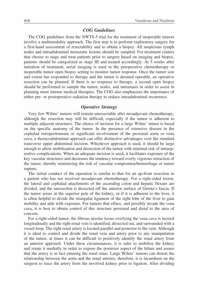
416 Vasudevan and Nuchtern
COG GuidelinesThe COG guidelines from the NWTS-5 trial for the treatment of inoperable tumors
involve a multimodality approach. The first step is to perform exploratory surgery fora first-hand assessment of resectability and to obtain a biopsy. All suspicious lymphnodes and intraabdominal metastatic lesions should be sampled. For treatment centersthat choose to stage and treat patients prior to surgery based on imaging and biopsy,patients should be categorized as stage III and treated accordingly. At 5 weeks afterinitiation of treatment, serial imaging is used in the preoperative chemotherapy orinoperable tumor open biopsy setting to monitor tumor response. Once the tumor sizeand extent has responded to therapy and the tumor is deemed operable, an operativeresection can be planned. If there is no response to therapy, a second open biopsyshould be performed to sample the tumor, nodes, and metastasis in order to assist inplanning more intense medical therapies. The COG also emphasizes the importance ofeither pre- or postoperative radiation therapy to reduce intraabdominal recurrence.
Operative StrategyVery few Wilms’ tumors will remain unresectable after neoadjuvant chemotherapy,
although the resection may still be difficult, especially if the tumor is adherent tomultiple adjacent structures. The choice of incision for a large Wilms’ tumor is basedon the specific anatomy of the tumor. In the presence of extensive disease in thecephalad retroperitoneum or significant involvement of the proximal aorta or venacava, a thoracoabdominal approach can offer distinctive advantages over the standardtransverse upper abdominal incision. Whichever approach is used, it should be largeenough to allow mobilization and dissection of the tumor with minimal risk of intraop-erative complications. When an adequate incision is used, it facilitates exposure of thekey vascular structures and decreases the tendency toward overly vigorous retraction ofthe tumor, thereby minimizing the risk of vascular compromise/hemorrhage or tumorrupture.
The initial conduct of the operation is similar to that for an up-front resection ina patient who has not received neoadjuvant chemotherapy. For a right-sided lesion,the lateral and cephalad attachments of the ascending colon and hepatic flexure aredivided, and the mesocolon is dissected off the anterior surface of Gerota’s fascia. Ifthe tumor arises in the superior pole of the kidney, or if it is adherent to the liver, itis often helpful to divide the triangular ligament of the right lobe of the liver to gainmobility and aide with exposure. For tumors that efface, and possibly invade the venacava, it is best to obtain control of this structure proximal and distal to the area ofconcern.
For a right-sided tumor, the fibrous areolar tissue overlying the vena cava is incisedlongitudinally and the right renal vein is identified, dissected out, and surrounded with avessel-loop. The right renal artery is located parallel and posterior to the vein. Althoughit is ideal to control and divide the renal vein and artery prior to any manipulationof the tumor, at times it can be difficult to positively identify the renal artery froman anterior approach. Under these circumstances, it is safer to mobilize the kidneyand rotate it medially in order to expose the posterior aspect of the hilum and assurethat the artery is in fact entering the renal sinus. Large Wilms’ tumors can distort therelationship between the aorta and the renal arteries; therefore, it is incumbent on thesurgeon to trace the artery from the involved kidney prior to ligation. After dividing

Chapter 24 / Wilms’ Tumor 417
the renal vessels, the ureter is identified, traced to the pelvic brim, suture ligated, anddivided. Finally the tumor and kidney are dissected out circumferentially and removed.It is standard practice to remove the adrenal gland if it is not separated from an upperpole tumor by a distinct tissue plane. The approach to a left-sided tumor is similar tothe right side; again, care should be taken to positively identify the renal vessels forlarge tumors that distort the retroperitoneal anatomy.
Wilms’ tumors rarely invade the liver, spleen, pancreas, or colon, although the tumormay be somewhat adherent to these organs. It is not normally necessary to performen-bloc resection of these organs, but rather it is better to seek a plane of dissectionthat will allow separation. For nonvital structures such as diaphragm, psoas muscle,or tail of pancreas, it may be advantageous to resect a small portion of these tissuesrather than risk entering the tumor.
OutcomeThe majority of patients with nonmetastatic, initially inoperable Wilms’ tumor
have stage III disease based on lymph node involvement, tumor spillage owing toinitial biopsy or difficulty of resection, peritoneal involvement, incomplete tumorresection (positive margins on surgical specimen), or piecemeal tumor removal. TheNWTSG encourages continued treatment of these patients as stage III regardless ofpreoperative chemotherapy effects on the tumor. The SIOP treatment protocols use apostchemotherapy staging system to determine the appropriate treatment regimens foreach patient. A SIOP trial performed to determine the necessity of abdominal radiationafter chemotherapy in patients with stage II, node-negative disease was stopped earlybecause of an unacceptably high rate of local recurrence in those subjects who did notreceive abdominal radiation (21).
Patients with stage III Wilms’ and favorable histology have a 4-year relapse-freesurvival of 90% and overall 4-year survival of 95% (22). The treatment recom-mendation for this group is: nephrectomy with lymph node sampling, abdominalradiation, and a 24-week chemotherapy regimen (NWTS Regimen DD-4A) consistingof vincristine, doxorubicin, and dactinomycin. For patients with diffuse anaplasia onhistology, the 4-year overall survival is decreased to 56%. They receive a modified24-week chemotherapy regimen (NWTS Regimen I) with the addition of etoposide,cyclophosphamide, and mesna (no dactinomycin). In NWTS-3, the complication ratereported for patients who underwent delayed resection after neoadjuvant chemotherapywas 19%, similar to that of patients subjected to primary nephrectomy without preop-erative therapy (4,23). However, in NWTS-4, the complication rate was lower, 12.7%,because of decrease in extensive intraoperative bleeding and major intraoperativecomplications (24).
WILMS’ TUMOR WITH MAJOR VENOUS EXTENSION
Epidemiology and Clinical PresentationIt is generally accepted that intravascular extension occurs in 4–10% of patients with
Wilms’ tumor (25). In NWTS-4, 165 out of 2731 (6%) patients had IVC and/or rightatrial involvement (26). Out of these patients, 19% had atrial involvement and 81% haddisease extending only into the IVC. Wilms’ tumor with caval and/or atrial extensionis considered stage II if it is resected en bloc with the primary tumor; however, ifthe primary tumor and intravascular extension are removed separately, the patient is
418 Vasudevan and Nuchtern
considered a stage III (2). Varicocele, heart failure, hepatomegaly, and ascites areunique clinical features of Wilms’ tumor with intravascular extension secondary toobstruction of venous return. However, these symptoms are only present in approx-imately 5% of the patients, with the majority presenting with the common Wilms’tumor symptoms discussed previously (26,27).
Diagnostic EvaluationUltrasound is the most appropriate initial study for an infant or child with an
abdominal mass. In Wilms’ tumor, this modality offers the additional advantage ofaccurate evaluation of the intravascular extension of the tumor (Fig. 2A). Specifically,amplitude-coded color Doppler sonography is recommended for this particular evalu-ation (5). CT can also reveal intravascular extension; however, this is a less sensitivetest because of artifact from nonopacified blood (Fig. 2B). MRI can be used to mapintravascular extension and may be superior to ultrasound for operative planning inpatients with intravascular tumor (28).
Operative StrategyPreoperative imaging will normally identify intravascular tumor extension, but the
surgeon should routinely palpate the renal vein and IVC prior to division of the veinto be certain that tumor is not present. If intravascular extension is limited to therenal vein and the vena cava below the level of the hepatic veins, it can normally beremoved en bloc with the kidney at the initial tumor resection. In this circumstance,the vena cava is dissected out and surrounded with vessel loops above the level oftumor extension and below the renal veins, and the contralateral renal vein is isolatedin a similar fashion. The kidney and tumor are then resected, except for division of therenal vein. Polypropylene stay sutures are placed in the vena cava proximal and distalto the renal vein, the vessel loops are cinched down, and the vena cava is opened at therenal vein junction. In most cases the tumor will be free-floating within the lumen andcan be gently manipulated back through the renal vein orifice. A Foley catheter canalso be used to aid with tumor extraction. In some circumstances, the tumor will betightly adherent to the venous intima. In these cases, the venotomy should be extendedproximally to near the cephalad extension of the tumor. The technique is similar to acarotid endarterectomy requiring use of a Freer periosteal elevator or fine hemostat toseparate the tumor from the wall of the vessel. If at all possible the tumor should notbe transected, as this will upstage the patient to stage III. After tumor extraction, thevena cava is closed with running polypropylene suture, with care taken to de-air thevessel prior to tying the suture and releasing the proximal vessel loop.
Primary resection of intravascular tumors that extend above the level of the hepaticveins is associated with a high rate of complications, especially when the right atriumis involved. These patients are treated with preoperative chemotherapy in an attempt toshrink the tumor thrombus below the hepatic outflow. If the tumor does not decreasebelow this level after chemotherapy, the resection should be performed with the aid ofcardiopulmonary bypass. The kidney and tumor are dissected out and the renal arteryand ureter are divided. The patient is then heparinized and placed on bypass. Afterhypothermic circulatory arrest is initiated, the right atrium and vena cava are openedand the tumor is removed either with traction or using a Foley catheter technique.The lumen should be carefully flushed prior to closure to remove any stray tumorfragments. Circulation is then restored.

Chapter 24 / Wilms’ Tumor 419
OutcomeBoth NWTSG and SIOP studies have determined that there is no significant survival
difference between Wilms’ tumor with or without intravascular extension (25,26). Theprimary differences lie in the frequency of complications. Caval and atrial surgeryrequire more extensive dissection, longer operative time, and, in most cases of atrialextension, cardiopulmonary bypass. Shamberger et al. report a complication rate of17% with IVC extension and 37% in disease with atrial involvement (26). The mostrecent summary of surgical complications in NWTS-4 identified caval and/or atrialextension as a significant risk factor for complications in both univariate (odds ratio= 5.66) and multivariate analysis (odds ratio = 3.77) (24). Complications include:intraoperative hemorrhage, bowel obstruction, tumor embolus, IVC obstruction, respi-ratory distress syndrome, wound dehiscence, aortic thrombosis, paraparesis, hepaticvein injury, cerebral ischemia, pancreatitis, and postpericardiotomy syndrome.
ThAo
Liv
Tu
Th
A)
B)
Fig. 2. (A) Ultrasound showing cross-sectional view of the liver (Liv), inferior vena cava (IVC)tumor thrombus (Th) extending from primary tumor, and the aorta (Ao). (B) Coronal reconstructionCT scan showing tumor thrombus (Th) extending from the right kidney primary tumor (Tu) into theIVC. Distal to the thrombus, the patent hepatic vein and IVC (black arrow) can be seen draininginto the right atrium.

420 Vasudevan and Nuchtern
The most recent NWTSG and SIOP recommendations are for preoperativechemotherapy in patients with tumor extension above the level of the renal veins.This can cause tumor regression from suprahepatic or atrial locations helping to avoidmore involved dissections and cardiopulmonary bypass. In NWTS-4, it was noted thatpatients who had received preoperative chemotherapy had more adherent intravasculartumor thrombus compared to those who were not treated preoperatively (62 versus44%, respectively) (26). In approximately one-half of the patients that had receivedpreoperative chemotherapy, unresectable tumor thrombus remained after resection;however, there was no increased risk of relapse. In addition, preoperative chemotherapywas associated with other rare complications, such as tumor embolism and respiratorydistress syndrome. In general, the complication rate and overall survival in the primaryresection group versus preoperative chemotherapy with subsequent surgery group didnot reach statistical significance.
BILATERAL WILMS’ TUMOR
Epidemiology and Clinical PresentationSynchronous, bilateral tumors account for approximately 5% of all nephroblastomas
(1). Patients presenting with synchronous, bilateral Wilms’ tumor are, on average,2–3 years of age (29–31). Metachronous lesions occur in approximately 1–2% of allpatients presenting with unilateral Wilms’ tumor (32). The incidence of metachronouslesions increases to approximately 4% in children diagnosed with unilateral Wilms’tumor at less than 1 year of age.
Symptoms at presentation are very similar to unilateral Wilms’ as discussed previ-ously. In a case series by Tomlinson et al., all of the patients presented with a palpableabdominal mass and some patients with palpable mass and macroscopic hematuria(29). Patients with bilateral disease can also present with hypertension and cardiomy-opathy (31).
Diagnostic EvaluationThe imaging strategy outlined previously also applies to diagnostic evaluation
of bilateral Wilms’ tumor. Because 5% of unilateral Wilms’ are associated witha synchronous lesion, thorough imaging analysis of the contralateral side must beperformed with ultrasound, CT scan, and/or MRI (Fig. 3A). A study by Ritchey et al.showed that CT is more sensitive than ultrasound for detecting synchronous lesions;however, 7% of the patients with bilateral disease would have been missed with imagingalone (33). Because of this, intraoperative evaluation of the contralateral kidney is notreserved for indeterminate imaging and clinical suspicion.
Prognostic FactorsThe same histologic, prognostic factors described previously apply to bilateral
disease. The most important risk factor for synchronous disease or development ofmetachronous disease is the presence of nephrogenic rests. Nephrogenic rests arethought to be precursor lesions for Wilms’ tumor. These can be subdivided into twocategories: perilobar and intralobar. Nephroblastomatosis refers to diffuse or multiple
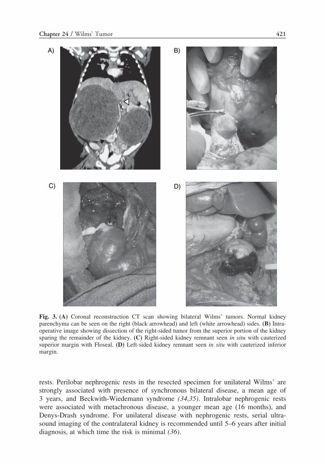
Chapter 24 / Wilms’ Tumor 421
A) B)
D)C)
Fig. 3. (A) Coronal reconstruction CT scan showing bilateral Wilms’ tumors. Normal kidneyparenchyma can be seen on the right (black arrowhead) and left (white arrowhead) sides. (B) Intra-operative image showing dissection of the right-sided tumor from the superior portion of the kidneysparing the remainder of the kidney. (C) Right-sided kidney remnant seen in situ with cauterizedsuperior margin with Floseal. (D) Left-sided kidney remnant seen in situ with cauterized inferiormargin.
rests. Perilobar nephrogenic rests in the resected specimen for unilateral Wilms’ arestrongly associated with presence of synchronous bilateral disease, a mean age of3 years, and Beckwith-Wiedemann syndrome (34,35). Intralobar nephrogenic restswere associated with metachronous disease, a younger mean age (16 months), andDenys-Drash syndrome. For unilateral disease with nephrogenic rests, serial ultra-sound imaging of the contralateral kidney is recommended until 5–6 years after initialdiagnosis, at which time the risk is minimal (36).

422 Vasudevan and Nuchtern
COG GuidelinesThe NWTS-5 originally recommended performing partial nephrectomy at the initial
surgery if the tumors could be completely removed while preserving adequate renalfunction. The most recent guidelines recommend not performing an initial operation atall. After the diagnosis is made radiographically, the patient should receive 6 weeks ofchemotherapy followed by imaging to determine tumor response. If the tumors seemamenable to partial nephrectomy, surgery should be performed at this time. If there isa partial response, the patient can be given more chemotherapy and reassessed afteran additional 6 weeks for surgery. If response is poor, bilateral open biopsies can beperformed at the 6-week mark to plan more intensive therapy. In the setting of extensive,unresponsive disease in one kidney and responsive or partially responsive disease inthe other kidney, therapy should be tailored to achieving a complete resection in thesalvageable kidney and radical nephrectomy of the unresponsive kidney. Radiationtherapy is reserved for the partially responsive or unresponsive tumors after second-look surgery.
Operative StrategyCT or MRI with reconstruction of the renal vascular anatomy can be very helpful
when planning partial nephrectomy. After an adequate skin incision, the kidney andtumor are mobilized in a standard fashion. The arterial and venous anatomies aredefined, and separate branches are tagged with vessel loops, should clamping orligation be needed. If the extent of tumor invasion into the kidney is unclear, intraop-erative ultrasound will help to define a plane of transection that will give an adequatemargin of normal kidney, ideally 2 mm. Sonographic imaging can also help predictthe involvement of the renal collecting system, which will help the surgeon antic-ipate the need for repair. The kidney is transected with cautery while the assistantcompresses the adjacent parenchyma to minimize bleeding (Fig. 3B). Hemostasis onthe cut surface is controlled with both fine Monocryl sutures for larger vessels andFloseal for parenchymal bleeding (Fig. 3C and D). The collecting system is repairedusing similar suture material and gently leak-tested by injecting it with dilute methyleneblue dye through a fine needle.
OutcomeFor bilateral Wilms’ tumor, the outcome is worse than the 90% overall survival seen
in all stages of unilateral disease. Data from NWTS-3 and -4 show that the 10-yearsurvival approaches 80% for synchronous disease, with no difference in survival notedbetween preoperative chemotherapy versus immediate resection (37,38). The utilityfor preoperative chemotherapy in bilateral Wilms’ tumor is to increase the likelihoodof a successful bilateral nephron-sparing nephrectomy (39). The main determinantsof prognosis are similar to unilateral disease: presence of unfavorable histology, ageat diagnosis, and highest stage of the individual tumors. Shamberger et al. haverecently reported a distinct survival difference between bilateral Wilms’ tumors thatare responsive to preoperative chemotherapy and those that are not (37). The overallsurvival for nonresponders is 67 versus 86% in those that respond to therapy. The mainreason for nonresponsive disease is undiagnosed anaplastic histology, which is bettertreated with immediate resection followed by intensive chemotherapy (40). The recom-mendations from this study are for open biopsy in patients with less than 50% tumor

Chapter 24 / Wilms’ Tumor 423
reduction after two courses of therapy and early surgical resection for nonresponsivedisease.
In metachronous disease, survival is much worse than synchronous bilateral disease(41,42). A more recent series of 108 patients with metachronous Wilms’ tumor showeda 10-year overall survival of 47%. Paulino et al. further analyzed the survival dataaccording to time interval to second tumor development and found that contralateraltumors arising less than 18 months from the first tumor have a 10-year overall survivalof 40%, and those developing at or after 18 months have a survival rate of 55% (43).
Bilateral Wilms’ tumor is a risk factor for end-stage renal disease (ESRD). Uremiacan be caused by progression of Wilms’ tumor, necessitating total nephrectomy,radiation injury, or hyperfiltration in patients with relatively small amounts of renalparenchyma. In contrast to patients with unilateral tumors, in whom the overallincidence of ESRD is 1.3%, the cumulative risk of ESRD in patients with bilateralWilms’ tumor is 15% at 20 years. The incidence is much higher in patients withDenys-Drash syndrome (50%), WAGR syndrome (90%), or with hypospadias orcryptorchidism (25%), whereas patients without these characteristics have an incidenceof 11.5% (43).
METASTATIC DISEASE
The most common sites for metastasis in stage IV Wilms’ tumor are the lung andliver. Lung metastasis are the most common, occurring in approximately 80% of allmetastatic disease; whereas liver metastasis occurs in approximately 15%. Breslowet al. determined that specific metastatic sites at diagnosis had little prognostic value.Survival was dependent on histology and staging of the primary tumor; however,metastasis developing during or after treatment had a significant effect on survival,where liver metastasis conferred a much worse prognosis than lung (44). More recently,Varan et al. reported that metastatic site at diagnosis did effect survival (45). Theyidentified an overall survival of 50% for patients with lung metastasis only and 17%for patients with liver metastasis. The NWTSG reports that the overall survival forpatients with stage IV, favorable histology is 80% using a treatment regimen consistingof immediate nephrectomy, postoperative whole lung irradiation, abdominal irradiationaccording to local stage of primary tumor, and 24 weeks of chemotherapy (22,46).Patients with unfavorable histology of the primary lesion receive the same therapy withthe addition of etoposide, cyclophosphamide, and mesna; however, the 4-year survivalrate is only 17% (40). In general, resection of metastasis is reserved for persistent orrelapsing disease, which will be discussed in the next section.
RECURRENT WILMS’
Disease relapse occurs in less than 20% of patients after treatment of the primaryWilms’ (47,48). The characteristics correlating with the best prognosis for relapseddisease include: favorable histology, abdominal recurrence without prior irradiation,recurrent disease confined to the lung, stage I primary disease, primary disease treatedwith two drug regimen, and recurrence occurring at or after 12 months from diagnosis ofprimary disease (49). Adverse prognostic factors of relapsed disease include: recurrentabdominal disease in setting of irradiation, tumor positive lymph nodes, two or morerecurrent lesions, relapse less than 12 months after diagnosis of primary disease, bone

424 Vasudevan and Nuchtern
or brain metastasis, stage IV primary disease, unfavorable histology, and thoracic andabdominal recurrence (50).
The prognosis for recurrent disease has improved over the years; however, overallsurvival rates are not equivalent to the greater than 90% survival seen with treatedprimary disease. The 3- to 4-year overall survival of relapsed patients observed inNWTS -1, -2, and -3 is less than 50%, with some subgroups identified with abetter prognosis than others (49,51). The latest report of the NWTS-5 focused on agroup of patients enrolled on NWTS-5 and subsequently registered on the NWTSGprotocol, “Treatment of Relapsed Patients” after recurrence. This protocol incorporatesa more intense 24-week therapeutic regimen consisting of radiation therapy, vincristine,doxorubicin, etoposide, and cyclophosphamide (50). Compared to relapsed patientsnot registered on this protocol, improved 4-year relapse-free was 71 versus 53%, andoverall survival rates of 82% versus 62% were observed.
The surgeon’s role in recurrent Wilms’ tumor lies in both prevention and therapy. Thepreventative role has to do with local recurrence. A thorough analysis of local recurrencein patients treated in NWTS-4 was completed by Shamberger et al. (52). They identifiedtumor spill (local or diffuse), age greater than 4 years, incomplete resection, and no lymphnode staging at primary operation as factors associated with a high risk of local recurrence.The association of tumor spill and lack of lymph node staging to local recurrence has leadto important staging and protocol alterations. As a result of this study, any tumor spilleage,local or diffuse, is automatically staged as a stage III in order to receive more intensivepostresection therapy to prevent recurrence and improve survival. In addition, resectionof stage I tumors without lymph node sampling was associated with a 6-fold higherrisk of recurrence compared to node-negative patients; therefore, all Wilms’ tumorresections for localized or invasive disease must include a sampling of the hilar and/orparaaortic lymph nodes to recognize patients at risk for recurrence.
The therapeutic role has to do with postrelapse therapy. Green et al. studied recurrentWilms’ confined to the lung in NWTS -1, -2, and -3, and found no benefit ofsurgical resection over whole-lung irradiation and chemotherapy (53). The guidelinesfor resection of recurrent disease and metastasis has not been extensively studied;however, it is generally accepted that open biopsy to confirm relapse and attemptedresection of intraabdominal recurrence to reduce tumor burden prior to radiation therapyand chemotherapy should be performed (54).
REFERENCES
1. Gommersall LM, Arya M, Mushtaq I, Duffy P. Current challenges in Wilms’ tumor management.Nat Clin Pract Oncol 2005;2:298–304.
2. Metzger ML, Dome JS. Current therapy for Wilms’ tumor. Oncologist 2005;10:815–826.3. Rivera MN, Haber DA. Wilms’ tumour: connecting tumorigenesis and organ development in the
kidney. Nat Rev Cancer 2005;5:699–712.4. Ritchey ML, Pringle KC, Breslow NE, et al. Management and outcome of inoperable Wilms’ tumor.
Ann Surg 1994;5:683–690.5. Riccabona M. Imaging of renal tumors in infancy and childhood. Eur Radiol 2003;13
(Suppl 4):L116–L129.6. Saarinen UM, Wikstrom S, Koskimies O, Sariola H. Percutaneous needle biopsy preceding preop-
erative chemotherapy in the management of massive renal tumors in children. J Clin Oncol 1991;9:406–415.
7. Vujanic GM, Kelsey A, Mitchell C, Shannon RS, Gornall P. The role of biopsy in the diagnosisof renal tumors of childhood: results of the UKCCSG Wilms’ tumor study 3. Med Pediatr Oncol2003;40:18–22.

Chapter 24 / Wilms’ Tumor 425
8. Lee IS, Nguyen S, Shanberg AM. Needle tract seeding after percutaneous biopsy of Wilms’ tumor.J Urol 1995;153:1074–1076.
9. Scott DJ, Wallace WH, Hendry GM. With advances in medical imaging can the radiologist reliablydiagnose Wilms’ tumours? Clin Radiol 1999;54:321–327.
10. Zoeller G, Pekrun A, Lakomek M, Ringert RH. Staging problems in the pre-operative chemotherapyof Wilms’ tumour. Br J Urol 1995;76:501–503.
11. Gow KW, Roberts IF, Jamieson DH, Bray H, Magee JF, Murphy JJ. Local staging of Wilms’ tumor-computerized tomography correlation with histological findings. J Pediatr Surg 2000;35:677–679.
12. Kullendorff CM, Wiebe T, Hayder S. National reevaluation of staging in Wilms’ tumor. Eur J PediatrSurg 1996;6:23–24.
13. Beckwith JB. National Wilms’ tumor study: an update for pathologists. Pediatr Dev Path 1998;1:79–84.
14. Beckwith JB, Palmer NF. Histopathology and prognosis of Wilms’ tumors: results from the firstnational Wilms’ tumor study. Cancer 1978;41:1937–1948.
15. Faria P, Beckwith JB, Mishra K, et al. Focal versus diffuse anaplasia in Wilms’ tumor-new definitionswith prognostic significance: a report from the National Wilms’ Tumor Study Group. Am J SurgPathol 1996;20:909–920.
16. Grundy PE, Telzerow PE, Breslow N, Moksness J, Huff V, Paterson MC. Loss of heterozygosityfor chromosomes 16q and 1p in Wilms’ tumors predicts an adverse outcome. Cancer Res 1994;54:2331–2333.
17. Grundy PE, Breslow NE, Li S, et al. Loss of heterozygosity for chromosomes 1q and 16q is anadverse prognostic factor in favorable-histology Wilms’ tumor: a report from the National Wilms’Tumor Study Group. J Clin Oncol 2005;23:7312–7321.
18. Breslow NE, Palmer NF, Hill LR, Buring J, D’Angio GJ. Wilms’ tumor: prognostic factors for patientswithout metastases at diagnosis: results of the national Wilms’ tumor study. Cancer 1978;41:1577–1589.
19. Breslow N, Churchill G, Beckwith JB, et al. Prognosis for Wilms’ tumor patients with nonmetastaticdisease at diagnosis-results of the second National Wilms’ Tumor Study. J Clin Oncol 1985;3:521–531.
20. Breslow N, Sharples K, Beckwith JB, et al. Prognostic factors in nonmetastatic, favorable histologyWilms’ tumor. Results of the Third National Wilms’ Tumor Study. Cancer 1991;68:2345–2353.
21. Tournade MF, Com-Nougue C, Voute PA, et al. Results of the Sixth International Society of PediatricOncology Wilms’ Tumor Trial and Study: a risk-adapted therapeutic approach in Wilms’ tumor.J Clin Oncol 1993;11:1014–1023.
22. NIH-NCI PDQ�. Wilms’ tumor and other childhood kidney tumors (PDQ�): Treatment. http://www.cancer.gov/cancertopics/pdq/treatment/Wilms’/healthprofessional. Date accessed: 7/24/07.
23. Ritchey ML, Kelalis PP, Breslow NB, et al. Surgical complications following nephrectomy for Wilms’tumor: a report of national Wilms’ tumor study-3. Surg Gynecol Obstet 1992;175:507–514.
24. Ritchey ML, Shamberger RC, Haase G, Horwitz J, Bergemann T, Breslow NE. Surgical complicationsafter primary nephrectomy for Wilms’ tumor: report from the National Wilms’ Tumor Study Group.J Am Coll Surg 2001;192:63–68.
25. Szavay P, Luithle T, Semler O, Graf N, Fuchs J. Surgery of cavoatrial tumor thombus in nephrob-lastoma: a report of the SIOP/GPOH study. Pediatr Blood Cancer 2004;43:40–45.
26. Shamberger RC, Ritchey ML, Haase GM, et al. Intravascular extension of Wilms’ tumor. Ann Surg2001;234:116–121.
27. Akyuz C, Emir S, Buyukpamukcu N, et al. Cavoatrial tumor extension in children with Wilms’ tumor.J Pediatr Hematol Oncol 2005;27:267–269.
28. Weese DL, Applebaum H, Taber P. Mapping intravascular extension of Wilms’ tumor with magneticresonance imaging. J Pediatr Surg 1991;26:64–67.
29. Tomlinson GS, Cole CH, Smith NM. Bilateral Wilms’ tumor: a clinicopathologic review. Pathology1991;31:12–16.
30. Kullendorff CM, Wiebe T. Bilateral Wilms’ tumor. Pediatr Surg Int 1999;15:46–49.31. Millar AJ, Davidson A, Rode H, et al. Bilateral Wilms’ tumors: a single-center experience with 19
cases. J Pediatr Surg 2005;40:1289–1294.32. Coppes MJ, Arnold M, Beckwith JB, et al. Factors affecting the risk of contralateral Wilms’ tumor
development: a report from the National Wilms’ Tumor Study Group. Cancer 1999;85:1616–1625.33. Ritchey ML, Green DM, Breslow NB, Moksness J, Norkool P. Accuracy of current imaging modalities
in the diagnosis of synchronous bilateral Wilms’ tumor. A report from the National Wilms’ TumorStudy Group. Cancer 1995;75:600–604.

426 Vasudevan and Nuchtern
34. Beckwith JB, Kiviat NB, Bonadio JF. Nephrogenic rests, nephroblastomatosis, and the pathogenesisof Wilms’ tumor. Pediatr Pathol 1990;10:1–36.
35. Heppe RK, Koyle MA, Beckwith JB. Nephrogenic rests in Wilms’ tumor patients with the Drashsyndrome. J Urol 1991;145:1225–1228.
36. Paulino AC, Thakkar B, Henderson WG. Metachronous bilateral Wilms’ tumor: the importance oftime interval to the development of a second tumor. Cancer 1998;82:415–420.
37. Shamberger RC, Haase GM, Argani P, et al. Bilateral Wilms’ tumors with progressive or nonre-sponsive disease. J Pediatr Surg 2006;41:652–657.
38. Montgomery BT, Kelalis PP, Blute ML, et al. Extended followup of bilateral Wilms’ tumor: resultsof the National Wilms’ Tumor Study. J Urol 1991;146:514–518.
39. McLorie GA, McKenna PH, Greenberg M, et al. Reduction in tumor burden allowing partialnephrectomy following preoperative chemotherapy in biopsy proved Wilms’ tumor. J Urol1991;146:509–513.
40. Green DM, Beckwith JB, Breslow NE, et al. Treatment of children with stages II to IV anaplasticWilms’ tumor: a report from the National Wilms’ Tumor Study Group. J Clin Oncol 1994;12:2126–2131.
41. Casale AJ, Flanigan RC, Moore PJ, McRoberts JW. Survival in bilateral metachronous (asynchronous)Wilms’ tumors. J Urol 1982;128:766–769.
42. Jones B, Hrabovsky E, Kiviat N, Breslow N. Metachronous bilateral Wilms’ tumor, National Wilms’Tumor Study. Am J Clin Oncol 1982;5:545–550.
43. Breslow NE, Collins AJ, Ritchey ML, Grigoriev YA, Peterson SM, Green DM. End stage renaldisease in patients with Wilms’ tumor: results from the National Wilms’ Tumor Study Group and theUnited States Renal Data System. J Urol 2005;174:1972–1975.
44. Breslow NE, Churchill G, Nesmith B, et al. Clinicopathologic features and prognosis for Wilms’tumor patients with metastases at diagnosis. Cancer 1986;58:2501–2511.
45. Varan A, Buyukpamukcu N, Caglar M, et al. Prognostic significance of metastatic site at diagnosisin Wilms’ tumor: results from a single center. J Pediatr Hematol Oncol 2005;27:188–191.
46. Green DM. The treatment of stages I-IV favorable histology Wilms’tumor. J Clin Oncol2004;22:1366–1372.
47. Groot-Loonen JJ, Pinkerton CR, Morris-Jones PH, Pritchard J. How curable is relapsed Wilms’tumour? The United Kingdom Children’s Cancer Study Group. Arch Dis Child 1990;65:968–970.
48. Dome JS, Liu T, Krasin M, et al. Improved survival for patients with recurrent Wilms’ tumor: theexperience at St. Jude Children’s Research Hospital. J Pediatr Hematol Oncol 2002;24:192–198.
49. Grundy P, Breslow N, Green DM, Sharlpes K, Evans A, D’Angio GJ. Prognostic factors for childrenwith recurrent Wilms’ tumor: results from the Second and Third National Wilms’ Tumor Study.J Clin Oncol 1989;7:638–647.
50. Green DM, Cotton CA, Malogolowkin M, et al. Treatment of Wilms’ tumor relapsing after intialtreatment with vincristine and actinomycin D: a report from the National Wilms’ Tumor Study Group.Pediatr Blood Cancer 2007;48(5):493–499.
51. Sutow WW, Breslow NE, Palmer NF, D’Angio GJ, Takashima J. Prognosis in children with Wilms’tumor metastases prior to or following primary treatment: results from the first National Wilms’Tumor Study (NWTS-1). Am J Clin Oncol 1982;5:339–347.
52. Shamberger RC, Guthrie KA, Ritchey ML, et al. Surgery-related factors and local recurrence ofWilms’ tumor in National Wilms’ Tumor Study 4. Ann Surg 1999;229:292–297.
53. Green DM, Breslow NE, Ii Y, et al. The role of surgical excision in the management of relapsedWilms’ tumor patients with pulmonary metastases: a report from the National Wilms’ Tumor Study.J Pediatr Surg 1991;26:728–733.
54. Grundy PE, Green DM, Coppes MJ, et al. Renal tumors. In: Pizzo PA, Poplack DG, (eds.), Principlesand practice of pediatric oncology, fourth edition. Philadelphia: Lippincott Williams & Wilkins;2002: 865–893.

25 Peripheral Neuroectodermal Tumors
Michael P. La Quaglia, MD
CONTENTS
Primitive Neuroectodermal Tumor of the Chest Wall
References
PRIMITIVE NEUROECTODERMAL TUMOR OF THE CHEST WALL
James Ewing described Ewing sarcoma in 1921 as an “endothelioma of bone” (1).He believed at the time that it arose from the osseous blood vessels and later realizedthat the tumor was more complex, describing it as an “endothelial myeloma” (2). Ewingis credited with differentiating this tumor from neuroblastoma and lymphoma. In 1979,Askin and Rosai described a “malignant small cell tumor of the thoracopulmonaryregion” in 20 children and adolescents with a median age of 14.5 years and with afemale rather than male predominance, as seen in Ewing sarcoma of bone (3). Theseauthors noted a predilection for local recurrence.
In the present era, most texts refer to the Ewing sarcoma family of tumors that includenot only the highly malignant small round cell tumors of bone originally describedby Ewing but also the chest wall tumors described by Askin, as well as extra-skeletalEwing sarcoma, and peripheral primitive neuroectodermal tumors (PNET) (peripheralneuroepithelioma). A unifying feature of these tumors is the presence of a reciprocaltranslocation between chromosomes 11 and 22 (t11;22)(q24;q12) in approximately85%. This is considered pathognomonic superseding other forms of pathologic analysis,like immunohistochemistry (4). Of great practical importance is that the surgeon mustassure that an adequate amount of biopsy tissue is preserved fresh and sterile sothat molecular genetic analysis can confirm the diagnosis. This chapter describes thediagnosis, treatment, reconstructive procedures, and late-treatment sequelae of PNETof the chest wall (Askin’s tumor, chest wall Ewing sarcoma). These chest wall tumorscomprise 16% of all Ewing sarcomas, with most arising from ribs (63%), followed byscapula (25%), clavicle (9%), and sternum (3%) (5). Ewing sarcoma family of tumorsgenerally metastasize to lung (10%) and bone (10%), or combinations of the two (6%).The rate of metastases for chest wall primary tumors is lower, and was commentedupon in the initial paper by Askin (3).
From: Reoperative Pediatric SurgeryEdited by: S. Teich and D. A. Caniano © Humana Press, Totowa, NJ
427

428 La Quaglia
Diagnostic Evaluation and Initial TherapyPatients with chest wall tumors may complain of a mass, bone pain referable to the
ribs or spine, or radicular pain from infiltration of the intercostal nerves (6). Anteriormasses may impinge on the pericardium, and be associated with chest pain in thisregion or a pericardial effusion. More posterior masses may invade not only the ribsand soft tissue but the bony spine as well. This could give rise to severe back pain.Diaphragmatic infiltration is possible and referred pain to the shoulder could ensue.Large tumors may also be associated with a significant pleural effusion and subsequentdyspnea, tachypnea, and/or orthopnea.
A chest wall mass or fullness is usually detectable on examination. Diminishedbreath sounds indicate a large pleural effusion, whereas heart tones may be decreasedor a rub heard with a pericardial effusion. Because these tumors develop peripherally atthe chest wall, the airway narrowing seen with large mediastinal masses like lymphomais not an issue.
After the initial clinical examination, computerized tomography (CT) of the chestwith and without intravenous contrast is recommended. The CT without contrast willdelineate the mass, and identify an adjacent pleural effusion or cardiac enlargement,suggesting pericardial effusion. The spine can also be evaluated as tumors near thecostovertebral junction can invade a vertebral body. A CT without contrast is alsothe best study to look for pulmonary metastases, which occur in less than 10% ofcases. The addition of intravenous contrast will give further anatomic detail regardingthe primary mass and its proximity to the aorta or other great vessels. These tumorsarise from a single rib and expand and distort the bone, so that this rib can beidentified as the epicenter of the tumor. This will have importance when resection iscontemplated. The rest of the mass is extraosseous soft tissue expansion and this canbe quite extensive. Magnetic resonance imaging (MRI) of the chest can also be usedand it too provides a good deal of anatomic information regarding the primary tumor.Evaluation of pulmonary metastases is less accurate however, and the technique mayrequire 30–60 minutes in the scanner for patients who may be having pain or dyspnea.
Since bony metastases occur in approximately 10% of patients with Ewingsarcoma/PNET, although this rate is probably less with chest wall tumors, a bone scanshould also be done. There are recent reports that positron emission tomography with18Fluorodeoxyglucose (PET) is more sensitive in identifying bony metastases fromEwing sarcoma/primitive ectodermal tumor (7). PET scans may also be useful in evalu-ating the response of primitive ectodermal tumors to neoadjuvant chemotherapy (8).Whole-body MRI may also be more sensitive in determining the number of osseousmetastases than bone scan (9,10).
Once imaging studies are done, the patient will be ready for biopsy of the primarytumor to definitively establish the diagnosis. Needle core biopsies with or without CTguidance can give adequate amounts of tissue. It should be remembered, however, thatmuch of the tumor before therapy can be necrotic and that an adequate amount of viabletissue must be obtained, not only for histology and immunohistochemistry but also formolecular genetics to document the (11;22) translocation. Molecular genetics alonerequires two viable core biopsies. An open approach under general anesthesia may alsobe elected, and has the advantage of not only getting a larger biopsy but also allowsplacement of a vascular access device, and bone marrow aspiration and biopsy, whichis part of the staging for these patients. Vascular access using an implanted double

Chapter 25 / Peripheral Neuroectodermal Tumors 429
lumen port is adequate and may be associated with a lower infection rate and improvedquality of life compared to external devices. This initial biopsy should be planned sothat the biopsy scar and underlying tissues can be incorporated into the incision fordefinitive resection and completely excised with the specimen. In addition, the pleuralspace should not be violated. Untreated chest wall PNETs can be very vascular, andmay also have a large necrotic core caused by rapid tumor growth. Frozen sectionsshould be obtained to verify that an adequate biopsy of viable tumor is being obtained.Packing with absorbable hemostatic agents may be required to control bleeding.
There is no formal staging system for Ewing sarcoma/PNET at present, except todescribe it as localized or metastatic. Some have used the rhadbomyosarcoma groupingsystem: Group I—complete gross and microscopic resection; Group II—microscopicresidual; and Group III—gross residual. Patients with metastases at diagnosis aregiven a separate category. This system is based on the results of resection rather thanparameters measured at the time of resection and is generally considered unsatisfactory.
Initial therapy for chest wall PNET is systemic, multiagent chemotherapy. Combi-nations of vincristine, doxorubicin, cyclophosphamide, ifosfamide, and etoposide areused at present. Actinomycin D has not been used in recent studies. No one regimen hasbeen accepted as optimal. Usually 4–5 cycles are administered prior to definitive localcontrol. The rate of complete resection with negative margins for chest wall PNETwas 50% when done prior to chemotherapy, and 77% after neoadjuvant chemotherapy(p = 0.043) (11).
Surgical Techniques for Excision After Biopsy and ChemotherapyThere is little disagreement that local control and cure from chest wall PNET
requires complete resection. Available Pediatric Oncology Group-Childrens CancerGroup Intergroup data has shown that neoadjuvant chemotherapy increases the rate ofcomplete microscopic resection of chest wall PNET, eliminating the need for adjuvantradiotherapy with subsequent pulmonary sequlae and the possibility of secondarytumors (11). The goal should be a complete microscopic resection after neoadjuvantchemotherapy.
Repeat imaging studies should be done prior to resection to document partial responsein the primary tumor and make sure the unlikely development of intercurrent metastaseshas not taken place. At this point, the tumor may be 25–50% of its initial volumeand much less vascular. This is illustrated in Fig. 1. The surgeon should know theprimarily involved rib and what interspaces are affected as well as whether anterioror posterior. Because a negative microscopic margin is crucial, an adequate amountof tissue surrounding the gross extension of the primary tumor is required. If thereis vertebral involvement, appropriate preoperative consultation with orthopedics andneurosurgery must be done so that an adequate contiguous resection is done at theprimary resection. Anesthetic management includes placement of a double lumenendotracheal tube, or bronchial blocker, so that one-lung ventilation can be instituted.This will make it easier to excise any attached pulmonary segments in continuitywith the primary chest wall tumor. Adequate intravenous access is necessary, as theseresections often bleed heavily when the chest wall is cut. Positioning depends on thesite of the primary, but the affected side is elevated so that the surgeon has access toall extensions of the tumor. Patients with anterior tumors may undergo less elevation,whereas those with very posterior tumors may be positioned in an almost semi-proneposition.

430 La Quaglia
BA
Fig. 1. (A) An MRI showing a large PNET of the upper sternum at diagnosis. A pleuraleffusion is evident posteriorly. (B) An extraordinary response in the same lesion after neoadjuvantchemotherapy. Intergroup data indicates that survival is not worsened by delaying resection to givechemotherapy, and this approach facilitates complete removal and avoids the need for chest wallirradiation.
Transverse, thoracotomy-like incisions are used and must incorporate the biopsysite and underlying tract, which is resected in continuity with the main specimen.This tract, which includes skin, fat, and the underlying fascia and muscle down tothe chest wall tumor, must be handled carefully as it is easy to avulse it from thespecimen. An elliptical extension around the tract works best. Once the tract is definedand the incision extended to the chest wall, the surgeon should refer to preoperativeimaging studies and enter the pleural cavity anterior or posterior to the tumor and twointerspaces away. Entry into the residual tumor will compromise the resection andrequire postoperative radiotherapy.
Once the pleural cavity is entered, the intrapleural extension of the mass is gentlypalpated and this usually is straight-forward. There are no generally agreed upon limitsto resection of the primary tumor, but excision of an interspace and a rib segmentaround the gross extension of the tumor superiorly and inferiorly works well and willgive a negative microscopic margin. The resection is then centered on the involved ribdetermined from imaging studies. One palpates the tumor, determines an interspaceand rib segment above and below, and begins by cutting the unaffected ribs aroundthe tumor to achieve a margin. Cutting the ribs above and below the affected rib givesmobility to the chest wall and allows visualization of the primary tumor. The intercostalvessels must be ligated when dividing the interspace. Titanium clips, stapling devices,bipolar electrocautery, and ultrasonic devices can be used.
The primarily involved rib, as noted previously, should be the center of the resectionand of the residual mass. This is the origin of the tumor, and in theory the marrow cavitycould be involved (Fig. 2). Although there is no definitive data, complete disarticulationof this rib at the costovertrebral junction is a good surgical practice, especially forposterior tumors. The involved rib can also be separated from the costal cartilageanteriorly. The surgeon must not consider the size of the defect when removing thetumor. Huge defects can be reconstructed, and any positive margin will require theaddition of radiotherapy with a resultant increase in morbidity. Similarly, segments of
Chapter 25 / Peripheral Neuroectodermal Tumors 431
A
B
C
Fig. 2. (A) Plain film depicting a large chest wall PNET at diagnosis. (B) CT scan showing the samelesion with black arrows pointing to the rib of origin, which is greatly expanded and distorted. (C)The result after delayed resection and rigid reconstruction using a mesh-cement composite (whitearrow).
lung, diaphragm, and pericardium should be removed in continuity with the specimen.Once the tumor has been removed, it should be carefully marked with multicoloredsutures to orient the pathologist. Frozen sections should be obtained on the tumormargins intraoperatively, and positive margins should undergo further excision.
Techniques of Chest Wall ReconstructionAdequate resection usually results in a significant chest wall defect and the possi-
bility of flail chest. The terminology regarding reconstruction is not well defined, butcan simply be divided into rigid and nonrigid techniques. Nonrigid approaches useprosthetic material to seal the pleural cavity, but do not provide chest wall stiffness.Examples include the use of expanded polytetrafluoroethylene (Gore-Tex; Gore Indus-tries) or polypropylene mesh to close the defect. These approaches, in themselves, donot provide chest wall rigidity and will result in a flail chest and subsequent ventilatordependence when used for large defects. Usually, the chest wall will stabilize overtime and patients can be weaned off the ventilator. Another disadvantage of nonrigidreconstruction is that chest contour is not preserved and a concave chest wall defectmay be visible. Rigid reconstruction requires that in addition to a barrier to isolate thepleural cavity, unbending materials are incorporated. This will prevent a flail chest andrestore contour to the chest wall. To accomplish this, polypropylene mesh is combinedwith methylmethacrylate cement to achieve the required rigidity. Two techniques areused: (1) A sandwich of mesh and cement is created and sutured into the defect;(2) Mesh is first sutured into the defect and then strengthened with rib-like struts of

432 La Quaglia
cement created by hand. More recently, there has been interest in the use of expandablevertically oriented titanium ribs in reconstructing the chest wall, although reports focuson chest wall repair in conjunction with spinal surgery and scoliosis repair (12,13).Finally, latissimus dorsi rotation flaps or free tissue transfer has been used (14).
Rigid repair is generally preferable for large defects, because it prevents flail chestand preserves chest wall contour (15–17). The simplest approach to closure of alarge chest wall defect is a rigid repair done with polypropylene mesh and methyl-methacrylate cement. Such a composite repair is easy to do and can be made frommaterials readily available in the operating room. The sandwich technique requiresthat a template of the defect be taken using a gauze pad. The outline of the defect isthen drawn out on a layer of polypropylene mesh. Methylmethacrylate cement is thenlayered onto the mesh and kept approximately 1 cm from the edges to allow a space forsuture placement. A second layer of mesh is then used to “sandwich” the cement. Theexcess mesh is trimmed and the surgeon molds the composite into an outwardly convexshape to mimic the chest wall contour. The cement will get warm and set. This canbe hastened by placing in warm saline. After the composite sandwich has cooled it issecurely sutured into the defect with nonabsorbable material. This sometimes requiresingenuity in suture placement, and often bites are taken around the ribs for security.Tobramycin-containing cement is available, although data on infection rates with orwithout antibiotic-containing cement are not reported. An example of an anterior chestwall repair using a mesh-cement sandwich is illustrated in Fig. 3.
A simplification of the composite sandwich technique requires the creation of cementstrips adherent to a single layer of mesh. In this variant, the mesh is securely suturedinto the defect to close the pleural cavity. Two to three pairs of ribs (usually two) areselected, and the marrow cavities hollowed out for a few centimeters with a curette.Cement is then injected into the marrow cavities while a strip of cement is createdby filling a .75-inch Penrose drain with cement as well. As the cement in the Penrosedrain begins to harden, the ends are placed into proximity with the filled ribs andconnection made by filling the intervening space with fresh cement. The drain can
A B
Fig. 3. (A) CT scan showing an anterior chest wall PNET that abutted the diaphragm and pericardium.Resection and rigid reconstruction using a mesh-cement sandwich was done after neoadjuvantchemotherapy at 5 years of age. (B) The status of the reconstruction 3 years later. The arrow pointsto the chest wall prosthesis.

Chapter 25 / Peripheral Neuroectodermal Tumors 433
easily be cut longitudinally and pealed away, taking care that it remains as one piece.Fresh cement is also placed between the cement ribs, which are kept convex outwardto restore contour and the underlying mesh. As the cement hardens, it will adhere to themesh and form an amazingly rigid and stable repair. The advantage of this techniqueis that smaller amounts of cement are used while rigidity and contour remain. It isalso easier to place the mesh and it is more forgiving if the mesh size is not estimatedcorrectly. Fig. 4 shows the result of this technique when applied to sternal resectionafter chemotherapy for the patient depicted in Fig. 1.
Complications and Late EffectsEarly surgical complications of chest wall resection are the same for those of
any major thoracotomy and include bleeding, pleural effusion (chlylous, reactive),pneumonia, wound infections, and thrombophlebitis. Early mobilization and chestphysiotherapy to promote lung expansion are mandatory. The major late effect of chestwall resection is scoliosis, with the convexity in the direction of the chest wall defect(18,19). Although there is no published data, this seems much more pronounced whennonrigid repairs are done. Bracing or formal scoliosis repair with spinal fusion may berequired.
Recurrent Chest Wall PNETRecurrence can be either local or in distant sites, or both, and is a very ominous
prognostic finding. Local recurrence is associated with a positive surgical margin andmay be heralded by increased local pain, or a mass appreciated clinically or on follow-up imaging studies. If a large mass is observed, the recurrence is obvious; smaller
A B
Fig. 4. The result after upper sternal resection of the patient whose initial and postchemotherapyMRI is shown in Fig. 1. First, a polypropylene mesh patch was sewn to ribs, intercostals, andperiosteum to provide a backing and close the sternal and chest wall defect. Then, a “neosternum” ofmethylmethacrylate cement was created in a .75-inch Penrose drain and attached to cement insertedinto each cut end of the sternum. These ends were first hollowed out with a curette for approximately2 cm from the end. Further cement was added behind the sternum to secure it to the mesh and tocreate attachments of two ribs.

434 La Quaglia
masses may require biopsy. Pleurocentesis of a recurrent effusion can show malignantcells and occasionally a more formal biopsy is required. Because the diagnosis andmolecular genetics have already been established, a fine needle aspiration can be usedand is less invasive.
REFERENCES
1. Ewing J. Diffuse endothelioma of bone. Proceedings of the NY Pathol. Society 1921;21:17–24.2. Ewing J. Further report of endothelial myeloma of bone. Proceedings of the NY Pathol Society
1924;24:93–100.3. Askin FB, Rosai J, Sibley RK, et al. Malignant small cell tumor of the thoracopulmonary region in
childhood: a distinctive clinicopathologic entity of uncertain histogenesis. Cancer 1979;43:2438–2451.4. Delattre O, Zucman J, Melot T, et al. The Ewing family of tumors—a subgroup of small-round-cell
tumors defined by specific chimeric transcripts. N Engl J Med 1994;331:294–299.5. Bernstein MKH, Paulussen M, Randall RL, Schuck A, Teot LA, Juergens H. Ewing sarcoma family
of tumors. In: Pizzo PA (ed.), Principles and practice of pediatric oncology, fifth ed. Philadelphia:Lipincott Williams & Wilkins; 2006: 1002–1032.
6. Saenz NC, Hass DJ, Meyers P, et al. Pediatric chest wall Ewing’s sarcoma. J Pediatr Surg2000;35:550–555.
7. Gyorke T, Zajic T, Lange A, et al. Impact of FDG PET for staging of Ewing sarcomas and primitiveneuroectodermal tumours. Nucl Med Commun 2006;27:17–24.
8. Hawkins DS, Schuetze SM, Butrynski JE, et al. [18F]Fluorodeoxyglucose positron emission tomog-raphy predicts outcome for Ewing sarcoma family of tumors. J Clin Oncol 2005;23:8828–8834.
9. Furth C, Amthauer H, Denecke T, et al. Impact of whole-body MRI and FDG-PET on stagingand assessment of therapy response in a patient with Ewing sarcoma. Pediatr Blood Cancer2006;47:607–611.
10. Mentzel HJ, Kentouche K, Sauner D, et al. Comparison of whole-body STIR-MRI and 99mTc-methylene-diphosphonate scintigraphy in children with suspected multifocal bone lesions. Eur Radiol2004;14:2297–2302.
11. Shamberger RC, LaQuaglia MP, Gebhardt MC, et al. Ewing sarcoma/primitive neuroectodermaltumor of the chest wall: impact of initial versus delayed resection on tumor margins, survival, anduse of radiation therapy. Ann Surg 2003;238:563–567; discussion 567–568.
12. Emans JB, Caubet JF, Ordonez CL, et al. The treatment of spine and chest wall deformities withfused ribs by expansion thoracostomy and insertion of vertical expandable prosthetic titanium rib:growth of thoracic spine and improvement of lung volumes. Spine 2005;30:S58–S68.
13. Hell AK, Campbell RM, Hefti F. The vertical expandable prosthetic titanium rib implant for thetreatment of thoracic insufficiency syndrome associated with congenital and neuromuscular scoliosisin young children. J Pediatr Orthop B 2005;14:287–293.
14. Horio H, Ohtsuka T, Kubota Y, et al. Large chest wall reconstruction using titanium micromesh anda pedicled latissimus dorsi musculocutaneous flap: report of a case. Surg Today 2005;35:73–75.
15. McCormack PM. Use of prosthetic materials in chest-wall reconstruction. Assets and liabilities. SurgClin North Am 1989;69:965–976.
16. McCormack PM, Bains MS, Martini N, et al. Methods of skeletal reconstruction following resectionof lung carcinoma invading the chest wall. Surg Clin North Am 1987;67:979–986.
17. McCormack P, Bains MS, Beattie EJ Jr, et al. New trends in skeletal reconstruction after resectionof chest wall tumors. Ann Thorac Surg 1981;31:45–52.
18. Soyer T, Karnak I, Ciftci AO, et al. The results of surgical treatment of chest wall tumors in childhood.Pediatr Surg Int 2006;22:135–139.
19. Rao BN, Hayes FA, Thompson EI, et al. Chest wall resection for Ewing’s sarcoma of the rib: anunnecessary procedure. 1988. Updated in 1995. Ann Thorac Surg 1995;60:1454–1455.

26 Osteosarcoma Metastases
Matthew T. Harting, MD, Richard J.Andrassy, MD, and Andrea Hayes-Jordan, MD
CONTENTS
Overview of Osteosarcoma
Primary Diagnosis and Treatment of Osteosarcoma
Outcomes and Prognosis After Treatment
Recurrent Disease
Prognosis After Recurrence/Management
Future/Ongoing Research
References
OVERVIEW OF OSTEOSARCOMA
Osteosarcoma is a high-grade malignant bone tumor arising from mesenchymaltissue. Approximately 400 children and adolescents are affected annually, making itthe most common primary malignant bone tumor among children and adolescentsless than 20, and the third most common malignancy among adolescents and youngadults (exceeded only by leukemia and lymphoma) (1). Although osteosarcoma canafflict patients of all ages, the peak incidence is seen during the second decade of life,consistent with the time of most rapid bone growth (Fig. 1).
Basic ScienceOsteosarcoma consists of malignant sarcomatous stroma producing tumor osteoid
and bone. The cellular origin is likely a mesenchymal stem cell capable of differenti-ating toward fibrous tissue, cartilage, or bone (2). The most common pathologic variantsof osteosarcoma are osteoblastic, chondroblastic, and fibroblastic, although numeroushistologic subtypes exist (3). At the molecular level, osteosarcomas are characterizedby an array of sequential and well-orchestrated genetic changes, including numeroustumor-suppressor genes and oncogenes (3). Two recessive oncogenes, p53 and RB,are thought to play major roles at the molecular level in the malignant transformationseen in osteosarcoma. Even still, the exact molecular pathogenesis remains unclear andmany other genes seem to be involved (3).
From: Reoperative Pediatric SurgeryEdited by: S. Teich and D. A. Caniano © Humana Press, Totowa, NJ
435

436 Harting et al.
Fig. 1. Bone cancer age-specific incidence rates by histology (Osteosarcoma in solid box) (1).
PRIMARY DIAGNOSIS AND TREATMENT OF OSTEOSARCOMA
Diagnosis and EvaluationOsteosarcoma is usually diagnosed after a plain radiograph reveals an osseous lesion
extending into the soft tissues with destruction of cancellous and cortical bone. Initialevaluation includes a magnetic resonance imaging (MRI) of the affected extremity toassess the extent of local disease, a computed tomography (CT) scan of the chest toidentify possible pulmonary metastases (most likely site of metastatic disease) (Fig. 2),and a radionuclide bone scan to identify osseous metastases (second most commonsite of disease). Chest CT scans are an important part of the initial evaluation, because15–20% of patients present with pulmonary metastatic disease (4,5).
TreatmentEven when synchronous metastases are identified, management of the primary
tumor is the initial priority. The treatment of the primary tumor is surgical, combinedwith neoadjuvant and adjuvant chemotherapy. Various chemotherapeutic agents,including cisplatin (administered intraarterially or intravenously), doxorubicin, high-dose methotrexate, cyclophosphamide, etoposide, and ifosfamide have been shown tobe effective. Neoadjuvant chemotherapy is critical in the management of osteosarcomafor many reasons. Neoadjuvant chemotherapy provides early therapy against occultmicrometastases, because up to 80% of patients presenting with osteosarcoma haveoccult disease (2,6). In addition, the primary tumor response to chemotherapy is animportant prognostic factor. Percentage of tumor necrosis seen in the primary tumorexplant accurately predicts prognosis. Patients with less than 99% necrosis in theprimary osteosarcoma site have a much higher incidence of bony and pulmonary metas-tasis. Adjuvant chemotherapy, first introduced in the early 1970s, is largely responsiblefor improved overall survival. Randomized, clinical trials have confirmed the favorableimpact of chemotherapy on the treatment of osteosarcoma (2,7,8).

Chapter 26 / Osteosarcoma Metastasis 437
Fig. 2. CT scan showing multiple pulmonary metastases.
Surgical ManagementThe goal of surgical management of the primary tumor is resection of all gross
and microscopic disease, along with surrounding normal tissue to ensure completeremoval. En bloc resection of the primary tumor is accomplished by an amputation ora limb salvage procedure. Limb salvage surgery has become the standard of care andcan usually be accomplished, with overall survival and local recurrence rates similarto that of amputation (2,9). However, if complete excision is in doubt, secondary totumor location, size, or extent, amputation is indicated to minimize the likelihood oflocal recurrence.
Patients with synchronous metastatic disease should undergo resection of the metas-tases after control of the primary tumor. The most common sites for metastatic diseaseare the lungs and bone, with pulmonary metastases being far more common. Morethan 80% of synchronous or metachronous metastases are located in the lung, and thelung is likely the only site of metastasis (10,11).
The fundamental principal guiding the management of metastatic pulmonaryosteosarcoma is the importance of complete resection of all disease. Surgicalapproaches to pulmonary metastasis vary and are based on location, size, and extentof metastatic disease, as well as personal preference (Table 1). Because the majorityof pulmonary metastases are located peripherally in the lung, they are amenable towedge resection, allowing complete removal of tumor with maximal preservation of

438 Harting et al.
Table 1Surgical Approaches to Resection of Bilateral Pulmonary Metastatic Disease
Surgical Approach Advantages Disadvantages
Staged bilateral • Optimal exposure • Multiple trips to ORthoracotomies • Short operative time • Potential for long delay in
• Best approach for repeat operations between surgeries• Less pain • Can delay chemotherapy• Minimal morbidity
Simultaneous • Optimal exposure • Lengthy operative timebilateral • Single trip to OR • More painfulthoracotomies • Best approach for repeat operations
and bilateral disease• Potential for postoperative
pulmonary complications• Rapid initiation of adjuvant
chemotherapyMediansternotomy
• Single incision to expose boththoracic cavities
• Poor exposure to posteriorlesions/LLL
• Single trip to OR • Increased morbidity with• Good cosmesis repeat operations
• Intra-op cardiac compromise
OR, operating room; LLL, left lower lobe.
pulmonary parenchyma. Occasionally, segmentectomy, lobectomy, or pneumonectomymay be required to completely resect extensive, large, or centrally located disease.
Given the importance of complete resection, recent reports of thoracoscopicresection, and the current radiographic gold standard for pulmonary imaging, helical CTscanning, it is important to understand the limitations of detecting metastatic pulmonarydisease. Although the speed and quality of resolution continue to improve, current CTscanning may miss small metastases (particularly those ≤5 mm in diameter) (12,13).The sensitivity of helical CT scanning is approximately 80% for all types of pulmonarymetastases and approximately 60% for those less than 6 mm (13). Between 15 and25% of metastases will not be identified on CT scan (13,14). A recent study foundthat that CT scanning underestimated the number of pathologically proven metastasesin 35% of thoracotomies (14). Given these data, minimally invasive procedures shouldnot be used in resection of pulmonary metastasis in osteosarcoma when the goal iscomplete resection.
Unilateral MetastasesThe most common approach to the patient with osteosarcoma and unilateral metas-
tases is unilateral thoracotomy with resection, followed by close surveillance (15).There is considerable evidence that a significant proportion of patients with presumedunilateral pulmonary disease diagnosed using current imaging techniques actually haveoccult bilateral disease (16). There is also ample evidence that complete resection ofall disease (image detected and undetected) improves prognosis and overall survival(11,17,18). Kempf-Bielack et al. studied 576 patients with osteosarcoma relapse (469included lung metastases) and found that patients with a second complete remission(defined as macroscopically complete surgery) had a 2-year and 5-year overall survivalof 60% and 38%, respectively (10). Those without a second complete remission had
Chapter 26 / Osteosarcoma Metastasis 439
2- and 5-year overall survivals of 4% and 0%, respectively (p < 0.0001 for both).Given the importance of complete resection and the possibility of occult metastasesafter imaging, repeat unilateral thoracotomies are warranted.
Bilateral MetastasesThere are multiple generally accepted strategies of surgically treating patients with
bilateral lung involvement from osteosarcoma (15). The surgical options for thesepatients include staged bilateral thoracotomies, simultaneous bilateral thoracotomies,and median sternotomy. Probably the most common approach is staged thoracotomies,which provides optimal exposure of each hemithorax, avoids the longer operative andanesthetic time associated with simultaneous thoracotomies, and potentially minimizespostoperative pulmonary complications. A disadvantage of this approach is the potentialfor delay between the first and second thoracotomies owing to slow recovery or otherpostoperative complications. This delay could allow progression of disease, makingeventual resection more difficult and postponing adjuvant therapy.
Simultaneous bilateral thoracotomies may be a safe and effective alternative totraditional staged bilateral thoracotomies. Rodeberg and colleagues recently comparedstaged and simultaneous bilateral thoracotomies in the pediatric population (19). Theyfound that simultaneous bilateral thoracotomies lead to a shorter intensive care unit(ICU) stay, shorter hospital stay, decreased number of days with tube thoracostomy,and decreased time to initiation of adjuvant chemotherapy (19). They also found lesspostoperative complications, although not statistically significant, refuting the beliefthat the longer single operation, which exposed both thoracic cavities, would lead tomore postoperative complications. They did note that patients with poor preoperativepulmonary function or patients who are anticipated to have marginal or inadequatepostoperative pulmonary function, secondary to major resections, may benefit fromthe staged procedure. In these cases, pulmonary rehabilitation may improve residuallung function after initial resection (19).
Median sternotomy is an effective approach for many patients with bilateralosteosarcoma pulmonary metastases, and is the preferred approach for some surgeons(20–22). As with any operation, careful patient selection is important to ensure optimaloutcome. Patient characteristics that make median sternotomy less ideal are: lunglesions that are located posteriorly; large or multiple metastases within the left lowerlobe; and prior thoracic resections. The advantages include one operation for bilateralexposure and resection and probably decreased pain as compared to thoracotomy.However, because of the limited exposure of the posterior lung fields, the authors’preferred approach is bilateral thoracotmies when bilateral disease is present.
Follow-UpGiven the high rate of disease recurrence, close follow-up should be maintained for
more than 1 decade after initial therapy completion. Patients should undergo a physicalexamination and radiographic (chest X-ray or CT scan) evaluation at 3-month intervalsfor 3 years, 6-month intervals through 5 years, and 8–12 month intervals through 10years (23). Because of the risk of long-term toxicity and possibility of late relapses(10,24,25), follow-up should continue into the second decade of disease-free survival.

440 Harting et al.
OUTCOMES AND PROGNOSIS AFTER TREATMENT
The first three-quarters of the twentieth century saw little change in the dismalprognosis facing patients with osteosarcoma. Over the subsequent three decades,however, greater understanding of the biology of the disease led to remarkable progressin the treatment of this malignant bone tumor. Multimodality therapeutic strategy(discussed previously) has been critical in the evolving treatment of this aggressivedisease.
Until the early 1970s, the vast majority of patients with osteosarcoma developedpulmonary metastases and few patients survived beyond 1 year. The 5-year overallsurvival was less than 20% (2,26,27). Current therapeutic strategy, including early,multiagent chemotherapy, primary tumor resection via limb salvage procedures, andaggressive pulmonary metastatectomy, has significantly altered the prognosis of thispreviously lethal disease. Current overall survival for all patients at 2, 5, and 10 yearsis approximately 80%, 65%, and 60%, respectively (28). Only 10–15% of patientspresent with synchronous metastases (18), and only 30–40% of the remainder everdevelop metastatic disease (24). Even those that develop metastatic disease have a5-year survival of 30–40% (18,24).
The identification of demographic, tumor, and therapy factors that affect theprognosis of patients with osteosarcoma has helped focus therapeutic goals, clarifiedindividual probability of survival, and stimulated basic science and clinical research.It is important to keep these prognostic factors in mind when treating and counselingpatients with osteosarcoma. Some prognostic factors such as patient age, tumor site,tumor size, and synchronous metastases are assessable at diagnosis, whereas otherssuch as chemotherapeutic response, extent of resection, or disease-free interval areassessable after primary treatment or during extended management.
Tumor response to preoperative chemotherapy has emerged as one of the mostimportant prognostic factors in osteosarcoma (28,29). Patients that have a goodresponse to neoadjuvant chemotherapy (>90% necrosis) have a 10-year overall survivalup to 70–80% (28). Synchronous metastatic disease is a poor prognostic indicator (28).Patients with axial tumors fare worse than patients with extremity tumors (28). Extent
Table 210-Year Overall Survival by Prognostic Factor (28)
Prognostic Factor 10-Year Overall Survival p Value
Age <40 years 60% 0.012≥40 years 42%
Tumor site Extremity 62% <0.0001Axial 29%
Tumor size <1/3 of bone 67% <0.0001≥1/3 of bone 53%
Synchronous metastases No 64% <0.0001Yes 27%
Tumor necrosis >90% 73% <0.0001≤90% 47%
Surgical remission Complete 65% <0.0001Incomplete 15%

Chapter 26 / Osteosarcoma Metastasis 441
Fig. 3. CT scan of an apical metastasis that was only resectable after radiation therapy.
of surgery (macroscopically complete versus incomplete) is also a very importantprognostic factor (28). Rate of overall survival at 10 years is highly variable, dependingon certain key prognostic factors (see Table 2, extracted from the analysis of 1702patients with osteosarcoma by Bielack et al. (28)).
A thorough understanding of the possible prognostic factors for patients undergoingpulmonary metastatectomy is particularly important for pediatric surgeons performingthese resections. Most studies agree that primary tumor response to chemotherapy anddisease-free interval (DFI) are the most significant prognostic indicators of long-termsurvival (28,30). Total tumor burden, as measured by number of metastases, locationof metastases, and size of metastases, has been shown to be a significant prognosticfactor (28). We recently found, however, that the measures of tumor burden (number,laterality, size) were not as significant as measures of tumor biology (response tochemotherapy and DFI) with respect to long-term survival (30). If all gross diseasecan be eliminated via operative resection, irrespective of the volume of disease, thenlong-term survival is possible through chemotherapeutic control of the microscopicdisease. Often, unresectable disease is encountered (Fig. 3), and other approaches topulmonary metastatic disease are warranted, such as radiation therapy, radiofrequencyablation, or high-dose chemotherapy.
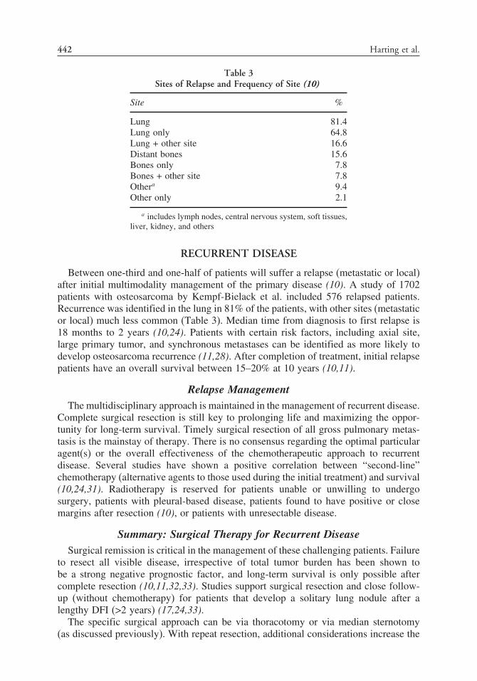
442 Harting et al.
Table 3Sites of Relapse and Frequency of Site (10)
Site %
Lung 81.4Lung only 64.8Lung + other site 16.6Distant bones 15.6Bones only 7.8Bones + other site 7.8Othera 9.4Other only 2.1
a includes lymph nodes, central nervous system, soft tissues,liver, kidney, and others
RECURRENT DISEASE
Between one-third and one-half of patients will suffer a relapse (metastatic or local)after initial multimodality management of the primary disease (10). A study of 1702patients with osteosarcoma by Kempf-Bielack et al. included 576 relapsed patients.Recurrence was identified in the lung in 81% of the patients, with other sites (metastaticor local) much less common (Table 3). Median time from diagnosis to first relapse is18 months to 2 years (10,24). Patients with certain risk factors, including axial site,large primary tumor, and synchronous metastases can be identified as more likely todevelop osteosarcoma recurrence (11,28). After completion of treatment, initial relapsepatients have an overall survival between 15–20% at 10 years (10,11).
Relapse ManagementThe multidisciplinary approach is maintained in the management of recurrent disease.
Complete surgical resection is still key to prolonging life and maximizing the oppor-tunity for long-term survival. Timely surgical resection of all gross pulmonary metas-tasis is the mainstay of therapy. There is no consensus regarding the optimal particularagent(s) or the overall effectiveness of the chemotherapeutic approach to recurrentdisease. Several studies have shown a positive correlation between “second-line”chemotherapy (alternative agents to those used during the initial treatment) and survival(10,24,31). Radiotherapy is reserved for patients unable or unwilling to undergosurgery, patients with pleural-based disease, patients found to have positive or closemargins after resection (10), or patients with unresectable disease.
Summary: Surgical Therapy for Recurrent DiseaseSurgical remission is critical in the management of these challenging patients. Failure
to resect all visible disease, irrespective of total tumor burden has been shown tobe a strong negative prognostic factor, and long-term survival is only possible aftercomplete resection (10,11,32,33). Studies support surgical resection and close follow-up (without chemotherapy) for patients that develop a solitary lung nodule after alengthy DFI (>2 years) (17,24,33).
The specific surgical approach can be via thoracotomy or via median sternotomy(as discussed previously). With repeat resection, additional considerations increase the

Chapter 26 / Osteosarcoma Metastasis 443
complexity of the operation. Repeat thoracotomy (after a previous thoracotomy ormedian sternotomy) is probably the most common procedure performed. The dissectionposes minimal risk and the exposure allows maximal exploration of the entire lung sothat all metastatic disease can be identified and resected. After satisfactory single-lungventilation, the collapsed lung should be very thoroughly palpated by the operatingsurgeon, because a preoperative CT scan with 5-mm cuts can miss 15–25% of lesions.Numerous repeated thoracotomies can be performed relatively safely. At the authors’institution, we have performed more than five sequential thoracotomies (on the sameside) for several children with repeated recurrent disease and have prolonged theirsurvival. However, recurrent thoracotomies are often complicated by increased bloodloss and increased operative time. The prospect of repeating a thoracotomy for thefourth or fifth time is not itself prohibitive, but patients should undergo preoperativepulmonary function tests and echocardiography to determine the feasibility of surgicalresection when a repeat thoracotomy is being considered.
Although median sternotomy offers several advantages, it is not recommended forrecurrent resection (irrespective of the initial operation). Even though it exposes thethoracic cavities bilaterally, it offers poor exposure to posterior pulmonary segments.Adhesions from previous thoracic operations can make entry via the sternum a difficulttask, and the approach has a higher morbidity with repeated surgeries.
Wedge resection is the preferred surgical procedure, even in reoperative thoraco-tomies, because complete resection can be combined with preservation of maximalrespiratory capacity. As discussed previously, lobectomy and pneumonectomy areusually reserved for very extensive disease or disease located very centrally (i.e., atthe hilum) (32).
Given the importance of complete surgical resection (10,11) and the possibilityof long-term, disease-free survival, even after multiple recurrences (10,11,17,30,34),aggressive pulmonary resection and repeat pulmonary resection should be attemptedif all gross disease is resectable and if adequate postoperative pulmonary reserve canbe anticipated (30,32). Our recent research revealed that patients with a good responseto chemotherapy and a DFI greater than 1 year had a 50–60% 5-year survival afteraggressive, complete metastasectomy (30). However, even those with a suboptimalresponse to chemotherapy (<98% necrosis) and a short DFI had a 12% 5-year survivalafter surgical remission. Briccoli and coworkers found that 5-year event-free actuarialsurvival from first metastectomy (38%) to be similar to the 5-year event-free actuarialsurvival after second metastectomy (32%) (32). Even after the fourth and fifth relapses,patients undergoing resection were experiencing long-term, disease-free survival.
PROGNOSIS AFTER RECURRENCE/MANAGEMENT
The prognosis after osteosarcoma disease recurrence and subsequent managementis not as promising as the prognosis after initial management. Ensuing relapses tendto occur after a shorter disease-free interval (10,32). Only one-quarter of patients areexpected to survive 5 years after recurrence and 10-year survival is approximately 20%(10,35). Among the prognostic factors associated with survival after recurrence, theonly significant factor from the initial disease presentation/management appears to beprimary tumor response to chemotherapy (10,30). Other prognostic factors associatedwith prolonged survival include late relapse (i.e., DFI or recurrence-free interval[RFI]), solitary lesions, unilateral lesions, and absence of pleural involvement (10,33).

444 Harting et al.
Various studies defined late relapse at different time points, including 18 months (10),24 months (33), 36 months (31), although they all found a correlation between longRFI and long-term survival.
A recent single institution study at MD Anderson Cancer Center found that thenumber of lesions, size of lesions, and location of lesions were factors that were notas important as the primary tumor response to chemotherapy or the DFI (30). Theseresults led to the theory that tumor burden was not as critical as tumor biology withrespect to long-term survival.
FUTURE/ONGOING RESEARCH
Although the prognosis for patients with osteosarcoma has improved significantlyover the last three decades, a disturbing trend is the more recent plateau in survival(36). Although the advances in chemotherapeutic and surgical strategy developed overthe last 30 years produced enormous strides in the management of osteosarcoma,innovative discoveries must continue at the molecular, cellular, and clinical levels.
The basic science, translational, and clinical research discoveries continue to broadenour understanding and therapeutic potential for this disease. As we learn more aboutthe biology of osteosarcoma, genetic markers of the host and/or the tumor could predictdisease progression and aid the selection and development of effective therapy (11,37).This type of “molecular risk-based treatment” could help focus therapy so that high-risk patients receive the most aggressive therapy and lower risk patients are perhapsspared the side effects of this more aggressive treatment.
It may also be possible to subgroup patients at the cellular level, based on thedetection of micrometastatic osteosarcoma cells (38). Immunomagnetic detectionassays have been used to assess the presence of tumor cells in bone marrow andperipheral blood. Such an assay has the clinical potential to evaluate the effectivenessof a particular chemotherapeutic approach or to group patients based on cellular risk.
In addition, multicenter trials and clinical studies are leading to larger cohorts andmore powerful conclusions. New chemotherapeutic and immunotherapeutic agents,used alone or in combination, are being critically assessed and more standardizedrecommendations are being generated. More patients are being enrolled in well-designed clinical trials, which yield valuable data and offer patients and their familiessome hope of prolonged survival.
Osteosarcoma remains a challenging disease to manage and treat, particularly whendisease recurrences lead to multiple surgeries and consecutive rounds of intense medicaltreatment. The current state of the multimodality approach leads to long-term survivalfor many patients, even after metastatic recurrences.
REFERENCES
1. Gurney JG, Swensen AR, Bulterys M. In: Ries LAG, Smith MA, Gurney JG, (eds.), Cancer incidenceand survival among children and adolescents: United States SEER Program 1975–1995. Bethesda:National Cancer Institute, SEER Program. NIH Pub. No. 99–4649, 1999:99–110.
2. Link MP, Gebhardt MC, Meyers PA. Osteosarcoma. In: Pizzo PA, Poplack DG, (eds.), Principlesand practice of pediatric oncology. Philadelphia: Lippincott Williams and Wilkins; 2002: 1051–1089.
3. Sandberg AA, Bridge JA. Updates on the cytogenetics and molecular genetics of bone and soft tissuetumors: osteosarcoma and related tumors. Cancer Genet Cytogenet 2003;145(1):1–30.
4. Mialou V, Philip T, Kalifa C, et al. Metastatic osteosarcoma at diagnosis. Prognostic factors andlong-term outcome—The French Pediatric experience. Cancer 2005;104:1100–1109.
Chapter 26 / Osteosarcoma Metastasis 445
5. Kaste SC, Pratt CB, Cain AM, et al. Metastases detected at the time of diagnosis of primary pediatricextremity osteosarcoma at diagnosis: imaging features. Cancer 1999;86:1602–1608.
6. Friedman MA, Carter SK. The therapy of osteogenic sarcoma: current status and thoughts for thefuture. J Surg Oncol 1972;4:482–510.
7. Meyers PA, Schwartz CL, Krailo M, et al. Osteosarcoma: a randomized, prospective trial ofthe addition of ifosfamide and/or muramyl tripeptide to cisplatin, doxorubicin, and high-dosemethotrexate. J Clin Oncol 2005;23(9):2004–2011.
8. Souhami RL, Craft AW, Van der Eijken JW, et al. Randomised trial of two regimens ofchemotherapy in operable osteosarcoma: a study of the European Osteosarcoma Intergroup. Lancet1997;350(9082):911–917.
9. Rougraff BT, Simon MA, Kneisl JS, et al. Limb salvage compared with amputation for osteosarcomaof the distal end of the femur. A long-term oncological, functional, and quality-of-life study. J BoneJoint Surg Am 1994;76:649–656.
10. Kempf-Bielack B, Bielack SS, Jurgens H, et al. Osteosarcoma relapse after combined modalitytherapy: an analysis of unselected patients in the Cooperative Osteosarcoma Study Group (COSS).J Clin Oncol 2005;23(3):559–568.
11. Crompton BD, Goldsby RE, Weinberg VK, et al. Survival after recurrence of osteosarcoma: a 20-yearexperience at a single institution. Pediatr Blood Cancer 2006;47(3):255–259.
12. Castagnetti M, Delarue A, Gentet JC. Optimizing the surgical management of lung nodules inchildren with osteosarcoma: thoracoscopy for biopsies, thoracotomy for resections. Surg Endosc2004;18(11):1668–1671.
13. Margaritora S, Porziella V, D’Andrilli A, et al. Pulmonary metastases: can accurate radiologicalevaluation avoid thoracotomic approach? Eur J Cardiothorac Surg 2002;21(6):1111–1114.
14. Kayton ML, Huvos AG, Casher J, et al. Computed tomographic scan of the chest underestimates thenumber of metastatic lesions in osteosarcoma. J Pediatr Surg 2006;41:200–206.
15. Harting MT, Blakely ML. Management of osteosarcoma pulmonary metastases. Semin Pediatr Surg2006;15(1):25–29.
16. Su WT, Chewning J, Abramson S, et al. Surgical management and outcome of osteosarcoma patientswith unilateral pulmonary metastases. J Pediatr Surg 2004;39(3):418–423.
17. Hawkins DS, Arndt CA. Pattern of disease recurrence and prognostic factors in patients withosteosarcoma treated with contemporary chemotherapy. Cancer 2003;98(11):2447–2456.
18. Kager L, Zoubek A, Potschger U, et al. Primary metastatic osteosarcoma: presentation and outcomeof patients treated on neoadjuvant Cooperative Osteosarcoma Study Group protocols. J Clin Oncol2003;21(10):2011–2018.
19. Zarroug AE, Hamner CE, Pham TH, et al. Bilateral staged versus bilateral simultaneous thoracotomyin the pediatric population. J Pediatr Surg 2006;41:647–651.
20. De Oliveira-Filho AG, Neto LS, Epelman S. Median sternotomy for the resection of bilateralpulmonary metastases in children. Pediatr Surg Int 1998;13(8):560–563.
21. Pastorino U, Valente M, Gasparini M, et al. Median sternotomy and multiple lung resections formetastatic sarcomas. Eur J Cardiothoracic Surg 1990;4(9):477–481.
22. Johnston MR. Median sternotomy for resection of pulmonary metastases. J Thorac Cardiovasc Surg1983;85(4):516–522.
23. Saeter G, Kloke O, Jelic S; ESMO Guidelines Task Force. ESMO minimum clinical recommendationsfor diagnosis, treatment, and follow-up of osteosarcoma. Ann Oncol 2005;16(Supp 1):171–172.
24. Ferrari S, Briccoli A, Mercuri M, et al. Postrelapse survival in osteosarcoma of the extremities:prognostic factors for long-term survival. J Clin Oncol 2003;21(4):710–715.
25. Strauss SJ, McTiernan A, Whelan JS, et al. Late relapse of osteosarcoma: implications for follow-upand screening. Pediatr Blood Cancer 2004;43:692–697.
26. Sweetnam R. The surgical management of primary osteosarcoma. Clin Orthop Relat Res 1975;111:57–64.
27. Price CH, Zhuber K, Salzer-Kuntschik M, et al. Osteosarcoma in children. A study of 125 cases.J Bone Joint Surg Br 1975;57(3):341–345.
28. Bielack SS, Kempf-Bielack B, Delling G, et al. Prognostic factors in high-grade osteosarcoma of theextremities or trunk: an analysis of 1,702 patients treated on neoadjuvant cooperative osteosarcomastudy group protocols. J Clin Oncol 2002;20:776–790.
29. Davis AM, Bell RS, Goodwin PJ. Prognostic factors in osteosarcoma: a critical review. J Clin Oncol1994;12:423–431.
446 Harting et al.
30. Harting MT, Blakely ML, Jaffe N, et al. Long-term survival after aggressive resection of pulmonarymetastases among children and adolescents with osteosarcoma. J Pediatr Surg 2006;41(1):194–199.
31. Saeter G, Hoie J, Stenwig AE, et al. Systemic relapse of patients with osteosarcoma: prognosticfactors for long-term survival. Cancer 1995;75:1084–1093.
32. Briccoli A, Rocca M, Salone M, et al. Resection of recurrent pulmonary metastases in patients withosteosarcoma. Cancer 2005;104:1721–1725.
33. Chou AJ, Merola PR, Wexler LH, et al. Treatment of osteosarcoma at first recurrence after contem-porary therapy. Cancer 2005;104:2214–2221.
34. Duffaud F, Digue L, Mercier C, et al. Recurrences following primary osteosarcoma in adolescentsand adults previously treated with chemotherapy. Eur J Cancer 2003;39:2050–2057.
35. Kandioler D, Kromer E, Tuchler H, et al. Long-term results after repeated surgical removal ofpulmonary metastases. Ann Thorac Surg 1998;65:909–912.
36. Gatta G, Capocaccia R, Stiller C, et al. Childhood cancer survival trends in Europe: a EUROCAREworking group study. J Clin Oncol 2005;23(16):3742–3745.
37. Ochi K, Daigo Y, Katagiri T, et al. Prediction of response to neoadjuvant chemotherapy forosteosarcoma by gene-expression profiles. Int J Oncol 2004;24:647–655.
38. Bruland OS, Hoifodt H, Saeter G, et al. Hematogenous micrometastases in osteosarcoma patients.Clin Cancer Res 2005;11(13):4666–4673.

27 Neurofibromatosis
Ravi S. Radhakrishnan, MD
and Richard J. Andrassy, MD
CONTENTS
Introduction
Cutaneous Manifestations
Gastrointestinal (GI) Manifestations
Vascular Manifestations
Thoracic Manifestations
MPNSTs and Pheochromocytomas
Conclusion
References
INTRODUCTION
Neurofibromatosis (NF) a common genetic disorder with an autosomal dominantpattern of inheritance with variable penetrance. NF is a complex disease with manydifferent systemic manifestations and complications. Based on symptoms and the regionof chromosomal abnormality, NF has been classified into two types: neurofibromatosisI (NF 1) and neurofibromatosis II (NF 2). NF 1, or von Recklinghausen’s disease, hasbeen localized to the long arm of chromosome 17 and occurs in nearly one in 3000 livebirths (1,2). Because NF 1 has many different manifestations, the National Institutes ofHealth (NIH) developed a standardized method to diagnose NF 1, which includes twoof the following criteria: six or more café-au-lait spots; two or more neurofibromasor one plexiform neurofibroma; axillary or inguinal freckling; optic gliomas; osseouslesions; lesions of the iris known as Lisch’s nodules; or a first-degree relative withNF 1 (3). In contrast, NF 2 has been localized to the long arm of chromosome 22and has a much lower incidence of only one in 55,000 live births (4). It has beendefined as either bilateral acoustic neuromas visualized with appropriate imaging ora first-degree relative with NF 2, in addition to either a unilateral eighth nerve palsyor two other lesions: neurofibroma, meningioma, glioma, schwannoma, or juvenileposterior subcapsular lenticular opacities (3). Despite the autosomal dominant patternof inheritance, it is important to note that nearly 50% of all cases of NF 1 and a greaterproportion of NF 2 cases are sporadic mutations (2). NF is a progressive, systemicdisease that can affect many different areas of the body. Depending on the location,
From: Reoperative Pediatric SurgeryEdited by: S. Teich and D. A. Caniano © Humana Press, Totowa, NJ
447

448 Radhakrishnan and Andrassy
size, and growth of lesions, patients may require multiple trips to the operating room,which may vary from simple excision of cutaneous lesions to aneurysm repair to largeresections of malignant peripheral nerve sheath tumors (MPNST).
CUTANEOUS MANIFESTATIONS
The most common manifestations of NF are cutaneous neurofibromas. In the dermis,neurofibromas often extend outward and become pedunculated, but may not be visiblewhen in the subcutaneous or deeper tissues. During periods of high hormonal activitysuch as puberty or pregnancy, neurofibromas may appear, grow, and increase innumber (2,5,6). The lesions may be tender to palpation or trauma, but they are rarelypainful. These lesions are described based on their distribution as discrete, segmental,or plexiform (2,7). Discrete neurofibromas are well-defined, rubbery masses withdistinct margins that develop at any point along peripheral nerves. These lesionsrange in size from a few millimeters to several centimeters and can be found atmultiple sites throughout the body. Segmental NF is defined as café-au-lait maculesor neurofibromas in a single, unilateral segment of the body, with no crossing of themidline, no familial history, and no systemic involvement (7). In general, segmental NFarises in a dermatomal distribution, typically in the cervical, followed by the thoracic,lumbar, and sacral dermatomes (8). With discrete and segmental NF, the morbidityis usually cosmetic, because of the number visibly apparent lesions. However, whenneurofibromas extend to major nerve roots, radiculopathy and weakness secondaryto compression is possible (2,9). In both cases, malignant transformation is rare,and surgical management is typically reserved for resection for cosmesis, functionalreasons, or pain control (10).
Plexiform neurofibromas do not refer to the involvement of a specific plexus ofnerves, but rather have a network-like growth of a neurofibroma involving multiplefascicles of a nerve and may include multiple branches of a large nerve, leading to adiffuse mass of thickened nerves (11). Plexiform neurofibromas can be debilitating inpatients with NF, and are one of the most common sources of morbidity. Plexiformneurofibromas can be found in nearly 30% of patients with NF by physical examinationalone (6). However, to determine the true incidence of plexiform neurofibromas in NFpatients, Tonsgard et al. conducted thorough radiological evaluation of 91 patients withNF. They determined that thoracic lesions were identified in 20% and abdominal lesionsin 44% of patients, which were largely asymptomatic (12). Plexiform neurofibromastypically have a “bag of worms” feel on palpation and can grow very large. They canextend for variable distances along a nerve or nerve plexus and can lead to hypertrophyof the tissue around the lesion (10,11). In addition, the overlying skin may also bethickened or discolored (11). Plexiform neurofibromas tend to grow in two distinctperiods: early childhood and periods of high hormonal activity, such as puberty orpregnancy (10,11,13). Plexiform neurofibromas may be tender to palpation but arerarely painful. In addition, plexiform neurofibromas may degenerate into MPNSTs, sorapid growth or constant pain should alert the clinician to the possibility of malignancy(10,11). Facial plexiform neurofibromas and pruritus were associated with increasedmortality in children (14).
Although no uniform guidelines exist, surgical excision is the mainstay ofmanagement for plexiform neurofibromas. The decision to excise lesions is focusedon both the timing and the extent of the operation; however, some controversy exists.

Chapter 27 / Neurofibromatosis 449
Some believe that it may be difficult to justify resection of a small, asymptomaticplexiform neurofibroma on the assumption that it may grow at a later date. In addition,others state that complete resection of the lesion is difficult with recurrence likely(11). Others have proposed that early resection of lesions may prevent functionaldamage at a later date. Jackson et al. have offered that early resection of plexiformneurofibromas may lead to better long-term functional outcome (15). In addition, aretrospective review of 121 patients with plexiform neurofibromatosis showed thatoverall tumor progression after surgical excision was 46% and that age less than 10years at operation and incomplete resection were independent prognostic for tumorprogression. It is interesting to note that the authors did show that more extensive resec-tions did slow tumor progression (16). Although early resection may avoid functionalor cosmetic impairments, it is important to consider the risk of recurrence of plexiformneurofibromas when considering surgical excision.
GASTROINTESTINAL (GI) MANIFESTATIONS
Although GI manifestations of NF 1 are well described, the true incidence andproportion that become symptomatic are unknown. Based on retrospective reviews ofpatients with NF 1, 10–25% of patients are estimated to have GI tumors, with lessthan 5% having symptoms from these tumors (6,17,18). There are reports of a subtypeof NF that has isolated GI involvement without other systemic features of NF (19,20).However, it is unclear whether this represents a distinct variant of NF or incompletepenetrance of the NF 1 gene.
GI manifestations of NF are divided into three groups: hyperplasia of the gutneural tissue, stromal tumors, and duodenal or periampullary endocrine tumors (21).The hyperplastic lesions are characterized by increase in Meissner’s and Auerbach’splexuses along the GI tract. Based on its patchy distribution, the lesions may appear asdiscrete or plexiform neurofibromas. These lesions typically cause constipation, whichis found in nearly 10% of all patients with NF and may be confused for Hirschsprung’sdisease (22,23). If conservative management fails to control symptoms, colectomy maybe necessary to relieve the symptoms (10).
GI stromal tumors (GIST) are the most common nonepithelial tumor of the GI tract(24). In patients with NF 1, there is a 45-fold increase in the incidence of GIST,suggesting that inactivation of the NF 1 tumor suppressor pathway is also involved inthe pathogenesis of GIST (25). This association with NF 1 and GIST has promptedexamination into the mutations seen in GIST tumors. Tazakawa et al. have recentlydemonstrated that 92% of sporadic GIST tumors have mutations of the gene forthe type III tyrosine kinase receptor (KIT). In contrast, only 8% of NF 1-associatedGIST tumors have the mutation of the KIT gene (24). These lesions are typicallyfound in the stomach and jejunum, and present with bleeding, obstruction, perfo-ration, instussusception, or volvulus (10,17). Treatment of GISTs involves resectionwith grossly negative margins and an intact pseudocapsule. Because lymph nodeinvolvement is rare with GISTs, lymph node dissection is not necessary. Typically,adjuvant chemotherapy is not necessary; however, clinical trials are examining theefficacy of these therapies.
An association has been observed between NF and duodenal or periampullarycarcinoid tumors (10,26–28). These tumors are usually located at or near the ampullaof Vater and are small, tan nodules. On histologic examination, these lesions are

450 Radhakrishnan and Andrassy
composed of glandular elements that involve surrounding tissue planes and are easilyconfused with adenocarcinoma (21). However, it is the presence of psammoma bodiesthat helps differentiate carcinoid tumors from adenocarcinoma (10,21).
VASCULAR MANIFESTATIONS
Hypertension is frequently seen in children with NF, and is typically caused byrenovascular abnormalities or aortic coarctations (29–32). Renal artery stenosis istypically the leading cause of secondary hypertension in patients with NF, with asmany as 25% also having associated coarctation of the abdominal aorta. As a result, itis typically amenable to surgical correction (33,34). However, these vasculopathies areprogressive and often require repeat angioplasty, stenting, or surgical reconstruction.Restenosis is very common after angioplasty and can be especially challenging inyoung patients, in whom grafts or stents may not allow for sufficient growth of thevessels (35,36).
NF vasculopathy was first described in 1945 by Reubi, was subsequently revisedby Salyer and Salyer in 1974, and is now the standard (10,37). They described pureintimal type, with focal nodules of spindle cells and media thinning; advanced intimaltype, with eccentric fibrous thickening in the intima and fibrosis of the media; intimal-aneurysmal type, with marked fibrous thickening of the intima and elastic fragmentationin the media; and nodular/epithelial type, with cluster of spindle cells throughout allthree layers. In addition to this description, Greene and coworkers have classified twoprimary types of vascular lesions associated with NF based on vessel diameter. Largevessels are affected by perivascular neurofibromas or ganglioneuromas that degeneratethe adjacent vascular walls, and small vessels are composed of nodules of smoothmuscle representing mesodermal dysplasia (38).
Peripheral vascular lesions are more common than central lesions in NF andmay lead to significant functional problems. One young girl, in whom femoral andpopliteal aneurysm reconstruction had failed, ultimately required bilateral below-the-knee amputations (39). Arterial aneurysms are frequently seen in the NF populationand may also be the result of trauma from vascular interventions (35). Another younggirl with lower limb hypoplasia and gangrene had chronic ischemia not amenable toreconstruction and required an above-the-knee amputation (40). Aneurysms have alsobeen reported in combination with renal artery stenosis and hypertension, underscoringthe importance of thorough examination of patients with NF for vascular abnormal-ities (41).
The vascular changes associated with NF often produce challenging hemostasisproblems when resection or debulking of complex lesions is attempted. Arteriovenousmalformations are common in large plexiform neurofibromas, often with multiple“feeding” vessels present, and aneurysms may actually be complex, multilobulatedmasses. Massive hemorrhage has been noted during many resections, frequentlyrequiring blood transfusions of several units (42–45). Based on their successful experi-ences, some authors routinely request angiography for patients with plexiform neurofi-bromas, and if indicated, preoperative embolization (43). Some choose to performstaged excisions with hypotensive anesthesia, preliminary sutures around the lesion,ligation of all feeding vessels, and autotransfusion as necessary, though significantblood loss is still possible (42). Although not routinely required, these modalitiesshould be considered during preoperative evaluation.

Chapter 27 / Neurofibromatosis 451
THORACIC MANIFESTATIONS
The association of cardiovascular malformations (CVM) and NF has been impliedfor years. Numerous case reports have described patients with NF and congenital heartdefects (2,46,47). A recent sonographic study demonstrated a significant increase incardiac abnormalities in children with NF 1 versus other adolescents (27% versus3.6%). In addition, there was a significant decrease in the isovolemic relaxation time(tau) in patients with NF 1, suggesting that NF changes the intrinsic properties of theheart as well (48). It has been noted that pulmonic stenosis does make up a largerproportion of the CVMs of patients with NF than those of patients without NF. Basedon a review of the National Neurofibromatosis Foundation International Database, 25of the 54 CVMs documented were pulmonic stenosis (46). The association of thisdefect with features of NF 1 has been well described in both Watson’s syndrome andNF 1-Noonan’s syndrome; but in the study mentioned previously, only four of the54 patients were diagnosed with either of these conditions (2,46). For these reasons,it may be reasonable to assume that there is a genetic predisposition for pulmonicstenosis in patients with NF and CVMs.
Though rare, NF can involve the heart, with plexiform neurofibromas invading thisand other surrounding structures. A 10-year-old girl in whom a mediastinal plexiformneurofibroma was causing a mass effect on the left mainstream bronchus and both atriaalso had echocardiographic evidence of right atrial and arteriovenous septal invasion(49). In addition, a 29-year-old man was reported with clinical biventricular failure;his open biopsy confirmed plexiform neurofibroma encasing both atria, resulting inabnormal function. Review of this patient’s history revealed that partial resection ofa mediastinal mass had been attempted 15 years earlier but was limited at that timesecondary to extension into the interatrial septa (50).
Intrathoracic noncardiac tumors are the most common manifestation of NF in thechest. One pediatric-based review of 260 patients over 10 years reported an incidenceof thoracic tumors of 3.5%, but this study was severely limited. The true incidenceof thoracic tumors is not known, but is probably closer to 10% (13). Six of the ninechildren with tumors were asymptomatic, so the long-term implications of minor orincidental tumors are not known. Thoracic tumors can be very aggressive, and havemajor functional implications. These tumors typically extend from spinal, phrenic,vagus, or intercostal nerves and progressively invest surrounding structures. Theycan produce dyspnea, dysphagia, invasion of the lung tissue, erosion of the ribs,arrhythmias, or diaphragm paralysis (13,51–55). In addition, tumors that develop fromthe structures of the chest wall can produce restrictive pulmonary disease, if theyenlarge sufficiently, and pose complex reconstructive problems (56).
MPNSTS AND PHEOCHROMOCYTOMAS
Long-term studies have revealed that patients with NF have a two- to 5-fold increasein relative risk of neoplasm than the general population (57). As a result, routineexamination and vigilance is necessary throughout the patient’s life in order to detectmalignancies early and improve patient outcome. Although patients with NF have beenknown to develop numerous different malignancies, the most common extracranialmalignancies are Malignant Peripheral Nerve Sheath Tumors (MPNSTs).
MPNSTs have been classified by the World Health Organization to include malignantschwannoma, neurogenic sarcoma, neurofibrosarcoma, and malignant neurilemmoma

452 Radhakrishnan and Andrassy
(58). Overall, MPNST is a rare form of cancer in the general population, representingonly 10% of all soft tissue sarcomas with an incidence of only 0.0001% (58,59).However, more than 50% of these tumors occur in patients with NF, with an estimatedlifetime risk of approximately 4%, or a 4600-fold increase in risk compared to thegeneral population (58,60). In NF, these tumors typically arise from a portion ofa plexiform neurofibroma and are very aggressive, frequently recurring locally ordistantly, with hematogenous spread as the route of metastasis. Doorn et al. examinedMPNST occurring in patients with and without NF and found that the median overallsurvival was only 24 months (61). Other authors have found similar results withsurvival ranging from 8–44 months, indicating the truly aggressive nature of the disease(59,62–64). Although some studies have suggested that the presence of NF has noeffect on the overall survival of patients with MPNSTs, the largest study of its kindfrom the Italian and German Soft Tissue Sarcoma Cooperative Group has suggestedthat MPNSTs in patients with NF are more aggressive, with a significantly lower5-year overall survival (32% versus 55%) and a significantly lower progression-freesurvival (19% versus 42%) than their counterparts without NF (59,61,64,65). Thesefindings support prior findings from other smaller studies (58,62,66).
Prognosis for patients with MPNST is dependent on several factors. Size of tumorsgreater than 5 cm, local invasion, presence of NF, and higher histologic gradehave been identified as factors indicating a poor prognosis (58,59,67,68). Improvedprognosis is associated with duration of symptoms less than 6 months and clear surgicalmargins (64).
Overall, the natural history of MPNST is such that the majority of patients willrelapse. A retrospective review of 57 years of experience with MPNST in children atMD Anderson Cancer Center revealed 38 documented cases (60). A group of 25 of the32 patients who initially had a complete response relapsed after therapy. Of these, 14occurred as distant disease, with the lung as the most common location (86%). Of the22 children with negative surgical margins, survival was 33% as compared with 23%in those with positive margins. Gender, site, margins, adjuvant therapy, or presenceof NF did not notably affect long-term outcome (60). In contrast, a recent study hasshown that primary site, age, and presence of NF 1 are prognostic factors in additionto size and invasiveness (65). Because of the rarity of MPNSTs in children, it maybe difficult to resolve the controversy in the literature. However, the current data dodemonstrate the aggressive nature and poor overall prognosis of MPNSTs.
The most important goal for successful management of MPNST is curative resectionwith negative margins. When attainable, this provides the best hope for long-termsurvival (58–61,63,64,68). Over the past several decades, a trend toward limb-sparingresection over amputation has developed, in part because of earlier diagnosis of smallertumors and the increased use of perioperative radiation therapy (58,59,69,70). Aprospective randomized trial has shown a decrease in local recurrence when adjuvantbrachytherapy radiation was used in conjunction with limb-sparing operation, providedthere was no evidence of gross residual disease (59). Although no significant differencein overall survival was found, it was felt that more long-term data were needed beforethis conclusion could be made. Others have also found an advantage in prolongeddisease-free survival when radiation was used postoperatively (58). The use of adjuvantchemotherapy continues to be controversial. Given the rare nature of the disease and thechanges in chemotherapy used over the years, it has been difficult, if not impossible,to conduct prospective randomized trials or systematic retrospective reviews. Although
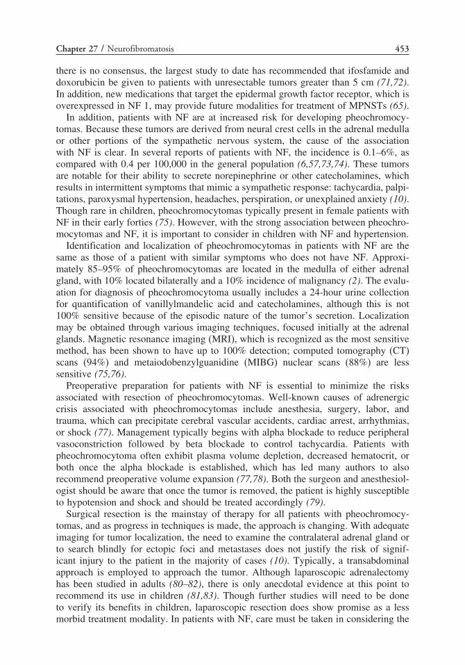
Chapter 27 / Neurofibromatosis 453
there is no consensus, the largest study to date has recommended that ifosfamide anddoxorubicin be given to patients with unresectable tumors greater than 5 cm (71,72).In addition, new medications that target the epidermal growth factor receptor, which isoverexpressed in NF 1, may provide future modalities for treatment of MPNSTs (65).
In addition, patients with NF are at increased risk for developing pheochromocy-tomas. Because these tumors are derived from neural crest cells in the adrenal medullaor other portions of the sympathetic nervous system, the cause of the associationwith NF is clear. In several reports of patients with NF, the incidence is 0.1–6%, ascompared with 0.4 per 100,000 in the general population (6,57,73,74). These tumorsare notable for their ability to secrete norepinephrine or other catecholamines, whichresults in intermittent symptoms that mimic a sympathetic response: tachycardia, palpi-tations, paroxysmal hypertension, headaches, perspiration, or unexplained anxiety (10).Though rare in children, pheochromocytomas typically present in female patients withNF in their early forties (75). However, with the strong association between pheochro-mocytomas and NF, it is important to consider in children with NF and hypertension.
Identification and localization of pheochromocytomas in patients with NF are thesame as those of a patient with similar symptoms who does not have NF. Approxi-mately 85–95% of pheochromocytomas are located in the medulla of either adrenalgland, with 10% located bilaterally and a 10% incidence of malignancy (2). The evalu-ation for diagnosis of pheochromocytoma usually includes a 24-hour urine collectionfor quantification of vanillylmandelic acid and catecholamines, although this is not100% sensitive because of the episodic nature of the tumor’s secretion. Localizationmay be obtained through various imaging techniques, focused initially at the adrenalglands. Magnetic resonance imaging (MRI), which is recognized as the most sensitivemethod, has been shown to have up to 100% detection; computed tomography (CT)scans (94%) and metaiodobenzylguanidine (MIBG) nuclear scans (88%) are lesssensitive (75,76).
Preoperative preparation for patients with NF is essential to minimize the risksassociated with resection of pheochromocytomas. Well-known causes of adrenergiccrisis associated with pheochromocytomas include anesthesia, surgery, labor, andtrauma, which can precipitate cerebral vascular accidents, cardiac arrest, arrhythmias,or shock (77). Management typically begins with alpha blockade to reduce peripheralvasoconstriction followed by beta blockade to control tachycardia. Patients withpheochromocytoma often exhibit plasma volume depletion, decreased hematocrit, orboth once the alpha blockade is established, which has led many authors to alsorecommend preoperative volume expansion (77,78). Both the surgeon and anesthesiol-ogist should be aware that once the tumor is removed, the patient is highly susceptibleto hypotension and shock and should be treated accordingly (79).
Surgical resection is the mainstay of therapy for all patients with pheochromocy-tomas, and as progress in techniques is made, the approach is changing. With adequateimaging for tumor localization, the need to examine the contralateral adrenal gland orto search blindly for ectopic foci and metastases does not justify the risk of signif-icant injury to the patient in the majority of cases (10). Typically, a transabdominalapproach is employed to approach the tumor. Although laparoscopic adrenalectomyhas been studied in adults (80–82), there is only anecdotal evidence at this point torecommend its use in children (81,83). Though further studies will need to be doneto verify its benefits in children, laparoscopic resection does show promise as a lessmorbid treatment modality. In patients with NF, care must be taken in considering the
454 Radhakrishnan and Andrassy
individual patient’s associated manifestations when planning the approach. Previousresections of intraabdominal disease or overlying cutaneous lesions may precludecertain techniques, so a thorough knowledge of the patient’s past surgical history is arequirement.
CONCLUSION
NF is a complex, systematic disease with a wide variety of sequelae and compli-cations. Because of its systemic sequelae, a surgeon may be called on to operatemultiple times on the same patient. These operations may range from simple excisionsof cutaneous neurofibromas for cosmesis to resections of a life-threatening malignancy.As a result, it is important that the surgeon approach these patients with a thoroughunderstanding of the progressive and ubiquitous nature of this disease in order toadequately manage the disease over the life of the patient.
REFERENCES
1. Barker D, Wright E, Nguyen K, et al. Gene for von Recklinghausen neurofibromatosis is in thepericentromeric region of chromosome 17. Science 1987;236(4805):1100–1102.
2. Friedman JM, Riccardi VM. Neurofibromatosis: phenotype, natural history, and pathogenesis, thirdedition. Baltimore: Johns Hopkins University Press; 1999.
3. Mulvihill JJ, Parry DM, Sherman JL, Pikus A, Kaiser-Kupfer MI, Eldridge R. NIH conference.Neurofibromatosis 1 (Recklinghausen disease) and neurofibromatosis 2 (bilateral acoustic neurofi-bromatosis). An update. Ann Intern Med 1990;113(1):39–52.
4. Rouleau GA, Wertelecki W, Haines JL, et al. Genetic linkage of bilateral acoustic neurofibromatosisto a DNA marker on chromosome 22. Nature 1987;329(6136):246–248.
5. Dugoff L, Sujansky E. Neurofibromatosis type 1 and pregnancy. Am J Med Genet 1996;66(1):7–10.6. Huson SM, Harper PS, Compston DA. Von Recklinghausen neurofibromatosis. A clinical and
population study in south-east Wales. Brain 1988;111 (Pt 6):1355–1381.7. Niiyama S, Satoh K, Kaneko S, Aiba S, Mukai H. Segmental neurofibromatosis. Acta Derm Venereol
2005;85(5):448–449.8. Tinschert S, Naumann I, Stegmann E, et al. Segmental neurofibromatosis is caused by somatic
mutation of the neurofibromatosis type 1 (NF1) gene. Eur J Hum Genet 2000;8(6):455–459.9. Levy WJ, Latchaw J, Hahn JF, Sawhny B, Bay J, Dohn DF. Spinal neurofibromas: a report of 66
cases and a comparison with meningiomas. Neurosurgery 1986;18(3):331–334.10. Seymour-Dempsey K, Andrassy RJ. Neurofibromatosis: implications for the general surgeon. J Am
Coll Surg 2002;195(4):553–563.11. Korf BR. Plexiform neurofibromas. Am J Med Genet 1999;89(1):31–37.12. Tonsgard JH, Kwak SM, Short MP, Dachman AH. CT imaging in adults with neurofibromatosis-1:
frequent asymptomatic plexiform lesions. Neurology 1998;50(6):1755–1760.13. Schorry EK, Crawford AH, Egelhoff JC, Lovell AM, Saal HM. Thoracic tumors in children with
neurofibromatosis-1. Am J Med Genet 1997;74(5):533–537.14. Khosrotehrani K, Bastuji-Garin S, Riccardi VM, Birch P, Friedman JM, Wolkenstein P. Subcutaneous
neurofibromas are associated with mortality in neurofibromatosis 1: a cohort study of 703 patients.Am J Med Genet A 2005;132(1):49–53.
15. Jackson IT, Carbonnel A, Potparic Z, Shaw K. Orbitotemporal neurofibromatosis: classification andtreatment. Plast Reconstr Surg 1993;92(1):1–11.
16. Needle MN, Cnaan A, Dattilo J, et al. Prognostic signs in the surgical management ofplexiform neurofibroma: the Children’s Hospital of Philadelphia experience, 1974–1994. J Pediatr1997;131(5):678–682.
17. Ghrist TD. Gastrointestinal Involvement In Neurofibromatosis. Arch Intern Med 1963;112:357–362.18. Riccardi VM. Von Recklinghausen neurofibromatosis. N Engl J Med 1981;305(27):1617–1627.19. Heimann R, Verhest A, Verschraegen J, Grosjean W, Draps JP, Hecht F. Hereditary intestinal
neurofibromatosis. I. A distinctive genetic disease. Neurofibromatosis 1988;1(1):26–32.20. Lipton S, Zuckerbrod M. Familial enteric neurofibromatosis. Med Times 1966;94(5):544–548.
Chapter 27 / Neurofibromatosis 455
21. Fuller CE, Williams GT. Gastrointestinal manifestations of type 1 neurofibromatosis (von Reckling-hausen’s disease). Histopathology 1991;19(1):1–11.
22. Saul RA, Sturner RA, Burger PC. Hyperplasia of the myenteric plexus. Its association with earlyinfantile megacolon and neurofibromatosis. Am J Dis Child 1982;136(9):852–854.
23. Staple TW, McAlister WH, Anderson MS. Plexiform neurofibromatosis of the colon simulatingHirschsprung’s disease. Am J Roentgenol Radium Ther Nucl Med 1964;91:840–845.
24. Takazawa Y, Sakurai S, Sakuma Y, et al. Gastrointestinal stromal tumors of neurofibromatosis typeI (von Recklinghausen’s disease). Am J Surg Pathol 2005;29(6):755–763.
25. Miettinen M, Fetsch JF, Sobin LH, Lasota J. Gastrointestinal stromal tumors in patients with neurofi-bromatosis 1: a clinicopathologic and molecular genetic study of 45 cases. Am J Surg Pathol2006;30(1):90–96.
26. Burke AP, Sobin LH, Shekitka KM, Federspiel BH, Helwig EB. Somatostatin-producing duodenalcarcinoids in patients with von Recklinghausen’s neurofibromatosis. A predilection for black patients.Cancer 1990;65(7):1591–1595.
27. Swinburn BA, Yeong ML, Lane MR, Nicholson GI, Holdaway IM. Neurofibromatosis associatedwith somatostatinoma: a report of two patients. Clin Endocrinol (Oxf) 1988;28(4):353–359.
28. Wheeler MH, Curley IR, Williams ED. The association of neurofibromatosis, pheochromocytoma,and somatostatin-rich duodenal carcinoid tumor. Surgery 1986;100(6):1163–1169.
29. Rowen M, Dorsey TJ, Kegel SM, Ostermiller WE. Thoracic coarctation associated with neurofibro-matosis. Am J Dis Child 1975;129(1):113–115.
30. Schurch W, Messerli FH, Genest J, et al. Arterial hypertension and neurofibromatosis: renal arterystenosis and coarctation of abdominal aorta. Can Med Assoc J 1975;113(9):879–885.
31. Tilford DL, Kelsch RC. Renal artery stenosis in childhood neurofibromatosis. Am J Dis Child1973;126(5):665–668.
32. West CA, Delis KT, Service GJ, Driscoll DJ, McPhail IR, Gloviczki P. Middle aortic syndrome:surgical treatment in a child with neurofibromatosis. J Vasc Surg 2005;42(6):1236.
33. Donaldson MC, Ellison LH, Ramsby GR. Hypertension from isolated thoracic aortic coarctationassociated with neurofibromatosis. J Pediatr Surg 1985;20(2):169–171.
34. Fossali E, Signorini E, Intermite RC, et al. Renovascular disease and hypertension in children withneurofibromatosis. Pediatr Nephrol 2000;14(8–9):806–810.
35. Khan MS, Moore JW. Treatment of abdominal aortic pseudoaneurysm with covered stents in apediatric patient. Catheter Cardiovasc Interv 2000;50(4):445–448.
36. Kurien A, John PR, Milford DV. Hypertension secondary to progressive vascular neurofibromatosis.Arch Dis Child 1997;76(5):454–455.
37. Salyer WR, Salyer DC. The vascular lesions of neurofibromatosis. Angiology 1974;25(8):510–519.38. Greene JF Jr, Fitzwater JE, Burgess J. Arterial lesions associated with neurofibromatosis. Am J Clin
Pathol 1974;62(4):481–487.39. Ilgit ET, Vural M, Oguz A, Ozdogan ME. Peripheral arterial involvement in neurofibromatosis type
1—a case report. Angiology 1999;50(11):955–958.40. Zachos M, Parkin PC, Babyn PS, Chait P. Neurofibromatosis type 1 vasculopathy associated with
lower limb hypoplasia. Pediatrics 1997;100(3 Pt 1):395–398.41. Han M, Criado E. Renal artery stenosis and aneurysms associated with neurofibromatosis. J Vasc
Surg 2005;41(3):539–543.42. Lin YC, Chen HC. Rare complication of massive hemorrhage in neurofibromatosis with arteriovenous
malformation. Ann Plast Surg 2000;44(2):221–224.43. Littlewood AH, Stilwell JH. The vascular features of plexiform neurofibroma with some observa-
tions on the importance of pre-operative angiography and the value of pre-operative intra-arterialembolisation. Br J Plast Surg 1983;36(4):501–506.
44. Pentecost M, Stanley P, Takahashi M, Isaacs H Jr. Aneurysms of the aorta and subclavian andvertebral arteries in neurofibromatosis. Am J Dis Child 1981;135(5):475–477.
45. Smith BL, Munschauer CE, Diamond N, Rivera F. Ruptured internal carotid aneurysm resulting fromneurofibromatosis: treatment with intraluminal stent graft. J Vasc Surg 2000;32(4):824–828.
46. Lin AE, Birch PH, Korf BR, et al. Cardiovascular malformations and other cardiovascular abnormal-ities in neurofibromatosis 1. Am J Med Genet 2000;95(2):108–117.
47. Lin AE, Garver KL. Cardiac abnormalities in neurofibromatosis. Neurofibromatosis 1988;1(3):146–151.
48. Tedesco MA, Di Salvo G, Natale F, et al. The heart in neurofibromatosis type 1: an echocardiographicstudy. Am Heart J 2002;143(5):883–888.

456 Radhakrishnan and Andrassy
49. Noubani H, Poon E, Cooper RS, Kahn E, Kazadevich M, Parnell VA. Neurofibromatosis with cardiacinvolvement. Pediatr Cardiol 1997;18(2):156–158.
50. Alaeddini J, Frater RW, Shirani J. Cardiac involvement in neurofibromatosis. Tex Heart Inst J2000;27(2):218–219.
51. Nordback P, Halkic N, Boumghar M. Intrathoracic tumours in von Recklinghausen’s neurofibro-matosis. Schweiz Med Wochenschr 8 2000;130(31–32):1105–1111.
52. Hassoun PM, Celli BR. Bilateral diaphragm paralysis secondary to central von Recklinghausen’sdisease. Chest 2000;117(4):1196–1200.
53. Chow LT, Shum BS, Chow WH. Intrathoracic vagus nerve neurofibroma and sudden death in apatient with neurofibromatosis. Thorax 1993;48(3):298–299.
54. Dabir RR, Piccione W Jr, Kittle CF. Intrathoracic tumors of the vagus nerve. Ann Thorac Surg1990;50(3):494–497.
55. Strickland B, Wolverson MK. Intrathoracic vagus nerve tumours. Thorax 1974;29(2):215–222.56. Salazar R, Robotti EB, Chin DH, Grossman JA. Giant neurofibromatosis of the chest wall: two patient
reports. Ann Plast Surg 1998;41(2):211–214.57. Sorensen SA, Mulvihill JJ, Nielsen A. Long-term follow-up of von Recklinghausen neurofibromatosis.
Survival and malignant neoplasms. N Engl J Med 1986;314(16):1010–1015.58. Das Gupta TK CP. Tumors of the soft tissue, second edition. Stamford: Appleton and Lang; 1998.59. Hruban RH, Shiu MH, Senie RT, Woodruff JM. Malignant peripheral nerve sheath tumors of the
buttock and lower extremity. A study of 43 cases. Cancer 1990;66(6):1253–1265.60. Neville H, Corpron C, Blakely ML, Andrassy R. Pediatric neurofibrosarcoma. J Pediatr Surg
2003;38(3):343–346; discussion 343–346.61. Doorn PF, Molenaar WM, Buter J, Hoekstra HJ. Malignant peripheral nerve sheath tumors in patients
with and without neurofibromatosis. Eur J Surg Oncol 1995;21(1):78–82.62. D’Agostino AN, Soule EH, Miller RH. Sarcomas of the peripheral nerves and somatic soft
tissues associated with multiple neurofibromatosis (von Recklinghausen’s disease). Cancer 1963;16:1015–1027.
63. Ducatman BS, Scheithauer BW, Piepgras DG, Reiman HM. Malignant peripheral nerve sheath tumorsin childhood. J Neurooncol 1984;2(3):241–248.
64. Wanebo JE, Malik JM, VandenBerg SR, Wanebo HJ, Driesen N, Persing JA. Malignant peripheralnerve sheath tumors. A clinicopathologic study of 28 cases. Cancer 1993;71(4):1247–1253.
65. Carli M, Ferrari A, Mattke A, et al. Pediatric malignant peripheral nerve sheath tumor: the Italianand German soft tissue sarcoma cooperative group. J Clin Oncol 2005;23(33):8422–8430.
66. Das Gupta TK, Brasfield RD. Solitary malignant schwannoma. Ann Surg 1970;171(3):419–428.67. Casanova M, Ferrari A, Spreafico F, et al. Malignant peripheral nerve sheath tumors in children: a
single-institution twenty-year experience. J Pediatr Hematol Oncol 1999;21(6):509–513.68. Raney B, Schnaufer L, Ziegler M, Chatten J, Littman P, Jarrett P. Treatment of children with
neurogenic sarcoma. Experience at the Children’s Hospital of Philadelphia, 1958–1984. Cancer1987;59(1):1–5.
69. Brennan MF, Casper ES, Harrison LB, Shiu MH, Gaynor J, Hajdu SI. The role of multimodalitytherapy in soft-tissue sarcoma. Ann Surg 1991;214(3):328–336; discussion 336–328.
70. Williard WC, Collin C, Casper ES, Hajdu SI, Brennan MF. The changing role of amputation for softtissue sarcoma of the extremity in adults. Surg Gynecol Obstet 1992;175(5):389–396.
71. Ferrari A, Brecht IB, Koscielniak E, et al. The role of adjuvant chemotherapy in children andadolescents with surgically resected, high-risk adult-type soft tissue sarcomas. Pediatr Blood Cancer2005;45(2):128–134.
72. Ferrari A, Casanova M. New concepts for the treatment of pediatric nonrhabdomyosarcoma soft tissuesarcomas. Expert Rev Anticancer Ther 2005;5(2):307–318.
73. Zoller ME, Rembeck B, Oden A, Samuelsson M, Angervall L. Malignant and benign tumors in patientswith neurofibromatosis type 1 in a defined Swedish population. Cancer 1997;79(11):2125–2131.
74. Brasfield RD, Das Gupta TK. Von Recklinghausen’s disease: a clinicopathological study. Ann Surg1972;175(1):86–104.
75. Lew JI, Jacome FJ, Solorzano CC. Neurofibromatosis-associated pheochromocytoma. J Am Coll Surg2006;202(3):550–551.
76. McGrath PC, Sloan DA, Schwartz RW, Kenady DE. Advances in the diagnosis and therapy of adrenaltumors. Curr Opin Oncol 1998;10(1):52–57.
77. Goldfien A. Phaeochromocytoma. Clin Endocrinol Metab 1981;10(3):607–630.

Chapter 27 / Neurofibromatosis 457
78. Cullen ML, Staren ED, Straus AK, et al. Pheochromocytoma: operative strategy. Surgery1985;98(5):927–930.
79. Lucon AM, Pereira MA, Mendonca BB, Halpern A, Wajchenbeg BL, Arap S. Pheochromocytoma:study of 50 cases. J Urol 1997;157(4):1208–1212.
80. Mobius E, Nies C, Rothmund M. Surgical treatment of pheochromocytomas: laparoscopic or conven-tional? Surg Endosc 1999;13(1):35–39.
81. Tarrado X, Ribo JM, Morales L. [Laparoscopic adrenalectomy]. Cir Pediatr 2004;17(1):33–35.82. Palazzo FF, Sebag F, Sierra M, Ippolito G, Souteyrand P, Henry JF. Long-term outcome following
laparoscopic adrenalectomy for large solid adrenal cortex tumors. World J Surg 2006;30(5):893–898.83. Kim HY, Lee HS, Jung SE, Lee SC, Park KW, Kim WK. Experience with surgical excision in
childhood pheochromocytoma. J Korean Med Sci 2004;19(3):401–406.

28 Tumors of the Liver
Rebecka L. Meyers, MD
CONTENTS
Benign Liver Tumors
Malignant Liver Tumors
Reoperation for Surgical Complications
Operative Procedures for Tumor Relapse
References
BENIGN LIVER TUMORS
The differential diagnosis of benign liver tumors in children varies tremendouslywith age. In infancy, the most common benign hepatic tumor is hepatic hemangioma.Infantile hepatic teratoma is exceedingly rare. Commonly seen benign tumors of thetoddler age group are mesenchymal hamartoma and focal nodular hyperplasia. Hepaticadenoma is almost exclusively a disease of older children and adolescents. There areseveral distinguishing characteristics of these benign tumors on radiographic evalu-ation; however, imaging techniques such as ultrasound scan, computed tomography(CT) angiography, and magnetic resonance imaging (MRI) are not always reliable indifferentiating benign from malignant tumors.
MALIGNANT LIVER TUMORS
The differential diagnosis of malignant liver tumors in children also varies withage. In infants and children, most malignant liver tumors are hepatoblastoma andpresent with an asymptomatic right upper quadrant mass. Symptoms of inanitionare more common in older children with hepatocellular carcinoma. Obstructivejaundice may be the presenting symptom in biliary rhabdomyosarcoma. Other raremalignant liver tumors of childhood include a spectrum of sarcomas (angiosarcoma,rhabdomyosarcoma, rhabdoid, undifferendiated), malignant germ cell tumors, andmetastatic Wilm’s or neuroblastoma. Ultrasound will identify the liver as the organof origin; additional testing is aimed at distinguishing benign from malignant disease.The serum alpha-fetoprotein (AFP) level is elevated in 90% of patients with hepato-blastoma and one-half of children with hepatocellular carcinoma. A CT scan outlinesthe anatomic extent of the tumor, clarifies its relationship to the central venous, arterial,
From: Reoperative Pediatric SurgeryEdited by: S. Teich and D. A. Caniano © Humana Press, Totowa, NJ
459
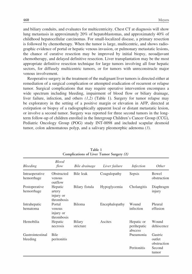
460 Meyers
and biliary conduits, and evaluates for multicentricity. Chest CT at diagnosis will showlung metastasis in approximately 20% of hepatoblastomas, and approximately 40% ofchildhood hepatocellular carcinomas. For small-localized disease, a primary resectionis followed by chemotherapy. When the tumor is large, multicentric, and shows radio-graphic evidence of portal or hepatic venous invasion, or pulmonary metastatic lesions,the chance of curative resection may be improved by initial biopsy, neoadjuvantchemotherapy, and delayed definitive resection. Liver transplantation may be the mostappropriate definitive resection technique for large tumors involving all four hepaticsectors, for diffusely multicentric tumors, or for tumors with unreconstrucle majorvenous involvement.
Reoperative surgery in the treatment of the malignant liver tumors is directed either atremediation of a surgical complication or attempted eradication of recurrent or relapsetumor. Surgical complications that may require operative intervention encompass awide spectrum including bleeding, impairment of blood flow or biliary drainage,liver failure, infection, and others (1,2) (Table 1). Surgery for tumor relapse maybe exploratory in the setting of a positive margin or elevation in AFP, directed atextirpation or biopsy of a radiographically apparent local or distant metastatic lesion,or involve a second tumor. Surgery was reported for three second tumors in the long-term follow-up of children enrolled in the Intergroup Children’s Cancer Group (CCG),Pediatric Oncology Group (POG) study INT-0098 and included scapular desmoidtumor, colon adenomatous polyp, and a salivary pleomorphic adenoma (3).
Table 1Complications of Liver Tumor Surgery (1)
BleedingBloodflow Bile drainage Liver failure Infection Other
Intraoperativehemorrhage
Obstructedvenousoutflow
Bile leak Coagulopathy Sepsis Bowelobstruction
Postoperativehemorrhage
Hepaticarteryinjury orthrombosis
Bilary fistula Hypoglycemia Cholangitis Diaphragminjury
Intrahepatichematoma
Portalvenousinjury orthrombosis
Biloma Encephalopathy Woundinfection
Pleuraleffusion
Hemobilia Hepaticnecrosis
Bilarystricture
Ascites Hepatic orperihepaticabscess
Wounddehiscence
Gastrointestinalbleeding
Bileperitonitis
Pneumonia Gastricoutletobstruction
Peritonitis Secondtumor

Chapter 28 / Tumors of the Liver 461
REOPERATION FOR SURGICAL COMPLICATIONS
BleedingOperation for bleeding after a biopsy, whether percutaneous or open, will depend
on the indications for the biopsy and the extent of bleeding. Bleeding from liver tissueat the site of needle entry can almost always be stopped with pressure, combined withfulguration, or with horizontal mattress suture of the needle entrance site at the livercapsule. Bleeding from a preoperative tumor rupture often produces an intracapsularhematoma and may stop spontaneously. Occasionally hepatoblastoma or hemangiomamay present with hemorrhage and free tumor rupture into the peritoneal cavity withlife-threatening hypovolemic shock. In these instances the initial operation is directedat control of the hemorrhage, either with resection if it can be performed in a controlled,expeditious manner, or a nonanatomic debulking procedure in combination with fulgu-ration, hemostatic agents, and if necessary packing followed by percutaneous tumorembolization (4). Inadvertent injury to vital structures can be minimized if heroic,uncontrolled procedures are avoided and definitive surgical resection is performed asa second-look procedure, once hemodynamic stability is fully restored and completeradiographic tumor staging has been completed. Massive blood transfusion should beavoided where possible, as the extent of blood transfusion has been correlated withpostoperative morbidity and has been reported to increase the risk of tumor recurrencein hepatocellular carcinoma (5).
Postoperative intrahepatic or perihepatic hemotoma may require surgical drainage,predisposes to the risk of abscess, and may delay the administration of adjuvantchemotherapy. The preferred treatment is prevention, and a postoperative hematomais best avoided by meticulous attention to hemostatic surgical technique. Resection oflarge tumors requires extensive preoperative planning by an experienced surgical team,wide exposure, the appropriate use of inflow and/or outflow occlusion, close commu-nication with anesthesia to keep the central venous pressure (CVP) relatively lowand clotting factors appropriately replaced, and the avoidance of unplanned, inappro-priately aggressive attempts at tumor resection in proximity to major vessels (6). Inthe event of a failed initial resection attempt, reoperative resection may be associatedwith increased perihepatic blood loss because of the density of liver adhesions to thediaphragm, retroperitoneum, and right adrenal gland. There is difficulty staying out ofthe liver and out of the diaphragm and adrenal gland when attempting to dissect thosestructures one from the other. Judicious use of electrocautery and good exposure areinvaluable. Special care should be exercised when approaching the liver hilum and theregions of the vena cava and the termini of the hepatic veins.
Vascular InjuryA normal liver can occasionally survive permanent interruption of arterial inflow or
portal venous inflow, but not both. In the rare instance that inflow of the portal vein,hepatic artery, or both are disrupted at surgery, salvage requires immediate revascu-larization. Fulminant hepatic failure will progress rapidly with total devascularizationof a normal liver and occurs with less than total devascularization of a cirrhotic orotherwise damaged liver. Alternatively, if these vital vessels are not gently handledwith care throughout the operative procedure, postoperative thrombosis can occur.Early diagnosis and prompt revascularization with some combination of percutaneouscatheter infusion of thrombolytic agents, thrombectomy, and/or operative vascular

462 Meyers
repair may be life-saving. When there is irretrievable liver devascularization, the onlyoption may be emergent liver transplantation.
Maintenance of venous drainage from the residual liver remnant is of paramountimportance, as liver tissue without adequate venous drainage will become congestedand not remain viable. Protection of venous drainage includes judicious planning ofanticipated surgical resection margins, and avoidance of inadvertent hepatic venousocclusion with ill-placed sutures into the hepatic parenchyma in an attempt to controlbleeding. In extreme cases, salvage transplantation or venous outflow reconstructionwith either autologous or synthetic grafts may be required (7).
Biliary InjuryThe bile ducts, particularly at the hilar plate, are more easily disrupted than the
vessels and are usually more difficult to dissect individually at that level. If injuryoccurs to an extrahepatic bile duct and the injury is minor, it can usually be directlyrepaired. If there is major injury, with loss of more than 50% of the wall, completedivision, or loss of length, the remaining ductal system should be debrided back tohealthy well-perfused ducts and drained with a Roux-en-Y jejunal limb.
Bile leak occurs in approximately 5–10% of patients after major liver resection andcan be lessened by meticulous dissection and closure of all sites of bile drainage onthe raw surface of the liver. Although placing drains at the time of operation does notlessen the rate of bile leakage, it may facilitate postoperative management (8). Bileperitonitis results from bile accessing the general peritoneal cavity rather than beingwalled off into a discrete collection (biloma) or external drain (fistula). Treatmentof large-volume bile peritonitis often requires a combination of peritoneal drainageand decompression of the biliary tree with a transhepatic or endobiliary drain. Bileleaks that do not respond to appropriate drainage are often associated with a distalobstruction. Etiologies of distal bile duct obstruction include a retained section ofviable liver excluded from the remaining biliary system, iatrogenic occlusion (clip,ligature, thermal injury), hematoma, stone, residual obstructing tumor, or an ischemicstricture. If the fistula is confined to a drain, the drain should be left in place for severalweeks for formation of a fibrous tract and resolution of self-limiting forms of distalobstruction (e.g., hematoma). If leakage persists and no distal obstruction is identifiedwith magnetic resonance cholangiopancreatography (MRCP), hepatobiliary radiotracer99mtc dimethyliminodiacetic acid (HIDA), or endoscopic retrograde cholangiopancre-atography (ERCP), the drain should be advanced slightly away from the liver edgeto encourage fibrous obliteration of the leak. If the leak or radiographically evidentsite of distal biliary obstruction persists, operative biliary reconstruction with Roux-en-Y jejunal limb is indicated (Fig. 1). When the treatment goal is curative ratherthan palliative, biliary stricture treatment with percutaneously placed stents provide atemporary (not long-term) solution, especially in children. Stents have a limited lifetime(usually <16 weeks) because of build-up of bile salts and proteinaceous material withocclusion, repeat obstrucion, and cholangitis. Most critical in treating and avoidingcholangitis is the operative establishment of adequate biliary drainage.
Liver FailureThe most common cause of postoperative liver failure following liver resection
is a small functioning liver remnant, generally a much more common concern in

Chapter 28 / Tumors of the Liver 463
•
•
Drainage is primarily through the external drain site
Only a small amount of activity in duodenum on delayed images suggests partial obstruction of the extrahepatic biliary tree
Fig. 1. Bilary fistula. HIDA scan shows all tracer exiting the liver through the external drain sitewith minimal activity in the extrahepatic biliary tree.
the adult population, where hepatocellular carcinoma often occurs in patients withpreexisting liver disease. The decision about how much liver may be removed whendoing liver resection or ablation is based on the anatomic extent of the tumor and theassociated liver anatomy. A guideline has been that long-term survival and appropriateliver regeneration can be anticipated with a remnant volume of 25–30% of normalliver, or 60–70% of cirrhotic liver. Other etiologies of postoperative liver failurediscussed previously include liver devascularization, interruption of venous drainage,excessive liver warm ischemia owing to prolonged operative vascular occlusion andmassive bleeding, major bile duct obstruction, halogenated anesthetic agents, viralinfections, and reactions to certain drugs. If vascular occlusion is used intraoperatively,there has been an increasing trend toward “ischemic preconditioning,” using an initial10-minute period of warm ischemia, followed a 10-minute period of reperfusion,after which a continuous longer stretch of warm ischemia seems to cause less livercell injury (9). Although warm ischemia up to 60 minutes may be tolerated, mostexperienced liver surgeons will limit inflow occlusion to periods of 15–20 minutes,interspersed with 5–10 minute intervals of reperfusion. If postoperative liver failuredoes not show definitive signs of improvement in the first few days, and is notamenable to revascularization or biliary decompression, the potential need for livertransplantation should be considered.
OPERATIVE PROCEDURES FOR TUMOR RELAPSE
Local Tumor Persistence or RelapseIn all patients, but especially in those whose initial operative resection results in
gross or microscopic tumor residual, careful follow-up with radiographic imaging andserial AFP levels is mandatory. Atypical or wedge resections—as opposed to anatomiclobectomy, trisegmentectomy, or mesohepatectomy—have been associated with anincreased risk of local relapse (10). Second-look laparotomy with biopsy and possiblyextension of the liver resection is indicated in the setting of a radiographically identi-fiable small focal mass lesion, or a persistent elevation in the AFP after completing theprescribed courses of postoperative chemotherapy. In SIOPEL–1, a multicenter trialfrom the International Childhood Liver Tumour Strategy Group (SIOPEL), there were

464 Meyers
five of 128 patients who underwent surgery for a local recurrence after having had adefinitive resection (11). Of these five patients, two showed no evidence of disease atlong-term follow-up, one died during surgery because of uncontrollable bleeding. Onepatient developed a second recurrence and then a third, underwent “rescue” transplan-tation and died shortly thereafter. The fifth survived after four recurrences, three localrecurrences were treated with resection, followed by prolonged chemotherapy for alung recurrence, followed by liver transplantation.
The potential role of positron emission tomographic (PET) imaging in screening fortumor recurrence when serum AFP is elevated, but CT or MRI imaging is normal,remains controversial. There are case reports of PET identification and subsequentsuccessful surgical resection of small occult lesions occurring locally, within theabdominal cavity, and in the lung (12). Caution must be exercised because of similarreports describing operative exploration and biopsy of histologically negative regener-ative liver nodules in patients with a positive PET scan (13) (Fig. 2).
Heroic resections with a high probability of leaving residual tumor should be avoidedwhenever possible (14). If large or extensive residual or recurrent tumor is presentin the remaining liver, a “rescue” liver transplant may be required (15,16). A recentanalysis of the SIOPEL 1 data and a review of the world experience on transplantationfor hepatoblastoma clearly showed that a primary transplant after good response tochemotherapy is now associated with a prognosis similar to a conventional completeresection, with a long-term survival of more than 80%. In contrast, “rescue” transplantfor recurrent tumor after resection had only a 30% chance of cure (15). Transplantationfor hepatocellular carcinoma and other malignant tumors is more problematic, and asa routine should be strictly reserved to tumors without extrahepatic extension (17).Recently, the SIOPEL study group, in collaboration with the COG, SPLIT pediatricliver transplant registry, and German cooperative liver tumor study group (GPOH),have established a worldwide electronic registry for transplantation in the treatment ofchildhood hepatoblastoma and hepatocellular carcinoma. This international cooperativeregistry is coined PLUTO (Pediatric Liver Unresectable Tumor Observatory), is admin-istered by the Italian group CINECA, and can be reached at www.PLUTO.org (18).
Whether to treat primary or relapse tumor that focally involves the hepatic venousoutflow tract with resection and extensive venous reconstruction, transplant, or both
• PET Scan shows increase in activity at the cut surface of the regenerating liver 2 months after lobectomy
Fig. 2. False-positive PET Scan. PET scan shows increase in activity at the cut surface of theregenerating liver.

Chapter 28 / Tumors of the Liver 465
is a matter of much debate. Certainly, the surgical techniques necessary to performcomplex venous outflow reconstruction have improved dramatically in recent years.Outflow reconstruction usually involves total vascular occlusion to provide adequatevisualization and prevent life-threatening hemorrhage. Other techniques described in theadult literature include veno-venous bypass, in situ cooling, or ex vivo resection (19–21).Several autologous and synthetic graft materials have been used for the venous recon-struction including saphenous vein, iliac vein, cryopreserved iliac vein or inferior venacava (IVC), polytetrafluoroethylene (PTFE) or Gore-Tex grafts (WL Gore & Assoc),and bovine pericardium (No-React, Shellhigh Inc.) (20,22–28). Synthetic grafts maycarry an increased risk of infection and thrombosis, although harvesting of autogenoustissue requires additional operative procedures with the risk of added morbidity.
Metastatic Tumor RelapseIn numerous large multicenter trials, the incidence of pulmonary metastatic disease
at diagnosis is approximately 20% for hepatoblastoma, and approximately 40% forchildhood hepatocellular carcinoma. The rate is less clearly defined for the rare hepaticsarcomas. Lung metastasis from hepatoblastoma, but not hepatocellular carcinoma,may resolve with platinum based chemotherapy. If focal pulmonary metastatic diseaseremains after chemotherapy, thoracotomy for wedge resection has been recom-mended (3). Nine of the 22 (41%) patients with lung metastasis at diagnosis inSIOPEL–1 were long-term survivors, and four of the survivors underwent pulmonarymetastectomy as well as chemotherapy (29). Thoracotomy and biopsy of a lung noduleor focal area of pulmonary atelectasis may be necessary in a patient with elevatedscreening AFP, or in the setting of impending liver transplantation. Transplantationshould never be performed in the face of potential persistent pulmonary metastaticdisease. Suspicious lung lesions must disappear completely on chemotherapy, beamenable to complete surgical resection, or be biopsied to prove that they are benign.
Two of five patients in SIOPEL–1, without metastatic disease at diagnosis, developedtumor relapse in the lungs after initial therapy and underwent pulmonary metastectomy.Both were long-term survivors (11). Although the role of pulmonary metastectomyremains unclear, surgical resection of pulmonary metastases seems to be a goodtreatment option if local control of the liver has been accomplished. The current COGmulticenter trial of hepatoblastoma will prospectively investigate the potential role ofthoracotomy in these patients.
Rarely, surgery may be required for metastatic disease at a site other than the lung. Thereis a case report in the literature of a long-term survivor who underwent surgical resectionof metastatic hepatoblastoma to the brain (30). The CCG/POG Intergroup-0098 studyincluded five patients who underwent surgical resection of extrapulmonary metastaticdisease; three had resection of a mediastinal tumor recurrence, two had resection of aspinal or paraspinous tumor recurrence—none were long-term survivors (3).
REFERENCES
1. Towu E, Kiely E, Pierro A, et al. Outcome and complications after resection of hepatoblatoma.J Pediatr Surg 2004;39:199–202.
2. Knol JA. Complications of hepatic surgery. In: Mullholland MW, Doherty GM, (eds.), Complicationsin surgery. Philadelphia: Lippincott Williams and Wilkins; 2006: 407–422.
3. Meyers RL, Megahren E, Malogolowkin MH, et al. Surgical resection of pulmonary metastaticlesions in children with hepatoblastoma. J Pediatr Surg, in press.

466 Meyers
4. Lee SC, Chung JW, Kim KH, et al. Successful transumbilical embolization of congenitally rupturedhepatoblastoma. J Pediatr Surg 1999;34:1851–1852.
5. Yamamoto J, Kosuge T, Takayama T, et al. Perioperative blood transfusion promotes recurrence ofhepatocellular carcinoma after hepatectomy. Surgery 1994;115:303–309.
6. Smyrniotis V, Kostopanagiotou G, Theodoraki K, et al. The role of central venous pressure and typeof vascular control in blood loss during major liver resections. Am J Surg 2004;187:398–402.
7. Sano K, Makuuchi M, Miki K, et al. Evaluation of hepatic venous congestion: proposed indicationcriteria for hepatic vein reconstruction. Ann Surg 2002;236:241–247.
8. Reed DN, Vitale GC, Wrightson WR, et al. Decreasing mortality of bile leaks after elective hepaticsurgery. Am J Surg 2003;185:316–318.
9. Clavien PA, Selzner M, Rudiger HA, et al. A prospective randomized study in 100 consecutivepatients undergoing major liver resection with versus without ischemic preconditioning. Ann Surg2003;238:843–852.
10. Fuchs J, Rydzynski J, Hecker H, et al. The influence of preoperative chemotherapy and surgicaltechnique in the treatment of hepatoblatoma: a report from the German Cooperative Liver TumorStudy HB 89 and HB 94. Eur J Pediatr Surg 2002;12:255–261.
11. Schnater JM, Aronson DC, Plaschkes J, et al. Surgical view of the treatment of patients withhepatoblastoma. Cancer 2002;94:1111–1120.
12. Philip I, Shun A, McCowage G. Positron emission tomoraphy in recurrent hepatoblastoma. Ped SurgInt 2005;21:341–345.
13. Wong KK, Lan LC, Lin SC, et al. The use of positron emission tomography in detecting hepato-blastoma recurrence—a cautionary tale. J Pediatr Surg 2004;39:1779–1781.
14. Czauderna P, Otte JB, Aronson DC, et al. Guidelines for surgical treatment of hepatoblastomain the modern era—recommendations from the Childhood Liver Tumour Strategy Group of theInternational Society of Paediatric Oncology (SIOPEL). Eur J Cancer 2005;41:1031–1036.
15. Otte JB, Pritchard J, Aronson DC, et al. Liver transplantation for hepatoblastoma: results fromthe International society of Pediatric Oncology (SIOP) study SIOPEL-1 and review of the worldexperience. Pediatr Blood Cancer 2004;42:74–83.
16. Tiao GM, Bobey N, Allen S, et al. The current management of hepatoblastoma: a combination ofchemotherapy, conventional resection, and liver transplantation. J Pediatr 2005;146:204–211.
17. Von Schweinitz D. Management of liver tumors in childhood. Semin Ped Surg 2006;15:17–24.18. Otte JB, Meyers RL. Pediatric liver unresectable tumor observatory (PLUTO): an international registry
for liver transplantation in children with unresectable tumors of the liver. J Pediatr Surg [In press].19. Lodge JPS, Ammori BJ, Prasad KR, et al. Ex vivo and in situ resection of inferior vena cava with
hepatectomy for colorectal metastases. Ann Surg 2000;231:471–479.20. Hemming AW, Reed AI, Langham MR, et al. Hepatic vein reconstruction for resection of hepatic
tumors. Ann Surg 2002;235:850–858.21. Kim YI, Hwang YJ, Lee JW, et al. 101 hepatectomies under continuous inflow occlusion
following simple in-situ liver cooling in patients with chronic liver disease. Hepatogastroenterology2004;51:1093–1098.
22. Aoki T, Sugawara Y, Imamura H, et al. Hepatic resection with reconstruction of the inferior venacava or hepatic venous confluence for metastatic liver tumor. J Am Coll Surg 2004;198:366–372.
23. Hashimoto T, Kokudo, N, Aoki T, et al. Reconstruction of middle hepatic vein using a rotating leftheptic vein flap. J Am Coll Surg 2004;199:656–660.
24. Sugawara Y, Makuuchi M, Akamatsu N, et al. Refinement of venous reconstruction using cryopre-served veins in right liver grafts. Liver Transpl 2004;10:541–547.
25. Hemming AW, Reed AI. Left trisegmentectomy with reconstruction of segment 6 hepatic venousoutflow using cryopreserved vein graft. J Gastrointest Surg 2005;9:353–356.
26. Sakamoto Y, Yamamoto J, Saiura A, et al. Reconstruction of heptic or portal veins by use of newlylcustomized great saphenous vein grafts. Langenbecks Arch Surg 2004;389:110–113.
27. Makuuchi M, Sugawara Y. Living-donor liver transplantation using the left liver, with specialreference to vein reconstruction. Transplantation 2003;75:S23–S24.
28. Asthana S, Jones D, Lodge JP. Surgical management of recurrent liver tumors involving hepaticvenous outflow. J Am Coll Surg 2006;202(4):711–713.
29. Perilongo G, Brown J, Shafford E, et al. Hepatoblastoma presenting with lung metastases. Cancer2000;89:1845–1853.
30. Robertson PL, Muraszko KM, Axtell RA. Hepatoblastoma metastatic to brain: prolonged survival aftermultiple surgical resections of a solitary brain lesion. J Pediatr Hematol Oncol 1997;19:168–171.

29 Sacrococcygeal Teratoma
Antoine De Backer, MD, PHD
and Frans W. J. Hazebroek, MD, PHD
CONTENTS
Introduction
Some Epidemiological Data on Recurrence
Factors Associated With Recurrence
Diagnosis of Recurrent SCT
Principles of Reoperative Surgery
Long-Term Results
References
INTRODUCTION
With an incidence of one in 30,000–40,000 live births, sacrococcygeal teratoma(SCT), one of the type I germ cell tumors (GCT) (1), is truly a very rare tumor. Yetit is the most frequent tumor in newborns. SCT is also the most frequent extragonadalGCT, far ahead of tumors in other extragonadal sites such as the brain, mediastinum,retroperitoneum, and neck.
DiagnosisSCT occurs most commonly in girls (75–80%). In approximately 70% of the patients,
the diagnosis is made at latest at birth, seeing that an increasing number of patients arebeing diagnosed antenatally. It follows that a still-important subset (around 30%) isbeing diagnosed at a later age. The Altman classification (2) divides tumors in functionof their anatomical extension into four groups (Fig. 1). Roughly one-third of patients canbe classified as Altman I (predominantly external tumor with minimal extension intopelvis), one-quarter as Altman II (external tumor with significant intrapelvic extension),15–20% as Altman III (external tumor with intra-abdominal extension), and finally,some 20% as Altman IV (intrapelvic tumor only, not visible externally) (3,4). A correctclinical diagnosis is usually made on the presence of a sacrococcygeal tumor thatdisplaces the anus ventrally. However, thediagnosisofAltmanIVtumors isoftendelayeduntil the second or third year of life, following investigation for intractable constipation orurinary symptoms. Imaging studies (computed tomography [CT] or magnetic resonanceimaging [MRI]) are crucial for determining the intrapelvic/intraabdominal extension of
From: Reoperative Pediatric SurgeryEdited by: S. Teich and D. A. Caniano © Humana Press, Totowa, NJ
467

468 De Backer and Hazebroek
the tumor, allowing for correct operative planning. Plasma levels of alpha-fetoprotein (�-FP)and, toa lesserextent,beta-humanchorionicgonadotropin(�-HCG),haveproventobevery reliable markers for malignancy (yolk sac tumor and choriocarcinoma, respectively).Unfortunately, as �-FP is high at birth and decreases steadily over the first year of life(5,6), it has hardly any value in discriminating between benign and malignant tumor atbirth and during the first months of life. The vast majority (i.e., 80–85%) of SCTsare histologically benign (mature and immature teratoma) (3,4,7). Malignancy (insome 15% of cases) is most frequently encountered in yolk sac tumor—embryoniccarcinomas and mixed malignant tumors are really exceptional diagnoses. Only 5% ofpatients show metastases at presentation (4).
TreatmentTreatment has been well standardized for many years already. For SCT diagnosed
at birth, complete surgical excision en bloc with the os coccyx is the golden standardand sole treatment. For large tumors, this is best achieved through an inverted Vincision; smaller tumors can be managed by a posterior sagittal incision. When theintraabdominal tumoral component is important (Altman III), the abdominal componentis first dissected using a Pfannenstiel incision, followed by the perineal approach,allowing for en-bloc removal of the entire tumor together with the coccyx. Newbornswill rarely have malignant tumors, and therefore seldom need adjuvant (chemo)therapy.Older children, especially in Altman IV SCT, have a greater likelihood of malignanthistology. Tumor resection may also be undertaken as the first step in the face ofmalignancy (elevated tumor markers, metastases), but if judged inoperable, biopsy ordebulking as well as up-front chemotherapy are valid options. In all such cases, theresidual tumor has to be removed after several courses of chemotherapy. Since the mid-1980s, very efficacious multiagent chemotherapy regimens have been made availablefor GCTs; these regimens all include platinum compounds (cisplatin or carboplatin).The PEB regimen of the Pediatric Oncology Group (POG)/Children’s Cancer Group(CCG) combines cisplatin (P) with etoposide (E) and bleomycin (B). The PEI regimenaccording to the MAKEI protocol of the German Association of Pediatric Oncologyprescribes cisplatin (P) in combination with etoposide (E) and ifosfamide (I). The JEBprotocol of the United Kingdom Children’s Cancer Study Group (UKCCSG) blendscarboplatin (J) with etoposide (E) and bleomycin (B). These are all very efficaciousprotocols that yield comparable results but differ as to number, type and severity ofcomplications.
Fig. 1. Altman classification of SCTs.

Chapter 29 / Sacrococcygeal Teratoma 469
ResultsOverall, with a survival rate of more than 95%, the prognosis for newborns and
children with SCT is excellent. However, a significant proportion will suffer fromfunctional sequelae (fecal soiling, constipation, fecal incontinence, urinary inconti-nence, and aesthetic sequelae) that impair quality of life (8). A small proportionwill show tumor recurrence, either histologically benign or malignant. Patients withmalignant SCT are facing a less favorable prognosis than those with benign histology,but substantial improvements have been noted over the past 20 years. Survival ratesfrom 80–90% are now achieved in subgroups with malignant tumors (9,10).
SOME EPIDEMIOLOGICAL DATA ON RECURRENCE
One of the most feared complications is recurrent tumor, as it spells impairedprognosis. Table 1 summarizes the percentages of recurrences according to histology asreported in the most recent series (3,4,7,11–20). Of course, these series show importantvariations. Overall, recurrence is reported in 3–35% of the patients (mean: 12.9%).Initial histological diagnosis of mature teratoma is associated with a mean 10.2%recurrence rate (range 0–26%). Recurrence rates double for immature teratoma (mean:20.2%, range 4–55%) and initial malignant histology (yolk sac tumor or embryoniccarcinoma) (mean: 19.2%, range 0–100%). Mean time between primary surgery anddiagnosis of recurrence is 10 months; however, extremes range from 1–35 months.More than 50% of recurrent SCTs show comparable histology after resection of theprimary SCT and after resection of the recurrent tumor, but a shift towards either a moreundifferentiated tumor or towards a more differentiated tumor has also been observed(4). Malignancy after resection of a benign teratoma could result from a samplingerror missing small foci of malignancy, or could develop from a small malignantfocus left in place after surgical excision. In a study of the CCG-POG, four of thesix children with malignant recurrence after neonatal resection of mature or immatureteratomas were noted to have microscopic foci of yolk sac tumor in the originalspecimen at reexamination (21). Most recurrences in children with SCT occur at theprimary site of the tumor, but distant metastases are seen as well. As mentioned earlier,recurrent tumor is associated with serious impairment of prognosis. Overall mortalityin patients with recurrent SCT (all histologies) is reported to equal 35% (3) (Fig. 2).However, malignant recurrence is associated with mortality rates as high as 55%(22). It is therefore of extreme importance to prevent recurrence whenever possible.This would require full knowledge of the factors that are possibly associated withrecurrence.
FACTORS ASSOCIATED WITH RECURRENCE
Insufficient local therapy has been documented to represent an important risk factorof recurrence (9,22). For quite a while already, surgeons have accepted the axiomthat complete en bloc resection of the primary sacrococcygeal tumor and the coccyxwithout tumor spillage offers the best perspective of tumor control. This cannot alwaysbe (easily) achieved. Histologically benign tumors (teratomas) are mostly well encap-sulated, but tumor borders may be difficult to identify in some regions. In particularanatomical sites, it is impossible to remove the tumor with a sheet of healthy tissue allaround. Also, if a substantial intrapelvic tumoral component is present, as in Altman

Tab
le1
Surv
eyof
mor
talit
yan
dre
curr
ence
rate
sre
port
edin
825
pati
ents
wit
hSC
T
Rec
urre
nce
Aut
hor
(ref
)Y
ear
ofpu
blic
atio
nPe
riod
No.
ofpa
tien
tsM
orta
lity
(%)
MT
IT“
Mal
igna
nt”
GC
TT
otal
recu
rren
ces
(%)
Com
men
t
Gon
zale
z-C
russ
iet
al11
1978
1946
–197
640
22%
4/22
(18%
)10
/18
(55%
)0
14/4
0(3
5%)
Exc
lusi
vely
beni
gnSC
TE
inet
al12
1980
1951
–197
633
3%1/
33(3
%)
00
1/33
(3%
)E
xclu
sive
lybe
nign
SCT
Eng
elsk
irch
enet
al13
1987
1960
–198
487
16%
9/76
(12%
)0
4/11
(36%
)13
/87
(15%
)
Schr
opp
etal
1419
9219
50–1
990
7316
%5/
57(9
%)
0/16
(0%
)5/
73(7
%)
Hav
rane
ket
al15
1992
1980
–198
932
6%4/
23(1
7%)
2/4
(50%
)1/
5(2
0%)
7/32
(22%
)B
ilik
etal
1619
9319
72–1
990
28?
6/28
(21%
)0
06/
28(2
1%)
Exc
lusi
vely
beni
gnSC
TR
esco
rla
etal
719
9819
72–1
994
126
7%9/
80(1
1%)
1/24
(4%
)2/
13(1
5%)
12/1
17(1
0.2%
)M
ultic
entr
icst
udy
(15
cent
ers)
Schm
idt
etal
1719
9919
76–1
995
2322
%5/
19(2
6%)
3/3
(60%
)8/
22(3
5%)
Wak
hlu
etal
1820
0219
83–2
000
7228
%0/
47(0
%)
05/
25(2
0%)
5/72
(7%
)Pe
relli
etal
1920
0219
85–1
998
1729
%2/
11(1
8%)
1/3
(33%
)0/
3(0
%)
3/15
(20%
)H
udda
rtet
al20
2003
1989
–199
751
4%5/
29(1
7%)
2/16
(12.
5%)
0/6
(0%
)7/
51(1
4%)
Exc
lusi
vely
new
born
s<
4w
kD
eB
acke
ret
al3
2005
1960
–200
370
8.5%
3/43
(7%
)2/
11(1
8%)
0/16
(0%
)5/
70(7
%)
Der
ikx
etal
420
0619
70–2
003
173
9.8%
6/11
0(5
%)
4/33
(12%
)8/
22(3
6%)
19/1
73(1
1%)
Tot
al82
559
/578
(10.
2%)
22/1
09(2
0.2%
)23
/120
(19.
2%)
105/
813
(12.
9%)
MT
mat
ure
tera
tom
aIT
imm
atur
ete
rato
ma
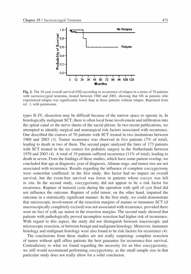
Chapter 29 / Sacrococcygeal Teratoma 471
Fig. 2. The 10-year overall survival (OS) according to occurrence of relapse in a series of 70 patientswith sacrococcygeal teratoma, treated between 1960 and 2003, showing that OS in patients whoexperienced relapse was significantly lower than in those patients without relapse. Reprinted fromref. 3, with permission.
types II–IV, dissection may be difficult because of the narrow space to operate in. Inhistologically malignant SCT, there is often local bone involvement and infiltration intothe spinal canal or the nerve sheets of the sacral plexus. In two recent publications, weattempted to identify surgical and nonsurgical risk factors associated with recurrence.One described the courses of 70 patients with SCT treated in two institutions between1960 and 2003 (3). Tumor recurrence was observed in five patients (7% of total),leading to death in two of them. The second paper analyzed the fates of 173 patientswith SCT treated in the six centers for pediatric surgery in the Netherlands between1970 and 2003 (4). A total of 19 patients suffered recurrence (11% of total), leading todeath in seven. From the findings of these studies, which have some patient overlap, weconcluded that age at diagnosis, year of diagnosis, Altman stage, and tumor size are notassociated with recurrence. Results regarding the influence of complete coccygectomywere somewhat conflicted: in the first study, this factor had no impact on overallsurvival, but the event-free survival was lower in patients whose coccyx was leftin situ. In the second study, coccygectomy did not appear to be a risk factor forrecurrence. Rupture of tumoral cysts during the operation with spill of cyst fluid didnot influence the outcome. Rupture of solid tumor, on the other hand, impaired theoutcome in a statistically significant manner. In the first study, we could demonstratethat microscopic involvement of the resection margins of mature or immature SCT (ifmacroscopically completely excised) was not associated with recurrence, provided therewere no foci of yolk sac tumor in the resection margins. The second study showed thatpatients with pathologically proved incomplete resection had higher risk of recurrence.With regard to this aspect, the study did not distinguish between macroscopic andmicroscopic resection, or between benign and malignant histology. Moreover, immaturehistology and malignant histology were also found to be risk factors for recurrence (4).
The conclusions from these studies are not really surprising: complete resectionof tumor without spill offers patients the best guarantee for recurrence-free survival.Contradictory to what we found regarding the necessity for en bloc coccygectomy,we still would recommend performing coccygectomy, as the small sample size in thatparticular study does not really allow for a solid conclusion.

472 De Backer and Hazebroek
DIAGNOSIS OF RECURRENT SCT
The necessity of following the patients after discharge is widely recognized. A strictfollow-up protocol for all the patients with SCT is in place in our department. Forpatients with benign SCT, this protocol includes regular control visits with clinicalexamination, determinations of serum �-FP, �-HCG, and pelvic ultrasound; patients areseen every 3 months during the first year, every 4 months during the second year, every6 months during the third year, and finally at 4 and 5 years after treatment. For thosewith malignant histology, the protocol is even more intense. As a consequence, mostrecurrent tumors will be diagnosed during these follow-up visits, either through clinicalrectal examination, elevated tumor markers, abnormal ultrasound scan, and/or otherimaging. The usual symptoms in local recurrence are constipation, tenesmus, and globusof the urinary bladder. Symptoms associated with distant metastases (usually lungs,liver, lymph nodes) are anorexia, weight loss, unexplained fever, respiratory insuffi-ciency, and others. Relapse calls for a complete clinical and radiological restaging,including imaging by either CT scan or MRI.
PRINCIPLES OF REOPERATIVE SURGERY
Presumably Benign RecurrenceThese are the patients showing a well-circumscribed tumor that does not infiltrate
into the sacrum, who have no distant metastases, and whose tumor markers are notraised. Complete surgical excision of the tumor is the only treatment. Rectal prepa-ration is carried out preoperatively. The patient is positioned in the prone, jack-knifeposition. The rectum is filled with a polividone iodine gauze. The approach dependson the precise location and extent of the recurrent tumor and of the former scar.A posterior sagittal incision usually provides adequate exposure for centrally locatedtumors. The operation should not be mutilating. Sometimes, the rectum appears to beinvaded by tumor, but there is no place for rectum amputation. Rarely, a combinedabdominoperineal approach may be necessary.
Presumably Malignant RecurrenceThese patients usually show raised tumor markers; tumors are very irregular, invade
the sacrum, and some show distant metastases. Surgery alone may be adequate therapyfor stage I lesions (7). In all other cases, surgery must be combined with chemotherapyand occasionally radiotherapy, autologous bone marrow transplantation, and/or locore-gional hyperthermia (22). If complete surgical excision is judged feasible, this may beundertaken. Otherwise, up-front chemotherapy (after biopsy, if requested) followed bysurgical resection of the residual tumoral mass is another valid option.
LONG-TERM RESULTS
Benign RecurrenceIn our experience, patients with recurrent mature teratoma all do well after complete
excision (3,4). However, we have observed some deaths among those with immaturerecurrent teratoma.

Chapter 29 / Sacrococcygeal Teratoma 473
Malignant RecurrenceThere are two reports detailing the outcomes of children with recurrent malignant
SCT. One describes six patients from the POG and CCG study with malignant recur-rence of mature or immature teratoma (21). Four responded to therapy, one died, andone was lost to follow-up. The second is a large series of 22 patients with recurrentmalignant sacrococcygeal GCTs, registered in the MAKEI protocols (22). Of these,17 presented with an isolated local recurrence, two showed a distant relapse, andthree patients suffered from combined local and distant recurrences. Those 22 patientspresented with 22 first, 14 second, five third, and two fourth relapses. A total of12 patients achieved complete remission, seven of whom remained in continuouscomplete remission. Five of these patients experienced a second relapse. All patientswho achieved only a partial remission after the first relapse developed a second relapse.Only three of 14 patients who developed a second relapse could be cured. Altogether,10 of 22 patients survived disease-free (45%), and 12 patients died as a result of tumorprogression or therapy-related complications.
REFERENCES
1. Oosterhuis JW, Looijenga L. Testicular germ cell tumours in a broader perspective. Nat Rev Cancer2005;5:210–222.
2. Altman RP, Randolph JG, Lilly JR. Sacrococcygeal teratoma: American Academy of PediatricsSurgical Section survey–1973. J Pediatr Surg 1974;9:389–398.
3. De Backer A, Madern GC, Hakvoort-Cammel FGAJ, Haentjens P, Oosterhuis JW, Hazebroek FWJ.Study of the factors associated with recurrence in children with sacrococcygeal teratoma. J PediatrSurg 2006;41:173–181.
4. Derikx J, De Backer A, van de Schoot L, et al. Factors associated with recurrence andmetastatic disease in sacrococcygeal teratoma: results in the Netherlands (1970–2003). Br J Surg2006;93(12):1543–1548.
5. Wu JT, Bool L, Sudar K. Serum alpha fetoprotein in normal infants. Pediatr Res 1981;15:50–52.6. Calaminus G, Vesterling-Horner D, Bokkerink JP, et al. The prognostic significance of serum alpha
1-fetoprotein in children and adolescents with malignant extracranial non-testicular germ cell tumors.Klin Pädiatr 1991;203:246–250.
7. Rescorla FJ, Sawin RS, Coran AG, Dillon PW, Azizkhan RG. Long-term outcome for infants andchildren with sacrococcygeal teratoma: a report of the Children’s Cancer Group. J Pediatr Surg1998;33:171–176.
8. Derikx J, De Backer A, van de Schoot L, et al. Long-term functional sequelae of sacrococcygealteratoma: a national study in the Netherlands. J Pediatr Surg 2007;42(6):1122–1126.
9. Göbel U, Schneider DT, Calaminus G, et al. Multimodal treatment of malignant sacrococcygeal germcell tumors: a prospective analysis of 66 patients of the German cooperative protocols MAKEI 83/86and 89. J Clin Oncol 2001;19:1943–1950.
10. Rescorla F, Billmire D, Stolar C, et al. The effect of cisplatin dose and surgical resection in childrenwith malignant germ cell tumors at the sacrococcygeal region: a Pediatric Intergroup Trial (POG9049/CCG 8882). J Pediatr Surg 2001;36:12–17.
11. Gonzalez-Crussi F, Winkler RF, Mirkin DL. Sacrococcygeal teratomas in infants and children.Relationship of histology and prognosis in 40 cases. Arch Pathol Lab Med 1978;102:420–425.
12. Ein SH, Adeyemi SD, Mancer K. Benign sacrococcygeal teratomas in infants and children. A 25 yearreview. Ann Surg 1980;191:382–384.
13. Engelskirchen R, Holschneider AM, Rhein R, Hecker WC, Höpner F. Sacrococcygeal teratomas inchildren. An analysis of long-term results in 87 children. Z Kinderchir 1987;42:358–361.
14. Schropp KP, Lobe TE, Rao B, et al. Sacrococcygeal teratoma: the experience of four decades.J Pediatr Surg 1992;27:1075–1079.
15. Havranek P, Rubenson A, Güth D, et al. Sacrococcygeal teratoma in Sweden: a 10-year nationalretrospective study. J Pediatr Surg 1992;27:1447–1450.

474 De Backer and Hazebroek
16. Bilik R, Shandling B, Pope M, Thorner P, Weitzman S, Ein SH. Malignant benign neonatal sacro-coccygeal teratoma. J Pediatr Surg 1993;28:1158–1160.
17. Schmidt B, Haberlik A, Uray E, RatschekM, Lackner H, Höllwarth ME. Sacrococcygeal teratoma:clinical course and prognosis with a special view to long-term functional results. Pediatr Surg Int1999;15:573–576.
18. Wakhlu A, Misra S, Tandon RK, Wakhlu AK. Sacrococcygeal teratoma. Pediatr Surg Int 2002;18:384–387.
19. Perelli L, D’Urzo C, Manzoni C, et al. Sacrococcygeal teratoma. Outcome and management. Ananalysis of 17 cases. J Perinat Med 2002;30:179–184.
20. Huddart SN, Mann JR, Robinson K, et al. Sacrococcygeal teratomas: the UK children’s cancer studygroup experience. I Neonatal. Pediatr Surg Int 2003;19:47–51.
21. Hawkins E, Issacs H, Cushing B, Rogers P. Occult malignancy in neonatal sacrococcygeal teratomas.A report from a combined Pediatric Oncology Group and Children’s Cancer Group study. Am JPediatr Hematol Oncol 1993;15:406–409.
22. Schneider DT, Wessalowski R, Calaminus G, et al. Treatment of recurrent malignant sacrococcygealgerm cell tumors: analysis of 22 patients registered in the German protocols MAKEI 83/86, 89, and96. J Clin Oncol 2001;19:1951–1960.

30 Recurrent Hernia, Hydrocele,and Varicocele
Robert E. Cilley, MD,Brett W. Engbrecht, MD,and Andreas H. Meier, MD
CONTENTS
Recurrent Inguinal Hernia
Recurrent Hydrocele
Recurrent Varicocele
References
RECURRENT INGUINAL HERNIA
Technical Variations in Pediatric Inguinal Hernia RepairThe technique of pediatric inguinal hernia repair varies among pediatric surgeons
according to training and personal habits developed over time (1). Variations intechnique include the degree to which the inguinal canal is exposed (stretching ofthe external ring or long incision in the external oblique), the extent of the retroperi-toneal dissection of the indirect sac (i.e., how high the high ligation is performed),the frequency of addition of a floor repair to indirect hernia repair (never, frequentlyin premature infants with “weak floors”), handling of the distal hernia/processusvaginalis/tunica vaginalis testis (ignore it every time, explore it every time, selectivelyexplore), the decision to create surgical fixation of a mobile testicle, type of suturematerial, open versus laparoscopic technique, and the use of prosthetic material inadult-size teenagers. Common to every pediatric hernia is the “high” ligation of theindirect hernia sac. The contribution of the technical variation to the risk of recur-rence is unknown. It is unknown whether laparoscopic or laparoscopic-assisted ligationof indirect inguinal hernias in infants and children will result in higher (or lower)recurrence rates with long-term follow-up.
Factors Affecting Inguinal Hernia RecurrenceRecurrence rates for inguinal hernia repair in infants and children are lower than in
adults, and vary from less than 0.5% to approximately 4% (2–6). The recurrence rate
From: Reoperative Pediatric SurgeryEdited by: S. Teich and D. A. Caniano © Humana Press, Totowa, NJ
475

476 Cilley et al.
for inguinal hernia repair may be lower when performed by trained pediatric surgeonscompared to adult general surgeons (7). Patient characteristics and biologic factorsmay predispose to much higher rates of recurrence. Premature neonates with repair ofsymptomatic hernias are at increased risk compared to other neonates and children.Chronic lung disease and the presence of ascites increase the complication rate forhernia repair in all ages. In the pediatric population, cystic fibrosis patients and thosewith ventriculoperitoneal shunts or peritoneal dialysis catheters are at higher risk forrecurrence. Patients with connective tissue disorders such as mucopolysaccharidosesand Ehlers-Danlos or Marfan syndrome are also at higher risk for recurrence. Patientswith irreducible incarcerated hernias requiring emergent surgery are at increased riskof recurrence. Difficult dissection, damage to surrounding structures, and tissue edemaare implicated. The use of rapidly absorbing suture material such as “catgut” mayalso predispose to recurrence. Abnormalities of the collagen matrix in patients withrecurrent inguinal hernias may account for recurrences in some patients (8).
Clinical Presentation and Diagnosis of Recurrent Inguinal Hernias
Swelling after indirect inguinal hernia repair
Inguinoscrotal swelling that occurs after pediatric inguinal hernia repair may ormay not be a result of recurrence of the hernia. Recurrent/persistent hydrocele afterhernia/hydrocele repair is discussed later in this chapter. All questions of recurrenceof an inguinal hernia in child should be addressed thoughtfully. One should notoverdiagnose the problem in patients operated on by others or deny or underdiagnosein patients operated on by oneself. Pediatric surgeons may be asked to assess patientspostoperatively with impressive inguinoscrotal swelling and concerns for early herniarecurrence. Significant edema may occur after the repair of giant inguinal herniasin premature infants. Careful physical exam, possibly supplemented by ultrasoundevaluation, is needed to distinguish tissue edema/swelling from early technical failure.
Direct and femoral hernias recognized after indirect inguinal
hernia repair
A direct inguinal hernia may occur after pediatric hernia repair for several possiblereasons. Excessive widening of the internal ring may predispose to direct herniarecurrence. It is not known whether maneuvers that tighten the internal ring or theaddition of an inguinal canal floor repair reduce this risk. The floor of the inguinalcanal can theoretically be injured by excessive dissection at the time of indirect repair.Minimizing the posterior dissection of the cremasteric fibers maintains the integrity ofthe floor of the inguinal canal. Weakness of the floor of the inguinal canal is likelya predisposing factor for direct recurrence in premature infants and may account fortheir much higher recurrence rate. It is unknown whether floor repair at the time ofthe initial repair of the premature infant with very large indirect hernia (large enoughto encroach on the direct space) increases or decreases recurrence rate. Experiencedpediatric surgeons are aware of the clinical scenario of a patient with a persistenthernia after indirect hernia repair that proved to be a femoral hernia or a direct hernia.Inguinofemoral masses that are present both before and after indirect inguinal herniarepair likely represent missed femoral or direct hernias (9). These are not true recurrenthernias, but rather represent “misdiagnosed” hernias. Pure direct and femoral herniasare relatively rare, and indirect inguinal hernias are very common in children. The

Chapter 30 / Recurrent Hernia, Hydrocele, and Varicocele 477
importance of a careful history and physical exam is obvious. One advantage of alaparoscopic hernia repair may be the ability to differentiate more accurately amongthe different defects.
Technical failures and recurrence
If the hernia sac is not dissected far enough and the “high ligation” of the herniasac retains enough of the proximal portion of the hernia sac, an indirect hernia maypersist. This condition is not technically a recurrent inguinal hernia, but rather shouldbe termed a “retained” hernia. The presence of visible bulging along the inguinal canal,near the level of the internal ring in slender patients without other symptoms, shouldbe observed. The natural history of this finding is unknown, and does not warrantreoperative surgery with its attendant risks. Other technical failures may result in a“recurrent” inguinal hernia. Immediate or early “recurrence” of an inguinal mass aftera pediatric hernia repair results from failure to control the hernia sac circumferentiallyin the ligation sutures or failure to recognize a tear in the sac. Viscera escape fromthe peritoneal cavity into the inguinal region after the procedure. Twisting and doubleligation of the sac with permanent or long-term absorbable suture minimizes thisproblem. Reoperation with control of the peritoneal opening is required. Excessivedissection has been implicated in damage to the floor of the canal and may increasethe direct recurrence rate; however, the sac must be dissected adequately to assure itsintegrity and the completeness of the high ligation. Inspection of the distal sac belowthe site of division of the sac helps confirm that the entire sac has been completelycircumferentially isolated. Incomplete isolation of the sac usually occurs with a portionof the sac overlying the cord left behind. Rents in the hernia sac, which usually occurlaterally, may extend deep into the retroperitoneum. All defects and rents in a herniasac can be fully assessed and repaired with adequate mobilization if recognized at thetime of initial repair.
True recurrent inguinal hernias
Any child who has had repair of an indirect inguinal hernia is at risk for recurrence.Progressive inguinal bulging may be either at the indirect space (probably relatedto technical failure as noted previously) or in the direct space, representing a trueweakness of the floor of the inguinal canal. The presence of a reducible mass thatcontains visceral contents is the most definitive indication for reoperative surgery.More subtle masses should be observed for progression. A progressively enlarginginguinal bulge is an indication for reoperation. Photographic documentation may assistin the objective assessment of progression. Unresolved questions of recurrence oftenpersist, especially in patients with other symptoms such as pain and in those with higherbody mass index. Diagnostic imaging is rarely helpful or indicated, but computedtomography and ultrasound may be considered. Diagnostic laparoscopy may be neededto definitively answer the question of recurrence. Indirect, direct, and femoral herniasare visualized definitively and repaired either laparoscopically or by open technique.This has been particularly helpful to rule out a recurrent hernia in patients withunremitting symptoms. When the definitive diagnosis of a recurrent hernia has beenmade, repair is recommended.

478 Cilley et al.
Surgical Correction of Recurrent Inguinal HerniasRisks of reoperation include greater likelihood of nerve injury, damage to the vas
deferens and/or testicular blood supply, and subsequent testicular atrophy. There isno data regarding the risks of second recurrence in pediatric hernia repair. It seemslikely that weakness of the floor of the inguinal canal will predispose these patients toadditional recurrence later in life. As the cadre of survivors of prematurity ages, thelong-term results of our pediatric surgical efforts will become apparent.
The standard principles of all reoperative surgery should be considered in recurrenthernia repair (10). An alternative anatomic approach to the condition should beconsidered. If the original inguinal approach is chosen, wider exposure of the reoper-ative field is used to allow clear anatomic visualization. Laparoscopic repair andpreperitoneal repair represent alternative anatomic approaches that allow the problem tobe visualized from a different orientation (11). These approaches may allow dissectionand repair to be performed with less risk of injury to the cord structures. The laparo-scopic approach is particularly appealing in the case a recurrent indirect hernia. A highligation of the indirect sac may be performed under direct vision. There are no outcomedata comparing the reoperative inguinal approach with either of these techniques.
An inguinal operative approach is used most often in cases of recurrent inguinalhernia. As noted previously, laparoscopy may be used as a diagnostic adjunct toconfirm the presence and type of hernia. An inguinal approach to a recurrent inguinalhernia is illustrated in Fig. 1. The original incision is expanded and initial dissectionis begun in unscarred tissue planes, which are usually found lateral and superior tothe original operative site. The cord structures may be identified inferior to the scarredexternal ring. The inguinal canal is opened where the least scarring is encountered. This
Fig. 1. (Continued).

Chapter 30 / Recurrent Hernia, Hydrocele, and Varicocele 479
Fig. 1. (Continued).
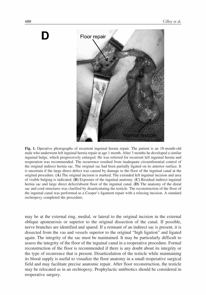
480 Cilley et al.
Fig. 1. Operative photographs of recurrent inguinal hernia repair. The patient is an 18-month-oldmale who underwent left inguinal hernia repair at age 1 month. After 3 months he developed a similaringuinal bulge, which progressively enlarged. He was referred for recurrent left inguinal hernia andreoperation was recommended. The recurrence resulted from inadequate circumferential control ofthe original indirect hernia sac. The original sac had been partially ligated on its anterior surface. Itis uncertain if the large direct defect was caused by damage to the floor of the inguinal canal at theoriginal procedure. (A) The original incision is marked. The extended left inguinal incision and areaof visible bulging is indicated. (B) Exposure of the inguinal anatomy. (C) Residual indirect inguinalhernia sac and large direct defect/absent floor of the inguinal canal. (D) The anatomy of the distalsac and cord structures was clarified by disarticulating the testicle. The reconstruction of the floor ofthe inguinal canal was performed as a Cooper’s ligament repair with a relaxing incision. A standardorchiopexy completed the procedure.
may be at the external ring, medial, or lateral to the original incision in the externaloblique aponeurosis or superior to the original dissection of the canal. If possible,nerve branches are identified and spared. If a remnant of an indirect sac is present, it isdissected from the vas and vessels superior to the original “high ligation” and ligatedagain. The integrity of the sac must be maintained. It may be particularly difficult toassess the integrity of the floor of the inguinal canal in a reoperative procedure. Formalreconstruction of the floor is recommended if there is any doubt about its integrity orthe type of recurrence that is present. Disarticulation of the testicle while maintainingits blood supply is useful to visualize the floor anatomy in a small reoperative surgicalfield and may facilitate precise anatomic repair. After floor reconstruction, the testiclemay be relocated as in an orchiopexy. Prophylactic antibiotics should be considered inreoperative surgery.

Chapter 30 / Recurrent Hernia, Hydrocele, and Varicocele 481
RECURRENT HYDROCELE
A true recurrent hydrocele is defined as the accumulation of scrotal fluid surroundingan otherwise normal testicle that persists beyond the immediate postoperative periodafter primary hydrocele repair or repair of inguinal hernia where the tunica vaginalistestis was manipulated at the time of initial surgery. The appearance of scrotal fluidafter hernia repair that involved only high ligation of the sac and no distal manip-ulation is not technically a recurrence. There is little information relating the typeof hydrocele procedure originally performed (puncture/aspiration, fenestration, partialexcision, eversion) and the risk of recurrence.
Postoperative Hydrocele Following Inguinal Hernia Repairand Hydrocelectomy
Inguinal hernia repair and hydrocele surgery are typically included together whendescribing postoperative hydrocele formation. Postoperative hydroceles typicallydevelop within several months of surgery. Morecroft et. al. reported a rate of 1.4%in their series of 556 boys (12). Most of these resolved without treatment within afew months. Davies et. al., in their series of inguinal hernias in boys less than 3 kg,reported a high incidence of postoperative hydrocele formation of 11% (five of 46) (13).Two hydroceles resolved spontaneously and one resolved after a single aspiration.Two patients underwent exploration and fenestration of the sack, with one hydrocelepersisting. Moss and Hatch reported two hydroceles requiring surgical treatment outof 328 boys less than 2 months of age requiring hernia repair (14). In a series of 5343male hernia repairs, Ein et. al. reported that only two boys required surgery for thiscomplication (15). The incidence of postoperative hydroceles and the use of aspirationwere not mentioned. The overall consensus seems to be that hydrocele formation isrelatively uncommon after inguinal hernia repair or hydrocele surgery and it shouldusually be treated by observation, with aspiration being therapeutic for the few that donot resolve spontaneously; reoperation is rarely required (16–18).
Hydrocleles That Occur After Varicocele RepairThe occurrence of hydrocele after surgical treatment of varicocele is common. These
are not truly recurrent hydroceles, because hydrocele was not recognized before thevaricocele procedure. Postoperative rates of hydrocele formation range from 1–40%(19–24). The median time to hydrocele formation is 2–22 months, with some hydro-celes appearing as late as 6 years after surgery (21,23). The large variability in thereports is partly explained by the differences in follow-up periods between the studies,with short-term studies likely missing a number of delayed presentations. Hydroceleformation after varicocele treatment presumably results from disruption of lymphaticchannels that follow the spermatic vessels (25). Disrupted lymphatics may regenerateor collaterals may develop, resulting in spontaneous resolution of some of these hydro-celes. Spontaneous resolution has been reported in 14–60% of postoperative hydroceles(21–23). The management of postoperative hydroceles that do not resolve sponta-neously includes aspiration and hydrocelectomy. There is no clear consensus regardingtreatment. When hydrocelectomy is performed after varicocele repair, true recurrenceis much higher than after simple hydrocele repair. Esposito et al. (23) reported truerecurrence in two of six children who underwent hydrocelectomy after varicocelectomy.

482 Cilley et al.
Management Strategies for Postoperative HydrocelesThe management of hydrocele formation after inguinal hernia repair, hydroc-
electomy, and varicocele surgery has neither clear consensus nor defined algorithm,and to date has been dictated by surgeon experience and preference. Because manysuch hydroceles will resolve spontaneously, expectant observation should be the initialapproach. Appropriate follow-up is necessary to assess for resolution. For hydrocelesthat do not resolve after months (six) of observation, are very large, or cause discomfort,percutaneous aspiration with local anesthetic should be the first treatment modality.Aspiration can be repeated if fluid reaccumulates. For those hydroceles that do notrespond to aspiration after several (up to five) attempts, hydrocelectomy should beconsidered. Inguinal or scrotal approach for hydrocelectomy is acceptable, and thedecision should depend on the surgeon’s experience and preference. An inguinalapproach is recommended if hydrocele recurs and is unresponsive to aspiration aftera transscrotal procedure. The inguinal approach confirms that a hernia/communicatinghydrocele is not responsible for the recurrence. If there is any concern for recurrenthernia or communication with the peritoneal cavity, aspirations should not be attemptedand an inguinal approach should be taken. These considerations argue for an inguinalapproach in all primary pediatric hydrocele repairs.
RECURRENT VARICOCELE
Background on Varicocele (26)
A varicocele is defined as swelling of the veins of the pampiniform plexus thatprovide venous drainage of the testicle via the testicular veins into the renal vein or venacava. Varicocele occurs much more commonly on the left and may be bilateral. Smallvaricoceles are visible only with valsalva maneuver, medium varicoceles are palpablemasses, and large varicoceles are visible masses within the scrotum. These severitiesof varicocele are also referred to as grade I, II, and III. Typically, the varicocelefills with blood when standing and becomes less prominent in the supine position.Untreated varicocele is associated with testicular atrophy and infertility. Patients mayexperience a sense of fullness, heaviness, or discomfort. Varicocele is thought toresult from absence or abnormality of the valves in the gonadal vein, which resultsin high venous pressure. Elevated venous pressure in the left renal vein has also beenimplicated. Obstructing retroperitoneal lesions such as tumors must be considered inunusual presentations such as young children. The testicle has additional blood supplybesides the testicular vessels, including arterial and venous channels that accompanythe vas deferens and cremasteric vessels. These collateral vessels provide adequatearterial and venous circulation to allow division of part or all of the testicular vessels inthe treatment of varicocele. Adolescent males with symptomatic varicocele or evidenceof testicular atrophy are treated.
Treatment Options for Adolescent VaricoceleEach of the treatment options includes the division or occlusion of the gonadal
veins on the affected side. The different procedural options are indicated in Table 1(20,27–31). There is no consensus on the primary treatment of varicocele in childrenand adolescents; however, laparoscopic division/ligation of the spermatic vessels (botharteries and veins) has a low recurrence rate as well as low risk of testicular atrophy,

Chapter 30 / Recurrent Hernia, Hydrocele, and Varicocele 483
Table 1Treatment Options for Varicocele
Procedure Variations Comments
“Palomo” procedure (massligation of testicular arteryand vein in retroperitoneumabove internal ring)
May be performed using openor laparoscopic techniques
Technicallystraight-forward, amenableto laparoscopy, collateralsresult in low atrophy rate.Conflicting data onincidence of postoperativehydrocele.
Ligation of testicular veinsin the inguinal canal
Magnification, microsurgicaltechniques, Doppler ultrasoundand intraoperative venographyhave been described to allowmore accurate identification ofveins, arteries and lymphatics
Division of lymphaticchannels at this level mayaccount for reports offrequent postoperativehydrocele. Microsurgicaltechnique results in lowcomplication rate.
Ligation of testicular veinsbelow the inguinal canal
Referred to as“subinguinal” approachMay be performedwith sedation and localanesthesia.
Retrograde venous injectionsclerotherapy (coils and/orsclerosants injected into thespermatic veins via a centralvenous approach)
Many technical variationsdescribed (type of vascularocclusion, exact level ofocclusion, inclusion ofcontralateral vessels) Usuallyperformed by interventionalradiologists.
Pampiniform phlebitis andthromboembolic problemsare potential complicationsnot present with surgicaltreatments. May beperformed with sedationand local anesthesia.Anatomic variations mayprevent use.
Antegrade sclerotherapy(injection of sclerosants intopampiniform plexus vesselsbelow the inguinal canal)
May be good choice forrecurrent varicocele.Requires operativeexposure of pampiniformvessels in upper scrotum.May be performedwith sedation and localanesthesia.
Techniques and procedures used to treat varicocele.
presumable owing to preservation of the collateral testicular blood supply. Previousreports of low risk of post-operative hydrocele have been questioned (24). I haveutilized the technique of ligation of the testicular veins within the inguinal canal usingmagnification and Doppler ultrasound (A theoretical advantage of this technique is thatit allows the processus vaginalis to be identified and ligated, eliminating a “missedcommunicating hydrocele” as a cause for postoperative hydrocele.).

484 Cilley et al.
Postoperative Care and RecurrenceAfter any procedure for varicocele, patients should be monitored for testicular growth
and size, recurrence of the varicocele, and hydrocele formation. Hydrocele formationmay be delayed and patients should be instructed of this. Recurrence and postop-erative hydrocele are the most important complications that may require reoperativetreatment. Postprocedural hydrocele may respond to aspiration or require operativehydrocelectomy. Recurrent hydrocele, even after operative treatment, is common asdiscussed previously. If varicocele recurs after primary treatment, it must be assumedthat some of the testicular veins were not identified and divided with the originalprocedure or that other collaterals exist. Although reoperation through the sameanatomic approach is feasible, reoperation via an inguinal approach, if this was usedfor the first procedure, puts the remaining testicular blood supply at risk. Antegradesclerotherapy has been applied to treat failed surgical ligation and failures after retro-grade sclerotherapy (32). Recurrence after failed retrograde sclerotherapy has beensuccessfully treated with repeat retrograde sclerotherapy (33). The Palomo procedurehas been used for sclerotherapy failures as well. Venography performed in conjunctionwith sclerotherapy can be used to identify the presence of persistent vessels and toidentify any vascular anomalies that might preclude success. Because the patterns ofrecurrence and the anatomic reasons for recurrence may differ with each technique,it seems reasonable to recommend an alternate anatomic approach for symptomaticrecurrence.
REFERENCES
1. Levitt MA, Ferraraccio D, Arbesman MC, et al. Variability of inguinal hernia surgical technique: asurvey of North American pediatric surgeons. J Pediatr Surg 2002;37(5):745–751.
2. Mestel AL, Turner HA. Recurrent Inguinal Hernia in Infants and Children. Int Surg 1966;45(5):575–578.
3. Grosfeld JL, Minnick K, Shedd F, et al. Inguinal hernia in children: factors affecting recurrence in62 cases. J Pediatr Surg 1991;26(3):283–287.
4. Vibits H, Pahle E. Recurrences after inguinal herniotomy in children. Long time follow-up. Ann ChirGynaecol 1992;81(3):300–302.
5. Wright JE. Recurrent inguinal hernia in infancy and childhood. Pediatr Surg Int 1994;91:64–66.6. Meier AH, Ricketts RR. Surgical complications of inguinal and abdominal wall hernias. Semin Pediatr
Surg 2003;12(2):83–88.7. Borenstein SH, To T, Wajja A, et al. Effect of subspecialty training and volume on outcome after
pediatric inguinal hernia repair. J Pediatr Surg 2005;40(1):75–80.8. Zheng H, Si Z, Kasperk R, et al. Recurrent inguinal hernia: disease of the collagen matrix? World
J Surg 2002;26(4):401–408.9. Al-Shanafey S, Giacomantonio M. Femoral hernia in children. J Pediatr Surg 1999;34(7):1104–1106.
10. Teich S, Caniano DA. Editor’s note: How we do it: repair of recurrent inguinal hernia (comment).Semin Pediatr Surg 2003;12(2):89.
11. Esposito C, Montupet P. Laparoscopic treatment of recurrent inguinal hernia in children. PediatriSurg Int 1998;14(3):182–184.
12. Morecroft JA, Stringer MD, Homes SJK, et al. Follow-up after inguinal herniotomy or surgery forhydrocele in boys. Br J Surg 1993;80:1613–1614.
13. Davies BW, Fraser N, Najmaldin AS, et al. A prospective study of neonatal inguinal herniotomy: theproblem of the postoperative hydrocele. Pediatr Surg Int 2003;19(1–2):68–70.
14. Moss RL, Hatch EI. Inguinal hernia repair in early infancy. Am J Surg 1991;161:596–599.15. Ein SH, Njere I, Ein A. Six thousand three hundred sixty-one pediatric inguinal hernias: a 35-year
review. J Ped Surg 2006;41(5):980–986.16. Glick PL, Boulanger SC. Inguinal hernias and hydroceles. In: Grosfeld JL, O’Neill JA Jr, Fonkalsrud EW,
Coran AG, (eds.), Pediatric surgery, sixth edition, volume two. Philadelphia: Mosby; 2006: 1172–1192.
Chapter 30 / Recurrent Hernia, Hydrocele, and Varicocele 485
17. Lloyd DA. Inguinal and femoral hernia. In: Ziegler MM, Azizkhan RG, Weber TR, (eds.), Operativepediatric surgery. New York: McGaw-Hill; 2003: 543–554.
18. Wolfson PJ. Inguinal hernia. In: Mattei P (ed.), Surgical directives: pediatric surgery. Philadelphia:Lippincott, Williams and Wilkins; 2003: 521–525.
19. Belloli G, Musi L, D’Argostino S. Laparoscopic surgery for adolescent varicocele: preliminary reporton 80 patients. J Pediatr Surg 1996;31:1488–1490.
20. Esposito C, Monguzzi GL, Gonzalez-Sabin MA, et al. Laparoscopic treatment of pediatric varicocele:a multicenter study of the Italian Society of Video Surgery in Infancy. J Urol 2000;163:1944–1946.
21. Misseri R, Gershbein AB, Horowitz M, et al. The adolescent varicocele. II: the incidence ofhydrocele and delayed recurrent varicocele after varicocelectomy in a long-term follow-up. BJU Int2001;87(6):494–498.
22. Esposito C, Monguzzi G, Gonzalez-Sabin MA, et al. Results and complications of laparoscopicsurgery for pediatric varicocele. J Pediatr Surg 2001;36:767–769.
23. Esposito C, Valla JS, Najmaldin A, et al. Incidence and management of hydrocele following varicocelesurgery in children. J Urol 2004;171(3):1271–1273.
24. Hassan JM, Adams MC, Pope JC, et al. Hydrocele formation following laparoscopic varicocelectomy.J Urol 2006;175:1076–1079.
25. Szabo R, Kessler R. Hydrocele following internal spermatic vain ligation: a retrospective study andreview of the literature. J Urol 1984;132(5):924–925.
26. Hutson JM. Undescended testis, torsion and varicocele. In: Grosfeld JL, O’Neill JA Jr, FonkalsrudEW, Coran AG, (eds.), Pediatric surgery, sixth edition, volume two. Philadelphia: Mosby Elsevier;2006: 1193–1214.
27. Palomo A. Radical cure of varicocele by a new technique: preliminary report. J Urol 1949;61(3):604–607.
28. Lord DJ, Burrows PE. Pediatric varicocele embolization (review). Tech Vasc Interv Radiol2003;6(4):169–175.
29. Greenfield SP, Seville P, Wan J. Experience with varicoceles in children and young adults. J Urol2002;168:1684–1688.
30. Alqahtani A, Yazbeck S, Dubois J, et al. Percutaneous embolization of varicocele in children: aCanadian experience. J Pediatr Surg 2002;37(5):783–785.
31. Mazzoni G. Adolescent varicocele: treatment by antegrade sclerotherapy. J Pediatr Surg2001;36(10):1546–1550.
32. Mazzoni G, Minucci S, Gentile V. Recurrent varicocele: role of antegrade sclerotherapy as first choicetreatment. Eur Urol 2002;41:614–618.
33. Sivanathan C, Abernethy LJ. Retrograde embolization of varicocele in the paediatric age group:a review of 10 years’ practice. Ann R Coll Surg Engl 2003;85(1):50–51.

31 Failed Orchidopexyand Complications of Circumcision
Seth A. Alpert, MD, Stephen A. Koff, MD,and Venkata R. Jayanthi, MD
CONTENTS
Failed Orchiopexy
Complications of Circumcision
References
FAILED ORCHIOPEXY
Orchiopexy for the correction of the undescended testicle is a commonly performedsurgical procedure and is generally very successful, implying that a normal-sized testisis located dependently in the scrotum. The overall success of the procedure is dependenton the initial location of the testis and the age of the patient (1). The incidence ofpersistent cryptorchidism after orchiopexy is estimated to be 7.5–13% (2–4). This canoccur because of either residual or recurrent cryptorchidism after the initial orchiopexyattempt. Residual cryptorchidism is recognized immediately at the time of the firstsurgery as an inability to bring the testis into a dependent scrotal location and requiresthat a planned second-stage procedure be undertaken at least 6 months later. Recurrentcryptorchidism noted on later follow-up after the initial procedure can be repairedelectively at that time. In several studies, the mean time between the initial surgery andreoperation for failed orchiopexy ranged from 3–4.7 years (3,5). After orchiopexy, thesize of the testis should be assessed to determine that testicular growth has occurred.Although most testes will grow normally after orchiopexy, some may have growthretardation, atrophy, or may involute altogether. This phenomenon may be caused bycompromise of the vascular supply to the testis during the initial orchiopexy attemptor may reflect intrinsic abnormality of a high intraabdominal testis as, for example, inthe prune belly syndrome.
Factors Predisposing to Unsuccessful OrchiopexyThe causes for failure of an orchiopexy can include technical factors, the proximal
nature of the original location, intrinsic testicular or spermatic cord abnormalities, orprior inguinal surgery. Most frequently, technical factors during the original surgery
From: Reoperative Pediatric SurgeryEdited by: S. Teich and D. A. Caniano © Humana Press, Totowa, NJ
487
488 Alpert et al.
are responsible for orchiopexy failure. Inadequate mobilization of the spermatic cordfar enough proximally is the chief factor predisposing to recurrent cryptorchidism(2,4,6,7). High ligation and division of the persistent patent processus vaginalis isa necessary step in most orchiopexies, and when this is not properly completed, ahigh-riding testis can result. Residual cremasteric or other fascial attachments createupward traction on the cord and may prevent successful scrotal placement of the testis.One study found that 60% of cord length was attributable to division of the patentprocessus and the remaining 40% was caused by separation of the vessels from theperitoneum and abdominal wall (division of the lateral bands) (8).
After corrective surgery for a hydrocele or hernia, a high-riding testicle may becreated if the surgeon fails to replace the testis in a low intrascrotal position (7). Thismay occur when the testis is pushed back into the hemiscrotum from above, instead ofpulling it back down from below by its scrotal and gubernacular attachments. Kaplanreviewed a series of cases of iatrogenic cryptorchidism that occurred after hernia orhydrocele repair; he postulated that excessive retractility of the involved testis withsubsequent formation of scar tissue may trap the testis in an elevated position (9). Healso noted inappropriate replacement of the testis in the wrong tissue plane as anothercause for this outcome.
Injury to the vascular supply of the testis may occur during the dissection to separatethe spermatic cord from the processus vaginalis, especially in a very young or prematureinfant or child (7). Furthermore, excessive tension on the spermatic vessels, which mayresult from an inadequately mobilized cord, may contribute to poor vascular flow tothe testis and subsequent atrophy and/or involution.
The distance that a testicle must be brought down into the scrotum has a bearingon the success of orchiopexy. In a metaanalysis of more than 8000 undescended testesperformed by Docimo, the preoperative location of testis position was directly relatedto the success rate of orchiopexy. The success rate increased as the initial locationof the testis was more distal. When all studies were included, the success rates forabdominal, peeping (at the internal ring), canilicular, and beyond the external ringtestes were 74, 82, 87, and 92%, respectively. When data before 1985 were excluded,the success rates were 77, 100, 96, and 100%, respectively (1). In the same study,one-stage Fowler-Stephens orchiopexies were successful in 68.5%, whereas two-stageprocedures were successful 76.8% of the time.
DiagnosisIn most cases, the diagnosis of a failed orchiopexy can be made on a thorough
physical examination by an experienced clinician to determine the position and size ofthe testis. A failed orchiopexy must be differentiated from a retractile, but otherwisenormal, testis, which may require an extended or repeat examination. Rarely, animaging study such as an ultrasound, computed tomography (CT) scan, or magneticresonance imaging (MRI) may be helpful to localize a small undescended testis,especially if the patient is obese (7). Measurement of testis size before and afterorchiopexy will allow testis atrophy to be distinguished from growth retardation; theformer reflects a testis that has become smaller than its preoperative size and the latterrepresents a testis that has grown but has not kept pace with its mate.
Chapter 31 / Failed Orchidopexy and Complications of Circumcision 489
Preoperative CounselingPrior to undertaking the reoperation for a failed orchiopexy, several issues must be
considered. The procedure will be more labor-intensive and challenging than the initialprocedure because of fibrosis and scarring present in the surgical field. There is anincreased risk of injury to the vas deferens or spermatic vessels for the same reason.Lastly, if the testis is found to be dysplastic or atrophic, then biopsy or orchiectomymay be required. It is of utmost importance that the parents understand these inherentissues prior to surgery so that complete informed consent is possible. It is usuallyrecommended that the reoperation be delayed at least 6 months from the time of theinitial procedure in order to allow postoperative edema and fibrosis to stabilize.
Techniques for Redo OrchiopexyIn most cases, although the prior inguinal incision can be used for the redo orchiopexy
procedure, adequate exposure is essential and one should not hesitate to extend thelength of the incision as needed or make a new incision if the original scar is too low.As mentioned previously, the most common reason for failure of initial orchiopexyis inadequate division of the patent processus vaginalis and/or incomplete proximaldissection of the spermatic cord. The first step is to rectify these deficiencies withcareful dissection of the spermatic cord proximally and high ligation and divisionof a persistent patent processus if present. This alone may provide adequate lengthto successfully bring the testis down into a dependent scrotal location. The cordstructures may be virgin higher up in the inguinal canal if no dissection was performedhere previously. In addition, aggressive retroperitoneal dissection and creation of anadequate subdartos pouch has been required in a large number of redo orchiopexies (2).
Opening the floor of the inguinal canal, dividing the inferior epigastric vessels toreroute the spermatic cord medially and straighten its course (the Prentiss maneuver)can all provide additional cord length and occasionally may be required to permitthe testis to reach a dependent scrotal location (10). Redman described reoperationfor persistent cryptorchidism or hernia via an inguinal approach to the spermatic cordthrough the cremasteric fascia (11). In this method, the external oblique fascia isincised laterally to medially through the external ring. The spermatic cord structuresare then approached caudally, where they may be less scarred than from a standardcranioventral approach. Further proximal dissection is then undertaken, up to and intothe retroperitoneum, if necessary. Using this technique, Redman reported 15 successfulrepeat orchiopexies in 13 patients with no evidence of testicular atrophy.
Cartwright et al. described a method for en-bloc mobilization of the spermatic cordin reoperative orchiopexy whereby a 1- to 2-cm-wide strip of external oblique fasciais left intact on top of the cord structures (12). This maneuver minimizes the risk ofinjury to cord structures by dissecting lateral and posterior to the spermatic vesselsand vas deferens instead of anteriorly beneath the fascia when these structures arethe most scarred and adherent. Standard techniques for completing an orchiopexy arethen used once en-bloc cord mobilization is completed, including further retroperi-toneal dissection and higher ligation of the patent processus. A total of 25 reoperativeorchiopexies where accomplished in this manner; one patient needed another procedureto bring the testis down completely and one patient had slight testis atrophy.
Palacio et al. built upon the en-bloc spermatic cord and external oblique fasciadissection technique by suturing a strip of the aponeurosis on the cord to the pubic

490 Alpert et al.
bone with nonabsorbable suture. They refer to this as “inguinal cordopexy,” becausethe proximal cord structures are fixed above the scrotum so that tension on the distalportion of the cord is minimized (13). This technique is not always feasible becausethe fascial adherence to the cord is sometimes too lax to permit proper fixation orthe testis is proximal to the external ring and the fascial strip would descend into thescrotum with the testis. In their study reporting this technique, these authors stated thatinguinal cordopexy was possible in 11 testes of 29 patients referred to their institutionwith malpositioned testes after initial orchiopexy or hernia repair. All testes after thisprocedure were present in the scrotum almost 2 years later.
The Jones technique for high undescended testicles is another option for reoperativeorchiopexy. Jones and Bagley described an abdominal extraperitoneal approach toorchiopexy that allows extensive retroperitoneal dissection of the spermatic vesselsand was originally advocated as a single-stage alternative to a staged Fowler-Stephensprocedure (14–16). The Jones technique starts with an incision medial to the anteriorsuperior iliac spine with splitting of the anterior abdominal wall muscles and medialreflection of the peritoneum. Once the hernia sac is located and opened, the testisis located within the abdomen. The hernia sac is then ligated and the peritoneum isclosed. The spermatic vessels are then dissected proximally in the retroperitoneumuntil enough length is obtained. A subcutaneous tunnel is then created from the inferioredge of the incision down to the pubic tubercle and the testis is delivered through anincision in the external oblique fascia here. Then the testis is brought down into asubdartos pouch in the scrotum through the standard method.
Multiple reoperative techniques have been described to deal with the failedorchiopexy. To avoid injury to the testis and spermatic cord, we have found that thetestis can be palpated and identified early in the procedure by initiating dissectioninferior to the external inguinal ring. En-bloc dissection of the testis and spermaticcord complex in a cephalad direction allows scar tissue and fibrosis to be separatedfrom the spermatic vascular supply and vas until the region of the internal ring hasbeen reached.
Testicular autotransplantation has been described as an alternative for Fowler-Stephens orchiopexy in high intraabdominal testes. Using microvascular surgicaltechniques, the spermatic vessels of the testis are anastamosed to the inferior epigastricvessels and the testis is placed into the scrotum. Silber and Kelly(17) first describedthis procedure in 1976, but it has not gained widespread popularity owing to themicrosurgical skill and instrumentation required.
Testis atrophy is a known complication of repeat orchiopexy and is reported inapproximately 4% of cases. Pesce et al. looked at the long-term follow-up after reoper-ative orchiopexy and found that, compared to controls, the volumes of the majority ofreoperated testes were smaller but fertility was only markedly affected in 15% of 20patients assessed (18).
COMPLICATIONS OF CIRCUMCISION
Circumcision is the most commonly performed surgical procedure in the UnitedStates and is probably one of the oldest of all surgical procedures (19–22). Data isnot available to estimate the total number of circumcisions done presently becausemany performed are ritual circumcisions outside of a healthcare setting. Therefore,the true rate of complications after circumcision cannot be precisely calculated. Major
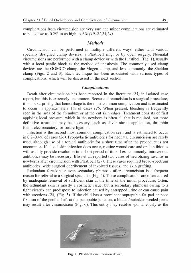
Chapter 31 / Failed Orchidopexy and Complications of Circumcision 491
complications from circumcision are very rare and minor complications are estimatedto be as low as 0.2% to as high as 6% (19–21,23,24).
MethodsCircumcision can be performed in multiple different ways, either with various
specially designed clamp devices, a Plastibell ring, or by open surgery. Neonatalcircumcisions are performed with a clamp device or with the Plastibell (Fig. 1), usuallywith a local penile block as the method of anesthesia. The commonly used clampdevices are the GOMCO clamp, the Mogen clamp, and less commonly, the Sheldonclamp (Figs. 2 and 3). Each technique has been associated with various types ofcomplications, which will be discussed in the next section.
ComplicationsDeath after circumcision has been reported in the literature (25) in isolated case
report, but this is extremely uncommon. Because circumcision is a surgical procedure,it is not surprising that hemorrhage is the most common complication and is estimatedto occur in approximately 1% of cases (26) When present, bleeding is frequentlyseen in the area of the frenulum or at the cut skin edges. Treatment consists of firstapplying local pressure, which in the newborn is often all that is required, but moredefinitive treatment may be necessary, such as silver nitrate application, thrombinfoam, electrocautery, or suture ligation.
Infection is the second most common complication seen and is estimated to occurin 0.2–0.4% of cases (26). Prophylactic antibiotics for neonatal circumcision are rarelyused, although use of a topical antibiotic for a short time after the procedure is notuncommon. If a local skin infection does occur, routine wound care and oral antibioticswill usually provide resolution in a short period of time. Less commonly, intravenousantibiotics may be necessary. Bliss et al. reported two cases of necrotizing fasciitis innewborns after circumcision with Plastibell (27). These cases required broad-spectrumantibiotics, wide surgical debribement of involved tissues, and skin grafting.
Redundant foreskin or even secondary phimosis after circumcision is a frequentreason for referral to a surgical specialist (Fig. 4). These complications are often causedby inadequate removal of sufficient skin at the time of the initial procedure. Often,the redundant skin is mostly a cosmetic issue, but a secondary phimosis owing to atight cicatrix can predispose to infection caused by entrapped urine or can cause painwith erections (28) (Fig. 5). If the child has a prominent suprapubic fat pad or poorfixation of the penile shaft at the penopubic junction, a hidden/buried/concealed penismay result after circumcision (Fig. 6). This entity may resolve spontaneously as the
Fig. 1. Plastibell circumcision device.
492 Alpert et al.
Fig. 2. GOMCO clamp.
Fig. 3. Mogen clamp.
Fig. 4. Redundant foreskin in a child who has previously undergone neonatal circumcision.

Chapter 31 / Failed Orchidopexy and Complications of Circumcision 493
Fig. 5. Secondary phimosis caused by a tight cicatrix, which formed subsequent to neonatalcircumcision.
Fig. 6. Buried (hidden) penis after circumcision.
child ages and body habitus changes, but persistent penile burying may require someform of penoplasty to correct poor fixation of the penile shaft to the penopubic fascia(29) (Fig. 7).
Adhesions of the shaft skin to the glans are fairly common after circumcision andare usually caused by inadequate genital hygiene. Mild adhesions caused by entrappedsmegma can often be separated manually in the office or can be treated medically witha several-week course of a steroid ointment (30). When penile adhesions epithializeand become more dense, these skin bridges do not respond to topical steroids and oftenrequire surgical lysis (Fig. 8). One study suggests that skin bridges are more likely toform when the adhesions involve the circumcision line (31).

494 Alpert et al.
Fig. 7. Appearance of penis immediately after penoplasty to correct buried penis.
Inclusion cysts at the circumcision line have been described (19,20,26). These areepidermoid cysts and are caused by entrapment of smegma in the circumcision woundor inadvertent rolling in of the epidermis during the initial procedure. These cysts cangrow to a large size and/or become infected (Fig. 9). Treatment consists of surgicalexcision.
Inadvertent injury to the urethra at the time of circumcision is a rare complicationand has been reported with a resulting fistula occurring either early or late afterthe procedure (19,20,32). Urethral injury may result from overzealous treatment of
Fig. 8. Penile skin bridge on the dorsal aspect of the penis: a probe can be seen being passed beneaththe shaft skin, which is adherent to the glans.

Chapter 31 / Failed Orchidopexy and Complications of Circumcision 495
Fig. 9. Penile inclusion cyst.
bleeding at the frenulum with either suturing or cautery (26). These fistulas tend tobecome apparent early on after circumcision because of ischemic injury of the urethra;however, one case report noted a urethral fistula in association with a penile skinbridge 13 years after neonatal circumcision (33). This fistula was felt to be subclinicaluntil puberty, when penile growth and more frequent erections may have caused itsunmasking. The urethra can be accidentally incised during a dorsal slit if the penis hasa 180° torsion and the glans is not appropriately visualized prior to circumcision (34).Surgical repair of urethral fistula must be individualized to each patient, but similarprinciples to that of hypospadias fistula repair apply. Formal urethroplasty should bedelayed at least 6–9 months after initial fistula formation to allow tissue inflammationand fibrosis to subside.
Partial or complete glans amputation is a very serious complication and is usuallyseen when a clamp is used for circumcision. This has been reported with a freehandprocedure as well as with Sheldon, Mogen, and GOMCO clamps (23,35–38) (Fig. 10).One case report in an older child noted an entire glans penis amputated duringa guillotine-type procedure (39). If minimal partial glans amputation has occurredwithout urethral involvement, local wound care and hemostasis may be adequatebecause this may granulate and heal with a satisfactory cosmetic result. If more seriousglans amputation with urethral involvement is found, immediate reanastamosis of theamputated glans and urethra after debridement of the wound base is required. Goodresults have been reported with glans reattachment surgery, although tissue loss, fistula,and/or meatal stenosis can result. Penile injury and glans necrosis have also occurredwhen diathermy was used to remove skin during metal clamp circumcision and thecurrent flows to the glans.
Meatitis or meatal stenosis occurs in 8–31% of circumcised boys, and is rarely seenin the uncircumcised (26). Inflammation and potential stenosis of the meatus are feltto occur because of inflammation from exposure to urine in the diaper. Meatitis canbe treated conservatively with observation or a brief course of topical antibiotics orsteroids. Symptomatic meatal stenosis (Fig. 11) is corrected in an office setting with ameatotomy after a local anesthetic block or with a meatoplasty under general anesthesiain older children or those in whom adequate local anesthesia or immobility cannot beachieved.

496 Alpert et al.
Fig. 10. Glans amputation after circumcision.
One complication specific to the Plastibell ring is dislocation onto the penile shaftproximal to the glans such that the glans becomes entrapped and compressed (40).This is believed to occur when too large a ring is used, so that the device can slideback over the glans. This situation causes compression of the penile shaft proximal tothe glans and subsequent glans edema similar to that seen in paraphimosis. The ringrequires removal by cutting the ring away from the underlying skin with a ring- or wire-cutting instrument, followed by local wound care (Fig. 12). Another rare complicationcaused by proximal Plastibell ring migration is urinary retention owing to edema andcompression of the urethra (41).
Fig. 11. Meatal stenosis: the urethral meatus is pinpoint in size and can cause irritative or obstructivevoiding symptoms.

Chapter 31 / Failed Orchidopexy and Complications of Circumcision 497
Fig. 12. Proximal migration of Plastibell circumcision device before (A) and after removal (B).
REFERENCES
1. Docimo SG. The results of surgical therapy for cryptorchidism: a literature review and analysis.J Urol 1995;154(3):1148–1152.
2. Maizels M, Gomez F, Firlit CF. Surgical correction of the failed orchiopexy. J Urol 1983;130(5):955–957.
3. Livne PM, Savir A, Servadio C. Re-orchiopexy: advantages and disadvantages. Eur Urol1990;18(2):137–139.
4. Adamsen S, Borjesson B. Factors affecting the outcome of orchiopexy for undescended testis. ActaChir Scand 1988;154(9):529–533.
5. Cohen TD, Kay R, Knipper N. Reoperation for cryptorchid testis in prepubertal child. Urology1993;42(4):437–439.
6. Hutcheson JC, Cooper CS, Snyder HM III. The anatomical approach to inguinal orchiopexy. J Urol2000;164(5):1702–1704.
7. Noseworthy J. Recurrent undescended testes. Semin Pediatr Surg 2003;12(2):90–93.8. Davey RB. Orchidopexy: the relative importance of each step of mobilisation. Pediatr Surg Int
1997;12(2/3):163–164.9. Kaplan GW. Iatrogenic cryptorchidism resulting from hernia repair. Surg Gynecol Obstet
1976;142(5):671–672.10. Prentiss RJ, Weickgenant CJ, Moses JJ, Frazier DB. Undescended testis: surgical anatomy of spermatic
vessels, spermatic surgical triangles and lateral spermatic ligament. J Urol 1960;83:686–692.11. Redman JF. Inguinal reoperation for undescended testis and hernia: approach to the spermatic cord
through the cremaster fascia. J Urol 2000;164(5):1705–1707.12. Cartwright PC, Velagapudi S, Snyder HM III, Keating MA. A surgical approach to reoperative
orchiopexy. J Urol 1993;149(4):817–818.13. Palacio MM, Sferco A, Garcia Fernanndez AE, Vilarrodona HO. Inguinal cordopexy: a simple
and effective new technique for securing the testes in reoperative orchiopexy. J Pediatr Surg1999;34(3):424–425.
14. Jones PF, Bagley FH. An abdominal extraperitoneal approach for the difficult orchidopexy. Br J Surg1979;66(1):14–18.
15. Gheiler EL, Barthold JS, Gonzalez R. Benefits of laparoscopy and the Jones technique for thenonpalpable testis. J Urol 1997;158(5):1948–1951.
16. Youngson GG, Jones PF. Management of the impalpable testis: long-term results of the preperitonealapproach. J Pediatr Surg 1991;26(5):618–620.
17. Silber SJ, Kelly J. Successful autotransplantation of an intra-abdominal testis to the scrotum bymicrovascular technique. J Urol 1976;115(4):452–454.
18. Pesce C, d’Agostino S, Costa L, Musi L, Manzi M. Reoperative orchiopexy: surgical aspects andfunctional outcome. Pediatr Surg Int 2001;17(1):62–64.
19. Williams N, Kapila L. Complications of circumcision. Br J Surg 1993;80(10):1231–1236.20. Kaplan GW. Complications of circumcision. Urol Clin North Am 1983;10(3):543–549.21. Mayer E, Caruso DJ, Ankem M, Fisher MC, Cummings KB, Barone JG. Anatomic variants associated
with newborn circumcision complications. Can J Urol 2003;10(5):2013–2016.
498 Alpert et al.
22. Reynolds RD. Use of the Mogen clamp for neonatal circumcision. Am Fam Physician 1996;54(1):177–182.
23. Ben Chaim J, Livne PM, Binyamini J, Hardak B, Ben-Meir D, Mor Y. Complications of circumcisionin Israel: a one year multicenter survey. Isr Med Assoc J 2005;7(6):368–370.
24. Hutcheson JC. Male neonatal circumcision: indications, controversies and complications. Urol ClinNorth Am 2004;31(3):461–467, viii.
25. Sullivan P. Infant’s death another nail in circumcision’s coffin, group says. CMAJ 2002;167(7):789.26. Lerman SE, Liao JC. Neonatal circumcision. Pediatr Clin North Am 2001;48(6):1539–1557.27. Bliss DP, Jr., Healey PJ, Waldhausen JH. Necrotizing fasciitis after Plastibell circumcision. J Pediatr
1997;131(3):459–462.28. Talarico RD, Jasaitis JE. Concealed penis: a complication of neonatal circumcision. J Urol
1973;110(6):732–733.29. Maizels M, Zaontz M, Donovan J, Bushnick PN, Firlit CF. Surgical correction of the buried penis:
description of a classification system and a technique to correct the disorder. J Urol 1986;136(1 Pt2):268–271.
30. Chu CC, Chen KC, Diau GY. Topical steroid treatment of phimosis in boys. J Urol 1999;162(3 Pt1):861–863.
31. Ponsky LE, Ross JH, Knipper N, Kay R. Penile adhesions after neonatal circumcision. J Urol2000;164(2):495–496.
32. Limaye RD, Hancock RA. Penile urethral fistula as a complication of circumcision. J Pediatr1968;72(1):105–106.
33. Yazici M, Etensel B, Gursoy H. A very late onset urethral fistula coexisting with skin bridge afterneonatal circumcision: a case report. J Pediatr Surg 2003;38(4):642–643.
34. Redman JF. Rare penile anomalies presenting with complication of circumcision. Urology1988;32(2):130–132.
35. Gluckman GR, Stoller ML, Jacobs MM, Kogan BA. Newborn penile glans amputation during circum-cision and successful reattachment. J Urol 1995;153(3 Pt 1):778–779.
36. Sherman J, Borer JG, Horowitz M, Glassberg KI. Circumcision: successful glanular reconstructionand survival following traumatic amputation. J Urol 1996;156(2 Pt 2):842–844.
37. Yilmaz AF, Sarikaya S, Yildiz S, Buyukalpelli R. Rare complication of circumcision: penileamputation and reattachment. Eur Urol 1993;23(3):423–424.
38. Neulander E, Walfisch S, Kaneti J. Amputation of distal penile glans during neonatal ritualcircumcision—a rare complication. Br J Urol 1996;77(6):924–925.
39. Ozkan S, Gurpinar T. A serious circumcision complication: penile shaft amputation and a newreattachment technique with a successful outcome. J Urol 1997;158(5):1946–1947.
40. Datta NS, Zinner NR. Complication from Plastibell circumcision ring. Urology 1977;9(1):57–58.41. Mihssin N, Moorthy K, Houghton PW. Retention of urine: an unusual complication of the Plastibell
device. BJU Int 1999;84(6):745.

32 Complications of Vaginoplastyand Clitoroplasty
Lesley L. Breech, MD
CONTENTS
Feminizing Genitoplasty
Vaginoplasty
References
Pelvic reconstructive surgery in a pediatric population is primarily undertaken in thetreatment of congenital anomalies or congenital conditions. Pelvic reconstruction isperformed in patients with conditions such as intersex conditions, complex urogenitalanomalies, or isolated reproductive anomalies. Preoperative recognition of potentialsurgical challenges is crucial. This chapter describes potential complications associatedwith genital reconstructive surgery and consideration of possible causes.
Surgical goals in pelvic reconstructive surgery are the establishment of an anatomi-cally appropriate, physiologically sensitive clitoris; an adequate, appropriately placedvagina for comfortable sexual relations in adulthood; and normal-appearing femaleexternal genitalia. The major reconstructive procedures include feminizing genito-plasty and vaginoplasty. Both procedures may be performed during the same operativeprocedure; however, because the postoperative complications are significantly different,each will addressed independently. As will be emphasized, it is also important toconsider the indication for surgical intervention. Certainly optimal function and normal-ization of anatomic appearance are of primary importance, but the underlying medicalcondition and the possible surgical and postoperative ramifications are necessaryto consider. In patients with the genital effects of androgen excess, intraoperativedissection and postoperative response may be more challenging because of the role ofhormonal stimulation. Surgical manipulations in conditions without hormonal influenceare more likely to proceed more smoothly, with less risk of postoperative bleedingcomplications.
FEMINIZING GENITOPLASTY
Feminizing genitoplasty as a surgical intervention has evolved significantly overrecent years. Much discussion has occurred relating to the appropriate timingand indications, in addition to the appropriate procedure. Surgical intervention for
From: Reoperative Pediatric SurgeryEdited by: S. Teich and D. A. Caniano © Humana Press, Totowa, NJ
499

500 Breech
clitoromegaly, as a component of genital ambiguity generally associated with intersexconditions, has been discussed and reviewed owing to concerns regarding long-termpatient outcome. Unfortunately, much of the potential controversy relates to the morevocal experiences of some patients. Public concern has influenced medical standardssuch that some providers and families contemplate deferring surgical manipulationsuntil the patient is able to make her own operative decision. This premise, whiledesirous in theory, is likely not realistic for most families. In the majority of cases, it isthe child’s parents who request surgical management of an enlarged clitoral structure.Often parents are less cognizant of the possible existence of a persistent urogenital sinus,but are acutely aware of clitoromegaly. Discomfort with genital appearance incon-sistent with chromosomal sex is the primary reason for early intervention in girls withfemale pseudohermaphroditism. In cases of extreme virilization, it is understandablewhy families or surgeons push for early surgical management. However, considerationof possible variation in outcome should also factor in operative decision-making aswell as in discussions with family.
Postoperative issues after surgery at the clitoris include nerve damage, sensation,pain, recurrent clitoromegaly caused by persistence of erectile tissue, body imageconcerns, and sexual function. Late outcomes following feminizing genitoplasty havenot been systematically reviewed to allow obvious pursuit or abandonment of individualprocedures. Consideration of the type of procedure performed and the combination ofintervention at the vagina for a single-stage procedure is essential.
The evolution of clitoral reconstruction began with clitorectomy, followed by clitoralrecession, and now most commonly, clitoral reduction. Clitorectomy results in theobvious loss of sensation and may jeopardize the ability of an orgasmic response.Clitoral recession maintains sensation; however, pain secondary to clitoral engorgementis a definite risk. Without adequate removal of erectile tissue, adolescent young womenmay experience pain, discomfort, and extreme enlargement of the clitoral area owingto simulation and engorgement of residual corporal tissue (see Fig. 1) (1). Recog-nition of potential sensory consequences that may affect pubertal adolescents or youngadult women are necessary to consider, despite the reality that most girls will undergoclitoral surgery as an infant or young child. The most appropriate procedure forfeminizing genitoplasty is based on the concept of maintaining the clitoral glans andsensory input, which facilitates orgasm (2–4). The technique involves resection ofthe corpora at the crura, with careful preservaton of dorsal nerves and vessels andventral mucosa that supplies the glans (5). Management of a persistent urogenital sinusand possible vaginoplasty are the next consideration in the management plan of girlswith genital ambiguity. Several techniques have been described, primarily predicatedon the level of confluence between the vagina and the urethra (6–8). Historically, alow vaginal insertion has been managed by a simple cut back incision or a posteriorflap vaginoplasty (9,10). The use of this technique when the confluence is higheris inappropriate because it leaves the urethral meatus on the anterior vaginal wall,resulting in vaginal voiding and difficult access to the meatus (see Fig. 2). A highvaginal insertion requires more elaborate operative intervention, likely performed as atwo-stage procedure. A pull-through vaginoplasty (11), intestinal neovaginal segment(12), perineal skin flaps (7), total urogenital sinus mobilization (TUM) (13), or posteriorsagittal transanorectal approach (14) have all been proposed to provide adequateurinary and reproductive function. The intimate relationship between the vagina andthe urogenital sinus mandates meticulous dissection of the vagina from the urethra

Chapter 32 / Complications of Genital Reconstruction 501
Fig. 1. A 15-year-old female with a history of CAH who underwent a single-stage feminizinggenitoplasty-reduction clitoroplasty and a pull-through vaginoplasty at 2 years of age. She presentedwith pain and enlargement of the clitoral region.
proximal to the confluence of the sinus. Some authors postulate that fibrosis in thisfield of dissection may play a pivotal role in the development of vaginal stenosis (seeFig. 3) (7,15). Procedures that rely upon the use of the distal (fibrotic) vaginal portionof the urogenital sinus may increase the risk of the development of stenosis or fistulaformation.
Several reviews have provided insight into both short- and long-term outcomesin this population. Eroglu and colleagues in Turkey reported on the results of 55intersex patients who underwent feminizing surgical management (16). They reportedthe outcomes of patients managed over a 16-year period with multiple proceduresincluding single-stage clitorovaginoplasty (29 patients), staging clitoral surgery andvaginoplasty (seven patients), and TUM (three patients). The mean age at surgerywas 3.5 years and the follow-up averaged 4.1 years (2 months to 17 years). Themost frequently encountered complication was vaginal stenosis; however, less vaginalstenosis was found in patients who underwent an early single-stage procedure (3.4%,1/29) than in those with a staged procedure (42.8%, 3/7). Comparison with TUM in thisstudy was impossible, as all patients were too young with a short postprocedure interval.Bocciardi and colleagues reported a large series of Passerini-Glazel clitorovaginoplastyprocedures in 66 children, of whom 46 were available for extended follow-up (17).Outcome regarding anatomical results of the vaginoplasty and the cosmetic effect ofthe entire procedure were described. All of the patients had good cosmetic outcomes.None of the patients had introital stenosis. The patients were divided relative to age at

502 Breech
Fig. 2. A 12-year-old female with CAH s/p reduction clitoroplasty at age 8 months followed by ahigh pull-through vaginoplasty at age 2 years. Of note, a persistent urogenital is present. Becausethe patient is experiencing pain and discomfort at the clitoris, she will undergo a repeat clitoroplastyand TUM.
surgical intervention. Of the prepubertal group, 45.5%, and of the postpubertal group,25% had vaginal stenosis at the anastomosis of the mucocutaneous cylinder and thetrue vagina. All stenoses were corrected by Y-V plasties. The authors proposed thatthe vaginal stenosis was related to persistence of dysplastic or ischemic tissue at thedistal edge of the vagina. They recommend aggressive removal of the distal vaginauntil soft, thick, highly vascularized native vagina is identified to avoid such stenosis.
Fig. 3. Photo of a 21.5-year-old female with CAH treated with a single-stage procedure at age 2with a reduction clitoroplasty and vaginal pull-through. The confluence of the urethra and vaginawas 2 cm into the urogenital sinus. The patient currently complains of the inability to engage invaginal intercourse because of vaginal stenosis. She did not previously perform vaginal dilatation.
Chapter 32 / Complications of Genital Reconstruction 503
Other reviewers dismiss this process and instead emphasize the detrimental role ofa circular anastomosis in constrictive healing and the development of stenosis (18).Krege and colleagues reported 27 females who underwent reconstructive surgery owingto congenital adrenal hyperplasia (CAH), with a special emphasis on the long-termresults of vaginoplasty (19). Vaginoplasty was performed in 25 girls, of whom 20underwent a single-stage approach and five had a two-stage procedure. Vaginoplastywas performed using the technique described by Fortunoff and others (9), using aperineal flap sutured to the margin of the vaginal introitus; one patient required apull-through vaginoplasty, described by Hendren and Crawford (10). During long-termfollow-up, nine of the 25 (36%) had secondary vaginal stenosis. The mean age ofdiagnosis of the stenosis was 14 years (10–19 years). Stenosis was treated with lasersurgery, Z-plasty, or a pedicled skin-fat island flap. Lack of flap advancement farenough into the vagina, lack of precise coaptation of the mucosa to the perineal skinflap, inadequate blood supply to the flap, inadequate marsupialization of the vaginalfloor, and imprecise suture placement have all been reported causes of vaginal stenosis(20,21). TUM was initially described by Peña to repair cloacas (22). This procedurehas been applied to children with urogenital sinus anomalies, including CAH (22,23).The most remarkable advantage of this procedure is the avoidance of the difficultdissection required for separation of the vagina from the urinary tract by treating theurethra and vagina as a single unit. Additionally, the mobilized portion of the urogenitalsinus may be split and placed anteriorly to create a mucosal surface or an additionalflap. Other advantages are reported to be a decrease in operating time, decreased riskof vaginal stenosis, no risk of fistula formation because there is no need for separationof the structures within the urogenital sinus, less urine pooling within the vagina, theabsence of postvoid dribbling, and the lack of skin flaps to reach the vagina (22,23).The main concern of TUM is the theoretical risk of stress urinary incontinence causedby the short and distally placed urethra (24). Jenak and colleagues report on six patientswho were treated with a modification of the TUM and did not find any changesin voiding habits postopertively in patients who were toilet-trained before surgery;however, the follow-up period was less than 1 year. Farkas and colleagues reporteda follow-up period of 4.7 ± 2.6 years for a group of 49 patients who underwent asingle-stage feminizing genitoplasty (25). Of the 49 patients, 46 were treated with anen bloc mobilization of the vagina and urethra via the perineal approach. All patientswere reported to have successful cosmetic and early functional results. In patients whoreached puberty, the authors reported normal menstruation, a wet and wide introitus,and no evidence of fibrosis or scarring of the perineum. No urinary tract infectionswere reported and all patients were continent. No patients with CAH had initiatedvaginal intercourse. Several reports confirm the technical ease of the TUM, especiallyin cases of a urogenital sinus less than 3 cm in length and improved cosmetic results(13,16,25). Longer-term follow-up will be necessary to confirm positive early results,because most patients will now be entering puberty and adolescence.
Possible complications after feminizing genital reconstruction include vaginalstenosis, meatal stenosis, vaginourethral fistula, female hypospadius, urinary tractinjuries, and recurrent clitoromegaly (especially in patients with difficulties incompliance with hormonal regimens). Limited surgical and long-term outcome data arecurrently available. Al-Bassam and Gado reported the development of vaginal stenosisin 16.2% patients who underwent either flap vaginoplasty or vaginal pull-through (26).Krege and colleagues reported 27 females who underwent reconstructive surgery owing

504 Breech
to CAH, with a special emphasis on the long-term results of vaginoplasty (19). Vagino-plasty was performed in 25 girls, of whom 20 underwent a single-stage approach andfive patients had a two-stage procedure. During follow-up, all patients were askedto complete a questionnaire. Questions regarded appearance of the external genitalia,overall body image, frequency of heterosexual intercourse or other forms of sexualactivity, dyspareunia, and ability to achieve orgasm. Nine patients were noted to havevaginal stenosis and of those nine patients, six completed the questionnaire. Only onepatient reported regular sexual intercourse. Of the remaining 18 patients, ten completedthe questionnaire. Four of nine patients 15 years and older reported sexual intercourseand seven reported having orgasm (including those having vaginal intercourse). Fewstudies have been performed to assess the sexual and social functions of patients inlate adolescence or adulthood (19,25,27,28).
The question of timing for feminizing genital reconstruction can be a difficult one.In conditions such as CAH and other intersex conditions, clitoral surgery and vagino-plasty are generally performed in infancy and childhood. Most authors recommendtiming between newborn to age 3 years. The prevailing opinion is that clitoroplastyshould be performed as early as possible; however, the timing of vaginoplasty remainscontroversial. The primary reason described for intervention before age 2 years is thepotential detrimental psychological effects for both children and parents (19,29). Othersurgeons defer surgery until puberty, if possible, to better differentiate the demar-cation between hair bearing and non-hair bearing skin and to potentially avoid possiblevaginal dilatation in childhood (19,29); however, bleeding complications can be morechallenging in the pubertal patient. Krege and colleagues argue for vaginoplasty tobe performed at puberty in patients with a longer confluence of the urethra and vagina,who may potentially need vaginal dilatation (19). They also note the potential advantageof a distensible vagina provided by estrogen stimulation. Conversely, Lobe and othersreported a decreasing complication rate in patients undergoing an early, single-stageprocedure (30). Although successful results from neonatal one-stage surgery havebeen reported (31), many still prefer to perform the single stage clitorovaginoplastyprocedure sometime before 3 years of age, citing better compliance with treatment andalleviating parent’s concerns (26,27).
VAGINOPLASTY
Vaginoplasty is a procedure relevant to the care of female patients of any age. Physi-cians may care for children with complex pelvic congenital anomalies or adolescentswith vaginal agenesis. Vaginal agenesis is an uncommon condition, which may resultfrom a variety of different underlying diagnoses. Mayer-Rokitansky-Kuster-Hauser(MRKH) Syndrome, the most common clinical scenario the general provider willencounter, describes vaginal agenesis with variable Müllerian duct development andpossible associated renal, skeletal, and auditory abnormalities. It is generally thoughtthat MRKH affects 1/5000 live-born females (32).
The timing of intervention is an important consideration. Vaginal agenesis is aparticularly challenging problem for genitourinary surgeons. In patients diagnosed withvaginal agenesis as a child with additional organ system involvement, life-threateningconditions should be stabilized and addressed before operative planning is undertakenfor neovaginal creation. Nonsurgical and surgical interventions that require continueddilation and maintenance have historically been deferred to adolescence or adulthood.

Chapter 32 / Complications of Genital Reconstruction 505
Reconstruction with the use of a bowel graft, as it often does not require continueddilation, can be accomplished at any age. Hendren and others have advocated forintervention during childhood (33). Hendren maintains that young girls suffer signif-icant psychological and social trauma from the knowledge of an anatomically absentvagina and from the necessary surgical intervention. He stresses that he has not seen atraumatic response to surgical revision of a previous vaginoplasty created as a child. Heemphasizes the outpatient nature of a minor revision and that it is regarded by patientsand families with less importance than original construction of a neovagina. This mustbe weighed against the psychological effects of potential daily vaginal dilation orstenting in prepubertal girls. Grafting or flap techniques that require persistent stentingor dilation may be unsuitable for younger patients because of the requisite maturity andmotivation required for maintenance and the avoidance of postoperative complications.Bowel substitutions may be applied to wider ranges of patients, even infants. Long-term functional studies are needed to assess introital size and sexual satisfaction withconsideration of timing of surgical intervention. An evaluation of the psychologicaloutcomes related to technique, the age at diagnosis, and age of repair is also necessary.
Over time, many methods of surgical vaginoplasty have not only been describedbut also advocated. Even today, there is no consensus regarding the best surgicalapproach. The primary goal of a neovaginoplasty is to create a vagina adequate forsatisfying sexual interactions. In patients with a uterus, adequate egress of menses andthe potential to accommodate a vaginal delivery are also of paramount importance.Additional goals should also include normal amounts of secretions/lubrication, theabsence of malodor, and minimal maintenance care. Unfortunately, one method has notyet been demonstrated to achieve all of these goals. Management options are describedaddressing procedure types, advantages, disadvantages, and outcomes. Controversycontinues regarding the best technique, as little complete long-term data, includingsexual identity and function data, about any procedure is truly available.
A well-established technique for vaginal reconstruction is the use of intestinalsegment substitution. Baldwin initially reported the use of ileum in 1904 and 1907followed by Wallace, who described the use of sigmoid colon in 1911 (34–36).Wesley and Coran modified Baldwin’s technique for the sigmoid colon (37). Prattfurther popularized the technique with sigmoid colon (38). The purported benefits ofusing intestinal segments include the lack of obligatory continued dilation or stenting,the potential for growth in small children, and beneficial lubrication. Rajimwale andassociates published a metaanalysis of seven recent series of neovaginal creation withbowel (33,37,39–43) . Patient ages ranged from 4 days to 26 years. Bowel vagino-plasty demonstrated a complication rate of 35%, with a reoperation rate of 4% in 202cases. The authors argue that the postoperative complications after bowel vaginoplastymay be managed more simply, using dilation or minor surgical repairs, than thoseassociated with skin grafting techniques. They also note that the use of bowel trans-position is broader, as it can be employed in a wider age range and for a variety ofdiagnoses compared to skin grafting techniques, which are primarily described in themore isolated anomaly of MRKH.
Several segments of intestinal vaginoplasty replacement have been described: ileum,cecum, sigmoid, and rectosigmoid. Selection may relate to the availability of a suitablesegment of an appropriate length; patients also undergoing bladder augmentation orother more complex anorectal or urogenital repairs may have any combination of smallor large bowel for reconstruction, dependent on the needs of other reconstruction.

506 Breech
Ileal or cecal segments may be problematic because of a shorter mesentery, whichmay produce tension on the neovagina; Hensle and Reiley reported a higher incidence ofstenosis attributed to this concern (39). In complex anomaly cases, the ileum may be theonly bowel segment available for use for vaginal construction. The rectum and sigmoidmay be more optimally used in other urinary or bowel reconstruction. It is important tobe cognizant of the role of the ileocecal valve within the gastrointestinal tract and thesubsequent morbidity associated with any disruption. Pratt reported a higher incidenceof dyspareunia and persistent mucus secretions with ileal vaginoplasty (38).
Sigmoid vaginoplasty has been the favored bowel segment due to its potentialadvantages: adequate length, natural lubrication, and early coitus in older patients. Thesigmoid neovagina is thought to be self-lubricating (see Fig. 4), without the excessmucus production associated with segments of small bowel (39). The risk of stenosisis small, without the requirement of continuous dilation or stenting. Mobilization ofthe vascular pedicle is usually straightforward and has been accomplished using aminimally invasive, laparoscopic approach (44,45). When replacement is performedduring childhood, the potential for adequate growth into adulthood remains unclear.Koyle and colleagues describe the sigmoid neovagina as aesthetically pleasing andmore compatible with sexual activity (46). The thickness of the sigmoid tissue maywithstand trauma associated with sexual intercourse better than small bowel or skingrafts. Freundt and colleagues reported on psychosexual and psychosocial performancein adult patients with a sigmoid neovagina (47). The series included eight patients withvaginal agenesis and one patient with androgen insensitivity. Psychosexual profiles,as determined by patient questionnaire, indicated that most of these patients achievedacceptable outcomes with respect to sexual relations, function, and satisfaction. Thevalidity of the evaluation is difficult to interpret as the same questionnaire was usedin transsexual patients. Communal and colleagues described the sexual outcomes of12 patients with MRKH syndrome who underwent sigmoid neocolpopoiesis usinga standardized questionnaire, the Female Sexual Function Index (FSFI), a validatedassessment of female quality of life and sexual function in adults (48). Six separatedomains of female sexual function are included in the questionnaire: desire disorder,arousal disorder, orgasmic disorder, sexual pain disorder, quality of vaginal lubrication,and sexual/relationship satisfaction. The authors also added details on sexual inter-
Fig. 4. Perineal photograph taken intraoperatively immediately after creation of the sigmoidneovagina in a 16-year-old female with MRKH syndrome.

Chapter 32 / Complications of Genital Reconstruction 507
course, vaginal discharge, self-esteem, and anxiety. The authors reported patients hada normal sexual life. A total of 72% of patients had vaginal intercourse at least onceweekly; however, 18% had severe dyspareunia. Vaginal discharge was described asalmost constant, but the daily discomfort score was “moderate.” Patients who were“dry” preoperatively later reported wearing one to two pads daily. Consideration ofdevelopment of colitis in the graft, which may play an integral role in the developmentof persistent vaginal discharge, is essential (49,50). In severe cases of colitis, pain,irritation, and bleeding may occur, necessitating vaginoscopy. Stenosis of the introitusmay occur related to excessive traction on the sigmoid transposition, or limited perinealcleavage may necessitate dilation or additional surgery.
More recent publications by both Hensle and Kapoor confirm the successfuloutcomes in patients treated with a sigmoid vaginoplasty (51,52). Hensle and colleaguesreviewed the records of 57 patients who underwent bowel replacement vagino-plasty over a 24-year period. Replacement vaginoplasty was performed using sigmoidcolon in 39 patients. The authors evaluated the postoperative quality of life, with aspecial emphasis on sexual function. Postoperative sexual function was evaluated in44 patients; however, only 36 completed the sexual function questionnaire. Of the36 patients who responded, 78% reported sexual desire, 33% sexual arousal, 33%sexual confidence, and 78% sexual satisfaction. A group of 20 patients (56%) reportedfrequent orgasms and eight (22%) reported occasional orgasms. Only 22% deniedhaving orgasm. A total of 32 patients (89%) reported adequate lubrication for inter-course, and two reported dyspareunia. A group of 34 patients used home douching,and 20 (56%) required pads for mucus production.
Sigmoid neocolporrhaphy has historically involved a major laparotomy and theassociated risks of bowel surgery, including perforation. In patients with more isolatedvaginal anomalies, sigmoid neovaginoplasty offers the potential of a laparoscopic-perineal approach to decrease operative morbidity and adverse aesthetic conse-quences associated with a laparotomy. Darai and others conducted a preliminarystudy with seven patients with MRKH syndrome, confirming the feasibility of thelaparoscopic approach when performed by surgeons with experience in both gyneco-logical and gastrointestinal laparoscopic surgery (53). The mean operating time was312 minutes (range 220–450 minutes). No intraoperative complications were reported,and conversion to laparotomy was never necessary. The mean fall in hemoglobin was3.6 g/dL (range 2.0–4.4 g/dL). The mean hospital stay was 7.7 days (range 6–12 days).The mean length of the neovagina was 11.5 cm without documented shrinkage over theinitial 6-month follow-up. Mean follow-up was 31 months (range 5–90 months). Themean interval between surgery and first intercourse was 4 months (range 3–6 months).Despite promising results in this small population, randomized and/or multicenter trialsare necessary to confirm the advantages.
Possible advantages of the use of sigmoid colon, as in other bowel neovaginoplasty,are the avoidance of long-term stenting or dilation and the rare contraction of theneovagina. Potential disadvantages include the complexity of the surgical procedure,generally requiring laparotomy or extensive laparoscopic skills, significant mucousdischarge reported by patients, and postoperative complications. Periumbilical painwith vaginal intercourse, attributed to traumatic stretching of the graft vascularity, andproblems with defecation (constipation) are not uncommon.
An alternative technique familiar to many gynecologic surgeons is the Abbe-McIndoe vaginoplasty (54). The procedure utilizes a split-thickness skin graft to line

508 Breech
a neovaginal canal, dissected in the potential space between the urethra and rectum.The three most important principles in performing this procedure are dissection of anadequate space, inlay of a proper split-thickness skin graft, and continuous, prolongeddilation during the contractile phase of healing. Many consider this technique unsuitablefor children, as it requires strict adherence to dilation until regular intercourse occurs.In addition, it is unclear how well this type of neovagina will grow with the child.It has successfully been reported in patients with vaginal agenesis who undergo theprocedure as late adolescents or young adults. Templeman and colleagues published areview of the procedure and included the results of multiple surgeons (55–61). Successrates vary from 80–100%, depending on the experience of the surgeon. Klingele andcolleagues reported on 71 patients who responded to a questionnaire after a McIndoeprocedure (62). Of the 71 patients, 10 (14%) had significant complications, includingvaginal prolapse, and rectovaginal and vesicovaginal fistula. In this series, 56% ofpatients reported improvement in self-image, 83% reported satisfactory vaginal functionduring intercourse, and 75% were able to achieve orgasm. Only 28% of patients hada disfiguring graft harvest site. Relevant complications to consider for this procedureinclude failure of graft take, rectal perforation during dissection, and fistula formation.
The advantages of the Abbe-McIndoe procedure include the long-term experiencewith excellent published results, low complication rates, and the avoidance of alaparotomy and intestinal complications. The disadvantages should include prolongedpostoperative hospitalization, the necessity for continued dilation, and the visible skinscar from acquisition of the skin graft. Some authors also report difficulties withlubrication and dyspareunia (39).
Several authors have described a similar approach as the Abbe-McIndoe; however,they have utilized synthetic materials to wrap around the vaginal mold and line theneovaginal space. Jackson and colleagues originally reported the use of oxidizedregenerated cellulose fabric (Interceed™) after a segment of amnion was inadver-tently contaminated during a surgical neovaginoplasty (63). Moyotama and associatesdescribed 10 patients who were diagnosed with vaginal agenesis and treated withoxidative regenerated cellulose fabric (Interceed) (64). Operative time (<30 minutes)and postoperative hospitalization (2 days) were significantly shorter than that for thetraditional skin graft method. Vaginal depth was 8–10 cm, and no significant operativecomplications were reported. Patients had only been followed for 6 months at thetime of publication; however, vaginal intercourse was already described as easy andsuccessful.
Benefits of this intervention include the short operative time and hospitalization.The procedure is technically easier without acquisition of a skin graft. No scarring,as occurs in a skin graft procedure, is visible to others. Although short follow-upis reported, patients have early satisfactory sexual interactions. As in the skin graftprocedure, continued postoperative dilation is necessary. It is essential to provideadditional long-term follow-up data regarding this procedure to truly assess its role inpatient care.
Skin grafts provide a thin lining for the neovaginal cavity, which does not alterthe length and diameter of the dissected neovaginal cavity during the operation.Split-thickness skin grafts have been used most often for adolescents being treatedby gynecologists, and long-term follow-up has demonstrated a patent and functionalvagina. The split skin segment retains most of its original morphological and histo-chemical characteristics, but atrophy of sweat glands and hair follicle has been

Chapter 32 / Complications of Genital Reconstruction 509
observed (65). The main concern is possible inadequate take of the graft leadingto a reduction in the size of the neovaginal cavity during the contractile phase ofhealing (66,67). Patients are required to wear a vaginal stent continuously for manymonths to prevent contracture and stenosis. Full-thickness skin grafts may have adecreased tendency to contract than split-thickness grafts during the contractile phaseof healing. Additionally, full-thickness grafts may successfully grow in children (68).Reduced stenosis without consistent dilation or intercourse may be especially beneficialin patients in whom vaginal construction is performed at an earlier age (66,67). Italso has some benefit even in select adolescents who may experience more signif-icant psychological distress with dilation. Because sebaceous and sweat glands arebetter preserved in full-thickness grafts, secretions produce vaginal lubrication within4–5 months postoperatively (68). Although the full-thickness graft method may beacceptable for neocolporrhaphy, most pediatric surgeons would have less expertise andrequire plastic surgery collaboration for optimal results.
The Vecchietti operation was first described in 1965 by Giuseppe Vecchietti (69).Veronikus and collegues reviewed the use of the technique and described a laparo-scopic modification, which combines cystoscopy to confirm bladder integrity (70). TheVecchietti procedure is a surgical technique that constructs a dilatation-type neovaginain 7–9 days. The procedure utilizes specialized equipment including a traction device,a ligature carrier, and an olive-shaped acrylic vaginal dilator. The process involvestwo steps with essential operative and postoperative components. The operative phaseinvolves positioning the olive at the perineum, and the traction sutures are positionedextraperitoneally. Classically performed through a Pfannenstiel incision, althoughmay be approached laparoscopically, the ligature carrier introduces the suture into anewly dissected vesicorectal space. During the postoperative invagination phase, theneovagina is created by applying constant traction to the olive. The process reportedlyoccurs at a rate of 1.0–1.5 cm/day. developing a 10–12 cm vagina in 7–9 days.
The Vecchietti procedure is an interesting procedure that applies the principlesof progressive dilation against the vaginal dimple in an innovative fashion. Notablebenefits are the avoidance of vesicorectal dissection and postprocedure visible scarring,in addition to the opportunity for a laparoscopic approach. Functional success rates varyfrom 98–100% (71,72). Disadvantages include poor lubrication, long-term dilation inpatients who are not sexually active, and a significant risk of neovaginal prolapse.
Davydov described a three-stage surgical creation of a neovagina in 1969 (73).The procedure involves dissection of the rectovesical space, abdominal mobilizationof the peritonem followed by attachment of the peritoneum to the introitus. Laparo-scopic modifications to perform the rectovesical dissection and the purse-string closureof the abdominal neovagina have been described. The most common postoperativeproblem involved the formation of granulation tissue at the vaginal vault. As withany open abdominal procedure, laparoscopic conversion has allowed shorter hospitalstays, decreased morbidity, and a substantial decrease in visible scarring. However, thetechnically challenging components of the laparoscopic approach may limit the use ofthis interesting technique. Additional follow-up information regarding this techniquewill be helpful in determining its role in the care of young women.
Carcinoma of the neovagina is an extremely rare disease, at least 10 times lesscommon than vaginal cancer, which represents only 1–2% of all gynecologicalcancers (74). The first cases of a neoplastic process in the neovagina involved the devel-opment of adenocarcinoma in small bowel grafts (75,76). Subsequently, neovaginal

510 Breech
squamous cell carcinoma was also described (77–80), as well as carcinoma in situ(81,82). The evidence supports a risk of neoplastic change in both skin and bowelgrafts, with a reported average interval from vaginoplasty to diagnosis on the orderof 19 years (83). The use of bowel grafts in children and teenagers could result inthe possibility of invasive adenocarcinoma developing in very young women. It isunclear if this represents a risk for any particular colonic segment independently devel-oping adenocarcinoma or whether it may be related to the transplantation of colonictissue to a new environment. It is therefore important that patients with any type ofneovagina have long-term follow-up examinations after vaginoplasty. Patients shouldalso undergo regular cytologic screening. Epithelium transplanted to the vagina willassume the oncogenic potential of the lower reproductive tract. Consideration of aplan for surveillance and counseling regarding transmission risk for virally relateddysplastic change in the lower genital tract is also extremely important for any patientsundergoing neovaginoplasty. Suspicious symptoms such as bleeding or a palpable massshould necessitate prompt evaluation.
Few cases of neovaginal prolapse have been reported in the literature. Prolapse maybe a concern in patients managed surgically or nonsurgically. Creation of a neovaginawith dilation may be at risk, as the apex is not anchored to any supporting structuresor ligaments. O’Connor and colleagues reported a patient that required conversionto a sigmoid neovagina after complete prolapse of the vagina after progressivedilatation (84). Some authors note a significant incidence of neovaginal prolapse,which may be attributed to the absence of normal supporting ligament (41). A similarphenomenon may also occur in bowel transpositions (39). Mucosal prolapse requiringtrimming of the exposed mucosa or anchoring of the bowel segment to the sacrumis known to occur. Hendren and Atala described 16 cases of eversion-prolapse of thebowel segment that required trimming (11). Hendren qualified the report as a simpleeversion of the distal segment, not an intussusception as in apical vaginal prolapse.He reported no significant change in the length of the neovagina with resection ofthe prolapsed tissue. Although the etiology of neovaginal prolapse remains unclear,retroperitoneal sacropexy may be a successful solution, especially when apical prolapseis the concern (85). Communal and colleagues incorporated anchorage of the distalend of the sigmoid neovagina to the fascia of the sacral promontory at the time of theneocolporrhaphy for prophylaxis of prolapse (48).
Regardless of the method of neocolpopoiesis performed, it is important that theoperation be performed correctly the first time. If the vagina becomes constrictedbecause of granulation tissue formation, injury to adjacent structures, or failure tocomply with postoperative maintenance, then subsequent attempts to create a satis-factory vagina will be more difficult. The first operation has the best chance of success.
Clearly, multiple techniques remain available for creation of the neovagina. Thereis truly no ideal procedure secondary to the lack of clearly defined and comparedmeasures of success. Commonly, the decision is based on the degree and area ofexpertise of the surgeon. The application of intestinal neovaginoplasty has widenedto treating patients with isolated vaginal agenesis, in addition to patients with morecomplex anomalies. The McIndoe vaginoplasty has been the most widely performed bygynecologic surgeons. The Vecchietti procedure, performed laparoscopically, has beenperformed more readily outside of the United States but has gained some popularityhere in recent years. Innovative neovaginoplasty techniques, such as use of Interceedor buccal mucosa, may also have promise as more data is gained regarding long-

Chapter 32 / Complications of Genital Reconstruction 511
term outcomes. Despite all the options, an appreciation of the varying methods ofvaginoplasty is essential to provide complete care and counseling to patients andfamilies regarding outcomes and potential reproductive consequences.
REFERENCES
1. Hutson J, Voigt R, Luthra M, Kelly J, Fowler R. Girth-reduction clitoroplasty—a new technique:experience with 37 patients. Pediatr Surg Int 1991;6:336–340.
2. Allen LE, Hardy BE, Churchill BM. The surgical management of the enlarged clitoris. J Urol1982;128(2):351–354.
3. Spence HM, Allen TD. Genital reconstruction in the female with the adrenogenital syndrome. Br J Urol1973;45(2):126–130.
4. Gearhart JP, Lee BR, Partin AW, Epstein JI, Gosling JA. A quantitative histological evaluation ofthe dilated ureter of childhood. II: ectopia, posterior urethral valves and the prune belly syndrome.J Urol 1995;153(1):172–176.
5. Donahoe PK, Hendren WH III. Perineal reconstruction in ambiguous genitalia infants raised asfemales. Ann Surg 1984;200(3):363–371.
6. Hendren WH, Donahoe PK. Correction of congenital abnormalities of the vagina and perineum.J Pediatr Surg 1980;15(6):751–763.
7. Passerini-Glazel G. A new 1-stage procedure for clitorovaginoplasty in severely masculinized femalepseudohermaphrodites. J Urol 1989;142(2 Pt 2):565–568; discussion 572.
8. Parrott TS, Woodard JR. Abdominoperitoneal approach to management of the high, short vagina inthe adrenogenital syndrome. J Urol 1991;146(2 Pt 2):647–648.
9. Fortunoff S, Lattimer JK, Edson M. Vaginoplasty technique for female pseudohermaphrodites. SurgGynecol Obstet 1964;118:545–548.
10. Hendren WH, Crawford JD. Adrenogenital syndrome: the anatomy of the anomaly and its repair.Some new concepts. J Pediatr Surg 1969;4(1):49–58.
11. Hendren WH, Atala A. Repair of the high vagina in girls with severely masculinized anatomy fromthe adrenogenital syndrome. J Pediatr Surg 1995;30(1):91–94.
12. Freitas Filho LG, Carnevale J, Melo CE, Laks M, Miranda EG. Sigmoid reconfigured vaginalconstruction in children. J Urol 2001;166(4):1426–1428.
13. Jenak R, Ludwikowski B, Gonzalez R. Total urogenital sinus mobilization: a modified perinealapproach for feminizing genitoplasty and urogenital sinus repair. J Urol 2001;165(6 Pt 2):2347–2349.
14. Peña A, Filmer B, Bonilla E, Mendez M, Stolar C. Transanorectal approach for the treatment ofurogenital sinus: preliminary report. J Pediatr Surg 1992;27(6):681–685.
15. Rink RC, Adams MC. Feminizing genitoplasty: state of the art. World J Urol 1998;16(3):212–218.16. Eroglu E, Tekant G, Gundogdu G, et al. Feminizing surgical management of intersex patients. Pediatr
Surg Int 2004;20(7):543–547.17. Bocciardi A, Lesma A, Montorsi F, Rigatti P. Passerini-Glazel feminizing genitoplasty: a long-term
followup study. J Urol 2005;174(1):284–288; discussion 288.18. Hurwitz R. Editorial comment on Passserini-Glazel feminizing genitoplasty: a long-term followup
study. J Urol 2005;174:288.19. Krege S, Walz KH, Hauffa BP, Korner I, Rubben H. Long-term follow-up of female patients with
congenital adrenal hyperplasia from 21-hydroxylase deficiency, with special emphasis on the resultsof vaginoplasty. BJU Int 2000;86(3):253–258; discussion 258–259.
20. Gearhart JP. Editorial comment. BJU Int 2000;86(3):258–259.21. Gonzalez R, Fernandes ET. Single-stage feminization genitoplasty. J Urol 1990;143(4):776–778.22. Peña A. Total urogenital mobilization—an easier way to repair cloacas. J Pediatr Surg 1997;32(2):
263–267; discussion 267–268.23. Rink RC. Total genital mobilization (TUM). Dialogues Peditar Urol 2000;23:2–4.24. Braz A. Posterior sagittal transanorectal approach in patients with ambiguous genitalia: report of eight
cases. Pediatr Surg Int 1999;15(2):108–110.25. Farkas A, Chertin B, Hadas-Halpren I. 1-Stage feminizing genitoplasty: 8 years of experience with
49 cases. J Urol 2001;165(6 Pt 2):2341–2346.26. Al-Bassam A, Gado A. Feminizing genital reconstruction: experience with 52 cases of ambiguous
genitalia. Eur J Pediatr Surg 2004;14(3):172–178.

512 Breech
27. Graziano K, Teitelbaum DH, Hirschl RB, Coran AG. Vaginal reconstruction for ambiguous genitaliaand congenital absence of the vagina: a 27-year experience. J Pediatr Surg 2002;37(7):955–960.
28. Kuhnle U, Bullinger M. Outcome of congenital adrenal hyperplasia. Pediatr Surg Int 1997;12(7):511–515.
29. Alizai NK, Thomas DF, Lilford RJ, Batchelor AG, Johnson N. Feminizing genitoplasty for congenitaladrenal hyperplasia: what happens at puberty? J Urol 1999;161(5):1588–1591.
30. Lobe TE. Acute abdomen. The role of laparoscopy. Semin Pediatr Surg 1997;6(2):81–87.31. de Jong TP, Boemers TM. Neonatal management of female intersex by clitorovaginoplasty. J Urol
1995;154(2 Pt 2):830–832.32. Laufer MR. Congenital absence of the vagina: in search of the perfect solution. When, and by what
technique, should a vagina be created? Curr Opin Obstet Gynecol 2002;14(5):441–444.33. Hendren WH, Atala A. Use of bowel for vaginal reconstruction. J Urol 1994;152(2 Pt 2):752–755;
discussion 756–757.34. Baldwin A. The formation of an artificial vagina by intestinal transplantation. Ann Surg 1904;40:
398–403.35. Baldwin A. Formation of an artificial vagina by intestinal transplantation. Am J Obstet Gynecol
1907;56:636–640.36. Goligher J. The use of pedicled transplants of sigmoid or the other parts of the intestinal tract for
vaginal construction. Ann Roy Coll Surg Engl 1983;65:353–355.37. Wesley JR, Coran AG. Intestinal vaginoplasty for congenital absence of the vagina. J Pediatr Surg
1992;27(7):885–889.38. Pratt JH. Vaginal atresia corrected by use of small and large bowel. Clin Obstet Gynecol
1972;15(3):639–649.39. Hensle TW, Reiley EA. Vaginal replacement in children and young adults. J Urol 1998;159(3):
1035–1038.40. Tillem SM, Stock JA, Hanna MK. Vaginal construction in children. J Urol 1998;160(1):186–190.41. Lenaghan R, Wilson N, Lucas CE, Ledgerwood AM. The role of rectosigmoid neocolporrhaphy.
Surgery 1997;122(4):856–860.42. Hitchcock RJ, Malone PS. Colovaginoplasty in infants and children. Br J Urol 1994;73(2):196–199.43. Turner-Warwick R, Kirby RS. The construction and reconstruction of the vagina with the colocecum.
Surg Gynecol Obstet 1990;170(2):132–136.44. Ikuma K, Ohashi S, Koyasu Y, Tei K, Haque SM. Laparoscopic colpopoiesis using sigmoid colon.
Surg Laparosc Endosc 1997;7(1):60–62.45. Ota H, Tanaka J, Murakami M, et al. Laparoscopy-assisted Ruge procedure for the creation of a
neovagina in a patient with Mayer-Rokitansky-Kuster-Hauser syndrome. Fertil Steril 2000;73(3):641–644.
46. Rajimwale A, Furness PD III, Brant WO, Koyle MA. Vaginal construction using sigmoid colon inchildren and young adults. BJU Int 2004;94(1):115–119.
47. Freundt I, Toolenaar TA, Huikeshoven FJ, Jeekel H, Drogendijk AC. Long-term psychosexualand psychosocial performance of patients with a sigmoid neovagina. Am J Obstet Gynecol1993;169(5):1210–1214.
48. Communal PH, Chevret-Measson M, Golfier F, Raudrant D. Sexuality after sigmoid colpopoiesis inpatients with Mayer-Rokitansky-Kuster-Hauser Syndrome. Fertil Steril 2003;80(3):600–606.
49. Toolenaar TA, Freundt I, Huikeshoven FJ, Drogendijk AC, Jeekel H, Chadha-Ajwani S. The occur-rence of diversion colitis in patients with a sigmoid neovagina. Hum Pathol 1993;24(8):846–849.
50. Syed HA, Malone PS, Hitchcock RJ. Diversion colitis in children with colovaginoplasty. BJU Int2001;87(9):857–860.
51. Hensle TW, Shabsigh A, Shabsigh R, Reiley EA, Meyer-Bahlburg HF. Sexual function followingbowel vaginoplasty. J Urol 2006;175(6):2283–2286.
52. Kapoor R, Sharma DK, Singh KJ, et al. Sigmoid vaginoplasty: long-term results. Urology2006;67(6):1212–1215.
53. Darai E, Toullalan O, Besse O, Potiron L, Delga P. Anatomic and functional results of laparoscopic-perineal neovagina construction by sigmoid colpoplasty in women with Rokitansky’s syndrome. HumReprod 2003;18(11):2454–2459.
54. Abbe R. New method of creating a vagina in a case of congenital absence. Med Rec 1898;54:836–838.55. Hojsgaard A, Villadsen I. McIndoe procedure for congenital vaginal agenesis: complications and
results. Br J Plast Surg 1995;48(2):97–102.
Chapter 32 / Complications of Genital Reconstruction 513
56. Rock JA, Reeves LA, Retto H, Baramki TA, Zacur HA, Jones HW Jr. Success following vaginalcreation for Mullerian agenesis. Fertil Steril 1983;39(6):809–813.
57. Buss JG, Lee RA. McIndoe procedure for vaginal agenesis: results and complications. Mayo ClinProc 1989;64(7):758–761.
58. Wiser WL, Bates GW. Management of agenesis of the vagina. Surg Gynecol Obstet 1984;159(2):108–112.
59. Alessandrescu D, Peltecu GC, Buhimschi CS, Buhimschi IA. Neocolpopoiesis with split-thicknessskin graft as a surgical treatment of vaginal agenesis: retrospective review of 201 cases. Am J ObstetGynecol 1996;175(1):131–138.
60. Garcia J, Jones HW Jr. The split thickness graft technic for vaginal agenesis. Obstet Gynecol1977;49(3):328–332.
61. Farber M, Mitchell GW. Surgery for congenital absence of the vagina. Obstet Gynecol1978;51(3):364–367.
62. Klingele CJ, Gebhart JB, Croak AJ, DiMarco CS, Lesnick TG, Lee RA. McIndoe procedure for vaginalagenesis: long-term outcome and effect on quality of life. Am J Obstet Gynecol 2003;189(6):1569–1572; discussion 1572–1573.
63. Jackson ND, Rosenblatt PL. Use of interceed absorbable adhesion barrier for vaginoplasty. ObstetGynecol 1994;84(6):1048–1050.
64. Motoyama S, Laoag-Fernandez JB, Mochizuki S, Yamabe S, Maruo T. Vaginoplasty with Interceedabsorbable adhesion barrier for complete squamous epithelialization in vaginal agenesis. Am J ObstetGynecol 2003;188(5):1260–1264.
65. Holmstrom H, Heyden G, Johanson B. Congenital absence of the vagina. A clinical, histological, andhistochemical study on 17 patients with graft constructed vaginas. J Plast Reconstr Surg 1976;10:231–236.
66. Sadove RC, Horton CE. Utilizing full-thickness skin grafts for vaginal reconstruction. Clin Plast Surg1988;15(3):443–448.
67. Chen YB, Cheng TJ, Lin HH, Yang YS. Spatial W-plasty full-thickness skin graft for neovaginalreconstruction. Plast Reconstr Surg 1994;94(5):727–731.
68. Rudolph R, Ballantyne Jr D. Skin grafts. In: McCarthy J, May J Jr, Littler J, (eds.), Plastic surgery.Philadelphia: WB Saunders; 1990: 221–274.
69. Vecchietti G. [Creation of an artificial vagina in Rokitansky-Kuster-Hauser syndrome]. Attual OstetGinecol 1965;11(2):131–147.
70. Veronikis DK, McClure GB, Nichols DH. The Vecchietti operation for constructing a neovagina:indications, instrumentation, and techniques. Obstet Gynecol 1997;90(2):301–304.
71. Fedele L, Busacca M, Candiani M, Vignali M. Laparoscopic creation of a neovagina in Mayer-Rokitansky-Kuster-Hauser syndrome by modification of Vecchietti’s operation. Am J Obstet Gynecol1994;171(1):268–269.
72. Brun JL, Belleannee G, Grafeille N, Aslan AF, Brun GH. Long-term results after neovagina creation inMayer-Rokitanski-Kuster-Hauser syndrome by Vecchietti’s operation. Eur J Obstet Gynecol ReprodBiol 2002;103(2):168–172.
73. Davydov SN, Zhvitiashvili OD. Formation of vagina (colpopoiesis) from peritoneum of Douglaspouch. Acta Chir Plast 1974;16(1):35–41.
74. Ramming KP, Pilch YH, Powell RD Jr, Ketcham AS. Primary carcinoma in an artificial vagina. AmJ Surg 1970;120(1):108–111.
75. Ritchie R. Primary carcinoma of the vagina following a Baldwin reconstruction operation forcongenital absence of the vagina. Am J Obstet Gynecol 1929;18:794–799.
76. Lavand’Homme P. Late carcinoma of the artificial vagina formed from the rectum. Brux Med1938;19:14–17.
77. Jackson GW. Primary carcinoma of an artificial vagina. Report of a case. Obstet Gynecol1959;14:534–536.
78. Duckler L. Squamous cell carcinoma developing in an artificial vagina. Obstet Gynecol1972;40(1):35–38.
79. Rotmensch J, Rosenshein N, Dillon M, Murphy A, Woodruff JD. Carcinoma arising in the neovagina:case report and review of the literature. Obstet Gynecol 1983;61(4):534–536.
80. Hopkins MP, Morley GW. Squamous cell carcinoma of the neovagina. Obstet Gynecol1987;69(3 Pt 2):525–527.
81. Gallup DG, Morley GW. Carcinoma in situ of the vagina. A study and review. Obstet Gynecol1975;46(3):334–340.

514 Breech
82. Lathrop JC, Ree HJ, McDuff HC Jr. Intraepithelial neoplasia of the neovagina. Obstet Gynecol1985;65(3 Suppl):91S–94S.
83. Lawrence A. Vaginal neoplasia in a male-to-female transsexual: case report, review of the literature,and recommendations for cytological screening. Int J Transgender 2001;5(1).
84. O’Connor JL, DeMarco RT, Pope JCT, Adams MC, Brock JW III. Bowel vaginoplasty in children:a retrospective review. J Pediatr Surg 2004;39(8):1205–1208.
85. Matsui H, Seki K, Sekiya S. Prolapse of the neovagina in Mayer-Rokitansky-Kuster-Hauser syndrome.A case report. J Reprod Med 1999;44(6):548–550.
33 Abdominal Complicationsof Ventricular-Peritoneal Shunts
Benedict C. Nwomeh, MD
and Scott Elton, MD
CONTENTS
Overview
Shunt Infection
Shunt Obstruction
Pseudocysts
Shunt Migration
Visceral Perforation
Laparotomy in Patients With VPS
Alternative Techniques for Shunt Drainage
Acknowledgment
References
OVERVIEW
The usual reoperative neurosurgical procedure that the pediatric surgeon encountersis revision of a failed ventricular drainage device, most commonly a ventriculoperi-toneal shunt (VPS). Shunt failure after primary insertion occurs frequently, withincidence of 40% at 1 year and 50% after 2 years. Shunt complications includeinfection and dysfunction caused by obstruction, mechanical failure, or abdominalcauses. This chapter will focus on the abdominal complications of VPS and theabdominal component of shunt revision. We will also discuss alternative drainage sitesfor those occasions when adhesions from prior laparotomy or previous shunt proceduresmake further peritoneal access difficult or impractical. For a comprehensive account ofthe pathophysiology of hydrocephalus and the different approaches to surgical drainage,the reader should consult a textbook of pediatric neurosurgery.
SHUNT INFECTION
The incidence of shunt infection is 8–10% and usually occurs within the first6 months after placement. Younger children, and particularly neonates, are at greater
From: Reoperative Pediatric SurgeryEdited by: S. Teich and D. A. Caniano © Humana Press, Totowa, NJ
515
516 Nwomeh and Elton
risk for infection. The most common pathogens are skin flora such as Staphylococcusepidermidis.
Clinical PresentationThe onset of illness is generally insidious, and approximately one-half of patients
will manifest evidence of shunt dysfunction. Other symptoms include fever, pain,swelling or redness along the shunt tract, meningeal irritation, and abdominal pain.Cellulitis at the shunt insertion site or tenderness along the shunt tract may be observed.Shunt infection is well known to masquerade as an acute surgical abdomen, thereforeany child with a recent VPS surgery who presents with abdominal pain and signs ofperitonitis must be thoroughly investigated for a shunt infection.
Diagnostic EvaluationThe diagnosis is made by Gram stain and culture of cerebrospinal fluid (CSF)
aspirated from the shunt system. If peritonitis is suspected abdominal imaging withcomputed tomography (CT) or ultrasound may help to detect intraperitoneal abscessesor pseudocyst. These abscesses usually occur near the VPS catheter, although theymay be sequestered in distant locations within the peritoneal cavity (Fig. 1).
Operative ManagementOnce the spinal fluid has been obtained to assess for shunt infection, intravenous
antibiotics are initiated. The shunt is externalized, either by removal of the shunt andplacement of an external ventricular drain, or simple externalization of the abdominalcatheter. Small intraabdominal abscesses may respond to antibiotics alone, but mostwill require drainage by the percutaneous ultrasound- or CT-guided technique or byoperative drainage. Revision of the shunt is performed once eradication of the infectionis proved by serial negative CSF cultures. Whenever possible, old incisions shouldbe used in order to reduce the number of abdominal scars in the child who mightrequire multiple revisions in the future. The peritoneal incision should be limited tothe minimum extent required to achieve safe placement of the catheter into a large freeperitoneal space. If extensive lysis of adhesions is necessary, a larger incision may berequired.
Laparoscopic-assisted revision of VPS offers the advantage that peritoneal incisionsare minimized and a superior view of the peritoneal cavity is usually possible comparedto open procedures. In addition, adhesiolysis can be safely performed and the tip ofthe catheter guided to a precise location. In laparoscopic VPS revision, it is advisableto place the initial trocar using the open Hasson technique in order to reduce the riskof iatrogenic perforation of viscus.
SHUNT OBSTRUCTION
Obstruction of the VPS can occur at any time after insertion and most frequentlyinvolves the ventricular catheter or valve, but may occur distally at any point alongthe course of the shunt. Shunt obstruction is the most frequent indication for surgicalrevision.

Chapter 33 / Ventricular-Peritoneal Shunts 517
A
B
Fig. 1. Intraperitoneal infection. (A) Contrast-enhanced CT scan shows a focal fluid collection(*) associated with the VPS (arrow). There is associated bowel wall thickening (arrowhead)and peritoneal enhancement indicating infection. (B) Contrast-enhanced CT shows an interloopabscess (*).
Clinical PresentationThe clinical presentation of an obstructed shunt is one of raised intracranial pressure
similar to the untreated hydrocephalus. The onset is usually insidious, although can berapid and life threatening. Symptoms may occur intermittently prior to frank failure. Theinfant usually presents with irritability, vomiting, and bulging fontanel. Older childrenoften complain of nausea, projectile vomiting, and headaches, frequently occurring in

518 Nwomeh and Elton
the morning. Physical examination may reveal fluctuance around the shunt, ataxia,cranial nerve palsies, papilledema, and mental status changes such as lethargy.
Diagnostic EvaluationClinical evaluation is paramount in determining the presence of shunt malfunction.
Other tests, including imaging studies and shunt taps, may assist in confirming thediagnosis and identifying the site of obstruction. Imaging studies are most helpful whencompared with baseline films, especially because early radiologic changes may be quitesubtle. A shunt series helps to rule out mechanical failure and CT scan will usuallyshow ventricular enlargement, although shunt failure in the absence of changes on CTis well known. Additional contrast study may help to identify the site of obstruction.
A shunt tap, provided a shunt reservoir is present, may be helpful to determinethe location of obstruction. Once the needle is inserted into the reservoir, an elevatedopening pressure signals distal obstruction, whereas reduced flow may point toobstruction more proximally. Relief of symptoms by shunt aspiration supports thediagnosis of obstructed CSF flow.
Operative ManagementWhen obstruction at the ventricular end, shunt valve obstruction, and mechanical
causes along the shunt tract have been ruled out, poor drainage at the abdominal end isthe most likely problem. In these instances, an exploratory laparotomy or laparoscopymay be necessary to detect and relieve the obstruction. Occasionally, the tip of thecatheter may be lodged in a preperitoneal pocket or there may be kinking or knottingof the tubing as it enters the peritoneal cavity, caused possibly by a tight anchoringsuture. The tip of the catheter may also be obstructed by omentum, or adhesions, orit may be wedged against the surface of abdominal viscera. Finally, the catheter tipmay be sequestered within an abdominal pseudocyst, preventing CSF drainage into thegeneral peritoneal cavity (see the next section).
PSEUDOCYSTS
In patients with a VPS, abdominal pseudocysts are loculated pockets filled withunabsorbed CSF, usually developing around the peritoneal catheter. They may developwithinafewweeksofshunt insertion,butcanalsopresent inadelayedfashionseveralyearsafterwards. The overall incidence is 1–5% of patients with VPS. Pseudocysts are lined bychronic inflammatory tissue rather than a mesothelial membrane. Although pseudocystsare commonly believed to result from low-grade chronic infection, the fluid is frequentlyfound to be sterile upon aspiration. Predisposing factors include previous shunt infectionsand peritoneal adhesions from previous abdominal operations. Elevated protein level inthe CSF, as may occur in children with intracranial tumors, has been associated withpseudocysts. This is presumably because protein-rich CSF is not well absorbed fromthe peritoneal space and is more likely to be trapped in loculations (Fig. 2).
Clinical PresentationPseudocysts may present as a localized abdominal mass or diffuse swelling. Patients
may complain of abdominal pain or discomfort, nausea, vomiting, anorexia, or fever.Large cysts may produce frank bowel obstruction. Interestingly, the vast majority ofpatients show no signs of shunt dysfunction.

Chapter 33 / Ventricular-Peritoneal Shunts 519
A
B
Fig. 2. CSF pseudocyst. (A) Barium study in a patient with spinal dysraphism and abdominaldistension and a VPS (arrowheads) shows a mass displacing loops of small bowel. (B) Longitudinalextended field of view sonogram shows a 22-cm-long pseudocyst associated with the VPS (arrow).There is a single septations within the cyst (arrowhead). RK = right kidney.
520 Nwomeh and Elton
Diagnostic EvaluationPlain films may show a large cyst causing bowel displacement or intestinal
obstruction. However, the best diagnostic modality is ultrasound, which shows thefluid collection adjacent to or surrounding the tip of the catheter, and the cyst contentscompletely anechoic. CT scan is essential only if distinction from an abscess isindicated; the cyst walls are thin and nonenhancing whereas an abscess wall is thickand enhances.
Operative ManagementWith a pseudocyst, the shunt should be removed and externalized in most cases.
The cyst fluid is aspirated and submitted to Gram stain and culture. Even if the fluid issterile, some surgeons will externalize the shunt and treat the patient with a course ofantibiotics with the presumption of an underlying low-grade infection. Other authorsrecommend that a sterile peritoneal shunt should be converted to an atrial or pleuralshunt. Usually, removal of the catheter from the peritoneal cavity allows the pseudocystto resolve spontaneously. Once the infection is eradicated and the pseudocyst collapsed,it is reasonable to reinsert the new catheter into the peritoneum. In the treatmentalgorithm proposed by Mobley (Fig. 2), only the abdominal portion of the shunt isremoved when no infection is detected, with immediate replacement in the peritonealcavity.
SHUNT MIGRATION
Shunt malfunction may be caused by migration of the shunt outside of the peritonealspace. Children with shunts inserted in early life will occasionally outgrow the lengthof their shunts, causing the tip to retract into the abdominal wall. The catheter may alsomigrate through preexistent peritoneal recesses such as a patent processus vaginalis, aBochdalek’s hernia, or other abdominal wall hernia.
Clinical PresentationThe clinical finding will depend on the nature of the shunt migration. If CSF
absorption is impeded, the child will develop signs of increased intracranial pressureas described previously. A large indirect hernia or hydrocele in an infant or child witha VPS is likely caused by a persistent processus vaginalis and not necessarily by shuntmigration into the sac.
Diagnostic EvaluationWhen shunt migration is suspected, plain radiographs in frontal and lateral projection
can be very helpful, or the catheter may be localized by ultrasound or CT scan.
Operative ManagementShunt revision is required in these cases so that the appropriate length of catheter
is used and optimal placement performed. Abdominal wall and diaphragmatic herniasshould be repaired in order to prevent recurrent migration.

Chapter 33 / Ventricular-Peritoneal Shunts 521
VISCERAL PERFORATION
Visceral perforation may occur surgically, with erroneous placement of a catheterinto a hollow organ such as the bowel, urinary bladder, vagina, gall bladder, andthrough the diaphragm into the bronchial tree or into the umbilicus. There may be anincreased risk of visceral perforation when the trocar technique is used for insertion.If the trocar is to be used, it is advisable to insert a nasogastric tube and Foley catheterto reduce the risk of perforation of stomach and bladder. A spontaneous mechanismfor visceral entry of catheter is chronic erosion of the tubing into the viscus, whichusually occurs after several months or years.
Clinical PresentationPerforation of hollow viscera, especially bowel, may present acutely with peritonitis;
but the catheter may completely plug the hole in the viscera, thus preventingfurther peritoneal leak and resulting in delayed presentation several days or weekslater. Depending on the organ perforated by the catheter, the child may presentwith watery diarrhea, pleural effusion, copious fluid drainage through the vagina orumbilicus, urinary frequency or incontinence, or shunt infections owing to Gram-negative organisms. Chronic erosion of catheter tubing into a hollow viscus usually hasa more insidious and occasionally dramatic presentation, including the extrusion of theshunt catheter through the anus or urethra. In these cases, there may be no associatedperitoneal reaction (Fig. 3).
Diagnostic EvaluationUltrasound or CT scan may also localize the catheter in a hernia or in an intraluminal
site. Bowel perforation can be confirmed by a contrast shunt study, which shows fillingof the bowel.
Operative ManagementIf bowel perforation is suspected, antibiotic therapy should be instituted. In acute
cases, emergent laparotomy and repair of viscera is indicated, and the shunt is exter-nalized. When the CSF is sterile, the shunt should be replaced into an alternativedrainage site, although the abdomen may be used again when peritoneal contaminationhas been eliminated. With delayed presentation there is usually a well-formed fistulatract, which closes spontaneously once the catheter is externalized or removed anddoes not require acute surgical intervention.
LAPAROTOMY IN PATIENTS WITH VPS
The major consideration when performing laparotomy in a child with known VPS isthe avoidance of injury to the shunt tubing and minimizing the risk of iatrogenic shuntinfection. The surgeon should be aware of the course of the tubing so that injury canbe avoided when placing the abdominal incision, including laparoscopic port sites. Therisk of shunt infection is directly proportional to the degree of intraperitoneal contam-ination associated with the surgical procedure. A thorough antiseptic skin preparationshould be performed and prophylactic antibiotic used against Gram-positive cocci. Inelective cases with minimal contamination, the shunt should not be disturbed. We haveperformed laparoscopic procedures including Nissen fundoplication, cholecystectomy,

522 Nwomeh and Elton
A
B
Fig. 3. Bowel perforation. (A) CT scan shows the VPS (arrow) inseparable from the small bowelwith minimal surrounding inflammation. (B) Shunt contrast study shows contrast passing from theshunt into the bowel (arrow) confirming bowel perforation.

Chapter 33 / Ventricular-Peritoneal Shunts 523
and gastrostomy in several children with VPS with no adverse sequelae. Several studiessupport the safety of elective percutaneous endoscopic gastrostomy (PEG) in patientswith VPS, but Gassas has pointed out the risk when PEG insertion occurs within a fewweeks of the VPS.
For elective surgery, in which significant contamination is anticipated, the shuntshould be externalized or converted to other sites before the abdominal procedure.In addition, when peritonitis or other peritoneal contamination is encountered duringemergent laparotomy, consideration should be given to externalizing the shunt. Subse-quently, the shunt may be relocated to other sites or returned to the abdomen withresolution of peritonitis. Whether the shunt should be externalized in all cases ofperitonitis arising from other organs is controversial. Some authors have reported lowrate of shunt infection when it was left in the abdomen.
Marked intraperitoneal adhesion formation is often encountered in patients withVPS, which in severe cases could cause a “frozen” abdomen. These adhesions rarelycause bowel obstruction, but could make abdominal reentry quite difficult. Extensiveadhesions will likely impair shunt drainage and necessitate relocation to other sites.When bowel obstruction occurs in the patient with VPS, potential causes includeintestinal volvulus around the shunt tubing.
ALTERNATIVE TECHNIQUES FOR SHUNT DRAINAGE
As discussed previously, there are several occasions when reinsertion of a VPS isimpractical or carries unacceptable risks of shunt infection. In those cases, a numberof alternative sites can be used.
Ventriculoatrial shunts are typically used with failure of the VPS. These carrysimilar risks to VPSs, although they have the added risk of bacteremia and organfailure should they become infected. Ventriculopleural shunts have been utilized withvariable success. In children lacking access to these sites, typically for various anatomicor pathologic reasons, we as well as others have successfully placed a number ofventriculogallbladder shunts. Imaging is performed to assess the gallbladder for bothstones and tone through ultrasound and stimulation tests. The gallbladder is typicallyapproached through an open laparotomy, irrigated for stones and debris, and the cathetersecured into the gallbladder via purse-string suture around a metal connector that hasbeen slid in to the distal end of the shunt catheter. Other anatomic sites have beenutilized, including the sagittal sinus, the right cardiac atrium, the kidneys, ureters, andbladder with varying degrees of success.
ACKNOWLEDGMENT
The authors are grateful for the radiographic images provided by Dr. Brian Coleyof the Department of Radiology at the Columbus Children’s Hospital, Columbus OH.
REFERENCES
1. Browd SR, Ragel BT, Gottfried ON, Kestle JR. Failure of cerebrospinal fluid shunts: part i: obstructionand mechanical failure. Pediatr Neurol 2006;34:83–92.
2. Browd SR, Gottfried ON, Ragel BT, Kestle JR. Failure of cerebrospinal fluid shunts: part ii:overdrainage, loculation, and abdominal complications. Pediatr Neurol 2006;34:171–176.
3. Coley BD, Kosnik EJ. Abdominal complications of ventriculoperitoneal shunts in children. SeminUltrasound CT MR 2006;27:152–160.

524 Nwomeh and Elton
4. Coley BD, Shiels WE II, Elton S, Murakami JW, Hogan MJ. Sonographically guided aspirationof cerebrospinal fluid pseudocysts in children and adolescents. AJR Am J Roentgenol 2004;183:1507–1510.
5. Gassas A, Kennedy J, Green G, et al. Risk of ventriculoperitoneal shunt infections due to gastrostomyfeeding tube insertion in pediatric patients with brain tumors. Pediatr Neurosurg 2006;42:95–99.
6. Mobley LW III, Doran SE, Hellbusch LC. Abdominal pseudocyst: predisposing factors and treatmentalgorithm. Pediatr Neurosurg 2005;41:77–83.
7. Pumberger W, Lobl M, Geissler W. Appendicitis in children with a ventriculoperitoneal shunt. PediatrNeurosurg 1998;28:21–26.
8. Reynolds M, Sherman JO, McLone DG. Ventriculoperitoneal shunt infection masquerading as anacute surgical abdomen. J Pediatr Surg 1983;18:951–954.
9. Patrick D, Marcotte P, Garber GE. Acute abdomen in the patient with a ventriculoperitoneal shunt.Can J Surg 1990;33:37–40.
10. Sanan A, Haines SJ, Nyberg SL, Leonard AS. Knotted bowel: small-bowel obstruction from coiledperitoneal shunt catheters. Report of two cases. J Neurosurg 1995;82:1062–1064.
11. Nabika S, Oki S, Sumida M, Isobe N, Kanou Y, Watanabe Y. Analysis of risk factors for infectionin coplacement of percutaneous endoscopic gastrostomy and ventriculoperitoneal shunt. Neurol MedChir (Tokyo) 2006;46:226–230.
12. West KW, Turner MK, Vane DW, Boaz J, Kalsbeck J, Grosfeld JL. Ventricular gallbladder shunts:an alternative procedure in hydrocephalus. J Pediatr Surg 1987;22:609–612.
13. Kirshtein B, Benifla M, Roy-Shapira A, et al. Laparoscopically guided distal ventriculoperitonealshunt placement. Surg Laparosc Endosc Percutan Tech 2004;14:276–278.
14. Bani A, Hassler WE. Laparoscopy-guided insertion of peritoneal catheters in ventriculoperitonealshunt procedures: analysis of 39 children. Pediatr Neurosurg 2006;42:156–158.
34 Revisional Bariatric Surgeryin Adolescents
Marc Michalsky, MD, Steven Teich, MD,and Bradley J. Needleman, MD
CONTENTS
Introduction
General Considerations
Primary Evaluation
Definitions of Success
Revision of Rygb
Revision of Lapband
References
INTRODUCTION
The recent surge of interest in various bariatric procedures over the past decade haveled to a much better understanding of the pathophysiology of morbid obesity, as wellas the complex physiologic and metabolic consequences of surgical weight-reductionprocedures. With the geometric increase in the number of patients undergoing bariatricsurgery both in this country and abroad, there has been an increased need to understandthe role for revisional bariatric surgery in order to optimize clinical outcomes andrecognize the need for potential reoperation. In addition to the expanding field of adultbariatric surgery, the surgical community is now faced with an increasing interest inthe application of such techniques in the morbidly obese adolescent population.
Childhood obesity has been called “a new pandemic of the new millennium” (1).Although the overall health status of the pediatric population in the US has improved,the last quarter century has witnessed a dramatic rise in the prevalence of childhoodobesity (15%) and its associated comorbid conditions, making the pediatric age groupthe fastest growing subpopulation to suffer the ravages of obesity (2). Data demon-strating a 10-fold increase in excess morbidity among the morbidly obese (body massindex [BMI] >35 kg/M2) lend support to a potential deceleration in life expectancy,rising obesity-related healthcare costs (70–300 billion dollars/year) and obesity-relateddeaths (300,000/year) (2,3). These health-related effects, as well as potential socioe-conomic impact (i.e., diminished educational attainment, reduced income trajectory,
From: Reoperative Pediatric SurgeryEdited by: S. Teich and D. A. Caniano © Humana Press, Totowa, NJ
525

526 Michalsky et al.
and job absenteeism) have resulted in a public health crisis never before seen in thiscountry.
Since its introduction in the 1960s and subsequent refinements, including the intro-duction of minimally invasive surgical techniques, bariatric suregry has been shown tobe both safe and effective in the adult population. The most commonly used weight-lossprocedures rely on a combination of gastric restriction and/or intestinal malabsorption toproduce 35–70% loss of excess body weight (EBW) (4–8). Data on adolescent bariatricsurgery consist of small case series, which offer almost no meaningful insight regardingthe potential clinical impact on obesity-related comorbidities (9,10). Furthermore, thereis little to no data examining potential differences seen in the adolescent populationwhen compared to an adult cohort (11–13). Despite the current paucity of data regardingthe applicability of various bariatric surgical techniques for the treatment of adolescentmorbid obesity, an increasing number of pediatric tertiary care centers have committedto establishing adolescent bariatric surgery programs, which will undoubtedly producethe evidence-based data required to help shape the future of weight-reduction surgeryin this controversial population. In response to the increasing interest in the applica-bility of bariatric surgery in the adolescent population, the following chapter addressescomplications that develop in both the short-term (within the first month) and long-term(after 1 month) time periods.
Although there are numerous bariatric procedures that can be applied to the morbidlyobese adolescent population, this chapter will focus on the complications and subse-quent need for reoperative surgery limited to Roux-en-Y gastric bypass (RYGB), thecurrent “gold standard” weight-reduction procedure in the United States and mostcommonly reported procedure for the treatment of childhood obesity, and the laparo-scopic adjustable gastric banding (LAPBAND�; Inamed, A Division of Allergan,Goleta, CA), a purely restrictive procedure that has been gaining wide attention inthe United States since its approval for the adult population by the Food and DrugAdministration (FDA) in 2001.
In light of the emerging nature of weight-reduction surgery in the adolescentpopulation, and subsequent paucity of large adolescent bariatric series, the authorsrecognize that much of the information reviewed in this chapter is based on the adultexperience and may therefore be subject to change in the next several years as largerscale multicenter longitudinal outcome data becomes available. In accordance with thecurrent consensus that the surgical treatment of adolescent morbid obesity should beundertaken by a team consisting of both an experienced pediatric and adult bariatricsurgeon, several institutions, including the Ohio State University School of Medicine,ascribe to a model of close collaboration with our adult colleagues. Because of thecomplex nature of various surgical weight-reduction procedures as well as an increasingbody of literature defining the correlation between surgical complications and clinicaloutcomes with the technical experience of individual surgeons, the authors recommendclose clinical collaboration with adult colleagues at the same institution in an effortto avoid technical misadventures and possibly improve the learning curve (14). Inaddition to the consideration of this model for primary operations, the authors stronglyrecommend close collaboration with adult bariatric surgical colleagues before under-taking any reoperative bariatric procedures.

Chapter 34 / Revisional Bariatric Surgery in Adolescents 527
GENERAL CONSIDERATIONS
Although the number of bariatric procedures has been increasing over the pastseveral years and will most likely continue to increase with the better understanding ofsurgical weight loss in the morbidly obese adolescent population, one should considerseveral aspects of revisional surgery before undertaking this challenging set of patients.As in the case of other complex gastrointestinal procedures, it is recommended that oneidentify clear goals and expectations well in advance of potential revisional surgery.Specifically, a clear determination of factors leading to the failure of any primaryprocedure is imperative before a secondary surgical intervention can be undertaken.Although early postoperative complications are often more clear cut and pertain towound infections and/or anastomotic problems, the management of long-term compli-cations require significant planning and consideration both on the part of the surgeon,the patient, and, in the case of adolescent bariatric surgery, the patient’s family Inaddition to the general considerations delineated in Table 1, ethical considerationsincluding the potential scenario of reversing or altering a previous operation in the faceof a new or “better” procedure should be anticipated as the use of bariatric surgerygains further acceptance among the adolescent population.
PRIMARY EVALUATION
Evaluation of a patient who has previously undergone bariatric surgery and presentswith one or more complications requiring a secondary operation can be a significantdiagnostic and therapeutic challenge. In an effort to minimize these difficulties, a step-wise approach should be undertaken. Initial evaluation should consist of a detailedhistory and physical examination in conjunction with a routine diagnostic work-up(Table 2) in order to determine the associated causes, direct additional diagnosticevaluations, and set clear therapeutic goals. Specific attention should be applied to thepatient’s perception of the problem with specific emphasis on the degree of weightreduction (i.e., too much or not enough) as well as the presence or absence of several
Table 1General Considerations
1. Review all anatomic considerations of prior surgeries.2. Formulate a thorough understanding regarding the suboptimal results of previous
operations.3. Consider endoscopy and/or contrast radiography in an attempt to better understand
possible associated anatomic abnormalities.4. Discuss the relevant findings with the patient and family in order to make clear your
understanding of why the previous operation failed.5. Lay out specific goals of revisional surgery for the patient and family, including expected
recovery time, possible complications, and expected future weight loss.6. Consider reinforcing educational components such as nutrition and exercise with the
patient and family prior to performing a revisional operation.7. Consider early consultation and/or referral to a more experienced bariatric surgeon when
faced with extremely complex revisoinal considerations.8. Employ behavior counseling early in the course of planning a revisional operation in order
to assess potential pitfalls that may lead to further failure.
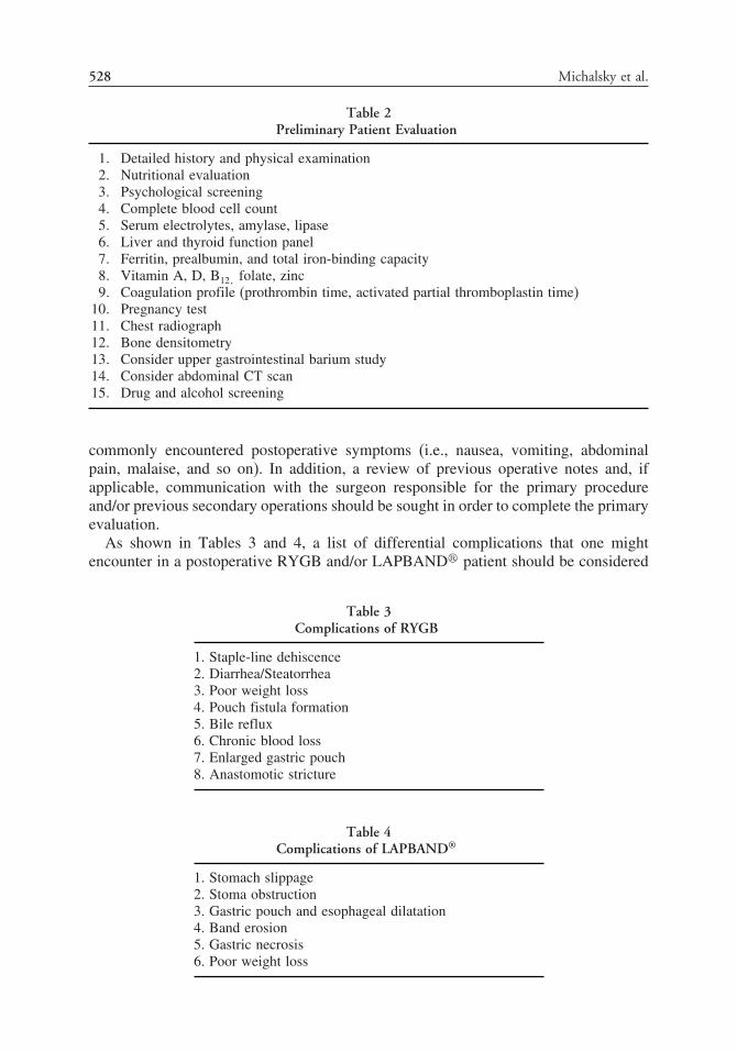
528 Michalsky et al.
Table 2Preliminary Patient Evaluation
1� Detailed history and physical examination2� Nutritional evaluation3� Psychological screening4� Complete blood cell count5� Serum electrolytes, amylase, lipase6� Liver and thyroid function panel7� Ferritin, prealbumin, and total iron-binding capacity8� Vitamin A, D, B12� folate, zinc9� Coagulation profile (prothrombin time, activated partial thromboplastin time)
10� Pregnancy test11� Chest radiograph12� Bone densitometry13� Consider upper gastrointestinal barium study14� Consider abdominal CT scan15� Drug and alcohol screening
commonly encountered postoperative symptoms (i.e., nausea, vomiting, abdominalpain, malaise, and so on). In addition, a review of previous operative notes and, ifapplicable, communication with the surgeon responsible for the primary procedureand/or previous secondary operations should be sought in order to complete the primaryevaluation.
As shown in Tables 3 and 4, a list of differential complications that one mightencounter in a postoperative RYGB and/or LAPBAND� patient should be considered
Table 3Complications of RYGB
1. Staple-line dehiscence2. Diarrhea/Steatorrhea3. Poor weight loss4. Pouch fistula formation5. Bile reflux6. Chronic blood loss7. Enlarged gastric pouch8. Anastomotic stricture
Table 4Complications of LAPBAND®
1. Stomach slippage2. Stoma obstruction3. Gastric pouch and esophageal dilatation4. Band erosion5. Gastric necrosis6. Poor weight loss

Chapter 34 / Revisional Bariatric Surgery in Adolescents 529
early in the course of a primary evaluation in order to direct diagnostic and therapeuticefforts in a timely and efficient manner.
DEFINITIONS OF SUCCESS
Before embarking on further discussion about revisional bariatric surgery, one mustdefine success and failure in terms of both the immediate postoperative period aswell as the long-term postoperative time frame. Failure in the immediate postoperativeperiod is more easily addressed and for the purposes of this discussion, will focus onanastomotic complications (i.e., leakage and/or stenosis) as well as internal herniation(which may be encountered both during the immediate and nonimmediate postoperativeperiods).
The definition of long-term success is a more complex issue and must take intoconsideration not only the overall efficacy of the primary operation (typically measuredby the degree of EBW loss) but the relative amelioration of various obesity-relatedcomorbid diseases. Even more complex, and beyond the scope of this chapter, is thedetermination of success as a measure of “quality of life” indicators. As an exampleof the individual considerations that need to be applied to definitions of success,patients who fail to obtain optimal weight loss within 2 years of undergoing RYGB(i.e., 50–70% EBW) may not require surgical revision if there has been a signif-icant improvement in obesity-related comorbid conditions and overall improvement inquality of life. As alluded to earlier, a team-oriented approach with input from varioushealthcare disciplines is highly recommended in order to accurately define success anddetermine the need for reoperation.
REVISION OF RYGB
The RYGB (Fig. 1) is an extremely effective means of establishing durable weightloss and reversing associated comorbid conditions in the morbidly obese population.Frequently referred to as the “gold standard” among numerous surgical weight-reduction surgeries, this procedure is the most commonly performed bariatric procedurein the United States as well as the primary procedure of choice among the variousbariatric procedures currently available. In addition, it has also been cited as thepreferred secondary procedure for patients requiring revisional surgery followingseveral well-described alternative primary operations. Despite the general safety andefficacy and of this procedure, several well-recognized complication of the RYGBhave resulted in a predictable need for revisional surgery (Table 3).
Immediate Postoperative Period (Anastomotic Complications)Although patients undergoing any form of bariatric surgery are subject to a host of
potential complications during the immediate and late postoperative period (i.e., deepvenous thrombosis, wound infection, pulmonary embolism, and so on) information inthis section will focus on more bariatric-specific issues that may result in the need for asecondary intervention (including reoperation) in the immediate postoperative period.
Anastomotic leak
Leakage from the various enteric anastomoses in the immediate postoperative periodmay pose a significant diagnostic challenge and should be considered in patients even

530 Michalsky et al.
Fig. 1. Roux-en-Y gastric Bypass.
in the face of a technically unremarkable operation (open or laparoscopic), a negativeintraoperative leak test, and/or a negative postoperative contrast study. Leakage mayoccur at the either the gastrojejunostomy or the jeunojejunostomy. Although patientswith early anastomotic leakage may present with gross peritoneal signs with or withoutassociated hemodynamic embarrassment, more subtle clinical findings may be the onlyclue that a complication has occurred. Such patients may be noted to have persistenttachycardia with or without fever, leukocytosis, poor oxygen saturation, left shoulderpain, hiccups, and a feeling of restlessness or agitation. Any combination of subtlefindings warrants the serious consideration of abdominal reexploration within the earlypostoperative period in order to rule out an anastomotic complication.
Anastomotic narrowing
In addition to leakage, narrowing of the anastomosis secondary to technical error orpostoperative inflammation may result in intestinal obstruction at the level of the gastro-jejunostomy or jejunojejunostomy. This complication may occur in the early postop-erative time period and result in signs and symptoms of acute bowel obstruction. Aswith patients experiencing postoperative anastomotic leakage, patients with intestinalobstruction often require the ability to distinguish associated symptoms (i.e., nausea,vomiting, and abdominal pain) from common complaints often encountered duringthe normal postoperative recovery period. Again, interpretation of subtle findings (i.e.,persistence of abdominal discomfort for a longer than usual time period) should be

Chapter 34 / Revisional Bariatric Surgery in Adolescents 531
interpreted as having the potential to represent a patient with an intestinal obstructionand, as stated earlier, should prompt early surgical reexploration.
Internal hernia
In addition to the anastomotic complications discussed previously, the creation ofseveral potential sites for intestinal herniation can also serve as a cause for problems inboth the immediate and late postoperative periods (15–17). Intestinal obstruction mayresult from herniation of small bowel through three commonly recognized sites: (1) themesocolic defect through which the Roux limb passes in cases of a retrocolic approach;(2) the “Petersen” defect between the posterior portion of the transverse mesocolonand the Roux limb; and (3) the mesenteric defect created at the jejunojejunostomy site(Fig. 2).
The initial goal of management of postoperative small bowel obstruction shouldbe patient resuscitation (fluid replacement and correction of metabolic derangement)and include intestinal decompression with an endoscopically placed nasogastric tube(NGT). The use of flexile endoscopy rather than blind placement of an NGT allows forinspection of the gastric pouch and enteric limb while avoiding the risk of inadvertentperforation of the gastric pouch. In addition to providing proximal decompression, theuse of an NGT will allow for further assessment using contrast radiography. The useof such conservative measures should be carefully weighed against a low threshold forreturn to the operating room in the face of no improvement within 24 hours or signs ofdeterioration and/or relative operative indications, including increasing leukocytosis,fever, or persistent abdominal tenderness. Once the decision to proceed to surgeryhas been made, one should decide whether to use a minimally invasive techniques
Fig. 2. Potential mesenteric defects involved in postoperative small bowel obstruction. (1) Transversemesocolon window; (2) Petersen’s space; (3) enteroenterostomy mesenteric defect.
532 Michalsky et al.
(i.e., laparoscopic) or an “open” approach based on the comfort level and experienceof the individual surgeon, because deciphering the abnormal anatomy in an effort tocorrect the herniation can be quite challenging. Once the defect is identified, carefulreduction of the herniated bowel and closure of the defect should be carried out inorder to prevent recurrent herniation in the future.
Late Postoperative PeriodIn contrast to the complications associated with the early postoperative period
outlined previously, the remainder of this chapter is devoted to a discussion of a numberof well-recognized anatomic and functional derangements that may be encounteredmonths or years after a seemingly successful primary operation.
Staple-line dehiscence
Most often encountered in the setting of a nondisconnected RYGB, dehiscence ofthe staple line results in direct communication between the gastric pouch and theremaining gastric remnant. As the restrictive component of the original operation issubsequently compromised, the patient often experiences weight gain, as well as thepossibility of bile reflux and ulceration within the Roux limb and/or anastomotic sites.Because of the 5–10% incidence of staple-line dehiscence encountered when using thistechnique, most bariatric surgeons advocate complete gastric division when fashioningthe restrictive pouch (Fig. 1). In the event of a patient that presents with staple-linedehiscence subsequent to a nondisconnected RYGB, complete division of the full stapleline with repair of the dehiscence is the revisonal procedure of choice. In addition,one should carefully assess the size of the proximal gastric pouch and be prepared torevise it with linear staplers in order to insure that it has total volume no greater than30 mL.
Diarrhea/Steatorrhea
When presented with a patient experiencing significant diarrhea and/or steatorrhea,one must take a very systematic approach and address both dietary concerns andpotential nutritional deficiencies. Before definitive surgical therapy can be undertaken,it is advisable to assess the degree of protein, calorie, and fat deficiencies as wellas potentially dangerous electrolyte derangements. Once the patient is nutritionallyand hemodynamically resuscitated, a systematic assessment of potential causes can beexplored. As alluded to previously, a comprehensive review of dietary input shouldbe undertaken by a registered dietician in order to rule out the possibility of diet-related dumping syndrome. This is recommended prior to undertaking potentially time-consuming diagnostic endeavors and possibly subjecting the patient to an unnecessaryreoperation in the face of an easily correctable problem. If dietary influences can beruled out as a potential cause, one should consider the overall length of the Roux limbas a potential cause for this complication. Recent evidence has advocated for the useof progressively longer limb lengths (150–200 cm) in association with increasing BMI(50 kg/M2) (18,19). The resultant shortening of the common channel, as a consequenceof the creation of a long Roux limb, is the most common anatomic factor responsible forpersistent nondiet-related diarrhea and/or steatorrhea. Once nutritional and metabolicderangements have been fully addressed, the patient should undergo a lengthening ofthe common channel in an effort to improve the absorptive capacity of the small bowel

Chapter 34 / Revisional Bariatric Surgery in Adolescents 533
while maintaining acceptable weight loss. In addition to lengthening of the commonchannel, one should consider insertion of a feeding tube into either the gastric remnantor proximal small bowel in an effort to help optimize the patient’s nutritional statusduring the recovery phase.
Poor weight loss
As with the scenario described previously, patients who present to a bariatric surgeonwith poor weight loss or weight gain months or years after under going RYGB requireimmediate nutritional assessment, because many of these patients can attribute poorweight reduction or weight regain to suboptimal dietary habits. A strong indication thatdietary habits are potentially playing a role in this scenario is inferred when encoun-tering a patient that demonstrated adequate weight loss during the early postoperativeperiod (i.e., within the fist 6–24 months). In addition to nutritional assessment, oneshould consider behavioral counseling with the bariatric team psychologist in order toaddress poor eating habits. Recommendations regarding reoperation for poor weightloss, in the face of an anatomically intact RYGB, are controversial at best owing tothe paucity of meaningful clinical data. Anecdotal reports of improved weight loss asa result of limb lengthening have been reported however.
Gastrogastric fistula formation
Formation of a fistula from the gastric pouch to the gastric remnant can presentas a formidable problem that is typically seen as a result of an anastamotic leak.Unlike several of the complications described earlier, a gastrogastric fistula cannot becontrolled through nonoperative means. Definitive surgical therapy in this situationmay require ligation of the fistula with limited or total proximal gastrectomy.
Bile reflux
In the case of RYGB bile reflux is typically encountered as a result of a Roux limbthat is too short. Most bariatric surgeons advocate making the Roux limb at least 85–100 cm in length in order to avoid this complication. If it is determined that a Roux limbis too short, reoperation should consist of revision of the enteroenterostomy in order tocreate a final limb length of at least 150 cm. As stated previously, other causes of bilereflux may be seen in the case of gastrogastric fistula formation as well as staple-linedehiscence in the case of a nondisconnected RYGB. Reoperative surgical strategiesdesigned to address these particular anatomic complications (described previously)should resolve exposure to bile.
Chronic blood loss
Chronic blood loss may be encountered in a small subset of patients who haveundergone RYGB. Associated causes include antral gastritis and duodenal ulceration.Although the exact pathophysiology involved in post bypass antral gastritis is uncertain,a trial of oral antibiotics for the treatment of presumed Helicobacter pylori infectionmay be warranted. This recommendation supports the argument for routine preoperativescreening and treatment of H. pylori in morbidly obese patients undergoing bariatricsurgery. In rare instances, chronic blood loss secondary to postbypass antritis mayrequire complete gastrectomy of the gastric remnant. As in the case of postbypassantritis, the development of ulceration at the level of the duodenum is another classiccause for chronic anemia, with an associated incidence of approximately 1–2 %. Again,
534 Michalsky et al.
treatment with antibiotics for presumed H. pylori infection is recommended prior toany reoperative surgical option. In cases when antibiotic therapy fails to improve thesituation, reoperative surgical options should be predicated on the classic surgicalapproach to chronic bleeding ulceration of the duodenum recalcitrant to medical therapy(i.e., antrectomy). In cases of an acutely bleeding duodenal ulceration one shouldconsider radiographic embolization or direct ligation of the associated feeding vessel.
Enlarged gastric pouch
Although the causes of pouch enlargement remain unclear, several authors havesuggested that this phenomenon may be more likely caused by the creation of a pouchthat is too large at the culmination of the original operation (i.e., greater than 30 mLcapacity) rather that an actual enlargement of the pouch subsequent to the procedure.A larger than normal pouch has been attributed to suboptimal weight loss as wellweight gain during the postoperative period. As discussed in the section on poor weightloss, reoperative surgery in the face of an anatomically intact RYGB is controversial.Surgical options consist of decreasing the size of the pouch after taking down thegastrojejunostomy and excluding any gastric fundus before reestablishing intestinalcontinuity. In addition, one should consider lengthening the size of the Roux limb byat least 50 cm in an effort to increase the degree of intestinal malabsorption capacity.A third but more controversial consideration consists of recent reports of the applicationof a laparoscopic adjustable band to the gastric pouch in the case of suboptimal weightloss and/or weight gain in patients with RYGB (20).
Anastomotic stricture
Anastomotic stricture following RYGB has been attributed to localized ischemiaat the level of the gastrojejunostomy owing to either tension on the anastamosisor ischemia at the terminal end of the Roux limb. In addition, stricture formationhas been seen in association with overuse or abuse of nonsteroidal antiinflammatoryagents (NSAIDs). The clinical consequences may be mild or may result in acuteobstructive symptoms that may dictate the appropriate level of surgical intervention.The first-line treatment for patients with significant stricture formation is endoscopicballoon dilatation. This procedure should only be performed by a bariatric surgeon orendoscopist familiar with this complication in order to avoid the mistake of overdilatingthe anastomosis. The use of this technique is often very successful in the immediatepostoperative period (i.e., first several months); however, it becomes significantly morechallenging later on as a result of significant ulcer formation and associated fibroticchanges. Failure of endoscopic balloon dilatation in the setting of severe fibrosis maydictate the need to perform a formal revision, if the gastrojejunostomy with resectionof surrounding fibrotic tissue. Fibrotic reaction involving the proximal pouch may beso severe that an esophagojejunostomy must be performed.
REVISION OF LAPBAND
As mentioned to earlier, placement of the LAPBAND� system is the most widelyperformed bariatric surgical procedure in Europe and Australia, and has been gainingpopularity within the United States since its approval by the FDA in 2001. Severalperceived advantages, including the lower overall complication rate, purely restrictivedesign (Fig. 3) and potential for reversibility when compared to a RYGB, make this optionincreasingly attractive to both the morbidly obese adult and adolescent populations.

Chapter 34 / Revisional Bariatric Surgery in Adolescents 535
Fig. 3. Laparoscopic-adjustable gastric band.
Immediate and Late Postoperative PeriodIn addition to acute complications generally associated with any abdominal surgery,
specific complications related to the use of the LAPBAND system are seen in boththe early and late postoperative periods, and include dilatation and perforation of thestomach and/or esophagus, necrosis of the proximal gastric pouch, stomal obstruction,band erosion, and malfunction and/or disruption of the silastic tubing and subcutaneousport system (Table 4).
Gastric PerforationWith an incidence ranging from 0.2–3.5%, perforation of the stomach after
LAPBAND� insertion is typically encountered in the immediate postoperative periodand has been associated with technical errors (i.e., perforation caused during theretrogastric dissection) seen during the learning-curve period (21–26). Symptoms aretypically the result of associated peritonitis and may range from vague abdominaltenderness to gross peritoneal signs. In the event that a gastric perforation is encoun-tered in the postoperative period, one should completely remove the band beforeperforming a formal repair. Reapplication of the LAPBAND� system in this settingshould be considered on an individual basis, but should be discouraged in the case oflarge peritoneal contamination and/or associated gastric inflammation.
Stomach SlippageWidely recognized as the most commonly encountered complication of the
LAPBAND� insertion, and typically seen in the late postoperative phase, slippageof the stomach (anterior or posterior) through the band is sometimes mistaken aspouch dilatation but rather, is deemed to be the result of an oversized gastric pouchcreated during the primary procedure. Although initial reports cited an incidence ashigh as 10%, modification of the surgical dissection technique from the previouslyendorsed “perigastric” technique to the more recently recommended “pars flaccida”

536 Michalsky et al.
(Fig. 3) dissection (encompassing minimal tissue dissection and a diminished incidenceof posterior slippage) has led to a recently improved incidence of 2–5% (26,27).Consideration that gastric slippage (both anterior or posterior) has occurred shouldtake place when one encounters typical associated symptoms. Specifically, patientsmay complain of one or several symptoms similar to those encountered with classicalgastroesophageal reflux disease (i.e., wheezing, dyspepsia, regurgitation, coughing orchoking spells, difficulty swallowing solid food, and so on).
Diagnosis of either posterior (the most common) or anterior slippage of the stomachis best confirmed by obtaining an upper gastrointestinal series. When performed afterthe band has been completely deflated, this radiographic modality can help differen-tiate dilatation of the gastric pouch (which may improve with simple band deflation)from true gastric slippage (which is unlikely to resolve following this diagnosticmaneuver). Once it has been established that the associated symptoms are caused bytrue gastric slippage, surgical options include readjustment or complete replacement ofthe LAPBAND system. Reoperation (laparoscopic or open) in the case of both anteriorand posterior gastric slippage begins with careful intraoperative inspection in an effortto confirm the radiological diagnosis, assess the viability of the involved stomach, anddetermine the specific anatomy (i.e., anterior versus posterior slippage).
Anterior gastric slippage is the result of inadequate fixation of the anterior portionof the band (which is obtained by the placement of several strategically placed fixationsutures). Once confirmed, and assuming that complete removal of the LAPBAND�
system is not required, the surgical goal should be to gently pull the herniated portionof stomach back into an optimal position prior to replacement and/or supplementationof previously placed anterior fixation sutures to resecure the band (Fig. 4).
Rather than attempting to pull the stomach back into place, as in the reoperativetreatment of an anterior slippage, posterior herniation invariably requires completeremoval of the band with its reapplication in a more cephalad position using the parsflaccida technique (incorporation of a portion of the lesser omentum) as shown in Fig. 3.
Fig. 4. Application of gentle traction on the stomach in an attempt to reduce an anterior slip.

Chapter 34 / Revisional Bariatric Surgery in Adolescents 537
Gastric Pouch and Esophageal DilatationThe pathophysiologic definition of gastric and/or esophageal dilatation in the case
of a patient with a LAPBAND is somewhat confusing, because there appears to beoverlap with band malposition and slippage (see previous section). Whether or notdilatation of the esophagus and proximal gastric pouch are independent entities orthe consequence of suboptimal band positioning versus band-related outlet obstructionremains controversial. In addition to such etiologic uncertainty, other potential causesthat require consideration include overinflation of the band as well as consumption ofinappropriate food or noncompliance with proper mastication techniques (i.e., failure tochew food well before swallowing), resulting in a state of chronic partial obstruction ofthe esophagus or proximal gastric pouch. Assuming that the patient has been thoroughlyevaluated (including an upper gastrointestinal series, careful assessment of the degreeof band inflation, and potential patient compliance issues) and has failed to showimprovement after a trial of band deflation, one should assume that gastric slippageand/or band malposition has occurred and proceed as described earlier.
Band ErosionThe incidence of band erosion, defined as a breach of the gastric wall underlying
the band, is currently reported to be between 0–14 (22,25,28,29). This complication isthought to be associated with various etiologic factors including local tissue ischemia(possibly secondary to microperforation during the primary procedure), chronic inflam-mation, and chronic ingestion of NSAIDs. Patients do not typically exhibit signs ofovert sepsis but rather present with fatigue, weight regain and/or inadequate weightloss, failure to improve weight loss after multiple adjustments or “tightening” of theinner diameter of the device, and port site infection. Bands that have eroded must beremoved either operatively (open or laparoscopic), endoscopically, or using a combi-nation of both modalities. The decision whether to insert a new band immediately or ata later time must take into consideration based on individual circumstances, includingthe surgeon’s overall level of technical experience.
REFERENCES
1. Kimm YS, Obarzanek E. Childhood obesity: a new pandemic for the new millennium. Pediatrics2002;110:1003–1007.
2. MacDonald K. Overview of the epidemiology of obesity and the early history of procedures to remedymorbid obesity. Arch Surg 2003;138:357–360.
3. Olshansky SJ, Passaro DJ, Hershow RC, et al. Potential decline in life expectancy in the United Statesin the 21st century. Obstet Gynecol Surg 2005;60:450–452.
4. Smith SC, Goodman GN, Edwards CB. Roux-en-Y gastric bypass. A 7-year retrospective review of3,855 patients. Obesity Surg 1995;5:314–318.
5. Bacci V, Basso M, Greco F, et al. Modifications of metabolic and cardiovascular risk factors afterweight loss induced by laparoscopic gastric banding. Obesity Surg 2002;12:77–82.
6. Griffen WO, Bivins BA, Bell RM, et al. Gastric bypass for morbid obesity. World J Surg 1981;5:817–822.
7. Gastrointestinal surgery for severe obesity. NIH consensus development conference, March 25–7,1991. Nutrition 1996;12:397–404.
8. Fobi MA, Lee H, Holness R, et al. Gastric bypass operation for obesity. World J Surg 1988;22:925–935.
9. Strauss RS, Bradley LJ, Brolin RE. Gastric bypass surgery in adolescents with morbid obesity.J Pediatrics 2001;138:499–504.

538 Michalsky et al.
10. Sugerman HJ, Sugerman EL, DeMaria EL, et al. Bariatric surgery for severely obese adolescents.J Gastrointest Surg 2003;7:102–107.
11. Inge T, Kreb N, Garcia V, et al. Bariatric surgery for severely overweight adolescents: concerns andrecommendations. Pediatrics 2004;114:217–223.
12. Ren C, Horgan S, Ponce J. US Experience with the LAP-BAND System. Am J Surg 2002;1684:46S–50S.
13. Dolan K, Creighton L, Hopkins G, et al. Laparoscopic gastric banding in morbidly obese adolescents.Obesity Surg 2003;13:101–104.
14. Weller W, Hannan E. Relationship between provider volume and postoperative complications forbariatric procedures in New York State. J Am Coll Surg 2006;202:753–761.
15. Higa KD, Boone KB, Ho T. Laparoscopic Roux-en-Y gastric bypass for morbid obesity: techniquesand preliminary results of our first 400 patients. Arch Surg 2000;135:1029–1033.
16. DeMaria EJ, Sugerman HJ, Kellum JM, et al. Results of 281 consecutive total laparoscopic Roux-en-Ygastric bypasses to treat morbid obesity. Ann Surg 2002;235:640–647.
17. Schauer PR, Ikramuddin S. Laparoscopic surgery for morbid obesity. Sur Clin North Am2001;81:1145–1179.
18. Brolin, RE. Long limb Roux en Y gastric bypass revisited. Surg Clin North Am 1985;4:807–817, vii.19. Lee S, Sahagian KG, Schriver JP. Relationship between varying Roux limb lengths and weight loss
in gastric bypass. Curr Surg 2006;63:259–263.20. Gumbs A, Margolis B, Bessler M. Laparoscopic banded-Roux-en-Y gastric bypass. Surg Obes Relat
Dis 2006;2:408–409.21. Chelala E, Cadiere GB, Favretti F, et al. Conversion and complications in 185 laparoscopic adjustable
silicone gastric banding cases. Surg Endosc 1997;11:268–271.22. Favretti F, Cadiere GB, Segato G, et al. Laparoscopic adjustable silicone gastric banding (Lap Band):
how to avoid complications. Obes Surg 1997;7:310.23. Natalini G, Carolini G, Capelletti S, et al. Laparoscopic adjustable silicone gastric banding (LASGB)
for treatment of morbid obesity. Obes Surg 1997;7:310.24. Dargent J. Laparoscopic adjustable gastric banding: lessons from the first 500 patients in a single
institution. Obes Surg 1999;9:446–452.25. Biesheuvel TH, Sintenie JB, Pels Rijcken TH, et al. Laparoscopic adjustable silicone gastric banding
for treating morbid obesity in the centre of Amsterdam. Obes Surg 1998;8:360.26. Rubin M. Prospective study of 250 patients undergoing laparoscopic gastric banding using the two-
step technique. Surg Endosc 2003;17:857–860.27. Kriwanek S, Schermann M, Abdullah S, et al. Band slippage—a potentially life threatening compli-
cation after laparoscopic adjustable gastric banding. Obes Surg 2005;15:133–136.28. Belachew M, Belva PH, Desaive C. Long-term results of laparoscopic adjustable gastric banding for
the treatment of morbid obesity. Obes Surg 2002;15:564–568.29. Fielding G, Rhodes M, Nathanson LK. Laparoscopic gastric banding for morbid obesity: surgical
outcomes in 335 cases. Surg Endosc 1999;13:550–554.
Index
Abbe-McIndoe vaginoplasty, 507. See also VaginoplastyAbdominal abscess
CT for, 2–3radiology of postoperative patient, 2–9ultrasound for, 3–4
Abdominal compartment syndrome (ACS), 183–184. Seealso Abdominal wall disruption
intra-abdominal pressure (IAP), 183Abdominal complications (VPS)
laparotomy, 521, 523pseudocysts, 518–520shunt drainage aspects, 523shunt migration, 520visceral perforation, 521VPS infection, 515, 516VPS obstruction, 516–518
Abdominal neovagina, 509. See also VaginoplastyAbdominal wall disruption, 175–186. See also
Abdominal abscessabdominal compartment syndrome (ACS) and,
183–184closure technique, 177–178, 182–183computed tomography (CT) for, 183dehiscence, presentation and management of, 178–179fascial closure and, 177incisional hernias and, 184–186intraoperative
aids, 181–182prevention, 176–177
open abdomen management, 179–180reoperative techniques, 180–181retention sutures, 178secondary closure of, 182–183vacuum-assisted closure (VAC) of open wounds, 180
Ablationhepatic tumor, 54–56lung tumor, 54–56radiofrequency (RFA), 53–56
Abscessabdominal, 2–9, 244–245chest, 2–9drainage, 7musculoskeletal, 3NEC, 8postappendectomy, 247–248recurrent perirectal, 331retropharyngeal, 5splenic, 6transrectal, 7
Accessfor antegrade irrigation, 227for feeding
gastrostomy, 225–227jejunostomy, 227
venous, 75–86angioplasty, 83catheterization of collateral veins, 82CVC insertion and, 76interventional techniques, 80–84PICCs, 83–84preoperative aspects, 75–80stent placement, 83surgical techniques, 84–86transhepatic catheterization, 81–82translumbar catheterization, 80–81
Achalasiaesophageal disorders and, 16–17surgery, 198–199
Acquired urethral atresia, 320. See also Anorectalmalformations
Acquired vaginal atresia, 320Acute recurrent pancreatitis, 376–381Adjuvant therapy, 393–394. See also
Rhabdomyosarcoma (RMS)Adolescents. See also Recurrent varicocele
varicocele, 482–483Altman classification, 467–468. See also Sacrococcygeal
teratoma (SCT)Anal. See also Anorectal malformations
condition, recurrent, 327warts, 333–334
Anal fistulasclinical presentation, 262–263diagnostic evaluation, 263–264long-term issues, 265MRI for, 264nonoperative techniques, 265operative techniques, 264–265recurrent Crohn’s disease and, 262–265
Anastomosisend-to-end (EEA), 305gastrointestinal (GIA), 305NEC and primary, 232–233repeat pull-through with stapled, 305–307
Anastomotic complications (RYGB revision)anastomotic leak, 529–530anastomotic narrowing, 530internal hernia, 531–532
539

540 Index
Anastomotic leakafter tracheoesophageal fistula repair,
193–194esophageal disorders, 20–21RYGB revision, 529–530
Anastomotic strictureafter tracheoesophageal fistula repair, 194bariatric surgery and, 534
Angiogenesis, good, 232Angioplasty. See also Access
venous access and, 83Anorectal malformations, 311
colostomy for, 313–315definitive repair
acquired urethral atresia, 320acquired vaginal atresia, 320persistent urogenital sinus, 320posterior urethral diverticulum, 321rectal problems, 318–319rectourinary fistulae, 319–320rectovaginal fistulae, 319–320
laparoscopic approach to, 322loop colostomy for, 316postoperative management, 323
fecal incontinence, 323–324sequelae from constipation,
324–325preoperative evaluation and management,
311–318rectoperineal fistula, 313rectourethral fistula, 313tube vaginostomy for, 312urologic injuries
neurogenic bladder, 322rectal prolapse, 322–323
Anorectal manometry, 290. See also Hirschsprungdisease (HD)
Antegrade irrigation access. See also Distal irrigationaccess
appendicostomy, 227small bowel surgery and, 227tube cecostomy, 227tube sigmoidostomy, 227
Antibiotics for appendicitis, 242–243Appendectomy
interval, 245laparoscopic, 243open, 243
Appendicitisantibiotics for, 242–243difficult appendiceal stump management
laparoscopic appendectomy, 243open appendectomy, 243
drains and, 242intra-abdominal abscess
diagnostic evaluation, 244interval appendectomy, 245nonoperative management, 244–245operative management, 245percutaneous abscess drainage, 244–245
intraoperative cultures, 244lavage versus drainage, 242long-term complications
fertility, 251SBO, 250
morbidity, 241perforation risk, 241short-term postoperative complications, 246
pathogenic organisms, 247postappendectomy abscess, 247postoperative SBO, 248–249stump appendicitis, 249–250suppurative pylephlebitis, 248wound infection, 246
wound closure, 242Appendicostomy. See also Small bowel surgery (SBS)
antegrade irrigation access aspects of, 227Arterial
access, 75embolization, 59
Arterial thrombosis, 41–46Arteriovenous malformation, 46Atresia
acquired urethral, 320acquired vaginal, 320biliary, 343–347
Atypical mycobacterial cervical lymphadenitis, 94–96.See also Reoperative head and neck surgery
Autologous gastrointestinal reconstruction (AGIR),237–238. See also Small bowel obstruction (SBO)
Bacterioides, 247Band erosion, 537. See also LAPBAND® revisionBariatric surgery
considerations, 527LAPBAND® procedure, 526, 528LAPBAND® revision, 534
band erosion, 537esophageal dilatation, 537gastric perforation, 535gastric pouch dilatation, 537immediate and late postoperative period, 535stomach slippage, 535–536
primary evaluation, 527–528revisional, 525RYGB procedure, 526, 528RYGB revision aspects
anastomotic complications, 529–532anastomotic stricture, 534bile reflux, 533chronic blood loss, 533–534diarrhea/steatorrhea, 532–533enlarged gastric pouch, 534gastrogastric fistula formation, 533immediate postoperative period, 529–532late postoperative period, 532–534poor weight loss, 533staple-line dehiscence, 532
success in, 529

Index 541
Barrett’s esophagus, 197–198. See also Esophagealdisorders
Benign liver tumors, 459Benign SCT recurrence
long-term results, 472reoperative surgery principles, 472
Bilateral osteosarcoma metastases, 439Bilateral Wilms’ tumor. See also Unresectable Wilms’
tumorCOG guidelines, 422diagnostic evaluation, 420epidemiology and clinical presentation, 420operative strategy, 422outcome, 422–423prognostic factors, 420–421
Bile, 34–35. See also Bariatric surgeryduct injury. See Hepatobiliary diseasereflux, 533
Biliary atresiaCT for, 347early reoperation indications, 345–347late reoperation indications, 347MRCP, 347
Biliary diseaseatresia, 343–347CBD
injury, 348–356stones, 351–356
choledochal cysts, 337–343liver tumors and, 462
Biliary leaks, 34–35. See also Hepatobiliarydisease
Biopsychest wall tumors, 429–431rectal, 291Wilms’ tumor, 413
Bladder, neurogenic, 322. See also Urologic injuriesBleeding, liver tumors and, 461Blood loss, chronic, 533–534Boix-Ochoa fundoplication, 149Branchial cleft cysts, 93, 94. See also Reoperative head
and neck surgeryBreast disorders
recurrent breast cystic disease, 172recurrent solid breast masses, 169–171
CAH. See Congenital adrenal hyperplasia (CAH)Cardiac surgery, pectus excavatum and, 133Cardiovascular malformations (CVM), 451. See also
Neurofibromatosis (NF)Catheterization
collateral veins, 82IVC
transhepatic, 81–82translumbar, 80–81
Catheters, 83–84. See also Peripherally inserted centralcatheters (PICCs)
displaced venous, 41CBD exploration (CBDE), 354–356
CDH. See Congenital diaphragmatic hernia(CDH)
Cecostomy, tube, 227Central venous catheters (CVCs)
insertion, 76surgical access aspects, 86venous access and, 76
Cervical dermoid cysts, 93. See also Reoperative headand neck surgery
Cervical lymphadenitis, atypical mycobacterial, 94–96Chemotherapy. See also Tumor
chest wall tumors, 429–431Chest abscess
CT for, 3radiology of postoperative patient, 2–9
Chest wallCDH and, 153–154defects following resections, 135deformities, 153–154PNET, 427problems, congenital, 133
Chest wall tumorscomplications and late effects, 433diagnostic evaluation and initial therapy,
428–429recurrent chest wall PNET, 433–434surgical techniques for excision after biopsy and
chemotherapy, 429–431wall reconstruction techniques, 431–433
Cholecystectomy, 348Cholecystitis, 348Cholecystostomy. See also Hepatobiliary disease
percutaneous transhepatic, 35Choledochal cysts, 337
classification, 338clinical presentation, 338operative management, 340reoperation indications, 340–343
Choledocholithiasis. See also Biliary diseasecommon duct stones and, 351
Cholelithiasis, 348Chronic blood loss, 533–534. See also Bariatric surgeryChronic pancreatitis, 376–381Chylothorax, CDH and, 152Circumcision. See also Clitoroplasty; Vaginoplasty
complications, 491–496methods, 491neonatal, 491
Clatworthy Shunt, 357Clitorectomy. See also Clitoroplasty
feminizing genitoplasty and, 500Clitoromegaly, 500Clitoroplasty, 500. See also Vaginoplasty
feminizing genitoplasty procedure, 499–502postoperative issues, 500
Clitorovaginoplasty, 501Closed-loop intestinal obstruction, 233. See also
ObstructionClostridia, 247, 354. See also InfectionClostridium welchii, 247

542 Index
Closureappendicitis wound, 242enterostomy, 229fascial, 177secondary, 182–183technique, 177–178vacuum-assisted closure (VAC), 180
Clover leaf deformity, 158Colitis
indeterminate, 273ulcerative, 266–267
Collateral veins catheterization, 82. See also IVCcatheterization
Colonic transit time, 291. See also Hirschsprungdisease (HD)
Colostomyfor anorectal malformations, 313–315loop, 316
Common bile duct (CBD) injuryCBDE (CBD exploration), 354–356cholecystectomy for, 348clinical classification, 349clinical presentation, 348–349common duct stones
CBD exploration (CBDE), 354–356clinical presentation, 351diagnostic evaluation, 352–353ERCP, 353–355HCT-C, 352helical CT scan, 352long-term issues, 355–356management, 353–354MRC, 352
ERCP for, 349, 353–355long-term issues, 351operative management, 349–350
Component separation (CS) technique, 182. See alsoAbdominal wall disruption
Computed tomography (CT). See also Magneticresonance imaging (MRI)
abdominal, 183abdominal abscesses, 2–3biliary atresia, 346chest abscesses, 3contrast, 80failed orchiopexy, 488high-resolution, 158Jeune’s syndrome, 158–160ulcerative colitis, 267
Congenital adrenal hyperplasia (CAH), 501–504feminizing genitoplasty concept, 501–504vaginoplasty and, 503–504
Congenital chest wall problems, 133. See also Pectusexcavatum
Congenital diaphragmatic hernia (CDH), 139chest wall deformities, 153–154chylothorax and, 152ECMO recannulation and, 151–152gastrostomy tube and, 150–151GER and, 146–150
GERD management in, 148–149laparoscopic approach for, 151PEG for, 150–151recurrent, 139reherniation aspects, 140–146repair techniques, 139, 144–146small bowel obstruction and, 152spinal deformities and, 153–154
Congenital duodenal obstruction, 220–222Constipation. See also Anorectal malformations
sequelae from, 324–325Contrast CT. See also Computed tomography (CT)
vascular access and, 80Contrast enema, 290. See also Hirschsprung
disease (HD)Contrast venography, conventional, 80. See also
Vascular accessCordopexy, inguinal, 490Crohn’s disease. See also Inflammatory bowel
disease (IBD)anal fistulas
clinical presentation, 262–263diagnostic evaluation, 263–264long-term issues, 265nonoperative techniques, 265operative techniques, 264–265
stricturesclinical presentation, 257–258diagnostic evaluation, 258–259long-term issues, 262nonoperative techniques, 261–262operative techniques, 259–261
Cryptorchidism, 487. See also Failed orchiopexyCutaneous neurofibromas, 448. See also Gastrointestinal
neurofibromatosis (NF); Thoracic neurofibromatosis(NF); Vascular neurofibromatosis (NF)
discrete, 448plexiform, 448–449segmental, 448
Cystic disease, recurrent breast, 172
Decompressing enterostomies, 228. See also Smallbowel surgery (SBS)
Deep venous thrombosis (DVT). See also Pulmonaryembolus (PE)
radiology of postoperative patient, 35–37Definitive repair
acquired urethral atresia, 320acquired vaginal atresia, 320persistent urogenital sinus, 320posterior urethral diverticulum, 321rectal problems, 318–319rectourinary fistulae, 319–320rectovaginal fistulae, 319–320
Dehiscence, 178–179. See also Abdominal walldisruption
Dermoid cysts, cervical, 93Diaphragmatic hernia (CDH), congenital
chest wall deformities, 153–154chylothorax and, 152

Index 543
ECMO recannulation and, 151–152gastrostomy tube and, 150–151GER and, 146–150laparoscopic approach for, 151recurrent, 139reherniation aspects, 140–146repair techniques, 139, 144–146small bowel obstruction and, 152spinal deformities and, 153–154
Diarrhea, 532–533. See also Bariatric surgeryDilatation
esophageal, 198Hirschsprung disease (HD) surgery and,
292–293LAPBAND® revision
esophageal dilatation, 537gastric pouch dilatation, 537
Direct hernia, 476Direct reconstruction portosystemic shunts, 358Discrete neurofibromas, 448. See also Neurofibromatosis
(NF)Displaced venous catheter. See also Catheterization
radiology of postoperative patient and, 41Dissociation, esophagogastric, 211. See also Failed
fundoplicationDistal irrigation access. See also Antegrade irrigation
accesssmall bowel surgery and, 228
Distal splenorenal shunt (DSRS), 357Diverticulum, posterior urethral, 321Drainage. See also Intra-abdominal abscess;
Ventricular-peritoneal shunts (VPS)percutaneous abscess, 244–245shunt, 523versus laparotomy, NEC and, 231
Drains, appendicitis and, 242Drooling, 96–97. See also Reoperative head and neck
surgeryDuhamel procedure. See also ERPT (endorectal
pull-through)Hirschsprung disease (HD) and, 300, 302
Duodenalatresia, 219fistulas, 219
Duodenal obstruction, 219. See also Small bowelobstruction (SBO)
incidence, 220repair
early complications after, 220–221late complications after, 221–222very late complications after, 222
ECMO recannulation. See also Congenital diaphragmatichernia (CDH)
complications, 151–152indications, 151technique, 151
Effusion, pleural, 38
Embolization. See also Deep venous thrombosis (DVT);Pulmonary embolus (PE); Pulmonarythromboembolism (PTE)
arterial, 59reoperative tumor, 56–57splenic, 59tumor, 53, 56–57
Empyema, radiology of postoperative patient and,38–41
Endoscopic retrograde cholangiopancreatography(ERCP)
CBD injury and, 349CBD stones and, 353–354
Endothelioma, bone, 427End-to-end anastomosis (EEA), 305Enema, radiographic contrast, 290Enterobacter, 354Enteropathy. See also Small bowel obstruction (SBO)
ischemic, 234Enterostomies
decompressing, 228Mikulicz, Bishop–Koop, and Santulli, 228
Enterostomyclosure, 229complications, 229, 230proximal diverting, 232
ERCP. See Endoscopic retrogradecholangiopancreatography (ERCP)
ERPT (endorectal pull-through), 302, 304. See alsoDuhamel procedure
Hirschsprung disease (HD) and, 299–300Escherichia coli, 247, 354. See also InfectionEsophageal atresia, 192Esophageal dilatation
LAPBAND® revision, 537perforation after, 198
Esophageal disorders. See also Stomach disordersachalasia, 16–17anastomotic leak, 20–21esophageal perforation, 17esophageal strictures, 19–20foreign body, 14, 15fundoplication complications, 17–18postoperative patient radiology aspects, 14–21recurrent fistula, 20–21
Esophageal replacement surgery, 199–200Esophageal stricture, recurrent reflux-induced, 196–197Esophageal surgery complications, 192Esophageal varices, 200, 201Esophagogastric dissociation, 211. See also Failed
fundoplicationEsophagus
anatomy, 191–192specific conditions requiring surgery of, 192–193
achalasia surgery, 198–199anastomotic leaks after tracheoesophageal fistula
repair, 193–194anastomotic stricture after tracheoesophageal fistula
repair, 194Barrett’s esophagus, 197–198

544 Index
esophageal replacement surgery, 199–200esophageal varices, 200–201gastroesophageal reflux after tracheoesophageal
fistula repair, 194, 196perforation after esophageal dilatation, 198recurrent after tracheoesophageal fistula, 193recurrent reflux-induced esophageal stricture,
196–197tracheoesophageal fistula, 192
Ewing sarcoma, 427Expansion outgrowth, 165. See also Jeune’s syndrome
Failed fundoplicationincidence and predisposing factors for, 208nonoperative approaches to, 208–209preoperative assessment, 209presentation, 207–208surgical approaches
esophagogastric dissociation, 211repeat fundoplication, 210–211surgical jejunostomy, 211
Failed orchiopexyCT for, 488diagnosis, 488factors predisposing to, 487–488MRI for, 488preoperative counseling, 489techniques for redo orchiopexy,
489–490Fascial closure. See also Closure
abdominal wall disruption and, 177Fecal incontinence, 323–324. See also Anorectal
malformationsFeeding access devices
gastrostomy and, 225–227jejunostomy and, 227small bowel surgery and, 225–227
Feminizing genitoplasty concept, 499–504CAH, 501–504clitorectomy, 500clitoroplasty and, 501TUM, 500–503vaginoplasty and, 500–501
Femoral hernias, 476Fertility complications, 251. See also AppendicitisFibrocystic breast. See also Breast disorders
proliferative, 172Fistula
anal, 262–265duodenal, 219gastrocutaneous, 213gastrogastric, 533pancreatic, 381rectoperineal, 313rectourethral, 313rectourinary, 319–320rectovaginal, 319–320recurrent
esophageal, 20–21tracheoesophageal, 193
small bowel surgery, 230tracheoesophageal, 192–194, 196
Fistula-in-ano, 331Fistulography, 264Foreign bodies, esophageal, 14–15. See also Esophageal
disordersFowler-Stephens procedure, 490. See also Failed
orchiopexyFractures. See also Jeune’s syndrome
strut, 164–165Fundoplication
Boix-Ochoa, 149complications, 17–18esophageal disorders and, 17–18failed, 207–211Nissen, 149repeat, 210–211Thal, 149Toupet, 149
Gastric empting. See also Stomach disordersgastroparesis and, 21, 22
Gastric operationsfailed fundoplication and, 207–212gastrostomy, 211–213pyloromyotomy, 214–215
Gastric perforation, LAPBAND® revisionand, 535
Gastric pouch. See also Bariatric surgerydilatation, 537enlarged, 534
Gastrocutaneous fistula, 213. See also Gastrostomy tubeGastroesophageal reflux (GER)
after tracheoesophageal fistula repair,194, 196
diagnosis, 147–148etiology, 146–147management in CDH, 148–149repair technique, 149–150surgical approach, 149
Gastrogastric fistula formation, 533. See also Bariatricsurgery
Gastrointestinal anastomosis (GIA), 305Gastrointestinal neurofibromatosis (NF), 449Gastrojejunostomy (G-J), 209. See also FundoplicationGastroparesis
gastric empting and, 21stomach disorders and, 21–23
Gastrostomy. See also Small bowel surgery (SBS)feeding access aspects of, 225–227
Gastrostomy tube, 211–213. See also Failedfundoplication
dislodgement, 25, 27, 213gastrocutaneous fistula and, 213indications, 150infection, 213leakage around, 212migration of, 212surgical approach, 150–151

Index 545
Genitoplasty concept. See also Clitoroplasty;Vaginoplasty
feminizing, 499GERD management in CDH, 148–149Germ cell tumors (GCT), 467Good-angiogenesis, 232Granulomata. See also Jeune’s syndrome
tracheal, 165–166
HD-associated enterocolitis (HAEC), 289. See alsoHirschsprung disease (HD)
Head surgery. See Reoperative head and neck surgeryHelical computed tomography cholangiography
(HCT-C), 352. See also Computedtomography (CT)
biliary disease and, 352Hemorrhoids, persistent, 331–333Hepatic tumor
ablation, 54–56RFA, 53–55
Hepatobiliary disease, 337. See also Biliary diseasebiliary leaks, 34–35diagnosis and intervention, 27–28obstructive jaundice, 29–33percutaneous transhepatic cholecystostomy, 35radiology of postoperative patient, 27–35
Herniacongenital diaphragmatic (CDH), 139–154incisional, 184–186parastomal, 282, 284–285recurrent inguinal
affecting factors, 475–476clinical presentation and diagnosis,
476–477pediatric inguinal hernia repair, 475surgical correction of, 478, 480
retained, 477RYGB revision and internal, 531–532
High-resolution computed tomography (HRCT), 158.See also Jeune’s syndrome
High-risk neuroblastoma disease, 400Hirschsprung disease (HD)
long-term complications, 287HAEC, 289persistent stooling problems, 288soiling/incontinence, 288
myectomy, 293–298permanent stoma, 307reoperative surgery, 292–293repeated dilatation, 292–293repeat pull-through procedures, 298
Duhamel procedure, 300, 302ERPT, 299–300, 304indications, 299results, 303techniques, 299–300, 302with stapled anastmosis, 305–307
work-up for complicationsanorectal manometry, 290colonic transit time, 291
contrast enema, 290history-taking and and clinical examination,
289–290rectal biopsy, 291
Hydrocele, recurrentafter varicocele repair, 481hydrocelectomy and, 481postoperative hydroceles management, 482
Hydrocolpos, 312. See also Anorectalmalformations
Hypertension, portal, 356–362Hypoglycemia after pancreatectomy, persistent,
368–371
Ileus, 10–14Image-guided drainage procedures, 8Incisional hernias, 184–186. See also Abdominal wall
disruptionIncontinence
fecal, 323–324Hirschsprung disease (HD), 288
Indeterminate colitis, 273. See also Ulcerative colitisoperative consideration and management, 273
Infectionappendicitis complications
diagnostic evaluation and treatment, 246incidence, 246
circumcision, 491gastrostomy tube, 213ventricular-peritoneal shunts (VPS), 515–516
Inflammatory bowel disease (IBD). See also Esophagealdisorders; Stomach disorders
indeterminate colitis, 273recurrent Crohn’s disease
anal fistulas, 262–266strictures, 257–262
ulcerative colitisfailed procedure, 266–271pouchitis, 271–272
Inguinal cordopexy, 490Inguinal hernia
postoperative hydrocele following, 481recurrent
affecting factors, 475–476clinical presentation and diagnosis, 476–477direct hernia, 476femoral hernia, 476repair, 475surgical correction of, 478, 480swelling after repair, 476technical failures, 477true, 477
Intermediate-risk neuroblastoma disease, 400Interval appendectomy, 245Interventional techniques for venous access
angioplasty, 83catheterization of collateral veins, 82IVC catheterization
transhepatic, 81–82translumbar, 80–81

546 Index
PICCs, 83–84stent placement, 83
Intestinal fistulaMRI for, 9postoperative patient radiology aspects, 9–10
Intestinal injuries. See also Small bowel surgery (SBS)NEC, 231–233perforations/leaks/fistulae, 230
Intestinal intraluminal nutrient transit. See also Smallbowel obstruction (SBO)
slowing, 236–237Intra-abdominal abscess, 2
diagnostic evaluationfollowing appendectomy, 244initial presentation, 244
interval appendectomy, 245nonoperative management, 244–245operative management, 245percutaneous abscess drainage, 244–245
Intra-abdominal pressure (IAP). See also Abdominalwall disruption
ACS and, 183Intraspinal extension, neuroblastoma, 405–406Intussusception, 234. See also Small bowel
obstruction (SBO)stoma complications, 282
Irrigation. See also Small bowel surgery (SBS)antegrade, 227distal, 228
Ischemia, 279–280Ischemic enteropathy, 234IVC catheterization
surgical access aspects, 84–85transhepatic, 81–82translumbar, 80–81
Jaundice. See also Hepatobiliary diseaseobstructive, 29–33
Jejunostomyfeeding access aspects of, 227surgical, 211
Jeune’s syndromeclinical presentation, 157–158computed tomography (CT), 158–160diagnostic evaluation, 158–160expansion outgrowth and, 165HRCT for, 158indications for operation, 160–161lateral thoracic expansion (LTE) approach, 161–166long-term issues, 165–166operative technique, 161–164postoperative management, 163reoperative or long-term surgical management,
164–165strut fractures and, 164–165synostosis creation and, 165tracheal granulomata and, 165–166
Jones technique, 490. See also Failed orchiopexy
Klebsiella species, 354
Laparoscopic adjustable gastric banding. SeeLAPBAND® revision
Laparoscopyanorectal malformations, 322CDH, 151laparoscopic appendectomy, 243
Laparotomyversus drainage, NEC and, 231VPS patients and, 521, 523
LAPBAND® revision, 534band erosion, 537esophageal dilatation, 537gastric perforation, 535gastric pouch dilatation, 537immediate and late postoperative period, 535stomach slippage, 535–536
Lateral pancreaticojejunostomy (LPJ), 377Lateral thoracic expansion (LTE), 161–164, 166.
See also Jeune’s syndromeLavage
appendicitis and, 242versus drainage, 242
Leakage around gastrostomy tube, 212Leaks, small bowel surgery, 230Liver
failure, 462–463system reoperation aspects, 337
Liver tumorsbenign, 459malignant, 459–460operative procedures for tumor relapse
local tumor persistence or relapse,463–465
metastatic tumor, 465PET for, 464reoperation for surgical complications
biliary injury, 462bleeding, 461liver failure, 462–463vascular injury, 461–462
Localized tumors, 400–402. See also NeuroblastomaLocoregional disease in patients with metastatic disease
delayed surgical intervention of locoregional disease,403–404
initial procedures, 402–403operative principles, 404–405
Loop colostomy, 316. See also Anorectalmalformations
Low-risk neuroblastoma disease, 399Lung
RFA, 56tumor ablation, 54–56
Lymphadenitisatypical mycobacterial cervical, 94–96tuberculous, 94–95
Lymphatic malformations (LM), 46–54. See also Venousmalformations (VM)

Index 547
Magnetic resonance cholangiography (MRC), 352Magnetic resonance imaging (MRI)
anal fistulae, 264for failed orchiopexy, 488fistulography, 264intestinal fistula, 9recurrent solid breast masses, 170ulcerative colitis, 267venography, 78–79
Malignant liver tumors, 459, 460Malignant peripheral nerve sheath tumors (MPNSTs),
451–454. See also PheochromocytomasMalignant SCT recurrence
long term results, 473reoperative surgery principles, 472
Malrotation with volvulus, 234. See also Small bowelobstruction (SBO)
Manometry, anorectal, 290Mayer-Rokitansky-Kuster-Hauser (MRKH) Syndrome,
504, 506–507. See also VaginoplastyMcIndoe vaginoplasty, 510Meatitis, 495. See also CircumcisionMesenterico-portal shunt, 358. See also REX shuntMesocaval shunt, 357Metastases, osteosarcoma, 435
bilateral, 439unilateral, 438–439
Metastatic disease, locoregional disease in patients with,402–405
Metastatic liver tumor relapse, 465Metastatic neuroblastoma
delayed surgical intervention of locoregional disease,403–404
initial procedures, 402–403operative principles, 404–405
Metastatic RMS, 391clinical presentation and risk factors, 392diagnostic evaluation, 392operative therapy, 392–393outcomes, 393
Metastatic Wilms’ tumor, 423Migration, shunt, 520. See also Ventricular-peritoneal
shunts (VPS)Mikulicz, Bishop–Koop, and Santulli enterostomies, 228Morbidity, appendicitis, 241MRCP, biliary atresia, 347MRI. See Magnetic resonance imaging (MRI)Mucosal surface area increase, 237–238. See also Small
bowel obstruction (SBO)Musculoskeletal abscesses
MRI for, 3ultrasound for, 3
MYCN protooncogene, 399. See also NeuroblastomaMycobacterial cervical lymphadenitis, atypical, 94–96Mycobacterium avium, 94Mycobacterium intracellulare, 94Mycobacterium tuberculosis, 94Myectomy. See also Hirschsprung disease (HD)
indications, 293–294posterior saggital approach, 296
results, 297–298technique
posterior saggital approach, 295–296transanal approach, 294
NECabscesses, 8small bowel surgery (SBS) and, 231
drainage versus laparotomy, 231patch, drain, and wait, 231–232proximal diverting enterostomy, 232resection and primary anastomosis, 232, 233
Neck surgery, reoperative. See Reoperative head andneck surgery
Necrosis, 279–280Necrotizing enterocolitis (NEC), 2Neocolpopoiesis, 510Neocolporrhaphy, sigmoid, 507Neonatal
circumcisions, 491neuroblastoma, 407
Neovagina. See also Vaginoplastycarcinoma, 509prolapse, 510
Nephroblastoma. See Wilms’ tumorNeuroblastoma, 397
metastatic, 402–405neonatal, 407paraspinal, 405–406prognostic variables
age, 398–399histopathology, 399MYCN protooncogene amplification, 399stage, 398
reoperative surgery, 408–409risk stratification
high-risk disease, 400intermediate-risk disease, 400low-risk disease, 399–400
surgical managementintraspinal extension of neuroblastoma, 405–406localized tumors, 400–402locoregional disease in patients with metastatic
disease, 402–405neonatal, 407recurrent, 408stage 4S neuroblastoma, 407–408
Neuroectodermal tumors, peripheral, 427Neurofibromatosis (NF), 447
cutaneous, 448discrete, 448plexiform, 448–449segmental, 448
gastrointestinal (GI), 449MPNSTs, 451–454pheochromocytomas and, 451–454thoracic, 451vascular, 450
Neurogenic bladder, 322. See also Urologic injuriesNissen fundoplication, 149

548 Index
Non-selective portosystemic shunts, 357. See alsoSelective portosystemic shunts
Nuss operation, 127–132. See also Recurrent pectusexcavatum
NWTSG protocols, 413–414. See also Wilms’ tumor
Obstructionduodenal, 219–222
early complications after repair, 220–221incidence, 220late complications after repair, 221–222very late complications after repair, 222
small bowel obstruction (SBO), 10–14, 152ventricular-peritoneal shunts (VPS), 516–518
Obstructive jaundice, 29–33Open abdomen. See also Abdominal wall disruption
management, 179–180vacuum-assisted closure (VAC) of, 180
Open appendectomy, 243Open operation, 124–126. See also Recurrent pectus
excavatumOrchiopexy
failed. See Failed orchiopexyredo, 489–490
Osteosarcomabasic science, 435bilateral metastases, 439diagnosis and evaluation, 436follow-up of, 439future/ongoing research, 444outcomes and prognosis after treatment, 440–441recurrent
prognosis after recurrence/management, 443–444relapse management, 442surgical therapy for, 442–443
surgical management, 437–438treatment, 436unilateral metastases, 438–439
Outgrowth of expansion, 165. See also Jeune’s syndrome
Pancreatectomy, persistent hypoglycemia after, 368–371Pancreatic fistula, 381Pancreatic injury
acute recurrent pancreatitis, 376–381chronic pancreatitis, 376–381pancreatic fistula, 381persistent hypoglycemia after pancreatectomy,
368–371PHHI, 367–371traumatic pancreatic pseudocyst, 372–375
Pancreatitisacute recurrent, 376–381chronic, 376–381
Parastomal hernia, 282, 284–285Parathyroid surgery, reoperative. See Reoperative
parathyroid surgery (RPS)Passerini-Glazel clitorovaginoplasty, 501Patch, drain, and wait strategy, 231–232. See also NECPathogenic organisms (appendicitis), 247
Pectus carinatum, 119Pectus excavatum, 119
cardiac surgery and, 133chest wall defects following resections and, 135congenital chest wall problems, 133floating sternum and, 134
PEGCDH and, 150–151small bowel surgery and, 226
Pelvic abscess, ultrasound guidance and, 7Pelvic reconstructive surgery
clitoroplasty, 499–500vaginoplasty, 499, 504–506
Penile growth, 487. See also Failed orchiopexyPercutaneous abscess drainage, 244–245. See also
Intra-abdominal abscessPercutaneous transhepatic cholecystostomy, 35Perforation
after esophageal dilatation, 198appendicitis, 241during pyloromyotomy, 214esophageal, 17gastric, 23–25, 535LAPBAND® revision and, 535small bowel surgery, 230visceral, 521
Perianal condition, recurrent, 327Peripherally inserted central catheters (PICCs), 83–84Peripheral neuroectodermal tumors, 427
chest wall, 427–433recurrent chest wall, 433–434
Perirectal abscess, recurrent, 331Peristaltic function, 237Peritoneal shunts. See Ventricular-peritoneal
shunts (VPS)Permanent stoma, 307. See also Hirschsprung
disease (HD)Persistent anal warts, 333–334Persistent hemorrhoids, 331–333Persistent hyperinsulinemic hypoglycemia of infancy
(PHHI), 367–371. See also Pancreatic injurypersistent hypoglycemia after pancreatectomy,
368–371Pheochromocytomas, 451–454. See also
Neurofibromatosis (NF)Pleural effusion, 38–41Plexiform neurofibromas, 448PNET. See Peripheral neuroectodermal tumorsPneumonia, 37–38Pneumothorax, 37Poland’s syndrome, 133Portal hypertension, 356–362Portosystemic shunts
direct reconstruction, 358non-selective, 357reoperation indications, 359–361selective, 357transjugular intrahepatic portosystemic shunt (TIPS),
361–362Postappendectomy abscess, 247–248

Index 549
Posterior saggital approach, 295. See also MyectomyPosterior urethral diverticulum, 321. See also Anorectal
malformationsPostoperative hydrocele
following inguinal hernia, 481management, 482
Postoperative SBO. See also Appendicitisdiagnostic evaluation, 248–249operative versus nonoperative management, 249
Pouchitis, 266. See also Ulcerative colitisclinical presentation, 271diagnostic evaluation, 271–272long-term issues, 272medical versus operative management, 272
Preoperative planning for vascular access, 75–76Pretreatment reexcision (PRE), 387–388. See also
Rhabdomyosarcoma (RMS)Primary anastomosis, NEC and, 232–233Primary tumor reoperation
resection at therapy end, 390–391resection during therapy, 389–390resection for recurrent disease, 391
Primitive neuroectodermal tumor (PNET)chest wall, 427–431diagnostic evaluation and initial therapy,
428–429surgical techniques for excision after biopsy and
chemotherapy, 429–431Prolapse. See also Anorectal malformations
rectal mucosal, 322–323recurrent rectal, 327–331stoma complications, 282
Proliferative fibrocystic disease, 172Protooncogene. See also Neuroblastoma
MYCN, 399Proximal decompression. See also Small bowel
surgery (SBS)distal irrigation access and, 228
Proximal diverting enterostomy, NEC and, 232Pseudocysts
traumatic pancreatic, 372–375ventricular-peritoneal shunts (VPS),
518–520Pull-through procedures
Duhamel procedure, 300, 302ERPT, 299–300HD long-term complications after, 287repeat, 298–307vaginoplasty, 500
Pulmonary embolus (PE). See also Deep venousthrombosis (DVT)
radiology of postoperative patient, 35–37Pulmonary thromboembolism (PTE), 36–37Pylephlebitis, suppurative, 248Pyloric stenosis, 214Pyloromyotomy, 214
incomplete, 214, 215perforation during, 214
Radiofrequency ablation (RFA), 53–56
hepatic tumor, 54–55lung, 56tumor, 53–56
Radiology of postoperative patient, 1abscess
abdominal, 2–9chest, 2–9
arterial thrombosis, 41–46biliary leaks, 34–35deep venous thrombosis (DVT), 35–37displaced venous catheter and, 41empyema, 38–41esophageal disorders
achalasia, 16–17anastomotic leak, 20–21esophageal perforation, 17esophageal strictures, 19–20foreign body, 14–15fundoplication complications, 17–18recurrent fistula, 20–21
hepatic and lung/tumor ablation, 54–56hepatobiliary diseases, 27–28ileus, 10–14intestinal fistula, 9–10obstructive jaundice, 29–33percutaneous transhepatic cholecystostomy, 35pleural effusion, 38–41pneumonia, 37–38postoperative pneumothorax, 37pulmonary embolus, 35–37reoperative tumor embolization, 56–57small bowel obstruction (SBO), 10–14stomach
gastric perforation, 23–25gastroparesis, 21–23gastrostomy tube dislodgement, 25, 27
thoracic management issues and, 37–41tumor ablation and embolization, 53, 56–57ultrasound guidance, 3–4vascular anomalies, 46–54vascular injury, 58–67venous thrombosis, 41–46
Ravitch operation, 124–126. See also Recurrent pectusexcavatum
Rectal. See also Anorectal malformationsbiopsy, 291problems, 318–319
Rectal prolapse, 322–323. See also Urologic injuriesrecurrent, 327–331
Rectoperineal fistula, 313Rectourethral fistula, 313Rectourinary fistula, 319–320Rectovaginal fistula, 319–320. See also Anorectal
malformationsRecurrent anal conditions, 327Recurrent breast cystic disease
clinical presentation, 172diagnostic evaluation, 172long-term issues, 172operative techniques, 172
550 Index
ultrasound, 172Recurrent chest wall PNET, 433, 434Recurrent Crohn’s disease
anal fistulasclinical presentation, 262–263diagnostic evaluation, 263–264long-term issues, 265nonoperative techniques, 265operative techniques, 264–265
stricturesclinical presentation, 257–258diagnostic evaluation, 258–259long-term issues, 262nonoperative techniques, 261–262operative techniques, 259–261
Recurrent hydroceleafter varicocele repair, 481postoperative, 481postoperative hydroceles management, 482
Recurrent inguinal herniaaffecting factors, 475–476clinical presentation and diagnosis, 476–477direct hernia, 476femoral hernia, 476repair, 475surgical correction of, 478, 480swelling after repair, 476technical failures, 477true, 477
Recurrent neuroblastoma, 408Recurrent osteosarcoma, 442
prognosis, 443, 444relapse management, 442surgical management, 442–443
Recurrent pancreatitis, acute, 376–381Recurrent pectus carinatum
after open operation, 132–133after treatment with bracing, 133
Recurrent pectus excavatumclinical presentation, 120–124operative techniques
Nuss operation, 127–132open or Ravitch operation, 124–126
unusual associations with, 134–135Recurrent perianal conditions, 327Recurrent perirectal abscess, 331Recurrent/persistent anal warts, 333–334Recurrent rectal prolapse, 327–331Recurrent reflux-induced esophageal stricture, 196–197Recurrent sacrococcygeal teratoma (SCT), 469–473
benign recurrencelong term results, 472reoperative surgery principles, 472
diagnosis, 472factors associated with, 469, 471malignant recurrence
long term results, 473reoperative surgery principles, 472
Recurrent solid breast massesclinical presentation, 169–170
diagnostic evaluation, 170long-term issues, 171MRI, 170operative techniques, 170–171ultrasound, 170
Recurrent tracheoesophageal fistula, 193Recurrent varicocele
background, 482postoperative care and, 484treatment options for adolescent varicocele, 482–483
Recurrent Wilms’ tumor, 423–424Redo orchiopexy, 489–490Reflux-induced esophageal stricture, recurrent, 196–197Reherniation. See also Congenital diaphragmatic
hernia (CDH)diagnostic tools, 141–142incidence, 140presentation, 140–141repair techniques, 144–146surgical approach, 142–144
Relapseliver tumor, 463, 464, 465metastatic liver tumor, 465recurrent osteosarcoma and, 442
Reoperative abdominal surgery, 180–181Reoperative head and neck surgery, 91–100
atypical mycobacterial cervical lymphadenitis,94–96
branchial cleft cysts, 93–94cervical dermoid cysts, 93drooling, 96–97sinuses, 93thyroglossal duct cysts, 91–93tracheostomy, 97–100
Reoperative parathyroid surgery (RPS),103, 109
indications for, 109–110localization studies, 111–114preoperative evaluation, 111reoperation results, 116reoperative approach, 114–116
Reoperative thyroid surgery, 103–105complications, 108operative approach, 107–108postoperative care, 108–109preoperative assessment, 106–107specific indications for, 105–106
Repeated dilatation, 292–293. See also Hirschsprungdisease (HD)
Repeat fundoplication, 210–211. See also Failedfundoplication
ResectionNEC, 232, 233primary tumor reoperation
during therapy, 389–390for recurrent disease, 391at therapy end, 390–391
Retained hernia, 477Retention sutures, 178. See also Abdominal wall
disruption

Index 551
Retraction, 281–282Retropharyngeal abscess, 5Revisional bariatric surgery, 525REX Shunt, 358–361Rhabdomyosarcoma (RMS), 385
adjuvant therapy, 393–394diagnostic evaluation, 387late complications, 394metastatic, 391
clinical presentation and risk factors, 392diagnostic evaluation, 392operative therapy, 392–393outcomes, 393
pretreatment reexcision (PRE), 387–388primary tumor reoperation, 388–389resection
during therapy, 389–390for recurrent disease, 391at therapy end, 390–391
second malignancies, 394surgical guidelines, 386
Roux-en-Y gastric bypass (RYGB), 526, 528. See alsoBariatric surgery
RYGB revision. See also LAPBAND® revisionanastomotic complications
anastomotic leak, 529–530anastomotic narrowing, 530internal hernia, 531–532
immediate postoperative period,529–532
late postoperative period complicationsanastomotic stricture, 534bile reflux, 533chronic blood loss, 533–534diarrhea/steatorrhea, 532–533enlarged gastric pouch, 534gastrogastric fistula formation, 533poor weight loss, 533staple-line dehiscence, 532
Sacrococcygeal teratoma (SCT), 467Altman classification, 467–468diagnosis, 467–468long term results
malignant recurrence, 473presumably benign recurrence, 472
recurrent, 469–470diagnosis, 472factors associated with, 469, 471
reoperative surgerypresumably benign recurrence, 472presumably malignant recurrence, 472
results, 469treatment, 468
Secondary closure, disrupted abdominal wall,182–183
Second look operation (SLO), 389. See alsoRhabdomyosarcoma (RMS)
Segmental neurofibromas, 448Selective portosystemic shunts, 357
Sequelae from constipation, 324–325Serial transverse enteroplasty (step), 238Shunt
Clatworthy, 357direct reconstruction, 358mesocaval, 357non-selective, 357Rex, 359selective, 357stenosis, 362thrombosis, 362VPS. See Ventricular-peritoneal shunts (VPS)Warren, 357
Sigmoidneocolporrhaphy, 507vaginoplasty, 506–507
Sigmoidostomy, tube, 227Sinus
reoperative head and neck surgery, 93urogenital, 320
Skin grafts, 508–509. See also VaginoplastySlippage. See also LAPBAND® revision
stomach, 535–536Small bowel obstruction (SBO), 233–238
AGIR for, 237–238CDH and, 152improving peristaltic function and, 237increase mucosal surface area and, 237–238intussusception and, 234ischemic enteropathy and, 234long-term complication of appendicitis, 250malrotation with volvulus and, 234postoperative
diagnostic evaluation, 248–249operative versus nonoperative management, 249patient radiology aspects, 10–14
SBS and, 234–238slowing intestinal intraluminal nutrient transit and,
236–237STEP and, 238
Small bowel surgery (SBS). See also Small bowelobstruction (SBO)
antegrade irrigation access aspects, 227decompressing enterostomies, 228distal irrigation access, 228enterostomy closure, 229enterostomy complications, 229–230feeding access
devices, 225gastrostomy issues and, 225–227jejunostomy issues and, 227
improving peristaltic function, 237increase mucosal surface area, 237, 238intestinal injuries
NEC, 231–233perforations/leaks/fistulae, 230
medical management, 234, 235Mikulicz, Bishop–Koop, and Santulli
enterostomies, 228operative management, 235

552 Index
PEG and, 226proximal decompression access, 228slowing intestinal intraluminal nutrient transit,
236, 237stoma and feeding access devices, 225
Soiling/incontinence complications, 288. See alsoHirschsprung disease (HD)
Solid masses, recurrent breast, 169–171Spinal deformities, CDH and, 153–154Splenic abscess, 6Splenic embolization, 59Stage 4S neuroblastoma, 407–408Staging
neuroblastoma, 398Wilms’ tumor, 413
Staphylococcus aureus, 247Staphylococcus epidermidis, 516Stapled anastmosis, repeat pull-through with, 305–307Steatorrhea, 532–533. See also Bariatric surgeryStenosis
pyloric, 214shunt, 362stoma complication, 280–281
Stent placement, venous access and, 83Stomach disorders. See also Esophageal disorders
gastric perforation, 23–25gastroparesis, 21–23gastrostomy tube dislodgement, 25, 27postoperative patient radiology aspects, 21–27
Stomach slippage, 535–536. See also LAPBAND®
revisionStoma complications
Hirschsprung disease, 307intussusception, 282ischemia, 279–280necrosis, 279–280operative technique
ischemia/necrosis, 280parastomal hernia, 285prolapse/intussusception, 282retraction, 281stenosis/stricture, 281
parastomal hernia, 282, 284–285prolapse, 282retraction, 281–282stenosis, 280–281stricture, 280–281
Stones, CBDCBD exploration (CBDE), 354–356clinical presentation, 351diagnostic evaluation, 352–353ERCP, 353–355HCT-C, 352helical CT scan, 352long-term issues, 355–356management, 353–354MRC, 352
Stooling problems, 288. See also Hirschsprungdisease (HD)
Streptococcus, 247
Streptococcus faecalis, 354Streptococcus pneumoniae, 38Strictures
bariatric surgery and anastomotic, 534esophageal, 19–20, 196–197recurrent Crohn’s disease, 257–262stoma complication, 280–281
Strut fractures, 164–165. See also Jeune’s syndromeStump appendicitis, 249–250
laparoscopic appendectomy for, 243open appendectomy for, 243
Suppurative pylephlebitis. See also Appendicitisdiagnostic evaluation and operative
management, 248Surgical techniques for venous access, 84–86Sutures. See also Abdominal wall disruption
retention, 178SVC, surgical access aspects, 84–86Synostosis, 165. See also Jeune’s syndrome
TEP, fundoplication after failed, 209Testiscular abnormalities, 487. See also Failed
orchiopexyThal fundoplication, 149Thoracic management issues
empyema, 38–41pleural effusion, 38–41pneumonia, 37–38postoperative pneumothorax, 37radiology of postoperative patient and, 37–41
Thoracic neurofibromatosis (NF), 451Thrombosis. See also Deep venous thrombosis (DVT)
arterial, 41–46shunt, 362venous, 41–46
Thyroglossal duct cysts, 91–93. See also Reoperativehead and neck surgery
Thyroid surgery, reoperative. See Reoperative thyroidsurgery
Total urogenital sinus mobilization (TUM)feminizing genitoplasty, 500–503vaginoplasty and, 503
Toupet fundoplication, 149Tracheal granulomata, 165–166. See also Jeune’s
syndromeTracheoesophageal fistula, 192–196
anastomotic leaks after, 193–194anastomotic stricture after, 194gastroesophageal reflux after, 194, 196recurrent, 193repair, 193–194, 196
Tracheostomy, 97–100. See also Reoperative head andneck surgery
Transanal approach, 294. See also MyectomyTransgluteal drainage, 4Transgluteal ultrasound guidance, 4Transhepatic catheterization. See also Translumbar
catheterizationof IVC, 80–81

Index 553
Transhepatic cholecystostomy. See also Hepatobiliarydisease
percutaneous, 35Transjugular intrahepatic portosystemic shunt (TIPS),
361–362Translumbar catheterization. See also Transhepatic
catheterizationof IVC, 80–81
Transrectal abscess drainage, 7Traumatic pancreatic pseudocyst, 372–375Tube cecostomy, 227. See also Small bowel
surgery (SBS)Tube dislodgement, gastrostomy, 25, 27. See also
Stomach disordersTuberculous lymphadenitis, 94, 95Tube sigmoidostomy, 227Tube vaginostomy for anorectal malformations, 312Tumor. See also Osteosarcoma; Rhabdomyosarcoma
(RMS); Sacrococcygeal teratoma (SCT)ablation
hepatic tumor, 54–56lung tumor, 54–56radiofrequency (RFA), 53–56radiology of postoperative patient, 53–57
embolization, reoperative, 56–57germ cell tumors (GCT), 467liver. See Liver tumorsneuroblastoma, 400–402peripheral neuroectodermal, 427primary tumor reoperation
resection at therapy end, 390–391resection during therapy, 389–390resection for recurrent disease, 391
Wilms’. See Wilms’ tumorTUM. See Total urogenital sinus mobilization (TUM)
Ulcerative colitis. See also Indeterminate colitisCT for, 267failed procedure
clinical presentation, 266–267diagnostic evaluation, 267–268long-term issues, 270–271operative techniques, 268–270
MRI, 267pouchitis
clinical presentation, 271diagnostic evaluation, 271–272long-term issues, 272medical versus operative management, 272
Ultrasound. See also Radiology of postoperative patientabdominal abscesses, 3–4guidance, 3–9
abdominal abscesses, 4abscess visulalization and, 3–4NEC abscess, 8pelvic abscess, 7retropharyngeal abscess, 5splenic abscess, 6transgluteal, 4
musculoskeletal abscesses, 3
recurrent breast cystic disease, 172for recurrent solid breast masses, 170vascular access, 77–78
Unilateral osteosarcoma metastases, 438–439Unresectable Wilms’ tumor, 412–417Urethral atresia, acquired, 320Urethral diverticulum, posterior, 321Urogenital sinus, 320. See also Anorectal malformationsUrologic injuries, 321
laparoscopic approach, 322neurogenic bladder, 322rectal prolapse, 322–323
Vacuum-assisted closure (VAC), 180Vaginal atresia. See also Anorectal malformations
acquired, 320Vaginoplasty, 499. See also Clitoroplasty
Abbe-McIndoe, 507abdominal neovagina, 509CAH and, 503–504feminizing genitoplasty, 500–501McIndoe, 510MRKH Syndrome, 504, 506–507postoperative problems, 509–510pull-through, 500sigmoid, 506–507skin grafts and, 508–509surgical approach, 505TUM and, 503Vecchietti procedure, 509
Vaginostomy, tube, 312Varices, esophageal, 200–201Varicocele
recurrent. See Recurrent varicocelerepair, 481
Vascular access. See also Accesscontrast CT for, 80conventional contrast venography for, 80MRI venography for, 78–79preoperative imaging, 76–80preoperative planning, 75, 76transhepatic catheterization of IVC, 81–82translumbar catheterization of IVC, 80–81ultrasound for, 77–78
Vascular anomalies, reoperative treatment, 46–54Vascular injury
liver tumors and, 461–462radiology of postoperative patient, 58–67
Vascular malformations, 46Vascular neurofibromatosis (NF), 450Vascular tumors, 46Vecchietti procedure, 509. See also VaginoplastyVenography, conventional contrast, 80Venography, MRI, 78–79Venous access, 75–86
CVC insertion and, 76interventional techniques
angioplasty, 83catheterization of collateral veins, 82

554 Index
peripherally inserted central catheters (PICCs),83–84
stent placement, 83transhepatic catheterization of IVC, 81–82translumbar catheterization of IVC, 80–81
preoperativeimaging, 76–80planning, 75–76
surgical techniques, 84–86Venous catheter, displaced, 41Venous malformations (VM), 46–47. See also
Lymphatic malformations (LM)reoperative treatment, 47–48, 50
Venous thrombosis. See also Deep venousthrombosis (DVT)
radiology of postoperative patient and, 41–46Ventricular-peritoneal shunts (VPS), 515
infection, 515clinical presentation, 516diagnostic evaluation, 516operative management, 516
laparotomy, 521, 523migration
clinical presentation, 520diagnostic evaluation, 520operative management, 520
obstruction, 516clinical presentation, 517diagnostic evaluation, 518operative management, 518
pseudocysts, 518–520clinical presentation, 518–519diagnostic evaluation, 520operative management, 520
shunt drainage aspects, 523visceral perforation
clinical presentation, 521diagnostic evaluation, 521operative management, 521
Visceral perforation, 521Volvulus, malrotation with, 234. See also Small bowel
obstruction (SBO)
Warren Shunt, 357Warts, anal, 333–334Weight loss, 533. See also Bariatric surgeryWilms’ tumor, 411
bilateralCOG guidelines, 422diagnostic evaluation, 420epidemiology and clinical presentation, 420operative strategy, 422outcome, 422–423prognostic factors, 420–421
metastatic, 423recurrent, 423–424unresectable
biopsy, 413clinical presentation, 412COG guidelines, 416diagnostic evaluation, 412operative strategy, 416outcome, 417prognostic factors, 414–415staging, 413surgical exploration, 413
with major venous extensiondiagnostic evaluation, 418epidemiology and clinical presentation, 417operative strategy, 418outcome, 419–420


![Systematic review on reoperative bariatric surgery...Surgery for Obesity and Related Diseases ] (2014) 00–00 Review article Systematic review on reoperative bariatric surgery American](https://img.dokumen.tips/doc/110x75/5e895b8ab6659820e03e3410/systematic-review-on-reoperative-bariatric-surgery-surgery-for-obesity-and-related.jpg)