Embed Size (px)
Citation preview

GLAUC MAJOHN PAUL A. TADAY, RN, MD-MPA LEVEL 2

GLAUCOMA: CLASSIFICATION
CONGENITAL & DEVELOPMENTAL GLAUCOMAS1
PRIMARY ADULT GLAUCOMAS1
SECONDARY GLAUCOMAS1
ABSOLUTE GLAUCOMA2

GLAUCOMA: CLASSIFICATION

PRIMARY GLAUCOMAGENERAL INFORMATION

PRIMARY GLAUCOMA: DEFINITION
OPTIC DISK CUPPING
VISUAL FIELD LOSS
OCULAR HYPERTENSION cases with constantly
IOP w/o any assoc. glaucomatous damage
NORMAL or LOW TENSION GLAUCOMA cases
with cupping of the disc and/or visual field
defects with a normal or IOP, NTG/LTG
PRIMARY OPEN-ANGLE GLAUCOMA & PRIMARY ANGLE-CLOSURE GLAUCOMA
• Refers to collection of diseases with chronic optic
neuropathy showing DISTINCTIVE CHANGES
• Mostly associated with IOP
• Normal or low-tension glaucoma also possible1

PRIMARY GLAUCOMA: EPIDEMIOLOGY
INCIDENCE ~60M people affected (Worldwide)
• Expected to from 64M (2015) to 76M (2020),
and 111M (2040)3
• 3M people (US) & 50% undiagnosed
RACE African Countries (highest prevalence)3
• Blacks & Whites (POAG > PCAG)
• China & Asians (PCAG – 90% of cases)
• Japan (Normal-tension glaucoma most common)
• Philippines (POAG = PCAG)4
AGE the mean IOP after 40 y/o possibly d/t
facility of aqueous outflow2
GENDER in older age
groups IOP with age
greater in females2

PRIMARY GLAUCOMA: PHYSIOLOGY
• Pathophysiology revolves around the
AQUEOUS HUMOUR DYNAMICS
• CILIARY BODY aqueous production
• ANGLE OF ANTERIOR CHAMBER formed by
root of iris, anterior-most part of ciliary body,
scleral spur, trabecular meshwork and
Schwalbe’s linePRINCIPAL OCULAR STRUCTURES:

PRIMARY GLAUCOMA: PHYSIOLOGY
• AQUEOUS OUTFLOW SYSTEM
TRABECULAR MESHWORK (CONVENTIONAL)

PRIMARY GLAUCOMA: PHYSIOLOGY
• AQUEOUS OUTFLOW SYSTEM
UVEOSCLERAL
OUTLOW
(UNCONVEN-
TIONAL)

PRIMARY GLAUCOMA: PHYSIOLOGY
• AQUEOUS OUTFLOW SYSTEM
UVEAL MESHWORK
CORNEOSCLERAL MESHWORK
JUXTACANALICULAR
(ENDOTHELIAL) MESHWORK
• TRABECULAR MESHWORK sieve-
like structure through which aqueous
humour leaves the eye

• SCHLEMM’S CANAL endothelial
lined oval channel to the aqueous
vein and intrascleral plexus
PRIMARY GLAUCOMA: PHYSIOLOGY
• AQUEOUS OUTFLOW SYSTEM
• COLLECTOR CHANNELS called
“Intrascleral Aqueous Vessels”, about
25-35 in number, terminate into
episcleral veins
AQUEOUS HUMOUR PRODUCTION:
• ULTRAFILTRATION
• SECRETION (ACTIVE TRANSPORT)
• DIFFUSION (PASSIVE TRANSPORT)

PRIMARY GLAUCOMA: PHYSIOLOGY
VACUOLATION THEORY
1 – Non-vacuolated stage
2 – Stage of early infolding of basal surface of
the endothelial cell
3 – Stage of macrovacuolar structure formation
4 – Stage of vacuolar transcellular channel
formation
5 – Stage of occlusion of the basal infolding
• “Most Accepted View” in aqueous humor flow
• Transcellular spaces exist in the endothelial
cells OPEN AS A SYSTEM OF VACUOLES
AND PORES, primarily in response to pressure

PRIMARY GLAUCOMA: MAJOR TYPES
OPEN ANGLE CLOSED ANGLE

PRIMARY OPEN ANGLEGLAUCOMA

PRIMARY OPEN ANGLE GLAUCOMA
DEFINITION• A type of primary glaucoma
• (–) obvious systemic or ocular cause
•IOP OPEN ANGLE of anterior chamber
• A.k.a. “CHRONIC SIMPLE GLAUCOMA OF
ADULT ONSET”
EPIDEMIOLOGY• Varies in different populations & 1/3 of all cases of glaucoma• RACE 4X more common & 6X more likely to cause blindness in BLACKS• AGE 5th & 7th decades• INCIDENCE affects ~ 1/100 of the population (of either sex) >40 y/o

PRIMARY OPEN ANGLE GLAUCOMA
ETIOLOGY &
PATHOPHYSIOLOGY
PREDISPOSING AND RISK FACTORS
• HEREDITY polygenic inheritance, 4%
risk in the offspring of patients
• AGE risk with age
• RACE more severe in black
• MYOPES nearsighted person
• DIABETICS higher prevalence
• SMOKING higher risk
• HIGH BLOOD PRESSURE
• THYROTOXICOSIS does not cause
IOP, but prevalence more in Graves’
ophthalmic patients than the normals IOP
AQUEOUS OUTFLOW FACILITY
RESISTANCE TO AQUEOUS OUTFLOW
TRABECULAR MESHWORK
THICKENING & SCLEROSIS
ABSENCE OF GIANT VACUOLES (CANAL
OF SCHLEMM)
CAUSE UNCERTAIN
UNCONTROLLED

PRIMARY OPEN ANGLE GLAUCOMA
VISUAL FIELD DEFECTS
FORCES THE LAMINA
CRIBROSA BACKWARDS
MECHANICAL EFFECTS
VASCULAR EFFECTS
DAMAGING CASCADE
ISCHAEMIC
ATROPHY
SQUEEZES THE
NERVE FIBRES
VISUAL FIELD LOSS
DEATH OF RETINAL
GANGLION CELLS (RGC’s)
LARGE CAVERNS OR LACUNAE ARE
FORMED (CAVERNOUS OPTIC ATROPHY)

PRIMARY OPEN ANGLE GLAUCOMA
CLINICAL MANIFESTATIONSSYMPTOMS
• Insidious & usually ASYMPTOMATIC
• Mild headache & eyeache
• Frequent changes in presbyopic
glasses
• Delayed dark adaptation
SIGNS• ANTERIOR SEGMENT SIGNS
pupil reflex becomes sluggish &cornea may show slight haze (LATESTAGES)
SIGNS
• IOP CHANGES IOP falls during
the evening (most patients) in Diurnal
Variation Test

PRIMARY OPEN ANGLE GLAUCOMA
SIGNS
• OPTIC DISC CHANGES seen on
fundoscopic exam
• VERTICALLY OVAL CUP d/t
selective loss of neural rim tissue in
the inferior and superior poles
• ASYMMETRY OF THE CUPS
difference of > 0.2 b/w two eyes
OPTIC DISK CUPPING
1. EARLY GLAUCOMATOUS CHANGES
Reveals one or more of the following
signs:
NORMAL

PRIMARY OPEN ANGLE GLAUCOMA
• LARGE CUP 0.6 or more
(Normal cup size – 0.3 to 0.4) may
occur d/t concentric expansion
• SPLINTER HAEMORRHAGES
present on/near optic disc margin
• PALLOR AREAS present on disc
• ATROPHY OF RETINAL NERVE FIBRE
LAYER seen with red free light
1. EARLY GLAUCOMATOUS CHANGES
Reveals one or more of the following
signs:
NORMAL EARLY CHANGE

PRIMARY OPEN ANGLE GLAUCOMA
• MARKED CUPPING cup size 0.7
to 0.9, excavation may even reach
the disc margin
• THINNING OF NEURORETINAL RIM
seen as a crescentric shadow
adjacent to the disc margin
• NASAL SHIFTING OF RETINAL
VESSELS appearance of being
broken off at the margin –
BAYONETTING SIGN
2. ADVANCED GLAUCOMATOUS CHANGES
Reveals one or more of the following signs:
NORMAL LATE CHANGE

PRIMARY OPEN ANGLE GLAUCOMA
• PULSATIONS OF THE RETINAL
ARTERIOLES may be seen at the
disc margin (a PATHOGNOMIC
SIGN of glaucoma), when IOP is
very high
• LAMELLAR DOT SIGN pores in
the lamina cribrosa are slit-shaped
and are visible up to the margin of
the disc
2. ADVANCED GLAUCOMATOUS CHANGES
Reveals one or more of the following signs:
NORMAL LATE CHANGE

PRIMARY OPEN ANGLE GLAUCOMA
• As the damage progresses, all the
NEURAL TISSUE of the disc is
destroyed and the OPTIC NERVE
HEAD appears white & excavated
3. GLAUCOMATOUS OPTIC ATROPHY
Reveals one or more of the following signs:
NORMAL OPTIC ATROPHY

PRIMARY OPEN ANGLE GLAUCOMA
• VISUAL FIELD DEFECTS usually run
parallel to the changes at optic nerve
head & progresses if IOP is not controlled
• SRF & IRF superior and inferiorradiating fibres from nasal half
• PMB PAPILLOMACULAR BUNDLEfrom macular area
• SAF & IAF superior & inferior
arcuate fibres from the temporal half
ANATOMICAL BASIS OF FIELD DEFECTS
• DISTRIBUTION OF RETINAL NERVE FIBRES

PRIMARY OPEN ANGLE GLAUCOMA
• VISUAL FIELD DEFECTS usually run
parallel to the changes at optic nerve
head & progresses if IOP is not controlled
• From peripheral part – lie deep in the
retina but occupy the most peripheral
(superficial) part of the optic disc
• Fibres originating closer to the nerve
head – lie superficially and occupy a
more central (deep) portion of the disc.
EARLY LOSS IN THE VISUAL FIELD REGIONS
RETENTION OF CENTRAL VISION TILL END
ANATOMICAL BASIS OF FIELD DEFECTS
• ARRANGEMENT OF NERVE FIBRES WITHIN
OPTIC NERVE HEAD
• ARCUATE FIBERS (SAF & IAF) occupy the
superior & inferior temporal half of optic
nerve head – most sensitive to damage
• MACULAR FIBRES most resistant to theglaucomatous damage

PRIMARY OPEN ANGLE GLAUCOMA
• VISUAL FIELD DEFECTS usually run
parallel to the changes at optic nerve
head & progresses if IOP is not controlled
PROGRESSION OF FIELD DEFECTS
• ISOPTER CONTRACTION mildgeneralized constriction of central aswell as peripheral field
EARLIEST
VISUAL FIELD
DEFECT
• BARING OF BLIND SPOT exclusion
of the blind spot from the central
field d/t inward curve of the outer
boundary of 30° central field
• SMALL WING-SHAPED PARACENTRAL
SCOTOMA appear below or above
the blind spot in BJERRUM'S AREA
EARLIEST
CLINICALLY
SIGNIFICANT
FIELD DEFECT
• SEIDEL’S SCOTOMA “Sickle-shaped”
in time, paracental scotoma joins the
blind spot

PRIMARY OPEN ANGLE GLAUCOMA
• ARCUATE OR BJERRUM’S SCOTOMA
formed by extension of Seidel’s scotoma
in an area either above or below the
fixation point to reach the horizontal line
• RING / DOUBLE ARCUATE SCOTOMA
develops when the two arcuate
scotomas join together
• ROENNE'S CENTRAL NASAL STEP two
arcuate scotomas run in different arcs and
meet to form a sharp right-angled defect
at the horizontal meridian
• PERIPHERAL FIELD DEFECTS
can appear in early and
late stages
• ADVANCED FIELD DEFECTS
eventually only a small
island of central vision
(TUBULAR VISION) are left

PRIMARY OPEN ANGLE GLAUCOMA

PRIMARY OPEN ANGLE GLAUCOMA
DIAGNOSTIC FACTORS• CRITERIA to diagnose EARLY, MODERATE and SEVERE glaucomatous field defects
PRIMARY OPEN ANGLE GLAUCOMA
• TONOMETRY measures the IOP
TWO BASIC TYPES OF TONOMETERS:
INDENTATION (IMPRESSION)
2. Schiotz TonometerAPPLANATION
1. Goldmann
Tonometer

PRIMARY OPEN ANGLE GLAUCOMA
• DIURNAL VARIATION TEST
useful in detection of early cases
A – Normal slight morning rise
B – Morning rise seen in 20% cases
C – Afternoon rise seen in 25%
D – Biphasic variation seen in 55%
• SLIT-LAMP EXAMINATION to R/O
causes of 2° Open Angle Glaucoma
• WATER DRINKING TEST eyes with
glaucoma with greater response to
water drinkinga. 8 hours fasting, then baseline IOP
b. Patient drinks 1L of water, then IOP noted
q 15min for 1 hour
c. Rise of 8 mmHg or more (DIAGNOSTIC)
• NERVE FIBRE LAYER ANALYZER to
detect damage in retinal nerve fibres

PRIMARY OPEN ANGLE GLAUCOMA
• PERIMETRY detect visual field defects
TWO CLASSIFICATIONS OF PERIMETERS:
GOLDMANN’S PERIMETER
1. Manual PerimeterLISTER’S PERIMETER
2. Automated Perimeter
HUMPHREY FIELD ANALYSER
• ADVANTAGES OF AUTOMATED:
1. Level of precision & consistency
2. data storage capability & ease
3. Statistical comparison

PRIMARY OPEN ANGLE GLAUCOMA
• GONIOSCOPY primary importance in POAG is
to rule out other forms of glaucoma
GOLDMANN’S GONIOLENS & TECHNIQUE OF GONIOSCOPY
• APPLICATIONS OF GONIOSCOPY:
1. Classification of glaucoma into open angle and
closed angle based on configuration of the angle
2. Localization of foreign bodies, abnormal blood
vessels or tumors in the angle.
3. Demonstration of extent of peripheral anterior
synechiae and hence planning of glaucoma surgery
4. Direct goniolens is used during goniotomy

PRIMARY OPEN ANGLE GLAUCOMA
SHAFFER’S
SYSTEM OF
GRADING THE
ANGLE WIDTH
MOST COMMONLY USED

OCULARHYPERTENSION
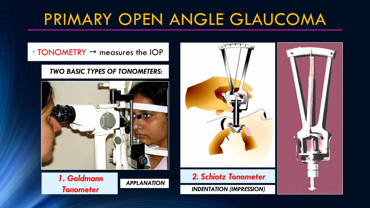
OCULAR HYPERTENSION
DEFINITION• “Glaucoma Suspect”
• IOP constantly >21mmHg but NO OPTIC
DISC or VISUAL FIELD CHANGES
ETIOLOGIC FACTORS
HIGH RISK FACTORS
• IOP CONSTANTLY >28 mmHg
• SIGNIFICANT DIURNAL VARIATION difference
of > 8mmHg
• Significantly positive WATER DRINKING TEST
• Association with SPLINTER HEMORRHAGES
• RETINAL NERVE FIBER LARGE DEFECTS
• PARAPAPILLARY CHANGES
• CENTRAL CORNEAL THICKNESS < 555 μm
Should be CAREFULLY MONITORED
by an ophthalmologist, should be
treated as cases of POAG in the
presence of HIGH RISK FACTORS
OCULAR HYPERTENSION
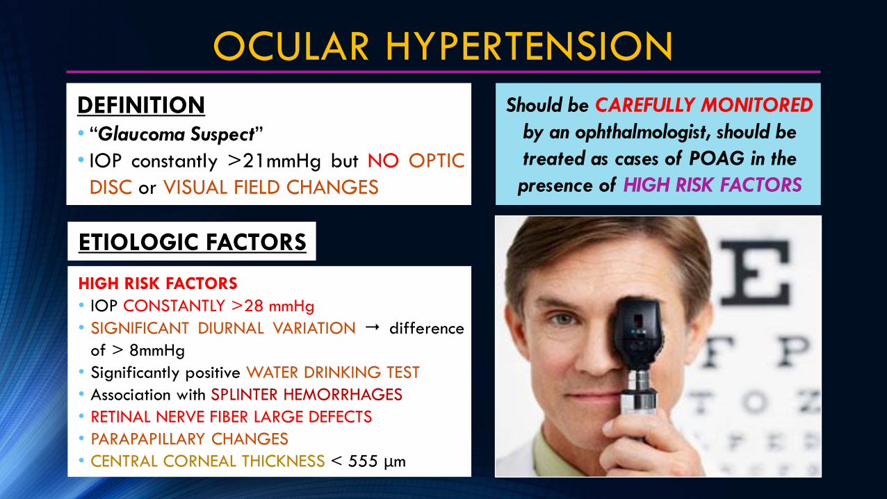
OTHER RISK FACTORS
• SIGNIFICANT ASYMMETRY in the cup size of
the two eyes difference of more than 0.2
• Strong FAMILY HISTORY of glaucoma
• When associated with HIGH MYOPIA,
DIABETES or PIGMENTARY CHANGES in the
anterior chamber
MANAGEMENT• WITH HIGH RISK FACTORS treated on
the lines of POAG (aim is to reduce IOP by20%)
• NO HIGH RISK FACTORS annuallyfollowed by examination of optic disc,perimetry and record of IOP, treatment notrequired till glaucomatous damage isdocumented

NORMAL TENSIONGLAUCOMA

NORMAL TENSION GLAUCOMA
DEFINITION• (NTG), A.k.a LOW TENSION GLAUCOMA,
typical glaucomatous DISC CHANGES, but
WITH / WITHOUT VISUAL FIELD DEFECTS
• Associated with IOP constantly <21 mmHg
EPIDEMIOLOGY
• Variant of POAG (16% of all
cases of POAG)
• AGE prevalence >40 y/o is
0.2%
ETIOLOGY & PATHOPHYSIOLOGY
OPTIC NERVE
SUSCEPTIBLE
CHRONIC LOW
VASCULAR
PERFUSION
PREDISPOSING AND RISK FACTORS
• Raynauld phenomenon
• Migraine
• Nocturnal systemic hypotension
• Overtreated systemic hypertension
• blood flow velocity (ophthalmic artery)
VASCULAR EFFECT ONLY!

NORMAL TENSION GLAUCOMA
CLINICAL MANIFESTATIONS• Disc changes & visual field defects
(Similar to POAG)
• NORMAL IOP
• Other features of NTG are some
ASSOCIATIONS mentioned
DIFFERENTIAL DIAGNOSIS• POAG early stages POAG may
present with normal IOP
• Congenital optic disc anomalies• APPROXIMATELY 60% HAVE
PROGRESSIVE VISUAL FIELD LOSS

NORMAL TENSION GLAUCOMA
MANAGEMENT• MEDICAL TREATMENT to IOP IOP
by 30% (about 12-14 mmHg)a. BETAXOLOL DOC d/t in addition toIOP, also optic nerve blood flow
b. Other Beta Blockers and Adrenergicdrugs (DIPIVERAFRINE) be avoided(causes nocturnal systemic hypotension& are likely to affect adversely theoptic nerve perfusion)
c. NEUROPROTECTIVE DRUGS maybe preferred like “Brimonidine”
d. PROSTAGLANDIN ANALOGUES
greater ocular hypotensive effect
• TRABECULECTOMY considered
when progressive field loss occurs
despite IOP in lower teens
• SYSTEMIC Ca2+ BLOCKERS for
confirmed peripheral vasospasm
• SYSTEMIC BP MONITORING

PRIMARY ANGLE-CLOSURE
GLAUCOMA

PRIMARY ANGLE-CLOSURE GLAUCOMA
DEFINITION• A type of primary glaucoma, (–) obvious
systemic or ocular cause
• IOP occurs d/t BLOCKAGE of the
aqueous humour outflow
• Closure of a NARROWER ANGLE of the
anterior chamber
EPIDEMIOLOGY• AGE more common in 5th decade of life
• GENDER F>M (Ratio 4:1)
• RACE South-East Asian, Chinese (50% of
all primary glaucoma) or Inuit/ Eskimos2
• SEASON higher in rainy season
• FAMILY HISTORY inherited
• TYPE OF PERSONALITY common in
individuals with unstable vasomotors

PRIMARY ANGLE-CLOSURE GLAUCOMA
ETIOLOGY & PATHOPHYSIOLOGY
I. ANATOMICAL FACTORS
• HYPERMETROPIA with shallow
anterior chamber
• Iris-lens DIAPHRAGM placed
anteriorly
• NARROW ANGLE of anterior
chamber, which may be d/t:
a. Small eyeball
b. Relatively large size of the
lens & smaller diameter of
the cornea
c. Bigger size of the ciliary
body
II. GENERAL FACTORS
• AGE• GENDER• RACESEASON• FAMILY HISTORY• TYPE OF PERSO-
NALITY
PREDISPOSING
RISK FACTORS
The following factors may
PRECIPITATE an attack:
• DIM ILLUMINATION
• EMOTIONAL STRESS
• MYDRIATIC DRUGS like
Atropine, Tropicamide
PRECIPITATING
FACTORS INCREASED
AMOUNT OF
APPOSITION B/W
IRIS
ANTERIORLY
PLACED LENS WITH
CONSIDERABLE
PRESSURE
NORMAL PUPILMILD PUPIL DILATATION

PRIMARY ANGLE-CLOSURE GLAUCOMA
RELATIVE
PUPIL
BLOCK
AQUEOUS HUMOR COLLECTS IN THE POSTERIOR CHAMBER
PUSHES THE PERIPHERAL FLACCID IRIS ANTERIORLY
IRIS
BOMBE
APPOSITIONAL
ANGLE CLOSURE
SYNECHIAL
ANGLE CLOSURE
ATTACK OF IOP
MAY LAST LONGER
ACUTE PACG
CHRONIC PACG
Results from the following
CIRCUMSTANCES:
• CREEPING SYNECHIAE
• SUBACUTE PACG ATTACKS
• MIXED MECHANISM

PRIMARY ANGLE-CLOSURE GLAUCOMA
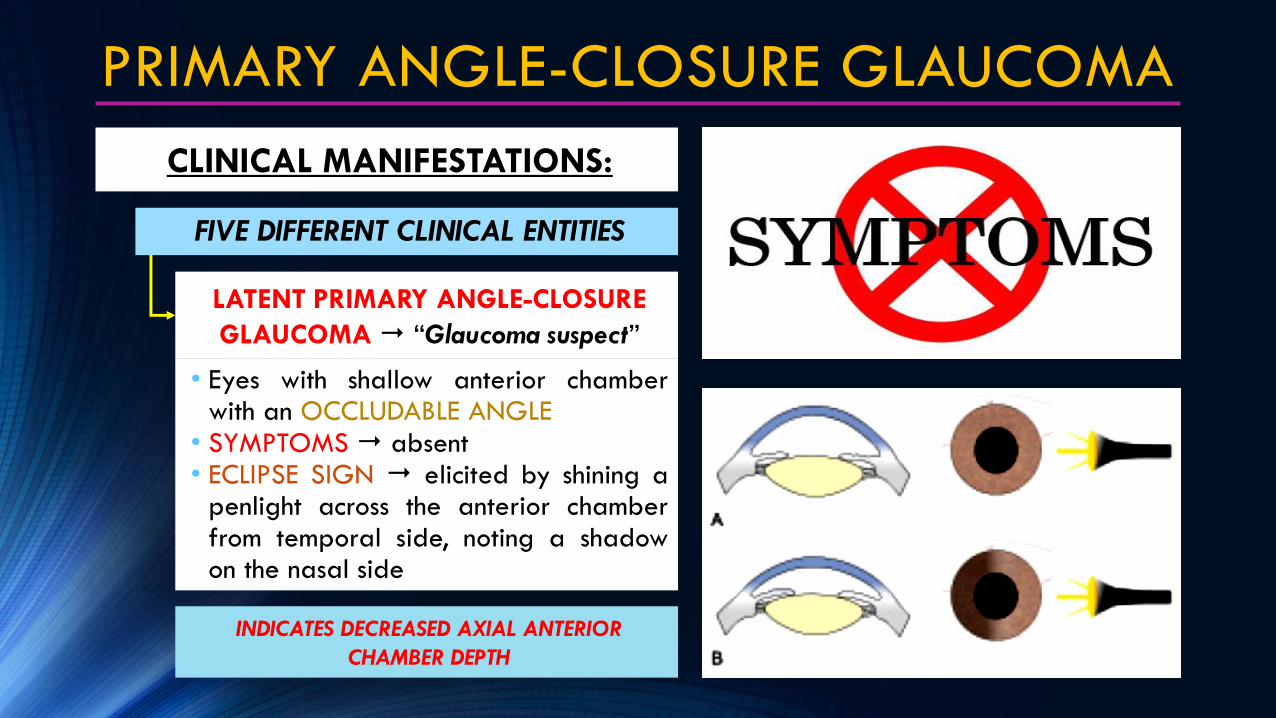
CLINICAL MANIFESTATIONS:
LATENT PRIMARY ANGLE-CLOSURE
GLAUCOMA “Glaucoma suspect”
SUBACUTE OR INTERMITTENT PACG
ACUTE ANGLE-CLOSURE GLAUCOMA
POSTCONGESTIVE
ANGLE-CLOSURE GLAUCOMA
CHRONIC PACG
FIVE DIFFERENT CLINICAL ENTITIES
PRIMARY ANGLE-CLOSURE GLAUCOMA
CLINICAL MANIFESTATIONS:
LATENT PRIMARY ANGLE-CLOSURE
GLAUCOMA “Glaucoma suspect”
INDICATES DECREASED AXIAL ANTERIOR
CHAMBER DEPTH
• Eyes with shallow anterior chamberwith an OCCLUDABLE ANGLE
• SYMPTOMS absent• ECLIPSE SIGN elicited by shining a
penlight across the anterior chamberfrom temporal side, noting a shadowon the nasal side
FIVE DIFFERENT CLINICAL ENTITIES

PRIMARY ANGLE-CLOSURE GLAUCOMA
CLINICAL MANIFESTATIONS:
• GONIOSCOPIC EXAMINATION it
shows very narrow angle (SHAFFER
GRADE 1)
• SLIT-LAMP BIOMICROSCOPIC SIGNS:
a.axial anterior chamber depth
b.Convex shaped iris lens diaphragm
c. Close proximity of the iris to cornea
in the periphery
LATENT PRIMARY ANGLE-CLOSURE
GLAUCOMA “Glaucoma suspect”
FIVE DIFFERENT CLINICAL ENTITIES

PRIMARY ANGLE-CLOSURE GLAUCOMA
CLINICAL MANIFESTATIONS:
LATENT PRIMARY ANGLE-CLOSURE
GLAUCOMA “Glaucoma suspect”
• VAN HERICK SLIT-LAMP GRADING
used when gonioscope is not available
(FAIR ACCURACY)
• Peripheral Anterior Chamber Depth
(PACD) compared to the adjacent
corneal thickness (CT) and the
presumed angle width
• CLINICAL COURSE if without Tx,
may follow any of the following:
a. IOP may remain NORMAL
b. SUBACUTE or ACUTE angle-closure
glaucoma may occur subsequently
c. CHRONIC angle-closure glaucoma
may develop without passing
through subacute or acute stage
FIVE DIFFERENT CLINICAL ENTITIES

PRIMARY ANGLE-CLOSURE GLAUCOMA
VAN HERICK METHOD OF
SLIT-LAMP GRADING
A – Grade IV
B – Grade III
C – Grade II
D – Grade I
E – Grade 0
GRADES:Grade 4 (WIDE OPEN ANGLE)
• PACD = 3/4 to 1 CT
Grade 3 (MILD NARROW)
• PACD = ¼ to ½ CT
Grade 2 (MODERATE NARROW)
• PACD = ¼ CT
Grade 1 (EXTREMELY NARROW)
• PACD < ¼ CT
Grade 0 (CLOSED ANGLE)
• PACD Nil

PRIMARY ANGLE-CLOSURE GLAUCOMA
CLINICAL MANIFESTATIONS:
FIVE DIFFERENT CLINICAL ENTITIES
• Attack of TRANSIENT IOP (40-50mmHg) (last for minutes to 1-2 hours)
• Usually PRECIPITATED by:a. PHYSIOLOGICAL MYDRIASIS
reading in dim light, watching TV orcinema in darkened room, or duringanxiety (Sympathetic Overactivity)
b. PHYSIOLOGICAL SHALLOWINGOF ANTERIOR CHAMBER afterlying in prone position
SUBACUTE OR INTERMITTENT PACG
• SYMPTOMS unilateral transient
blurring of vision, coloured halos
around light, headache, browache
and eyeache on the affected side
COLOURED HALOS AROUND LIGHT
PRIMARY ANGLE-CLOSURE GLAUCOMA
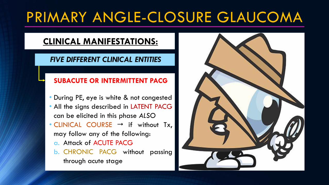
CLINICAL MANIFESTATIONS:
FIVE DIFFERENT CLINICAL ENTITIES
• During PE, eye is white & not congested
• All the signs described in LATENT PACG
can be elicited in this phase ALSO
• CLINICAL COURSE if without Tx,
may follow any of the following:
a. Attack of ACUTE PACG
b. CHRONIC PACG without passing
through acute stage
SUBACUTE OR INTERMITTENT PACG

PRIMARY ANGLE-CLOSURE GLAUCOMA
CLINICAL MANIFESTATIONS:
FIVE DIFFERENT CLINICAL ENTITIES
• Attack of Acute PACG occurs d/t a
sudden total angle closure leading to
SEVERE RISE in IOP
ACUTE ANGLE-CLOSURE GLAUCOMA
SIGHT THREATENING EMERGENCY!
SYMPTOMS• PAIN sudden onset of very severe pain
that radiates along the CN-V branches
• NAUSEA, VOMITING, PROSTRATIONS
• Rapid progress of VISION LOSS also
with redness, photophobia & lacrimation
(PRESENT IN ALL CASES)
• PAST HISTORY ~5% (+)Hx of typical
previous transient attacks of subacute
angle-closure glaucoma

SIGNS
• LIDS may be edematous
• CONJUNCTIVA congested
• CORNEA edematous & insensitive
• ANTERIOR CHAMBER very shallow
• ANGLE OF ANTERIOR CHAMBER
closed completely (SHAFFER GRADE0)
• IRIS may be discoloured
PRIMARY ANGLE-CLOSURE GLAUCOMA
CLINICAL MANIFESTATIONS:
FIVE DIFFERENT CLINICAL ENTITIES
ACUTE ANGLE-CLOSURE GLAUCOMA
•NOTE CILIARY CONGESTION,
•CORNEAL EDEMA & MIDDILATED PUPIL
DISCOLOURED IRISVERY SHALLOW –ANTERIOR CHAMBER

SIGNS
• IOP it is usually markedly elevated,
b/w 40-70 mmHg (NV:10-21mmHg)
• PUPIL semi-dilated, vertically oval
and fixed, usually non-reactive to both
light & accommodation
• OPTIC DISC edematous, hyperemic
• FELLOW EYE shows shallow anterior
chamber and a narrow angle
PRIMARY ANGLE-CLOSURE GLAUCOMA
CLINICAL MANIFESTATIONS:
FIVE DIFFERENT CLINICAL ENTITIES
ACUTE ANGLE-CLOSURE GLAUCOMA
PUPIL NON-REACTIVE TO BOTH LIGHT & ACCOMMODATION
EDEMATOUS & HYPEREMIC OPTIC DISC

PRIMARY ANGLE-CLOSURE GLAUCOMA
CLINICAL MANIFESTATIONS:
FIVE DIFFERENT CLINICAL ENTITIES
POSTCONGESTIVE
ANGLE-CLOSURE GLAUCOMA
• VOGT’S TRIAD seen with any type
of post-congesive glaucoma & in
treated acute congestive glaucoma:
a. GLAUCOMFLECKEN an anterior
sub-capsular lenticular opacity
b. PATCHES OF IRIS ATROPHY
c. SLIGHTLY DILATED NON-REACTING
PUPIL due to sphincter atrophy PATCHES OF IRIS ATROPHYSLIGHTLY DILATED NON-
REACTING PUPIL
GLAUCOMFLECKEN
VOGT’S TRIAD

PRIMARY ANGLE-CLOSURE GLAUCOMA
CLINICAL MANIFESTATIONS:
FIVE DIFFERENT CLINICAL ENTITIES
POSTCONGESTIVE
ANGLE-CLOSURE GLAUCOMA
1.POSTSURGICAL POSTCONGESTIVE
PACG after laser peripheral
iridotomy (PI) treatment for an
attack of acute PACG
FOUR CLINICAL SETTINGS
• Clinical status of eye after an attack of
acute PACG with or without treatment
• With normalized IOP post-laser Tx,
the eye usually “QUITENS” after some
time with/without s/s of acute attack
• With raised IOP after unsuccessful Tx,
there occurs a STATE OF CHRONIC
CONGESTIVE GLAUCOMA

PRIMARY ANGLE-CLOSURE GLAUCOMA
CLINICAL MANIFESTATIONS:
FIVE DIFFERENT CLINICAL ENTITIES
POSTCONGESTIVE
ANGLE-CLOSURE GLAUCOMA
2.SPONTANEOUS ANGLE OPENING
may very rarely occur in some cases
and the attack of acute PACG may
subside itself without treatment
FOUR CLINICAL SETTINGS
• Clinical status of eye after an attack of
acute PACG with or without treatment
3. CHRONIC CONGESTIVE PACG
continuation of acute congestive angle-
closure glaucoma when no Tx or when is
unsuccessful
a. EYE permanently congested, pain
reduced d/t “ACCLAMATIZATION”
b. IOP remains constantly raised
c. LID & CONJUCTIVAL EDEMA
d. OPTIC DISC may show cupping
e. Other features are similar to acute
congestive angle-closure glaucoma
4. CILIARY BODY SHUT DOWN
temporary cessation of aqueous humor
secretion due to ischemic damage
a. IOP is low, PAIN is “MARKEDLY”

• Similar to POAG, EXCEPT that the
angle in Chronic PACG is narrow
• IOP constantly raised
• EYEBALL it is usually remains white
(without congestion) & PAINLESS
• OPTIC DISC may show cupping
• VISUAL FIELD DEFECTS like POAG
• GONIOSCOPY variable degree of
angle closure
PRIMARY ANGLE-CLOSURE GLAUCOMA
CLINICAL MANIFESTATIONS:
FIVE DIFFERENT CLINICAL ENTITIES
CHRONIC PACG
PAINLESS EYEBALL

PRIMARY ANGLE-CLOSURE GLAUCOMA
ABSOLUTE PACG if no Tx for chronic
phase, with/without sub-acute attacks,
gradually passes into “FINAL PHASE”
a. PAINFUL BLIND EYE irritability & now
completely blind (NO LIGHT PERCEPTION)
b. PERILIMBAL REDDISH BLUE ZONE slight
ciliary flush around the cornea d/t dilated
anterior veins
c. CORNEA clear but insensitive
d. ANTERIOR CHAMBER very shallow
e. IRIS becomes atrophic
f. PUPIL fixed, dilated, greenish hue
g. OPTIC DISC shows atrophy
h. INTRAOCULAR PRESSURE high
i. EYEBALL becomes stony hard
PERILIMBAL REDDISH BLUE ZONE
PAINFUL BLIND EYE INSENSITIVE
CORNEA

PRIMARY ANGLE-CLOSURE GLAUCOMA
DIAGNOSTIC FACTORS:
CLINICAL ENTITY DIAGNOSIS
LATENT PRIMARY ANGLE-
CLOSURE GLAUCOMA
DIAGNOSIS MADE BY:
• CLINICAL SIGNS described beforehand
• PROVOCATIVE TESTS designed to precipitate closure of
the angle in the ophthalmologist’s office, where it can be
treated promptly
a. PRONE-DARKROOM TEST it is the most popular & best
physiological provocative test for PACG
b. MYDRIATIC PROVOCATIVE TEST not preferred now
SUBACUTE PRIMARY ANGLE-
CLOSURE GLAUCOMA
ACUTE, POSTCONGESTIVE,
CHRONIC, ABSOLUTE PACG*DIAGNOSIS USUALLY OBVIOUS FROM THE CLINICAL SIGNS1

PRIMARY ANGLE-CLOSURE GLAUCOMA
PRONE-DARKROOM TEST
MYDRIATIC PROVOCATIVE TEST
DIAGNOSTIC FACTORS:

PRIMARY ANGLE-CLOSURE GLAUCOMA
CORNEAL ULCERATION
STAPHYLOMA FORMATION
ATROPHIC BULBI
D/T prolonged epithelial edema & insensitivity
Sclera becomes very thin and atrophic,
ultimately bulges out (CILIARY staphyloma &
EQUATORIAL staphyloma)
Ciliary body degenerates, IOP and the
eyeball shrinks
COMPLICATIONS
If untreated, d/t prolonged IOP the following may occur:
CORNEAL
ULCERATION
STAPHYLOMA
FORMATION
ATROPHIC BULBI

REFERENCES
1 – Khurana's Ophthalmology - 4th Edition 2007
2 – Vaughan and Asbury's Ophthalmology - 17th Edition 2007
3 – http://eyewiki.aao.org/Glaucoma_in_the_Developing_World
4 – http://roqueeyeclinic.com/eye-conditions/glaucoma/80-glaucoma-classification-epidemiology

-- DR. JOSE RIZAL
Philippine National Hero
Ophthalmologist

END