Embed Size (px)
Citation preview

Presented by: Ligi XavierFirst year MSc nursing
Govt. College of nsg,Kottayam

nervous system includes brain, cranial nerves, spinal cord, spinal nerves, enteric plexus, sensory receptors.

Sensory functions
Integrative functions: mainly perception
Motor functions: muscle contraction ,gland
secretion.

Nervous System
Central Nervous
System
Brain Spinal Cord
Forebrain Midbrain Hindbrain
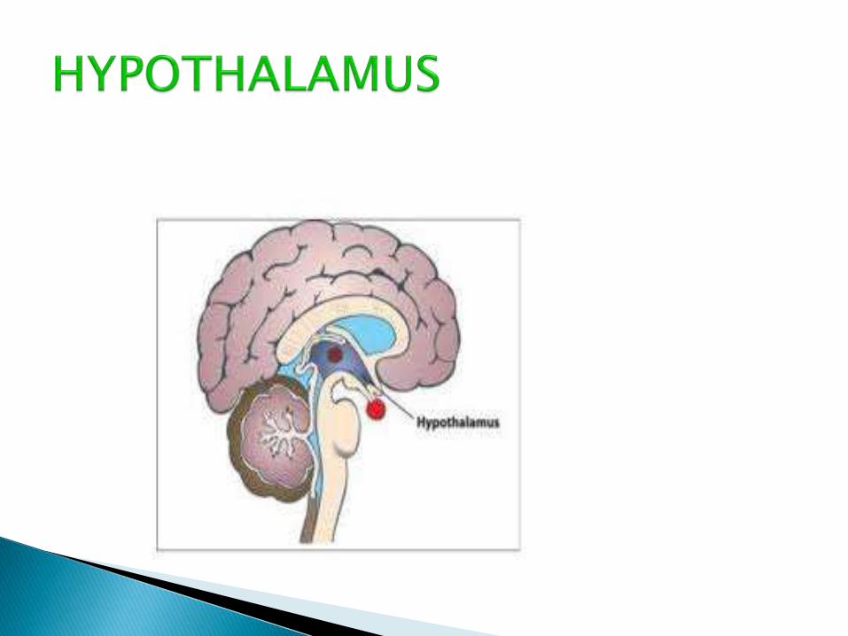
Thalamus Hypothalamus
Cortex Limbic SystemCorpus
Callosum
Peripheral Nervous
System
Somatic
System
Autonomic
System
Parasympathetic
Division
Sympathetic
Division
organization of nervous system


Produced by oligo dendrocytes in cns and Schwann cells in the cns.
It appears to be white, shiny

White matter composed of myelinated axons.
Gray matter composed of dendrites,cell bodies, neuroglia ,nissl bodies


Faster communication
synchronisation


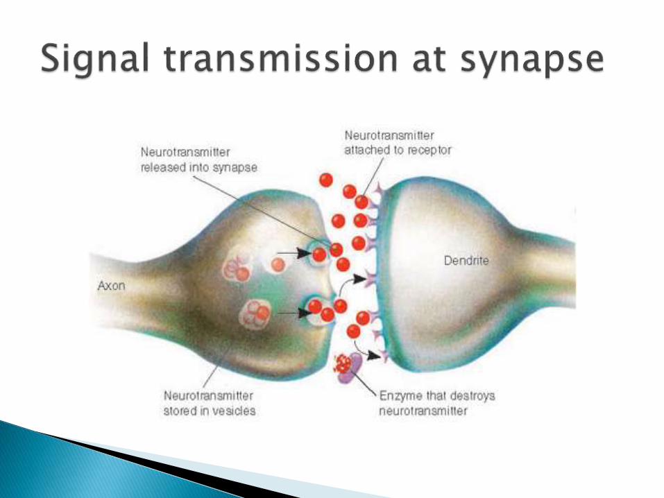
Small molecule neuro transmitters◦ Acetyl choline
◦ Aminoacids: glutamate, aspartate
◦ Biogenic amines:norepinephrine,epinephrine,dopamine
◦ ATP:ATP,ADP,AMP
◦ Purines
◦ Nitric oxide

Neuropeptide◦Enkephalins
◦Dynorphins
◦Endorphins
◦Substance P


It includes brain and Spinalcord


Meninges
Three layers of meninges◦ Dura mater
◦ Arachnoid mater
◦ Piua mater


Volume: 80 ml
Functions◦ Mechanical protection
◦ Chemical protection
◦ circulation


Part of brain between diencephalon and spinal cord.


Located between the pons and spinal cord.
Containsgray matter which has centers that play an important role in many involuntary actions such as respiration.
The centers are called vital centers.
Therespiratorycenter
The cardiaccenter
The vasomotorcenter


Part of reticular activating system is called RAS(reticular activating system)
It helps to maintain consciousness, & active during awakening from sleep.


Second largest part of Brain. It coordinates Movements, muscle contraction

Divided into two hemispheres, and one middle part(vermis).
Outer layer gray matter, inner layer white matter.
Located above the brainstem, and beneath the occipital lobes.

Coordination in voluntary movement.
Helps maintain balanceand equilibrium.
Helps maintain muscle tone.


NUCLEIANTERIOR MEDIALLATERALVENTARLINTRALAMINARMIDLINE NUCLEUS RETICULAR

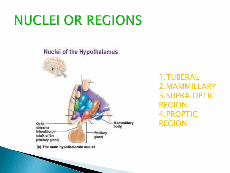
1.TUBERAL2.MAMMILLARY3.SUPRA OPTIC REGION4.PROPTIC REGION

Functions◦ Regulator of homeo stasis
◦ Production of hormones
◦ Regulation of behavioural and emotional patterns
◦ Regulation of eating & drinking
◦ Control body temperature
◦ Regulates circardian rhythm
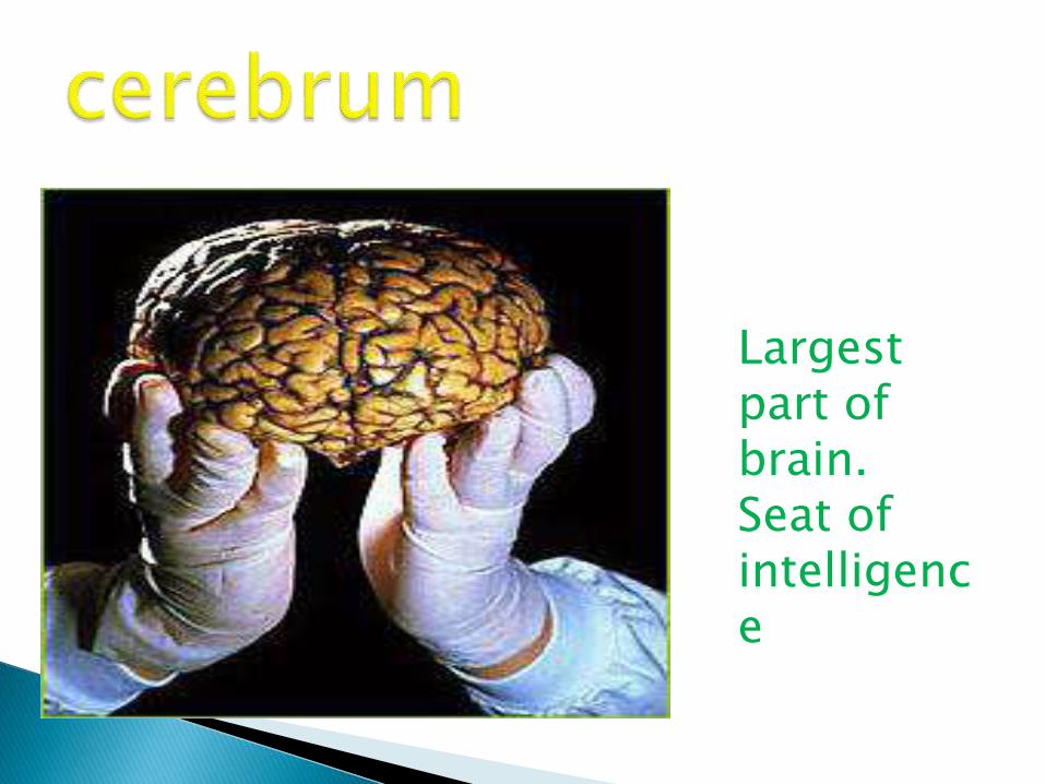
Largest part of brain.Seat of intelligence


FrontalParietalTemporalOccipital

The Frontal Lobe lies
anterior to the centralsulcus.

Contains an area that provides the conscious control of skeletal muscles.
Contains two areas that are important in speech

The Parietal Lobe
occupies the
superior
part of each
hemisphere
and lies posterior
to the
central nucleus

Contains a primary sensory area where
impulses from the skin are interpreted
Estimates distanceandsize

The Temporal Lobe lies inferior the lateral sulcussnd folds under the hemi-sphere on each side

Responsible for receiving and
interpreting
Auditory impulses from the ear.
An olfactory area that concerns the
sense of smell.

The Occipital Lobelies posterior to theparietal lobe and extends over thecerebellum.

Visual receiving area and visual
association for interpreting
impulses from the retina of the eye.


Connects the gray matter areas with one another and with other parts of the brain.
Dispersed in a tree like pattern
Made of myelinatedfibers
3 types of tracts: association tracts, commissural tracts, projection tracts

major function is to regulate initiationAnd termination of movements.controls the
contraction of skeletal muscles

known as emotional brainIt plays role in emotions likePain, pleasure, anger, affection etc


It is known as circle of willis.Circulation is divided into anteriorand posterior
cerebralcirculation

2 sub divisions :deep,superficial


2 pathwaysMedial lemniscalSpinothalamic pathway


• Measures synaptic potentials produced at cell bodies and dendrites.– Create electrical
currents.
• Used clinically do diagnose epilepsy and brain death.

Alpha: ◦ Recorded from parietal and occipital regions.
Person is awake, relaxed, with eyes closed. 10-12 cycles/sec.
Beta:◦ Strongest from frontal lobes near precentral gyrus.
Produced by visual stimuli and mental activity. Evoked activity.
13-25 cycles/sec.
Theta:◦ Emitted from temporal and occipital lobes.
Common in newborn. Adult indicates severe emotional stress.
5-8 cycles/sec. Delta:◦ Emitted in a general pattern.
Common during sleep and awake infant. In awake adult indicate brain damage.
1-5 cycles/sec.


The brain loses 5-10 % of it’s volume between the ages of 20-90.
The grooves widen and the surface shrinks


It is cylindrical in shape.it extends from the medulls oblongata to the 2nd lumbar vertebra.
Length:42-45cm


An area of skin thatProvides sensory Input to the CNS.


Maintains homeostasis by nerve impulse
propagation and integration of information
Reflex activity