Embed Size (px)
Citation preview

TROPICAL MEDICINE TROPICAL MEDICINE DEPARTEMENTDEPARTEMENT
Medical Education ProgramsMedical Education Programs
الله بسمالرحيم الرحمن

ByMahmoud Saad Desoky

To a very large extent, the health and well-being of an individual is dependent on the proper functioning
of this important detoxifying organ. …

Portal hypertensioncommon clinical syndrome, which is
hemodynamically defined as pathological increase of the portal pressure gradient(the pressure difference between the portal vein and the inferior vena cava)
And by the formation of portal–systemic collaterals that shunt part of the portal blood flow to the systemic circulation by passing the liver, Normal values of the portal pressure gradient are of 1–5 mm Hg.
Portal hypertension is a frequent complication of cirrhosis, and plays a crucial role in the transition from the pre clinical to the clinical phase of the disease.
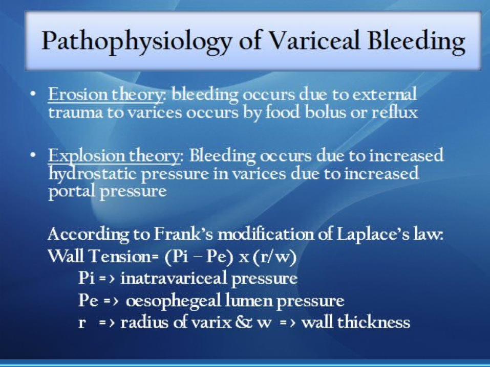
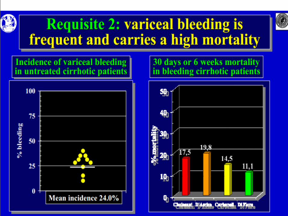
Portal hypertension is a contributing factor for the development of ascites and hepatic encephalopathy and a direct cause of variceal haemorrhage and of bleeding-related death, Bleeding from ruptured oesophago-gastric varices is the most severe complication of cirrhosis, and is the cause of death in about one third of cirrhotic patients.

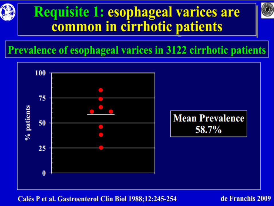
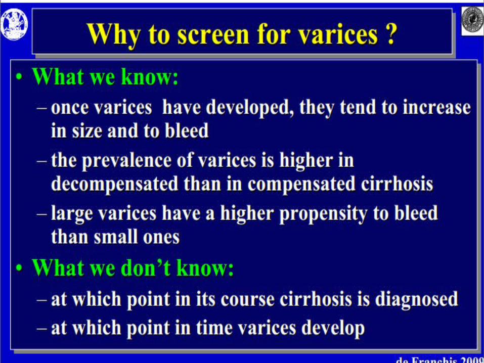
The yearly rate of development of ‘‘new” varices is about 5–10% per year in patients with cirrhosis, and the progression from small to large varices occur in 10% to 20% of cases after 1 year. In the 2 years following the first detection of EV, the risk of variceal bleeding ranges between 20% to 30% and results in 25% to 50% mortality within a week of the first bleeding episode

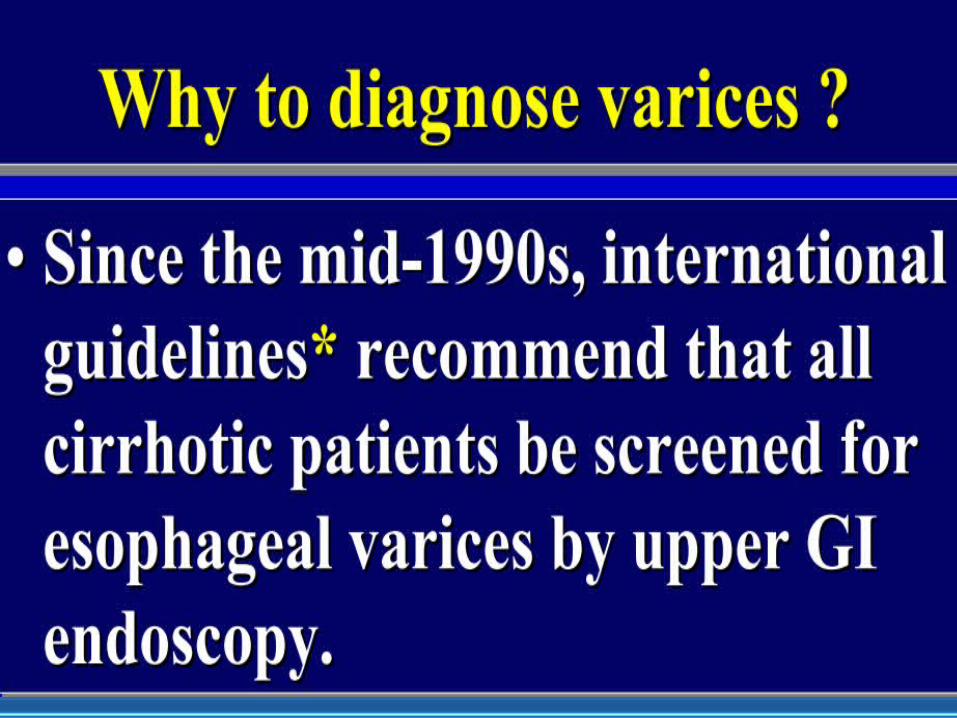
The current recommendations states that all cirrhotic patients should be screened for the presence of varices at the time of initial diagnosis of cirrhosis. Follow-up endoscopy should be performed at 2-3 years intervals in compensated patients with no varices, and at 1-2 years intervals in compensated patients with small varices

Clinically significant portal hypertension (CSPH) is diagnosed when clinical manifestations of the disease appear or when portal pressure gradient (in case of cirrhosis) determined by its equivalent, the hepatic venous pressure gradient (HVPG)) exceeds a threshold value of 10 mm Hg. Values of portal pressure gradient between 5 and 9 mm Hg correspond to preclinical portal hypertension


Portal veinis
not a true vein
The intrahepatic circulation has some unique features. One is the dual blood supply from portal vein and hepatic artery. Thirty percent of the flow and 30% to 60% of the oxygen consumed by the liver comes from the hepatic artery while the rest comes from the portal vein
2 The dual hepatic blood supply makes the normal liver resistant to anoxia. Ligation of the portal vein, for example, will not cause hepatocellular necrosis. Similarly, accidental ligation of the hepatic artery or its major branches does not necessarily lead to hepatic failure, except in the transplant setting, where the organ is much more dependent on hepatic arterial blood flow. There is also a unique interrelationship between hepatic artery and portal vein blood flow
In both animals and humans, a decrease in portal venous flow or sinusoidal pressure causes a reflex increase in hepatic arterial flow. Conversely, an increase in sinusoidal flow or pressure causes a reflex decrease in hepatic arterial flow. This buffer response may be mediated by adenosine, and the response maintains a constant hepatic blood flow despite changes in portal venous flow that occur during digestion.
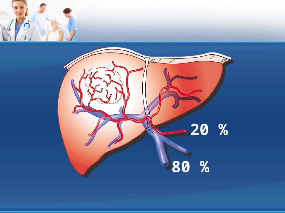
20 %
80 %
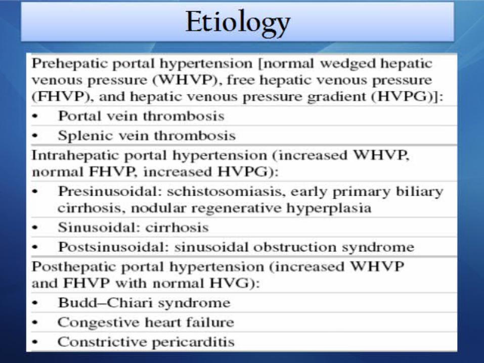

Prehepatic
Hepatic
Posthepatic

Prehapatic(portal vein obstruction)
•Thrombosis of portal vein•Congenital atresia / stenosis
•Extrinsic compression
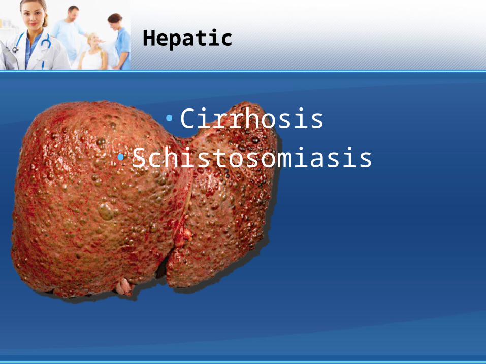
Hepatic
•Cirrhosis•Schistosomiasis

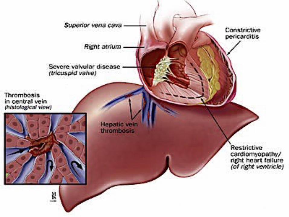
Posthepatic
•Budd-Chiari syndrome•Cardiac disease
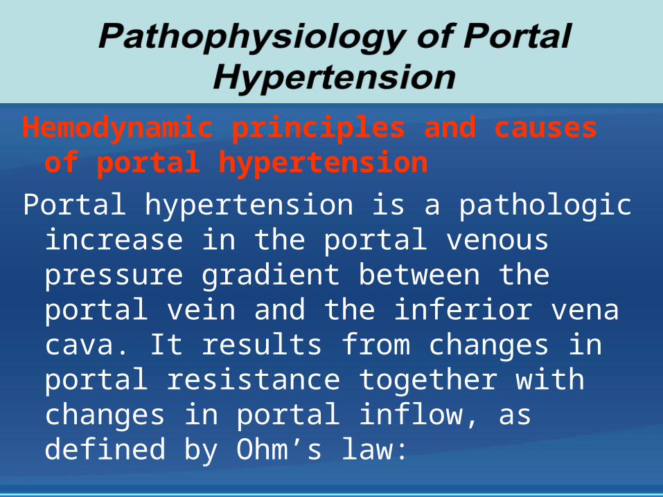
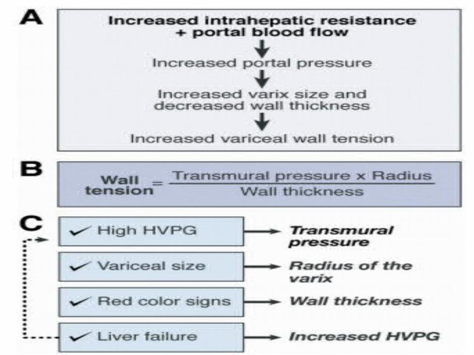
Hemodynamic principles and causes of portal hypertension
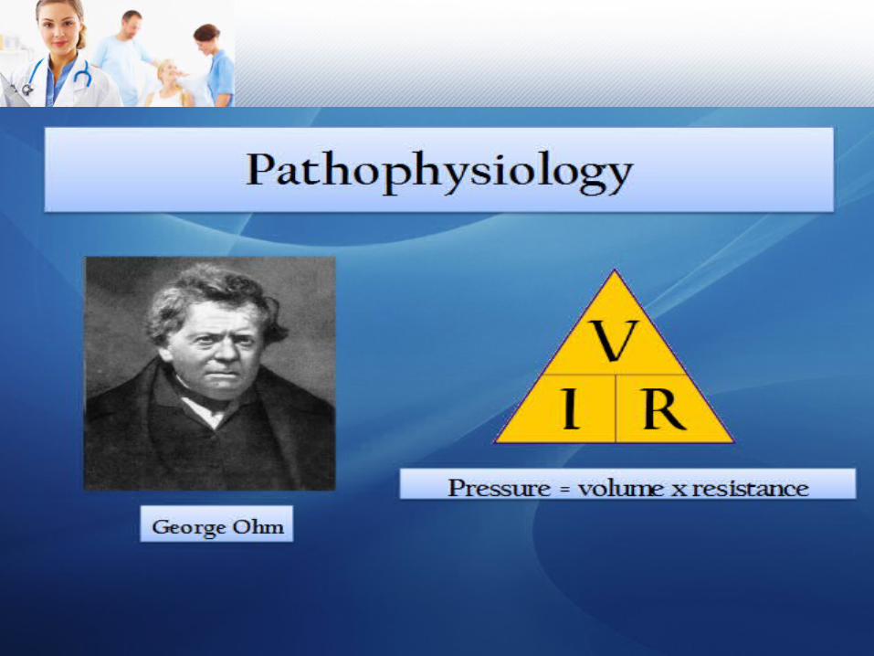
Portal hypertension is a pathologic increase in the portal venous pressure gradient between the portal vein and the inferior vena cava. It results from changes in portal resistance together with changes in portal inflow, as defined by Ohm’s law:


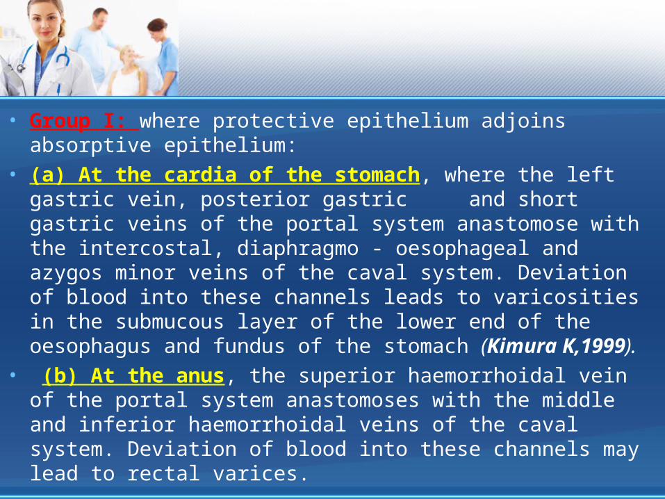
• Group I: where protective epithelium adjoins absorptive epithelium:
• (a) At the cardia of the stomach, where the left gastric vein, posterior gastric and short gastric veins of the portal system anastomose with the intercostal, diaphragmo - oesophageal and azygos minor veins of the caval system. Deviation of blood into these channels leads to varicosities in the submucous layer of the lower end of the oesophagus and fundus of the stomach (Kimura K,1999).
• (b) At the anus, the superior haemorrhoidal vein of the portal system anastomoses with the middle and inferior haemorrhoidal veins of the caval system. Deviation of blood into these channels may lead to rectal varices.

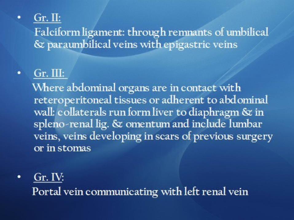
• Group II: in the falciform ligament through the paraumbilical veins, relics of the umbilical circulation of the fetus
• Group III: where the abdominal organs are in contact with retroperitoneal tissues or adherent to the abdominal wall. These collaterals run from the liver to diaphragm and in the splenorenal ligament and omentum. They include lumbar veins and veins developing in scars of previous operations or in small or large bowel stomas.
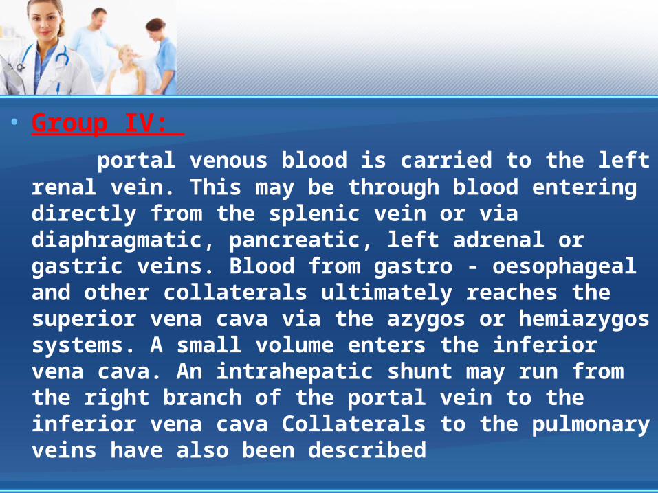
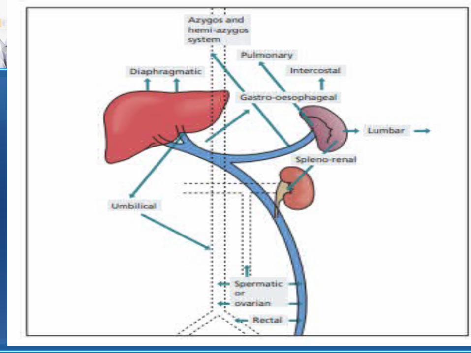
• Group IV: portal venous blood is carried to the left
renal vein. This may be through blood entering directly from the splenic vein or via diaphragmatic, pancreatic, left adrenal or gastric veins. Blood from gastro - oesophageal and other collaterals ultimately reaches the superior vena cava via the azygos or hemiazygos systems. A small volume enters the inferior vena cava. An intrahepatic shunt may run from the right branch of the portal vein to the inferior vena cava Collaterals to the pulmonary veins have also been described


Hippocrates said that: some patients get well only by the goodness of their physicians.


![Portal hypertension: Imaging of portosystemic collateral ...€¦ · portal hypertension[3-5]. Clinically significant portal hypertension is defined as an increase in HVPG to ≥](https://img.dokumen.tips/doc/110x75/5f03e1347e708231d40b3854/portal-hypertension-imaging-of-portosystemic-collateral-portal-hypertension3-5.jpg)