Embed Size (px)
Citation preview
04/15/2023
1
RESOURCE FACULTY PRESENTED BY
DR.ASHISH SHRESTHA ASSOCIATED PROFESSOR AND HOD
DR.TARAKANT BHAGATASSISTANT PROFESSOR (DEPARTMENT OF PUBLIC HEALTH DENTISTRY) BPKIHS,NEPAL
PAPULAR CHAUDHARY ROLL NO:491 [email protected]
SANTOSH PANDIT ROLL NO:502 [email protected] BATCH 2011
ORAL MANIFESTATIONS OF SYSTEMIC DISEASE
04/15/20232
Oral Manifestations of Systemic Diseases
Many systemic diseases are reflected in the oral mucosa, maxilla, and mandible.
Mucosal changes may include ulceration or mucosal bleeding.
Immunodeficiency can lead to opportunistic diseases such as infection and neoplasia.
Bone disease can affect the maxilla and mandible.
Systemic disease can cause dental and periodontal changes
Drugs prescribed for a systemic disease can affect oral tissue.
04/15/20233
Oral Manifestations of Systemic Diseases (cont.)
Local factors may be involved in the manifestation of systemic disease in oral mucosa.
The mucosa may be more easily injured due to a systemic disease, and mild irritation and chronic inflammation may cause lesions that otherwise would not occur.
These may include Endocrine disorders, disorders of red and white blood cells,
disorders of platelets and other bleeding and clotting disorders, and immunodeficiency disorders
04/15/2023
4
Oral manifestations of systemic diseases
Auto immune diseasesAIDS
04/15/2023
5
Systemic lupus erythematous
Lupus characterized by acute and chronic inflammation of various tissues of the body.
When the skin is involved, the condition is called lupus dermatitis or cutaneous lupus erythematosus.
A form of lupus dermatitis that can be isolated to the skin, without internal disease, is called discoid lupus.
When internal organs are involved, the condition is referred to as systemic lupus erythematosus (SLE)
04/15/2023
6
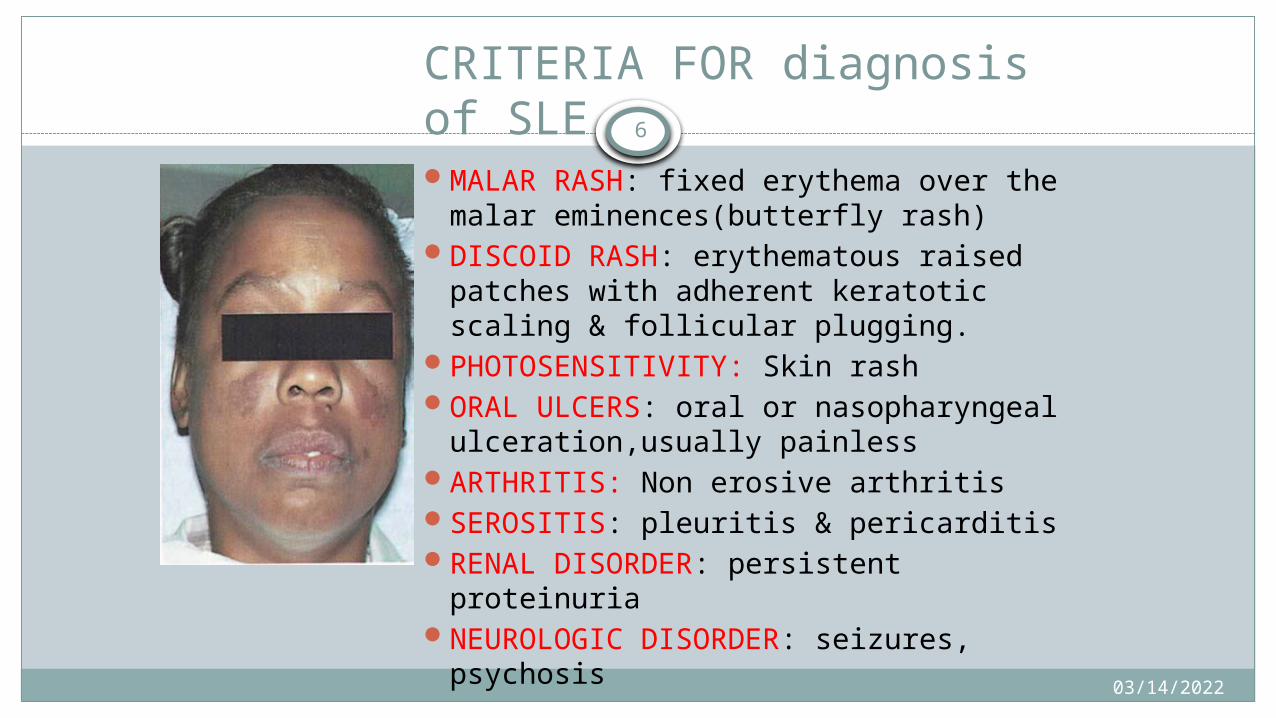
CRITERIA FOR diagnosis of SLEMALAR RASH: fixed erythema over the
malar eminences(butterfly rash)DISCOID RASH: erythematous raised
patches with adherent keratotic scaling & follicular plugging.
PHOTOSENSITIVITY: Skin rashORAL ULCERS: oral or nasopharyngeal
ulceration,usually painlessARTHRITIS: Non erosive arthritisSEROSITIS: pleuritis & pericarditisRENAL DISORDER: persistent
proteinuriaNEUROLOGIC DISORDER: seizures,
psychosis

04/15/2023
7 HEMATOLOGICAL DISORDER: hemolytic anemia, leucopenia,
lymphopenia, thrombocytopenia.
IMMUNOLOGIC: anti- ds DNA, anti Sm & antiphospholipid antibodies
ANTINUCLEAR ANTIBODY: abnormal
(for the diagnosis of SLE four or the above criteria should either be present simultaneously or serially)
04/15/2023
8Oral aspects
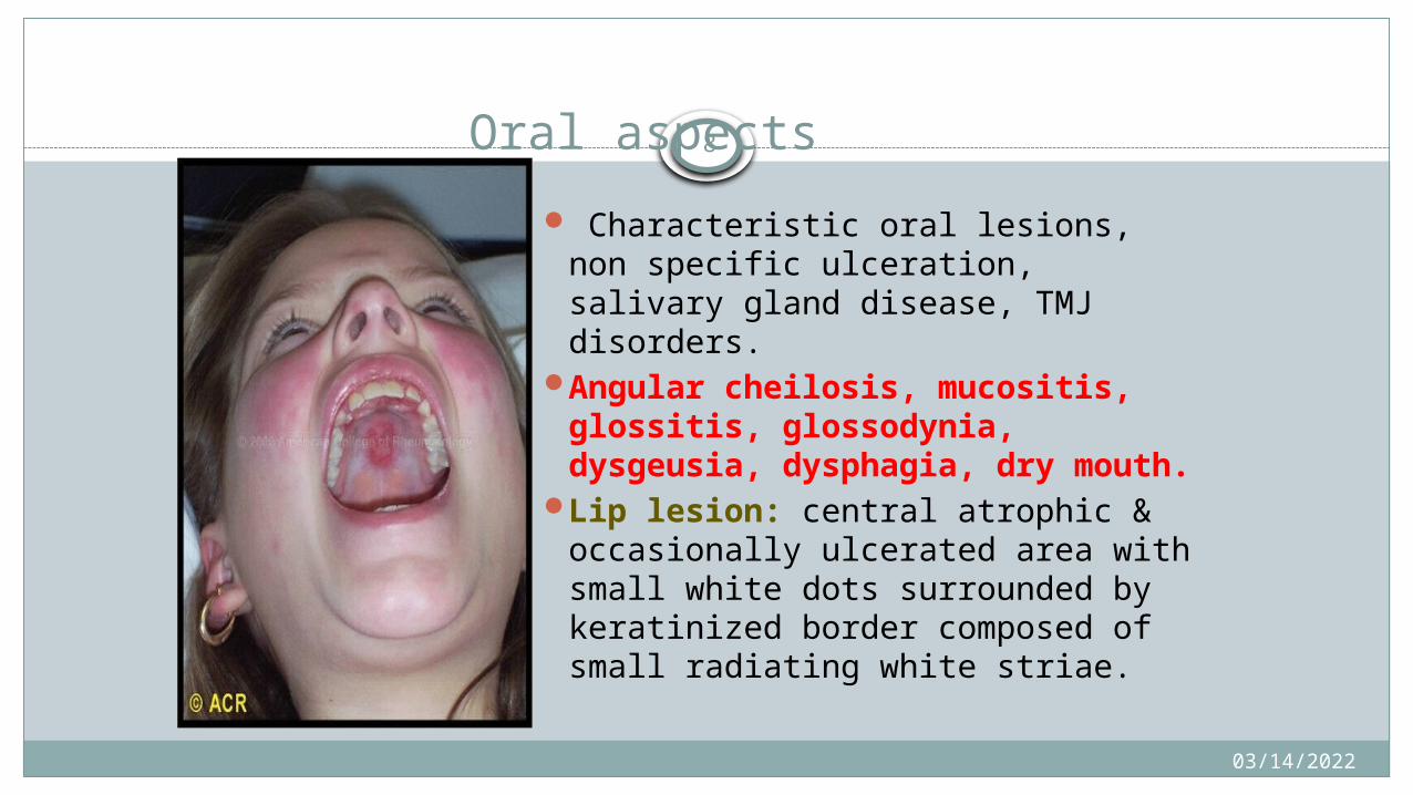
Characteristic oral lesions, non specific ulceration, salivary gland disease, TMJ disorders.
Angular cheilosis, mucositis, glossitis, glossodynia, dysgeusia, dysphagia, dry mouth.
Lip lesion: central atrophic & occasionally ulcerated area with small white dots surrounded by keratinized border composed of small radiating white striae.
04/15/2023 9
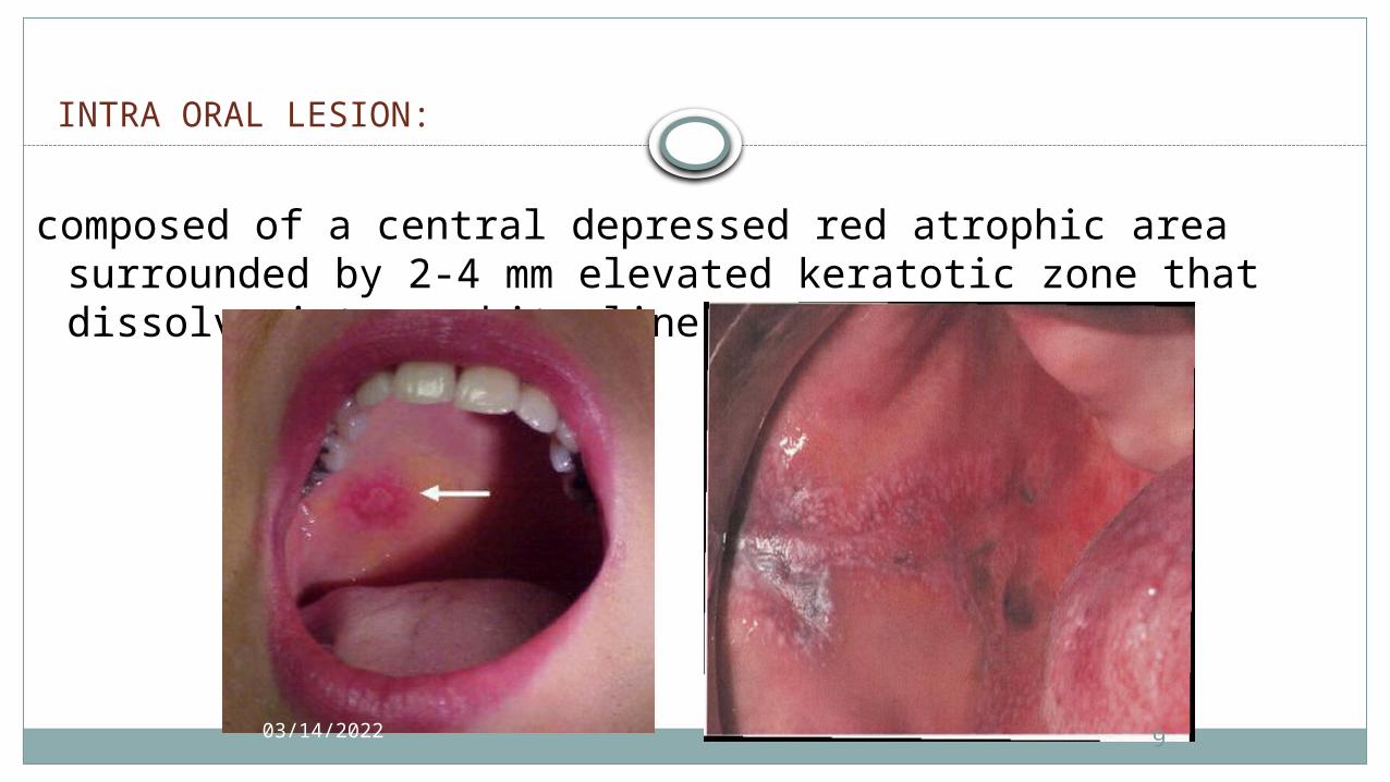
INTRA ORAL LESION:
composed of a central depressed red atrophic area surrounded
by 2-4 mm elevated keratotic zone that dissolve into a white line.

04/15/2023
10
Discoid lupus erythematus
DLE is a chronic, scaring, atrophy producing, photosensitive dermatosis.
DLE may occur in patients with SLE.
<5% DLE progress to SLE .
• PATHOPHYSIOLOGY Suggested that heat shock protein is induced in
keratinocyte followed by UV light exposure or stress, and this protein act as target for T-cell mediated epidermal cell cytotoxicity.

04/15/2023
11
Clinical features
Age: 3rd -4th decade.
Sex female>male.
Common sites:• Face, oral mucous membranes, chest, back and extremities.
• Cutaneous lesion are slightly elevated red of purple macules that are often covered by grey or yellow adherent scales.

04/15/2023
12
Contd…
• Forceful removal of scale result in “carpet track” extension .
• Typical “butterfly” distribution on malar region and across the bridge of the nose.
• Epidermoid carcinoma and less commonly basal cell carcinoma is reported in healed ulcer.
04/15/2023
13
Oral manifestation
Oral mucous membrane involvement rate is 20-50%.
Oral lesion in discoid form begin as erythematous area, some times slightly elevated but more often depressed.
No induration.
Occasionally superficial ,painful ulceration may occur with crusting or bleeding.
No scale formation .
04/15/2023
14
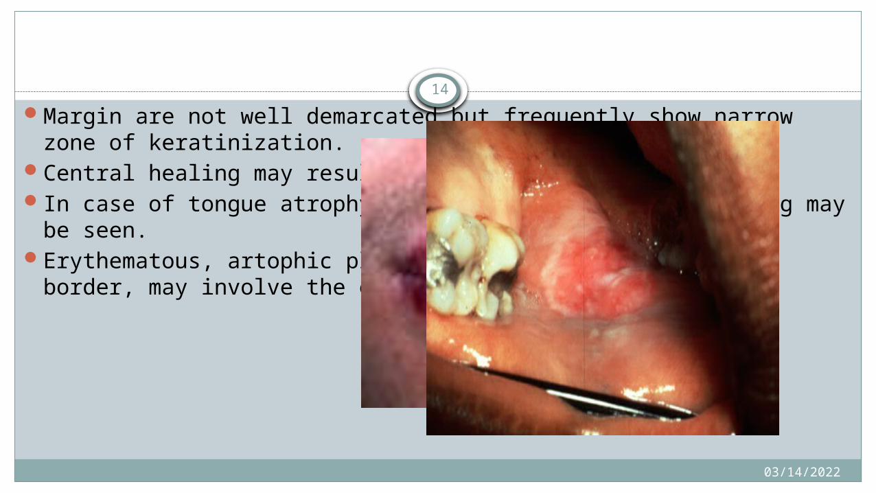
Margin are not well demarcated but frequently show narrow zone of keratinization.
Central healing may result in depressed scarring In case of tongue atrophy of papilla and severe fissuring may be seen.Erythematous, artophic plques , surrounded by kertotic border, may
involve the entire lip.
04/15/2023
15 Sjogren Syndrome
an autoimmune disorder in which immunocytes damage the salivary and lacrimal glands, and other exocrine glands.
-primary Sjogren syndrome(SS-1)-dry mouth and dry eyes are seen in the absence of connective tissue disease. Also termed as SICCA Syndrome. Less common
-Secondary Sjogren syndrome(SS-2)-Dry mouth dry eyes along with the connective tissue disease and is more common.
Common connective tissue disorder are Rheumatoid arthritis, SLE, Polymyositis,
04/15/202316
incidence
Can affect any age, but onset is more common 40-60 yrsF>M
etiologySpecific antigen in SS is unknownPossible etiological factors may be
Genetic association to HLA class II antigens connective tissue disease Viruses such as EBV, Hep C, HIV Graft versus Host disease Liver disease

04/15/2023
17
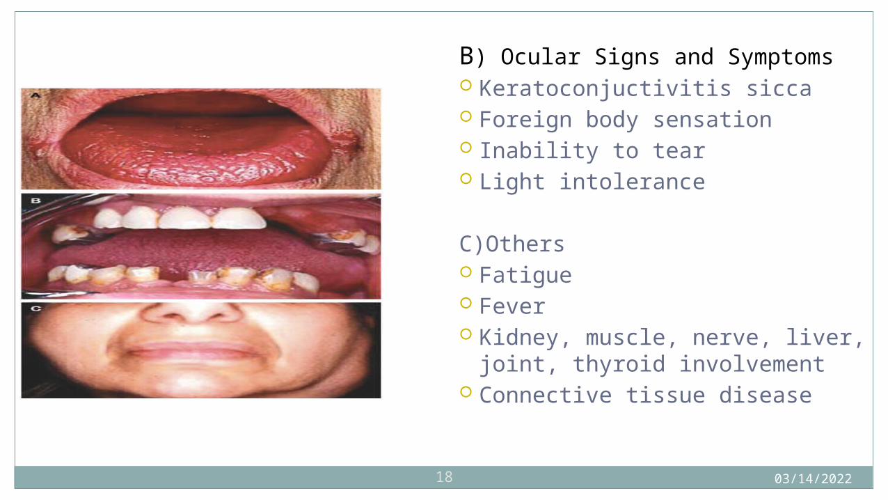
Clinical presentationsA)oral signs and symptoms Dry mouth
and erythematous mucosa Cracker sign Burning sensation Salivary gland swelling and sialedinitis Caries Candidiasis Abnormal taste and malodour
04/15/202318
B) Ocular Signs and Symptoms Keratoconjuctivitis sicca Foreign body sensation Inability to tear Light intolerance
C)Others Fatigue Fever Kidney, muscle, nerve, liver, joint,
thyroid involvement Connective tissue disease
04/15/2023
19
SYSTEMIC SCLEROSIS
Autoimmune collagen vascular disease characterized by fibrosis, vascular alterations
Systemic sclerosis describes the multisystem nature.Clinical types: 1)circumscribed plaques or bands 2)linear morphea 3)fronto parietal lesion with or without hemiatrophy of face
04/15/202320
IncidenceGreatest incidence in 30-50 years of age.F:M = 3-6:1
EtiologyGenetic predisposition Microchimerism.CytomegalovirusOrganic solvent and other chemicalsChronic renal failure.Drugs such as Bleomycin
04/15/2023
21
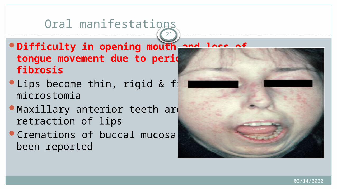
Oral manifestations
Difficulty in opening mouth and loss of tongue movement due to perioral and lingual fibrosis
Lips become thin, rigid & fixed producing microstomiaMaxillary anterior teeth are exposed due to retraction
of lipsCrenations of buccal mucosa and tongue has been
reported

04/15/2023
22
Tongue, soft palate & larynx are usually involved causing mild edema followed by atrophy & induration of mucosal & muscular tissues.
Tongue becomes stiff & border like causing difficulty in eating & speaking.
Gingival tissues are pale & firm.Dysphagia, limited mouth opening, TrismusSalivary changes similar to Sjogren syndrome

04/15/2023 23
Classic radiographic finding is generalized symmetric widening of periodontal ligament space(2-4 times) which is seen in almost all the patient.
Bone resorption at the angle of mandibleAnd may destroy condylar and coronoid process of mandible
04/15/2023
24
Dental aspect
Dysphagia and pulmonary, cardiac or renal disease as potential C.I for G.A
Constriction of oral orifice leading to limited mouth opening(fish mouth)
Tongue become stiff and less mobile due to submucosal tissue involvement(chicken tongue)
Involvement of peri-articular tissue of TMJ together with microstomia limits assess to the mouth.
04/15/2023
25
The mandibular angle may be resorbed of rarely there is resorption of TMJ.
Penicillamine therapy may cause loss of taste, oral ulceration, lichenoid reaction and other complications

04/15/2023
26 pemphigus
Derieved from Greek word; pemphix: bubble\blister)
Refers to group of auto immune disease characterized by development of vesiculobullous lesion in the skin and mucous membrane.

04/15/2023
27Clinical features
Vesiculo – bullous lesions
Oral mucosa involvement (50-70%)
Nikolsky’s sign (+)
Bulla spread sign (+)
Sites – scalp, face, trunk, groin

04/15/2023
28
Oral manifestation
About 90% of the patients show oral lesion whereas 60% of the patients present as the first sign in oral mucosa.
May occur anywhere in the oral mucosa however common sites are palate, buccal, mucosa, gingiva
In gingiva may give rise to desquamative gingivitis
04/15/2023
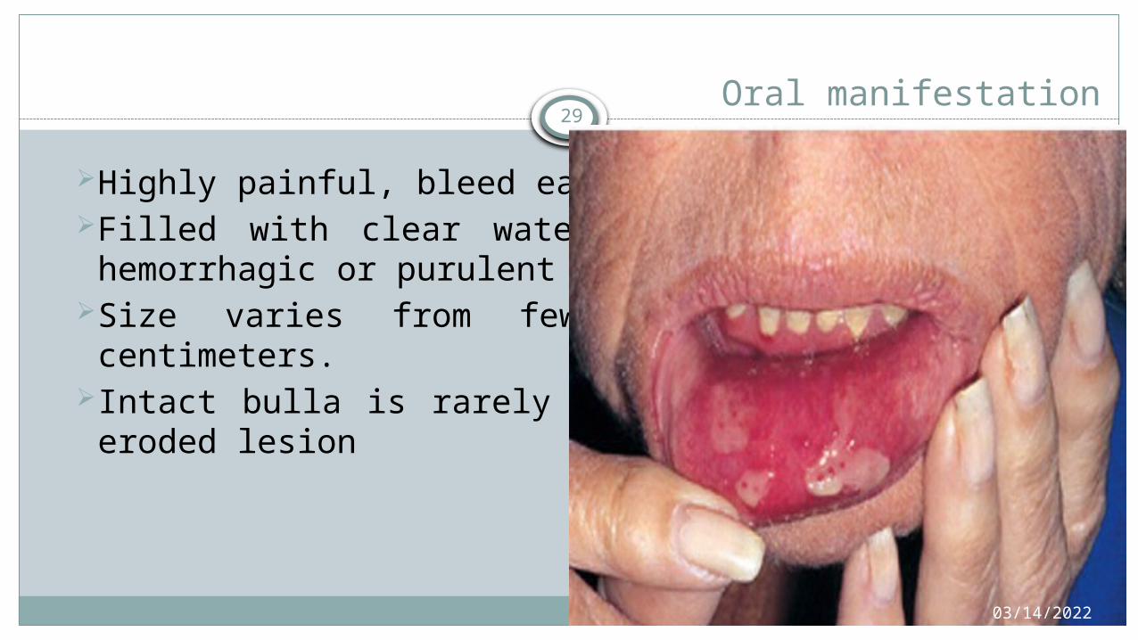
29 Oral manifestation
Highly painful, bleed easilyFilled with clear watery fluid which may become hemorrhagic
or purulent with ragged borders.Size varies from few millimeters to several centimeters.Intact bulla is rarely seen, most of the time its eroded lesion
04/15/2023 30
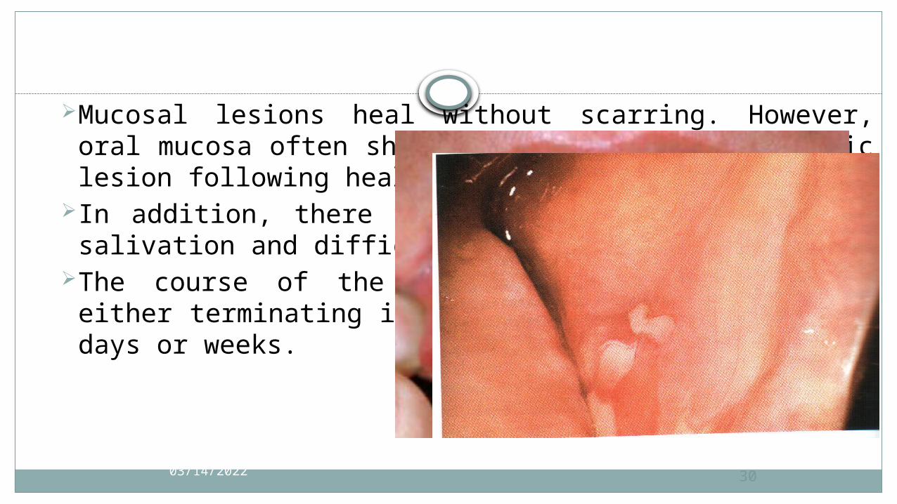
Mucosal lesions heal without scarring. However, oral mucosa often shows the formation of keratotic lesion following healing
In addition, there will be foul smell, excessive salivation and difficulty in taking food
The course of the disease is very rapid, one either terminating in death or recovery within few days or weeks.
04/15/2023
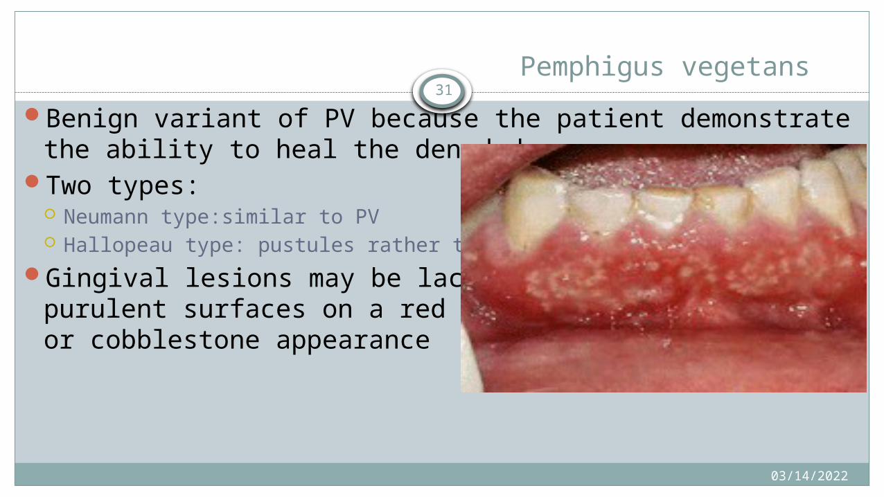
31 Pemphigus vegetans
Benign variant of PV because the patient demonstrate the ability to heal the denuded areas.
Two types: Neumann type:similar to PV Hallopeau type: pustules rather than bullae
Gingival lesions may be lace-like ulcers with purulent surfaces on a red base or have a granular or cobblestone appearance
04/15/2023
32
Pemphigoid
Two major types of pemphigoidI. Bullous pemphigoid.II. Mucous membrane pemphigoid.
Though in contrast to pemphigus, lesion are larger and leave denuded area while they brust, do heal and rarely fatal.

04/15/2023
33
Bullous pemphigoid (BP)
Age - 60 – 80 years
Sex - M : F = 2 : 1Pathogenesis:
Interaction of auto-antibody with BP antigen on the
lamina lucida of the basement membrane
Complement activation and attraction of neutrophils and
eosinophils
04/15/2023
34Clinical features
Bullous lesions
Sites - Axillae, acral region, trunk
Oral mucosa - usually not often
04/15/2023
35
04/15/202336
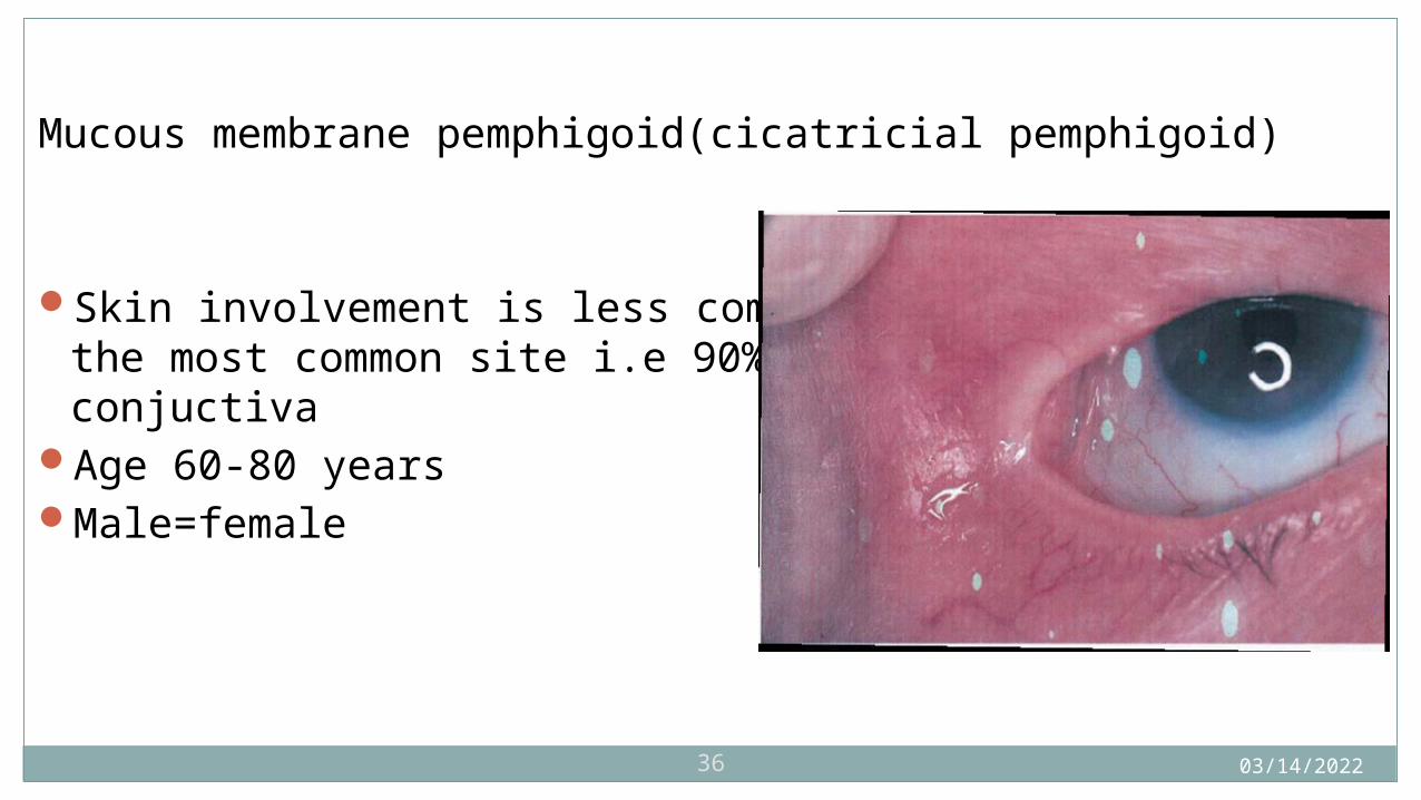
Mucous membrane pemphigoid(cicatricial pemphigoid)
Skin involvement is less common, oral mucosa being the most common site i.e 90% and 2nd being the conjuctiva
Age 60-80 yearsMale=female
04/15/2023
37 Clinical presentations– Common sites: limbs, abdomen, axilla, face and groin
– Urticaria or eczema may persist for several weeks to several months before vesico-bullous lesion appear.
– These vesicles and bullae are thick-walled and tensed and may remain for some days. Rupture doesn’t always occur.
– Oral, eye and skin lesions are present.
– Bulla spread sign and Nikolsky’s sign are negative.
– Lesions heal with milia formation.
04/15/2023
38
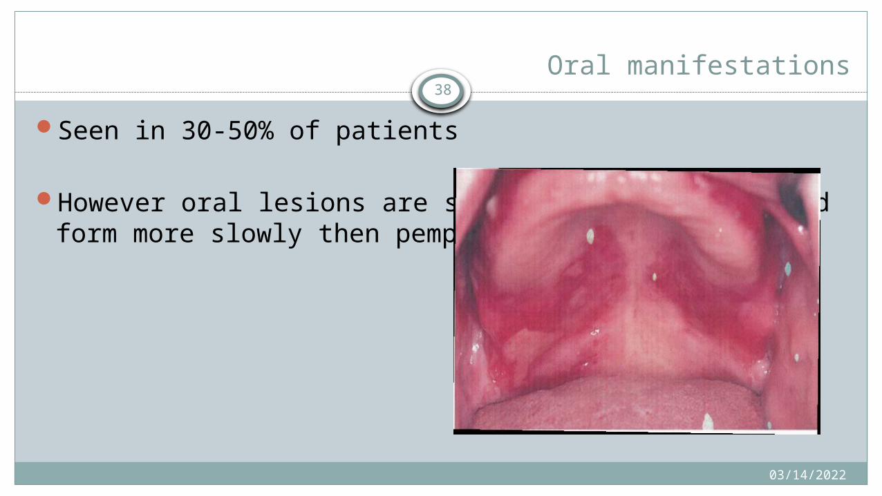
Oral manifestations
Seen in 30-50% of patients
However oral lesions are smaller, less painful and form more slowly then pemphigus vulgaris
04/15/2023
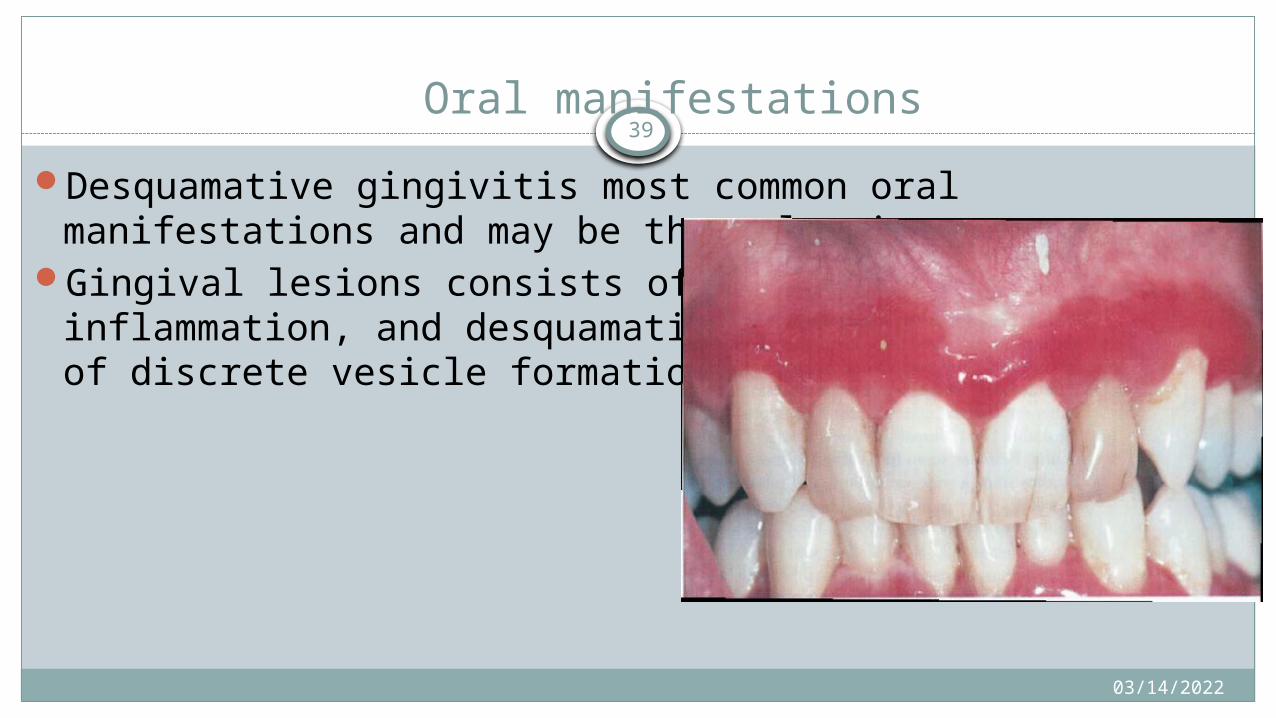
39Oral manifestations
Desquamative gingivitis most common oral manifestations and may be the only site
Gingival lesions consists of generalized edema, inflammation, and desquamation with localized areas of discrete vesicle formation

04/15/2023
40
Lichen planus
It is a rather common chronic mucocutaneous disease, which probably arises due to an abnormal immunological reaction.
Disease presented with characterstic volaceous polygonal puritic papules.
Malignant tranformation potential = 0.4-2%.
04/15/2023
41
Etiology
Exact etiological cause is unknown. Predisposing factors:I. Emotional stress.II. Trauma and infection.III. Grinspan’s syndrome.IV. Hereditary.
04/15/2023
42
Clinical feature
I. Age >40 years.II. Female to male ratio 1.4:1.III. Skin lesions. Small angular flat-topped only few mm in diameter. On course of
disease they may be discrete or gradually coalesce into larger plaque cover by fine glistening scale.
Initially disease lesion appears red, but they soon take reddish , purple and violaceous hue. Later on dirty brown colour develops.
Wickham’s straie.

04/15/2023
43
Oral manifestations- Commonly occurs in mucosal surface of buccal mucosa , vestibules,
tongue,lips, floor of the mouth, palate.
Appears weeks or months before the skin lesions.
Lesions are bilaterally symmetrical.
04/15/2023
44
Usually asymptomatic but patient may complaint of pain, burning sensation and intolerance to spicy food
Begins as small white papules that coalesce to form interlacing network of white lines (Wickham’s striae)
Erythematous atrophic and erosive (erosive OLP) white lesions are common

04/15/2023 45
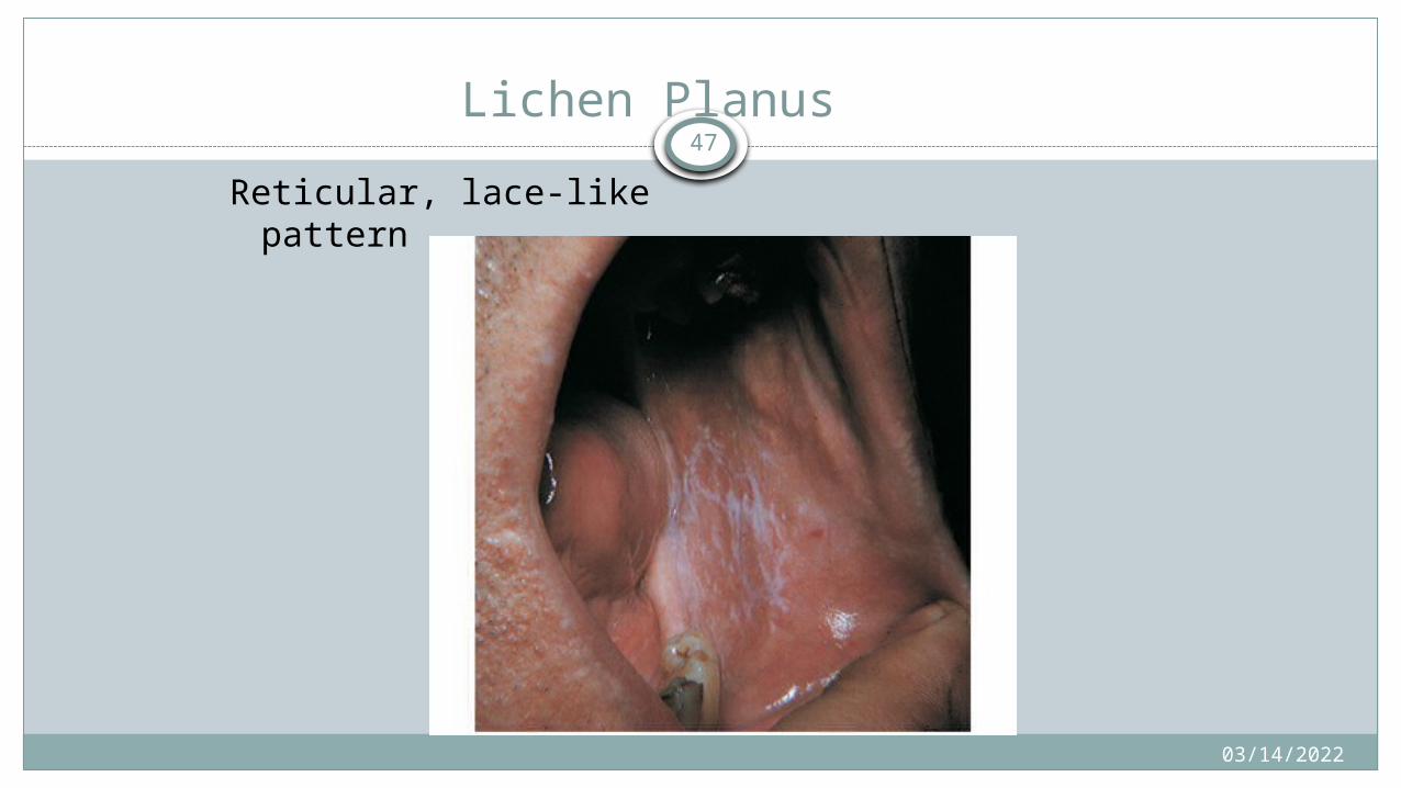
Reticular OLP has classical features and do not require biopsy to diagnose
Hypertrophic white plaques may occur sometimes and intact bullae (bullous OLP) are rarely seen
04/15/2023
46
Common site is buccal mucosa(78-87%),followed by tongue and gingiva
Koebner’s phenomenon present
Oral lesions tend to be bilateral and symmetrical
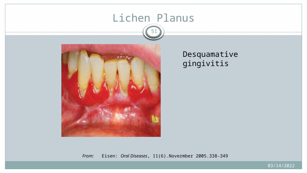
Erosive lesion on gingiva resembles desquamative gingivitis
04/15/2023
47
Lichen Planus
Reticular, lace-like pattern

04/15/2023
48
Atrophic
Lichen Planus
04/15/2023
49
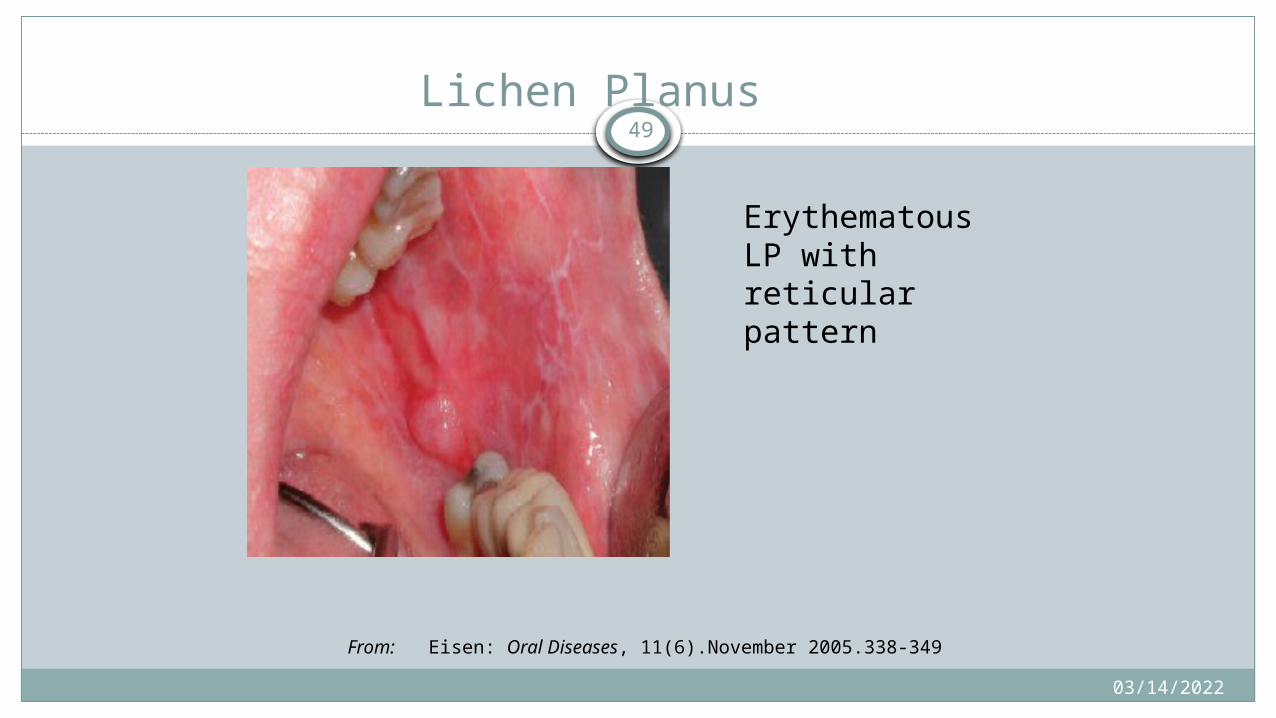
Lichen Planus
Erythematous LP with reticular pattern
From: Eisen: Oral Diseases, 11(6).November 2005.338-349

04/15/2023
50
Lichen Planus
Erosive
04/15/2023
51
Lichen Planus
Desquamative gingivitis
From: Eisen: Oral Diseases, 11(6).Novermber 2005.338-349
04/15/2023
52
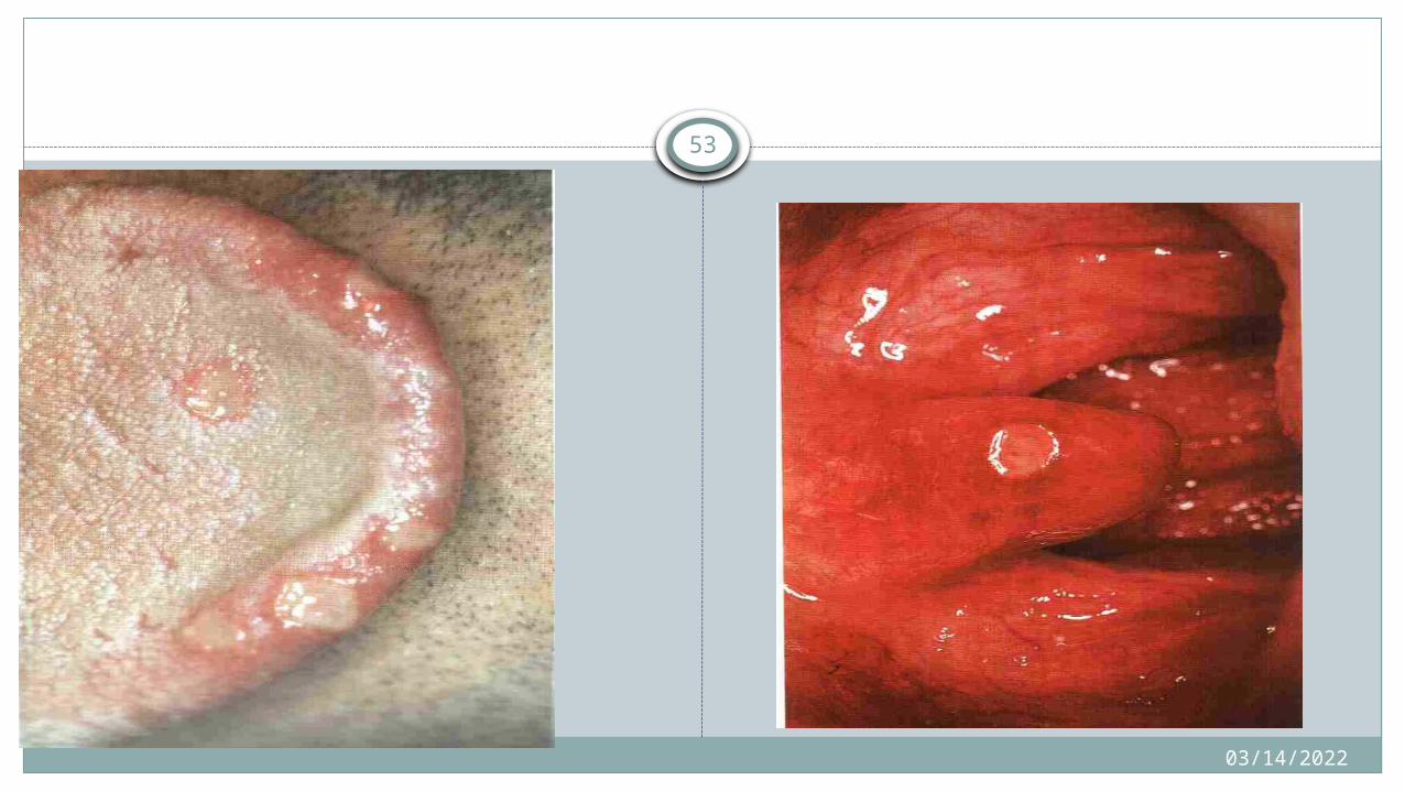
Recurrent aphthous stomatitis
Definition: Recurring episodes of small, round or oval ulcers with circumscribe margin, erythematous halo and a yellow or grey floor, present from childhood or adolescence and each episode last for 1 to 4 weeks before healing
04/15/2023
53
04/15/2023
54
Clinical Features
Common disease afflicts 20% of the population
High prevalence: higher socio-economic classes
Three main clinical types of RAS:1. Minor aphthous ulcers (Mikulicz ulcers): 80% 2. Major aphthous utcer (Sutton's ulcers): 10%
3. Herpetiform type ulceration: 10%
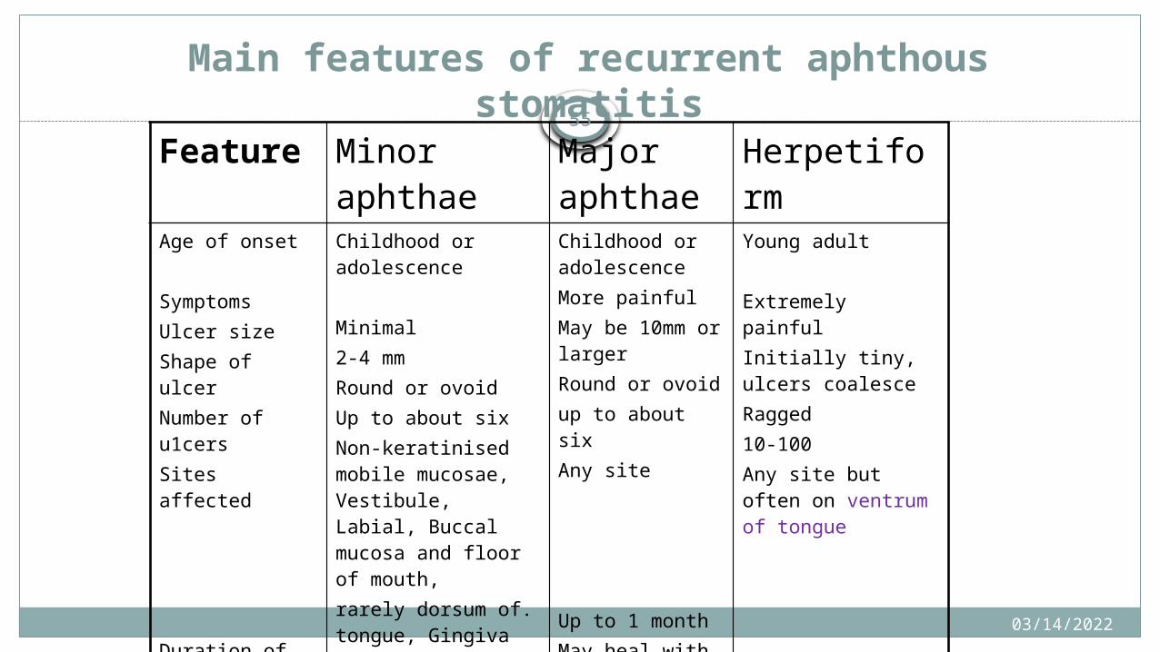
Main features of recurrent aphthousstomatitis
Feature Minor aphthae Major aphthae
Herpetiform
Age of onset
Symptoms Ulcer sizeShape of ulcerNumber of u1cers Sites affected
Duration of each UlcerOther comments
Childhood or adolescence
Minimal2-4 mm Round or ovoidUp to about sixNon-keratinised mobile mucosae, Vestibule, Labial, Buccal mucosa and floor of mouth,rarely dorsum of. tongue, Gingiva or palateUp to 10 daysHeal with little or no scar
Childhood or adolescenceMore painfulMay be 10mm or largerRound or ovoidup to about sixAny site
Up to 1 monthMay heal withscarring
Young adult
Extremely painfulInitially tiny, ulcers coalesceRagged10-100Any site but often on ventrum of tongue
Up to I monthAffect females predominantly
04/15/2023
55
04/15/2023
56
BEHCET’S DISEASE
Rare multisystem, chronic disease characterized by oral and genital mucocutaneous ulcerations, skin rashes, arthritis, thrombophlebitis, uveitis, colitis, and neurologic symptoms.
Also known adamatiades syndrome.

04/15/2023
57
Reiter’s syndrome
Disease of unknown etiology. Common on age group of 25-35 years.Consider as an important complication of non gonococcal urethritis.Oral lesion occurs in less than 5% to 50% of patient with disease.Consist of tetrad of :1. Urethritis 2. Arthritis. 3. Conjunctivities.4. Mucocutaneous lesion.
04/15/2023
58 Oral manifestationsLesions are painless, red, slightly elevated areas with a white
circinate border on the buccal mucosa, lips and gingiva.
Palatal lesions appears as small ,bright red purpuric spots which darken and coalese. Tongue represents “geographic tongue”.
Note: Clinically similar lesion are seen on the glans penis, producing a circinate balanitis

04/15/2023
59Wegner granulomatosis
Disease of unknown etiology.Involve vascular renal and respiratory systems.Disease characterize by necrotizing and granulomatous vasculitis.Clinical types:1. Generalize or classic wegener’s granulomatosis.2. Localized or limited wegener’s granulomatosis.
3. Superficial wegener’s granulomatosis.
04/15/2023
60Oral manifestations
Starts with tumor like vegetations in mouth and nose, then inflammatory process starts in nose and inter dental papilla spreading upto periodontium.
Ulceration can occur, usually perforating in nature.
Strawberry gingiva characterized by ulceration, friable granular lesions.
Mobile teeth .
Palatal destruction resulting in oro nasal communication.
04/15/2023
61Oral manifestations of HIV
CLASSIFICATIONGroup 1 : LESIONS STRONGLY ASSOCIATED WITH HIV INFECTIONS Candidiasis : Eythematous Pseudomembranous Hairy leukoplakia Kaposi’s sarcoma Non- Hodgkin’s lymphoma
04/15/2023
62
Periodontal diseases : Linear gingiva erythema Necrotizing ulcerative gingivitis Necrotizing ulcerative periodontitis
Group 2 LESIONS LESS COMMONLY ASSSOCIATED WITH HIV Bacterial infections: M.avium - intracellulare M. tuberculosis Melanotic hyperpigmentation Necrotizing ulcerative stomatitis
04/15/2023
63
LESIONS SEEN IN HIV INFECTION Bacterial infections :Actinomyces israeli Escherichia coli Kliebsella pneumoniae Cat scratch disease
Drug reactions: Ulcerative Erythema multiforme Lichenoid reaction Toxic epidermolysis
04/15/2023
64
• Fungal infections other than candidiasis Cryptococcus neoformans Geotrichum candidiasis Histoplasma capsulatum Zygomycosis Aspergillus flavus
• Neurologic disturbances : Facial palsy Trigeminal neuralgia• Recurrent apthous stomatitis
04/15/2023
65
Oral manifestation can be broadly clasified as
Fungal lesions Viral lesions Bacterial lesions Neoplastic lesions HIV associated other lesions
04/15/2023
66
Fungal lesions
CandidiasisHistoplasmosisCryptococcus neoformans
04/15/2023
67
Viral lesions
Herpes simplexHerpes zosterHuman papilloma virusCytomegalovirusEpstein-Barr Virus(oral hairy leukoplakia)

04/15/2023
68
Bacterial lesion
Periodontal diseases : Linear gingival erythema Necrotizing periodontal disease(NUG,NUP)
TuberculosisSyphilis

04/15/2023
69
Neoplastic lesions
Kaposi’s sarcoma Oral squamous cell carcinoma Non –Hodgkin’s lymphoma
04/15/2023
70
Other lesions
Recurrent apthous ulcerHIV associated salivary gland disease
04/15/2023
71
CANDIDIASIS
The most common opportunistic infectionCD4 cells counts< 400 cells/mm^3 4 clinical presentation: Pseudomombranous or oral thrush Erythematous Hyperplastic Angular cheilitis
04/15/2023
72
Pseudomembranous candidiasis
Most common type. Usually asymptomatic. Usually extensive involving more than one site Manifests as white or yellowish single or confluent plaque that is
scrappable, exposing erythematous area. May also extend to oropharynx and esophagus.
04/15/2023
73
Erythematous/ atrophic
Site: dorsum of the tongue,palate and buccal mucosa. Appears as a reddish macular lesion, or depappilation on the dorsum
of the tongue. May present alone or in combination with pseudomembranous
candidiasis. Tongue lesion are also referred as central papillary atrophy. Long standing lesion:mucosal ulceration Some may complain of occasional burning sensation .

04/15/2023
74
Hyperplastic
Uncommon and found in severly immunocompromised. Manifests as white or discolored plaques which cannot be scrapped
out Burning sensation, Dysphagia Feeling of having large piece of cotton in mouth
04/15/2023
75
Angular cheilitis
Can be due to mixed infection of candida albicans and staphylococcus aureus.
Manifests as erythema &/or fissuring and/ or scaling of angle of mouth.
May be present with intraoral candidiasis.

04/15/202376
pseudomembranous Erythematous
Hyperplastic

04/15/2023
77
Thank you

04/15/2023
78
Oral Manifestations of systemic diseases II
04/15/2023
79
Learning objectives
Be able to enumerate the major oral
manifestations of blood disorders,renal
disorders,GI disorder, diabetes, respiratory
diseases
Be able to list the dental considerations and
management of these systemic disorders

04/15/202380
Blood Disorders
Disorders of Red Blood Cells and Hemoglobin
Disorders of White Blood Cells
Bleeding Disorders

04/15/202381
Disorders of Red Blood Cells and Hemoglobin
Iron Deficiency AnemiaPernicious AnemiaFolic Acid and Vitamin B12 Deficiency AnemiaThalassemiaSickle Cell AnemiaAplastic AnemiaPolycythemia
04/15/202382
Anemia
A reduction in the oxygen-carrying capacity of blood
Most often related to a decrease in the number of circulating red blood cells
Nutritional anemiasA deficiency in a substance required for the normal development of
red blood cells, commonly vitamins
Suppression of bone marrow stem cells
04/15/202383
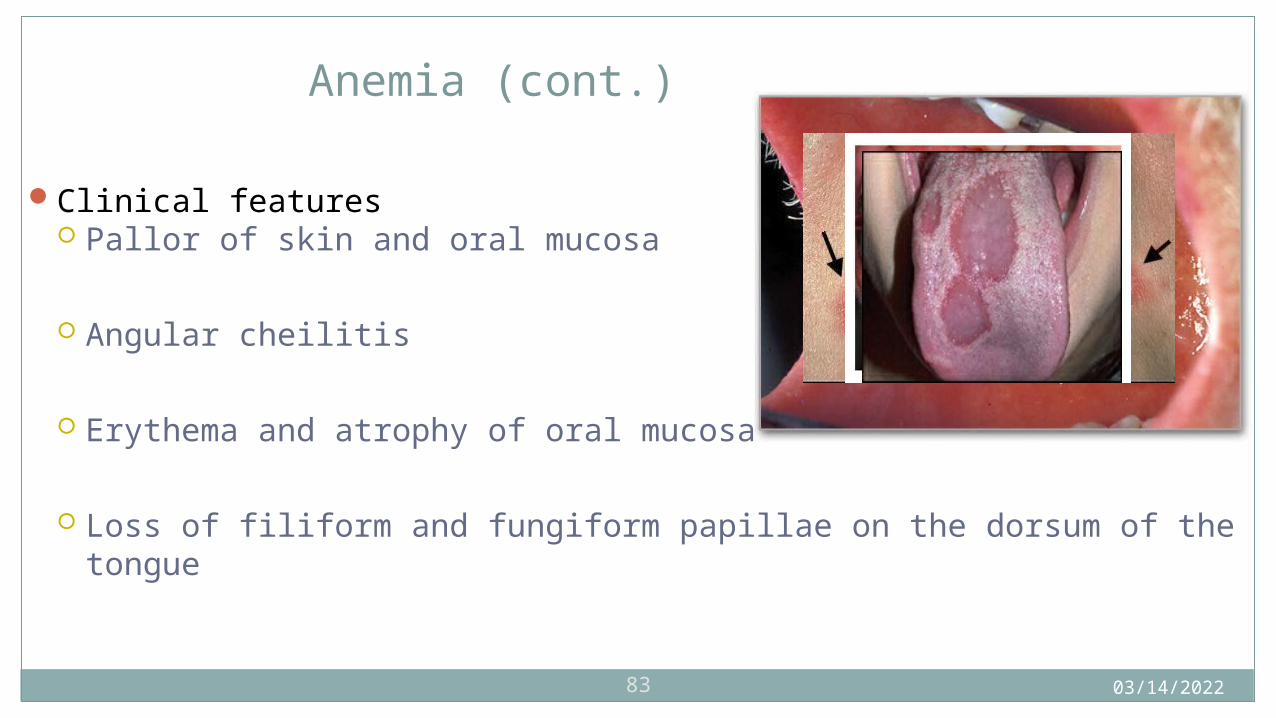
Anemia (cont.)
Clinical features Pallor of skin and oral mucosa
Angular cheilitis
Erythema and atrophy of oral mucosa
Loss of filiform and fungiform papillae on the dorsum of the tongue
04/15/202384
Iron Deficiency Anemia
An insufficient amount of iron is supplied to bone marrow for red blood cell development.
May occur as a result of deficient iron intake, blood loss from heavy menstrual bleeding or chronic gastrointestinal bleeding, poor iron absorption, or an increased requirement for iron in situations such as pregnancy or infancy
Plummer-Vinson syndrome may result from long standing iron deficiency anemia. Includes dysphagia, atrophy of the upper alimentary tract, and a predisposition to
developing oral cancer
04/15/202385
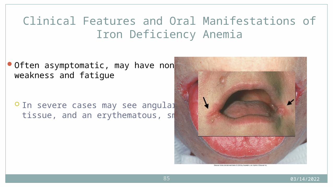
Clinical Features and Oral Manifestations of Iron Deficiency Anemia
Often asymptomatic, may have nonspecific symptoms such as weakness and fatigue
In severe cases may see angular cheilitis, pallor of oral tissue, and an erythematous, smooth, painful tongue
04/15/202386
Pernicious Anemia
Probably an autoimmune disorder in most situations
May be caused by removal of the stomach, gastric cancer, or gastritis
Caused by a deficiency in intrinsic factor
Intrinsic factor is secreted by parietal cells in the stomach; it is necessary for absorption of vitamin B12
04/15/202387
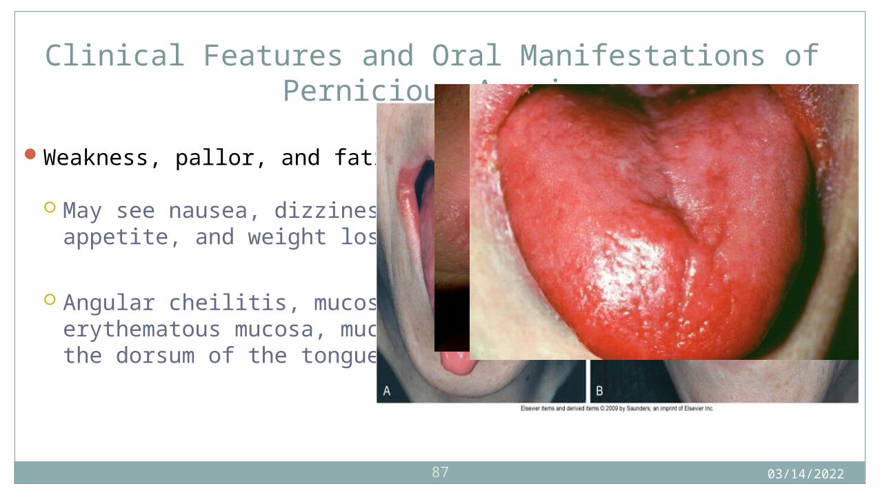
Clinical Features and Oral Manifestations of Pernicious Anemia
Weakness, pallor, and fatigue on exertion
May see nausea, dizziness, diarrhea, abdominal pain, loss of appetite, and weight loss
Angular cheilitis, mucosal pallor, painful atrophic and erythematous mucosa, mucosal ulceration, loss of papillae on the dorsum of the tongue, and burning and painful tongue
04/15/202388
Folic Acid and Vitamin B12 Deficiency Anemia
From dietary deficiencies
Can occur in association with malnutrition
May be found with alcoholism or pregnancy

04/15/202389
Oral Manifestations of Folic Acid and Vitamin B12 Deficiency Anemia
Oral manifestations are indistinguishable from those of pernicious anemia.

04/15/202390
Thalassemia (Mediterranean or Cooley Anemia)
A group of inherited disorders of hemoglobin synthesis
An autosomal dominant inheritance pattern
The heterozygous form may be mildly symptomatic or asymptomatic.
The homozygous form is associated with severe hemolytic anemia.

04/15/202391
Clinical Features and Oral Manifestations of Thalassemia
Yellow skin pallor, fever, malaise, and weakness
The face includes prominent cheekbones, depression of the bridge of the nose, a prominent maxilla, and protrusion or flaring of maxillary anterior teeth.
Radiographs may show a “salt and pepper” pattern.
Some trabeculae are prominent, and others are blurred.
04/15/202392
Sickle Cell Anemia
An inherited blood disorder
When someone is heterozygous, it is called sickle cell trait.
When someone is homozygous, they are much more severely affected.
04/15/2023
93
Occurs before age 30 and is more common in women than in men
The red blood cells develop a sickle shape when there is decreased oxygen.
This can be triggered by exercise, exertion, administration of a general anesthetic, pregnancy, or even sleep.
04/15/202394
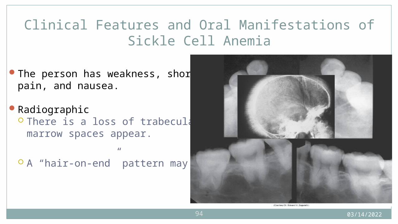
Clinical Features and Oral Manifestations of Sickle Cell Anemia
The person has weakness, shortness of breath, fatigue, joint pain, and nausea.
Radiographic There is a loss of trabeculation, and large, irregular marrow spaces
appear.
A “hair-on-end” pattern may be seen in the skull.
04/15/202395
Aplastic Anemia
A severe depression of bone marrow activity causes a decrease in all circulating blood cells. – pancytopenia
Primary aplastic anemia – the cause is unknown Secondary aplastic anemia – a result of a drug or chemical agent

04/15/202396
Oral Manifestations of Aplastic Anemia
Infection, spontaneous bleeding, petechiae, and purpuric spots
04/15/202397
Polycythemia
Polycythemia Vera
Secondary Polycythemia
Relative Polycythemia
04/15/202398
Oral Manifestations of Polycythemia
The oral mucosa may appear deep red to purple; the gingiva may be edematous and bleed easily.
Submucosal petechiae, ecchymosis, hematoma formation may be present.
04/15/202399
Disorders of White Blood Cells
Agranulocytosis
Cyclic Neutropenia
Leukemia
04/15/2023100
Clinical Features and Oral Manifestations of Agranulocytosis
Sudden onset of fever, chills, jaundice, weakness, sore throat
Oral infection
04/15/2023101
Leukemia
Malignant neoplasms of hematopoietic stem cells
Characterized by an excessive number of abnormal white blood cells in circulating blood
Unknown cause; some are investigating oncogenic viruses
There are many different types categorized as to whether they are acute or chronic.
04/15/2023102
Acute Leukemias
Characterized by very immature cells and a rapidly fatal course if not treated
Acute lymphoblastic leukemia – involves immature lymphocytes
Primarily affects children and young adults
Good prognosis Acute myeloblastic leukemia – involves immature granulocytes
Primarily affects adolescents and young adults.
Prognosis is not as good.

04/15/2023103
Clinical Features of Acute Leukemias
Weakness, fever, enlargement of lymph nodes, bleeding
04/15/2023104
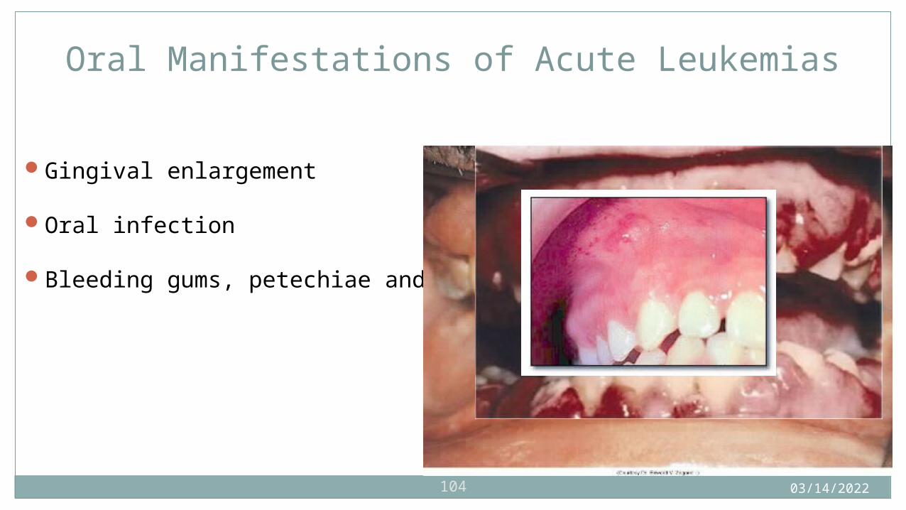
Oral Manifestations of Acute Leukemias
Gingival enlargement
Oral infection
Bleeding gums, petechiae and ecchymosis
04/15/2023105
Chronic Leukemias
Slow onset
Primarily affect adults
04/15/2023106
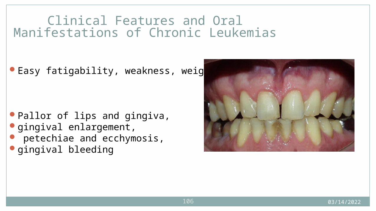
Clinical Features and Oral Manifestations of Chronic Leukemias
Easy fatigability, weakness, weight loss, anorexia
Pallor of lips and gingiva, gingival enlargement, petechiae and ecchymosis, gingival bleeding
04/15/2023107
Bleeding Disorders
Purpura
Hemophilia
04/15/2023108
Purpura
A reddish-blue or purplish discoloration of skin or mucosa from spontaneous extravasation of blood
May be due to a defect or deficiency in blood platelets
Blood may ooze from gingival margins.
04/15/2023109
Thrombocytopenic Purpura
A bleeding disorder that results from a severe reduction in circulating platelets
Idiopathic thrombocytopenic purpura
If the cause is unknown
Immune thrombocytopenia
An autoimmune type of process
Secondary thrombocytopenic purpura
Often associated with drugs
04/15/2023110
Clinical and Oral Manifestations of Thrombocytopenic Purpura
Spontaneous purpuric or hemorrhagic lesions on the skin
Patients bruise easily, may have blood in urine, have frequent nose bleeds.

04/15/2023111
Nonthrombocytopenic Purpura
Bleeding disorders that can result from either a defect in capillary walls or disorders of platelet function
Vitamin C deficiency and infections or chemicals and allergy may be the cause of alterations in vascular walls.
Drugs, allergy, and autoimmune disease may cause disorders of platelet function.
Von Willebrand disease is an autosomal dominant disorder of platelet function.

04/15/2023112
Oral Manifestations of Nonthrombocytopenic Purpura
Spontaneous gingival bleeding
petechiae
Ecchymoses
hemorrhagic blisters
04/15/2023113
Hemophilia
A disorder of blood coagulation
Results in severely prolonged clotting time
Due to a deficiency in plasma proteins involved in coagulation

04/15/2023114
Types of Hemophilia
The two most common types are type A and type B.
Transmitted as X-linked diseases through an unaffected carrier daughter to a son
Type ACaused by a deficiency of plasma thromboplastinogen or factor VIII
Type BChristmas diseaseLess common, the clotting defect is plasma thromboplastin or factor
IX
04/15/2023115
Oral Manifestations of Hemophilia
Spontaneous gingival bleeding,
petechiae,
ecchymosis
04/15/2023
116
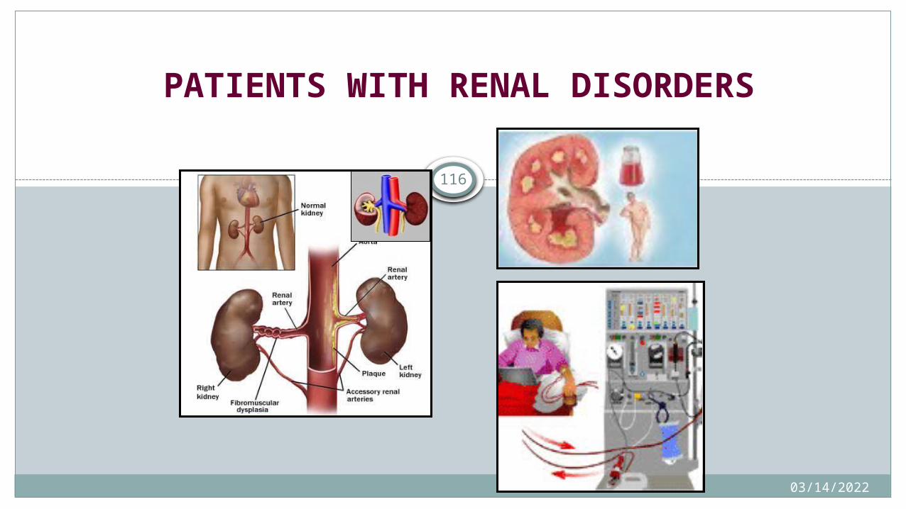
PATIENTS WITH RENAL DISORDERS
04/15/2023
117
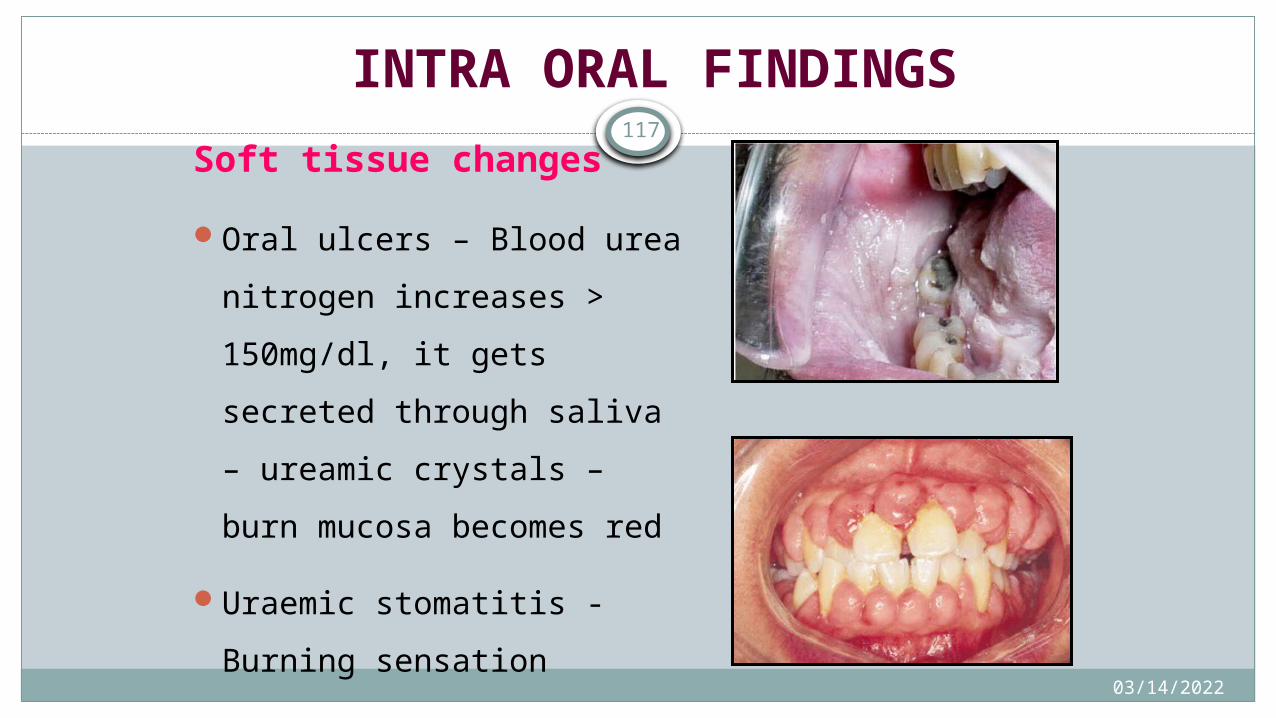
INTRA ORAL FINDINGS
Soft tissue changes
Oral ulcers – Blood urea
nitrogen increases >
150mg/dl, it gets secreted
through saliva – ureamic
crystals – burn mucosa
becomes red
Uraemic stomatitis - Burning
sensation
04/15/2023
118
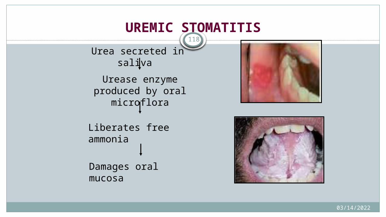
UREMIC STOMATITIS
Urea secreted in saliva
Urease enzyme produced by oral microflora
Liberates free ammonia
Damages oral mucosa

04/15/2023
119
INTRA ORAL FINDINGS
Soft tissue changes
Ulcers secondary to
anaemia, viral infections
(immunosupressed)
Gingival hyperplasia -
cyclosporine & nifedepine
04/15/2023
120
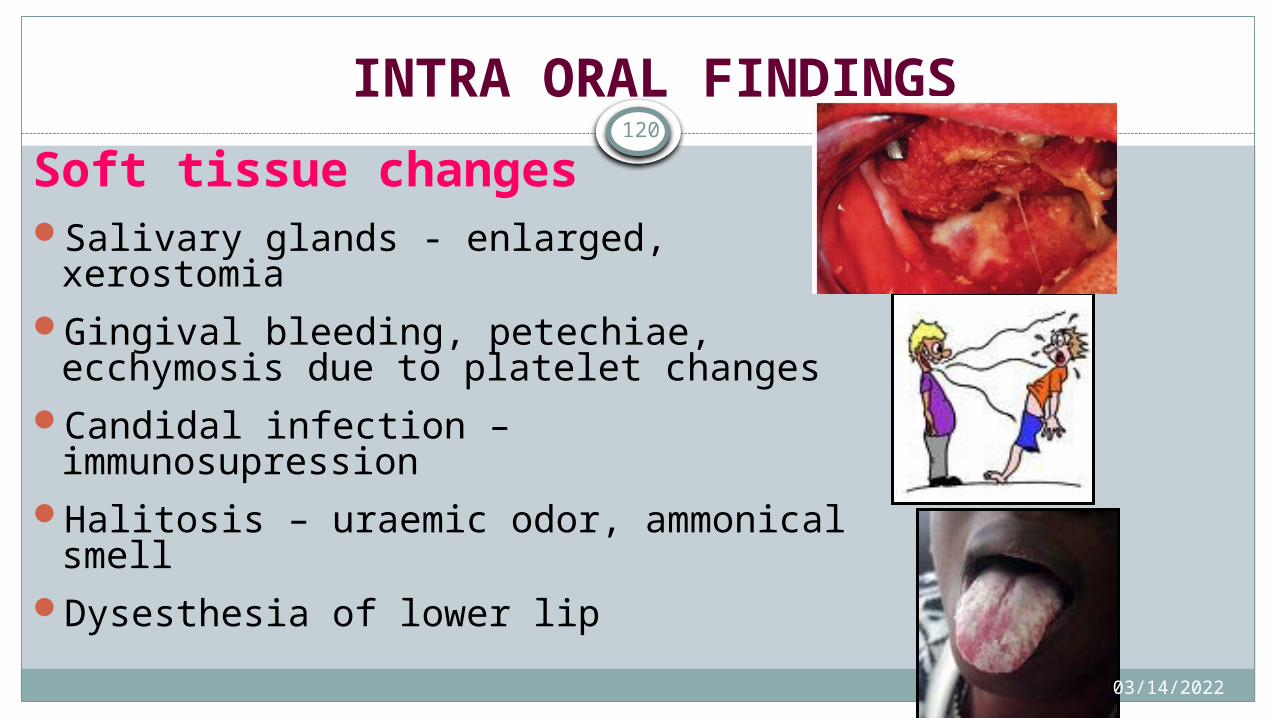
INTRA ORAL FINDINGS
Soft tissue changesSalivary glands - enlarged, xerostomiaGingival bleeding, petechiae, ecchymosis
due to platelet changesCandidal infection – immunosupressionHalitosis – uraemic odor, ammonical smellDysesthesia of lower lip
04/15/2023
121
Hard tissue changes
Staining in teeth – due to iron supplements
Reduced caries due to urea in saliva
Delayed tooth eruption
Enamel hypoplasia
Tooth Mobility
04/15/2023
122
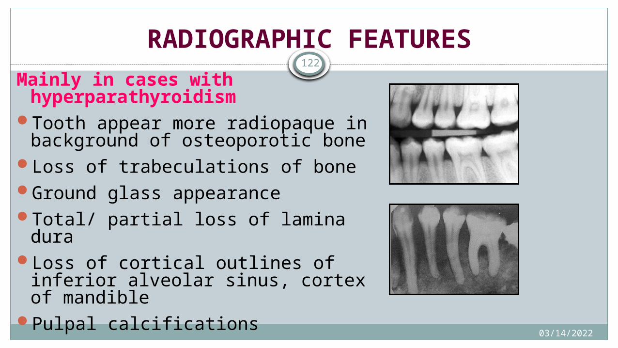
RADIOGRAPHIC FEATURESMainly in cases with
hyperparathyroidismTooth appear more radiopaque in
background of osteoporotic boneLoss of trabeculations of boneGround glass appearanceTotal/ partial loss of lamina duraLoss of cortical outlines of inferior
alveolar sinus, cortex of mandiblePulpal calcifications

04/15/2023
123
RADIOGRAPHIC FEATURES
Mainly in cases with hyperparathyroidism
Multilocular radiolucency – osteitis fibrosa cystica/ browns tumor
Arterial & oral calcifications
04/15/2023
124
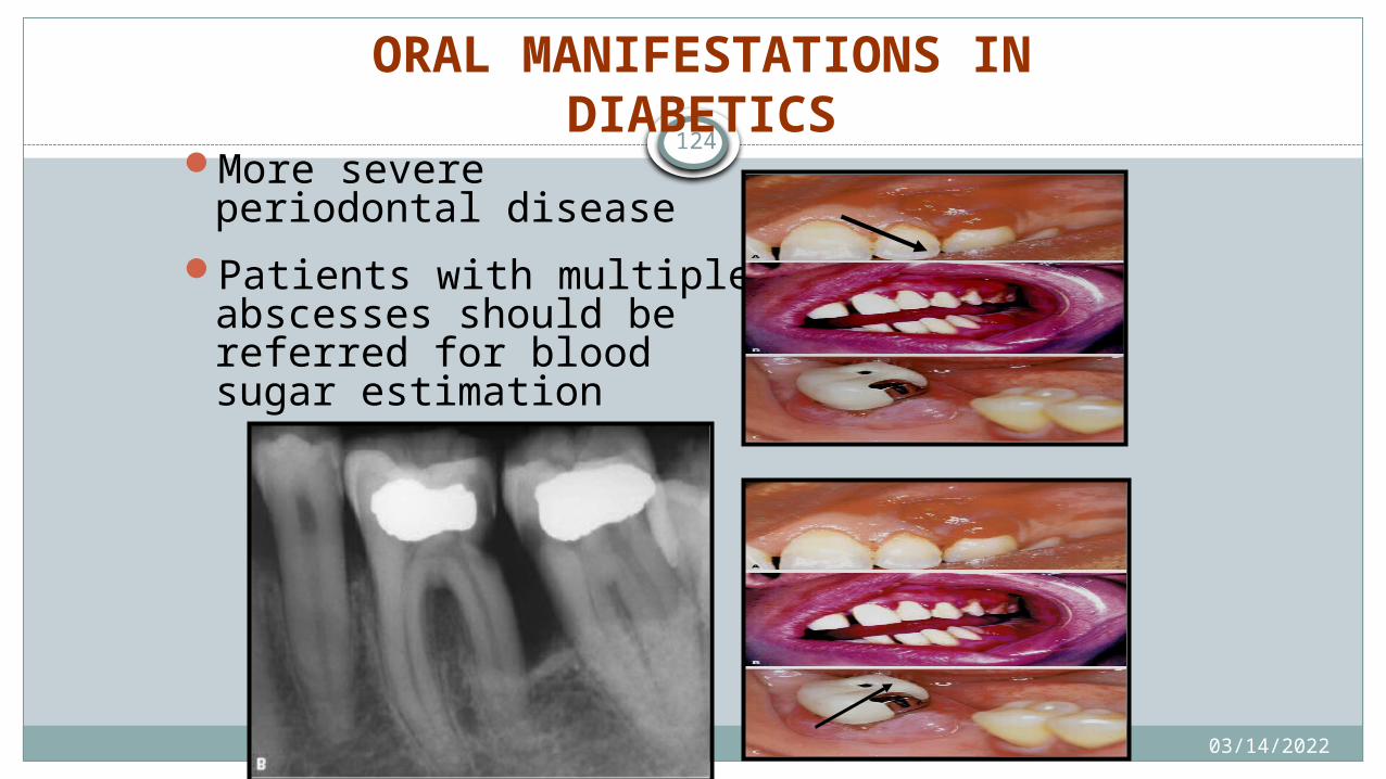
ORAL MANIFESTATIONS IN DIABETICS
More severe periodontal disease
Patients with multiple abscesses should be referred for blood sugar estimation
04/15/2023
125
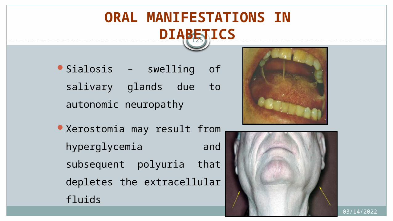
ORAL MANIFESTATIONS IN DIABETICS
Sialosis – swelling of salivary
glands due to autonomic
neuropathy
Xerostomia may result from
hyperglycemia and
subsequent polyuria that
depletes the extracellular
fluids
04/15/2023
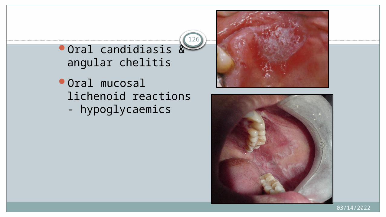
126Oral candidiasis &
angular chelitis
Oral mucosal lichenoid reactions - hypoglycaemics
04/15/2023
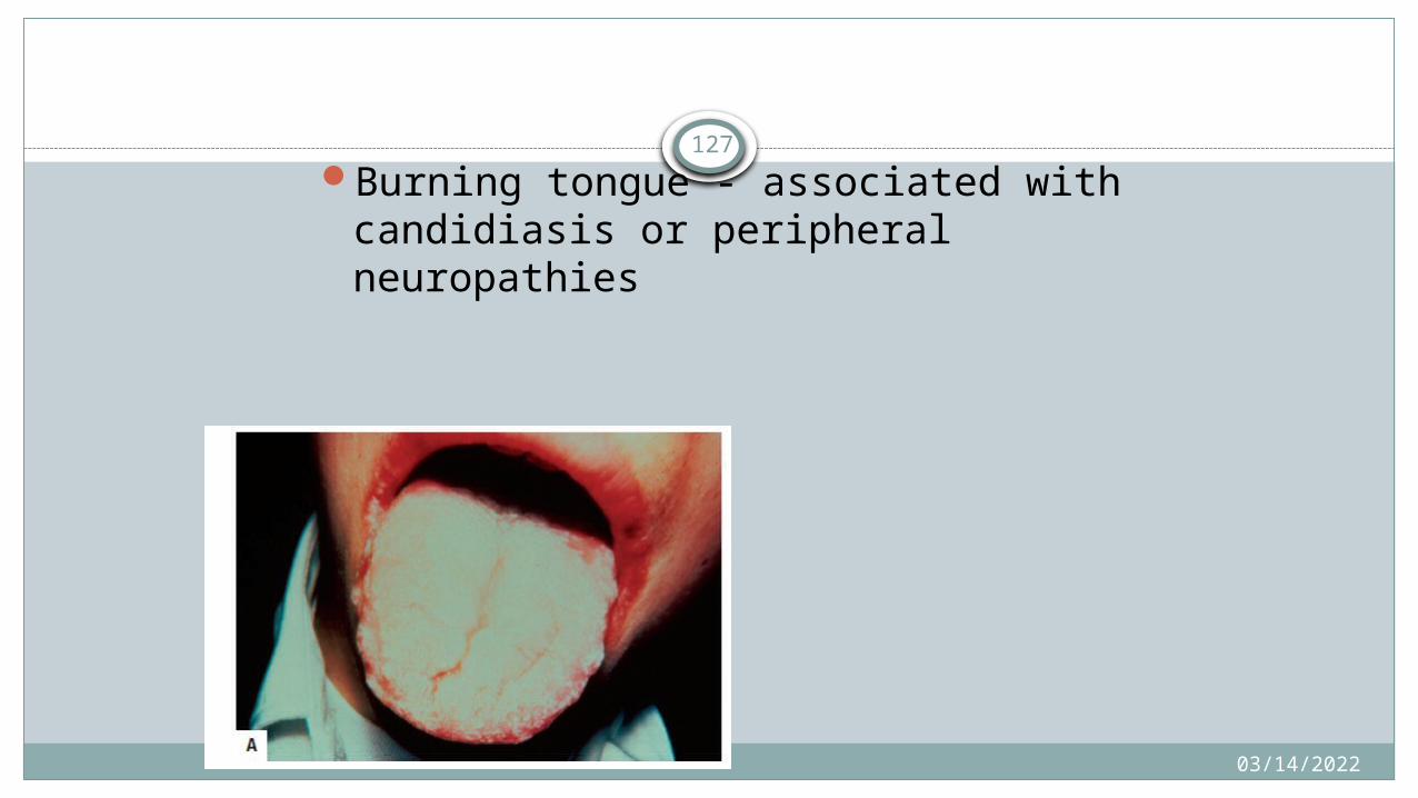
127Burning tongue - associated with
candidiasis or peripheral neuropathies
04/15/2023
128
Diseases of Respiratory tract
04/15/2023
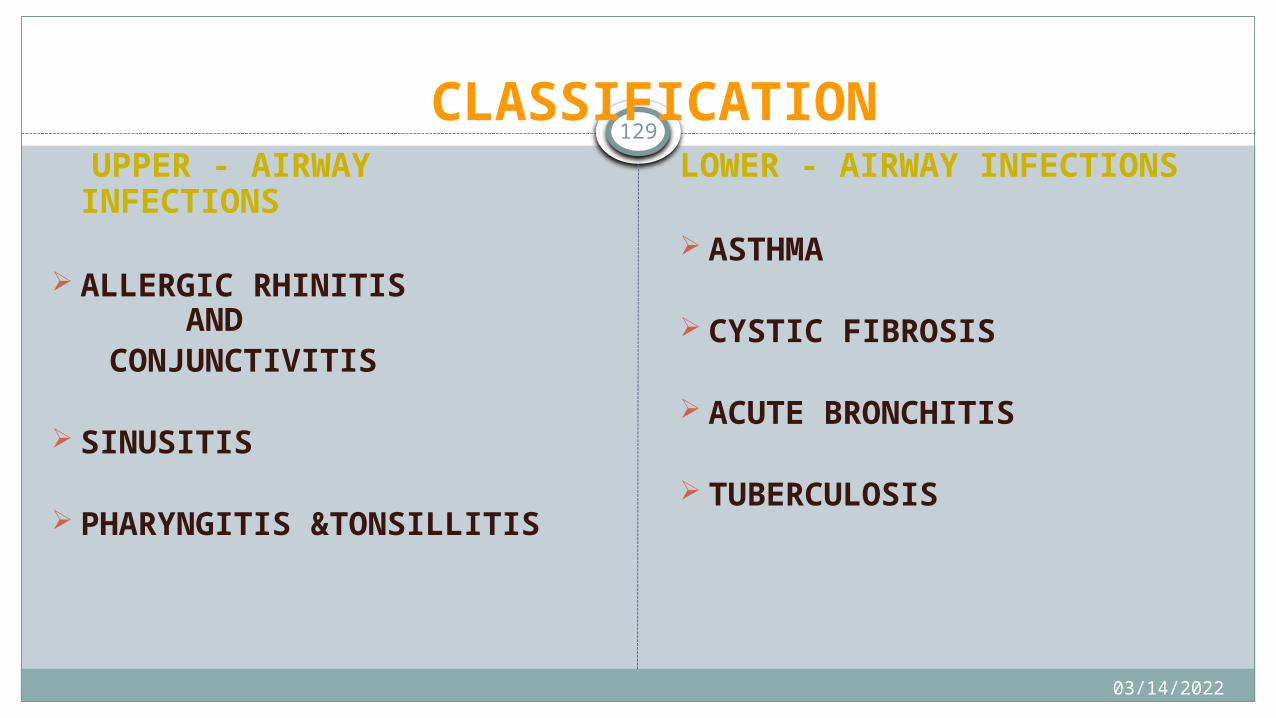
129 CLASSIFICATION
UPPER - AIRWAY INFECTIONS
ALLERGIC RHINITIS AND
CONJUNCTIVITIS
SINUSITIS
PHARYNGITIS &TONSILLITIS
LOWER - AIRWAY INFECTIONS
ASTHMA
CYSTIC FIBROSIS
ACUTE BRONCHITIS
TUBERCULOSIS
04/15/2023
130
ORAL HEALTH CONSIDERATIONS for allergic rhinitis
ORAL DRYNESS
ORAL CANDIDIASIS (CORTICOSTEROIDS)
04/15/2023
131
ORAL HEALTH CONSIDERATIONS for sinusitis
Tooth Pain - Differentiate Between An Odontogenic Infection And Sinus Pain.
Sinus Infections Usually Present With Pain Involving More Than One Tooth.
Chronic Sinus Infections Are Often Accompanied By Mouth Breathing -
Gingivitis.
Prolonged Use Of Antibiotics - The Potential Development Of Bacterial
Resistance .
04/15/2023
132
ORAL MANIFESTATIONS of Asthma
Candidiasis
Decreased Salivary Flow
Increased Calculus
Increased Gingivitis
Increased Periodontal Disease
Increased Incidence Of Caries
04/15/2023
133
CONSIDERATION FOR DENTAL CARE
Fluoride Supplements - Particular Those
Taking Β2-agonists.
The Patient Should Be Instructed To Rinse His
Or Her Mouth With Water After Using Inhalers.
Oral Hygiene Should Be Reinforced To Reduce
Of Gingivitis And Periodontitis.
04/15/2023
134
ORAL MANIFESTATION of TB
RARE
MAINLY SEEN IN MIDDLE AGED & OLDER PEOPLE
SEX-MALES
MOST COMMONLY-TONGUE

04/15/2023
135
ORAL MANIFESTATION of TB
ORAL LESION –Ulcer,nodule,vesicle,granuloma,fissure
Ulceration ,Ragged Border,minimal Induration,granular Base
Sentinal Tubercle; nodules Seen Around Ulceration.
04/15/2023
136
Pregnancy- Oral Manifestations
04/15/2023
137
Dental Considerations(First Trimester)
Dental treatment is best avoided
Assess the current oral health of the patient
Educate the patient regarding the oral changes

04/15/2023138
Nausea and vomiting – caused by increased levels of gonadotropins in the first trimester
Enamel erosion- gastric acids present in the vomit erode the inner surface of anterior teeth
Pregnancy induced gingivitis

04/15/2023
139
SECOND TRIMESTER
Organogenesis is complete and the risk to the foetus is at its lowest
safest period for any necessary elective dental treatment
140
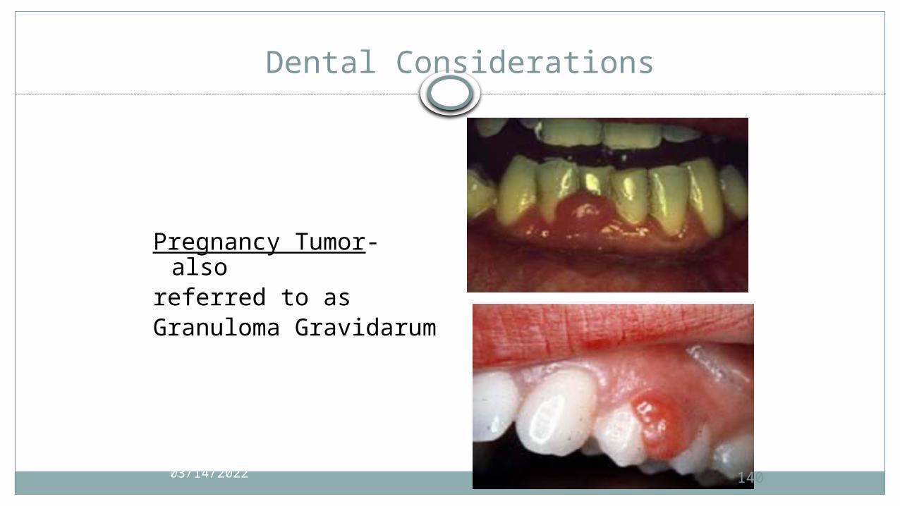
Dental Considerations
Pregnancy Tumor- also
referred to asGranuloma
Gravidarum
04/15/2023
141
Diseases of GIT
04/15/2023
04/15/2023
142GASTROESOPHAGEAL REFLUX DISEASE
Most common disease of upper GI tract
gastric contents passively move up from the stomach into the esophagus
Symptoms/signsHeart burn (pain/burning sensation extending from
epigastrium to the neck) – commonly felt after a mealesophagitis, esophageal ulceration & strictureChest pain (mimics anginal pain)Dysphagia

143
Oral Manifestations & Dental Considerations
erythema and mucosal atrophy
dysgeusia, sensitivity & erosion
(palatal aspects of upper
anteriors and premolars)
erosion leads to dentin
sensitivity & irreversible pulpal
involvement (gastric content
pH – as low as 1)04/15/2023
04/15/2023
144
Inflammatory Bowel Disease
Ulcerative colitisCrohn’s disease (regional enteritis, ileitis)
Ulcerative colitis
Inflammatory process usually extends from rectumproximally in a continuous fashion involvingvariable lengths of large intestine but confined tomucosa & superficial submucosa.
Bloody diarrhea – cardinal symptom
04/15/2023
145
CROHN’S DISEASE
Affects entire thickness of intestinal wall, in segments forms strictures and scarring
Small intestine - 40%Large intestine - 30%Both intestines- 30%
Pain & diarrheaFistulas which connect different sites in GIT, urinary
bladder,vagina, prostate and skin
Arthritis, uveitis & erythema nodosum of skin are common
in both diseases
04/15/2023
146
Oral Manifestations (Crohn’s disease)
Oral granulomatous lesions as a nodular mass in the mucobuccal fold
Lesions resembling aphthous ulcersCobblestone appearance on buccal mucosaLinear hyperplastic folds with ulcers in the vestibule
147
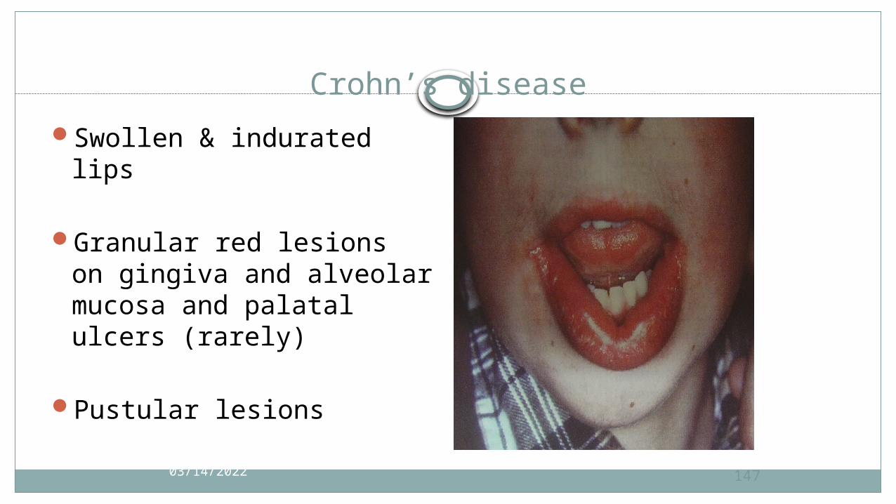
Crohn’s disease
Swollen & indurated lips
Granular red lesions on gingiva and alveolar mucosa and palatal ulcers (rarely)
Pustular lesions
04/15/2023

04/15/2023
148
EATING DISORDERS
ANOREXIA NERVOSA AND BULIMIAAnorexiaindividuals who intentionally starve themselvesintense fear of becoming fat Anorexia usually develops between 14 - 18 years
Bulimia nervosa Individuals consume large amounts of food and
preventweight gain by vomiting, laxatives, diuretics,dieting, and/or exercising aggressively.

04/15/2023
149
Oral Considerations
erosion of the enamel on the lingual surfaces of the maxillary teeth
Parotid enlargement may develop as a sequela of starvation.
04/15/2023
150
REFERENCES
BURKETS-ORAL MEDICINETEXT BOOK OF ORAL MEDICINE by Anil Govindrao GhomSHAFER-TEXT BOOK OF ORAL PATHOLOGYNEVILLE-TEXT BOOK OF ORAL PATHOLOGYCARANZZA’S-CLINICAL PERIODONTOLOGYWIKEPEDIA
04/15/2023
151
Thank you