Embed Size (px)
DESCRIPTION
Ophthalmic drug delivery system
Citation preview

Ocular Drug Delivery System
Presented By
Patel Bindiya Ramesh
M.Pharm – (II semister)
(Pharmaceutics)

OCULAR DDS 2
Agenda...• Objective• Introduction• Anatomy and physiology of the eye• Mechanism of ocular absorption• Factors affecting intraocular bioavailability• Approaches to improve ocular drug delivery• Classification of ocular drug delivery systems• Non erodible inserts• Erodible inserts• Control delivery systems• Conclusion

OCULAR DDS 3
OBJECTIVE
To review and study ocular drug delivery system.
To study various factors affecting ocular
absorption.
To know various approaches ot improve drug
absorption.
To study mechanism of drug absorption.

OCULAR DDS 4
INTRODUCTION Definition
They are specialized dosage forms designed to be instilled onto the
external surface of the eye (topical), administered inside
(intraocular) or adjacent (periocular) to the eye or used in
conjunction with an ophthalmic device.
The most commonly employed ophthalmic dosage forms are
solutions, suspensions, and ointments.
But these preparations when in stilled into the eye are rapidly drained
away from the ocular cavity due to tear flow and lacrimal nasal
drainage.

OCULAR DDS 5
CONT... Ocular administration of drug is primarily associated with the need
to treat ophthalmic diseases.
Eye is the most easily accessible site for topical administration of a
medication.
Ideal ophthalmic drug delivery must be able to sustain the drug
release and to remain in the vicinity of front of the eye for prolong
period of time.
The newest dosage forms for ophthalmic drug delivery are: gels, gel-
forming solutions, ocular inserts , intravitreal injections and
implants.
OCULAR DDS 6
CONT...Major classes of drugs used are
Miotics e.g. pilocarpine Hcl
Mydriatics e.g. atropine
Cycloplegics e.g. atropine
Anti-inflammatories e.g. corticosteroids
Anti-infectives (antibiotics, antivirals and antibacterials)
Anti-glucoma drugs e.g. pilocarpine Hcl
Surgical adjuncts e.g. irrigating solutions
Diagnostic drugs e.g. sodiumfluorescein
Anesthetics e.g. tetracaine

OCULAR DDS 7
COMPOSITION OF EYE
Water - 98%,
Solid -1.8%,
Organic element
Protein - 0.67%,
sugar - 0.65%,
NaCl - 0.66%
Other mineral element
sodium, potassium and ammonia - 0.79%.

OCULAR DDS 8
ANATOMY AND PHYSIOLOGY OF THE EYE

OCULAR DDS 9
Human eye Diameter 23 mm Structure comprises of three layers1. Outermost coat : The clear, transparent cornea and the
white, opaque sclera2. Middle layer : The iris anteriorly, the choroid posteriorly,
and the ciliary body at the intermediate part3. Inner layer : Retina (extension of CNS)
Cornea Epithelium-stroma-endothelium (fat-water-fat structure) Penetration of the drug depends on Oil-water partition
coefficient

OCULAR DDS 10
Corneal Cross Section

OCULAR DDS 11
Fluid systems in eye1. Aqueous humor
Secreted from blood through epithelium of the ciliary body.
Secreted in posterior chamber and transported to anterior
chamber.2. Vitreous humor
Secreted from blood through epithelium of the ciliary body.
Diffuse through the vitreous body.
Lacrimal glands
Secrete tears & wash foreign bodies.
Moistens the cornea from drying out.

OCULAR DDS 12
The sclera : The protective outer layer of the eye, referred to as the
“white of the eye” and it maintains the shape of the eye.
The cornea : The front portion of the sclera, is transparent and allows
light to enter the eye. The cornea is a powerful refracting surface,
providing much of the eye's focusing power.
The choroid : is the second layer of the eye and lies between the sclera
and the retina. It contains the blood vessels that provide nourishment
to the outer layers of the retina.
The iris : is the part of the eye that gives it color. It consists of muscular
tissue that responds to surrounding light, making the pupil, or circular
opening in the center of the iris, larger or smaller depending on the
brightness of the light.

OCULAR DDS 13
The lens is a transparent, biconvex structure, encased in a thin
transparent covering. The function of the lens is to refract and
focus incoming light onto the retina.
The retina is the innermost layer in the eye. It converts images into
electrical impulses that are sent along the optic nerve to the brain
where the images are interpreted.
The macula is located in the back of the eye, in the center of the
retina. This area produces the sharpest vision.

OCULAR DDS 14
MECHANISM OF OCULAR ABSORPTION
Non-Corneal Absorption
• Penetration across Sclera & Conjuctiva into Intra Ocular tissues
• Non-Productive: because penetrated drug is absorbed by general circulation
Corneal Absorption
• Outer Epithelium: rate limiting barrier, with pore size 60å,Only access to small ionic & lipohilic molecules
• Trans cellular transport: transport between corneal epithelium & stroma.

OCULAR DDS 15
General Pathway For Ocular Absorption
OCULAR DDS 16
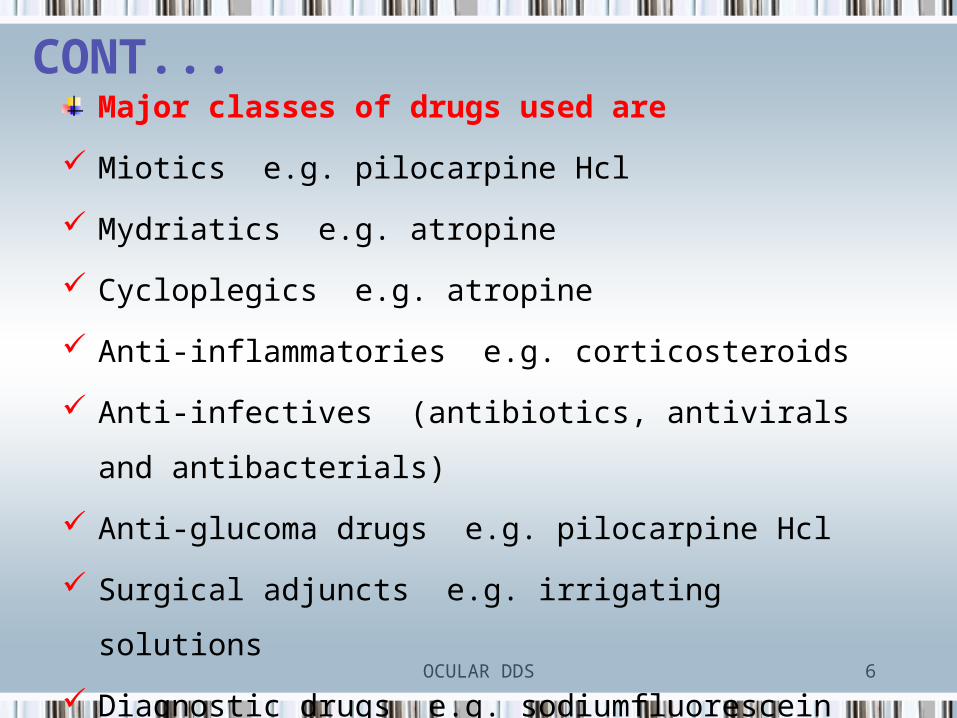
Factors Affecting Intraocular Bioavailability
Includes- • Pre corneal • Corneal• Interior of the eye
1. Inflow & outflow of lacrimal fluids.2. Efficient naso-lacrimal drainage.3. Interaction of drug with proteins of lacrimal fluid.4. Dilution with tears.5. Corneal barriers6. Physico-chemical properties of drugs7. Active ion transport at cornea,8. Limited and poor corneal permeability 9. Metabolism

OCULAR DDS 17
Absorption Of Drugs In The Eye
Factors affecting drug availability:
1. Rapid solution drainage by gravity, induced lachrymation,
blinking reflex, and normal tear turnover.
The normal volume of tears = 7 ul,
The blinking eye can accommodate a volume of up to 30 ul
without spillage,
The drop volume = 50 ul
2. Superficial absorption of drug into the conjunctiva and
sclera and rapid removal by the peripheral blood flow

OCULAR DDS 18
3. Low corneal permeability (act as lipid barrier)
In general:
- Transport of hydrophilic and macromolecular drugs
occurs through scleral route.
- Lipophilic agents of low molecular weight follow
transcorneal transport by passive diffusion.
4. Metabolism
Enzymatic biotransformation Esterases, oxidoreductases, Peptidases, Glucuronide Sulfate transferases, Lysosomal enzymes

OCULAR DDS 19
Corneal Absorption
Poor Bioavialability
Protective mechanism (short residence time)
BlinkingReflex lacrimation,
Nasolacrimal dranaige
Anatomy of eye
Barrier properties of cornea

OCULAR DDS 20
Approaches To Improve Ocular Drug
Delivery1. Viscosity enhancers 2. Eye ointments3. Gel 4. Prodrug5. Penetration enhancers 6. Liposomes 7. Niosomes 8. Nanosuspension 9. Microemulsion10. Nanoparticles/nanospheres11. In situ-forming gel

OCULAR DDS 21
Enhancement of bioavailability
1. Increase in viscosity of formulation leads to decrease in drainage.
2. Slows elimination rate from the precorneal area and enhance contact
time.
3. Generally hydrophilic polymers, eg. Methyl cellulose, polyvinyl
alcohols, polyacrylic acids, sodium carboxy methyl cellulose,
carbomer is used.
4. A minimum viscosity of 20 cst is needed for optimum corneal
absorption.
OCULAR DDS 22
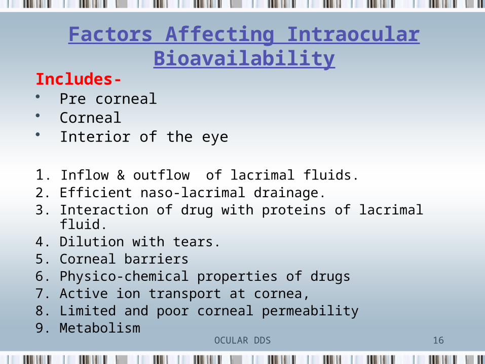
Use of penetration enhancers
1. Act by increasing corneal uptake by modifying the integrity of the
corneal epithelium.
2. Substances which increases the permeability characteristics of the
cornea by modifying the integrity of corneal epithelium are known
as penetration enhancers.
Modes of actions
1. By increasing the permeability of the cell membrane.
2. Acting mainly on tight junctions.
e.g. Caprylic acid, sodium caprate, Azone

OCULAR DDS 23
PRODRUGS
1. Prodrugs enhance corneal drug permeability through
modification of the hydrophilic or lipophilicity of the drug.
2. The method includes modification of chemical structure of the
drug molecule, thus making it selective, site specific and a safe
ocular drug delivery system.
3. Drugs with increased penetrability through prodrug
formulations are epinehrine, phenylephrine, timolol,
pilocarpine.

OCULAR DDS 24
USE OF MUCOADHESIVES IN OCULAR DRUG DELIVERY
1. Polymereric mucoadhesive vehicle: Retained in the eye due to
noncovalent bonding with conjuctival mucine.
2. Mucine is capable of picking of 40-80 times of weight of water.
3. Thus prolongs the residence time of drug in the conjuctival sac.
4. Mucoadhesives contain the dosage form which remains adhered to
cornea until the polymer is degraded or mucus replaces itself.
Types
1. Naturally Occurring Mucoadhesives - Lectins, Fibronectins
2. Synthetic Mucoadhesives - PVA,Carbopol, carboxy methyl cellulose,
cross-linked polyacrylic acid.

OCULAR DDS 25
Phase Transition System
1. Solution that are liquid in the container and thus can be instilled
as eye drop becomes gel on contact with the tear fluid and
provide increased contact time with the possibility of improved
drug absorption and increased duration of therapeutic effect.
2. Liquid-gel phase transition-dependent delivery system vary
according to the particular polymer employed and their
mechanism for triggering the transition to a gel phase in the eye
take advantage of change in temperature, pH, ion sensitivity, or
lysozymes upon contact with tear fluid.

OCULAR DDS 26
POLYMER MECHANISM
Lutrol FC – 127 and Poloxamer 407 Viscosity increased when their temperature raised to eye temperature.
Cellulose acetate phthalate latex Coagulates when its native pH 4.5 raised by tear fluid to pH 7.4
Gelrite Forms clear gel in the presence of cations
EXAMLE OF POLYMER

OCULAR DDS 27
OCULAR DELIVERY SYSTEMS
CONVENTIONAL VESICULAR
CONTROL RELEASE PARTICULATE
o IMPLANTSo HYDROGELSo DENDRIMERSo IONTOPORESISo COLLAGEN SHIELDo POLYMERIC
SOLUTIONSo CONTACT LENSESo CYCLODEXRINo MICROONEEDLEo MICROEMULSIONSo NANO
SUSPENSION
o MICROPARTICLESo NANOPARTICLES
o LIPOSOMESo NIOSOMESo DISCOMESo PHARMACOSOMES
ADVANCED
o SCLERAL PLUGS
o GENE DELIVERY
o Si RNAo STEM CELL
o SOLUTIONo SUSPENSIONo EMULSIONo OINTMENTo INSERTo GELS

OCULAR DDS 28
Classification Of Ocular Drug Delivery Systems
LIQUIDS
Solutions
Suspensions
Powders for reconstitution
Sol to gel systems
SOLID
Ocular inserts
Contact lenses

OCULAR DDS 29
Ideal Ophthalmic Delivery SystemGood corneal penetration.
Prolong contact time with corneal tissue.
Simplicity of instillation for the patient.
Non irritative and comfortable form.
Appropriate rheological properties.
Inert and stable.

OCULAR DDS 30
Eye Drops
o Drugs which are active at eye or eye surface are widely administered
in the form of Solutions, Emulsion and Suspension.
o Various properties of eye drops like hydrogen ion concentration,
osmolality, viscosity and instilled volume can influence retention of a
solution in the eye.
o Less than 5 % of the dose is absorbed after topical administration
into the eye.
o The dose is mostly absorbed to the systemic blood circulation via
the conjunctival and nasal blood vessels.

OCULAR DDS 31
Advantages And Disadvantages Of Eye Drops
Dosage form Advantages Disadvantages
Solutions 1. Convenience2. Usually do not interfere
with vision of patient.
1. Rapid precorneal elimination.2. Non sustained action.3. To be Administered at frequent
intervals.Suspension 1. Patient compliance.
2. Best for drug with slow dissolution.
3. Longer contact time
1. Drug properties decide performance loss of both solutions and suspended particles.
2. Irritation potential due to the particle size of the drug.
Emulsion 1. Prolonged release of drug from vehicle
1. Blurred vision.2. patient non compliance.

OCULAR DDS 32
Ointment • Prolongation of drug contact time with the external ocular surface
can be achieved using ophthalmic ointment vehicle.• The ointment base is sterilized by heat and appropriately filtered
while molten to remove foreign particulate matter.
Ointment base is sterilized by heat and filtered while molten
to remove foreign particulate matter.
It is then placed into a sterile steam jacketed
to maintain the ointment in a molten state and excipients
are added
The entire ointment may be passed
through a previously sterilized colloid mill

OCULAR DDS 33
• Advantages1. Longer contact time and greater storage stability.2. Flexibility in drug choice.3. Improved drug stability.
• Disadvantages1. Sticking of eyes lids.2. Blurred vision.3. Poor patient compliance4. Interfere with the attachment of new corneal epithelial cells
to their normal base.5. Matting of eyelids

OCULAR DDS 34
• Gels
1. Ophthalmic gels are composed of mucoadhesive polymers that
provide localized delivery of an active ingredient to the eye. Such
polymers have a property known as bioadhesion.
2. These polymers are able to extend the contact time of the drug
with the biological tissues and there by improve ocular
bioavailability.Advantages3. Longer contact time.4. Greater storage stability.
Disadvantages5. Blurred vision but less then ointment.6. Poor patient compliance.

OCULAR DDS 35
Vesicular System• LIPOSOMES Liposomes are biocompatible and biodegradable lipid vesicles made
up of natural lipids and about 25 –10 000 nm in diameter.
They are having an intimate contact with the corneal and conjunctival
surfaces which is desirable for drugs that are poorly absorbed, the drugs
with low partition coefficient, poor solubility or those with medium to
high molecular weights and thus increases the probability of ocular drug
absorption.
Vesicle composed of phospholipid bilayer enclosing aqueous
compartment in alternate fashion.
It is Biodegradable, Non-toxic in nature.

OCULAR DDS 36
Types : 1. MLV
2. ULV-SUV(upto 100 nm)
3. LUV(more than 100 nm)
Polar drugs are incorporated in aqueous compartment while
lipophilic drugs are intercalated into the liposome membrane.
Phospholipids used- Phosphotidylcholine, Phosphotidic acid,
Sphingomyline, Phosphotidyleserine, Cardiolipine.

OCULAR DDS 37
ADVANTAGESDrugs delivered intact to
various body tissues.
Liposomes can be used for
both hydrophilic and
hydrophobic drug.
Possibility of targeting and
decrease drug toxicity.
The size, charge and other
characteristics can be
altered according to drug
and desired tissue.
DISADVANTAGESTheir tendency to be
uptaken by RI system.
They need many
modification for drug
delivery to special organs.
Costly.
Stability problem and
oxidative degradation.
Requires special
packaging and storing
facility.

OCULAR DDS 38
Degradation And Drug Release Of Liposomes
1. Endocytosis 2. Fusion

OCULAR DDS 39
NIOSOMES The major limitations of liposomes are chemical instability, oxidative
degradation of phospholipids, cost and purity of natural
phospholipids.
To avoid this niosomes are developed as they are chemically stable
as compared to liposomes and can entrap both hydrophobic and
hydrophilic drugs.
Niosomes are non-ionic surfactant based multilamellar (>0.05µm),
small unilamellar (0.025-0.05µm) or large unilamellar vesicles
(>0.1µm) in which an aqueous solution of solute(s) is entirely
enclosed by a membrane resulted from organization of surfactant
macromolecules as bilayers.

OCULAR DDS 40
They are non toxic and do not require special handling techniques.
Niosomes are nonionic surfactant vesicles that have potential
applications in the delivery of hydrophobic or amphiphilic drugs.
STRUCTURAL COMPONENTS USED
• Surfactants (dialkyl polyoxy ethylene ether non ionic surfactant)
• Cholesterol.

OCULAR DDS 41
ADVANTAGES
The vesicle suspension being water based offers greater patient
compliance over oil based systems.
Since the structure of the niosome offers place to accommodate
hydrophilic, lipophilic as well as ampiphilic drug moieties, they can
be used for a variety of drugs.
The characteristics such as size, lamellarity etc. of the vesicle can be
varied depending on the requirement.
The vesicles can act as a depot to release the drug slowly and offer a
controlled release.

OCULAR DDS 42
They are osmotically active and stable.
They increase the stability of the entrapped drug.
Improves therapeutic performance of the drug by protecting
it from the biological environment and restricting effects to
target cells, thereby reducing the clearance of the drug.
Drawbacks
Physical instability.
Aggregation.
Leaking of entrapped drug.

OCULAR DDS 43
Pharmacosomes
This term is used for pure drug vesicles formed by the amphiphilic
drugs.
The amphiphilic prodrug is converted to pharmacosomes on dilution
with water.
Since many drugs are also amphiphiles, they can form the vesicles.
Advantages
Drug metabolism can be decreased.
Controled release profile can be achieved.

OCULAR DDS 44
DISCOMES• Soluble surface active agents when added in critical amount to
vesicular dispersion leads to solubilization or breakdown of vesicles
& translates them into mixed micellar systems
e.g: Egg yolk phosphatidyl choline liposomes by the addition
of non ionic surfactants of poly oxy ethylene cetyl ether till the
lamellar and mixed lamellar coexist
Advantages
• Minimal opacity imposes no hinderance to vision
• Increased patient compliance
• Zero order release can be easily attained

OCULAR DDS 45
Advantages Of Vesicular Systems1. No difficulty of insertion as in the case of ocular inserts.
2. No tissue irritation and damage as caused by penetration enhancers.
3. Provide patient compliance as there is no difficulty of insertion as
observed in the case of inserts.
4. The vesicular carriers are biocompatable and have minimum side effects.
5. Degradation products formed after the release of drugs are
biocompatable.
6. They prevent the metabolism of drugs from the enzymes present at
tear/corneal epithelium interface.
7. Provide a prolong and sustained release of drug.

OCULAR DDS 46
OCULAR INSERTS
NONERODABLE
OCUSERT
ERODABLE
SODI
COLLAGEN SHIELDSOCUFIT SR

OCULAR DDS 47
NON ERODIBLE INSERTS
OCUSERT
The Ocusert therapeutic system is a flat, flexible, elliptical device
designed to be placed in the inferior cul-de-sac between the sclera
and the eyelid and to release Pilocarpine continuously at a steady
rate for 7 days.
The device consists of 3 layers…..
1. Outer layer - ethylene vinyl acetate copolymer layer.
2. Inner Core - Pilocarpine gelled with alginate main polymer.
3. A retaining ring - of EVA impregnated with titanium di oxide

OCULAR DDS 48
OCUSERT

OCULAR DDS 49
• ADVANTAGES
Reduced local side effects and toxicity.
Around the clock control of drug.
Improved compliance.
• DISADVANTAGES
Retention in the eye for the full 7 days.
Periodical check of unit.
Replacement of contaminated unit
Expensive.

OCULAR DDS 50
ERODIBLE INSERTS The solid inserts absorb the aqueous tear fluid and gradually erode
or disintegrate. The drug is slowly leached from the hydrophilic
matrix.
They quickly lose their solid integrity and are squeezed out of the
eye with eye movement and blinking.
Do not have to be removed at the end of their use.
Three types :
1. Lacriserts
2. Sodi
3. Minidisc

OCULAR DDS 51
• LACRISERTS Sterile rod shaped device made up of hydroxyl propyl cellulose
without any preservative. For the treatment of dry eye syndromes. It weighs 5 mg and measures 1.27 mm in diameter with a
length of 3.5 mm. It is inserted into the inferior fornix.• SODI Soluble ocular drug inserts. Small oval wafer. Sterile thin film of oval shape. Weighs 15-16 mg. Use – glaucoma. Advantage – Single application.•
Lacriserts

OCULAR DDS 52
• Minidisc
Countered disc with a convex front and a concave back surface.
Diameter – 4 to 5 mm.
• Composition
Silicone based prepolymer-alpha-w-dis (4-methacryloxy)-butyl poly
di methyl siloxane. (M2DX)
M-Methyl a cryloxy butyl functionalities.
D – Di methyl siloxane functionalities.
Pilocarpine, chloramphenicol.
Minidisc

OCULAR DDS 53
CONTACT LENS• Contact lenses can be a way of providing extended
release of drugs into the eye.
• Conventional hydrogel soft contact lenses have the
ability to absorb some drugs and release them into
the post lens lachrymal fluid, minimizing clearance
and sorption through the conjunctiva.
• Their ability to be a drug reservoir strongly
depends on the water content and thickness of the
lens, the molecular weight of the drug, the
concentration of the drug loading solution and the
time the lens remains in it.
OCULAR DDS 54
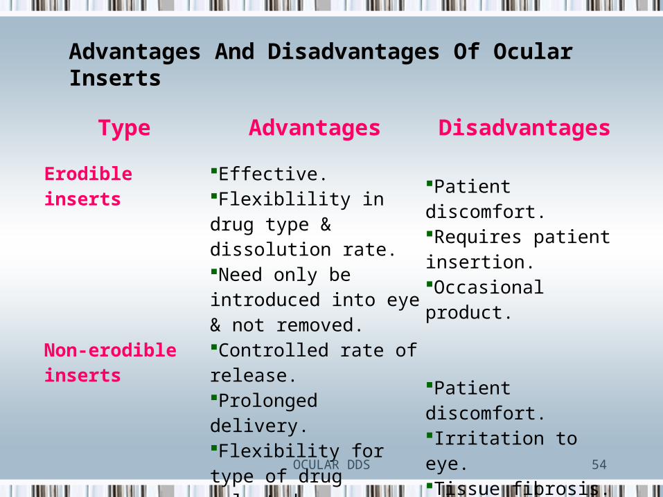
Type Advantages Disadvantages
Erodible inserts Effective.Flexiblility in drug type & dissolution rate.Need only be introduced into eye & not removed.
Patient discomfort.Requires patient insertion.Occasional product.
Non-erodible inserts Controlled rate of release.Prolonged delivery.Flexibility for type of drug selected.Sustained release.
Patient discomfort.Irritation to eye.Tissue fibrosis.
Advantages And Disadvantages Of Ocular Inserts

OCULAR DDS 55
Control Delivery Systems• 1. Implants
1. Implants have been widely employed to extend the release of drugs
in ocular fluids and tissues particularly in the posterior segment.
2. Implants can be broadly classified into two categories based on their
degradation properties:
(1) Biodegradable
(2) Nonbiodegradable
3. With implants, the delivery rate could be modulated by varying
polymer composition.
4. Implants can be solids, semisolids or particulate-based delivery
systems.

OCULAR DDS 56
For chronic ocular diseases like cytomegalo virus (CMV)
retinitis, implants are effective drug delivery system. Earlier
non biodegradable polymers were used but they needed
surgical procedures for insertion and removal.
Presently biodegradable polymers such as Poly Lactic Acid
(PLA) are safe and effective to deliver drugs in the vitreous
cavity and show no toxic signs.

OCULAR DDS 57
Iontophoresis
In Iontophoresis direct current drives ions into cells or tissues. For
iontophoresis the ions of importance should be charged
molecules of the drug.
If the drug molecules carry a positive charge, they are driven into
the tissues at the anode; if negatively charged, at the cathode.
Requires a mild electric current which is applied to enhance
ionized drug penetration into tissue.
Ocular iontophoresis offers a drug delivery system that is fast,
painless, safe, and results in the delivery of a high concentration
of the drug to a specific site.

OCULAR DDS 58
Ocular iontophoresis delivery is not only fast, painless and safe but
it can also deliver high concentration of the drug to a specific site.
Ocular iontophoresis has gained significant interest recently due to
its non-invasive nature of delivery to both anterior and posterior
segment.
Iontophoretic application of antibiotics may enhance their
bactericidal activity and reduce the severity of disease
Can overcome the potential side effects associated with intraocular
injections and implants.
Iontophoresis is useful for the treatment of bacterial keratitis.

OCULAR DDS 60
Dendrimer Dendrimers can successfully used for different routes of drug
administration and have better water-solubility, bioavailability and
biocompatibility.
Microemulsion
Microemulsion is dispersion of water and oil stabilized using
surfactant and co- surfactant to reduce interfacial tension and
usually characterized by small droplet size (100 nm), higher
thermodynamic stability and clear appearance.
Selection of aqueous phase, organic phase and surfactant/co-
surfactant systems are critical parameters which can affect stability
of the system.

OCULAR DDS 61
Nanosuspensions
Nanosuspensions have emerged as a promising strategy for the
efficient delivery of hydrophobic drugs because they enhanced not
only the rate and extent of ophthalmic drug absorption but also the
intensity of drug action with significant extended duration of drug
effect.
For commercial preparation of nanosuspensions, techniques like
media milling and high-pressure homogenization have been used.

OCULAR DDS 62
Advance System• Design of Punctal Plug
1. Punctal plugs are placed in the tear duct (punctum) to
release a variety of drugs.
2. Currently targeting the treatment of glaucoma and ocular
hypertension

OCULAR DDS 63
• Design of Replenish Mini Pump
1. Micro-electromechanical system that delivers continuous or bolus-targeted drugs to both the anterior and posterior segments.
2. Refillable drug reservoir (via 31 gauge needle) that is capable of storing and delivering up to 12 months.

OCULAR DDS 64
• Design of ODTx
1. Non-biodegradable implant that is comprised of multiple sealed
reservoirs containing individual doses of drugs.
2. Implant is injected into the vitreous.
3. Drug is released by creating an opening via laser.
OCULAR DDS 65
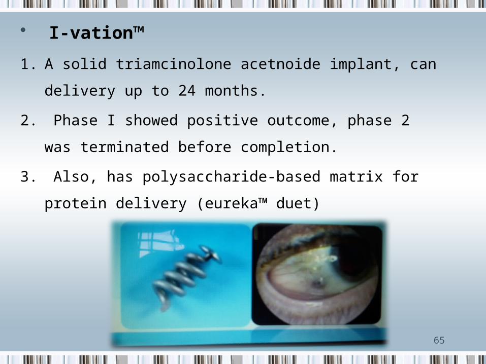
• I-vation™
1. A solid triamcinolone acetnoide implant, can delivery up to 24
months.
2. Phase I showed positive outcome, phase 2 was terminated
before completion.
3. Also, has polysaccharide-based matrix for protein delivery
(eureka™ duet)

OCULAR DDS 66
• Vitrasert®
1. (Ganciclovir-CMV retinitis) and Retisert® (fluocinolone acetonide-
chronic non infectious uveitis).
2. Are FDA approved
3. These devices are solid sustained-release devices, typically made
of PLGA, capable of delivering drug for up to 30 months.

67