Embed Size (px)
Citation preview

Noncompaction Cardiomyopathy
“A DIAGNOSTICALLY CHALLENGING CARDIOMYOPATHY”

LV Non Compaction•Noncompaction of the left ventricular
myocardium (LVNC) is being increasingly recognized and its diagnosis has moved from the autopsy table or previously poorly recognized entity to a widely recognized cardiomyopathy.
•Lot of research work- publications recently
•Advances in non invasive diagnostic technologies- better delineation of morphology

Isolated left ventricular noncompaction in an autopsy specimen, shown in short-axis view. Note the compacted epicardial layer and noncompacted endocardial layer with marked hypertrabeculation and deep recesses.
William D. Edwards, MD, Department of Laboratory Medicine and Pathology, Mayo Clinic, Rochester, MN.
Characterized by:An altered myocardial wall with prominent trabeculae and deep intertrabecular recesses resulting in thickened myocardium with two layers consisting of compacted and noncompacted myocardium.Continuity between the left ventricular cavity and the deep intratrabecular recesses, which are filled with blood from the ventricular cavity without evidence of communication to the epicardial coronary artery system.

HISTORY •First described in association with other
congenital abnormalities▫Obstruction of LVOT/RVOT
Pulmonary atresia with intact ventricular septum
▫Complex cyanotic congenital heart disease▫Anomalous coronary arteries
•Intertrabecular recesses communicate with ventricular cavity and coronary circulationLauer RM et al, NEJM 1964

HISTORY•Dusek first described the postnatal
persistence of spongy myocardium in 1975 pathologically,
•Engberding and Bender made the first clinical recognition with two-dimensional (2D) echocardiography in 1984
•Three decades later, with only morphologic assessment available and no definitive genetic pathway, isolated left ventricular noncompaction (LVNC) remains a diagnostic and management challenge.
Dusek J et al, Arch Pathol 1975

CASE SCENARIO•26 YEARS OLD FEMALE PRESENTED
WITH PROGRESSIVELY INCRAESING DYSPNEA FOR PAST 3 YAERS
•CLASS 3 PRESENTLY•INTERMITTENT EPISODES OF PND
•SENT TO US FOR ECHOCARDIOGRAPHY

ECHOCARDIOGRAPHY


MRI


TREATMENT
•BETA BLOCKERS•DIURETICS•ACE-INHIBITORS•ANTICOAGULATION

Noncompaction Cardiomyopathy
“A DIAGNOSTICALLY CHALLENGING CARDIOMYOPATHY”

OutlineDefinitionsPathomorphogenesisAssociations with other disease Isolated LV noncompactionEpidemiologyGeneticsClinical FeaturesDiagnosis
EchocardiographyCardiovascular magnetic resonance
ManagementPrognosis

TypesLeft ventricular noncompaction in
association with congenital abnormalities
Isolated left ventricular noncompaction
AKA▫Hypertrabeculation Syndrome▫Persistent myocardial sinusoids▫Spongy myocardium

Definition• Congenital heart
disease• Myocardial wall
distortion▫ Prominent trabeculae▫ Deep intertrabecular
recesses• Continuity between
LV cavity and recesses
• Primary cardiomyopathy in 2006 World Heath Organization classification
Ritter M et al, Mayo Clin Proc 1997

EMBRYOLOGY and PATHO-MORPHOGENESIS•Because myocardial noncompaction represents
a congenital anomaly of the ventricular myocardium, insight into the underlying pathomorphogenesis can be helpful in clarifying diagnostic criteria and understanding its clinical presentation and course.
•Need to review the literature on normal and
abnormal myocardial development in an attempt to characterize the disease as a morphogenetic entity.

Early Embryology, <5 weeksAnterolateral
mesoderm
Epithelium
Endocardium Myocardium
N-Cadherin
Cardiac Tube
Trabeculations
Neuregulin growth factors
3 weeks
↓N-Cadherin
The majority of cardiomyocytes originate from the anterolateral mesoderm soon after gastrulation. Soon after their specification, the cardiac myocytes migrate along the ventral midline of the embryo, begin to express Ncadherin, and differentiate into an epithelium. A small population of these cells then downregulates Ncadherin and dissociates from the epithelium to form the endocardium. The majority of epithelial cells, in turn, will give rise to the myocardium. At 3 weeks of human gestation, the myocardium and the endocardium fuse into a single beating cardiac tube .
During the early developmental stages, the heart tube is still without epicardial overlay. As an adaptation to improve nourishment of the rapidly growing heart, the myocardium forms a loose network of interwoven fibers separated by deep recesses that link the myocardium with the left ventricular cavity. This formation of ventricular trabeculae is restricted to the free wall and the lower part of the ventricular septum, sparing the regions of the atrioventricular canal and outflow tract septum . Neuregulin growth factors secreted from the endocardium and their myocardial receptors ErbB2 and ErbB4 are required for the development of the trabeculae . The resulting increase in surface facilitates myocardial supply by exchange diffusion . It is likely that during this developmental period the trabeculae generate much of the contractile force of the heart .

Embryology, 5-8 weeksEndocardium
Sub-epicardial space
Microvessels coronary
circulation
Vascular endothelial growth factorAngiopoietin-1
Compaction• Base apex
• Epi- endocardium• Intratrabecular
recesses myocardial capillaries
Once the myocardium has become covered with epicardium, the coronary vasculature develops. In the subepicardial space, an endothelial network is formed that gradually covers the heart and develops microvessels within the myocardium [62]. Arteriogenesis of the coronary arteries includes the formation of media from smooth muscle cells that originate from epitheliumderived cells . As soon as a continuity has been established by connection of the vessels to the aortic root as well as to the right atrium, the initial embryonic means of myocardial nourishment by diffusion from the cardiac lumen switches to the permanent form of active circulation through coronary vessels . At the same time—between 5 and 8 weeks of embryonic life—gradual compaction of the spongy meshwork of fibers and intertrabecular recesses occurs. This process results in the normal prominent outer compact layer of the mature myocardiumwith only few residual trabeculae remaining present subendocardially. Similar to the direction of coronary arterial development, the recession of the trabeculae proceeds from the epicardium to the endocardium and from the base of the heart to the apex

Srivastava D, Nature 2000; and RP, Nature Rev Genetics 2002
• In patients with myocardial noncompaction, the normal process of myocardial compaction has undergone a premature arrest.
• The fact that a normally developed myocardium is of immense importance for the viability of the rapidly developing embryo, may explain why primary myocardial noncompaction is such a rare disease.
• Failure of normal progression of myocardial morphogenesis results in the persistence of the early embryonic, largely trabeculated myocardium in certain ventricular regions.
• Referring to the prominent spongious layer of the myocardium in these cases, the disease has also been termed “spongy myocardium.”
• Histologically, patchy areas with loosely organized, wavy to angulated myocardial fibers that are thinner than normal, with a reduced content of myofibrils, and central cytoplasmic clearing can be found in these ventricles.
• Secondary to the premature arrest of myocardial maturation the inner, trabeculated, myocardial layer remains abnormally thick with a reduced thickness of the outer, compact, myocardial layer.
• Therefore, the noncompacted subendocardial myocardial layer is always much thicker than the outer compacted myocardium in primary myocardial noncompaction.
• This results in the typical echocardiographic picture of multiple prominent trabeculations with deep intertrabecular recesses invaginating deeply into the outer one third of the ventricular myocardium.
• Magnetic resonance imaging (MRI) provides a good correlation with echocardiography for localization and extent of noncompaction and can be useful in cases with poor echocardiographic image quality.

• Corresponding to the course of normal cardiovascular morphogenesis, a stop of progression of normal myocardial maturation will lead to different extensions of myocardial noncompaction.
• Because the inner part of the ventricular apex is always last to complete the compaction process, and minor trabeculations remain present in this segment even in the normal heart, primary myocardial noncompaction always affects the left ventricular apex.
• However, the abnormal myocardium can extend toward the ventricular base and involve additional regions of the left ventricle, such as inferior and lateral wall segments.
• Although the disease process typically occurs in the left ventricle, involvement of the right ventricle has been described in less than half of patients.
• Because of the difficulty in distinguishing normal variants of the highly trabeculated right ventricle with its per se thinner compact myocardial layer from the pathological noncompacted ventricle, several authors dispute the existence of right ventricular noncompaction


Compaction continues into the postnatal period with continued growth and increasing systemic pressures.The final process is development of the spiral pattern of the myocardial fibers, which is responsible for the twisting nature of contraction.Without the completion of compaction, there is myocardial dysfunction secondary to the failure of the efficient rotational ventricular system to develop for contractile performance. This concept has been demonstratedby abnormal speckle tracking
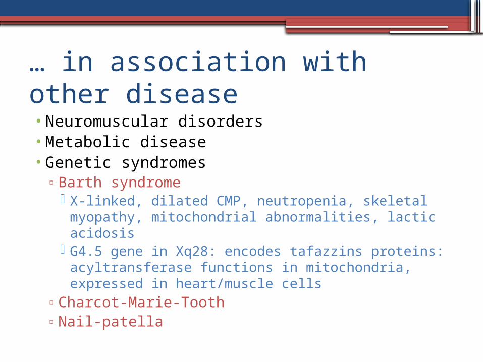
… in association with other disease• Neuromuscular disorders• Metabolic disease• Genetic syndromes
▫Barth syndrome X-linked, dilated CMP, neutropenia, skeletal myopathy,
mitochondrial abnormalities, lactic acidosis G4.5 gene in Xq28: encodes tafazzins proteins:
acyltransferase functions in mitochondria, expressed in heart/muscle cells
▫Charcot-Marie-Tooth▫Nail-patella

Similar phenotypes•Dilated cardiomyopathy•HCM•Restrictive cardiomyopathy•Left-dominant arrhythmogenic
cardiomyopathy▫42 patients with unexplained IL TWI,
arrhythmia of LV origin, and/or LDAC or familial myocardial fibrosis
▫5 patients fulfilled echocardiographic criteria for LVNC
Sen-Chowdhry S et al., JACC 2008

Epidemiology of Isolated LV Noncompaction•Children Adults, elderly•0.05% (Ritter M et al, Mayo Clin Proc 1997)
▫37,555 echocardiograms 17 cases▫Prominent, excessive trabeculations
•0.014% (Oechslin EN et al, JACC 2000)▫242,857 echocardiograms 34 cases▫Noncompacted/compacted ≥ 2:1
•Men >> women

Genetics
•Sporadic or familial•Familial in 18-50% (Oechslin et al, JACC 2000, Chin et al,
Circ 1990, Xing et al, Mol Genet Metab 2006)• Systematic review by Bhatia et al. identified a
familial occurrence rate of 30% in family members that were screened based on an index case
• There are multiple genetic proposals for the phenotypic development of noncompaction. None of them have been consistently identified to be the single gene abnormality causing LVNC.

Genes Identified.The identified genes are as follows:
•Fbkp1a/Notch pathway.•G4.5 gene/TAZ protein.•14-3-3 deletion.•ZASP protein.•TNNT2 protein.•MYH7 protein.•TPM1 protein.•MYBPC3 protein.•ACTC1 protein.

•The current genes available for testing are variants of established dilated cardiomyopathy and hypertrophic cardiomyopathy genes.
•Oechslin and Jenni have proposed an acquired pathogenesis in patients with prior normal cardiac structure and function that develop LVNC later in life.
•This supports the hypothesis that LVNC may represent a morphologic continuum of genetic cardiomyopathies, including dilated and hypertrophic cardiomyopathies

Clinical features• Depending on the exact time of failure of normal progression of the
myocardial maturation process, the hearts of patients with primary myocardial noncompaction show different extensions of the abnormally thickened trabeculated myocardial layer within the ventricle.
• The amount of normal myocardium will determine the functional relevance of the disease, thus explaining the highly variable clinical course.
• The more severe cases present during the first years of life, and myocardial noncompaction was initially described as a cardiomyopathy of children.
• It is likely that the most severe forms are lethal at an early stage of embryonic development.
• At the other end of the spectrum are patients with very localized forms of myocardial noncompaction that can be asymptomatic for prolonged periods, with the diagnosis only being made at an old age

Clinical Features•Heart failure
▫Dyspnea▫Chest pain
•Arrhythmia▫Atrial fibrillation▫Ventricular
tachycardia•Thromboembolism
▫CVA/TIA▫Pulmonary
embolism

Heart FailureDiastolic Systolic• Restrictive hemodynamics
on catheterization• Initial presentation as
restrictive cardiomyopathy• Pathophysiology
▫ Abnormal relaxation▫ Decreased compliance
due to volume of trabeculations
• No significant epicardial coronary disease
• Subendocardial hypoperfusion
• chronic microvascular ischemia
Ichida F et al, JACC 1999; Sen-Chowdhry et al, Curr Opin Card 2008

Electrophysiology• Atrial fibrillation• Ventricular tachycardia
ECG:• Left or right axis deviation• PR prolongation• Left ventricular hypertrophy• LBBB, RBBB, IVCD• Repolarization abnormalities• In pediatric population:
▫ Sinus bradycardia▫ WPW
Duru F et al, J Cardiovasc Electrophysiol 2000

Thromboembolism• Stroke• TIA• Pulmonary embolus• Mesenteric infarction• Reported 21-38% • Etiology
▫ Stasis of blood in deep recesses/trabeculations
▫ Atrial fibrillation
Chin TK et al, Circ 1990Ritter M et al., Mayo Clin Proc 1997Oechslin E et al, JACC 2000

DIAGNOSTIC APPROACH

•There is much debate regarding the diagnostic criteria for LVNC and the predilection for overdiagnosis.
•ECHO vs CMR vs MDCT –NO GOLD STANDARD
•No lab tests/ genetic tests confirmatory of LVNC
•Multiple modalities may be required for complete assessment.

Two-Dimensional Echocardiography.
•The traditional diagnostic study for evaluation of LVNC is echocardiography.
•It is still the most common initial test that identifies the characteristic findings of LVNC and may lead to further evaluation.
•There are three proposed diagnostic criteria that are most utilized in the literature. These criteria are :
1. Chin et al2. Jenni et al3. Stollberger et al


1990-First diagnostic criteria
•LVNC X (distance between epicardial surface
and trough of the intertrabecular recesses) Y (distance between epicardial surface and
peak of the trabeculations)If X/Y< 0.5if it progressively decreased from the
papillary muscles toward the apexChin TK, Perloff JK, Williams RG, Jue K, Mohrmann R. Isolated noncompaction of left ventricular myocardiu. A study of eight cases. Circulation 1990;82:507–13.

Diagnosis- Echocardiography I
• X/Y ≤ 0.5• Apex at end-diastole
▫ PSAX▫ Apical 4Ch
0.59+0.05 0.20±0.040.92+0.07
Chin TK et al, Circ 1990

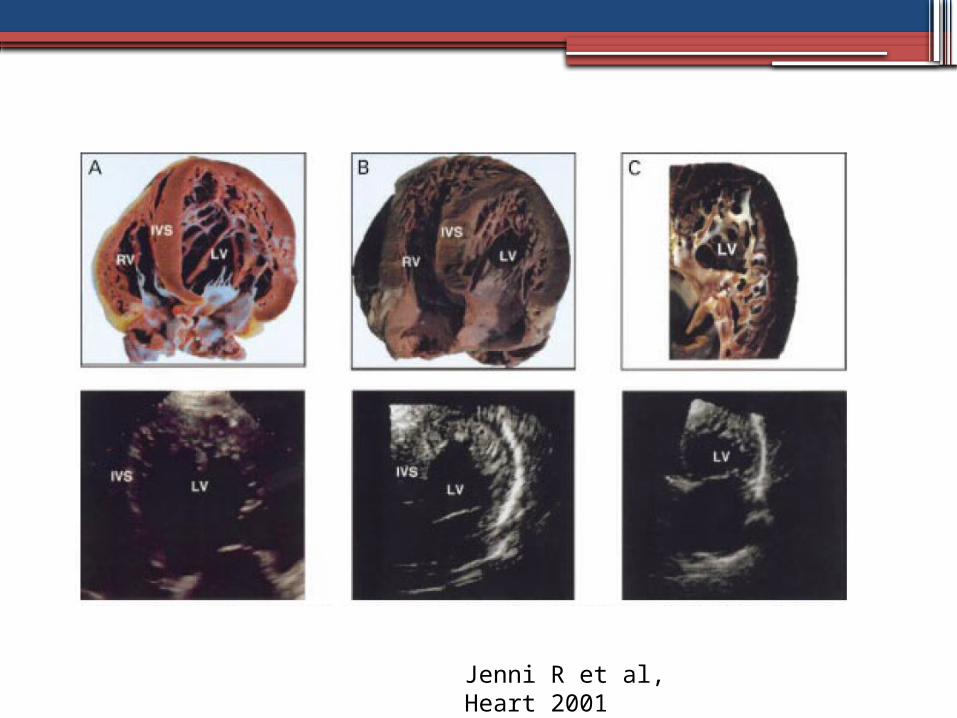
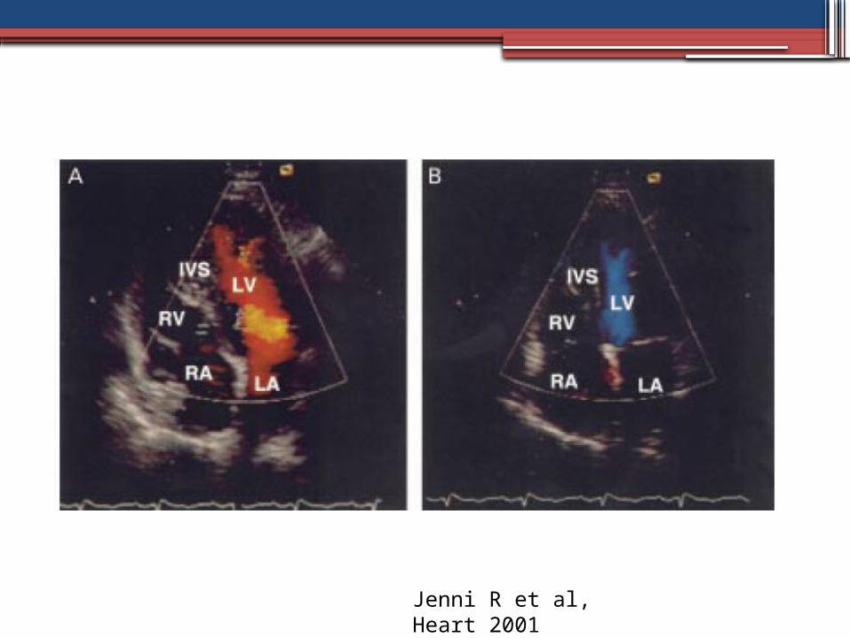
Diagnosis- Echocardiography 2nd criteria
• Compacted and noncompacted layers of ventricular wall▫ Thickened endocardial layer▫ Prominent trabeculations▫ Deep recesses▫ Ratio noncompacted to
compacted >2:1▫ End-systole
• Trabecular meshwork in apex or midventricular segments of inferior and lateral wall
• Absence of any other cardiac anomaly. Jenni R et al, Heart
2001


Noncompacted/ Compacted Ratio Mean±SD
Noncompacted/ Compacted RatioRange
Noncompaction (n=34)
3.5±0.8 2.3-5
Dilated CMP (n=10)
0.8±0.4 0.4-2.0
Hypertensive heart dz (n=9)
1.1±0.5 0.4-2.0
• All p <0.001 vs. noncompaction group • Autopsy validation in 7 of 34 noncompaction patients• Autopsy validation in all dilated cardiomyopathy patients
Jenni R et al, Heart 2001

Validation of Jenni criteria•Blinded retrospective review of records
comparing patients with:▫LVNC (n=19)▫Dilated cardiomyopathy (n=31)▫Hypertensive heart disease (n=22)▫Chronic severe valvular disease (n=86)
Mitral regurgitation (n=22) Aortic regurgitation (n=20) Aortic stenosis (bi- and tri-leaflet valves,
n=44)Frischknecht B et al, J Am Soc Echocardiogr 2005


Frischknecht B et al, J Am Soc Echocardiogr 2005

Frischknecht B et al, J Am Soc Echocardiogr 2005
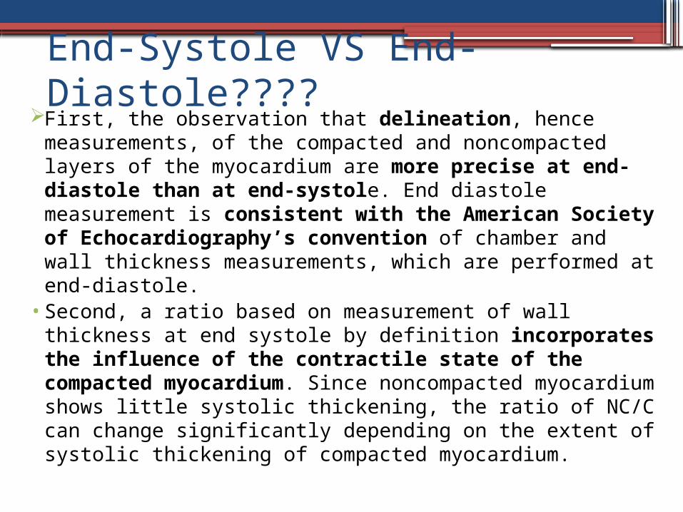
End-Systole VS End-Diastole????
First, the observation that delineation, hence measurements, of the compacted and noncompacted layers of the myocardium are more precise at end-diastole than at end-systole. End diastole measurement is consistent with the American Society of Echocardiography’s convention of chamber and wall thickness measurements, which are performed at end-diastole.
• Second, a ratio based on measurement of wall thickness at end systole by definition incorporates the influence of the contractile state of the compacted myocardium. Since noncompacted myocardium shows little systolic thickening, the ratio of NC/C can change significantly depending on the extent of systolic thickening of compacted myocardium.

2014-Latest:All 4 criteria to be fulfilledLVNC
1. >3 Prominent trabeculous formations along the left ventricular endocardial border visible in end-diastole, distinct from papillary muscles, false tendons or aberrant bands 2. Trabeculations move synchronously with the compacted myocardium3. Trabeculations form the noncompacted part of a two-layered myocardial structure, best visible at end-systole4. Perfusion of the intertrabecular spaces from the ventricular cavity is present at end-diastole on colour Doppler echocardiography or contrast echocardiography
Refinement of echocardiographic criteria for left ventricular noncompaction Claudia Stöllberger,Birgit Gerecke,Josef Finsterer,Rolf Engberding.International Journal of Cardiology 165 (2013) 463–467
They suggested that measurement of the myocardial layers is notfeasible due to the lack of uniformly accepted standards for measurements.


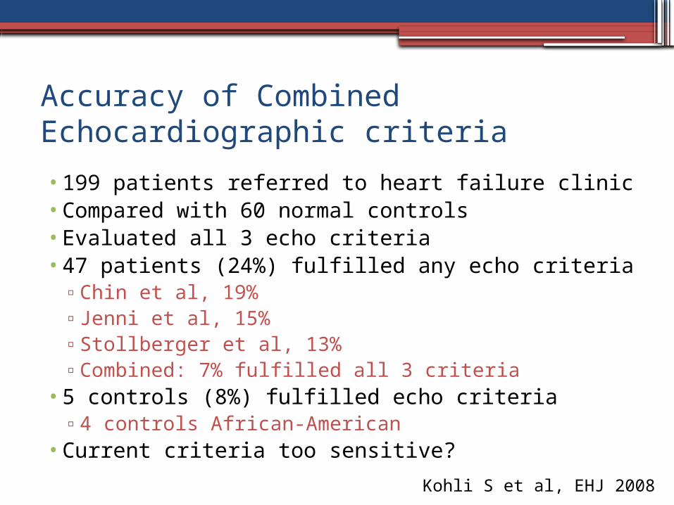
Accuracy of Combined Echocardiographic criteria• 199 patients referred to heart failure clinic• Compared with 60 normal controls• Evaluated all 3 echo criteria• 47 patients (24%) fulfilled any echo criteria
▫Chin et al, 19%▫Jenni et al, 15%▫Stollberger et al, 13%▫Combined: 7% fulfilled all 3 criteria
• 5 controls (8%) fulfilled echo criteria▫4 controls African-American
• Current criteria too sensitive?Kohli S et al, EHJ 2008

Echocardiographic criteria of Chin et al, Jenni et al and Stöllberger et al were used for identification of LVNC in HF and control groups.There was meager correlation among the 3 sets of echocardiographic criteria, with only 30% of patients satisfying all of them. Additionally, 8% of controls met at least 1 of the diagnosticcriteria for LVNC. This information raises the concern that the current echocardiographic criteria are too sensitive, particularly in individuals of African heritage, resulting in overdiagnosis of LVNC (false positive).

Why is it so difficult to validate the diagnosis of LVNC withcertainty?• Lack of awareness and meticulous imaging technique• Failure to differentiate prominent physiologic trabeculations to disease.
The complex meshwork of muscle bundles of the apical- third of the LV and muscle bundles aligning the border of the myocardium are normal structures.
• These normal trabecular patterns can mimic LVNC. The current echocardiographic diagnostic criteria often fail to distinguish the boundary of normal morphologic features and disease.
• Lastly, the difficulty in diagnosing LVNC is due to the lack of clarity between morphologic findings of “left ventricular noncompaction” and the disease entity LVNC cardiomyopathy. The disease should not be defined by the rigid criteria of measurements, such as ratios of the bilayered myocardium. Clearly, we cannot identify a NC/C ratio equal to 2 at end-diastole or end-systole as disease and a ratio of 1.9 as no disease.
• There must be recognition that any NC/C ratio with normal LV systolic and diastolic function and normal myocardial mechanics should not be defined as a disease.
• This set of circumstances must be characterized as morphologic findings that need close follow-up over time to determine if and when there is a transition to disease phenotype (ie, LVNC cardiomyopathy).

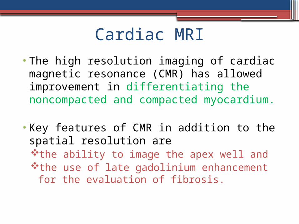
Cardiac MRI•The high resolution imaging of cardiac
magnetic resonance (CMR) has allowed improvement in differentiating the noncompacted and compacted myocardium.
•Key features of CMR in addition to the spatial resolution are the ability to image the apex well and the use of late gadolinium enhancement for
the evaluation of fibrosis.


• In 2005, Petersen et al. compared the noncompacted to compacted layers of myocardium on CMR of healthy volunteers and patients with hypertrophic cardiomyopathy, dilated cardiomyopathy, hypertensive heart disease, and aortic stenosis and patients previously diagnosed with LVNC based on other findings .
• They found that pathological noncompaction had a NC/C >2.3 in end-diastole and that the specificity and negative predictive values were both 99%.
• Later in 2010, Jacquier et al. proposed that the trabeculated mass be taken into consideration when they found that the percentage of trabeculated mass was three times higher in patients with LVNC compared to other groups including controls.
• LV trabecular mass >20% of the global mass predicted the diagnosis of LVNC with a sensitivity and specificity of 93.7%. The advantage of this later method is not depending on the evaluation of specific myocardial segments, but rather the entire mass.

CARDIAC MRI• Disadvantages of this modality include
the availability of MRI and the time to complete the exam and required breath
holding that pose challenges to the patients.There is also an inability to image patients with
some devices/implants.
CMR should play a major role in the evaluation of patients with LVNC when 1. The diagnosis by echocardiogram is not confirmed; 2. A good quality echocardiogram cannot be
obtained; and/or 3. The degree of fibrosis may help delineate the
severity of disease.

MANAGEMENT- no prospective studies/ large cohorts
• Screening 1st degree family members• Treatment of heart failure
▫Medical rx: Guideline based mx for heart failure
▫Consideration of biventricular PPM/ICD• Screening for arrhythmias
▫Consideration of ICD• Anticoagulation
▫Atrial fibrillation and/or LVEF <40% / high CHADS2 score (The event rate of stroke is 1-2% per year or a total risk thromboembolismof 21–38%)
• Heart transplantation

OUTCOME/PROGNOSIS

Oechslin et al, JACC 2000
Not so poor prognosis?•45 patients referred for cardiomyopathy
▫28M, 17F▫37±17 yrs (13-83)▫Majority in NYHA Class I-II CHF (64%)▫20% NSVT, no sustained arrhythmias▫Medical rx:
60% anticoagulation for EF <25% or thromboembolism 90% ACE-I 47% beta blockers
▫At 46 month followup, 97% mean survival from death or transplantation
Murphy RT et al, EHJ 2005

• 65 pts with suspected noncompaction• 74% symptom-based referral, 26% asymptomatic• Followed for mean 46 ± 44 mos (6-193 mos)• Non-symptom group more benign characteristics
▫Younger, fewer ECG abnormalities, greater LVEF, lower left atrial size
• No difference in extent of noncompaction• No major CV events in asymptomatic group• 31% symptomatic group CV death, transplantation• Independent predictors of CV death, transplantation:
▫NYHA III-IV, ventricular arrhythmias, LA size

Conclusions•Rare congenital heart disease thought to result
from an arrest in early cardiac embryogenesis▫Genetic and sporadic forms
•Clinical manifestations:▫Heart failure▫Arrhythmias▫Thromboembolism
•Diagnosis by echocardiography or CMR▫Advances in imaging increased recognition
•Variable prognosis.•Treatment based on clinical manifestations

THANKYOU

• Although distribution and development of the main coronary arteries typically are normal , there is increasing evidence of disturbances in microcirculation in hearts with myocardial noncompaction.
• Keeping in mind the intimate relation of embryonic morphogenesis of the myocardium and the coronary vasculature, this is not surprising.
• With MRI, positron emission tomography, and scintigraphy with thallium201, areas of disturbed microcirculation and transmural perfusion defects corresponding to the zones of noncompacted myocardium have been revealed.
• • Coronary flow reserve has been found to be reduced in the noncompacted
segments but also in other segments with wall motion abnormalities.
• Postmortem analysis has shown subendocardial ischemic lesions and interstitial fibrosis with necrotic myocytes within the trabeculae and compensatory hypertrophy of other myocytes .
• From the available data, it is difficult to differentiate whether the persistence of the embryonic pattern of trabeculated myocardium is secondary to a failure of the coronary microcirculation to grow with the increasing ventricular mass or whether the abnormal myocardial development did not allow normal progression of coronary artery development.

1st Report of Isolated Noncompaction


Genetics• Sporadic or familial• Familial in 18-50% (Oechslin et al, JACC 2000, Chin et al,
Circ 1990, Xing et al, Mol Genet Metab 2006)• Autosomal dominant with incomplete penetrance >
X-linked or autosomal recessive• G4.5 gene of Xq28 region (Bleyl SB et al, Am J Med Genet
1997): taffazin• α-dystrobrevin gene (Ichida F et al, Circ 2001)
▫Links cytoskeleton of myocytes to extracellular matrix• LIM domain binding protein 3/ZASP• Sarcomere genes: β myosin heavy chain (MYH7), α
cardiac actin (ACTC), cardiac troponin T (TNNT2) (Klaassen S et al., Circ 2008)

Genetics•Sporadic or familial•Familial in 18-50% (Oechslin et al, JACC
2000, Chin et al, Circ 1990, Xing et al, Mol Genet Metab 2006)

Oechslin et al, JACC 2000
Clinical Manifestations• Largest
comprehensive study in adults to date
• Review of all echocardiograms 1/84-12/98
• 34 adults with noncompaction

Oechslin et al, JACC 2000

Weiford et al, Circ 2004
Jenni R et al, Heart 2001
Jenni R et al, Heart 2001

Weiford et al, Circ 2004