Embed Size (px)
Citation preview

Development/Plasticity/Repair
Nogo Receptor 1 Confines a Disinhibitory Microcircuit to theCritical Period in Visual Cortex
X Celeste-Elise Stephany,1* X Taruna Ikrar,2* Collins Nguyen,2 Xiangmin Xu,2 and X Aaron W. McGee1
1Developmental Neuroscience Program, Saban Research Institute, Children’s Hospital Los Angeles, Department of Pediatrics, Keck School of Medicine,University of Southern California, Los Angeles, California 90027, and 2Department of Anatomy and Neurobiology, School of Medicine, University ofCalifornia, Irvine, California 92697
A characteristic of the developing mammalian visual system is a brief interval of plasticity, termed the “critical period,” when the circuitry ofprimary visual cortex is most sensitive to perturbation of visual experience. Depriving one eye of vision (monocular deprivation [MD]) duringthe critical period alters ocular dominance (OD) by shifting the responsiveness of neurons in visual cortex to favor the nondeprived eye. Adisinhibitory microcircuit involving parvalbumin-expressing (PV) interneurons initiates this OD plasticity. The gene encoding the neuronalnogo-66-receptor 1 (ngr1/rtn4r) is required to close the critical period. Here we combined mouse genetics, electrophysiology, and circuit mappingwith laser-scanning photostimulation to investigate whether disinhibition is confined to the critical period by ngr1. We demonstrate that ngr1mutant mice retain plasticity characteristic of the critical period as adults, and that ngr1 operates within PV interneurons to restrict the loss ofintracortical excitatory synaptic input following MD in adult mice, and this disinhibition induces a “lower PV network configuration” in bothcritical-period wild-type mice and adult ngr1�/� mice. We propose that ngr1 limits disinhibition to close the critical period for OD plasticity andthat a decrease in PV expression levels reports the diminished recent cumulative activity of these interneurons.
Key words: disinhibition; interneuron; ocular dominance; parvalbumin; plasticity
IntroductionThe developing circuitry of the mammalian visual system is in-structed by experience during an early critical period (Levelt and
Hubener, 2012). Abnormal experience, such as brief monoculardeprivation (MD), within the critical period, but not thereafter,alters the ocular dominance (OD) of neurons in primary visualcortex (Wiesel and Hubel, 1963; Hubel and Wiesel, 1970; Gordonand Stryker, 1996). In the mouse, this critical period extends fromthe third week (�P19) to the fifth week (�P32) postnatal. Fouror more days of MD during this interval yields saturating shifts inOD toward the nondeprived eye. In adult mice, longer periods ofdeprivation are required to shift OD (Sawtell et al., 2003; Fischeret al., 2007a; Morishita and Hensch, 2008; Sato and Stryker,2008). The OD plasticity in adult mice is smaller in magnitudethan during the critical period and operates through distinctmechanisms. Studies using a range of experimental techniquesreveal that OD plasticity engages both excitatory and inhibitory
Received March 21, 2016; revised Aug. 16, 2016; accepted Sept. 2, 2016.Author contributions: X.X. and A.W.M. designed research; C.-E.S., T.I., and C.N. performed research; X.X. and
A.W.M. contributed unpublished reagents/analytic tools; C.-E.S., T.I., C.N., X.X., and A.W.M. analyzed data; C.-E.S.,X.X., and A.W.M. wrote the paper.
This work was supported by the National Eye Institute 1R01EY021580 and Children’s Hospital Los AngelesResearch Development Career Award to A.W.M., and in part by National Institutes of Health Grants NS078434 andMH105427 to X.X. C.-E.S. received a Saban Research Institute predoctoral fellowship. A.W.M. received a BurroughsWellcome Fund Career Award in the Biomedical Sciences.
The authors declare no competing financial interests.*C.-E.S. and T.I. contributed equally to this work.Correspondence should be addressed to either of the following: Dr. Xiangmin Xu, Department of Anatomy and
Neurobiology, School of Medicine, University of California, Irvine, CA 92697, E-mail: [email protected]; or Dr. AaronW. McGee, Department of Anatomical Sciences & Neurobiology, University of Louisville, Louisville, KY 40292. E-mail:[email protected].
A.W. McGee’s present address: Department of Anatomical Sciences and Neurobiology, University of Louisville,Louisville, KY 40202.
DOI:10.1523/JNEUROSCI.0935-16.2016Copyright © 2016 the authors 0270-6474/16/3611006-07$15.00/0
Significance Statement
Life experience refines brain circuits throughout development during specified critical periods. Abnormal experience duringthese critical periods can yield enduring maladaptive changes in neural circuits that impair brain function. In the developingvisual system, visual deprivation early in life can result in amblyopia (lazy-eye), a prevalent childhood disorder comprisingpermanent deficits in spatial vision. Here we identify that the nogo-66 receptor 1 gene restricts an early and essential step in ODplasticity to the critical period. These findings link the emerging circuit-level description of OD plasticity to the genetic regulationof the critical period. Understanding how plasticity is confined to critical periods may provide clues how to better treat amblyopia.
11006 • The Journal of Neuroscience, October 26, 2016 • 36(43):11006 –11012
rich3/zns-neusci/zns-neusci/zns04316/zns9109d16z xppws S�5 10/18/16 11:30 4/Color Figure(s): F1-F4 Art: 0935-16 Input-jjp
AQ: auAQ: or
AQ: A
Fn1
AQ:G-H
AQ:I-J
neurons in visual cortex (Taha et al., 2002; Gandhi et al., 2008;Stephany et al., 2014).
Mice lacking a functional gene for the nogo-66 receptor 1(ngr1) retain critical-period OD plasticity as adults. Adult ngr1constitutive mutants (ngr1�/�) display OD plasticity with 4 d ofMD at P60 indistinguishable from wild-type (WT) mice at P24(McGee et al., 2005; Stephany et al., 2014, 2015). The speed,magnitude, and sensitivity to anesthetics of OD plasticity of adultngr1�/� mice mirror that of WT mice during the critical period(Pham et al., 2004; McGee et al., 2005; Fischer et al., 2007b). Adecrease in the ratio of excitatory to inhibitory neurotransmis-sion (E/I balance) also coincides with the closure of the criticalperiod (Morales et al., 2002). Adult ngr1�/� mice exhibit slightlylower cortical inhibition resulting from a modest reduction inexcitatory drive onto parvalbumin (PV)-expressing interneurons(Stephany et al., 2014). This higher E/I balance manifests as alower frequency but not amplitude of spontaneous EPSCs ontoPV inhibitory neurons and spontaneous IPSCs onto nearby py-ramidal excitatory neurons. Critical period OD plasticity is alsounaffected by diazepam (DZ), a positive allosteric modulator ofthe gamma-aminobutyric acid receptor A (GABA-A) (Hensch etal., 1998).
Disinhibition initiates OD plasticity (Kuhlman et al.,2013). One day of MD during the critical period elevates thevisually evoked activity of cortical excitatory neurons in alertmice. This increase in visual responsiveness results from areduction in activity by fast-spiking PV-positive interneuronsthat in turn can be attributed to a decrease in local excitatorysynaptic drive onto these inhibitory neurons. Importantly,this disinhibition precedes shifts in OD and appears to bepermissive rather than instructive for subsequent OD plastic-ity. How this disinhibitory microcircuit is confined to thecritical period is not yet known.
Here we investigated whether ngr1 may contribute to closingthe critical period by reducing E/I balance and/or by restrictingdisinhibition (Morales et al., 2002; Morishita and Hensch, 2008;Kuhlman et al., 2013). We demonstrate that OD plasticity inconstitutive ngr1�/� adults is resistant to treatment with DZ,both adult ngr1�/� mice and adult mice lacking ngr1 selectivelyin PV interneurons (ngr1flx/flx; PV-Cre) display a decrease in ex-citatory drive onto PV interneurons with 1 d MD that is confinedto the critical period in WT mice, and that a lower relative distri-bution of PV expression by interneurons reports this disinhibi-tion within visual cortex in both juvenile WT mice and adultngr1�/� mice. These findings support the model that ngr1 closesthe critical period by preventing the loss of excitatory synapsesonto PV interneurons during MD.
Materials and MethodsAll procedures and care were performed in accordance with the guide-lines of the Institutional Animal Care and Use Committees at Children’sHospital Los Angeles and the University of California, Irvine.
Mice. Constitutive ngr1 �/� mutant mice and the conditional ngr1flx/flx
mutant mice were a generous gift from Dr. Stephen Strittmatter, YaleUniversity School of Medicine (Kim et al., 2004; Wang et al., 2011).Mutant mice were repeatedly backcrossed onto the C57BL6 backgroundto at least F8. To identify PV interneurons for laser scanning photostimu-lation (LSPS) experiments, we examined WT and ngr1 mutant mice thatcarried alleles for the Cre-dependent td-Tomato reporter (Ai14) andPV-Cre (Hippenmeyer et al., 2005; Madisen et al., 2010). The Ai14 re-porter and PV-Cre driver lines were both obtained from Jackson Labo-ratories (strain #007908 and #0017320, respectively). Genotyping wasperformed using custom primer sets for PCR amplification. Experimentswere performed on both male and female mice.
MD. One eye was closed using a single mattress suture tied with 6-0polypropylene monofilament (Prolene 8709H; Ethicon) under brief iso-flurane anesthesia (2%). The knot was sealed with cyanoacrylate glue.The duration of eye closure was either 4 d before multiunit electrophys-iology (4 d MD), or 1 d before laser scanning photostimulation orperfusion for immunostaining (1 d MD). Before electrophysiologic re-cordings, the suture was removed, and the eye was flushed with sterilesaline and examined under a stereomicroscope. Mice with scarring of thecornea were eliminated from the study.
DZ treatment. A subset of juvenile WT mice (P24-P28) and adultngr1 �/� mice (P60-P90) were administered DZ (2 mg/kg) or saline bydaily intraperitoneal injection, concurrent with 4 d MD before multiunitelectrophysiology (Deidda et al., 2015).
Electrophysiologic recordings in visual cortex. Recording methods wereadapted from previously published methods (McGee et al., 2005;Stephany et al., 2014) and mice were anesthetized with isoflurane (4%induction, 2% maintenance in O2) rather than barbiturates (Nembutal)as we have used in preceding studies. Recordings were performed blindto genotype and drug treatment.
The ocular dominance index (ODI) was calculated for each unit bycomparing the number of action potentials (APs) elicited in a given unitwhen showing the same visual stimulus to each eye independently. Unitswere assigned to one of seven OD categories (1–7). Units assigned tocategory 1 respond predominantly to input from the contralateral eye,and units assigned to category 7 respond predominantly to input fromthe ipsilateral eye (Wiesel and Hubel, 1963). To categorize each unit, theaverage number of APs elicited by the blank was subtracted from theaverage number of APs elicited by the gratings for the contralateral eye(CE) and the ipsilateral eye (IE). Next, the ODI, given by ODI � (IE �CE)/(IE � CE) was calculated for each unit and assigned to OD catego-ries 1–7 as follows: 1, �1 to �0.6; 2, �0.6 to �0.4; 3, �0.4 to �0.1; 4,�0.1 to 0.1; 5, 0.1– 0.4; 6, 0.4 – 0.6; and 7, 0.6 –1. Finally, the sum of thenumber of cells in each category was used to calculate the contralateralbias index (CBI) for each animal with the following formula: CBI �[(n1 � n7) � (2/3) (n2 � n6) � (1/3) (n3 � n5) � N]/2 N where N is thetotal number of units and nx is the number of units with OD scores equalto x (Gordon and Stryker, 1996).
LSPS for circuit mapping. Electrophysiological recordings and photo-stimulation were performed as described previously (Xu et al., 2010).Electrophysiological data were acquired with a Multiclamp 700Bamplifier (Molecular Devices), data acquisition boards (models PCIMIO16E-4 and 6713, National Instruments), and custom-modified ver-sion of Ephus software34 (Ephus; https://openwiki.janelia.org/). Datawere digitized at 10 kHz.
The LSPS procedures were similar to those described previously(Weiler et al., 2008; Kuhlman et al., 2013). LSPS was performed througha 4� objective lens. Stock solution of MNI-caged-L-glutamate (TocrisBioscience) was added to 20 ml ACSF for a concentration of 0.2 mM cagedglutamate. The cortical slice image, acquired through the 4� objective,was visualized using a high-resolution digital CCD camera, and this im-age, in turn, was used to guide and register photostimulation sites. Anelectro-optical modulator and a mechanical shutter controlled the deliv-ery of 1.5ms duration, 15 mW pulses from a 350 nm UV laser (DPSSLasers) to the slice. Focal laser spots approximated a Gaussian profilewith a lateral width of 50 –100 �m. Under our experimental conditions,LSPS-evoked APs were recorded from stimulation locations within 100�m of targeted excitatory neuronal somata and occurred within 150 msafter photostimulation. This indicates that LSPS has a sufficient resolu-tion for V1 laminar circuit mapping. LSPS-evoked EPSCs in patchedneurons were detected under voltage clamp at an empirically determinedmembrane potential of �70 mV. By systematically surveying synapticinputs from hundreds of different sites across a large cortical region,aggregate synaptic input maps were generated for individual neurons.For our mapping experiments, a standard stimulus grid (16 � 16 stim-ulation sites, 65 �m 2 spacing) was used to tessellate V1 from pia to whitematter. The LSPS site spacing was empirically determined to capture thesmallest predicted distance in which photostimulation differentially ac-tivates adjacent neurons. Glutamate uncaging was delivered sequentiallyin a nonraster, nonrandom sequence, following a “shifting-X” pattern
Stephany, Ikrar et al. • ngr1 Limits Disinhibition in Visual Cortex J. Neurosci., October 26, 2016 • 36(43):11006 –11012 • 11007
rich3/zns-neusci/zns-neusci/zns04316/zns9109d16z xppws S�5 10/18/16 11:30 4/Color Figure(s): F1-F4 Art: 0935-16 Input-jjp
AQ: B

designed to avoid revisiting the vicinity of recently stimulated sites(Shepherd et al., 2003).
Laminar circuit input analysis. Photostimulation induces two forms ofexcitatory responses: (1) those that result from direct activation of therecorded neuron’s glutamate receptors; and (2) synaptic responses(EPSCs) resulting from the suprathreshold activation of presynaptic ex-citatory neurons (see Fig. 2A–C). Responses that occur within 10 ms oflaser pulse onset were considered direct; these responses exhibited a dis-tinct shape and occurred immediately after glutamate uncaging. Synapticcurrents with such short latencies are not possible because they wouldhave to occur before the generation of APs in photostimulated neurons.Therefore, direct responses were excluded from local synaptic input anal-ysis. At some locations, synaptic responses were overriding on relativelysmall direct responses; such responses were identified and included insynaptic input analysis as described previously (Kuhlman et al., 2013). Tocheck for any systematic differences across treatment conditions, thespatial extent and frequency of APs elicited in response to direct photo-stimulation were determined in a subset of the experiments by perform-ing whole-cell recordings in current-clamp mode using an 8 � 8mapping grid. Photostimulation excitation profiles assessed by gluta-mate uncaging were found to be similar for control and MD.
For data map analysis, LSPS-evoked EPSCs were quantified across the16 � 16 mapping grid for each cell, and 2–4 individual maps were averagedper recorded cell, reducing the likelihood of incorporating noise events in theanalysis window (150 ms). Averaged maps were then analyzed using the 4�DIC image to bin responses according to laminar cytoarchitectonic land-marks. Synaptic events were binned from locations spanning �195 �mtangential to the targeted soma location and from the top of layer 2/3 to thebottom of layer 6 across the radial vector. Data were plotted as the averageintegrated EPSC amplitude per map location.
Immunohistochemistry. Mice were deeply anesthetized with ketamineHCl (200 mg/kg, Phoenix Pharmaceuticals)/xylazine (20 mg/kg, LloydLaboratories) and transcardially perfused with PBS (ChemCruz, SC-362299) followed by a buffered 4% PFA/PBS (Acros Organics,416780030). Brains were postfixed overnight in 4% PFA/PBS. Free-floating 50 �m sections were cut on a vibrating microtome (Leica, VT1000S) in cool PBS and preserved in PBS containing 0.05% sodium azide(Sigma-Aldrich, S8032).
Coronal sections containing visual cortex were washed in PBS (3 � 5min) and incubated in blocking solution, 3% normal donkey serum(Jackson ImmunoResearch Laboratories) in PBS containing 0.1% TritonX-100 (Sigma-Aldrich T9284) (PBS-T) for 1 h at room temperature. Theprimary antibody sheep anti-PV (R&D Systems, AF5058) was diluted inblocking solution to 1 �g/ml and sections incubated in primary antibodyovernight at 4°C. After repeated washing in PBS-T (3 � 30 min), sectionswere incubated in Alexa-488-conjugated secondary antibody (JacksonImmunoResearch Laboratories, 1:200 in blocking solution) overnight at4°C. The first among a final series of washes contained Hoechst (1:10,000in PBS-T, Santa Cruz Biotechnology) (1 � 10 min), followed by PBS-T(2 � 30 min) and PBS (1 � 10 min). Sections were mounted ontoSuperFrost Plus slides (Fisher Scientific) with SlowFade Gold anti-fadereagent (Invitrogen).
Analysis of PV cell intensity. Analysis of PV cell intensity was adaptedfrom previous reports (Donato et al., 2013). Images from coronal sec-tions stained with anti-PV were captured with an LSM-710 confocalmicroscope with a 20� 0.4 NA objective (Zeiss). Hoechst staining wasused to demarcate visual cortex before capturing images of PV cells. Theacquisition settings (laser intensity and gain) were identical for all sec-tions processed in parallel throughout each set of experiments. For eachseries, sections from a nondeprived WT mouse (P60) were used to opti-mize the settings, maximizing the dynamic range while ensuring that nomore than 20% of pixels within any PV cell were saturated. An internalstandard was processed as described during each series. Two images wererequired to span the distance from the subcortical white matter to the pialsurface. Images were merged in Fiji using the 3D stitching macro. Sec-tions with dampening �30% between the first and last confocal planewere excluded from further analysis.
The intensity of each PV cell whose somas were completely containedin the slice of tissue was obtained using IMARIS software (8.0.0, Bit-
plane). Three-dimensional spheres with a diameter of 7.5 �m weredrawn around the center of each PV cell (smoothness, 0.5 �m; quality,�175). Spheres of consistent diameter were used rather than isosurfacesto eliminate errors introduced by the point spread function in theZ-plane. The intensity of immunohistochemical PV labeling was quan-tified as the average pixel intensity in arbitrary units (au) of pixels con-tained in the 7.5 �m sphere. PV neurons were classified into fourcategories by their average intensity: low PV (1300 au), mid-low PV(1300 –2200 au), mid-high PV (2200 –3100 au), and high PV (�3100 au).
Statistical analysis. All statistical analyses were performed using Prismsoftware (version 6.0, GraphPad). Unless otherwise stated, group com-parisons were made using unpaired, nonparametric Mann–Whitneytests. Where multiple groups are compared, one-way nonparametricANOVA was used (Kruskal–Wallis test with Dunn’s multiple compari-sons test). Error bars indicate � SEM.
ResultsFirst, to test whether the moderate increase in E/I balance dis-played by adult ngr1�/� mice contributes to the sustained criticalperiod for OD plasticity, we augmented cortical inhibition withDZ. DZ elevates cortical inhibition to open a precocious criticalperiod in juvenile WT mice but does not block OD plasticityduring the critical period as measured with single-unit recordings(Hensch et al., 1998). We treated juvenile WT and adult ngr1�/�
mice with saline or DZ daily during 4 d of MD preceding electro-physiologic recording (2 mg/kg/d) (Fig. 1A,E). DZ did not blockOD plasticity for WT mice during the critical period or ngr1mutants at P60, as mice receiving either DZ or saline displayedrightward shifts toward the nondeprived eye in OD histograms(Fig. 1B,F), decreased CBI scores (Fig. 1C,G; WT CP vs WT CP4 d MD�saline, p � 0.01; WT CP vs WT CP 4 d MD�DZ, p �0.005; KO vs KO 4 d MD�saline, p � 0.02; KO vs KO 4 dMD�DZ, p � 0.02), and rightward shifts in the cumulative dis-tribution of OD scores for individual units relative to nonde-prived controls (Fig. 1D,H). This finding is consistent withngr1�/� mice retaining critical-period visual plasticity that ismechanistically distinct from the slower and more limited visualplasticity resident in the adult visual system (Hensch et al., 1998;Sawtell et al., 2003; Pham et al., 2004; Fischer et al., 2007a;Harauzov et al., 2010).
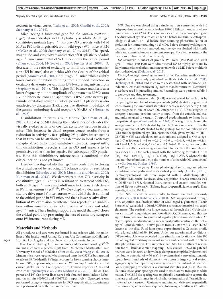
Second, we examined whether the circuit plasticity for disin-hibition is sustained in adult WT or ngr1�/� mice. During thecritical period, 1 d of MD reduces excitatory synaptic input ontoPV interneurons to induce a pronounced disinhibition of visualresponsiveness (Kuhlman et al., 2013). We used LSPS to assessthe spatial distribution and total strength of synaptic inputs ontoPV interneurons in visual cortex of adult WT and ngr1�/� mice(Fig. 2A–C). To identify PV interneurons, we used a Cre-dependent td-Tomato reporter strain (Ai14) in combinationwith PV-Cre (Hippenmeyer et al., 2005; Madisen et al., 2010). Inadult WT mice, 1 d of MD did not appreciably alter the excitatorysynaptic input onto layer (L) 2/3 PV interneurons, and the totalsynaptic current was not different between nondeprived animalsand those receiving MD (Fig. 2D–F). Thus, disinhibition withMD is confined to the critical period.
Conversely, in adult ngr1�/� mice, 1 d of MD significantlyreduced the magnitude of LSPS-induced EPSCs onto L2/L3 PVneurons compared with WT mice and nondeprived ngr1�/�
mice (p � 0.001 and p � 0.03, respectively; Fig. 2E). L2/L3, L4,and L5 provide excitatory synaptic input onto L2/L3 PV in-terneurons in visual cortex (Kuhlman et al., 2013). We comparedthe strength of synaptic input across cortical layers between adultWT and ngr1�/� mice, with and without 1 d of MD (Fig. 2F). Thereduction of total synaptic current in adult ngr1�/� mice follow-
11008 • J. Neurosci., October 26, 2016 • 36(43):11006 –11012 Stephany, Ikrar et al. • ngr1 Limits Disinhibition in Visual Cortex
rich3/zns-neusci/zns-neusci/zns04316/zns9109d16z xppws S�5 10/18/16 11:30 4/Color Figure(s): F1-F4 Art: 0935-16 Input-jjp
F1
F2

ing 1 d of MD was a result of significantly decreased synapticinput across these layers. The average strength of excitatory syn-aptic drive was greater for nondeprived WT mice relative to WTmice following 1 d MD in L5, but this difference was not statisti-cally significant (p � 0.55), whereas 1 d MD reduced total syn-aptic current from L2/L3, L4, and L5 (ngr1�/� vs ngr1�/� 1 dMD, p � 0.05, p � 0.02, and p � 0.01, respectively). We concludethat ngr1 functions to restrict the loss of excitatory synaptic inputonto PV interneurons with MD in adult animals.
Third, to explore where NgR1 functions within cortical cir-cuits to limit the reduction of synaptic drive onto PV interneu-rons by MD, we examined mice lacking ngr1 selectively in PVinterneurons. Deleting ngr1 either in the majority of cortical in-hibitory neurons with Dlx5/6-Cre, or in PV interneurons withPV-Cre, sustains critical-period OD plasticity in adult mice(Stephany et al., 2014). Similar to adult ngr1�/� mice, 1 d of MDalso significantly reduced the magnitude of LSPS-induced EPSCsonto L2/L3 PV neurons in mice lacking ngr1 in PV interneurons(p � 0.0005) (Fig. 3). Therefore, NgR1 can operate within PVinterneurons to limit the loss of excitatory intracortical synapticinput associated with critical-period OD plasticity.
Last, we tested whether the dramatic decrease in synaptic in-put onto PV interneurons following 1 d of MD alters the overallPV network in visual cortex. Recent studies have reported that theintensity of PV immunoreactivity reflects the relative strength ofinhibitory circuitry. PV expression correlates with GAD67 ex-pression, interneurons with low PV expression display lower ra-tios of excitatory to inhibitory synaptic density, and this “low-PVconfiguration” is associated with enhanced learning and struc-tural plasticity (Donato et al., 2013). In hippocampus, motor
cortex, and somatosensory cortex, the distribution of PVexpression is both developmentally regulated and experience-dependent (Donato et al., 2013, 2015). However, whether thisenhanced learning is accompanied by alterations in the spikingactivity of excitatory or inhibitory neurons has not been deter-mined. Given that 1 d MD during the critical period decreases thefiring rate of PV neurons in vivo (Kuhlman et al., 2013), we as-sessed whether the distribution of PV immunoreactivity reflectsdisinhibition associated with critical-period visual plasticity.
We measured the intensity of PV immunoreactivity acrosspopulations of PV interneurons in the binocular zone of primaryvisual cortex in WT mice and ngr1�/� mice at different ages (Fig.4A). We subdivided the population of interneurons into catego-ries of PV intensity: low, mid-low, mid-high, and high (Fig.4A,B) (Donato et al., 2013). The percentage of interneurons inthe low category decreases from P20 to P60 across all corticallayers (Fig. 4C). However, there was no difference in the distri-bution of PV immunoreactivity intensity between adult nonde-prived WT and ngr1�/� mice despite the modest decrease in totalsynaptic input onto L2/L3 PV interneurons in ngr1 mutants (Fig.4D). One day of MD in juvenile WT mice (P24) significantlydecreased the percentage of interneurons with high PV immuno-reactivity (p � 0.018) (Fig. 4E,F). Similarly, 1 d of MD in adult(P60) ngr1�/� mice also yielded a significant decrease in the per-centage of interneurons with high PV immunoreactivity (p �0.004). This shift in PV network configuration was accompaniedby an increase in the proportion of interneurons with low PVexpression (Fig. 4F). However, 1 d MD did not alter the distribu-tion of PV immunoreactivity intensity in adult WT mice. Thus,PV expression levels reflect the recent cumulative activity of PV
Figure 1. Increasing use-dependent GABA-A neurotransmission with DZ does not block OD plasticity in critical period and ngr1�/� mice. A, Schematic of the timeline for DZ injection and MDbefore assessment of OD in WT critical period (WT CP) mice (black). B, OD histograms and CBI score for nondeprived WT CP mice (n � 6), and WT CP mice receiving 4 d of MD (4 d MD) concurrent withsaline injection (n � 6) or DZ injection (n � 6). A black ellipse under the histogram indicates MD. There is a rightward shift toward the open eye in OD histograms for WT CP mice receiving saline orDZ injection with 4 d MD. C, The median CBI score for WT CP mice is lower after 4 d MD concurrent with daily DZ or saline injection (i.p.) compared with nondeprived controls. Each point indicates theCBI for an individual animal. Bars represent the average for each group. Error bars indicate SEM. Gray box represents the typical range of CBI scores for nondeprived mice. D, Cumulative histogramsof OD scores for groups reported in B, C. E–H, Same as A–D for adult (P60) ngr1 �/� mice (blue). F, Nondeprived (n � 4); 4 d MD � saline (n � 7); 4 d MD � DZ (n � 7). G, The median CBI scorefor ngr1 �/� mice is lower after 4 d MD concurrent with daily DZ or saline injection (i.p.) compared with nondeprived controls. * p 0.05; ** p 0.01.
Stephany, Ikrar et al. • ngr1 Limits Disinhibition in Visual Cortex J. Neurosci., October 26, 2016 • 36(43):11006 –11012 • 11009
rich3/zns-neusci/zns-neusci/zns04316/zns9109d16z xppws S�5 10/18/16 11:30 4/Color Figure(s): F1-F4 Art: 0935-16 Input-jjp
F3
AQ:D-E
F4

interneurons and the shift to a low PV network reports disinhi-bition of cortical circuitry.
DiscussionHere we demonstrate that the disinhibitory microcircuit initiat-ing OD plasticity is confined to the critical period by ngr1 oper-ating in PV interneurons, and this disinhibition is accompaniedby a decrease in the relative distribution of PV expression acrossthis population of interneurons in visual cortex. These find-ings link the emerging circuit-level description of experience-
dependent visual plasticity to the genetic regulation of the closureof the critical period.
How might ngr1 restrict disinhibition and close the criticalperiod? The molecular and cellular characterization of the NgR1protein provides limited insight into potential mechanisms bywhich the receptor gates disinhibition. NgR1 is a neuronal recep-tor attached to the neuronal surface by a glycosylphosphatidylin-isotol anchor, but the subcellular localization of NgR1 remainsunclear, as the protein has been reported to be enriched in eitherdendritic spines or axons (Raiker et al., 2010; Zemmar et al., 2014;
Figure 2. NgR1 restricts disinhibition to the critical period. A, A schematic of the recording configuration. PV interneurons are patched in the whole-cell configuration while UV laser directs thefocal release of glutamate over the soma of excitatory neurons distributed throughout the tissue section. Glutamate uncaging drives the firing of APs by neurons under the region of brief UVillumination. B, An example of the 16 � 16 grid (aqua dots) and the position of a recorded PV interneuron on L2/L3 (red circle). C, An example of (1) the current induced by direct somatic stimulationof the recorded PV interneuron and (2) excitatory synaptic currents. D, LSPS mapping of excitatory synaptic inputs onto PV interneurons in L2/L3 of adult (P55-P65) WT and ngr1 �/� mice (WT, n �13; WT 1 d MD, n � 9; ngr1 �/�, n � 9; ngr1 �/� 1 d MD, n � 16). Adult ngr1 �/� mice display a loss of excitatory drive with 1 d MD similar to WT mice during the critical period. E, Quantificationof total synaptic input for WT and ngr1 �/� mice with and without 1 d MD. *p � 0.03. **p � 0.0007. F, Comparison of the laminar distribution of average excitatory synaptic input to L2/L3 PVinterneurons across genotypes and conditions. Data are mean � SE. Black bar represents WT. Gray bar represents WT � 1 d MD. Dark blue bar represents ngr1 �/� without MD. Light blue barrepresents ngr1 �/� � 1 d MD. *p 0.05. **p .01.
Figure 3. NgR1 operates in PV interneurons to limit disinhibition following 1 d MD. A, LSPS mapping of excitatory synaptic inputs onto PV interneurons in L2/L3 of nondeprived adult (P55-P85)ngr1flx/flx; PV-Cre mice and following 1 d MD (nondeprived, n � 8; 1 d MD, n � 11). Adult ngr1flx/flx; PV-Cre exhibit a decrease of intracortical excitatory synaptic input onto PV interneurons following1 d MD similar to adult ngr1 �/� mice. B, Quantification of total synaptic input for nondeprived adult ngr1flx/flx; PV-Cre mice and after 1 d MD. ***p � 0.0005.
11010 • J. Neurosci., October 26, 2016 • 36(43):11006 –11012 Stephany, Ikrar et al. • ngr1 Limits Disinhibition in Visual Cortex
rich3/zns-neusci/zns-neusci/zns04316/zns9109d16z xppws S�5 10/18/16 11:30 4/Color Figure(s): F1-F4 Art: 0935-16 Input-jjp
AQ: F

Stephany et al., 2015). NgR1 is expressed in visual cortex before eyeopening (�P14), and expression levels are unchanged by the close ofthe critical period (McGee et al., 2005).
Recent studies have explored whether NgR1 limits the forma-tion and stability of dendritic spines by excitatory pyramidal neu-rons. Unfortunately, these results do not support a unifyingconclusion. Reducing the expression of NgR1 in transfected or-ganotypic hippocampal cultures doubles dendritic spine density,whereas overexpressing NgR1 reduces spine density by half invitro (Wills et al., 2012). However, spine density is normal in both
ngr1�/� mice and transgenic mice over-expressing NgR1 (Lee et al., 2008; Karlenet al., 2009). Likewise, ngr1�/� mice havebeen reported to display both dramati-cally elevated spine formation and newspine stability in vivo (Akbik et al., 2013).Yet we have performed similar, in somecases nearly identical, experiments withthe same strain of ngr1�/� mice, but weare unable to reproduce these findings. Inour hands, the basal synaptic structuralplasticity of ngr1�/� is not different fromWT mice (Park et al., 2014; Frantz et al.,2016). Experiments are still required todetermine whether MD yields increasedspine dynamics in ngr1�/� mice beyondthat reported for WT mice (Hofer et al.,2009). Yet, although ngr1 is not a promi-nent regulator of basal synaptic turn-over in adult brain, it may function in amore specific role to limit activity-dependent and experience-dependentsynaptic refinement.
As deleting ngr1 selectively with PV-Creis sufficient to permit the loss of excitatorysynaptic input onto PV interneurons withMD in adult mice, NgR1 functions in theseinterneurons to stabilize excitatory synapsesduring the reduced overall cortical activitythat induces disinhibition in V1 during thecritical period. The distribution of ligandsfor NgR1, both myelin-associated proteinsand chondroitin sulfate proteoglycans(CSPGs), increases as the critical periodcloses (Huang et al., 1999; McGee et al.,2005; Dickendesher et al., 2012). Interest-ingly, CSPGs are enriched in perineuronalnets that ensheath PV interneurons, and en-zymatic digestion of sugar chains fromCSPGs partially reactivates OD plasticity inrats (Pizzorusso et al., 2002). Perhaps NgR1closes the critical period by interacting withCSPGs in perineuronal nets to stabilize ex-citatory synapses onto PV interneurons orcounteract signaling pathways that promotedisinhibition in visual cortex following 1 dMD.
Disinhibition is a conserved compo-nent of cortical plasticity that is impli-cated in associative learning and memoryin hippocampus, amygdala, auditory cor-tex, and motor cortex (Letzkus et al.,2015). NgR1 is also expressed in these
brain regions (Barrette et al., 2007). Moreover, transgenic over-expression of NgR1 disrupts hippocampal-dependent spatiallearning (Karlen et al., 2009), whereas ngr1�/� mice display ab-errant extinction following auditory fear conditioning and a def-icit in overall performance on the rotarod (Park et al., 2014).Whether ngr1 also limits disinhibition in these neural circuits isnot known. Future work will be required to determine how cor-tical circuits change during this transient period of elevated ex-citatory neurotransmission to yield the enduring alterations in
Figure 4. PV network configuration is altered by disinhibition in V1. A, Representative population of PV intensities for onemouse from each age and genotype. Dots represent the intensity of one PV interneuron in arbitrary units (au). Dashed linesindicate the boundaries for expression level categories. B, Immunostaining for PV in V1 of P24 and P60 ngr1 �/� mice. C, Relativedistribution of PV intensity in P24 (n � 9 and 8), P40 (n � 6 and 6), and P60 (n � 7 and 5) WT and ngr1 �/� mice (n � numberof mice per group in parentheses). D, There is no significant difference in the fraction of PV neurons in the high- or low-PVconfiguration between WT and ngr1 �/� mice. E, Relative distribution of PV intensity in P24 WT mice and P60 WT and ngr1 �/�
mice with 1 d MD (MD). F, The fraction of PV neurons in the high-PV configuration is lower with MD in P24 WT and in P60 ngr1 �/�
mice compared with age-matched nondeprived control mice (Ctrl), whereas the fraction of PV neurons in the low-PV configurationincreases with MD. * p 0.05; ** p 0.01.
Stephany, Ikrar et al. • ngr1 Limits Disinhibition in Visual Cortex J. Neurosci., October 26, 2016 • 36(43):11006 –11012 • 11011
rich3/zns-neusci/zns-neusci/zns04316/zns9109d16z xppws S�5 10/18/16 11:30 4/Color Figure(s): F1-F4 Art: 0935-16 Input-jjp
AQ: C
AQ: G

cortical responsiveness associated with OD plasticity, a model ofthe prevalent childhood visual disorder amblyopia.
ReferencesAkbik FV, Bhagat SM, Patel PR, Cafferty WB, Strittmatter SM (2013) Ana-
tomical plasticity of adult brain is titrated by Nogo receptor 1. Neuron77:859 – 866. CrossRef Medline
Barrette B, Vallieres N, Dube M, Lacroix S (2007) Expression profile of re-ceptors for myelin-associated inhibitors of axonal regeneration in theintact and injured mouse central nervous system. Mol Cell Neurosci 34:519 –538. CrossRef Medline
Deidda G, Allegra M, Cerri C, Naskar S, Bony G, Zunino G, Bozzi Y, Caleo M,Cancedda L (2015) Early depolarizing GABA controls critical-period plas-ticity in the rat visual cortex. Nat Neurosci 18:87–96. CrossRef Medline
Dickendesher TL, Baldwin KT, Mironova YA, Koriyama Y, Raiker SJ, Askew KL,Wood A, Geoffroy CG, Zheng B, Liepmann CD, Katagiri Y, Benowitz LI,Geller HM, Giger RJ (2012) NgR1 and NgR3 are receptors for chondroitinsulfate proteoglycans. Nat Neurosci 15:703–712. CrossRef Medline
Donato F, Rompani SB, Caroni P (2013) Parvalbumin-expressing basket-cell network plasticity induced by experience regulates adult learning.Nature 504:272–276. CrossRef Medline
Donato F, Chowdhury A, Lahr M, Caroni P (2015) Early- and late-bornparvalbumin basket cell subpopulations exhibiting distinct regulationand roles in learning. Neuron 85:770 –786. CrossRef Medline
Fischer QS, Aleem S, Zhou H, Pham TA (2007a) Adult visual experiencepromotes recovery of primary visual cortex from long-term monoculardeprivation. Learn Mem 14:573–580. CrossRef Medline
Fischer QS, Graves A, Evans S, Lickey ME, Pham TA (2007b) Monoculardeprivation in adult mice alters visual acuity and single-unit activity.Learn Mem 14:277–286. CrossRef Medline
Frantz MG, Kast RJ, Dorton HM, Chapman KS, McGee AW (2016) Nogoreceptor 1 limits ocular dominance plasticity but not turnover of axonalboutons in a model of amblyopia. Cereb Cortex 26:1975–1985. CrossRefMedline
Gandhi SP, Yanagawa Y, Stryker MP (2008) Delayed plasticity of inhibitoryneurons in developing visual cortex. Proc Natl Acad Sci U S A 105:16797–16802. CrossRef Medline
Gordon JA, Stryker MP (1996) Experience-dependent plasticity of binocu-lar responses in the primary visual cortex of the mouse. J Neurosci 16:3274 –3286. Medline
Harauzov A, Spolidoro M, DiCristo G, De Pasquale R, Cancedda L, Pizzo-russo T, Viegi A, Berardi N, Maffei L (2010) Reducing intracortical in-hibition in the adult visual cortex promotes ocular dominance plasticity.J Neurosci 30:361–371. CrossRef Medline
Hensch TK, Fagiolini M, Mataga N, Stryker MP, Baekkeskov S, Kash SF(1998) Local GABA circuit control of experience-dependent plasticity indeveloping visual cortex. Science 282:1504 –1508. CrossRef Medline
Hippenmeyer S, Vrieseling E, Sigrist M, Portmann T, Laengle C, Ladle DR,Arber S (2005) A developmental switch in the response of DRG neuronsto ETS transcription factor signaling. PLoS Biol 3:e159. CrossRef Medline
Hofer SB, Mrsic-Flogel TD, Bonhoeffer T, Hubener M (2009) Experienceleaves a lasting structural trace in cortical circuits. Nature 457:313–317.CrossRef Medline
Huang ZJ, Kirkwood A, Pizzorusso T, Porciatti V, Morales B, Bear MF, MaffeiL, Tonegawa S (1999) BDNF regulates the maturation of inhibition andthe critical period of plasticity in mouse visual cortex. Cell 98:739 –755.CrossRef Medline
Hubel DH, Wiesel TN (1970) The period of susceptibility to the physiolog-ical effects of unilateral eye closure in kittens. J Physiol 206:419 – 436.CrossRef Medline
Karlen A, Karlsson TE, Mattsson A, Lundstromer K, Codeluppi S, Pham TM,Backman CM, Ogren SO, Aberg E, Hoffman AF, Sherling MA, Lupica CR,Hoffer BJ, Spenger C, Josephson A, Brene S, Olson L (2009) Nogo re-ceptor 1 regulates formation of lasting memories. Proc Natl Acad SciU S A 106:20476 –20481. CrossRef Medline
Kim JE, Liu BP, Park JH, Strittmatter SM (2004) Nogo-66 receptor preventsraphespinal and rubrospinal axon regeneration and limits functional re-covery from spinal cord injury. Neuron 44:439 – 451. CrossRef Medline
Kuhlman SJ, Olivas ND, Tring E, Ikrar T, Xu X, Trachtenberg JT (2013) Adisinhibitory microcircuit initiates critical-period plasticity in the visualcortex. Nature 501:543–546. CrossRef Medline
Lee H, Raiker SJ, Venkatesh K, Geary R, Robak LA, Zhang Y, Yeh HH, Shrager
P, Giger RJ (2008) Synaptic function for the Nogo-66 receptor NgR1:regulation of dendritic spine morphology and activity-dependent synap-tic strength. J Neurosci 28:2753–2765. CrossRef Medline
Letzkus JJ, Wolff SB, Luthi A (2015) Disinhibition, a circuit mechanism forassociative learning and memory. Neuron 88:264 –276. CrossRef Medline
Levelt CN, Hubener M (2012) Critical-period plasticity in the visual cortex.Annu Rev Neurosci 35:309 –330. CrossRef Medline
Madisen L, Zwingman TA, Sunkin SM, Oh SW, Zariwala HA, Gu H, Ng LL,Palmiter RD, Hawrylycz MJ, Jones AR, Lein ES, Zeng H (2010) A robustand high-throughput Cre reporting and characterization system for thewhole mouse brain. Nat Neurosci 13:133–140. CrossRef Medline
McGee AW, Yang Y, Fischer QS, Daw NW, Strittmatter SM (2005)Experience-driven plasticity of visual cortex limited by myelin and Nogoreceptor. Science 309:2222–2226. CrossRef Medline
Morales B, Choi SY, Kirkwood A (2002) Dark rearing alters the develop-ment of GABAergic transmission in visual cortex. J Neurosci 22:8084 –8090. Medline
Morishita H, Hensch TK (2008) Critical period revisited: impact on vision.Curr Opin Neurobiol 18:101–107. CrossRef Medline
Park JI, Frantz MG, Kast RJ, Chapman KS, Dorton HM, Stephany CE, ArnettMT, Herman DH, McGee AW (2014) Nogo receptor 1 limits tactile taskperformance independent of basal anatomical plasticity. PLoS One9:e112678. CrossRef Medline
Pham TA, Graham SJ, Suzuki S, Barco A, Kandel ER, Gordon B, Lickey ME (2004)Asemi-persistentadultoculardominanceplasticityinvisualcortexisstabilizedbyactivated CREB. Learn Mem 11:738–747. CrossRef Medline
Pizzorusso T, Medini P, Berardi N, Chierzi S, Fawcett JW, Maffei L (2002)Reactivation of ocular dominance plasticity in the adult visual cortex.Science 298:1248 –1251. CrossRef Medline
Raiker SJ, Lee H, Baldwin KT, Duan Y, Shrager P, Giger RJ (2010)Oligodendrocyte-myelin glycoprotein and Nogo negatively regulateactivity-dependent synaptic plasticity. J Neurosci 30:12432–12445.CrossRef Medline
Sato M, Stryker MP (2008) Distinctive features of adult ocular dominanceplasticity. J Neurosci 28:10278 –10286. CrossRef Medline
Sawtell NB, Frenkel MY, Philpot BD, Nakazawa K, Tonegawa S, Bear MF(2003) NMDA receptor-dependent ocular dominance plasticity in adultvisual cortex. Neuron 38:977–985. CrossRef Medline
Shepherd GM, Pologruto TA, Svoboda K (2003) Circuit analysis ofexperience-dependent plasticity in the developing rat barrel cortex. Neu-ron 38:277–289. CrossRef Medline
Stephany CE, Chan LL, Parivash SN, Dorton HM, Piechowicz M, Qiu S, McGee AW(2014) Plasticity of binocularity and visual acuity are differentially limited byNogo receptor. J Neurosci 34:11631–11640. CrossRef Medline
Stephany CE, Frantz MG, McGee AW (2015) Multiple roles for Nogo recep-tor 1 in visual system plasticity. Neuroscientist. Advance online publica-tion. Retrieved Nov. 9, 2015. doi: 10.1177/1073858415614564. CrossRefMedline
Taha S, Hanover JL, Silva AJ, Stryker MP (2002) Autophosphorylation ofalphaCaMKII is required for ocular dominance plasticity. Neuron 36:483– 491. CrossRef Medline
Wang X, Duffy P, McGee AW, Hasan O, Gould G, Tu N, Harel NY, Huang Y,Carson RE, Weinzimmer D, Ropchan J, Benowitz LI, Cafferty WB, Strit-tmatter SM (2011) Recovery from chronic spinal cord contusion afterNogo receptor intervention. Ann Neurol 70:805– 821. CrossRef Medline
Weiler N, Wood L, Yu J, Solla SA, Shepherd GM (2008) Top-down laminarorganization of the excitatory network in motor cortex. Nat Neurosci11:360 –366. CrossRef Medline
Wiesel TN, Hubel DH (1963) Single-cell responses in striate cortex of kit-tens deprived of vision in one eye. J Neurophysiol 26:1003–1017. Medline
Wills ZP, Mandel-Brehm C, Mardinly AR, McCord AE, Giger RJ, GreenbergME (2012) The Nogo receptor family restricts synapse number in thedeveloping hippocampus. Neuron 73:466 – 481. CrossRef Medline
Xu X, Olivas ND, Levi R, Ikrar T, Nenadic Z (2010) High precision and fastfunctional mapping of cortical circuitry through a novel combination ofvoltage sensitive dye imaging and laser scanning photostimulation. J Neu-rophysiol 103:2301–2312. CrossRef Medline
Zemmar A, Weinmann O, Kellner Y, Yu X, Vicente R, Gullo M, Kasper H,Lussi K, Ristic Z, Luft AR, Rioult-Pedotti M, Zuo Y, Zagrebelsky M,Schwab ME (2014) Neutralization of Nogo-A enhances synaptic plastic-ity in the rodent motor cortex and improves motor learning in vivo.J Neurosci 34:8685– 8698. CrossRef Medline
11012 • J. Neurosci., October 26, 2016 • 36(43):11006 –11012 Stephany, Ikrar et al. • ngr1 Limits Disinhibition in Visual Cortex
rich3/zns-neusci/zns-neusci/zns04316/zns9109d16z xppws S�5 10/18/16 11:30 4/Color Figure(s): F1-F4 Art: 0935-16 Input-jjp