Embed Size (px)
Citation preview

CHC 812: CELL BIOLOGY, GENETICS, IMMUNOLOGY AND TERATOLOGY
Lecture 01, 2015Molecular Diagnostics
Lecturer: Dr. G. Kattam Maiyoh
GKM/M.MEDPAEDS/LECT 01/2015

Molecular Diagnostics
The use of molecular biology techniques to
expand scientific knowledge of the natural
history of diseases, identify people who are at
risk for acquiring specific diseases, and
diagnose human diseases at the molecular
level.
GKM/M.MEDPAEDS/LECT 01/2015

Molecular Diagnostic
• USE OF:– Sequence Specific INFORMATION • in
– MACROMOLECULES• for
– Risk identification– Diagnosis– Prognosis– Prediction of response to therapy– Monitoring therapeutic responses
GKM/M.MEDPAEDS/LECT 01/2015
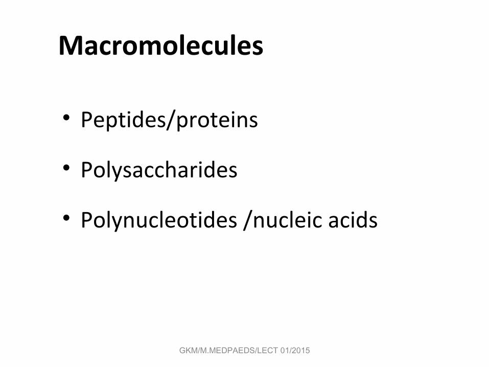
Macromolecules
• Peptides/proteins
• Polysaccharides
• Polynucleotides /nucleic acids
GKM/M.MEDPAEDS/LECT 01/2015

Molecular Diagnostics: Significance
To face the near future, the medical practitioner not only understand molecular biology, but must also embrace the use of this rapidly expanding body of information in his medical practice, whether practicing family medicine pediatrics, oncology, obstetrics and gynecology, pathology, or any other medical specialty.
GKM/M.MEDPAEDS/LECT 01/2015

Molecular Diagnostics are Transforming Medicine
Pre-natal testing
Disease predisposition
Disease detection
Drug selection
Recurrence monitoring
Key questions
-> Need for Molecular tests
“Is the baby healthy? “
“What diseases is this patient at risk for?”
“Does this patient have a disease?”
“What drugs should I prescribe?”
“Has the disease returned?”
Molecular diagnostics is >$3 billion
market WW and growing at >20%
annually
GKM/M.MEDPAEDS/LECT 01/2015

Old vs. New Molecular Diagnostics
• Old: grow cells/pathogen->test• Such growth can be a problem as it is
sometimes slow AND costly.• New: direct test (either immunological or
DNA/RNA based)
GKM/M.MEDPAEDS/LECT 01/2015
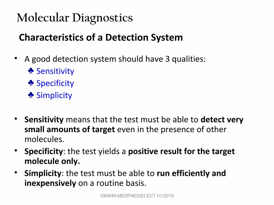
Characteristics of a Detection System
• A good detection system should have 3 qualities:♣ Sensitivity♣ Specificity♣ Simplicity
• Sensitivity means that the test must be able to detect very small amounts of target even in the presence of other molecules.
• Specificity: the test yields a positive result for the target molecule only.
• Simplicity: the test must be able to run efficiently and inexpensively on a routine basis.
Molecular Diagnostics
GKM/M.MEDPAEDS/LECT 01/2015

Molecular DiagnosticsImmunological Diagnostics Methods
1. Radioimmunoassay
2. Enzyme-Linked ImmunoSorbent Assay (ELISA)
3. Western Blotting
4. Immunoprecipitation
5. Immunofluorescence
6. Flow Cytometry and Fluorescence
7. Alternatives to Antigen-Antibody Reactions
8. Immunoelectron Microscopy
GKM/M.MEDPAEDS/LECT 01/2015

Target antigens and polyclonal versus monoclonal antibodies
Polyclonal antibodies are made against and react withmultiple antigenic sites (epitopes) on a target antigen.Monoclonal antibodies are directed against a particularantigenic site.
Target antigenwith various antigenicdeterminants (epitopes)1
2 3 4
5
67
GKM/M.MEDPAEDS/LECT 01/2015

Targets for diagnostic monoclonal antibodies
• Polypeptide hormones (chorionic gonadotropin, growth hormone)
• Tumor markers (Prostate-specific antigen)• Cytokines (interleukins 1-8)• Drug monitoring (cyclosporin)• Miscellaneous targets (Vitamin B12)• Infectious diseases (Chlamydia, Herpes, Rubella,
Hepatitis B, Legionella, HIV)
GKM/M.MEDPAEDS/LECT 01/2015

Radioimmunoassay
GKM/M.MEDPAEDS/LECT 01/2015

GKM/M.MEDPAEDS/LECT 01/2015

Enzyme-Linked Immunosorbent Assay (ELISA): Immunological detection
Target molecule
antigenic site
i i i i i i i i i i i i i i i
Support
A. Bind sample to the support (commonly plastic or a membrane)
B. Add primary antibody; washC. Add secondary antibody-enzyme conjugate; wash
D. Add substrate
Y
YY
Y
bound primaryantibody
Y
E Y EYEYE
enzyme linkedsecondary antibody
colorless substrate
colored product
GKM/M.MEDPAEDS/LECT 01/2015

Immunological Diagnostics Methods - ELISA
• Addition of a specific antibody (primary antibody) which will bind to the test molecule if it is present.
• Washing to remove unbound molecules.• Addition of secondary antibody which will
bind to the primary antibody.• The secondary antibody usually has attached
to it an enzyme e.g. alkaline phosphatase.• Wash to remove unbound antibody.• Addition of a colourless substrate which will
react with the secondary antibody to give a colour reaction which indicates a positive result.
-> can be used for quasi High-throughput!!!
GKM/M.MEDPAEDS/LECT 01/2015

ELISA -Variants
Detection based on enzyme catalyzed reactions:
1.alkaline Phosphatase2.horseradish peroxidase3. β-galactosidase
Detection based on fluorescent labeled secondary antibody
GKM/M.MEDPAEDS/LECT 01/2015

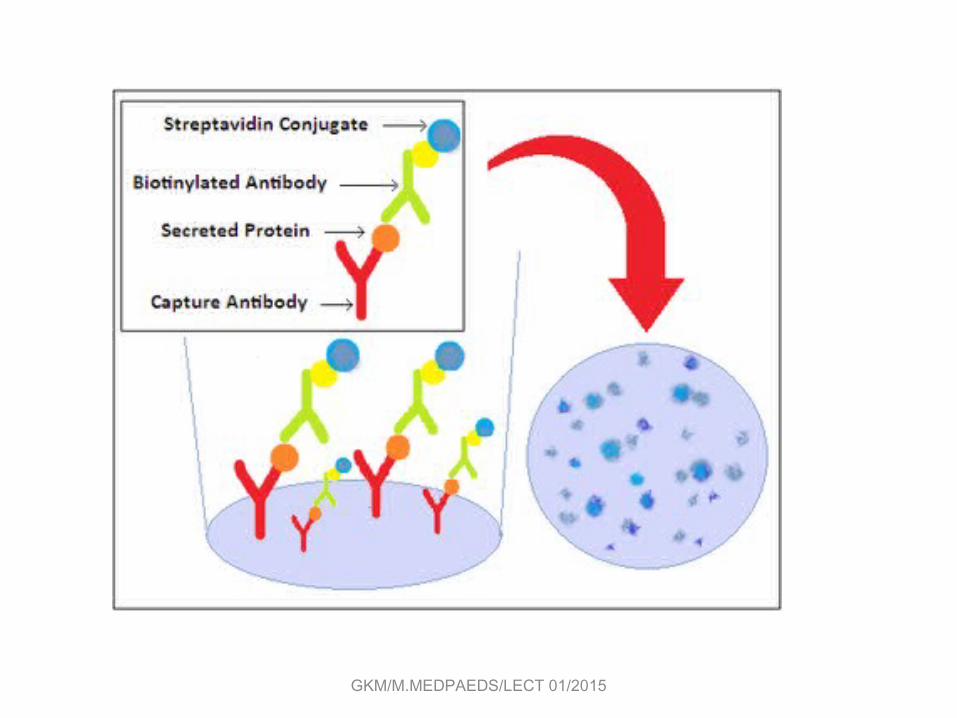
ELISA –VariantsThe ELISPOT
The ELISPOT assay -> to determine
quantitatively the # of cells in a population
that are producing specific Ab or cytokine.
-> precipitates & forms a spot only on the areas of the well where cytokine-secreting cells had been deposited.
P.T.O
GKM/M.MEDPAEDS/LECT 01/2015
GKM/M.MEDPAEDS/LECT 01/2015

Western blot
SDS-Page: separates the components according to their molecular weight.
Blot: the proteins in the gel are transferred to the sheet of nitrocellulose or nylon by the passage of an electric current.
Immunoreaction: probed with Ab & then radiolabeled or enzyme-linked 2nd Ab.
Detection: a position is visualized by means of an ELISA reaction.
GKM/M.MEDPAEDS/LECT 01/2015

GKM/M.MEDPAEDS/LECT 01/2015
GKM/M.MEDPAEDS/LECT 01/2015

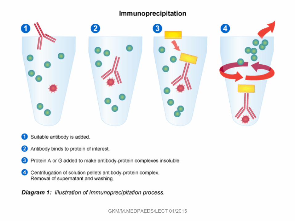
ImmunoprecipitationImmuno-precipitates can be collected using magnetic beads coupled to a secondary antibody.
GKM/M.MEDPAEDS/LECT 01/2015

Immunofluorescence
Fluorochromes-Fluorescein (490 517nm)→-Rhodamine (515 546nm)→-Phycoerythrin
mIgM-producing B cells indirectly stained with rhodamine-conjurated secondary Ab under a fluorescence microscope.
Protein A has the ability to bind to IgG
GKM/M.MEDPAEDS/LECT 01/2015

Immuno Electron Microscopy
electron-dense labelsabsorb electrons.
An immunoelectronmicrograph
of the surface of a B-cell
lymphoma was stained with two
antibodies (Ab against class II
MHC labeled with 30nm gold
particles, & another Ab against
class I MHC w/ 15nm gold
particles.
(The density of class I exceeds
that of class II)
- Electron-dense label (ferritin
or colloidal gold) is conjugated
to the Fc
portion.GKM/M.MEDPAEDS/LECT 01/2015

We now know how God wrote the book of life
Bill Clinton

But do we know how to read the book ?

Molecular Genetic Tests• Genetic test:– Analyis of human• DNA• RNA• chromosomes• proteins• metabolites
– to detect heritable disease-related• genotype, • phenotype• karyotype
– for clinical purposes.
GKM/M.MEDPAEDS/LECT 01/2015

Genetic Diagnosis“Purpose”
• Diagnostic Testing• Screening • Presymptomatic Testing• Prenatal testing• Preimplantation Diagnosis• Pharmacogenetic testing • Susceptibility to environmental agents
GKM/M.MEDPAEDS/LECT 01/2015

Genetic Alterations
• Chromosomal alterations• “Gene-level” alterations.
GKM/M.MEDPAEDS/LECT 01/2015
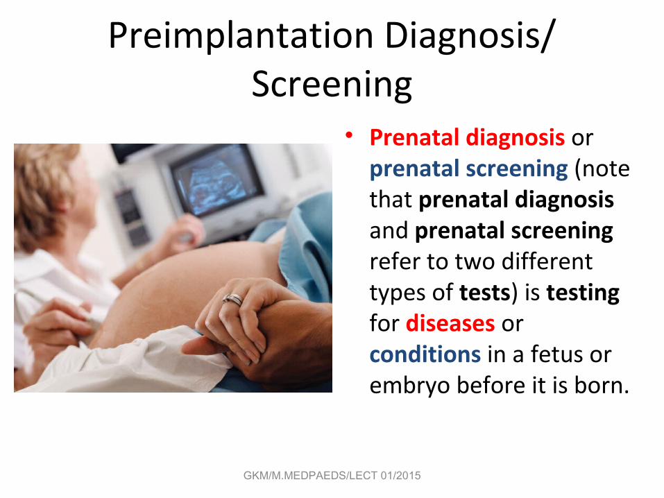
Preimplantation Diagnosis/ Screening
• Prenatal diagnosis or prenatal screening (note that prenatal diagnosis and prenatal screening refer to two different types of tests) is testing for diseases or conditions in a fetus or embryo before it is born.
GKM/M.MEDPAEDS/LECT 01/2015

GKM/M.MEDPAEDS/LECT 01/2015
Preimplantation Diagnosis
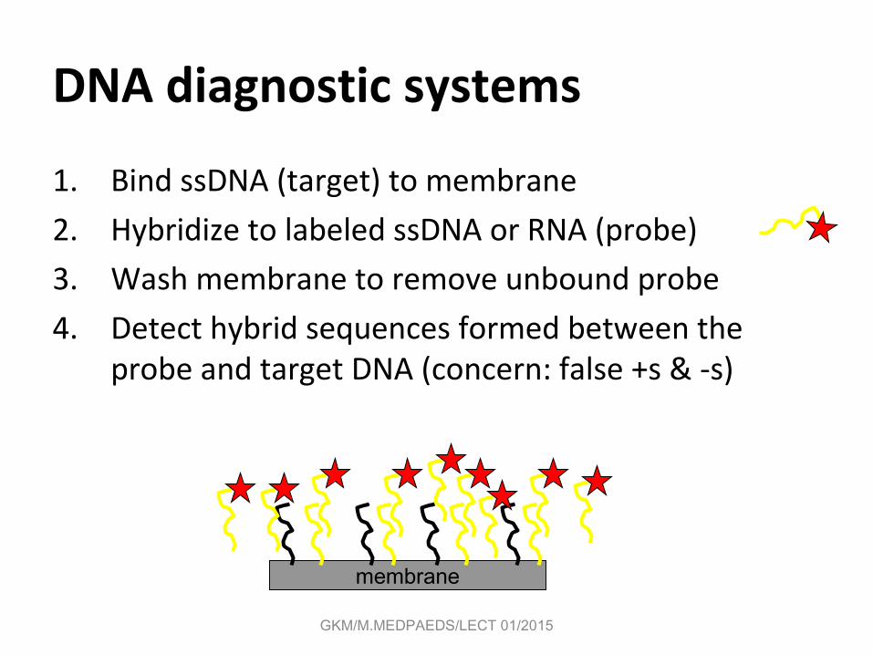
DNA diagnostic systems
1. Bind ssDNA (target) to membrane
2. Hybridize to labeled ssDNA or RNA (probe)
3. Wash membrane to remove unbound probe
4. Detect hybrid sequences formed between the probe and target DNA (concern: false +s & -s)
membrane
GKM/M.MEDPAEDS/LECT 01/2015

Ordering Molecular Tests• Patient preparation: None
– Avoid heparin: interferes with PCR.
• Specimens:– Fresh whole blood: EDTA/Citrate– Fresh tissues– Frozen tissues– Paraffin embeded tissues– Slides etc.
GKM/M.MEDPAEDS/LECT 01/2015

Ordering Molecular genetic Tests
• Specimen Handling• DNA-based tests:
– Room temperature, up to 72 hours (maybe more with special buffers)
• RNA-based tests:– Deliver ASAP (4-6 hours)– Special considerations for proprietary test.
GKM/M.MEDPAEDS/LECT 01/2015

Ordering Molecular genetic Tests • Essential info:
– Clinical information – pedigree, if possible– Race– reason for testing.
• Informed consent:• Nature of test; availability of genetic counseling;
implications of positive and negative tests, etc.
GKM/M.MEDPAEDS/LECT 01/2015

Molecular Diagnosis of Genetic Disease
• Cystic fibrosis Sickle-cell anemia
GKM/M.MEDPAEDS/LECT 01/2015

DNA based diagnosis of Malaria and Typanosoma cruzi
1. A DNA probe from a highly repeated DNA sequence of Plasmodium falciparum, the parasite that causes malaria, is used to screen blood samples via hybridization assays
2. DNA primers are made against the ends of a 188 bp repeated sequence contained in the protozoan parasite Typanosoma cruzi, the causative agent of Chagas disease and used in a PCR/polyacrylamide gel electrophoresis detection method
• Other examples of DNA-based detection: Salmonella typhi (food poisoning), certain E. coli (gastroenteritis), Mycobacterium tuberculosis (tuberculosis), etc. GKM/M.MEDPAEDS/LECT 01/2015

Nonradioactive Hybridization Procedures
• Use of biotin-labeled nucleotides in DNA probes instead of 32P, then add avidin (streptavidin) which binds to biotin, and then add biotin attached to an enzyme like alkaline phosphatase for detection (see Fig. 9.11)
• Note that fluorescent dyes can also be attached to DNA primers for detecting amplified DNA products (see Fig. 9.12)
GKM/M.MEDPAEDS/LECT 01/2015

Nonradioactive Hybridization Procedures
GKM/M.MEDPAEDS/LECT 01/2015
In case of lack of Hybridization probesAre washed away henceNo signal

Fig. 9.13 Nonradioactive Hybridization Procedures: Molecular Beacons
Target DNA
.
Molecular beacon probe
HybridizationFluorophore Quencher
Fluorescence!!!
(No Fluorescence)
GKM/M.MEDPAEDS/LECT 01/2015

DNA Fingerprinting & Forensics
• History• Uses of DNA Profiling • Hypervariable DNA sequences examined (RFLPs, VNTRs,
STRs, SNPs, mitochondrial DNA, Y chromosomal DNA)• Methods (Southerns & PCR)• Statistical considerations• Technical considerations• Databases and Privacy
GKM/M.MEDPAEDS/LECT 01/2015

DNA Fingerprinting• You're 99.9% identical• But of course, you are unique--in a genome of three
billion letters, even a 0.1 % difference translates into three million differences.
• These differences (or polymorphisms) reside in several places in the genome, often in microsatellites
• Examples of such polymorphisms include VNTRs, STRs, RFLPs and SNPs
– Variable number tandem repeats– Short Tandem Repeats– Restriction fragment length polymorphism– Single Nucleotide Polymorphism
GKM/M.MEDPAEDS/LECT 01/2015

DNA Fingerprinting• Focuses on the 0.1-1.0% of human DNA that is
unique• First described in 1985 by Dr. Alec Jeffreys in
England• DNA evidence is admissible in courts
GKM/M.MEDPAEDS/LECT 01/2015

Uses of DNA fingerprinting• Paternity testing• Identification of criminals (e.g. murderers, rapists,
letter bombers)• Immigration disputes (family relationships)• Identification of deceased individuals with mutilated
or decomposed bodies (e.g., the military, bomb blast)• Identifying the sperm donor who “decorated” Monica
Lewinsky’s blue dress
GKM/M.MEDPAEDS/LECT 01/2015

Preparation of a DNA fingerprintStep 1
• Specimen collection– blood, semen, etc– Easy to contaminate a DNA sample with DNA from
other sources (bacteria, DNA of person collecting sample)
– DNA is not stable for very long-it degrades• sunlight• heat• moisture
January 23, 2015 47GKM/Forensic and Clinical Bioc./Lec
03/2013

• DNA fingerprinting is a comparative process:– DNA from crime scene is compared with DNA of a
suspect– So minimum of two samples must be prepared
Step 2• DNA extraction
– standardized methods have been developed– need to separate DNA from other cell material
and debris from crime scene.
January 23, 2015 48GKM/Forensic and Clinical Bioc./Lec
03/2013

Step 3
• PCR using primers targeting STRs at different loci• PCR amplify STRs using target sites on
chromosome
January 23, 2015 49GKM/Forensic and Clinical Bioc./Lec
03/2013
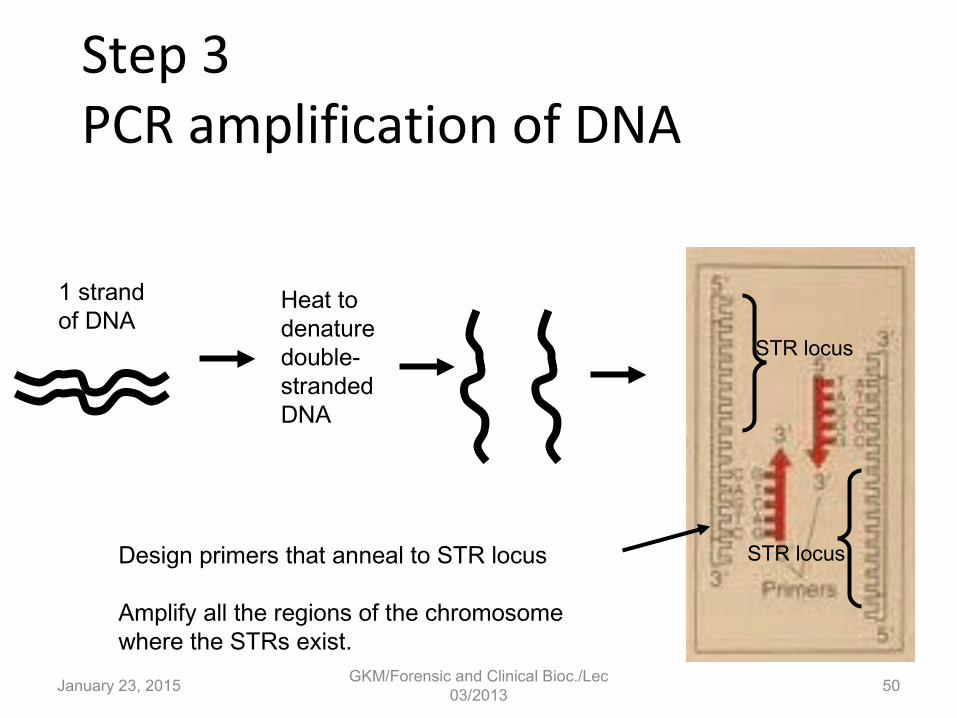
Step 3 PCR amplification of DNA
1 strandof DNA
Heat todenaturedouble-strandedDNA
Design primers that anneal to STR locus
Amplify all the regions of the chromosomewhere the STRs exist.
STR locus
STR locus
January 23, 2015 50GKM/Forensic and Clinical Bioc./Lec
03/2013

PCR allows you to make millions of copies of the STR region from a single copy of DNA you recovered from crime scene.
January 23, 2015 51GKM/Forensic and Clinical Bioc./Lec
03/2013

• Since the # of times sequence is repeated is different for each person, fragment size will be different.
• This is done for 13 different STR sequences• Differences occur among individuals at each of
the 13 loci on the chromosome where the STRs occur
• This allows for a lot of variation
January 23, 2015 52GKM/Forensic and Clinical Bioc./Lec
03/2013

Restriction Fragment Length Polymorphism
G-G-C-C-X-X-X-G-G-C-C-X-X.. G-G-G-C-C-X-X-G-G-C-C-X-X…..
STR
C-C-X-X-X-G-G C-C-X-X-G-G
PCR amplifySTR region
STR
well well
Gelelectrophoresis
Person A Forensic sample
For 1 STR sequence at 1 locus
January 23, 2015 53GKM/Forensic and Clinical Bioc./Lec
03/2013

• If you do this for 13 different repeat sequences at 13 different loci on the chromosome, each person produces a different band pattern when the fragments are separated by gel electrophoresis
• Banding patterns are identified using specific probes (see next slide)
• Since the patterns are unique to an individual, they are referred to as DNA finger prints
January 23, 2015 54GKM/Forensic and Clinical Bioc./Lec
03/2013
Banding Patterns

Some examples of DNA fingerprinting
• Paternity cases• Crime scenes
GKM/M.MEDPAEDS/LECT 01/2015

Example
January 23, 2015GKM/Forensic and Clinical Bioc./Lec
03/201356
E – reference sample, S1 – suspect 1 and S2 – suspect 2

GKM/M.MEDPAEDS/LECT 01/2015

GKM/M.MEDPAEDS/LECT 01/2015
Technical Considerations
• Preserve the integrity of DNA sample• Avoid DNA contamination & degradation• Avoid incomplete digestions if REs are used• Use standard hybridization conditions• Use standard PCR primers and procedures• Gel analysis is less reproducible than capillary
electrophoresis of PCR products
GKM/M.MEDPAEDS/LECT 01/2015

Test Choice
• Cost• Sample requirements• Turnaround time• Sensitivity/Specificity• Positive/ Negative predictive value• Type of mutation detected• Genotyping vs mutation scanning
GKM/M.MEDPAEDS/LECT 01/2015

DNA databases
• Already in place in the FBI for convicted felons (i.e., CODIS-COmbined DNA Index System, involves 13 STR loci) and the Dept. of Defense for armed service personnel and the Virginia saliva and blood bank of convicted felons
• A national DNA database has been suggested. What do you think?
• Could current or potential employers or insurance companies base decisions they make on this kind of data?
GKM/M.MEDPAEDS/LECT 01/2015

• A way to quantitate DNA in a PCR
• Involves the use of SYBR green dye
• SYBR green only binds to and fluoresces with dsDNA (detect product)
GKM/M.MEDPAEDS/LECT 01/2015

Bacterial biosensors
• One example involves using Pseudomonas fluorescens (genetically engineered for bioluminescence) to monitor pollutants
• If pollutants are present in a sample, then cell death occurs and “the light goes out”
lux genes in thechromosomal DNA
GKM/M.MEDPAEDS/LECT 01/2015

Bacterial biosensors (another example)
• Green fluorescent protein (GFP) can be used a reporter gene under the control of some inducible promoter (e.g., one that responds to some environmental signal such as a toxin)
• If the signal is present GFP will be produced
GKM/M.MEDPAEDS/LECT 01/2015

THE END
GKM/M.MEDPAEDS/LECT 01/2015