Embed Size (px)
Citation preview

Management of Japanese Encephalitis
Dr. Samiul Ahsan Hussain
IAP, Barak valley

• Japanese encephalitis (JE) is a leading cause of viral encephalitis in Asia.
• It has been controlled effectively through national vaccination programs in several countries like Japan, Korea, China and Thailand
IAP, Barak valley
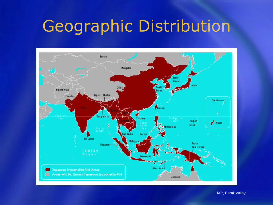
Geographic Distribution
IAP, Barak valley

4
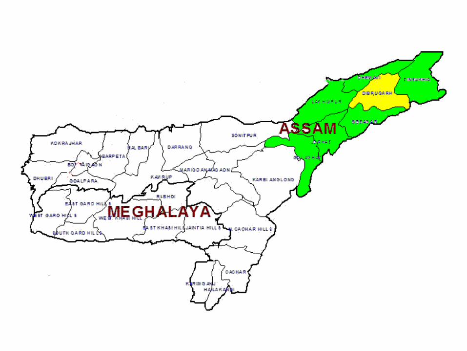
JE ENDEMIC AREAS IN INDIA
JE affected areas
• Andhra Pradesh
• Assam
• Bihar
• Haryana
• Kerala
• Karnataka
• Maharashtra
• Manipur
• Nagaland
• Tamil Nadu
• Uttar Pradesh
• West Bengal
IAP, Barak valley
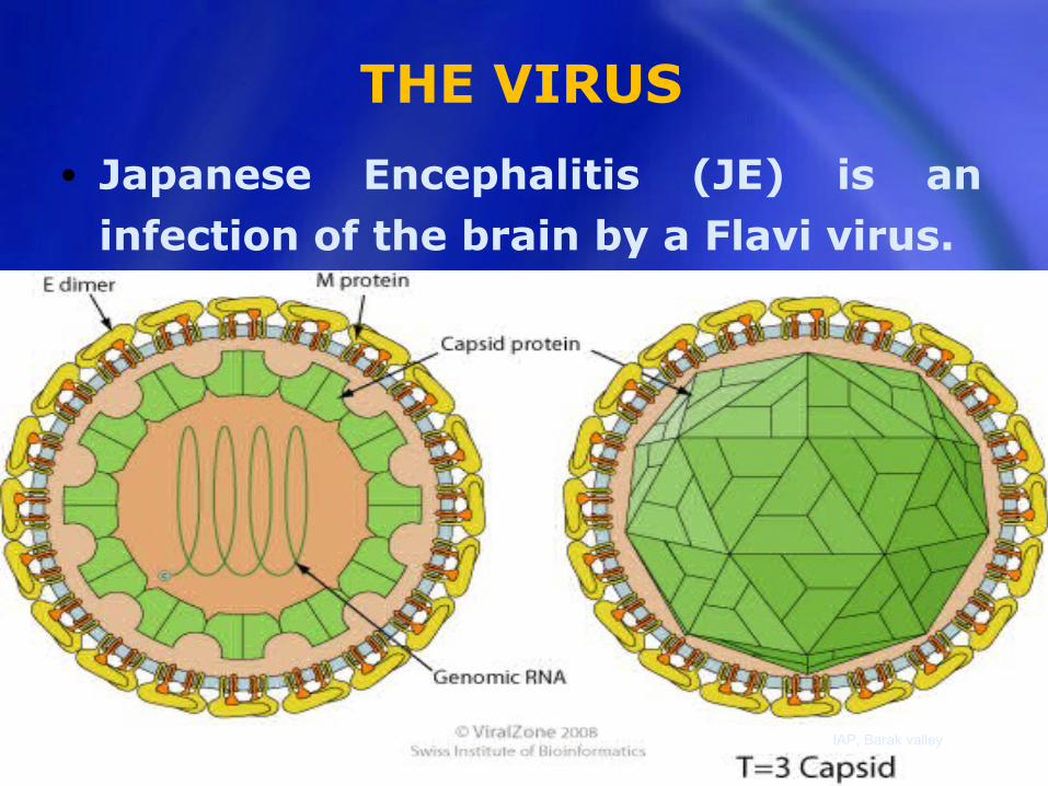
THE VIRUS
• Japanese Encephalitis (JE) is an infection of the brain by a Flavi virus.
IAP, Barak valley
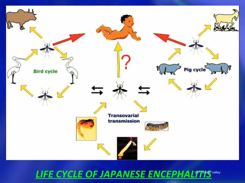
LIFE CYCLE OF JAPANESE ENCEPHALITISIAP, Barak valley
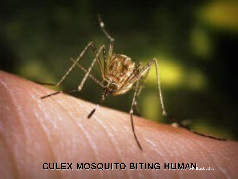
CULEX MOSQUITO BITING HUMANCULEX MOSQUITO BITING HUMANIAP, Barak valley
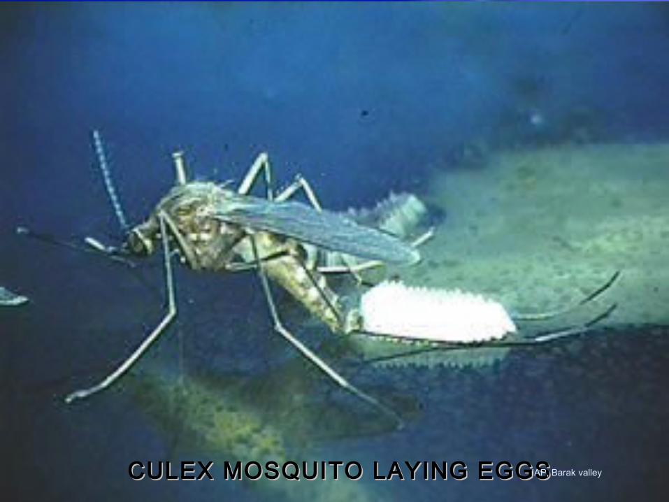
CULEX MOSQUITO LAYING EGGSCULEX MOSQUITO LAYING EGGSIAP, Barak valley
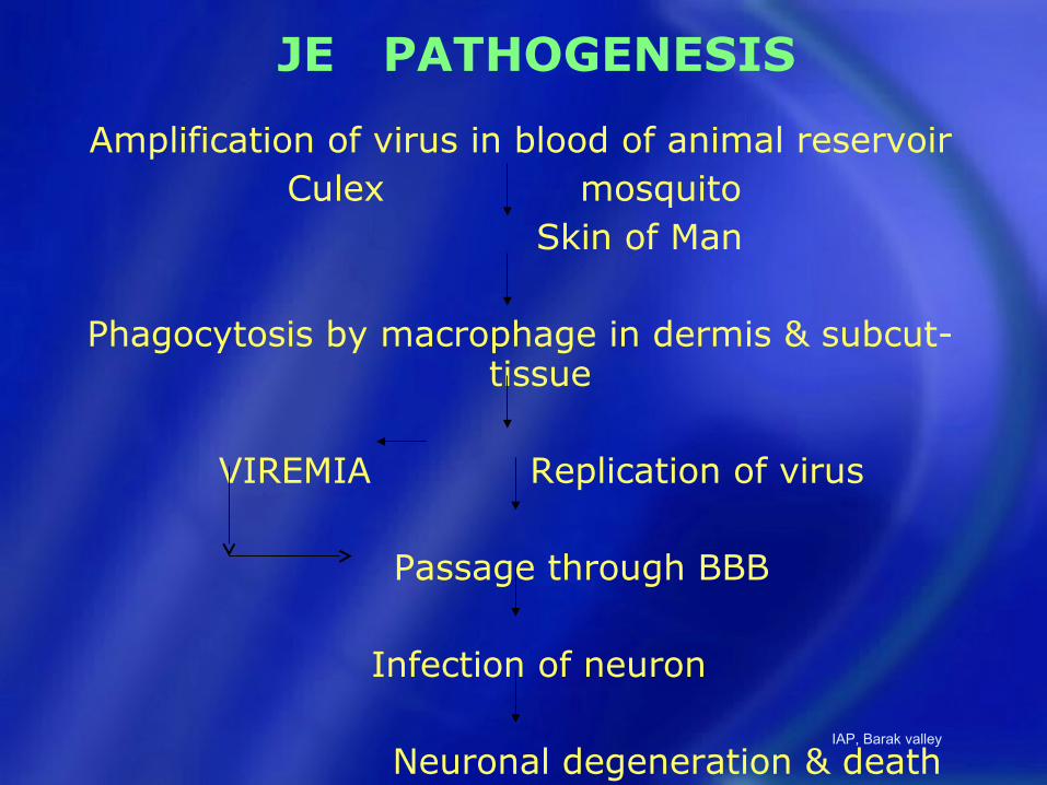
JE PATHOGENESIS
Amplification of virus in blood of animal reservoirCulex mosquito
Skin of Man
Phagocytosis by macrophage in dermis & subcut- tissue
VIREMIA Replication of virus
Passage through BBB
Infection of neuron
Neuronal degeneration & deathIAP, Barak valley

JE PATHOGENESIS (Contd.)
Neuronal degeneration & death
Microglial & astrocytic proliferation
Inflammatory & immune response
Congestion of blood vessels, edema, perivascular cuffing of mononuclear cells
IAP, Barak valley
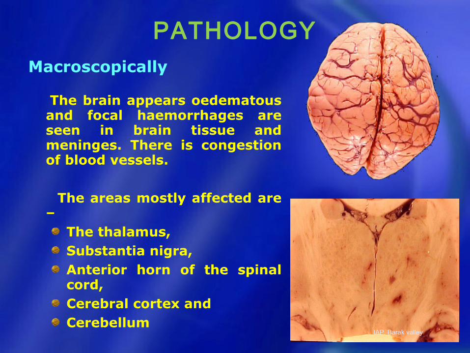
PATHOLOGYMacroscopically
The brain appears oedematous and focal haemorrhages are seen in brain tissue and meninges. There is congestion of blood vessels.
The areas mostly affected are –
The thalamus,Substantia nigra,Anterior horn of the spinal cord,Cerebral cortex andCerebellum
IAP, Barak valley
Acute encephalitis syndrome
(AES) is a term used by WHO for
syndromic surveillance in the
context of Japanese encephalitis
(JE)
IAP, Barak valley
JAPANESE ENCEPHALITIS (ACUTE ENCEPHALITIS SYNDROME )
• Clinical Case Definition:
Clinically a case of AES is defined as a person of any age at any time of year with acute onset of fever and a change in mental status (including symptoms such as confusion, disorientation, coma, or inability to talk).
• And/Or
• New onset of seizures (excluding simple febrile seizures).
IAP, Barak valley
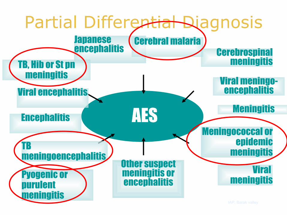
Partial Differential DiagnosisJapanese encephalitis
Viral meningitisPyogenic or
purulent meningitis
Meningococcal or epidemic
meningitis
TB, Hib or St pn meningitis
Encephalitis
TBmeningoencephalitis
Viral encephalitis
Cerebral malariaCerebrospinal
meningitis
Viral meningo-encephalitis
Other suspect meningitis or encephalitis
MeningitisAES
IAP, Barak valley
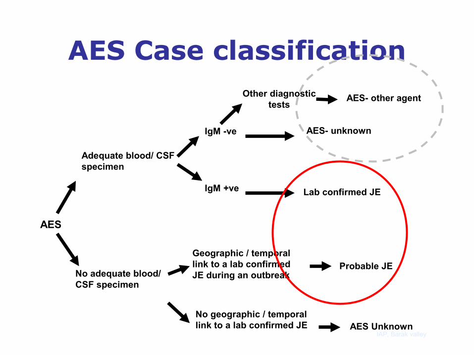
Adequate blood/ CSF specimen
No adequate blood/ CSF specimen
AES
IgM -ve
IgM +ve
Geographic / temporal link to a lab confirmed JE during an outbreak
No geographic / temporal link to a lab confirmed JE
AES- unknown
Lab confirmed JE
AES Unknown
Other diagnostic tests
AES- other agent
Probable JE
AES Case classification
IAP, Barak valley

Evaluation and Management
Step I: Rapid assessment
and stabilization
IAP, Barak valley
IAP, Barak valley
AIRWAY

BREATHING
IAP, Barak valley

CIRCULATION• Circulatory failure: fluid bolus (20 mL/kg-
Normal saline)
• Hypoglycemia is present:intravenous glucose
• Seizure :intravenous benzodiazepine followed by phenytoin loading 20 mg/kg
• Acid base and electrolyte abnormalities should be corrected
• Normothermia should be maintained.
IAP, Barak valley
Step 2: Detailed History
and Examination
IAP, Barak valley
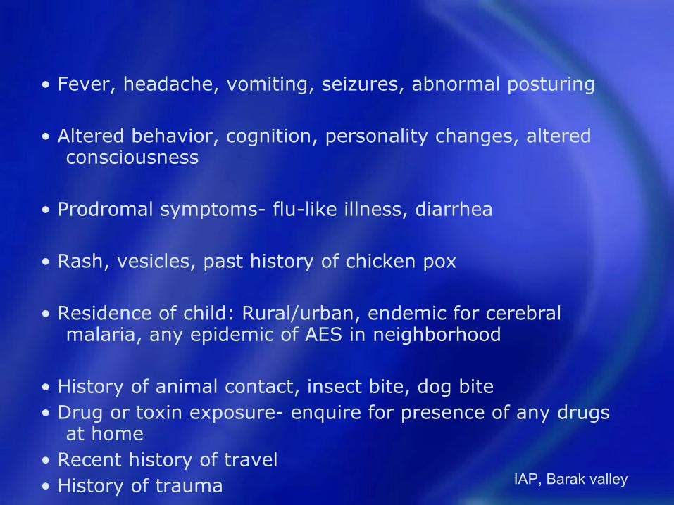
• Fever, headache, vomiting, seizures, abnormal posturing
• Altered behavior, cognition, personality changes, altered consciousness
• Prodromal symptoms- flu-like illness, diarrhea
• Rash, vesicles, past history of chicken pox
• Residence of child: Rural/urban, endemic for cerebral malaria, any epidemic of AES in neighborhood
• History of animal contact, insect bite, dog bite• Drug or toxin exposure- enquire for presence of any drugs
at home • Recent history of travel• History of trauma IAP, Barak valley

• Personal or family history of seizure disorder
• Recent immunizations
• History of recurrent episodes of encephalopathy: These are characteristic of some inborn errors of metabolism (urea cycle defects, organic acidemias and fatty acid oxidation defects), but may also be present in migraine, epilepsy, substance abuse, and Munchausen syndrome by proxy
• Other concurrent systemic illness e.g. jaundice (hepatic failure), pneumonia (hypoxic encephalopathy), diarrhea (dyselectrolytemia), dysentery (shigella encephalopathy)
• Past medical illness: Diabetes, congenital heart disease, chronic kidney or liver disease
• Family history of previous infant/child deaths• Pre-morbid developmental/ neurological status of the child• Risk factors for immunodeficiency- HIV risk factors, cancer
treatment, steroid/immunosuppressant treatmentIAP, Barak valley
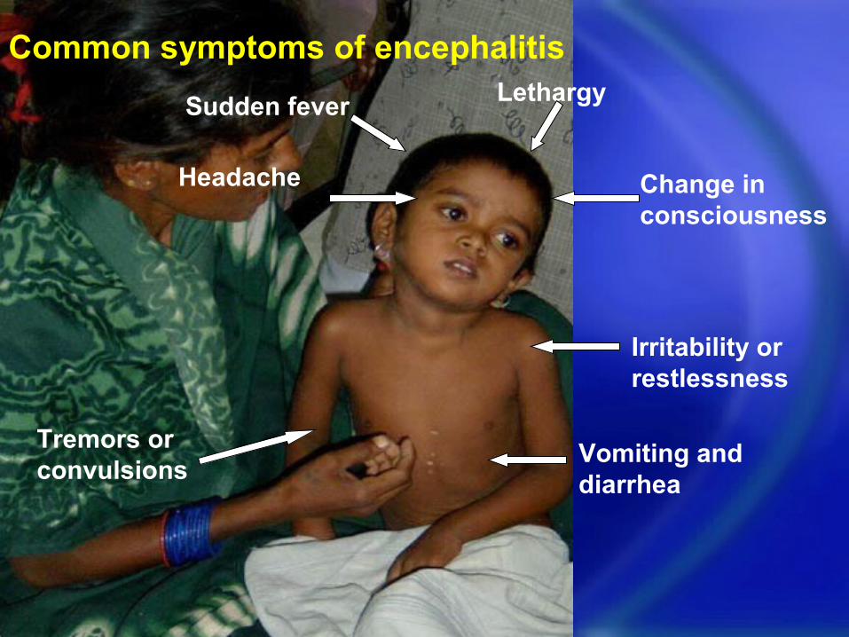
LethargySudden fever
Vomiting and diarrhea
Tremors or convulsions
Headache Change in consciousness
Irritability or restlessness
Common symptoms of encephalitis
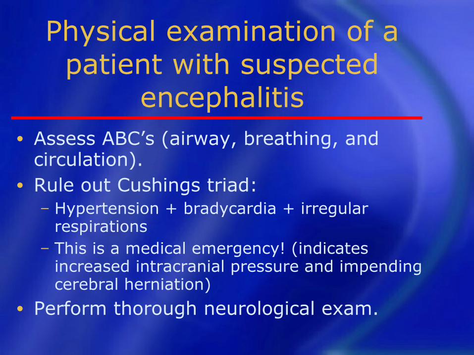
Physical examination of a patient with suspected
encephalitis• Assess ABC’s (airway, breathing, and
circulation).• Rule out Cushings triad:
− Hypertension + bradycardia + irregular respirations
− This is a medical emergency! (indicates increased intracranial pressure and impending cerebral herniation)
• Perform thorough neurological exam.
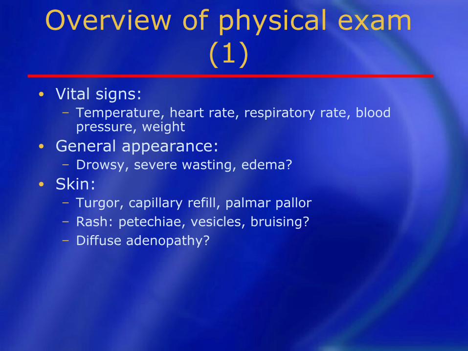
Overview of physical exam (1)
• Vital signs:− Temperature, heart rate, respiratory rate, blood
pressure, weight• General appearance:
− Drowsy, severe wasting, edema?• Skin:
− Turgor, capillary refill, palmar pallor− Rash: petechiae, vesicles, bruising?− Diffuse adenopathy?
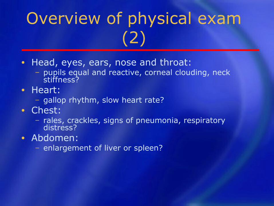
Overview of physical exam (2)
• Head, eyes, ears, nose and throat:− pupils equal and reactive, corneal clouding, neck
stiffness?• Heart:
− gallop rhythm, slow heart rate?• Chest:
− rales, crackles, signs of pneumonia, respiratory distress?
• Abdomen:− enlargement of liver or spleen?
The neurologic exam
1. Mental status− Level of alertness:
AVPU scale for rapid assessment: Alert / Responds to voice / Reacts to pain / Unconscious
Glasgow Coma Scale or other coma scale
− Orientation, memory, speech, etc.− Irritability, aphasia?

The neurologic exam (2)
3. Motor exam− Assess strength, tone of upper and lower
extremities Compare sides
− Abnormal movements or posturing?4. Sensory system
− Assess pain, vibration, temperature sensation Compare sides
2. Cranial nerves— Pupil reactivity, eye movements, fundoscopic
exam for papilledema, facial muscles
Testing facial nerve (VII)
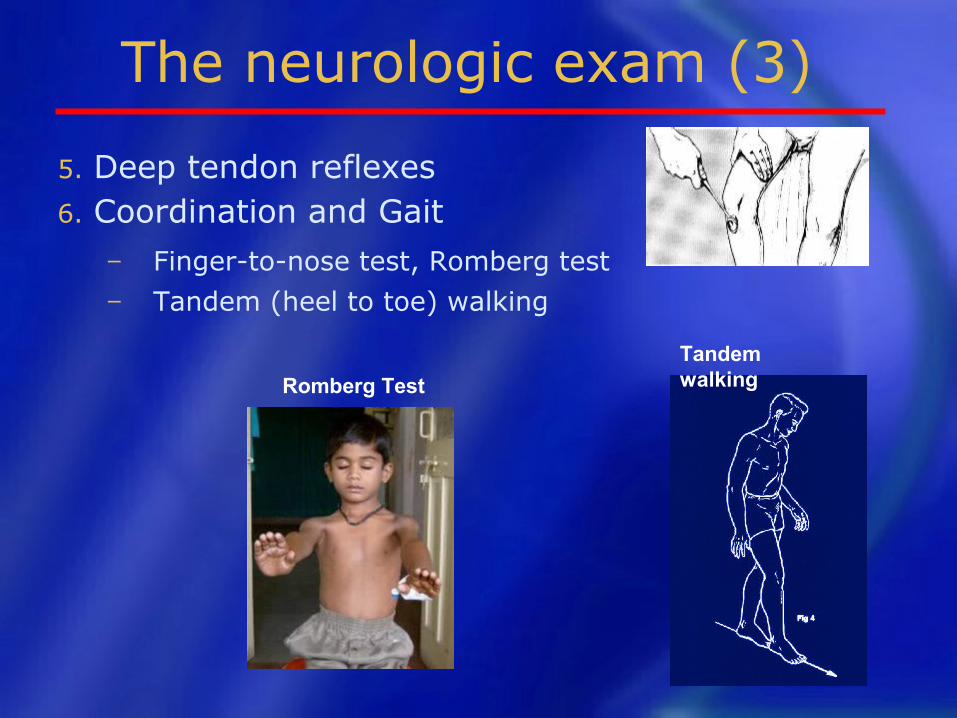
The neurologic exam (3)
5. Deep tendon reflexes6. Coordination and Gait
− Finger-to-nose test, Romberg test− Tandem (heel to toe) walking
Romberg Test
Tandem walking

Step 3: Investigations
IAP, Barak valley
SAMPLE COLLECTION
SAMPLES: CSF, Serum First specimens (blood and CSF) should
be collected on admission to hospital or when patient first seen.
Brain biopsy obtained post morterm
by Vim Silverman needle – pass through cribriform plate.
SAMPLE COLLECTION (CONT’D.)
• A follow up specimen (blood) should be collected at least 10 days after onset (before discharge or death).
• The collection of CSF should only be performed by experienced personnel under aseptic conditions in the hospital
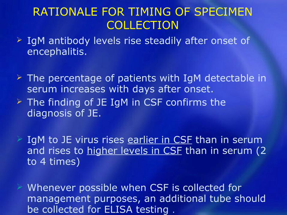
RATIONALE FOR TIMING OF SPECIMEN COLLECTION
IgM antibody levels rise steadily after onset of encephalitis.
The percentage of patients with IgM detectable in serum increases with days after onset.
The finding of JE IgM in CSF confirms the diagnosis of JE.
IgM to JE virus rises earlier in CSF than in serum and rises to higher levels in CSF than in serum (2 to 4 times)
Whenever possible when CSF is collected for management purposes, an additional tube should be collected for ELISA testing .
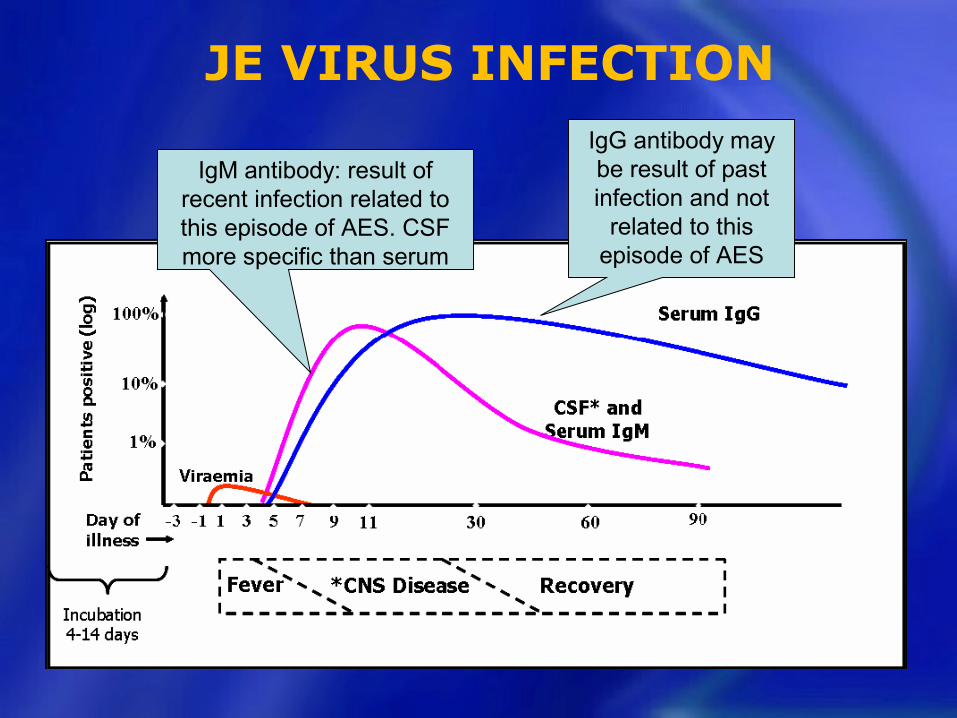
JE VIRUS INFECTION IgG antibody may be result of past infection and not
related to this episode of AES
IgM antibody: result of recent infection related to this episode of AES. CSF more specific than serum
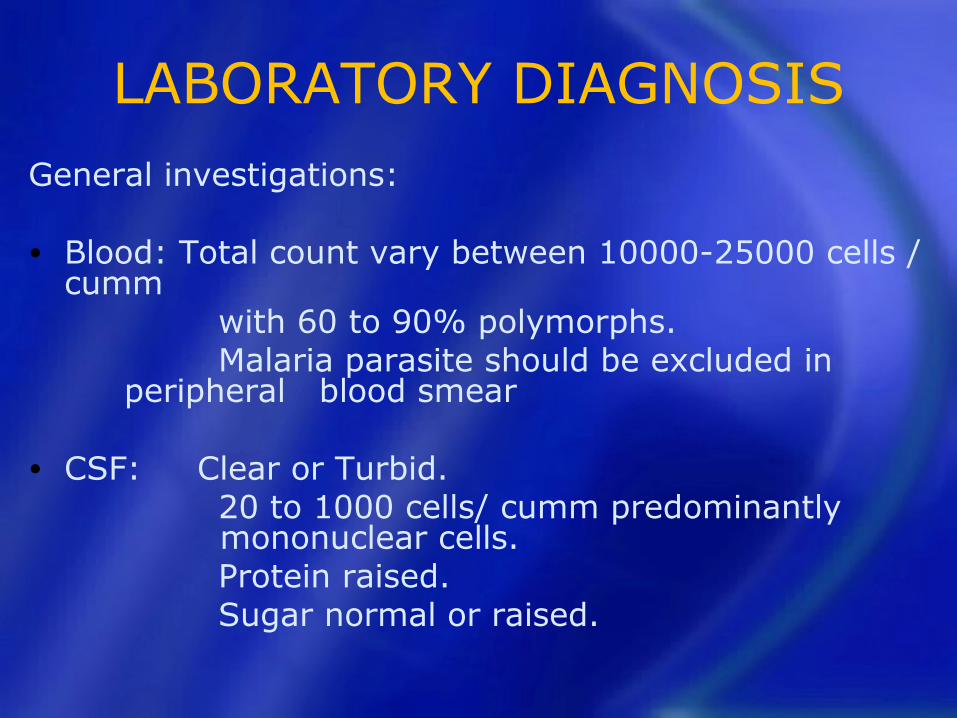
LABORATORY DIAGNOSISGeneral investigations:
• Blood: Total count vary between 10000-25000 cells / cumm
with 60 to 90% polymorphs. Malaria parasite should be excluded in
peripheral blood smear
• CSF: Clear or Turbid. 20 to 1000 cells/ cumm predominantly
mononuclear cells. Protein raised. Sugar normal or raised.
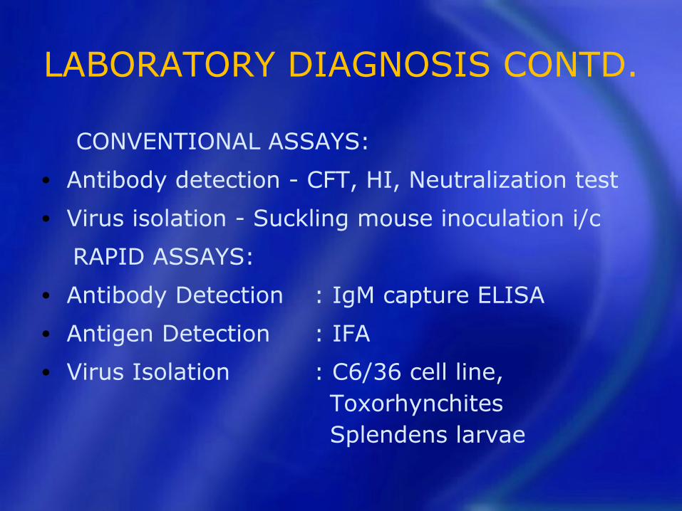
LABORATORY DIAGNOSIS CONTD.
CONVENTIONAL ASSAYS:
• Antibody detection - CFT, HI, Neutralization test
• Virus isolation - Suckling mouse inoculation i/c
RAPID ASSAYS:
• Antibody Detection : IgM capture ELISA
• Antigen Detection : IFA
• Virus Isolation : C6/36 cell line, Toxorhynchites
Splendens larvae
How does the JE IgM capture enzyme linked immunosorbent assay (ELISA) work
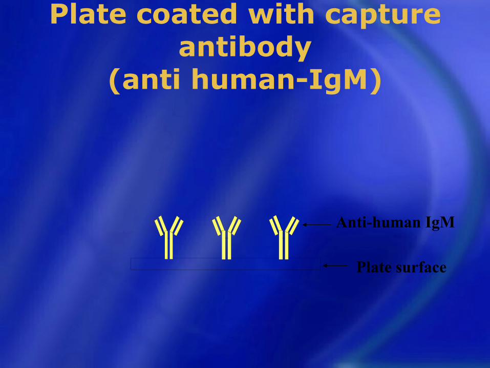
Plate coated with capture antibody
(anti human-IgM)
Anti-human IgM
Plate surface
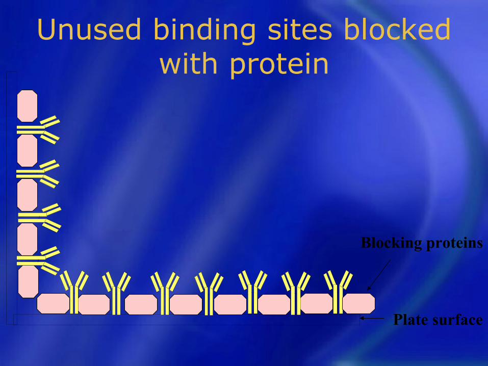
Unused binding sites blocked with protein
Blocking proteins
Plate surface
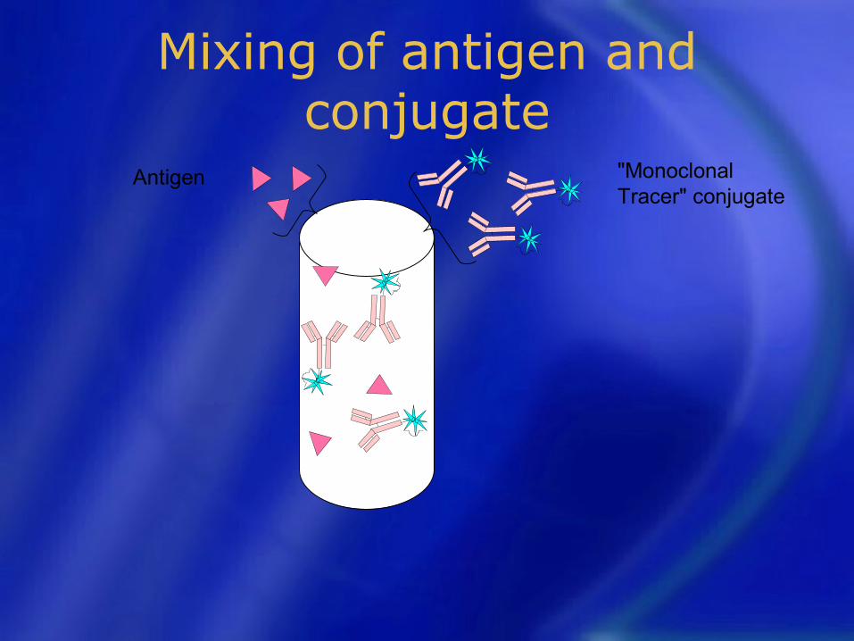
Mixing of antigen and conjugate
Antigen "Monoclonal Tracer" conjugate
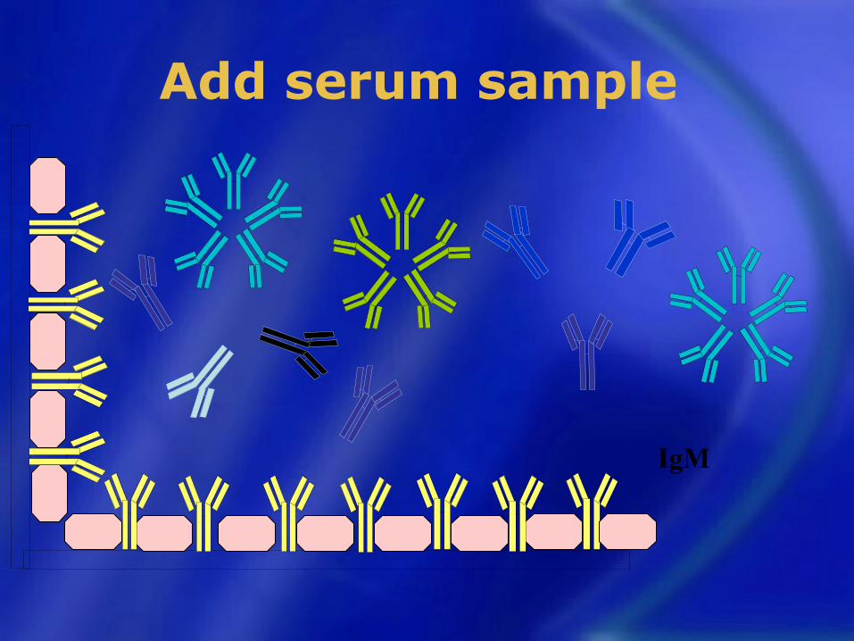
Add serum sample
IgM
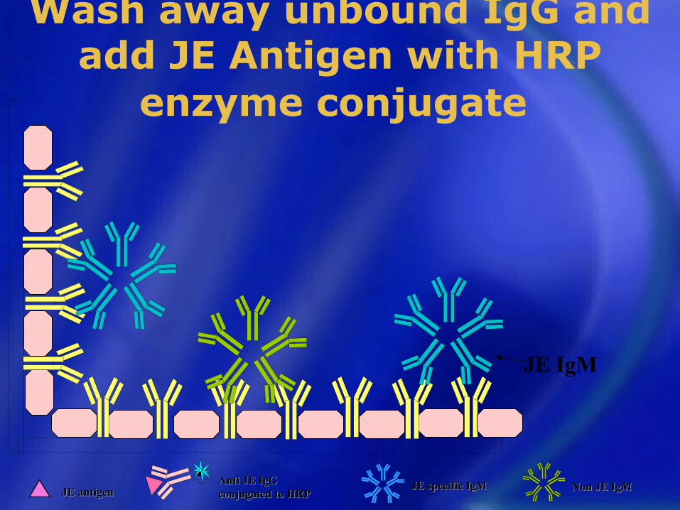
Wash away unbound IgG and add JE Antigen with HRP
enzyme conjugate
JE IgM
JE antigenJE antigenAnti JE IgG Anti JE IgG conjugated to HRPconjugated to HRP
JE specific IgMJE specific IgM Non JE IgMNon JE IgM
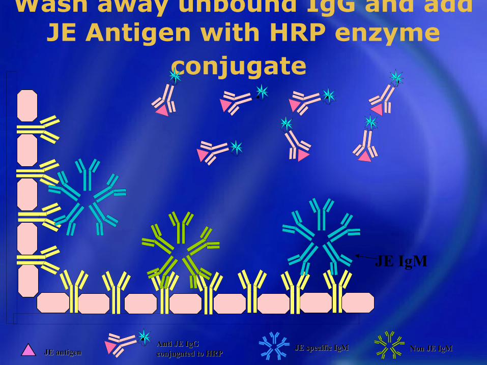
Wash away unbound IgG and add JE Antigen with HRP enzyme
conjugate
JE IgM
JE antigenJE antigenAnti JE IgG Anti JE IgG conjugated to HRPconjugated to HRP
JE specific IgMJE specific IgM Non JE IgMNon JE IgM
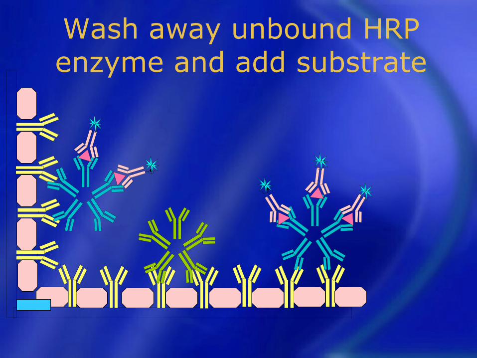
Wash away unbound HRP enzyme and add substrate
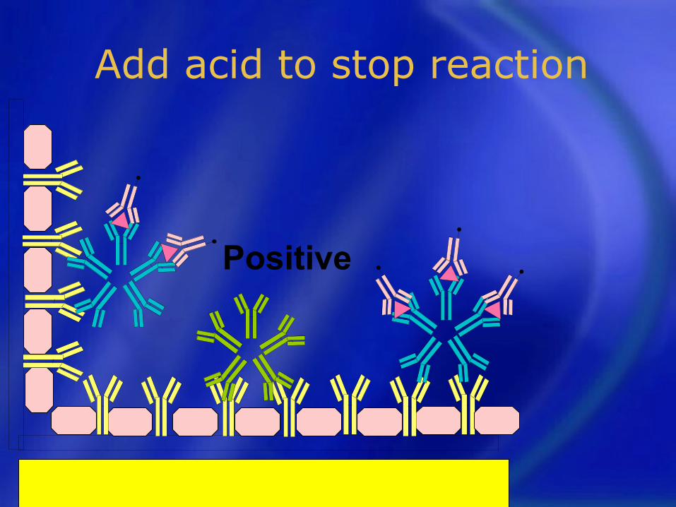
Add acid to stop reaction
Positive

Basic investigations:• complete blood count • blood glucose• serum electrolytes• liver and kidney function tests• blood culture• arterial blood gas, and lactate • malarial parasite • chest X-ray
IAP, Barak valley

Lumbar puncture:
• Cytology• Biochemistry• Gram stain• Ziehl-Nielsen stain for acid fast bacilli• Bacterial culture• Latex agglutination, PCR for HSV 1
and 2• IgM antibodies for JE and for
Dengue virus (if suspected)IAP, Barak valley

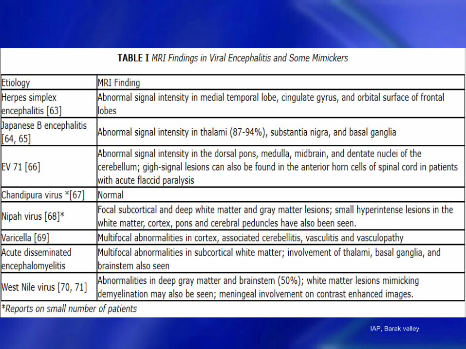
Neuroimaging: CT
• Presence of bleed• Cerebral edema• Temporal lobe hypodensities in
herpes simplex encephalitis• Thalamic abnormalities in JE• Basal exudates and hydrocephalus in
tubercular meningitis• Brain herniation• Brain abscesses and subdural
empyema.IAP, Barak valley
IAP, Barak valley
Step 4: Empirical
Treatment
IAP, Barak valley

Antibiotic
• Ceftriaxone
• Acyclovir
• Anti-malarial (artemisin-based combination therapy)
IAP, Barak valley

Step 5: Supportive
Care
IAP, Barak valley
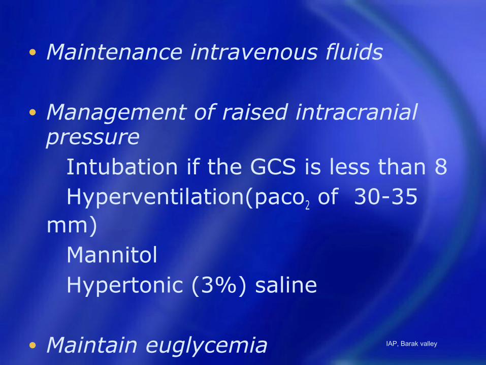
• Maintenance intravenous fluids
• Management of raised intracranial pressure
Intubation if the GCS is less than 8 Hyperventilation(paco2 of 30-35
mm) Mannitol Hypertonic (3%) saline
• Maintain euglycemia IAP, Barak valley
• Treatment and prevention of seizures• Corticosteroids ???
• Oral ribavirin was not found to be useful in children with Japanese B encephalitis in a randomized controlled trial [56].
• There is experimental evidence of benefit of minocycline in JE [57]. Movement disorders such as dystonia may need treatment with trihexyphenidyl. IAP, Barak valley

Step 6: Prevention/treatment of complications and rehabilitation
IAP, Barak valley
• (i) Surveillance for cases of AES;
• (ii) Vector control;
• (iii) Reduction in man-vector contact; and
• (iv) Vaccination
IAP, Barak valley
• In any AES outbreak, pediatricians will see affected patients. They should be aware of what information and samples they should collect, and whom to inform. All pediatricians need to be aware of the case definition of AES. The cases should be notified to the District Surveillance Unit.
IAP, Barak valley
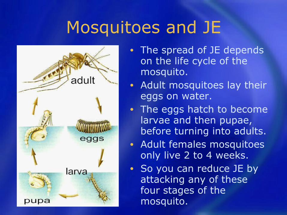
Mosquitoes and JE• The spread of JE depends
on the life cycle of the mosquito.
• Adult mosquitoes lay their eggs on water.
• The eggs hatch to become larvae and then pupae, before turning into adults.
• Adult females mosquitoes only live 2 to 4 weeks.
• So you can reduce JE by attacking any of these four stages of the mosquito.
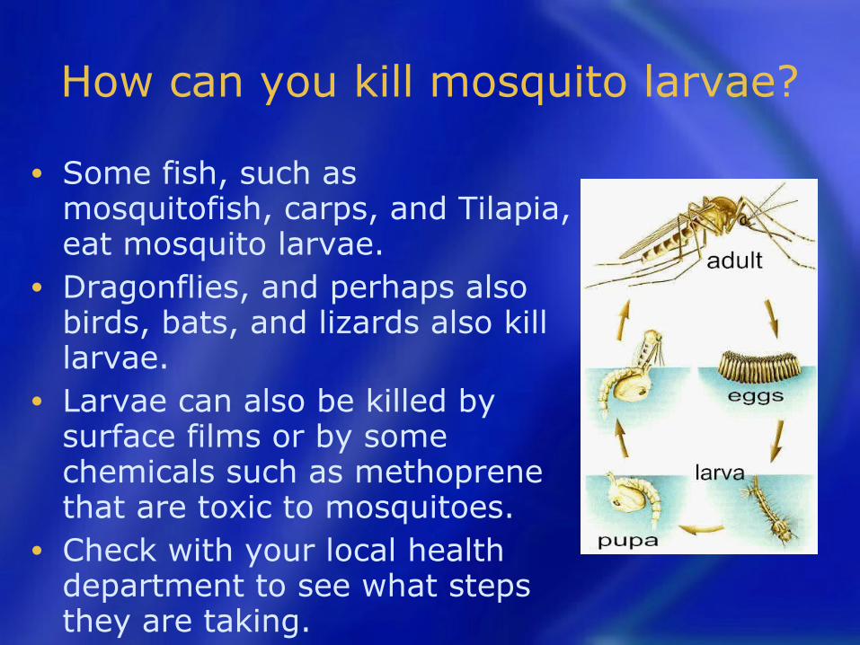
How can you kill mosquito larvae?
• Some fish, such as mosquitofish, carps, and Tilapia, eat mosquito larvae.
• Dragonflies, and perhaps also birds, bats, and lizards also kill larvae.
• Larvae can also be killed by surface films or by some chemicals such as methoprene that are toxic to mosquitoes.
• Check with your local health department to see what steps they are taking.
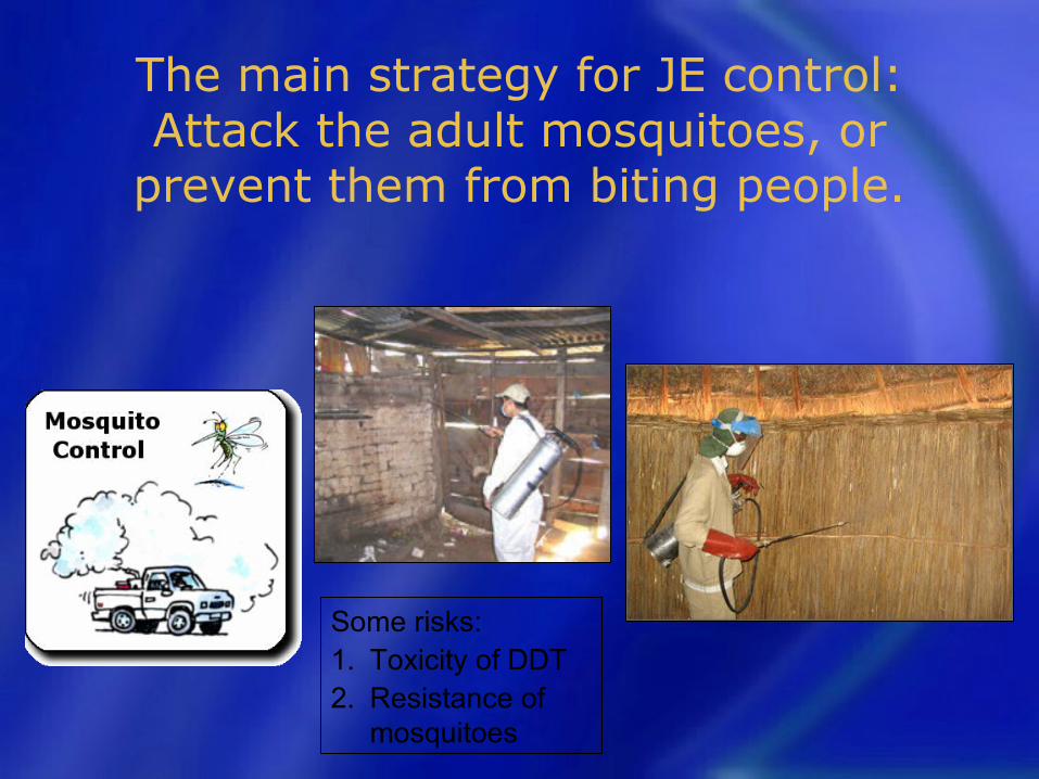
The main strategy for JE control: Attack the adult mosquitoes, or
prevent them from biting people.
Some risks:1. Toxicity of DDT2. Resistance of
mosquitoes
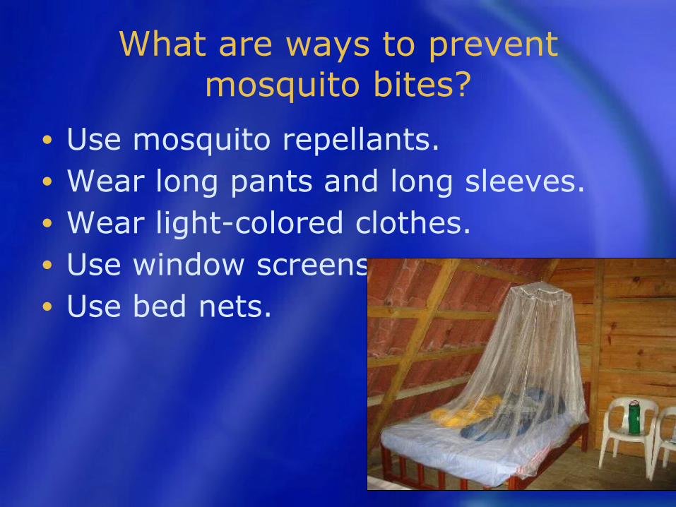
What are ways to prevent mosquito bites?
• Use mosquito repellants.• Wear long pants and long sleeves.• Wear light-colored clothes.• Use window screens• Use bed nets.
JE vaccination• Recommended only for individuals living in
the rural areas of endemic districts
Three types :• live attenuated, cell culture-derived SA-
14-14-2• inactivated JE vaccines, namely ‘vero cell
culture-derived SA 14-14-2 JE vaccine’ (JEEV® by BE India) and
• ‘vero cell culture derived,821564XY, JE vaccine’ (JENVAC® by Bharat Biotech)IAP, Barak valley
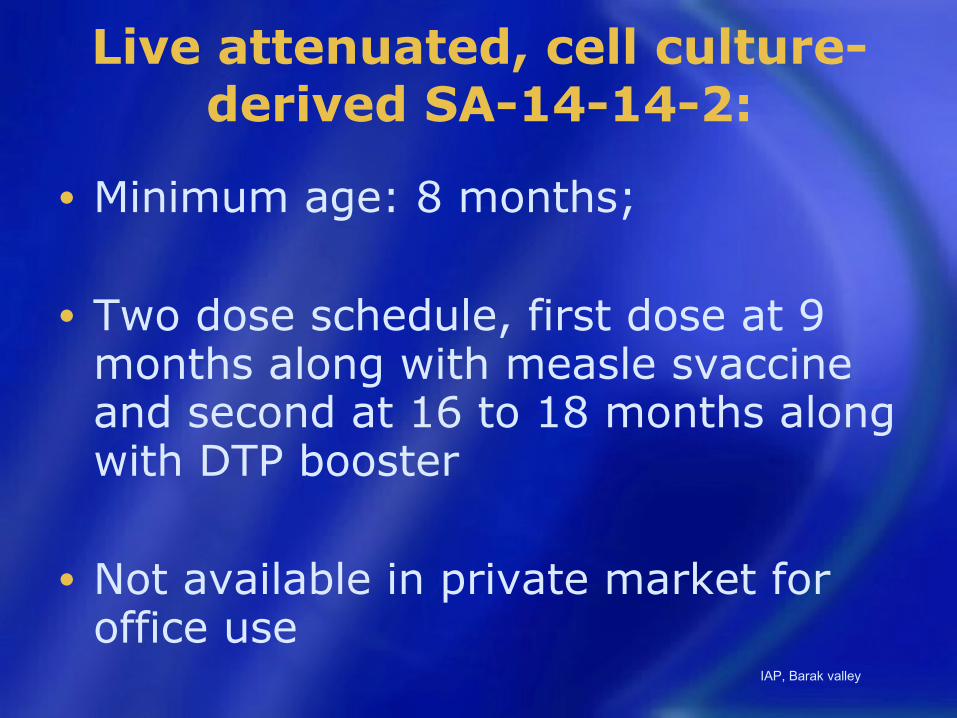
Live attenuated, cell culture-derived SA-14-14-2:
• Minimum age: 8 months;
• Two dose schedule, first dose at 9 months along with measle svaccine and second at 16 to 18 months along with DTP booster
• Not available in private market for office use
IAP, Barak valley

Inactivated Vero cell culture-derived Kolar strain, 821564XY, JE
vaccine (JENVAC® by Bharat Biotech)
• Minimum age: 1 year• Primary immunization schedule: 2
doses of 0.5 mL each
• administered intramuscularly at 4 weeks interval
• Need of boosters still undetermined.IAP, Barak valley
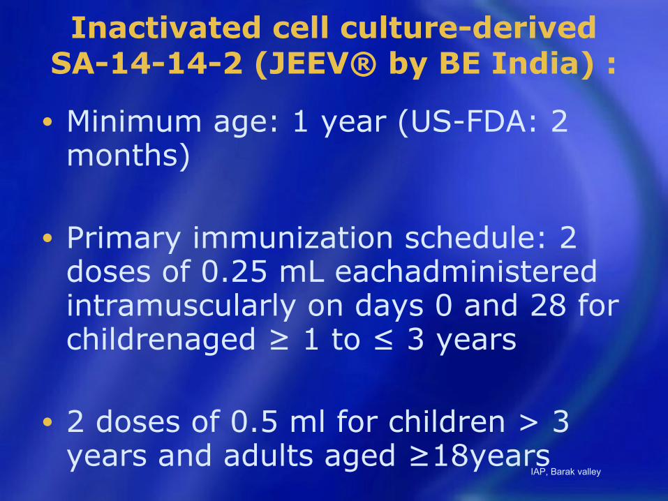
Inactivated cell culture-derived SA-14-14-2 (JEEV® by BE India) :
• Minimum age: 1 year (US-FDA: 2 months)
• Primary immunization schedule: 2 doses of 0.25 mL eachadministered intramuscularly on days 0 and 28 for childrenaged ≥ 1 to ≤ 3 years
• 2 doses of 0.5 ml for children > 3 years and adults aged ≥18years
IAP, Barak valley
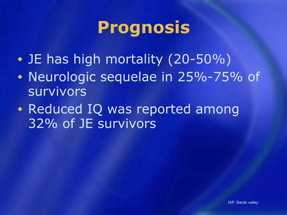
Prognosis
• JE has high mortality (20-50%)• Neurologic sequelae in 25%-75% of
survivors• Reduced IQ was reported among
32% of JE survivors
IAP, Barak valley

Decrease morbidity by early recognition of danger
• Pupils, tone and posture, respiration and doll’s eye movement examination helps us in assessing severity .
IAP, Barak valley

Misery of Mystery of Muzaffarpur
Most children reported being apparently well in the evening with a sudden onset of altered consciousness in the early hours of next day, with or without seizures and hypoglycemia with absence of clues for an infection such as prodromal symptoms, fever, brain edema or inflammatory response in the cerebrospinal fluid
IAP, Barak valley
• Heat stroke• Lychee seeds• Japanese encephalitis
IAP, Barak valley
• Importance of case reporting and sharing experience with peers.
• Information on clustering of cases.
• All pediatricians are requested to report unusual outbreak cases and keep an accurate clinical and laboratory records for data analysis when needed by the investigators.IAP, Barak valley