Embed Size (px)
Citation preview

1

IS THE SCHNEIDERIAN MEMBRANE THICKNESS AFFECTED BY PERIODONTAL DISEASE?
A CBCT- BASED EXTENDED CASE SERIES
DOROTHEA C, NICOLA U, THOMAS L, ROLAND W, CLEMENS W et al. J International Academy Of Periodontology 2013.
Presented By: Shilpa Shivanand I MDS

3
INTRODUCTION
Periodontal diseases prevalent chronic infectious diseases affecting primarily the tooth supporting structures
(Page and Korman 1997)Occurrence symmetrical patternMore pronounced maxillary posterior region
(Mombelli et al 2001)Maxillary molars highest risk complex morphology with
multiple roots, root fusion or root proximity and furcation entrances difficult to access with self performed oral hygiene
(Walter et al 2010)

4
Introduction…
Enhanced biofilm accumulation in furcation area advanced inflammation horizontal and vertical periodontal destruction with furcation involvement (FI) in multi-rooted teeth
Pneumatisation of paranasal sinus increases with ageMaxillary sinus extend into area of maxillary alveolar boneRoots of maxillary molars and/or bicuspids project into floor of
the sinus as small conical processes
(Gray 1970)

5
Introduction…
Close proximity of maxillary molars and sinus floor spread of inflammatory process from teeth and periodontium into maxillary sinus Atypical Odontalgia and TMJ pain
(Logan, Blocklebank et al 1999)
These process affect later implant placement because reduction in the amount of maxillary alveolar bone height and residual sinus infection, particularly when sinus augmentation has to be planned.

6
SCHNEIDERIAN MEMBRANE
Maxillary sinus lined by mucoperiostium, with cilia that beat toward the ostia.
Membrane referred to as the “Schneiderian Membrane”(mean thickness: 2.16-3.11mm Simone et al 2011)
Histologically bilaminar membrane with ciliated columnar epithelial cells on the internal (or cavernous) side and periosteum on the osseous side.
Postganglionic parasympathetic nerve innervation of mucous membrane for mucous secretion greater petrosal nerve (branch…facial nerve).
Sensory innervation superior alveolar (anterior, middle, and posterior) nerves (branch…maxillary nerve).

7

8
Intact schneiderian membrane.mp4

9
CBCT
Introduced into dentistry – Mozzo et al 1998
Clinically been validated for assessment of furcation invasion in maxillary molars
(Walter et al 2010)
CBCT enables exact estimation and classification of furcation involvement and a visualization of decisive features such as root proximities, root fusions or periapical lesions
(Walter et al 2009,2010)

10
AIM
To analyze the thickness of the Schneiderian membrane (SM) using CBCT in patients with advanced periodontal destruction in the distal maxillary area and to compare these data with edentulous regions.

11
MATERIALS AND METHODS
SUBJECTS:17 dentate patients (DG) diagnosed for generalized
advanced/severe chronic periodontal disease were recruitedDuration : April 2007- January 2010.Periodontal diagnosis was based on complete dental and
periodontal examinations (including sensitivity testing of all teeth, probing pocket depth (PPD), probing attachment level (PAL), furcation involvement (FI), and radiographic examinations (periapical radiographs)
(Walter et al 2009)
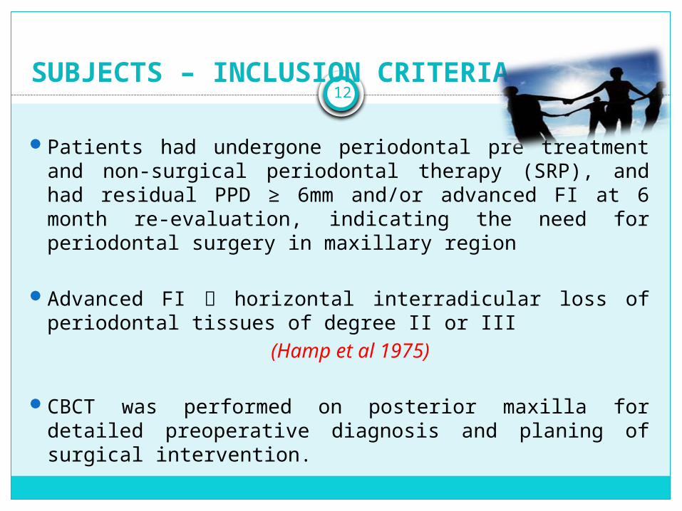
12SUBJECTS – INCLUSION CRITERIA
Patients had undergone periodontal pre treatment and non-surgical periodontal therapy (SRP), and had residual PPD ≥ 6mm and/or advanced FI at 6 month re-evaluation, indicating the need for periodontal surgery in maxillary region
Advanced FI horizontal interradicular loss of periodontal tissues of degree II or III
(Hamp et al 1975)
CBCT was performed on posterior maxilla for detailed preoperative diagnosis and planing of surgical intervention.
13
Subjects – inclusion criteria….
21 patients (EG) who required CBCT – based planing of implant placement in edentulous posterior maxillary area
(free end situations), served as control.Tomography was performed at least 8 weeks after tooth
extraction in posterior maxilla.Study was approved by Ethics Research Committee of University
of Basel SwitzerlandPatients were thoroughly informed and consent was takenFrom 38 patients enrolled, total of 44 CBCT scans were analyzed
(20 from DG and 24 from EG)

14
SUBJECTS – EXCLUSION CRITERIA
Patients with history of any bone disease (including osteoporosis), bisphosphonate intake, oro-antral fistula and inflammation or previous surgery affecting the maxillary sinus were excluded.

15
ANALYSES OF DENTAL CBCT’s
CBCT were performed in the posterior maxilla using high resolution 3D imaging system 3D Accuitomo 60 and 3D Accuitomo 80 (Morita, Kyoto, Japan).
Cylindrical volumes of 4 x 4 cm, 6 x 6 cm and 8x8 cm, settings in the range of 74-90 kV, 5-8 mA and voxel size of 0.125 mm were used depending on the region of interest.
All images were analyzed with the same monitor (Viewmedic 19C, 48 cm, 19°, Totoku, Japan).
The software i-Dixel-3DX (Morita, Kyoto, Japan) with a linear measurement tool and a digital magnification lens was used.

17
Analyses Of Dental CBCT’S……
Images were analyzed in the horizontal (axial), sagittal and/or transverse (coronal) sections.
All measurements were performed twice by one of the authors within one week, and an intra-class correlation coefficient (ICC) was determined to compare the repeated measurements of membrane thickness
(Shrout and Fleiss 1979) An ICC of 0.99 was calculated, revealing a high similarity of the
measurements.

18
Analyses Of Dental CBCT’S……
In DG, the following parameters were analyzed from CBCT
i) the horizontal dimension of furcation involvement
ii) the dimension of vertical furcation involvement
iii) the minimal bone height above each root tip to the sinus floor
iv) the bone height in the interfurcal region defined as minimal distance of the interradicular alveolar bone crest to the sinus floor
v. the mucous membrane (SM) thickness in second premolar, first molar and second molar sites
vi. Additional radiographic findings, including periapical lesions, combined periodontal-endodontic lesions and other findings, such as root perforation, fenestration defects, missing buccal/palatal bone plate, or overfill of the root canal.
19
HORIZONTAL DIMENSIONS OF FURCATION INVOLVEMENT
The horizontal furcation involvement (FI) was calculated in the horizontal (axial) plane by measuring the distance between the outer root surface and the interradicular bone to the nearest millimeter
Walter et al 2009Furcation involvement was
graduated according to Hamp et al 1975
Degree 0: no horizontal loss of periodontal tissue support, i.e., no radiolucency in the furcation area.
Degree I: horizontal loss of periodontal tissue support up to 3 mm.
Degree II: horizontal loss of support exceeding 3 mm, but no "through and through" destruction.
Degree III: horizontal "through and through" — destruction of the periodontal tissue in the furcation

20
DIMENSIONS OF VERTICAL FURCATION INVOLVEMENT
The assessment of the vertical dimension of FI was performed with a modified classification according to Tarnow and Fletcher, 1984 and the maximal distance between the roof of the furcation and the interradicular alveolar bone crest was measured.
Subclasses A, B and C.A : probeable vertical depth
of 1-3 mmB : 4-6 mmC : 7mm or more of
probeable depth from the roof of the furca (fornix) apically.
Furcation's would thus be classified as IA, IB, IC, IIA, IIB, IIC and IIIA, IIIB, IIIC.
21
BONE HEIGHT BETWEEN ROOT TIPS AND SINUS FLOOR
The images were re-sliced in order to get an orthogonal examination plane through the respective tooth.
By scrolling through the volume, the tip of each root was located in the transverse (coronal) and sagittal plane.
The shortest distance between each root tip and the sinus floor was estimated.
22
SM THICKNESS AT 2nd PREMOLAR, 1st MOLAR AND 2nd MOLAR SITES
In the center line of each tooth position (second premolar, first molar and second molar), the most caudal point of the sinus was identified in the sagittal plane.
The distance from the sinus floor to the top of SM was measured. In case of an interfering septum, the centerline was moved slightly
to a more distal position.The examination of the CBCT scans in the edentulous control
group (EG) included:
i) measurement of the vertical thickness of SM
ii) minimal alveolar bone height
iii) recording of additional findings such as metal-like radiopaque material in the alveolar bone crest or in the SM, a visible lamina dura of the alveolus after tooth extraction.

23
MINIMAL ALVEOLAR BONE HEIGHT
The minimal vertical distance from the alveolar bone crest to the sinus floor was determined by scrolling through the scan in the transverse (coronal) and sagittal plane.
The most caudal point of the maxillary sinus was identified and a perpendicular was dropped caudal to the bone crest. The mean of both measurements in the sagittal and transverse planes was calculated and used for further analyses.

24
Reference points for analysis of mucous membrane thickness in the maxillary sinus in the EDENTULOUS CONTROL GROUP (EG). RpC, control reference point at the medial sinus wall; RpP, sinus membrane thickness in premolar region; RpFM, sinus membrane thickness in first molar region; RpSM, sinus membrane thickness in second molar region.
Click icon to add pictureIn the sagittal plane the most caudal position of the sinus floor was identified and marked as reference point Rpl.
• An imaginary reference point Rp2 was defined 1.5 cm above and perpendicular to Rpl.
A horizontal line from Rp2 to the medial sinus wall defined RpC, which served as a control reference point apart from the alveolar bone and teeth.
• From RpC, a perpendicular line was drawn downwards, which served as a reference plane.
1.3 cm

25
Click icon to add picture• Perpendicular to this reference plane, three measurement points were constructed, which represented the position of the second maxillary premolar (RpP) at an average distance of 0.5 cm, the first molar (RpFM) at a distance of 1.3 cm, and the second molar (RpSM) at a distance of 2.1 cm.
Then the transverse (coronal) plane was used to measure the thickness of the SM by dropping a perpendicular from the most caudal point of the sinus floor to the top of the membrane at RpP, RpFM and RpSM.
• For comparison the thickness of the membrane was also measured at RpC.
Further, the vertical alveolar bone height between the alveolar bone crest and the sinus floor was measured at the respective points
1.3 cm

26
MEASUREMENTS PERFORMED IN DENTATE SUBJECTS (DG)
Click icon to add picturea) Assessment of the horizontal dimension of furcation involvement
b) Measurement of the dimension of vertical furcation involvement
c) Measurement of the minimal bone height above each root tip to the sinus floor
d) Assessment of the minimal distance of the interradicular alveolar bone crest to the sinus floor
e) Measurement of the mucous membrane thickness in a first molar

27

28
STATISTICAL ANALYSIS
Corresponding p-values were calculated by Fisher's exact test.Metric variables (eg. age) were reported as means with standard
deviation (SD). A Student's t- test was used for comparisons between groups
(DG and EG).

29

30

31
RESULTS
In the area of the first (RpFM) and second (RpSM) maxillary molars, SM thickness was significantly greater in DG than in EG.
In the medial area of the sinus (RpC) and at the second premolar position (RpP), no group differences were observed.
ASSOCIATION OF SYSTEMIC FACTORS WITH SM THICKNESS IN DG AND EG
Although no association between the systemic factors age or current smoking and SM thickness was found in DG, these factors were significantly associated with an increased SM thickness in EG.

32
RESULTS….
ASSOCIATION OF CLINICAL PARAMETERS OR ANATOMICAL FACTORS WITH SM IN THE DG
None of the clinical parameters (PPD and FI), or vertical bone loss within the furcal area, showed an association with SM thickness
Alveolar bone height was not related to the SM thickness (p= 0.157)
Distance of the root tips to the maxillary sinus revealed a significant association with smaller bone distances accompanying thicker sinus membranes (p = 0.036)

33
DISCUSSION
The present case series indicated that the thickness of the Schneiderian membrane is affected by periodontally diseased maxillary molars as compared to edentulous areas.
Although small bone layers separating the root tips from the sinus floor were associated with increased SM thickness, no clear relation was determined for the clinical periodontal parameters.
Because of the restricted indications for CBCT scans, the sample size was small and subjects requiring CBCT scans for implant planning were used as controls.
This control group was defined so as to have tooth extraction performed in the posterior maxilla at least two months previously

34
DISCUSSION…..
Cone-based computed tomography is widely used to analyze the bone quantity in the maxillofacial region, while its application for soft-tissue imaging has been questioned
(Scarfe et al 2006)The gold standard to visualize the inflammatory changes in the
nasal and paranasal sinus mucosa is the computed tomography (CT) or magnetic resonance imaging (MRI).
Both techniques are proven accurate for evaluation of the pathophysiology of paranasal sinusitis
(Larheim 2006)

35
DISCUSSION…..
Recent data confirmed the usefulness of CBCT in assessing and evaluating soft tissues
(Briillmann et at 2011; Janner et al 2011; Larheim 2006; Barriviera etal 2009; Januario etal 2008; Fu etal 2010)
Data from the present analysis on SM thickness demonstrated the potential use of CBCT imaging for the evaluation of the soft tissue conditions in the maxillary sinus.
The strong association of a significantly greater sinus membrane thickness and age in EG was confirmed by several previous data sets
(Iwabuchi etal 1997; Vallo etal 2010; Photikum etal 2012)

36
DISCUSSION…..
Briillmann et al reported a strong association between SM thickening and periodontitis (with PPD > 3 mm) or decayed and non-vital teeth
In this study comparing CBCTs from subjects with and without apical pathology at maxillary premolars or molars, SM thickening was found particularly in the vicinity of roots with apical lesions
(Bornstein etal 2012)
37
CONCLUSION
The current study was initiated based on the assumption of an association of periodontal disease and furcation involvement with sinus membrane thickness assessed from CBCT images.
While the 2nd premolar site did not reveal differences between the dentate and the edentulous group, SM adjacent to molar sites showed a thickening in EG, particularly with close proximity of the root tips to the sinus floor.
The clinical parameters PPD and FI, however, failed to predict SM thickening in this case series with advanced periodontal destruction in the maxillary posterior region.

38
CONCLUSION……
The Schneiderian membrane revealed a more pronounced thickness in CBCT images when comparing maxillary molar sites with the corresponding edentulous regions.
In contrast, the presence of additional findings, such as periapical lesions, overfill of the root canal or foreign bodies in the DG revealed a significant association with sinus membrane thickness

39
CRITICAL EVALUATION
1. CBCT …not indicated for soft tissue imaging…..questionable….could have used gold standard imaging techniques like CT, MRI etc….
2. Sensitivity testing of all teeth done….not discussed why

CROSS REFERENCES

41
1. CORRELATION BETWEEN GINGIVAL PHENOTYPE AND SCHNEIDERIAN MEMBRANE THICKNESS.
Aimetti M, Massei G, Morra M, Cardesi E, Romano FThe International Journal of Oral & Maxillofacial Implants 2008
PURPOSE:The most common complication during sinus graft surgery is tearing or perforation of the Schneiderian membrane. Perforations are most likely to occur if the sinus membrane is thin. Preoperative prediction of the antral membrane thickness may be of practical importance during maxillary sinus augmentation procedures. The purpose of this investigation was to analyze a possible association between gingival phenotypes and thickness of the healthy sinus mucosa.
MATERIALS AND METHODS: Twenty consecutive patients without preoperative clinical or radiologic signs of maxillary sinus pathologies were enrolled in the study. During otorhinolaringologic surgical interventions, maxillary mucosal biopsy specimens were endoscopically obtained from the sinus floor, and gingival thickness was measured at the maxillary anterior teeth.

42
RESULTS: Eleven out of 20 individuals had thick gingival tissues, and the remaining 9 presented with thin gingival phenotype. The average thickness of the Schneiderian membrane was 0.97 +/- 0.36 mm with a wide inter-individual variability. Thickness of the sinus mucosa amounted to 1.26 +/- 0.14 mm in individuals with thick gingival architecture and 0.61 +/- 0.15 mm in subjects with thin gingival tissues. The association between thickness of the antral mucosa and periodontal phenotypes was statistically significant (P <.0001).CONCLUSIONS: Gingival thickness seems to represent a reliable parameter to predict sinus membrane thickness. Further investigations are needed to support these preliminary data.

43
2. CHARACTERISTICS AND DIMENSIONS OF THE SCHNEIDERIAN MEMBRANE: A RADIOGRAPHIC ANALYSIS USING CBCT IN PATIENTS REFERRED FOR DENTAL IMPLANT SURGERY IN THE POSTERIOR MAXILLA
Simone Janner, Marco D, Patrick Dubach, Pedram Sendi, Daniel Buser, Michael Bornstein
Clinical Oral Implants Research 2011Objectives: To determine the dimensions of the Schneiderian
membrane using limited CBCT in individuals referred for dental implant surgery, and to determine factors influencing the mucosal thickness.
Material and methods: The study included 143 consecutive patients referred for dental implant placement in the posterior maxilla. A total of 168 CBCT images were taken using a limited field of view of 4 *4cm, 6 *6 cm, or 8 *8 cm. Reformatted coronal CBCT slices were analyzed with regard to the thickness and characteristics of the Schneiderian membrane in nine standardized points of reference.

44
Results: The thickness of the Schneiderian membrane exhibited a wide range, with a minimum value of 0.16mm and a maximum value of 34.61 mm. The highest mean values, ranging from 2.16 to 3.11 mm, were found for the mucosa located in the mid-sagittal regions of the maxillary sinus. The most frequent mucosal findings diagnosed were flat thickenings of the Schneiderian membrane (62 positive findings, 37%). For the multivariate linear regression model, only gender had a statistically significant influence on the mean overall and mid-sagittal thickness of the sinus mucosa.
Conclusion: There is great inter-individual variability in the thickness of the Schneiderian membrane. Gender seems to be the most important parameter influencing mucosal thickness in asymptomatic patients. Future studies are needed to assess the therapeutic and prognostic consequences of mucosal alterations in the maxillary sinus.

45