Embed Size (px)
Citation preview
BIRADS AND IMAGING OF BREAST
PATHOLOGIES
Dr.Archana Koshy
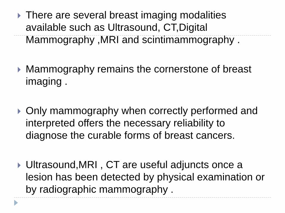
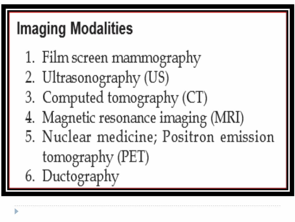
There are several breast imaging modalities
available such as Ultrasound, CT,Digital
Mammography ,MRI and scintimammography .
Mammography remains the cornerstone of breast
imaging .
Only mammography when correctly performed and
interpreted offers the necessary reliability to
diagnose the curable forms of breast cancers.
Ultrasound,MRI , CT are useful adjuncts once a
lesion has been detected by physical examination or
by radiographic mammography .
INDICATIONS
Screening of asymptomatic women
Screening of high risk women
Follow up of patients after mastectomy of same and
opposite breast / same breast with implant .
Investigations of benign breast diseases with
eczematous skin,nipple discharge , skin thickening .
Investigation of a breast lump
Investigation of occult primary with secondaries .
Male breast evaluation .
BIRADS Breast imaging- reporting and
data system
BIRADS
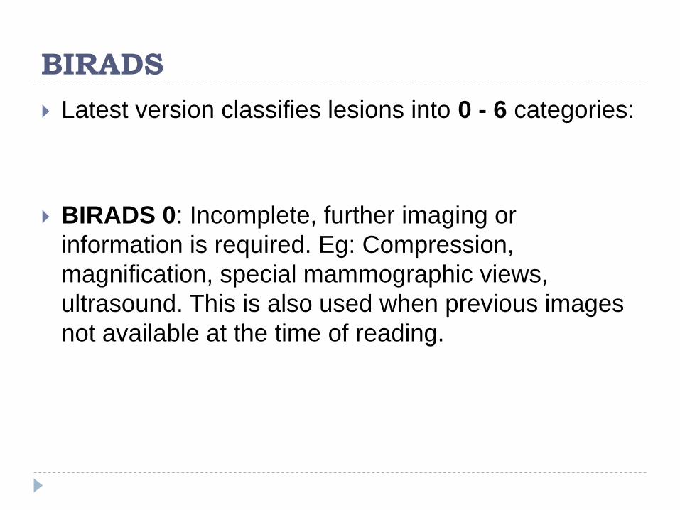
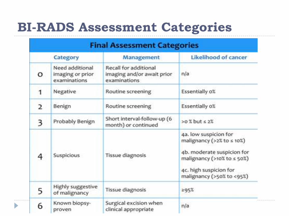
Latest version classifies lesions into 0 - 6 categories:
BIRADS 0: Incomplete, further imaging or
information is required. Eg: Compression,
magnification, special mammographic views,
ultrasound. This is also used when previous images
not available at the time of reading.
BIRADS
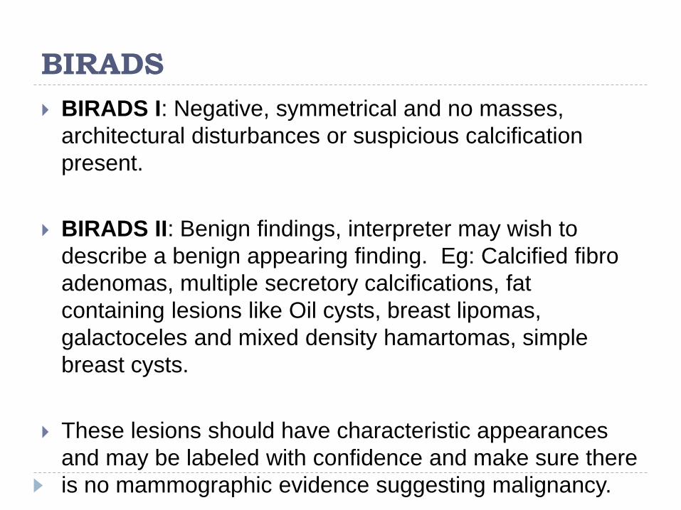
BIRADS I: Negative, symmetrical and no masses,
architectural disturbances or suspicious calcification
present.
BIRADS II: Benign findings, interpreter may wish to
describe a benign appearing finding. Eg: Calcified fibro
adenomas, multiple secretory calcifications, fat
containing lesions like Oil cysts, breast lipomas,
galactoceles and mixed density hamartomas, simple
breast cysts.
These lesions should have characteristic appearances
and may be labeled with confidence and make sure there
is no mammographic evidence suggesting malignancy.

BIRADS
BIRADS III: probably benign, short interval follow up
suggested.
BIRADS IV: suspicious abnormality.
There is mammographic appearance which is
suspicious of malignancy.
Biopsy should be considered.
BIRADS IVa: low level of suspicion
BIRADS IVb: intermediate level of suspicion
BIRADS IVc: moderate level of suspicion for
malignancy

BIRADS
BIRADS V: there is a mammographic appearance which is highly suggestive of malignancy, action should be taken.
BIRADS VI: known biopsy proven malignancy
The vast majority of mammograms fall into BIRADS I or II.
Risk of Cancer:
BIRADS III: ~ 2%
BIRADS IV: ~ 30%
BIRADS V : 95%
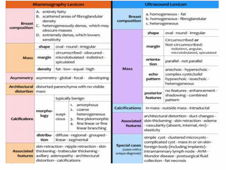
Mammography and Ultrasound Lexicon
BI-RADS Assessment Categories
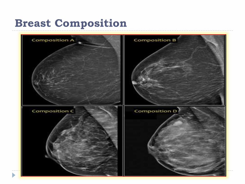
Breast Composition
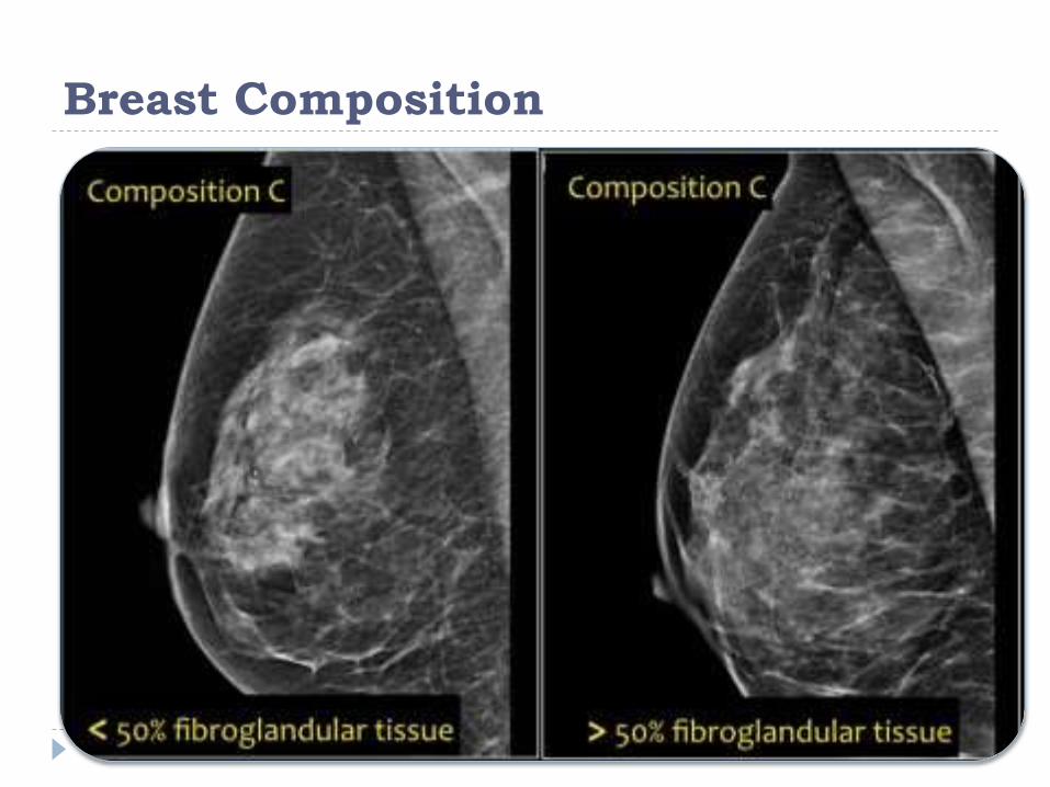
Breast Composition

SHAPE
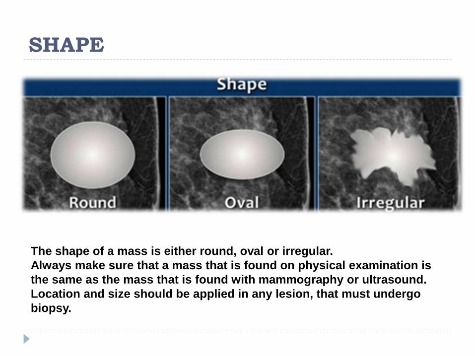
The shape of a mass is either round, oval or irregular.
Always make sure that a mass that is found on physical examination is
the same as the mass that is found with mammography or ultrasound.
Location and size should be applied in any lesion, that must undergo
biopsy.
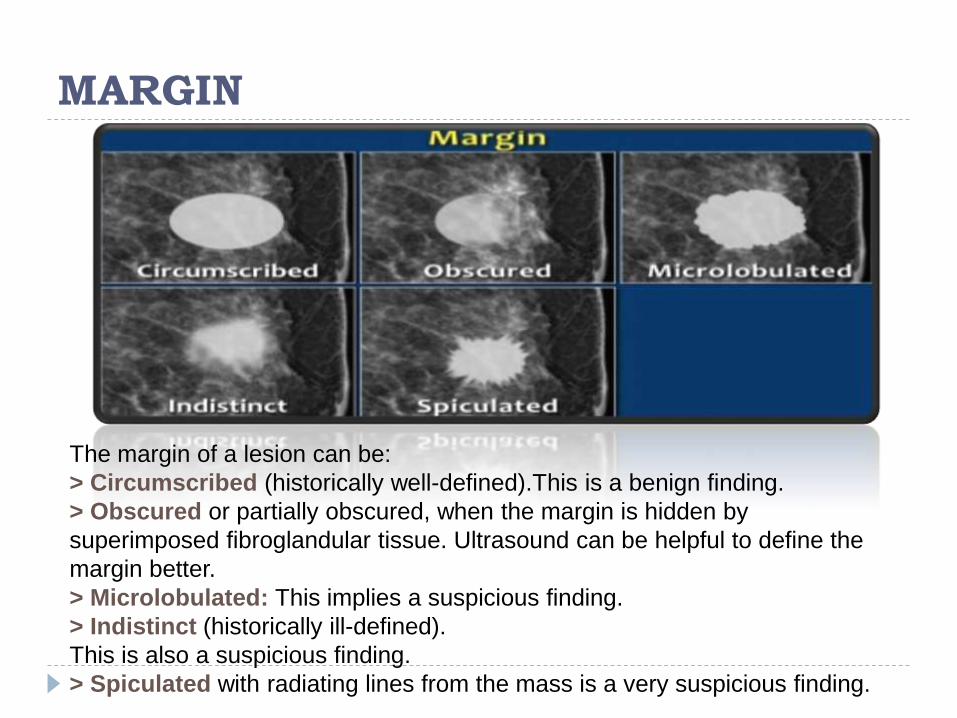
MARGIN
The margin of a lesion can be:
> Circumscribed (historically well-defined).This is a benign finding.
> Obscured or partially obscured, when the margin is hidden by
superimposed fibroglandular tissue. Ultrasound can be helpful to define the
margin better.
> Microlobulated: This implies a suspicious finding.
> Indistinct (historically ill-defined).
This is also a suspicious finding.
> Spiculated with radiating lines from the mass is a very suspicious finding.

DENSITY
The density of a mass is related to the expected attenuation of an
equal volume of fibroglandular tissue.
High density is associated with malignancy.
It is extremely rare for breast cancer to be low density.

Architectural distortion
The term architectural distortion is used, when the normal
architecture is distorted with no definite mass visible.
This includes thin straight lines or spiculations radiating
from a point, and focal retraction, distortion or
straightening at the edges of the parenchyma.
The differential diagnosis is scar tissue or carcinoma.
Architectural distortion can also be seen as an
associated feature.
For instance if there is a mass that causes architectural
distortion, the likelihood of malignancy is greater than in
the case of a mass without distortion.

Architectural distortion
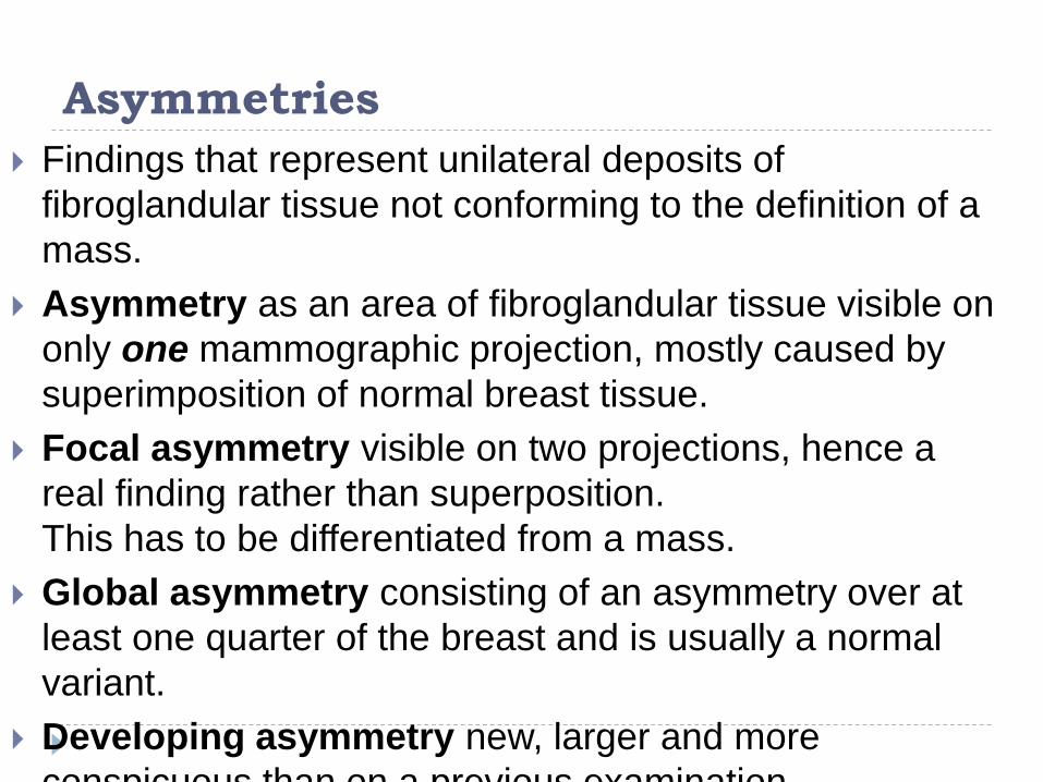
Asymmetries
Findings that represent unilateral deposits of
fibroglandular tissue not conforming to the definition of a
mass.
Asymmetry as an area of fibroglandular tissue visible on
only one mammographic projection, mostly caused by
superimposition of normal breast tissue.
Focal asymmetry visible on two projections, hence a
real finding rather than superposition.
This has to be differentiated from a mass.
Global asymmetry consisting of an asymmetry over at
least one quarter of the breast and is usually a normal
variant.
Developing asymmetry new, larger and more
conspicuous than on a previous examination.
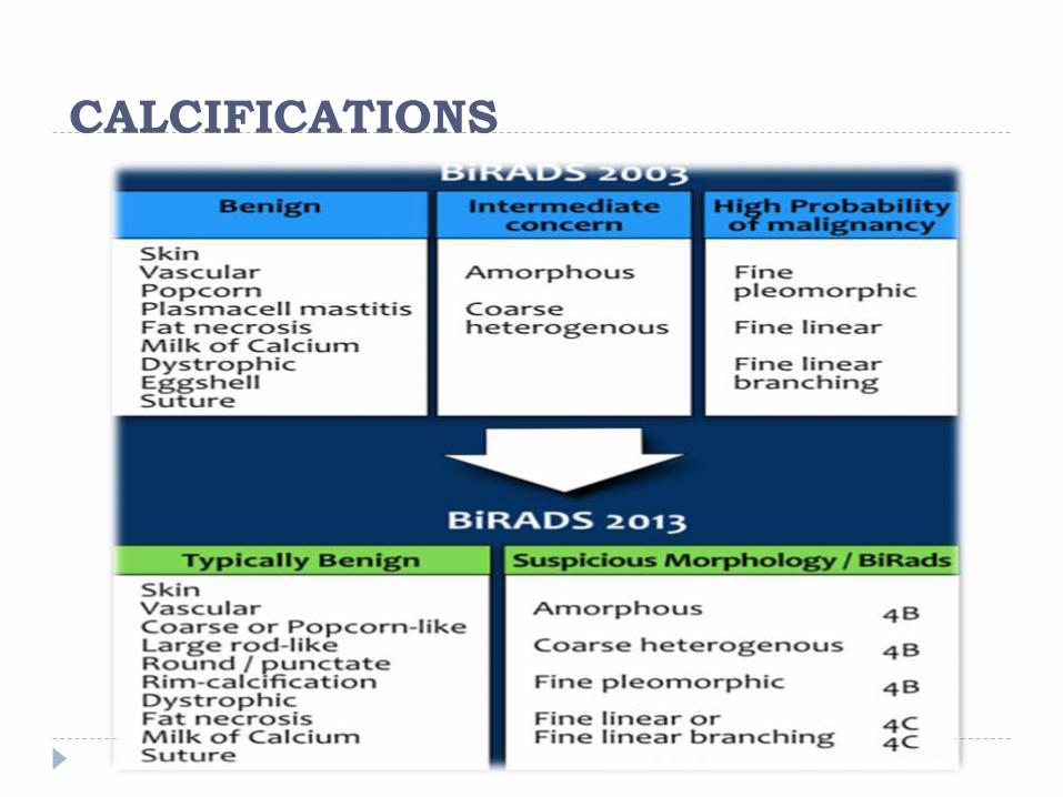
CALCIFICATIONS

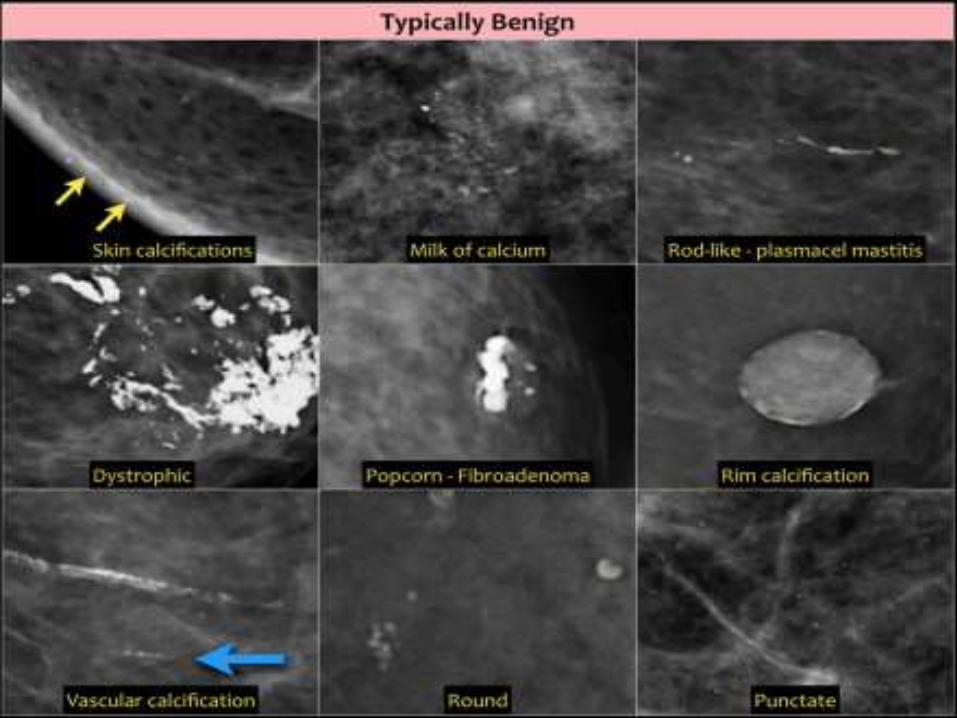
Typically Benign
Skin, vascular, coarse, large rodlike, round or
punctate (< 1mm), rim, dystrophic, milk of calcium
and suture calcifications are typically benign.
There is one exception of the rule: an isolated group
of punctuate calcifications that is new, increasing,
linear, or segmental in distribution, or adjacent to a
known cancer can be assigned as probably benign
or suspicious.

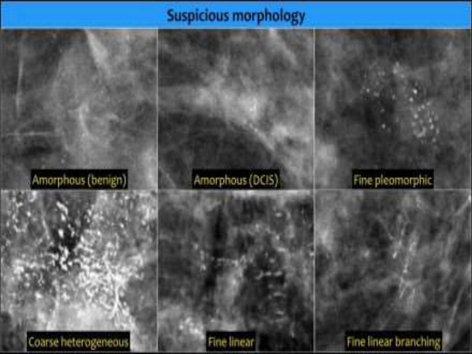
Calcifications of Suspicious Morphology
Amorphous (BI-RADS 4B)So small and/or hazy in appearance that a more specific particle shape cannot be determined.
Coarse heterogeneous (BI-RADS 4B)Irregular, conspicuous calcifications that are generally between 0,5 mm and 1 mm and tend to coalesce but are smaller than dystrophic calcifications.
Fine pleomorphic (BI-RADS 4C)Usually more conspicuous than amorphous forms and are seen to have discrete shapes, without fine linear and linear branching forms, usually < 0,5 mm.
Fine linear or fine-linear branching (BI-RADS 4C)Thin, linear irregular calcifications, may be discontinuous, occasionally branching forms can be seen, usually < 0,5 mm.
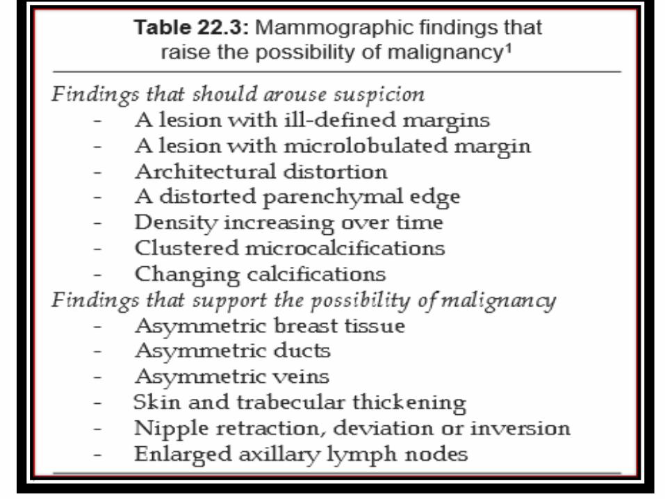
Associated features
Associated features are things that are seen in
association with suspicious findings like masses,
asymmetries and calcifications.
Associated features play a role in the final
assessment.
For instance a BI-RADS 4-mass could get a BI-
RADS 5 assessment if seen in association with skin
retraction.

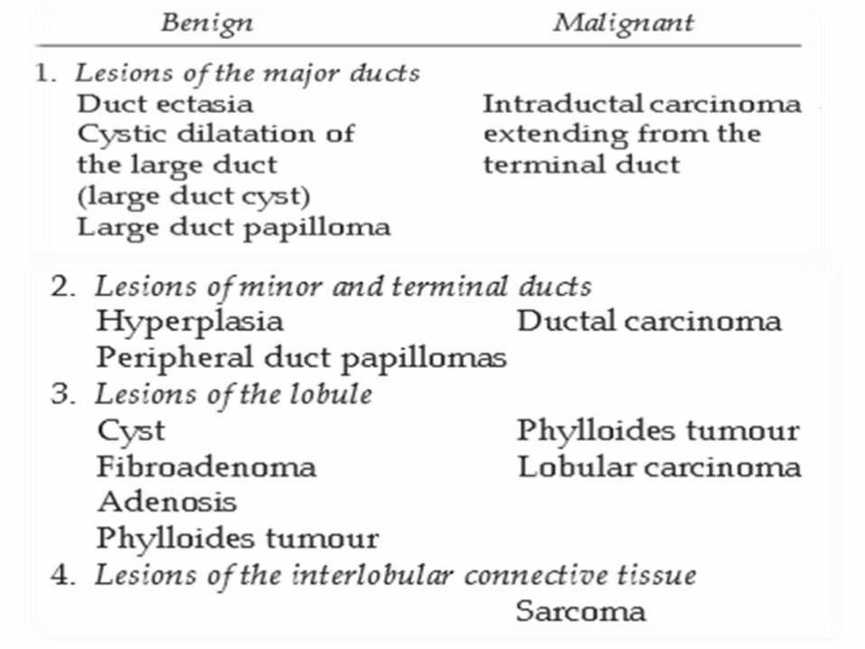

• BENIGN BREAST LESIONS
Lesions of the Major Ducts
Large Duct Papilloma (Intraductal Papilloma)
Papilloma is a benign mass lesion that results from proliferation of the
ductal epithelium that projects into the lumen of the duct.
These lesions are connected by a fibrovascular stalk to the epithelial
lining.
Papillomas may show areas of necrosis, haemorrhage and
occasionally calcification.
The duct around them can dilate forming a cystic structure giving the
appearance of an “intracystic papilloma”.
Benign papilloma is the single most common cause of serous or
bloody discharge from the nipple.
Almost all of these lesions are located in the major subareolar ducts
and are usually single
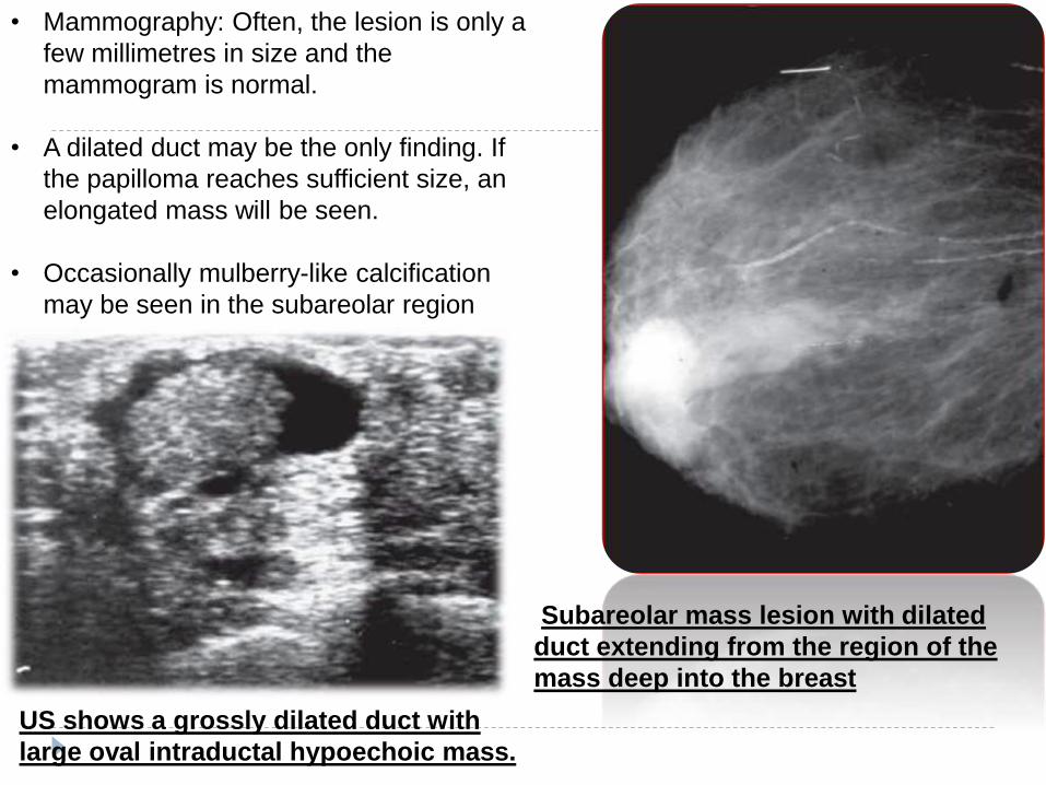
• Mammography: Often, the lesion is only a
few millimetres in size and the
mammogram is normal.
• A dilated duct may be the only finding. If
the papilloma reaches sufficient size, an
elongated mass will be seen.
• Occasionally mulberry-like calcification
may be seen in the subareolar region
Subareolar mass lesion with dilated
duct extending from the region of the
mass deep into the breast
US shows a grossly dilated duct with
large oval intraductal hypoechoic mass.
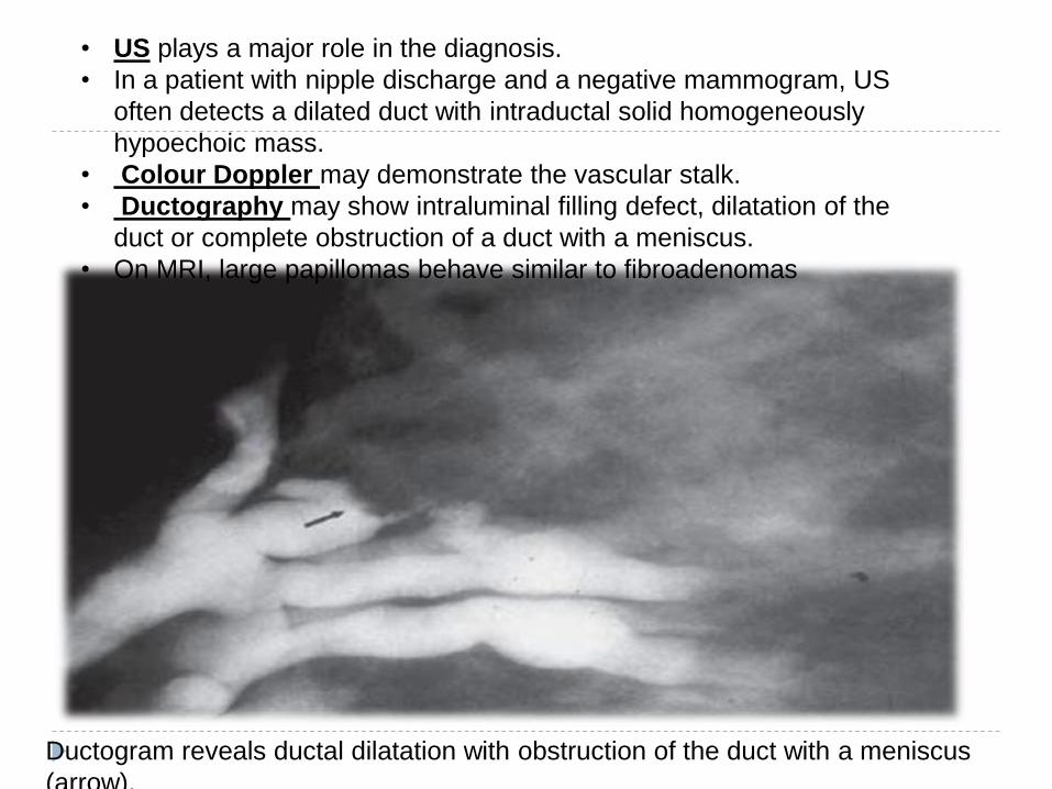
Ductogram reveals ductal dilatation with obstruction of the duct with a meniscus
(arrow).
• US plays a major role in the diagnosis.
• In a patient with nipple discharge and a negative mammogram, US
often detects a dilated duct with intraductal solid homogeneously
hypoechoic mass.
• Colour Doppler may demonstrate the vascular stalk.
• Ductography may show intraluminal filling defect, dilatation of the
duct or complete obstruction of a duct with a meniscus.
• On MRI, large papillomas behave similar to fibroadenomas
DUCT ECTASIA Duct ectasia primarily affects the major ducts in the subareolar region.
There is non-specific dilatation of one or more ducts.
The distended ducts are filled with fluid or thick secretions and cellular
debris.
Periductal fibrosis/inflammatory infiltrate usually may be found.
Normal ducts are usually too small to be resolved by mammography.
Ectatic ducts with thickened walls or periductal fibrosis become more
visible.
Dilated, thickened ducts are relatively common, and when symmetrically
distributed, are of no concern.
Intraluminal debris may calcify and produce calcifications called secretory
deposits
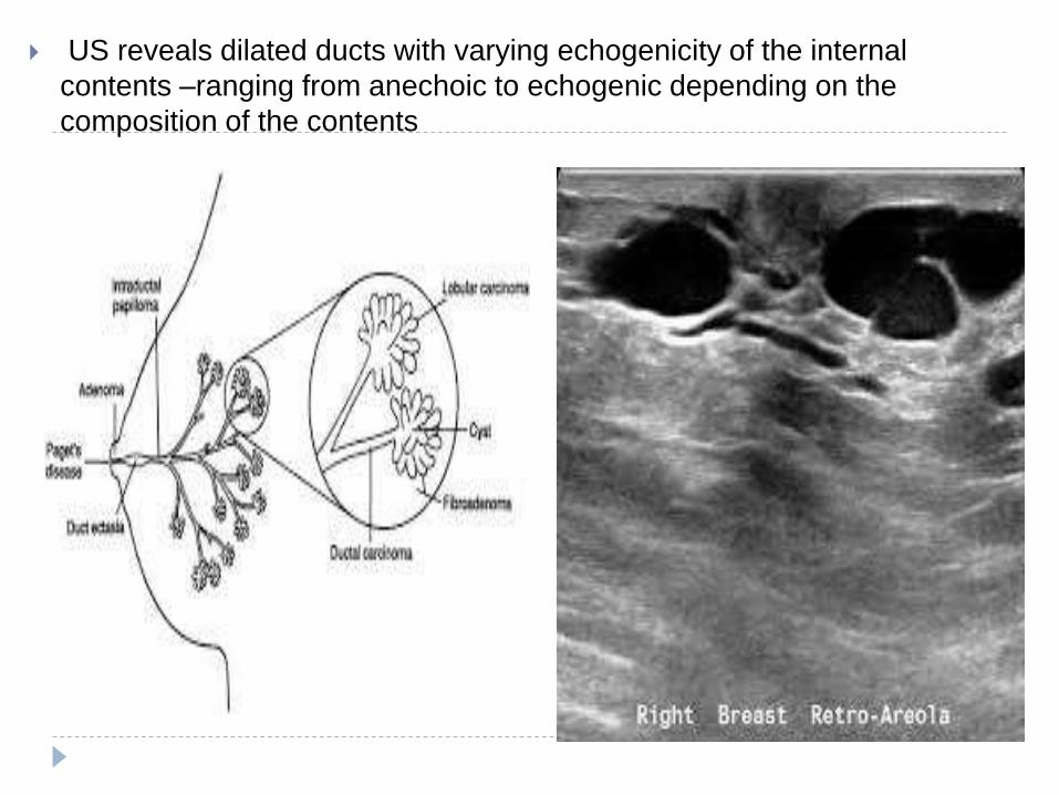
US reveals dilated ducts with varying echogenicity of the internal
contents –ranging from anechoic to echogenic depending on the
composition of the contents
FIBROADENOMA
Most common benign tumour of the breast in women of child bearing
age.
Fibroadenoma is essentially the result of overgrowth of the stromal
connective tissue within the lobule.
This idiopathic proliferation of collagen expands the lobule while
simultaneously surrounding an.
Physical examination reveals a firm, mobile, non-tender mass.
Fibroadenomas are hormone dependent lesions.
They regress with age and necrosis within the tumour results in coarse
nodular calcifications compressing the acini and terminal ducts.
Carcinoma is reported in less than 0.5 per cent of fibroadenomas.
Although fibroadenomas are not premalignant lesions, carcinomas can
incidentally arise alongside a fibroadenoma and envelope the lesion.
Also, because there is epithelium within fibroadenoma, cancer can
develop just as it can in normal ductal epithelium, and this is not a
malignant transformation of the lesion.
Hence ill-defined margins, microcalcification and large size or an
increase in size of a fibroadenoma should arouse concern
• On mammography, fibroadenoma is seen as a well-defined, homogeneous round
or oval mass with smooth margins.
• Fibroadenomas may have somewhat flattened contours which if present help to
distinguish them from cysts.
• fibroadenomas follow the structure of the lobule, their margins are often
lobulated.
• Occasionally they have microlobulated margins.
• In the presence of microlobulation however cancer should be suspected.
• The calcification of fibroadenoma can be differentiated from that of carcinoma by
its density, architecture and location
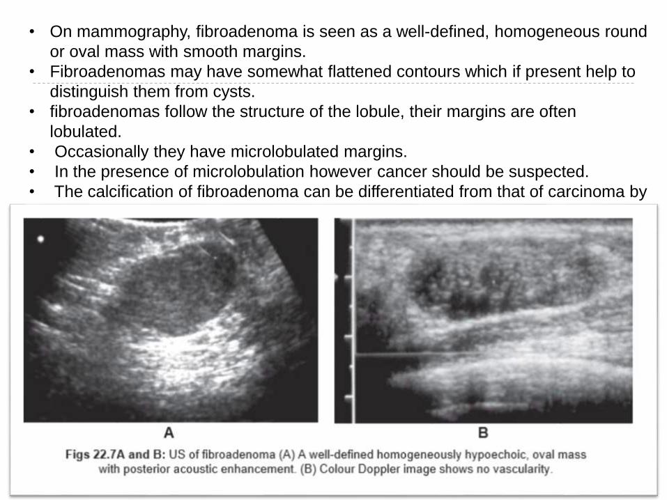
On ultrasonography, fibroadenomas are typically solid, ovoid, well-
circumscribed, homogeneously hypoechoic lesions that are wider
than they are high, with margins that are usually sharply
demarcated from the surrounding tissue.
As with other masses that are round or oval, fibroadenomas may
exhibit lateral wall refractive shadowing.
Posterior acoustic enhancement is frequently seen particularly when
the adenoma is cellular.
Fibroadenomas can however produce varying sonographic
appearances including ill defined margins and posterior acoustic
shadowing in more fibrotic adenomas simulating malignancy.
On Doppler imaging fibroadenomas are either avascular or display
minimal to moderate vascularity (in 20% cases).
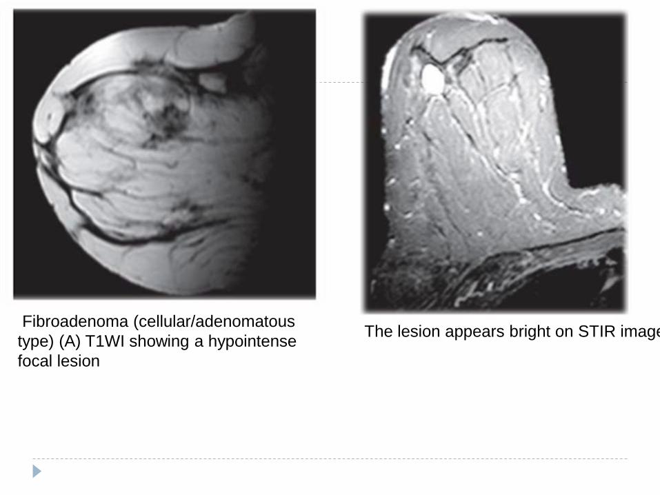
Fibroadenoma (cellular/adenomatous
type) (A) T1WI showing a hypointense
focal lesion
The lesion appears bright on STIR image
• MRI is only useful in the diagnosis of sclerosed lesions, i.e. predominantly
fibrous fibroadenomas.
• Such tumours are hypointense on all sequences and show no enhancement.
• Fibroadenomas, which are cellular and contain a fair amount of adenomatous or
myxoid tissue, show an intermediate to high-signal intensity on T2-weighted
images and most have well-circumscribed contours with low intensity internal
septae.
• The enhancement is significant and usually delayed, with absence of washout or
rim enhancement.
• The septi do not enhance. Because fibroadenomas develop multiple lobules,
different lobulations may develop different characteristics.
• Some can be oedematous, whereas others may be hyalinised. These criteria,
however, are not useful in differentiating cellular fibroadenomas from malignant
tumours.
• Hence, needle biopsy is more cost effective for their characterisation and MRI is
not recommended for a mammographically well-defined lesion suspected to be
a fibroadenoma in a premenopausal woman
PHYLLODES TUMOUR A rare tumour of fibroepithelial origin.
It is likely a variant of the benign fibroadenoma.
The basic histological features suggest a fibroadenoma with added branching
cystic cleft-like spaces of myxoid fluid and monotonous cellular stroma giving
a sarcomatous appearance.
The term “cystosarcoma phylloides” is a misnomer because most of these
tumours are benign and only a small percentage becomes malignant.
Approximately 25 per cent recur locally if not completely excised, and as
many as 10 per cent may metastasise to lung or bone.
Recurrence or metastases indicates presence of malignancy. Histological
establishment of malignancy is unreliable.1,14

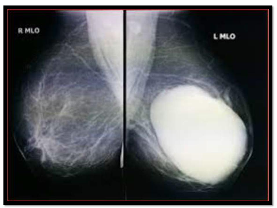
• Presents as a well circumscribed mass
in relatively young females (mean age
about 45 years).
• It may be of any size and may fill up
most of the breast. It has smooth,
lobulated contours and remains
relatively mobile even when very large.
• Mammographically, the tumour
resembles a large lobulated
fibroadenoma, some part of the margin
may be irregular suggesting local
breast invasion .
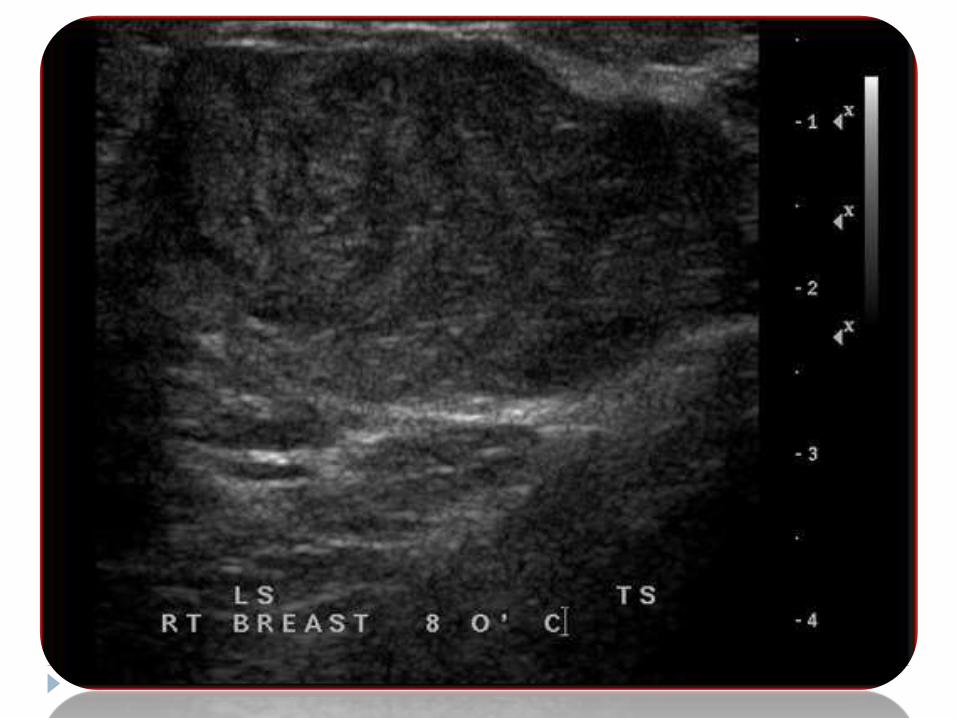
• Ultrasonography shows a mass with
very even internal echoes like
fibroadenoma but may show the
additional features of fluid clefts.
• On Doppler examination these lesions
show increased vascularity with high
peak systolic velocity and RI
resembling malignant masses.
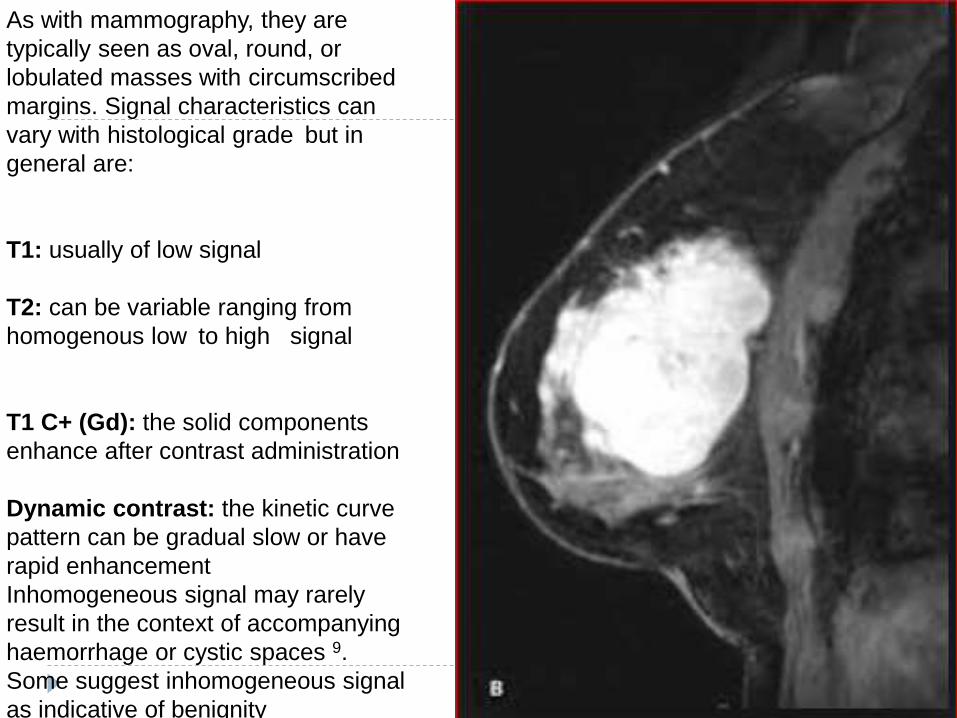
As with mammography, they are
typically seen as oval, round, or
lobulated masses with circumscribed
margins. Signal characteristics can
vary with histological grade but in
general are:
T1: usually of low signal
T2: can be variable ranging from
homogenous low to high signal
T1 C+ (Gd): the solid components
enhance after contrast administration
Dynamic contrast: the kinetic curve
pattern can be gradual slow or have
rapid enhancement
Inhomogeneous signal may rarely
result in the context of accompanying
haemorrhage or cystic spaces 9.
Some suggest inhomogeneous signal
as indicative of benignity
CYSTS
Breast cysts develop when lumina of ducts or acini become dilated and
lined by atrophic epithelium.
Simple cysts are common lesions and vary in size from microscopic to
larger palpable masses.
They are usually bilateral and multiple but only one may be identified
clinically or by imaging.
Cysts are common in perimenopausal age but may be seen in women of
all ages.
Cysts are benign lesions, with intracystic cancer found in < 0.2 per cent of
cysts.
Intracystic tumours if present are commonly intracystic papillomas.
Cysts may remain stable for many years or spontaneously resorb.
On mammography-A cyst is a homogeneous, well-defined mass, denser
than the surrounding more atrophic glandular tissue (in perimenopausal age).
The cyst may be of variable size, solitary or may occur in clumps.
Borders are smooth, but may appear lobulated when clumps of cysts are
present.
Calcification is infrequent, may be seen as a thin peripheral rim or flecks of
calcium near the periphery.
Rarely microcysts may contain milk of calcium fluid which on erect lateral
mammography layers on the cyst floor forming so-called “tea-cup”
calcification.
Cysts cannot be accurately diagnosed by mammography, because they
cannot be distinguished from other well-circumscribed masses unless they
display characteristic pattern of calcification
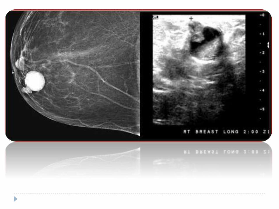
Ultrasonography has a very important role in the
diagnosis,therapeutic aspiration and follow-up of breast cysts.
Cysts should be sharply marginated, anechoic with posterior acoustic
enhancement.
Internal echoes if present should not be ignored.
Solid lesions, including cancer, may have only subtle internal echoes
and be otherwise indistinguishable from cysts. However internal debris
may be seen floating within the cyst.
Posterior enhancement may not be seen if the cyst is small or close to
the chest wall

Complex cyst
When internal echoes or
debris are seen, the cyst
is called a complex cyst.
These internal echoes
may be caused by
floating cholesterol
crystals, pus, blood or
milk of calcium crystals
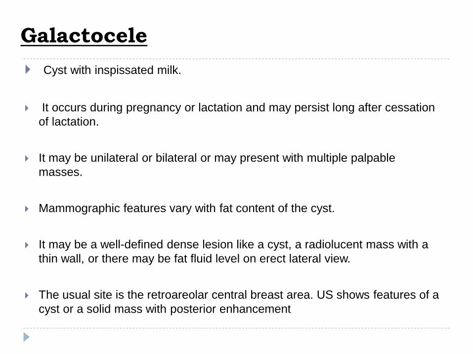
Galactocele
Cyst with inspissated milk.
It occurs during pregnancy or lactation and may persist long after cessation
of lactation.
It may be unilateral or bilateral or may present with multiple palpable
masses.
Mammographic features vary with fat content of the cyst.
It may be a well-defined dense lesion like a cyst, a radiolucent mass with a
thin wall, or there may be fat fluid level on erect lateral view.
The usual site is the retroareolar central breast area. US shows features of a
cyst or a solid mass with posterior enhancement
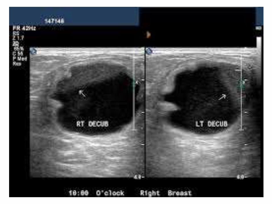
LIPOMA
As fat is frequently the preponderant tissue in the breast, it is difficult
to differentiate a true lipoma from normal fat.
Superficial and always encapsulated.
Freely movable and generally soft.
Liposarcoma is a rare lesion. Clinically it is firm and radiographically
dense, and hence is not confused with a lipoma.
On mammography- typical radiolucent appearance with a thin
capsule.
Harder, round, lucent lesions are generally either posttraumatic oil
cysts secondary to fat necrosis or galactoceles.
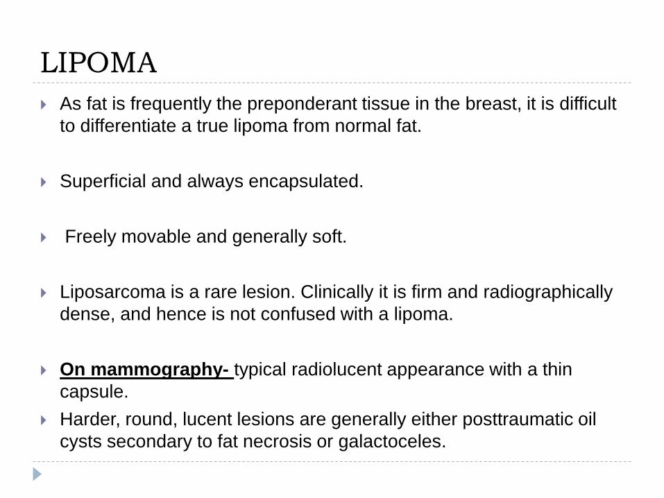
On US, lipoma are hypoechoic, and similar in echotexture to subcutaneous
fat. They may be distinguished from subcutaneous fat by the presence of
specular reflection from the capsule. Calcification may occur in necrotic
areas

MASTITIS/ABSCESS Breast infections may be in the form of acute mastitis
associated with lactation or a breast abscess.
Acute mastitis may progress to form an abscess.
Patient presents with painful localised or diffuse enlargement of the breast, with erythematous and oedematous overlying skin.
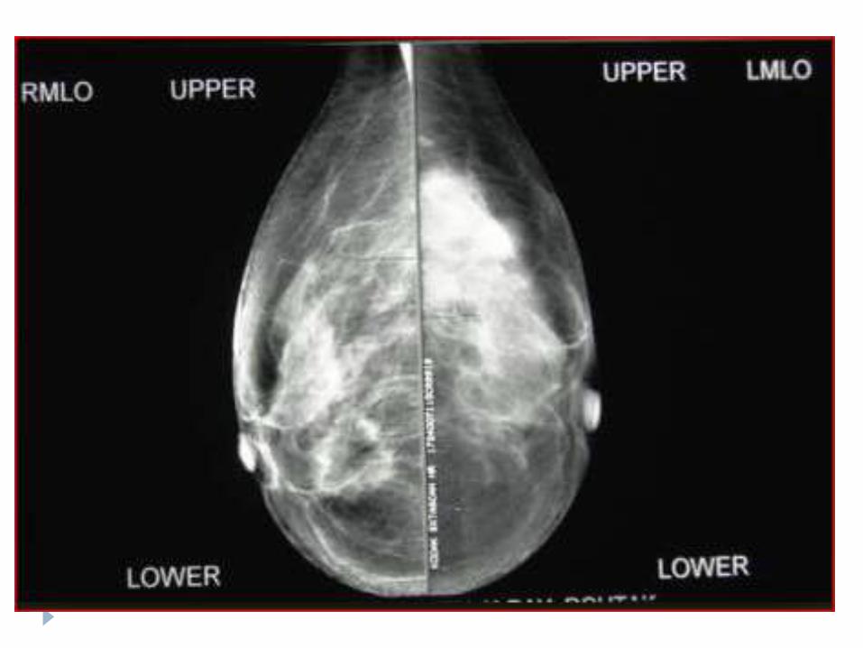
Mammography is seldom performed in acute mastitis.
If there is an underlying abscess formation, it may easily be missed through dense breast.
Abscess is usually well to ill-defined with a spiculatedmargin and overlying skin thickening is often present.
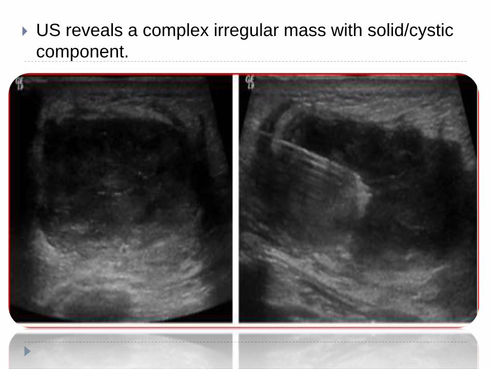
US reveals a complex irregular mass with solid/cystic
component.
CARCINOMA BREAST
Worldwide, breast cancer is the most common
invasive cancer in women.Breast cancer comprises
22.9% of invasive cancers in women and 16% of all
female cancers.
In 2012, it comprised 25.2% of cancers diagnosed in
women, making it the most common female cancer.]
GLOBAL TREND OF BREAST CANCER
The incidence of breast cancer in women has continued to rise. The
rate of increase has slowed recently, however, with the exception of
in situ breast cancer. Breast cancer death rates have decreased
since the early 1990s, with decreases of 2.5% per year among white
women.
Decreased breast cancer deaths have been attributed in part to
breast cancer screening, adjuvant chemotherapy, and
adoption of healthy standard of living
Randomized, population- controlled breast cancer screening trials
using mammography have shown an approximately 30% reduction in
breast cancer deaths in the women invited to screening compared to
women in the control group.
Because of this data, the American Cancer Society recommends
annual screening mammography for women age 40 years and older.
Risk Factors
Female
Older age
Family History
Early menarche
Late menopause
Nulliparity
First birth after age 30
Atypical ductal hyperplasia
BRCA1, BRCA2
Radiation exposure

Signs and Symptoms of Breast Cancer
Breast lump
Nipple discharge (new and spontaneous)
Bloody
Serosanguineous
Serous but copious
New nipple inversion
Skin retraction or tethering
Peau d’orange
Nothing (cancer detected on screening
mammography)

DUCTAL CARCINOMA IN SITU (DCIS)
The pathological classification of DCIS is based on the nuclear grade of the tumour cells (low, intermediate, or high), the architectural pattern of tumourgrowth (solid, papillary, micropapillary, or cribriform), and the presence or absence of comedonecrosis.
Ductal carcinoma in situ originates in a single glandular structure but may spread within the breast through the ductal system.
Two thirds of patients with low-to-intermediate grade ductal carcinoma in situ have multifocal disease, characterised by discontinuous intraductalgrowth.
In contrast, high-grade lesions tend to be continuous.
Most patients are asymptomatic, some may present with nipple discharge or palpable mass.
Currently, nearly 90 per cent of ductal carcinomas in situ are diagnosed while they are clinically occult because of mammographic detection of microcalcifications (in 76% of cases), soft-tissue densities (11%), or both (13%).
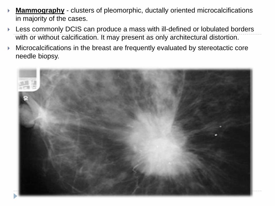
Mammography - clusters of pleomorphic, ductally oriented microcalcifications
in majority of the cases.
Less commonly DCIS can produce a mass with ill-defined or lobulated borders
with or without calcification. It may present as only architectural distortion.
Microcalcifications in the breast are frequently evaluated by stereotactic core
needle biopsy.
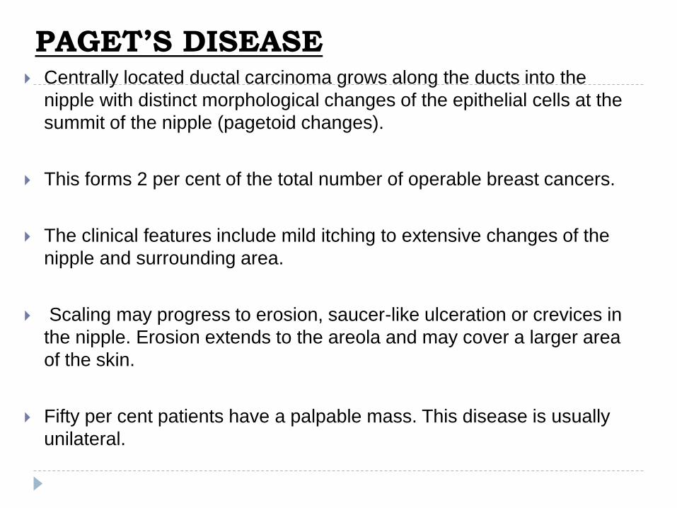
PAGET’S DISEASE Centrally located ductal carcinoma grows along the ducts into the
nipple with distinct morphological changes of the epithelial cells at the
summit of the nipple (pagetoid changes).
This forms 2 per cent of the total number of operable breast cancers.
The clinical features include mild itching to extensive changes of the
nipple and surrounding area.
Scaling may progress to erosion, saucer-like ulceration or crevices in
the nipple. Erosion extends to the areola and may cover a larger area
of the skin.
Fifty per cent patients have a palpable mass. This disease is usually
unilateral.

On Mammography-nipple and areolar thickening is present.
A subareolar mass may or may not be seen.
Malignant type of calcification may be seen extending from deeper carcinoma to the nipple.
Paget’s disease is not well delineated on US. The underlying mass may be seen on US with features similar to any other malignant lesio

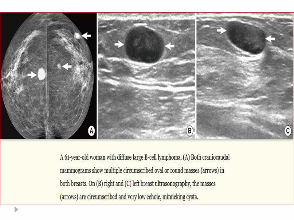
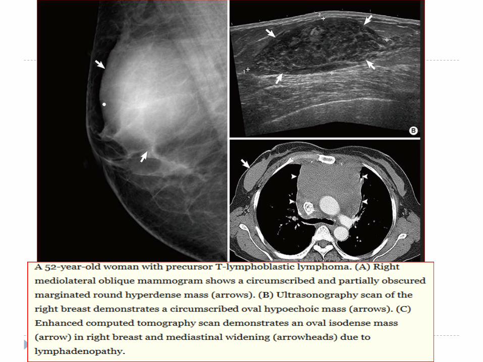
LYPMHOMA
Lymphoma of the breast can occur primarily or as a
metastatic lesion from elsewhere in the body.
Primary lymphoma is rare accounting for only 0.1 per
cent of breast malignancy
Mammary lymphoma may produce a single discrete
nodule or multiple nodules.
It may also produce a diffuse increase in radiographic
density.
Nodules may be well-defined or illdefined but spiculations
are not a feature of lymphoma.
Presence of large axillary nodes should raise the
possibility of lymphoma

THANK YOU