Embed Size (px)
Citation preview

Dr. Teffy JoseProf.Dr.DHANDAPANI’S UNIT

45 yr old , Mr.Ibrahim ,came with c/o 2 months cough with expectoration 1 episode of hemoptysis o/e afebrile PR- 82/mt BP-120/80 mmhg CVS –S1 S2 + RS -clear


CXR –PA view; Adequate penetration ;
Trachea in midline ;Bone and soft tissues normal;
‘well defined smooth bordered radio opaque lesion seen in the left lower zone with lobular contour ‘
‘lateral superior &inferior borders are well defined ‘ ‘left heart border is seen through the opacity ‘ ‘descending thoracic aorta is obscured by the opacity
IMP; suggestive of homogenous opacity located posteriorly
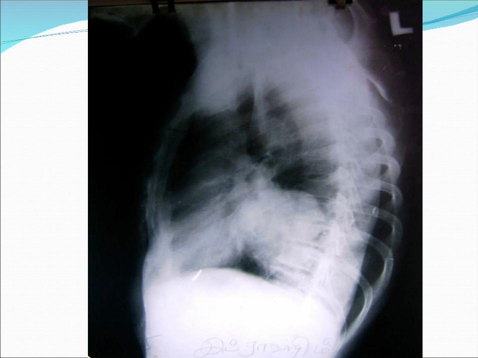

The lateral x ray confirmed the posterior location of the opacity
X ray wise this could thought of as a mediastinal mass / solitary pulmonary opacity > 4 cm size the diff.diag of which is
_bronchial carcinoma _lung abscess _wegener’s granulomatosis _lymphoma _round pneumonia
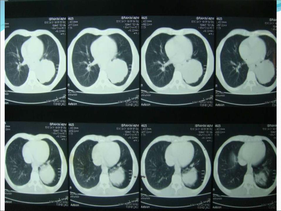

Ct picture

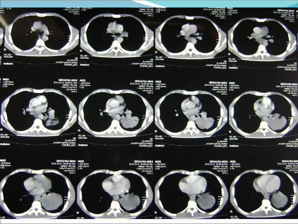

CT shows: 7*7.5*6 cm sized lobulated non enhancing cystic
density lesion of 0-25 HU noted in posterobasal segment of left lower lobe.
The lesion shows surrounding consolidatory changes with air bronchogram
IMP:Infected bronchogenic cyst with consolidation in the left lower lobe


Bronchogenic cyst “During development a portion of the tracheo bronchial tree
gets separated “
Can be ----- a)pulmonary b) mediastinal 10-15% 65-90%Radiology ; sharply demarcated round /oval, nodule /mass, usually in the medial 1/3 of lungs with a lower lobe predilection usually don’t communicate with the tracheobronchial tree unless infected
Ct findings Non enhancing homogenous opacity With attenuation density approximately of water 0-20 HU With smooth thin wall
Sometimes the density may be high due to varied contents with high protein or calcium
MRI is superior to CT in diagnosing
Mediastinal bronchogenic cysts five types -paratracheal -carinal -hilar -paraeosophageal -miscellaneous -thymus,pericardium etc ,

Clinical picture Usually asymptomatic When infected may present with cough and sputum
productionHemoptysis is also a common presenting feature
Rarely complications like pneumothorax air emoblism adenocarcinoma

Thank you