Embed Size (px)
Citation preview

Hindawi Publishing CorporationInternational Journal of NephrologyVolume 2012, Article ID 420247, 15 pagesdoi:10.1155/2012/420247
Review Article
Emergency Management of Hypertension in Children
Dinesh Singh,1 Olugbenga Akingbola,1 Ihor Yosypiv,2 and Samir El-Dahr2
1 Division of Pediatric Critical Care, Department of Pediatrics, Tulane University Health Sciences Center, 1430 Tulane Avenue,SL-37, New Orleans, LA 70112, USA
2 Division of Pediatric Nephrology, Department of Pediatrics, Tulane University Health Sciences Center, New Orleans,LA 70112, USA
Correspondence should be addressed to Dinesh Singh, [email protected]
Received 29 August 2011; Revised 13 December 2011; Accepted 12 January 2012
Academic Editor: Alberto Edefonti
Copyright © 2012 Dinesh Singh et al. This is an open access article distributed under the Creative Commons Attribution License,which permits unrestricted use, distribution, and reproduction in any medium, provided the original work is properly cited.
Systemic arterial hypertension in children has traditionally been thought to be secondary in origin. Increased incidence ofrisk factors like obesity, sedentary life-styles, and faulty dietary habits has led to increased prevalence of the primary arterialhypertension (PAH), particularly in adolescent age children. PAH has become a global epidemic worldwide imposing hugeeconomic constraint on health care. Sudden acute increase in systolic and diastolic blood pressure can lead to hypertensive crisis.While it generally pertains to secondary hypertension, occurrence of hypertensive crisis in PAH is however rare in children.Hypertensive crisis has been further subclassified depending on presence or absence of end-organ damage into hypertensiveemergency or urgency. Both hypertensive emergencies and urgencies are known to cause significant morbidity and mortality.Increasing awareness among the physicians, targeted at investigation of the pathophysiology of hypertension and its complications,better screening methods, generation, and implementation of novel treatment modalities will impact overall outcomes. In thispaper, we discuss the etiology, pathogenesis, and management of hypertensive crisis in children. An extensive database searchusing keywords was done to obtain the information.
1. Definitions and Epidemiology
Primary arterial hypertension is a global epidemic affectingpredominantly adult population [1]. Although secondaryetiologies of hypertension predominate in children, theprevalence of primary arterial hypertension has been increas-ing at an alarming rate particularly in adolescents and olderchildren [2]. Recent survey conducted by the National Healthand Nutrition Examination Survey (NHANES) in 8–17-year-old children showed a prevalence of prehypertensionand hypertension of about 10% and 4%, respectively,with a higher incidence in African American and MexicanAmericans [3]. Increase in the prevalence of hypertensionhas paralleled the increased prevalence of childhood obesity[4]. Childhood obesity has increased by more than threetimes in the past three decades [5]. The Fourth Report on theDiagnosis, Evaluation, and Treatment of High Blood Pressurein Children and Adolescents classified pediatric hypertensioninto various stages [6] (Table 1). In one study the incidenceof stage 1 and 2 hypertension was reported to be 2.6%
and 0.6%, respectively, in adolescent students [7]. The JointNational Committee on Detection, Evaluation, and Treatmentof Hypertension, JNC7, has labeled acute severe elevationof blood pressure above 180/120 mmHg (about 20 mmHgabove the Stage II hypertension) as “Hypertensive Crisis”in adults [8]. It is further subclassified based on presenceof target organ abnormalities like seizures, intracranialhemorrhage, Posterior reversible encephalopathy syndrome(PRES), focal neurological deficit, congestive cardiac failure,papilledema, retinal hemorrhages, and acute vision loss intoHypertensive Emergency and in the absence of target organabnormalities as Hypertensive Urgency [8]. Unfortunately, nosuch clear definition has been proposed for HypertensiveCrises in children. Sequelae of hypertension emergencieslike left ventricular failure, encephalopathy, aortic dissection,myocardial ischemia, acute renal insufficiency, and retinaldamage are well known in adults [9]. However, target organdamage and adverse effects have also been demonstratedin older children and adolescents and are an evidence oflong-term blood pressure elevation [10–17]. In addition,
2 International Journal of Nephrology
Table 1: Definitions of normal and elevated blood pressure inchildren.
Normal blood pressureSystolic and diastolic blood pressurebelow 90th centile
PrehypertensionSystolic or diastolic blood pressureabove the 90th centile (or 120/80mmHg), but below the 95th centile
Stage I hypertension
Systolic or diastolic blood pressurehigher than or equal to the 95thcentile, but lower than the 99th centileplus 5 mm Hg
Stage II hypertensionSystolic or diastolic BP higher than orequal to the 99th centile plus 5 mm Hg
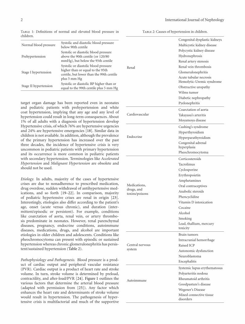
target organ damage has been reported even in neonatesand pediatric patients with prehypertension and whitecoat hypertension, implying that any age and any level ofhypertension could result in long-term consequences. About1% of all adults with a diagnosis of hypertension developHypertensive crisis, of which 76% are hypertensive urgenciesand 24% are hypertensive emergencies [18]. Similar data inchildren is not available. In addition, although the prevalenceof the primary hypertension has increased over the pastthree decades, the incidence of hypertensive crisis is veryuncommon in pediatric patients with primary hypertensionand its occurrence is more common in pediatric patientswith secondary hypertension. Terminologies like AcceleratedHypertension and Malignant Hypertension are obsolete andshould not be used.
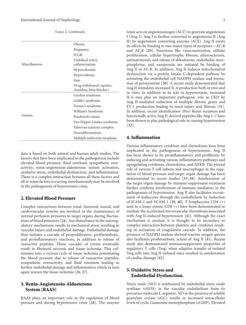
Etiology. In adults, majority of the cases of hypertensivecrises are due to nonadherence to prescribed medication,drug overdose, sudden withdrawal of antihypertensive med-ications, and so forth [19–22]. In comparison, majorityof pediatric hypertensive crises are renal in origin [23].Interestingly, etiologies also differ according to the patient’sage, onset (acute versus chronic), and duration (inter-mittent/episodic or persistent). For example, conditionslike coarctation of aorta, renal vein, or artery thrombo-sis predominate in neonates. However, renal parenchymaldiseases, pregnancy, endocrine conditions, autoimmunediseases, medications, drugs, and alcohol are importantetiologies in older children and adolescents. Conditions likepheochromocytoma can present with episodic or sustainedhypertension whereas chronic glomerulonephritis has persis-tent/sustained hypertension (Table 2).
Pathophysiology and Pathogenesis. Blood pressure is a prod-uct of cardiac output and peripheral vascular resistance(PVR). Cardiac output is a product of heart rate and strokevolume. In turn, stroke volume is determined by preload,contractility, and after-load/PVR [24]. Figure 1 outlines thevarious factors that determine the arterial blood pressure(adapted with permission from [25]). Any factor whichenhances the heart rate and determinants of stroke volumewould result in hypertension. The pathogenesis of hyper-tensive crisis is multifactorial and much of the supportive
Table 2: Causes of hypertension in children.
Renal
Congenital dysplastic kidneys
Multicystic kidney disease
Polycystic kidney disease
Hydronephrosis
Renal artery stenosis
Renal vein thrombosis
Glomerulonephritis
Acute tubular necrosisHemolytic-Uremic syndrome
Obstructive uropathy
Wilms tumor
Diabetic nephropathy
Pyelonephritis
CardiovascularCoarctation of aorta
Takayasu’s arteritis
Moyamoya disease
Endocrine
Cushing’s syndrome
Hyperthyroidism
Hyperparathyroidism
Congenital adrenalhyperplasia
Pheochromocytoma
Medications,drugs, andtoxins/poisons
Corticosteroids
Tacrolimus
Cyclosporine
Erythropoietin
Amphetamines
Oral contraceptives
Anabolic steroids
Phencyclidine
Vitamin D intoxication
Cocaine
Alcohol
Smoking
Lead, thallium, mercurytoxicity
Central nervoussystem
Brain tumors
Intracranial hemorrhage
Raised ICP
Autonomic dysfunction
Neuroblastoma
Encephalitis
Autoimmune
Systemic lupus erythematosus
Polyarteritis nodosa
Rheumatoid arthritis
Goodpasture’s disease
Wegener’s Disease
Mixed connective tissuedisorders
International Journal of Nephrology 3
Table 2: Continued.
Miscellaneous
Obesity
Pregnancy
IUGR
Umbilical arterycatheterization
Hypercalcemia
Hypervolemia
Pain
Drug withdrawal (opiates,clonidine, beta-blocker)
Genetic
Gordon syndrome
Liddle’s syndrome
Turner’s syndrome
William’s Syndrome
Friedreich’s ataxia
Von Hippel-Lindau syndrome
Tuberous sclerosis complex
Neurofibromatosis
Multiple endocrine neoplasia
data is based on both animal and human adult studies. Thefactors that have been implicated in the pathogenesis includeelevated blood pressure, fluid overload, sympathetic over-activity, renin-angiotensin-aldosterone system activation,oxidative stress, endothelial dysfunction, and inflammation.There is a complex interaction between all these factors andall or some factors occurring simultaneously may be involvedin the pathogenesis of hypertensive crisis.
2. Elevated Blood Pressure
Complex interactions between renal, humoral, neural, andcardiovascular systems are involved in the maintenance ofnormal perfusion pressures to target organs during fluctua-tions of blood pressures [24]. Any disturbance in the autoreg-ulatory mechanisms results in mechanical stress resulting invascular injury and endothelial damage. Endothelial damagethus initiates a cascade of proproliferative, prothrombotic,and proinflammatory reactions, in addition to release ofvasoactive peptides. These cascades of events eventuallyresult in fibrinoid necrosis and tissue ischemia. This cul-minates into a vicious cycle of tissue ischemia potentiatingthe blood pressure due to release of vasoactive peptides,sympathetic overactivity, and fluid retention leading tofurther endothelial damage and inflammation which in turnagain worsen the tissue ischemia [26, 27].
3. Renin-Angiotensin-AldosteroneSystem (RAAS)
RAAS plays an important role in the regulation of bloodpressure and during hypertensive crisis [28]. The enzyme
renin acts on angiotensinogen (AGT) to generate angiotensinI (Ang I). Ang I is further converted to angiotensin II (AngII) by angiotensin converting enzyme (ACE). Ang II exertsits effects by binding to two major types of receptors—AT1Rand AT2R [29]. Functions like vasoconstriction, cellularproliferation, cellular hypertrophy, fibrosis, atherosclerosis,antinatriuresis, and release of aldosterone, endothelin, nore-pinephrine, and vasopressin are initiated by binding ofAng II to AT1R. In addition, Ang II induces mitochondrialdysfunction via a protein kinase C-dependent pathway byactivating the endothelial cell NADPH oxidase and forma-tion of peroxynitrite [30]. A recent study demonstrated thatAng II stimulates increased IL-6 production both in vivo andin vitro. In addition to its role in hypertension, increasedIL-6 may play an important pathogenic role in CKD byAng II-mediated induction of multiple fibrotic genes andET-1 production leading to renal injury and fibrosis [31].In addition, recent identification (Pro) Renin receptors andfunctionally active Ang II-derived peptides like Ang 1–7 havebeen shown to play pathological role in causing hypertension[32].
4. Inflammation
Various inflammatory cytokines and chemokines have beenimplicated in the pathogenesis of hypertension. Ang IIhas been shown to be proinflammatory and profibrotic byinducing and activating various inflammatory pathways andupregulating cytokines, chemokines, and NFKB. The pivotalrole of various T-cell subsets and macrophages in the regu-lation of blood pressure and target organ damage has beendemonstrated in recent studies [33–38]. Amelioration ofthe target organ damage by immune-suppressant treatmentfurther confirms involvement of immune mediators in thepathogenesis of hypertension. Ang II also facilitates recruit-ment of leukocytes through the endothelium by inductionof ICAM-1 and VCAM-1 [39, 40]. T lymphocytes CD4 (+)and, to a lesser extent, CD8 (+) have been demonstrated tomediate the accelerated microvascular thrombosis associatedwith Ang II-induced hypertension [41]. Although the exactmechanism is unclear, it is thought to be secondary tocomplex interaction between platelets and cytokines result-ing in activation of coagulation cascade. In addition, thepresence of NADPH oxidase-derived reactive oxygen speciesalso facilitates prothrombotic action of Ang II [41]. Recentstudy also demonstrated immunosuppressive properties ofregulatory T cells (Treg) when adaptive transfer of isolatedTreg cells into Ang II–infused mice resulted in ameliorationof cardiac damage [42].
5. Oxidative Stress andEndothelial Dysfunction.
Nitric oxide (NO) is synthesized by endothelial nitric oxidesynthase (eNOS) in the vascular endothelium from itsprecursor molecule L-arginine. NO in the presence of solubleguanylate cyclase (sGC) results in increased intracellularlevels of cyclic Guanosine monophosphate (cGMP). Elevated

4 International Journal of Nephrology
cGMP causes decrease in intracellular calcium ion levelsleading to decreased vascular tone. Oxidative stress may playa causal role in the development of hypertension by alteringthe vascular tone either by oxidative modification of proteinsand nucleic acids or by decreasing the bioavailability of nitricoxide. Superoxide anions generated by various enzymes suchas NADPH oxidase, xanthine oxidase, and enzymes involvedin mitochondrial respiratory chain may directly inactivateNO and inhibit sGC. Increased angiotensin II levels facilitatefurther generation of superoxide anions by stimulating theseenzymes. In addition, superoxide anions lead to uncouplingof eNOS by oxidating the BH4 (tetrahydrobiopterin). BH4is an essential cofactor necessary in generation of NO byeNOS enzyme. Uncoupling of eNOS also facilitates furthergeneration of superoxide anions. These superoxide anionscause increased vascular cell proliferation and migration,apoptosis, inflammation, extracellular matrix alterations,and endothelial dysfunction [43–46].
6. Central Nervous System
The role of central nervous system in the regulation of bloodpressure via modulation of sympathetic and parasympatheticnervous system is well known. However, recent studiessuggest that increased sodium intake results in increase inendogenous ouabain (EO) levels in the paraventricular andsupraoptic nuclei and at the circumventricular organs suchas subfornical organ. This induces an acute but transientAng-II- mediated increase in peripheral sympathetic nervoussystem resulting in elevation in blood pressures. Experimentsconducted in rats reveal complex interactions involvingsodium ions, epithelial sodium channels (ENaCs), RAAS,and EO in the brain which activate sympathetic nervoussystem. Thus, a brain Na+-ENaC-RAAS-EO pathway and aneuromodulatory pathway involving Aldosterone-EO-Ang IIhave been proposed in explaining the mechanism of actionof hypertension. Further research and understanding ofthese novel mechanisms will help in newer antihypertensivetherapies [47].
In addition, genetic mutations and polymorphisms [48],and insulin resistance [49], and abnormalities involvingthe sodium transport mechanisms like Na+/H+ exchanger,Na+/K+/2Cl− cotransporter, Na+Cl− cotransporter, Na+/K+
ATPase, and sodium-phosphate cotransporter [50, 51] havealso been implicated in the pathogenesis of hypertension.A possible mechanism of hypertensive crisis is shown inFigure 2.
6.1. Clinical Features and Target-Organ Damage. Clinicalpresentation varies depending upon age, the target organinvolved, and etiology. Neonates may present with apnea,cyanosis, irritability, and poor feeding [52].In addition,clinical features may reflect specific etiologies like endocrinediseases, autoimmune conditions, pregnancy, and drugabuse. Older children with long-term hypertension or acuteexacerbation of chronic hypertension or sudden severeelevation of blood pressure may present with symptomsrelated to end organ abnormalities involving the heart, eye,kidney, and brain [53].
6.2. Cardiovascular Manifestations. Depending on the dura-tion and acuity of the symptoms, the cardiac involvementcan be in the form of left ventricular hypertrophy (LVH),left ventricular failure, or left ventricular ischemia [54, 55].Although left ventricular hypertrophy has traditionally beendefined in pediatric population as left ventricular mass index(LVMI) greater than 38.6 g/m2.7 and has been recommendedin the fourth report, a recent study has demonstrated thatLVMI varies significantly in children particularly those <9years of age. The study which was performed in 2,273nonobese, healthy children demonstrated that in childrenaged >9 years the 50th percentile values of LVMI rangedfrom 27 g/m2.7(girls) to 32 g/m2.7(boys) and varied little withage. The 95th percentile values of LVMI in the >9 yearsage group ranged from 40 g/m2.7(girls) to 45 g/m2.7(boys).The authors concluded that values >40 g/m2.7in girls and>45 g/m2.7 in boys should be considered abnormal. Incontrast, the 50th percentile values of LVMI in children <9years age group varied significantly from 56.44 g/m2.7(boys)and 55.38 g/m2.7(girls) in infants less than 6 months ofage to 31.79 g/m2.7(boys) and 29.71 g/m2.7(girls) in childrenless than 8years of age. Similarly, the 95th percentilevalues of LVMI in the <9 years age group varied from80.1 g/m2.7(boys) and 85.6 g/m2.7(girls) in infants less than6 months of age to 44.6 g/m2.7(boys) and 43.5 g/m2.7(girls) inchildren less than 8 years of age (Table 3). This study providesnormal percentile values for young children and emphasizesthe need for age–appropriate LVMI cut points and useof appropriate percentile curves particularly in children<9 years of age [56]. The LVH is common in childrenwith hypertension with an incidence of 41.1% particularlyin children with high Body-Mass index (BMI) and inHispanic population [57]. Left ventricular failure can leadto symptoms such as increased work of breathing, shortnessof breath, chest pain, palpitations, decreased urine output,and poor appetite. Sudden acute increase in blood pressuremay precipitate left ventricular failure in any pediatric agegroup but is more commonly reported in neonates [58–60].Carotid intima media thickness (CIMT), measured by B-mode ultrasound at end diastole, has emerged as a surrogatemarker of early atherosclerotic changes and is predictive ofadult cardiovascular structural damage. Indeed, increasingnumber of studies in children with hypertension, dyslipi-demia, diabetes, and obesity have shown an increased CIMT.However, a recent study showed that nonobese childrenwith primary hypertension had increased CIMT comparedwith BMI-matched controls. Although obesity may playa significant role in vascular changes, this study providesstrong and interesting evidence that CIMT may be increasedin nonobese children with primary hypertension. The majordrawback of this surrogate marker is that the reference valuesare not available for children younger than 10 years andfurther studies are needed to determine reference values ofCIMT in this age group [61–63].
6.3. Neurological Manifestations. Loss of cerebral autoreg-ulation leading to disruption of the blood brain barrierand endothelial dysfunction results in imbalance in oxygendelivery, edema formation, and microhemorrhages [64].

International Journal of Nephrology 5
β − 1
agonistsagonist(SNS)
β − 1agonist(SNS)
β − 2agonists
Force ofcontraction
++
Fillingpressure
Venouscapacitance
α− 2
agonist(SNS)
α − 1
Heartrate ×
×
Strokevolume
Aorticdistensibility
DiuresisIntravascular
volume
Cardiacoutput
Totalperipheralresistance
Arterial blood pressure
ANP
ADH
Aldo
A II
Sodium
RBCmass
Colloid
Renin
K, PGE, β − 2 agonists
PSNS(Ach)
Figure 1: Factors that determine the arterial blood pressure (adapted with permission from [25]).
Endothelial injury/dysfunction Increase vasoconstrictors and
decrease in vasodilators
Pressure natriuresis
Activation of procoagulation cascade and inhibition of
fibrinolytic mechanism
Release of inflammatory markers and reactive oxygen
species
Volume depletion and positive feedback to renin-
angiotensin system
Sudden severe increase in blood pressure
Fibrinoid necrosis andmyo-proliferation
Further increase in blood pressure
Figure 2: Mechanism of hypertensive crisis.
Table 3: Age-specific reference values of the LVMI in boys and girls Adapted from [56].
Age Sex LVMI (50th percentile) LVMI (95th percentile)
>9 yearsBoys 32.0 g/m2.7 45.0 g/m2.7
Girls 27.0 g/m2.7 40.0 g/m2.7
<8 yearsBoys 31.79 g/m2.7 44.6 g/m2.7
Girls 29.71 g/m2.7 43.5 g/m2.7
<6 monthsBoys 56.44 g/m2.7 80.1 g/m2.7
Girls 55.38 g/m2.7 85.6 g/m2.7

6 International Journal of Nephrology
These changes may lead to seizures, altered mental status,PRES, vomiting, signs of raised intracranial pressure, focalneurological deficits, and headache. Headache is the mostcommon symptom [65–69]. In one study seizures occurredin 25% of children, encephalopathy in 25%, facial palsyin 12%, and hemiplegia in 8% [18]. Posterior reversibleencephalopathy syndrome has been reported to predomi-nantly affect the occipitoparietal white matter with occa-sional spread to basal ganglia, cerebellum, and brainstem.Various etiologies like post-chemotherapy, posttransplant,postinfectious, autoimmune conditions, and posthyperten-sive crisis have been known to cause PRES. Clinical featuresinclude headache, altered mental status, nausea, vomiting,seizures, cortical blindness, and focal neurological deficits.Magnetic Resonance Imaging shows bilateral, symmetrical,involvement of white matter in occipitoparietal regionswhich appear as hyperintense lesions on T2-weighted imagesand hypointense or isointense lesions on Diffusion-WeightedImages. PRES is a completely reversible condition withoccasional reports of neurological deficits [70–72].
6.4. Renal Manifestations. Hematuria, flank pain, and olig-uria would indicate renal involvement. The most commonetiology leading to hypertension in children is renal diseasebut hypertension itself can result in renal injury and failuresecondary to loss of autoregulation of renal blood flow.But the data exploring the impact of hypertension on therenal function and structural injury in pediatric populationis limited. Histologically fibrinoid necrosis with thrombosisinvolving the intrarenal arteries has been demonstrated inadult studies which result in clinical presentation consistentwith microangiopathic hemolytic anemia [73, 74]. A recentstudy, however, has demonstrated increased microalbumin-uria and decreased glomerular filteration rate in prehyper-tensive children particularly with high blood pressure load[75]. In addition, another study demonstrated a reductionin microalbuminuria and LVH when hypertension wascontrolled with ACE inhibitors [76]. These findings suggestthat renal dysfunction and structural injury may occur earlyeven in pediatric population with hypertension and continueinto adult life and studies are needed to further elucidatethese findings.
6.5. Ophthalmological Manifestations. Retinal bleeds,papilledema, loss of visual acuity, acute ischemic opticneuropathy, and cortical blindness have been reportedsecondary to hypertensive crisis [77]. Loss of vision canbe serious and permanent. Traditionally hypertensiveretinopathy assessed by direct fundoscopy has beendescribed based on Keith-Wagner-Barkar’s classification(1939) which was subsequently modified by Scheie (1953)[78, 79]. The major drawback of these classifications is thatdirect fundoscopic examination is limited by physician’sexperience and high inter- and intraobserver variability.More recently Wong and Mitchell (2004) (Table 4) proposeda new classification which stratified cardiovascular risksassociated with different grades of hypertensive retinopathyin adults [80]. The data regarding the prevalence of
hypertensive retinopathy in general pediatric population islargely unknown. However, in two studies the prevalenceof hypertensive retinopathy in children with hypertensionvaried from 8.9% (assessed by direct fundoscopy) to 50%(assessed by retinal photographs) [13, 81]. Majority of thechildren in both studies had mild retinopathy and none hadhigher-grade retinopathy. In addition, evidence of grade IIIand grade IV hypertensive retinopathy was lacking in 32%of adults with hypertensive encephalopathy [82]. Althoughmany studies involving the risk stratification and prognosticimportance based on the retinopathy grades are availablein adults, no such data exists in pediatric population.In general, moderate-to-severe grades of retinopathy arerelatively rare in children and further studies are needed toelucidate the importance of mild retinopathy and long-termprognosis. But newer techniques like digital imaging andcomputer analysis of the early retinal changes and newergrading systems will further help in risk stratification anddisease progression [83].
7. Clinical Assessment
Clinical assessment begins with obtaining relevant present,past medical history. Potential risk factors include history oflow birth weight, intrauterine growth retardation, prema-turity, oligo or polyhydramnios, umbilical artery catheter-ization, recurrent urinary tract infections, weak stream ofthe urine in male child, hematuria, flank pain, polyuria,failure to thrive, joint pains, skin rashes, headaches, visualdisturbances, chest pain, palpitations, and poly or oliguria.Family history of diabetes, hypertension, obesity, hyperc-holesteremia, early strokes, coronary artery diseases, suddencardiac deaths, malignancies, autoimmune conditions, orhereditary conditions involving the kidneys, liver, and brainshould be assessed. A detailed endocrine-related historyshould also be obtained. Medication history involvingsteroids, antihypertensives, tacrolimus, cyclosporine, oralcontraceptives, and dietary and life-style history regardingsmoking, alcohol, and drug abuse should be elucidated.In addition in teenage adolescent girls, pregnancy-relatedsymptoms should be elicited. In obese children, history ofsleep apnea and daytime somnolence should be obtained.
Physical examination should involve general exami-nation to look for edema, skin rashes, neurocutaneousmarkers, cyanosis, elfin facies, webbing of neck, hirsutism,cushingoid features, thyroid enlargement, proptosis, andso forth. Heart rate, respiratory rate, and four-extremityblood pressure preferably both in lying and sitting posi-tion, peripheral pulses, height, weight, and BMI should berecorded. A detailed cardiovascular, respiratory, abdominal,and neurological examination should be performed to lookfor any evidence of coarctation, LVH, pulmonary edema,pleural, and pericardial effusions. In addition, examine forhepatosplenomegaly, intra-abdominal masses, ascites, andgenitourinary abnormalities. Look for any evidence of spinabifida, hydrocephalus, signs of raised ICP, papilledema, focalneurological deficits, and cranial nerve palsies particularly3rd and 7th cranial nerves.

International Journal of Nephrology 7
Table 4: Wong and Mitchell’s classification (adapted from [80]).
Grading Retinal signs Sytemic associations∗
Mild retinopathy
Generalized arteriolar narrowing, focalarteriolar narrowing, arteriovenousnicking, opacity (copper wiring) ofarteriolarwall, or a combination ofthese signs
Modest association with risk of clinicalStroke, subclinical stroke,coronaryheart disease, and death
Moderate retinopathy
Hemorrhage (blot, dot, or flameshaped), microaneurysm cotton-woolspot, hard exudates or a combinationof these signs
Strong association with risk of clinicalstroke, subclinical stroke, cognitivedecline, and cardiovascular mortality
Severe retinopathyModerate retinopathy plus optic discswelling # Strong association with mortality
∗A modest association is defined as an odds ratio of greater than 1 but less than 2. A strong association is defined as anodds ratio of 2 or greater.
#Anterior ischemic optic neuropathy, characterized by unilateral swelling of the optic disk, visual loss, and sectorial visual field loss, should be ruled out.
Table 5: Initial workup for hypertension.
Complete blood count
Basic metabolic panel including magnesium and phosphate
Serum uric acid
Fasting lipid profile
Fasting blood glucose
Urine analysis/culture
Urine electrolytes, creatinine, protein
Chest X-ray
EKG and echocardiogram
Renal ultrasound with doppler
8. Laboratory Workup and Evaluation
Successful management of the elevated blood pressure inchildren depends on the accurate diagnosis and evaluation ofthe etiology of hypertension. It is well known that the clinicalspectrum of the etiology of hypertension is varied dependingon the age of the child. Thus, it is vitally important that aftera thorough history and physical examination, an extensivelaboratory workup should be undertaken particularly ina young child who has extremely high pressure to ruleout secondary causes of hypertension. In addition, workupshould include tests and imaging studies to rule out end-organ damage. Some of the common initial workup andadvanced investigatory studies based on the etiology havebeen outlined (Tables 5 and 6).
9. Treatment
Prompt recognition and treatment is of utmost importanceto prevent target organ damage. Hypertensive emergency inchildren or adults is an indication for admission to intensivecare unit for close monitoring and prompt initiation ofappropriate intravenous antihypertensive therapy dependingupon the etiology. Blood pressure should be preferablymonitored continuously by invasive intra-arterial line orintermittently by noninvasive methods if intra-arterial line
Table 6: Further workup if needed depending upon the etiology.
TSH, Free T4. Free T3
Serum cortisol
Serum aldosterone
Serum renin levels
HbA1C
24 hr urinary catecholamine and metanephrine levels
(Pheochromocytoma)
Serum parathyroid hormone levels
Urine and serum toxicology screen
Urine pregnancy test
CT/MRI scan
DMSA/DTPA scan (renal scars)
MIBG scan (pheochromocytomas)
ANA/ESR/CRP/anti-dsDNA/anti-smith/rheumatoid
factor/pANCA/cANCA
cannot be obtained for any reason. Patient’s cardiac, respira-tory, and neurological status should be constantly monitoredand prompt interventions implemented in case of any dete-rioration. Hypertensive urgency may be managed on regularpediatric unit and with oral antihypertensive medications.Moving the patient to intensive care unit may be consideredin case of worsening of clinical condition (Figure 3). As perthe Fourth Report on the Diagnosis, Evaluation, and Treatmentof High Blood Pressure in Children and Adolescent, theprimary aim of antihypertensive treatment is to reduce theblood pressure to <95th percentile and to <90th percentile inthe presence of comorbid conditions like diabetes, cardiac, orrenal disease. Furthermore, the mean arterial blood pressureshould be lowered no more than ≤25% of the initial valuein the first 1 hr and a gradual reduction should be obtainedover the next 24–48 hrs to normalize the blood pressure [6].However, no clear guidelines exist about the rate of loweringthe blood pressure in the presence of ischemic stroke.
Majority of the randomized clinical trials and dataon anti-hypertensive medications are obtained from adultstudies. Data regarding the safety, efficacy, and adverse events

8 International Journal of Nephrology
Hypertensive crisis
Hypertensive emergency Hypertensive urgency
Admit to PICU
Target organ damage
Yes No
Consider intra-arterial blood pressure monitoring and commence immediate
and appropriate IV antihypertensive treatment depending on the cause
Decrease blood pressure carefully by about 25% of the initial value in
the first 8 hours
Gradual reduction of blood pressure to about 90th centile for
age in the next 48 hours
Blood pressure stable, change to oral antihypertensive medications, and
discontinue continuous intra-arterial blood pressure monitoring
Admit to pediatric floor
Frequent noninvasive blood pressure monitoring and treatment using oral antihypertensive medications
Worsening of blood pressure
Normalization or good control of blood pressure
Review medications, ensure compliance education and
prevention of risk factors, and close out-patient follow-up
Figure 3: Proposed algorithm for the management of hypertensive crisis in children.
about most of the anti-hypertensive agents is not availablein pediatric population and is either extrapolated from adultclinical trials or has been limited to expert opinions. Someof the intravenous antihypertensive medications used inthe treatment of hypertensive emergencies include sodiumnitroprusside, nicardipine, esmolol, hydralazine, labetalol,fenoldopam, and phentolamine. Some of the medicationsused in the treatment of hypertensive urgencies includeenalapril, nifedipine, clonidine, minoxidil, and angiotensinII receptor blockers. In our institute depending upon theetiology and contraindications, we prefer to use sodiumnitroprusside, nicardipine, and esmolol as our first, second,and third choices in the management of hypertensiveemergency. In case of hypertensive urgencies, we prefer touse amlodipine, nifedipine, ACE inhibitors, angiotensin IIreceptor blockers, labetalol, or clonidine either alone or incombination. The details of the mechanisms of action andadverse effects are outlined in Table 7. However, the resultsof a recent Cochrane review showed that there is no evidencedemonstrating that antihypertensive drugs reduce mortalityor morbidity in patients with hypertensive emergencies. Theauthors were unable to determine which drug or drug classis most effective in reducing mortality and morbidity. Theauthors concluded that RCTs are needed to assess differentdrug classes to determine initial and longer-term mortalityand morbidity outcomes [84]. In the next section, we havesummarized the management of hypertension crisis in someof the important pediatric conditions which we think may bemore useful to the clinicians.
9.1. Hypertensive Crisis due to Medications or Drugs. Suddencessation of opiates, benzodiazepines, and clonidine can also
lead to withdrawal syndrome and present with hypertensivecrisis. Reintroduction of medications causing the withdrawalfollowed by gradual weaning of the medications will result inresolution of hypertensive crisis in most cases. Cocaine toxi-city is a well-known cause of hypertensive crisis particularlyamong adolescents. Mechanism of hypertensive crisis dueto cocaine toxicity involves potentiation of catecholamineeffects by inhibition of the presynaptic uptake of nore-pinephrine. Intravenous alpha-blocker like phentolamine istreatment of choice and beta-blockers may be added ifneeded later in the treatment [85–87]. Hypertensive crisisis also known to occur when medications like monoamineoxidase inhibitors interact with food containing tyramineand other medications like dextromethorphan, methyleneblue, selective serotonin reuptake inhibitors, and linezolid.Hypertension due to MAOI interaction can be treated witheither an alpha blocker or sodium nitroprusside [88–91].Hypertensive crisis due to amphetamine toxicity is due tosympathomimetic and serotonergic effects of amphetamines.In addition to decontamination, cooling, sedation, intra-venous alpha-blockers, or sodium nitroprusside is the treat-ment of choice [92, 93]. Beta-blockers alone are absolutelycontraindicated in all these toxidromes as they will worsenthe hypertensive crisis due to unopposed action on alphareceptors.
9.2. Hypertensive Crisis due to Pheochromocytoma. Pheo-chromocytoma is rare tumor arising from chromaffin cells inthe adrenal medulla and extra adrenal paraganglionic tissuein children. About 80% of the pheochromocytomas arisefrom adrenal medulla in children. Approximately 40% of

International Journal of Nephrology 9
Ta
ble
7:C
omm
only
use
dm
edic
atio
ns
for
hyp
erte
nsi
vecr
isis
.
Med
icat
ion
Dos
ean
dR
oute
Mec
han
ism
ofac
tion
Du
rati
onof
acti
onA
dver
seeff
ects
Con
trai
ndi
cati
ons
and
prec
auti
ons
Sodi
um
nit
ropr
uss
ide
0.5–
10μ
g/kg
/min
I.V
Act
sby
rele
asin
gn
itri
cox
ide
1-2
min
ute
shy
pote
nsi
on,p
alpi
tati
ons,
hea
dach
e,n
ause
a,vo
mit
ing,
rais
edin
trac
ran
ialp
ress
ure
,th
iocy
anat
ean
dcy
anid
eto
xici
ty,
thyr
oid
supp
ress
ion
Intr
acra
nia
lhyp
erte
nsi
on
Nic
ardi
pin
e1–
3μ
g/kg
/min
IVC
alci
um
Ch
ann
elB
lock
er15
–30
min
ute
s;m
ayla
stfo
ru
pto
3-4
hrs
flu
shin
g,hy
pote
nsi
on,p
alpi
tati
ons,
angi
na,
syn
cope
,pe
riph
eral
edem
a,h
eada
che,
vom
itin
gre
quir
esla
rge
flu
idvo
lum
e
Esm
olol
125–
500μ
g/kg
/min
intr
aven
ousl
yB
eta-
bloc
ker
10–2
0m
inu
tes
brad
ycar
dia,
hypo
ten
sion
,bro
nch
ocon
stri
ctio
n,s
kin
nec
rosi
saf
ter
extr
avas
atio
n,R
ayn
aud’
sph
enom
enon
Ast
hm
a,co
nge
stiv
eca
rdia
cfa
ilure
,coc
ain
eto
xici
ty
Lab
etal
ol0.
25–3
mg/
kg/h
rin
trav
enou
sly
Com
bin
edal
pha
and
beta
bloc
ker
Up
to4
hrs
brad
ycar
dia,
hypo
ten
sion
,atr
iove
ntr
icu
lar
con
duct
ion
dist
urb
ance
s,h
eada
che,
bron
chos
pasm
,nas
alco
nge
stio
n
Hyd
rala
zin
e0.
1–0.
6m
g/kg
/dos
eev
ery
4–6
hrs
intr
aven
ousl
yD
irec
tva
sodi
lata
tion
ofar
teri
oles
1–4
hrs
palp
itat
ion
s,fl
ush
ing,
tach
ycar
dia,
feve
r,ra
sh,h
eada
che,
arth
ralg
ia,S
LE-l
ike
syn
drom
e,po
siti
veA
NA
,per
iph
eral
neu
ropa
thy
Fen
oldo
pam
0.8–
1.2μ
g/kg
/min
intr
aven
ousl
yD
opam
ine
D1
rece
ptor
agon
ist
1h
rta
chyc
ardi
a,hy
pote
nsi
on,fl
ush
ing,
hea
dach
e,hy
poka
lem
ia,
nas
alco
nge
stio
n
Ph
ento
lam
ine
0.05
–0.1
mg/
kg/d
ose
Intr
aven
ousl
y(m
axim
um
of5
mg
per
dose
)
Alp
ha-
adre
ner
gic
bloc
ker
15–3
0m
inu
tes
tach
ycar
dia,
palp
itat
ion
s,hy
pote
nsi
on,fl
ush
ing,
hea
dach
e,n
asal
con
gest
ion
,exa
cerb
atio
nof
pept
icu
lcer
En
alap
rila
t5–
10m
cg/k
g/do
seev
ery
8–24
hrs
intr
aven
ousl
y
An
giot
ensi
n-
conv
erti
ng
enzy
me
inh
ibit
or4–
6h
rshy
pote
nsi
on,h
yper
kale
mia
,olig
uri
a,ra
sh,a
ngi
oede
ma,
agra
nu
locy
tosi
s,n
eutr
open
ia,c
ough
,fat
alh
epat
icn
ecro
sis
(rar
e)
pati
ents
wit
hsu
pra-
ren
alao
rtic
sten
osis
and
bila
tera
lre
nal
sten
osis
;mos
tva
luab
lein
neo
nat
alhy
per
ten
sion
Nif
edip
ine
0.1–
0.25
mg/
kg/d
ose
ever
y4–
6h
rs(m
axim
um
10m
g/do
se)
oral
Cal
ciu
mch
ann
elbl
ocke
r4–
8h
rsFl
ush
ing,
hypo
ten
sion
,tac
hyca
rdia
,pal
pita
tion
s,sy
nco
pe,
peri
pher
aled
ema,
hea
dach
e,th
rom
bocy
top
enia
,ras
h,
urt
icar
ia,e
leva
ted
liver
enzy
mes
Clo
nid
ine
0.05
–0.1
mg/
dose
oral
lyC
entr
alal
pha-
agon
ist
6–8
hrs
brad
ycar
dia,
hypo
ten
sion
,reb
oun
dhy
pert
ensi
onw
ith
abru
ptw
ith
draw
al,s
edat
ion
,dry
mou
th,
Avo
idsu
dden
disc
onti
nu
atio
n
Min
oxid
il0.
1-0.
2m
g/kg
/day
(max
imu
m5
mg/
day)
oral
ly
Hyp
erpo
lari
zati
onof
K+
chan
nel
sre
sult
ing
insm
ooth
mu
scle
rela
xati
on
Up
to24
hrs
tach
ycar
dia,
flu
idre
ten
tion
,ras
h,h
eada
che,
wei
ght
gain
,pu
lmon
ary
edem
a,St
even
s-Jo
hn
son
syn
drom
e,ph
otos
ensi
tivi
tyPe
rica
rdia
leff
usi
on
Losa
rtan
dose
for
less
than
6ye
ars
isn
otes
tabl
ish
ed;f
orch
ildre
n>
6ye
ars
0.7
mg/
kgon
ceda
ily(m
axim
um
dose
100
mg/
day)
oral
ly
An
giot
ensi
nII
rece
ptor
bloc
ker
24h
rshy
pote
nsi
on,c
hes
tpa
in,h
yper
kale
mia
,ele
vati
onin
BU
N/C
reat
inin
e,h
eada
che,
feve
r,sy
nco
pe,
diar
rhea
,flu
-lik
eill
nes
s
Pati
ent
wit
hsu
prar
enal
aort
icst
enos
isan
dbi
late
ral
ren
alst
enos
is.
Cle
vidi
pin
e0.
5–3.
5m
cg/k
g/m
inin
trav
enou
sly
L-ty
pe
calc
ium
Ch
ann
elbl
ocke
ru
pto
15m
inu
tes
Hea
dach
e,n
ause
a,vo
mit
ing,
hypo
ten
sion
Pati
ents
wit
hlip
iddi
sord
ers
and
egg
and
soy
prot
ein
alle
rgie
s

10 International Journal of Nephrology
pheochromocytomas are associated with genetic mutationsand about 19–38% are bilateral. About 1% of pediatrichypertensive patients have pheochromocytoma with a peakincidence at around 11 years. But it is more commonlydiagnosed in adults with a peak incidence between 4thand 6th decade of life. Multiple endocrine neoplasia type2, Von Hippel-Lindau syndrome, neurofibromatosis type1, and germline mutations of succinate dehydrogenase(SDH) gene are some of the common causes of heredi-tary pheochromocytomas. Clinical presentation of pediatricpheochromocytoma is varied and ranges from asymptomaticto sustained hypertension in about 60–90%. Paroxysmalhypertension is less common when compared to adultsand its presence should raise a suspicion of pheochro-mocytoma. Children with dopamine secreting pheochro-mocytomas are usually normotensive. Symptoms such asheadache, palpitations, sweating, vomiting, pallor, weightloss, polyuria, visual disturbances, anxiety, and attentiondeficit-hyperactivity are common presenting features inchildren. Quantification of plasma-free metanephrine andnormetanephrine based on age-specific reference intervalsand 24 hr urinary-fractionated metanephrines preferablyin supine position are most sensitive diagnostic tests forpheochromocytoma. After the biochemical confirmation ofthe diagnosis, tumor localization studies like magnetic reso-nance imaging (MRI), 123metaiodobenzylguanidine (MIBG)scintigraphy, and occasionally in rare cases studies like[111In]-Octreotide scintigraphy, [18F]-fluorodopamine, or[18F]-fluorodeoxyglucose positron emission tomography(FDG-PET) may be required. Surgery is the treatmentof choice but good preoperative medical management ofhypertension for at least 10–14 days is recommended.Adequate perioperative management of hypertension willresult in decrease in postoperative complications. α2-blockade with noncompetitive α2-blocker like phenoxyben-zamine is the medication of choice. For localized tumorslaparoscopic cortical-sparing adrenalectomy is the preferredsurgery in children. Treatment options for invasive andmetastatic tumors include embolization, systemic therapywith 131MIBG, or chemotherapy [94–97].
9.3. Hypertensive Crisis due to Renovascular Disease. Ren-ovascular hypertension is an uncommon but importantcause of correctable hypertension in children. 3–10% ofthe children evaluated for hypertension are found to haverenovascular hypertension. Unlike atherosclerosis which isthe most common cause of renovascular hypertension inadults, the most common etiology of the renovascular diseasein children in Western countries is fibromuscular dysplasia.On the contrary, in developing Asian countries and SouthAfrica, Takayasu’s arteritis is the most common cause and isprobably related to high incidence of tuberculosis in thesecountries. The other important etiologies of renovasculardisease include syndromes like William’s syndrome, Marfan’ssyndrome, Neurofibromatosis type 1, rubella syndrome,tuberous sclerosis, Klippel Trenaunay Weber syndrome,Linear sebaceous nevus syndrome and vasculitides likeKawasaki’s disease, and Polyarteritis nodosa. In addition,
extrinsic compression due to neuroblastoma, Wilms’ tumor,and other tumors may lead to renal artery stenosis (RAS).Miscellaneous causes of RAS include irradiation, Crohn’sdisease, postrenal transplant, trauma and umbilical arterycatherization. Children with the aforementioned conditionsand hypertension should be meticulously evaluated for RAS.In addition, children with severe hypertension which isunresponsive or uncontrolled with multiple antihyperten-sion medications or have renal arterial bruit, absent pulses,hypokalemia, elevated plasma renin activity, and unequalsizes of kidneys on ultrasound should also be investigatedfor RAS. RAS can be unilateral or bilateral, extrarenal, orintrarenal or both. Incidence of bilateral RAS is higher thanunilateral RAS. It is important to know that renal artery bruitis an infrequent finding and may be absent in as many as 70%of the patients with RAS. Although renal arteriography is thegold standard in the investigation of RAS, other less invasivemethods like Doppler ultrasonography, magnetic resonanceangiography, computerized tomographic angiography, cap-topril renography, and digital substraction angiography maybe used initially. CO2 angiography has been advocatedin patients with renal insufficiency to minimize contrast-induced renal injury but pediatric experience is limited.When the index of suspicion is high or when noninvasivemethods have been tried, invasive techniques like renalarteriography and renal vein renin assays can be obtained toconfirm the diagnosis. In the presence of unilateral diseasea renal vein renin ratio of >1.5 between the affected andnonaffected kidney is considered significant. In absence ofany difference in the ratios bilateral RAS may be suspected.Treatment depends upon the etiology, severity, and extent ofthe disease. In cases of RAS secondary to Takayasu’s arteritissteroids, immunosuppressive therapy and antituberculoustreatment are the mainstay. In general it is advisable toavoid diuretic therapy as it may increase plasma reninlevels secondary to volume depletion. In children who haveRAS which is amenable to surgical treatment, percutaneoustransluminal renal angioplasty, stenting, segmental ethanolablation, revascularization, and partial or total nephrectomyof the affected kidney are some of the options. In selectedcases with diffuse and extensive bilateral intrarenal arteriallesions where surgical correction cannot be performed, a trialof angiotensin converting enzyme inhibitor or angiotensinreceptor blocker has been advocated under close supervisionby an experienced specialist in a tertiary hospital [98, 99].
9.4. Hypertension in Chronic Kidney Disease (CKD). Hyper-tension is a common occurrence in CKD increasing in sever-ity with worsening in renal function. Both renoparenchymaldiseases and tubulointerstitial diseases lead to renal scarringand reduction in nephron mass which results in activationof RAAS. The activation of RAAS leads to sodium and fluidretention and sympathetic hyperactivation contributingto hypertension. Volume overload is the most importantfactor leading to hypertension and hypertension-relatedmorbidity and mortality in patients with CKD. In additionto the previous mechanism, several other factors like uremictoxins, increased circulating endogenous NOS inhibitors

International Journal of Nephrology 11
like asymmetric dimethylarginine, secondary hyperparathy-roidism leading to hypercalcemia, and increased expressionof endothelin A receptors have also been implicatedin the pathogenesis of hypertension in CKD. Maintainingdry weight by strict volume control, dietary salt restriction,and frequent dialysis will markedly reduce the need for anti-hypertensive therapy. RAAS antagonist like ACE inhibitorsand ARB are considered as first choice in the treatmentof hypertension in this group of patients. RAAS antagonistsnot only lower transglomerular pressure and proteinuriabut also suppress cytokines and chemokine release. Theseeffects confer nephroprotection by reduction in glomerularhypertrophy and sclerosis and tubulointerstitial inflamma-tion and fibrosis. In addition to RAAS antagonist, calciumchannel blocker and beta-blockers are other therapeuticoptions [100, 101].
9.5. Hypertension in Microangiopathies. Thromboticmi-croangiopathy (TMA) is characterized by Coomb’snegative microangiopathic hemolytic anemia (MAHA),thrombocytopenia, and elevated lactate dehydrogenaselevels. Histopathologically the lesion is characterized byendothelial injury and complete or partial obliteration ofthe arteriolar lumen by fibrin and platelet clots. Three typesof TMA have been described—Glomerular TMA, Acutecortical necrosis and Arterial TMA—and have prognosticvalue. Acute cortical necrosis type of TMA is irreversible,glomerular TMA is partially reversible, and arterial TMA ispredominantly seen in adults, is associated with recurrenceor relapse and severe hypertension, and portends poorprognosis. Similar histopathological lesions have beendescribed in conditions like Hemolytic-Uremic syndrome(HUS), Thrombotic Thrombocytopenic Purpura (TTP),and Hypertensive crisis of varied etiologies. Clinical featuresof these conditions can overlap and may be difficult todistinguish one from another. In general, thrombocytopeniais more severe in TMA induced by HUS/TTP comparedto TMA secondary to hypertensive crisis. Von Willebrandfactor cleaving protease enzyme (ADAMTS 13) deficiency isfrequently associated with TTP or atypical HUS and normalin typical HUS. Renal involvement is common in all theseconditions. Hypertensive crisis can be the end result of renaldisease or can result in acute renal failure. HUS is one ofthe most common causes of acute renal failure in childhoodand is characterized by a triad of Coomb’s negative MAHA,thrombocytopenia, and acute renal failure. It has beenclassified as typical or D+ HUS (diarrhea associated) oratypical or DHUS (nondiarrhea associated). D+ HUSis more common, frequently secondary to infectiousetiology like Shiga-toxin-producing enterohemorrhagicEscherichia coli, Shigella dysenteriae type 1, and Streptococcuspneumoniae, accounts for 90% of all cases, and carriesmore favorable prognosis when compared to the atypicalHUS. Atypical HUS accounts for the remaining 10%of cases and is secondary to genetic mutations inthe proteins involved in the regulation of alternativecomplement pathway, or deficiency of Von Willebrandfactor cleaving protease enzyme (ADAMTS 13), and vitamin
B12 metabolic defects. Renal failure is common in bothtypical and atypical HUS but the severity of the diseaseand hypertension is more pronounced in atypical HUS.Management of hypertension in these conditions includesavoidance of volume overload and maintenance ofnormal volume status, peritoneal dialysis or continuousrenal replacement therapy, and etiology-specific therapies.In addition, in children with atypical HUS and TTP earlyplasmapheresis and plasma exchange has been used withvariable results. Prolonged therapy with Eculizumab,a monoclonal antibody against C5 preventing the formationof membrane attack complex has been used in someatypical HUS cases. In patients with HUS secondary tovitamin B12 metabolic defects hydroxocobalamin therapyhas been advocated [102–105].
9.6. Hypertension in Kidney Transplant Recipients. Hyperten-sion is common in pediatric patients with renal transplantand occurs in up to 90% of the recipients. Hypertensionin postrenal transplant recipients increases the risks ofgraft dysfunction and also cardiovascular morbidity andmortality. Some of the important factors responsible forthe hypertension after renal transplant include preexist-ing hypertension, native kidney disease, medications likesteroids, calcineurin inhibitors, both cold and warm ischemiatimes, graft dysfunction, renal artery stenosis, thromboticmicroangiopathy, and postbiopsy arteriovenous fistula, Thepathogenesis of the hypertension includes sodium and waterretention, activation of RAAS, sympathetic overactivity,inhibition of atrial natriuretic peptide, imbalance in thesynthesis and degradation of Nitric Oxide, endothelial dys-function, and oxidative stress. In general, the hypertensioninduced by cyclosporine is more severe when comparedto tacrolimus. Management of hypertension in this groupof patients includes close monitoring of the drug levelsof immunesuppressive medications like cyclosporine andtacrolimus and using least amount of medication or switch-ing to another medication (like cyclosporine to tacrolimusor sirolimus) needed for graft survival. Monitoring fordrug interactions and avoiding or minimizing medicationscan increase the serum levels of the immune-suppressivemedications. Calcium-channel blockers decrease the vaso-constriction induced by the calcineurin inhibitors and areconsidered in the management of hypertension in thesepatients. However, care should be taken in monitoring thelevels of cyclosporine and tacrolimus as some of the CCBlike diltiazem, verapamil, and nicardipine are known tointerfere with the drug metabolism. In addition, thiazideand loop diuretics, ACE inhibitors, and ARB are alsoused either alone or in combination with control resistanthypertension. ACE inhibitors and ARB because of theirantiproteinuric and antierythrocytosis effect are particularlyuseful. However, they can cause decrease in GFR and causeanemia and potentiate hyperkalemia. These medicationsshould be avoided in patients with decreased GFR secondaryto graft dysfunction and also in patients with posttransplantrenal artery stenosis. Minoxidil may be considered in patientswith tacrolimus-induced alopecia [106–108].

12 International Journal of Nephrology
10. Conclusions
Hypertensive crisis is an important pediatric emergencyassociated with significant morbidity and mortality. Earlydiagnosis, aggressive but careful management dependingon specific etiologies, and long-term follow-up will helpin decreasing some of this burden. Both basic and clinicalresearch efforts to further delineate risk factors, patho-physiology, and epidemiology should be prioritized. Thusimproved knowledge will contribute to better therapies.Dietary and lifestyle modifications do not seem as importantas the implementation of guidelines and policies in changingthe prognosis of hypertensive crisis.
Abbreviations
CRP: C-reactive proteinIL: InterleukinIF: InterferonTNF: Tumor necrotic factorICAM: Intercellular adhesion moleculeVCAM: Vascular cell adhesion moleculeGFR: Glomerular filtration rate.
Conflict of Interests
The authors declare that there is no conflict of interests.
References
[1] P. M. Kearney, M. Whelton, K. Reynolds, P. K. Whelton,and J. He, “Global burden of hypertension: analysis ofworldwide data,” The Lancet, vol. 365, no. 9455, pp. 217–223,2005.
[2] J. M. Sorof, D. Lai, J. Turner, T. Poffenbarger, and R.J. Portman, “Overweight, ethnicity, and the prevalence ofhypertension in school-aged children,” Pediatrics, vol. 113,no. 3, part 1, pp. 475–482, 2004.
[3] R. Din-Dzietham, Y. Liu, M. V. Bielo, and F. Shamsa,“High blood pressure trends in children and adolescents innational surveys, 1963 to 2002,” Circulation, vol. 116, no. 13,pp. 1488–1496, 2007.
[4] J. Sorof and S. Daniels, “Obesity hypertension in children: aproblem of epidemic proportions,” Hypertension, vol. 40, no.4, pp. 441–447, 2002.
[5] C. Ogden and M. Carroll, “Prevalence of Obesity AmongChildren and Adolescents: United States, Trends 1963–1965 Through 2007-2008,” 2010, http://www.cdc.gov/nchs/data/hestat/obesity child 07 08/obesity child 07 08.htm
[6] B. Falkner and S. R. Daniels, “Summary of the fourth reporton the diagnosis, evaluation, and treatment of high bloodpressure in children and adolescents,” Hypertension, vol. 44,no. 4, pp. 387–388, 2004.
[7] K. L. McNiece, T. S. Poffenbarger, J. L. Turner, K. D. Franco, J.M. Sorof, and R. J. Portman, “Prevalence of hypertension andpre-hypertension among adolescents,” Journal of Pediatrics,vol. 150, no. 6, pp. 640–e1, 2007.
[8] D. W. Jones and J. E. Hall, “Seventh report of the jointnational committee on prevention, detection, evaluation,and treatment of high blood pressure and evidence from new
hypertension trials,” Hypertension, vol. 43, no. 1, pp. 1–3,2004.
[9] M. Aggarwal and I. A. Khan, “Hypertensive crisis: hyperten-sive emergencies and urgencies,” Cardiology Clinics, vol. 24,no. 1, pp. 135–146, 2006.
[10] M. Litwin, A. Niemirska, J. Sladowska et al., “Left ventric-ular hypertrophy and arterial wall thickening in childrenwith essential hypertension,” Pediatric Nephrology, vol. 21,no. 6, pp. 811–819, 2006.
[11] M. Litwin, J. Trelewicz, Z. Wawer et al., “Intima-mediathickness and arterial elasticity in hypertensive children:controlled study,” Pediatric Nephrology, vol. 19, no. 7, pp.767–774, 2004.
[12] N. Prasad, S. Gulati, R. K. Gupta, R. Kumar, K. Sharma,and R. K. Sharma, “Is reversible posterior leukoencephalopa-thy with severe hypertension completely reversible in allpatients?” Pediatric Nephrology, vol. 18, no. 11, pp. 1161–1166, 2003.
[13] B. J. Foster, H. Ali, S. Mamber, R. C. Polomeno, and A. S.MacKie, “Prevalence and severity of hypertensive retinopathyin children,” Clinical Pediatrics, vol. 48, no. 9, pp. 926–930,2009.
[14] M. E. Leder Skalina, W. L. Annable, R. M. Kliegman, andA. A. Fanaroff, “Hypertensive retinopathy in the newborninfant,” Journal of Pediatrics, vol. 103, no. 5, pp. 781–786,1983.
[15] B. McCoy, M. King, D. Gill, and E. Twomey, “Childhoodposterior reversible encephalopathy syndrome,” EuropeanJournal of Paediatric Neurology, vol. 15, no. 2, pp. 91–94,2011.
[16] K. Ishikura, Y. Hamasaki, T. Sakai et al., “Posterior reversibleencephalopathy syndrome in children with kidney diseases,”Pediatric Nephrology, vol. 27, no. 3, pp. 375–384, 2012.
[17] R. S. Trompeter, R. L. Smith, and R. D. Hoare, “Neurologicalcomplications of arterial hypertension,” Archives of Disease inChildhood, vol. 57, no. 12, pp. 913–917, 1982.
[18] B. Zampaglione, C. Pascale, M. Marchisio, and P. Cavallo-Perin, “Hypertensive urgencies and emergencies: prevalenceand clinical presentation,” Hypertension, vol. 27, no. 1, pp.144–147, 1996.
[19] J. E. Tisdale, M. B. Huang, and S. Borzak, “Risk factors forhypertensive crisis: importance of out-patient blood pressurecontrol,” Family Practice, vol. 21, no. 4, pp. 420–424, 2004.
[20] S. Shea, D. Misra, M. H. Ehrlich, L. Field, and C. K.Francis, “Predisposing factors for severe, uncontrolled hyper-tension in an inner-city minority population,” The NewEngland Journal of Medicine, vol. 327, no. 11, pp. 776–781,1992.
[21] S. Shea, D. Misra, M. H. Ehrlich, L. Field, and C. K. Francis,“Correlates of nonadherence to hypertension treatment inan inner-city minority population,” American Journal ofPublic Health, vol. 82, no. 12, pp. 1607–1612, 1992.
[22] Y. Ostchega, J. P. Hughes, J. D. Wright, M. A. McDowell,and T. Louis, “Are demographic characteristics, health careaccess and utilization, and comorbid conditions associatedwith hypertension among US adults?” American Journalof Hypertension, vol. 21, no. 2, pp. 159–165, 2008.
[23] J. E. Deal, T. M. Barratt, and M. J. Dillon, “Management ofhypertensive emergencies,” Archives of Disease in Childhood,vol. 67, no. 9, pp. 1089–1092, 1992.
International Journal of Nephrology 13
[24] M. Singh, G. A. Mensah, and G. Bakris, “Pathogenesis andclinical physiology of hypertension,” Cardiology Clinics, vol.28, no. 4, pp. 545–559, 2010.
[25] B. J. Sobel and L. B. George, Hypertension: A Clinician’s Guideto Diagnosis and Treatment, Hanley and Belfus, Philadelphia,Pa, USA, 1999.
[26] H. P. Patel and M. Mitsnefes, “Advances in the pathogenesisand management of hypertensive crisis,” Current Opinion inPediatrics, vol. 17, no. 2, pp. 210–214, 2005.
[27] S. Oparil, M. A. Zaman, and D. A. Calhoun, “Pathogenesisof hypertension,” Annals of Internal Medicine, vol. 139, no. 9,pp. 761–776, 2003.
[28] T. A. Kotchen and G. P. Guthrie Jr., “Renin-angiotensin-aldosterone and hypertension,” Endocrine Reviews, vol. 1, no.1, pp. 78–99, 1980.
[29] G. P. Guthrie Jr., “Angiotensin receptors: physiology andpharmacology,” Trends in Pharmacological Sciences, vol. 18,no. 6, supplement 3, pp. 29–34, 1995.
[30] A. K. Doughan, D. G. Harrison, and S. I. Dikalov, “Molec-ular mechanisms of angiotensin II-mediated mitochondrialdysfunction: linking mitochondrial oxidative damage andvascular endothelial dysfunction,” Circulation Research, vol.102, no. 4, pp. 488–496, 2008.
[31] W. Zhang, W. Wang, H. Yu et al., “Interleukin 6 underliesangiotensin II-induced hypertension and chronic renal dam-age,” Hypertension, vol. 59, pp. 136–144, 2012.
[32] C. Cousin, D. Bracquart, A. Contrepas, and G. Nguyen,“Potential role of the (pro)renin receptor in cardiovascularand kidney diseases,” Journal of Nephrology, vol. 23, no. 5, pp.508–513, 2010.
[33] M. S. Madhur, H. E. Lob, L. A. McCann et al., “Interleukin 17promotes angiotensin II-induced hypertension and vasculardysfunction,” Hypertension, vol. 55, no. 2, pp. 500–507, 2010.
[34] P. Stenvinkel, M. Ketteler, R. J. Johnson et al., “IL-10,IL-6, and TNF-α: central factors in the altered cytokinenetwork of uremia—the good, the bad, and the ugly,” KidneyInternational, vol. 67, no. 4, pp. 1216–1233, 2005.
[35] B. Chamarthi, G. H. Williams, V. Ricchiuti et al., “Inflamma-tion and hypertension: the interplay of interleukin-6, dietarysodium, and the renin-angiotensin system in humans,”American Journal of Hypertension, vol. 24, no. 10, pp. 1143–1148, 2011.
[36] D. G. Harrison, T. J. Guzik, H. Lob et al., “Inflammation,immunity, and hypertension,” Hypertension, vol. 57, no. 2,pp. 132–140, 2011.
[37] D. N. Muller, H. Kvakan, and F. C. Luft, “Immune-relatedeffects in hypertension and target-organ damage,” CurrentOpinion in Nephrology and Hypertension, vol. 20, no. 2, pp.113–117, 2011.
[38] D. G. Harrison, T. J. Guzik, J. Goronzy, and C. Weyand, “Ishypertension an immunologic disease?” Current CardiologyReports, vol. 10, no. 6, pp. 464–469, 2008.
[39] A. Papayianni, E. Alexopoulos, P. Giamalis et al., “Circulatinglevels of ICAM-1, VCAM-1, and MCP-1 are increasedin haemodialysis patients: association with inflammation,dyslipidaemia, and vascular events,” Nephrology DialysisTransplantation, vol. 17, no. 3, pp. 435–441, 2002.
[40] C. A. Desouza, D. R. Dengel, R. F. Macko, K. Cox, and D. R.Seals, “Elevated levels of circulating cell adhesion moleculesin uncomplicated essential hypertension,” American Journalof Hypertension, vol. 10, no. 12, part 1, pp. 1335–1341, 1997.
[41] E. Y. Senchenkova, J. Russell, E. Kurmaeva, D. Ostanin, andD. N. Granger, “Role of T lymphocytes in angiotensin II-mediated microvascular thrombosis,” Hypertension, vol. 58,no. 5, pp. 959–965, 2011.
[42] H. Kvakan, F. C. Luft, and D. N. Muller, “Role of theimmune system in hypertensive target organ damage,” Trendsin Cardiovascular Medicine, vol. 19, no. 7, pp. 242–246, 2009.
[43] E. Schulz, T. Gori, and T. Munzel, “Oxidative stressand endothelial dysfunction in hypertension,” HypertensionResearch, vol. 34, no. 6, pp. 665–6673, 2011.
[44] A. Virdis, E. Duranti, and S. Taddei, “Oxidative stress andvascular damage in hypertension: role of angiotensin II,”International Journal of Hypertension, vol. 2011, Article ID916310, 7 pages, 2011.
[45] R. Rodrigo, J. Gonzalez, and F. Paoletto, “The role ofoxidative stress in the pathophysiology of hypertension,”Hypertension Research, vol. 34, no. 4, pp. 431–440, 2011.
[46] U. Forstermann and W. C. Sessa, “Nitric oxidesynthases:regulation and function,” European Heart Journal. 2011 Sep.1, In press.
[47] H. Takahashi, M. Yoshika, Y. Komiyama, and M. Nishimura,“The central mechanism underlying hypertension: a reviewof the roles of sodium ions, epithelial sodium channels, therenin-angiotensin-aldosterone system,oxidative stress andendogenous digitalis in the brain,” Hypertension Research,vol. 34, no. 11, pp. 1147–1160, 2011.
[48] M. G. Butler, “Genetics of hypertension,” Journal MedicalLibanais, vol. 58, no. 3, pp. 175–178, 2010.
[49] N. K. Lima, F. Abbasi, C. Lamendola, and G. M. Reaven,“Prevalence of insulin resistance and related risk factors forcardiovascular disease in patients with essential hyperten-sion,” American Journal of Hypertension, vol. 22, no. 1, pp.106–111, 2009.
[50] M. P. Kelly, P. A. Quinn, J. E. Davies, and L. L. Ng, “Activityand expression of Na+-H+ exchanger isoforms 1 and 3 inkidney proximal tubules of hypertensive rats,” CirculationResearch, vol. 80, no. 6, pp. 853–860, 1997.
[51] F. Lang, G. Capasso, M. Schwab, and S. Waldegger, “Renaltubular transportand the genetic basis of hypertensive dis-ease,” Clinical and Experimental Nephrology, vol. 9, no. 2, pp.91–99, 2005.
[52] J. T. Flynn, “Neonatal hypertension: diagnosis and manage-ment,” Pediatric Nephrology, vol. 14, no. 4, pp. 332–341, 2000.
[53] M. M. Mitsnefes, “Hypertension in children and adoles-cents,” Pediatric Clinics of North America, vol. 53, no. 3, pp.493–512, 2006.
[54] B. M. Chavers and C. A. Herzog, “The spectrum of cardio-vascular disease in children with predialysis chronic kidneydisease,” Advances in Chronic Kidney Disease, vol. 11, no. 3,pp. 319–327, 2004.
[55] A. C. Wilson and M. M. Mitsnefes, “Cardiovascular diseasein CKD in children: update on risk factors, risk assessment,and management,” American Journal of Kidney Diseases, vol.54, no. 2, pp. 345–360, 2009.
[56] P. R. Khoury, M. Mitsnefes, S. R. Daniels, and T. R.Kimball, “Age-specific reference intervals for indexed leftventricular mass in children,” Journal of the American Societyof Echocardiography, vol. 22, no. 6, pp. 709–714, 2009.
[57] C. Hanevold, J. Waller, S. Daniels, R. Portman, and J.Sorof, “The effects of obesity, gender, and ethnic group onleft ventricular hypertrophy and geometry in hypertensive
14 International Journal of Nephrology
children: a collaborative study of the international pediatrichypertension association,” Pediatrics, vol. 113, no. 2, pp. 328–333, 2004.
[58] P. W. de Leeuw and A. A. Kroon, “Hypertension and thedevelopment of heart failure,” Journal of CardiovascularPharmacology, vol. 32, supplement 1, pp. S9–S15, 1998.
[59] J. Imperato-McGinley, T. Gautier, K. Ehlers, M. A. Zullo,D. S. Goldstein, and E. D. Vaughan Jr., “Reversibility ofcatecholamine-induced dilated cardiomyopathy in a childwith a pheochromocytoma,” The New England Journal ofMedicine, vol. 316, no. 13, pp. 793–797, 1987.
[60] K. C. Hawkins, A. R. Watson, and N. Rutter, “Neonatalhypertension and cardiac failure,” European Journal of Pedi-atrics, vol. 154, no. 2, pp. 148–149, 1995.
[61] C. Lamotte, C. Iliescu, C. Libersa, and F. Gottrand,“Increased intima-media thickness of the carotid artery inchildhood: a systematic review of observational studies,”European Journal of Pediatrics, vol. 170, no. 6, pp. 719–729,2010.
[62] O. Elkiran, E. Yilmaz, M. Koc, A. Kamanli, B. Ustundag, andN. Ilhan, “The association betweenintima media thickness,central obesity and diastolic blood pressure in obese andowerweight children: a cross-sectional school-based study,”International Journal of Cardiology. 2011 Oct. 17, In press.
[63] M. B. Lande, N. L. Carson, J. Roy, and C. C. Meagher, “Effectsof childhood primary hypertension on carotid intima mediathickness: a matched controlled study,” Hypertension, vol. 48,no. 1, pp. 40–44, 2006.
[64] R. V. Immink, B. J. H. Van Den Born, G. A. Van Montfrans,R. P. Koopmans, J. M. Karemaker, and J. J. Van Lieshout,“Impaired cerebral autoregulation in patients with malignanthypertension,” Circulation, vol. 110, no. 15, pp. 2241–2245,2004.
[65] R. R. Wright and K. D. Mathews, “Hypertensiveencephalopathy in childhood,” Journal of Child Neurology,vol. 11, no. 3, pp. 193–196, 1996.
[66] M. H. Yang, J. N. Sheu, and S. J. Wang, “Cortical blindnessin a boy with acute glomerulonephritis,” Journal of theFormosan Medical Association, vol. 102, no. 1, pp. 52–54,2003.
[67] D. Taylor, J. Ramsay, S. Day, and M. Dillon, “Infarction of theoptic nerve head in children with accelerated hypertension,”British Journal of Ophthalmology, vol. 65, no. 3, pp. 153–160,1981.
[68] J. A. Hulse, D. S. Taylor, and M. J. Dillon, “Blindness andparaplegia in severe childhood hypertension,” The Lancet,vol. 2, no. 8142, pp. 553–556, 1979.
[69] A. C. Browning, L. S. Mengher, R. M. Gregson, and W.M. Amoaku, “Visual outcome of malignant hypertensionin young people,” Archives of Disease in Childhood, vol. 85,no. 5, pp. 401–403, 2001.
[70] R. S. Kandt, A. Q. Caoili, W. B. Lorentz, and A. D. Elster,“Hypertensive encephalopathy in children: neuroimagingand treatment,” Journal of Child Neurology, vol. 10, no. 3,pp. 236–239, 1995.
[71] A. Kahana, H. A. Rowley, and J. M. Weinstein, “Corticalblindness: clinical and radiologic findings in reversibleposterior leukoencephalopathy syndrome—case reportand review of the literature,” Ophthalmology, vol. 112,no. 2, pp. e7–e11, 2005.
[72] C. J. Gardner and K. Lee, “Hyperperfusion syndromes:insight into the pathophysiology and treatment of hyperten-sive encephalopathy,” CNS Spectrums, vol. 12, no. 1, pp. 35–42, 2007.
[73] L. Shavit, C. Reinus, and I. Slotki, “Severe renal failureand microangiopathic hemolysis induced by malignanthypertension—case series and review of literature,” ClinicalNephrology, vol. 73, no. 2, pp. 147–152, 2010.
[74] Y. Otsuka, K. Abe, and Y. Sato, “Malignant hypertensionand microangiopathic hemolytic anemia,” Japanese HeartJournal, vol. 17, no. 2, pp. 258–264, 1976.
[75] R. Lubrano, E. Travasso, C. Raggi, G. Guido, R. Masciangelo,and M. Elli, “Blood pressure load, proteinuria and renalfunction in pre-hypertensive children,” Pediatric Nephrology,vol. 24, no. 4, pp. 823–831, 2009.
[76] F. Assadi, “Effect of microalbuminuria lowering on regres-sion of left ventricular hypertrophy in children and adoles-cents with essential hypertension,” Pediatric Cardiology, vol.28, no. 1, pp. 27–33, 2007.
[77] P. Logan, P. Eustace, and R. Robinson, “Hypertensiveretinopathy: a cause of decreased visual acuity in children,”Journal of Pediatric Ophthalmology & Strabismus, vol. 29, no.5, pp. 287–289, 1992.
[78] R. E. Meetz and T. A. Harris, “The optometrist’s role in themanagement of hypertensive crises,” Optometry, vol. 82, no.2, pp. 108–116, 2011.
[79] J. B. Walsh, “Hypertensive retinopathy. Description, classifi-cation, and prognosis,” Ophthalmology, vol. 89, no. 10, pp.1127–1131, 1982.
[80] T. Y. Wong and P. N. Mitchell, “Hypertensive retinopathy,”The New England Journal of Medicine, vol. 351, no. 22, pp.2310–2317, 2004.
[81] S. R. Daniels, M. J. Lipman, M. J. Burke, and J. M. H.Loggie, “The prevalence of retinal vascular abnormalitiesin children and adolescents with essential hypertension,”American Journal of Ophthalmology, vol. 111, no. 2, pp. 205–208, 1991.
[82] F. Amraoui, G. A. van Montfrans, and B. J. H. van den Born,“Value of retinal examination in hypertensive encephalopa-thy,” Journal of Human Hypertension, vol. 24, no. 4, pp. 274–279, 2010.
[83] J. T. DellaCroce and A. T. Vitale, “Hypertension and the eye,”Current Opinion in Ophthalmology, vol. 19, no. 6, pp. 493–498, 2008.
[84] M. I. Perez and V. M. Musini, “Pharmacological interven-tions for hypertensive emergencies,” Cochrane Database ofSystematic Reviews, vol. 19, no. 6, pp. 493–498, 2008.
[85] K. Heard, R. Palmer, and N. R. Zahniser, “Mechanismsof acute cocaine toxicity,” The Open Pharmacology Journal,vol. 2, no. 9, pp. 70–78, 2008.
[86] L. R. Goldfrank and R. S. Hoffman, “The cardiovasculareffects of cocaine,” Annals of Emergency Medicine, vol. 20,no. 2, pp. 165–175, 1991.
[87] M. Shannon, P. G. Lacouture, J. Roa, and A. Woolf, “Cocaineexposure among children seen at a pediatric hospital,”Pediatrics, vol. 83, no. 3, pp. 337–342, 1989.
[88] M. Thorp, D. Toombs, and B. Harmon, “Monoamine oxidaseinhibitor overdose,” Western Journal of Medicine, vol. 166,no. 4, pp. 275–277, 1997.

International Journal of Nephrology 15
[89] D. G. Wells and A. R. Bjorksten, “Monoamine oxidaseinhibitors revisited,” Canadian Journal of Anaesthesia, vol. 36,no. 1, pp. 64–74, 1989.
[90] H. C. Bethune, R. H. Burrell, R. H. Culpan, and G. J.Ogg, “Vascular crises associated with monoamine-oxidaseinhibitors,” The American Journal of Psychiatry, vol. 121, no.3, pp. 245–248, 1964.
[91] E. Ciocatto, G. Fagiano, and G. L. Bava, “Clinical features andtreatment of overdosage of monoamine oxidase inhibitorsand their interaction with other psychotropic drugs,” Resus-citation, vol. 1, no. 1, pp. 69–72, 1972.
[92] S. R. White, “Amphetamine toxicity,” Seminars in Respiratoryand Critical Care Medicine, vol. 23, no. 1, pp. 27–36, 2002.
[93] G. Smets, K. Bronselaer, K. De Munnynck, K. De Feyter, W.Van de Voorde, and M. Sabbe, “Amphetamine toxicity inthe emergency department,” European Journal of EmergencyMedicine, vol. 12, no. 4, pp. 193–197, 2005.
[94] S. M. Zuber, V. Kantorovich, and K. Pacak, “Hyperten-sion in pheochromocytoma: characteristics and treatment,”Endocrinology & Metabolism Clinics of North America, vol. 40,no. 2, pp. 295–311, 2011.
[95] A. D. Ludwig, D. I. Feig, M. L. Brandt, M. J. Hicks, M.E. Fitch, and D. L. Cass, “Recent advances in the diagnosisand treatment of pheochromocytoma in children,” AmericanJournal of Surgery, vol. 194, no. 6, pp. 792–797, 2007.
[96] B. Havekes, J. A. Romijn, G. Eisenhofer, K. Adams, and K.Pacak, “Update on pediatric pheochromocytoma,” PediatricNephrology, vol. 24, no. 5, pp. 943–950, 2009.
[97] P. Laje and P. A. Mattei, “Laparoscopic adrenalectomyfor adrenal tumors in children: a case series,” Journal ofLaparoendoscopic and Advanced Surgical Techniques, vol. 19,no. 1, supplement, pp. S27–S29, 2009.
[98] K. Tullus, E. Brennan, G. Hamilton et al., “Renovascularhypertension in children,” The Lancet, vol. 371, no. 9622, pp.1453–1463, 2008.
[99] M. Kanitkar, “Renovascular hypertension,” Indian Pediatrics,vol. 42, no. 1, pp. 47–54, 2005.
[100] F. Schaefer, “Hypertension in chronic kidney disease,” inClinical Hypertension and Vacular Diseases: Pediatric Hyper-tension, J. T. Flynn, J. R. Ingelfinger, and R. J. Portman, Eds.,Springer, LLC, 2011.
[101] W. H. Horl, “Hypertension in end-stage renal disease: differ-ent measures and their prognostic significance,” NephrologyDialysis Transplantation, vol. 25, no. 10, pp. 3161–3166, 2010.
[102] K. Benz and K. Amann, “Thrombotic microangiopathy: newinsights,” Current Opinion in Nephrology and Hypertension,vol. 19, no. 3, pp. 242–247, 2010.
[103] S. Johnson and C. M. Taylor, “Hemolytic uremic syndrome,”in Pediatric Nephrology, E. D. Avner and D. Ellis, Eds.,Springer, 6th edition, 2009.
[104] J. Scheiring, S. P. Andreoli, and L. B. Zimmerhackl, “Treat-ment and outcome of Shiga-toxin-associated hemolyticuremic syndrome (HUS),” Pediatric Nephrology, vol. 23, no.10, pp. 1749–1760, 2008.
[105] O. Boyer and P. Niaudet, “Hemolytic uremic syndrome: newdevelopments in pathogenesis and treatment,” InternationalJournal of Nephrology, vol. 2011, Article ID 908407, 10 pages,2011.
[106] M. Mangray and J. P. Vella, “Hypertension after kidneytransplant,” American Journal of Kidney Diseases, vol. 57,no. 2, pp. 331–341, 2011.
[107] C. Ponticelli, D. Cucchiari, and G. Graziani, “Hypertensionin kidney transplant recipients,” Transplant International, vol.24, no. 6, pp. 523–533, 2011.
[108] T. Seeman, “Hypertension after renal transplantation,” Pedi-atric Nephrology, vol. 24, no. 5, pp. 959–972, 2009.

Submit your manuscripts athttp://www.hindawi.com
Stem CellsInternational
Hindawi Publishing Corporationhttp://www.hindawi.com Volume 2014
Hindawi Publishing Corporationhttp://www.hindawi.com Volume 2014
MEDIATORSINFLAMMATION
of
Hindawi Publishing Corporationhttp://www.hindawi.com Volume 2014
Behavioural Neurology
EndocrinologyInternational Journal of
Hindawi Publishing Corporationhttp://www.hindawi.com Volume 2014
Hindawi Publishing Corporationhttp://www.hindawi.com Volume 2014
Disease Markers
Hindawi Publishing Corporationhttp://www.hindawi.com Volume 2014
BioMed Research International
OncologyJournal of
Hindawi Publishing Corporationhttp://www.hindawi.com Volume 2014
Hindawi Publishing Corporationhttp://www.hindawi.com Volume 2014
Oxidative Medicine and Cellular Longevity
Hindawi Publishing Corporationhttp://www.hindawi.com Volume 2014
PPAR Research
The Scientific World JournalHindawi Publishing Corporation http://www.hindawi.com Volume 2014
Immunology ResearchHindawi Publishing Corporationhttp://www.hindawi.com Volume 2014
Journal of
ObesityJournal of
Hindawi Publishing Corporationhttp://www.hindawi.com Volume 2014
Hindawi Publishing Corporationhttp://www.hindawi.com Volume 2014
Computational and Mathematical Methods in Medicine
OphthalmologyJournal of
Hindawi Publishing Corporationhttp://www.hindawi.com Volume 2014
Diabetes ResearchJournal of
Hindawi Publishing Corporationhttp://www.hindawi.com Volume 2014
Hindawi Publishing Corporationhttp://www.hindawi.com Volume 2014
Research and TreatmentAIDS
Hindawi Publishing Corporationhttp://www.hindawi.com Volume 2014
Gastroenterology Research and Practice
Hindawi Publishing Corporationhttp://www.hindawi.com Volume 2014
Parkinson’s Disease
Evidence-Based Complementary and Alternative Medicine
Volume 2014Hindawi Publishing Corporationhttp://www.hindawi.com