Embed Size (px)
Citation preview
Humoral Immunity

B Cells and Humoral immunity
• The humoral response is carried out by antibodies which are produced by Plasma cells.
• Plasma cells are derived from activated B-cells that are produced in the bone marrow
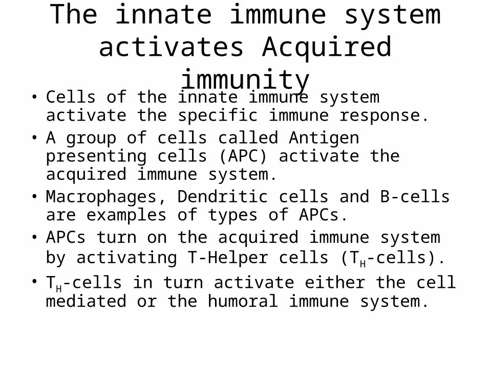
The innate immune system activates Acquired immunity
• Cells of the innate immune system activate the specific immune response.
• A group of cells called Antigen presenting cells (APC) activate the acquired immune system.
• Macrophages, Dendritic cells and B-cells are examples of types of APCs.
• APCs turn on the acquired immune system by activating T-Helper cells (TH-cells).
• TH-cells in turn activate either the cell mediated or the humoral immune system.
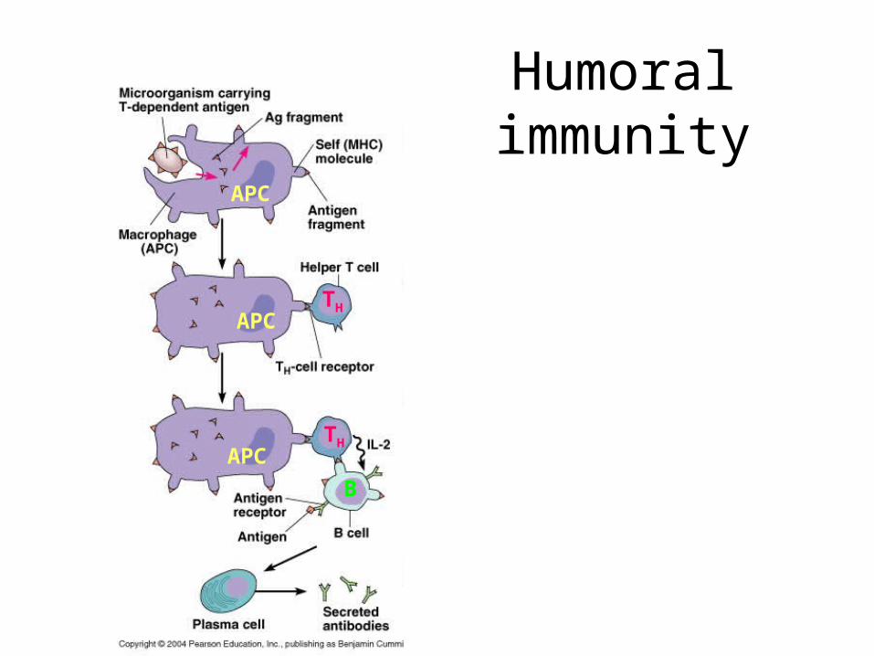
Humoral immunity
APC
APC
APC
TH
TH
B

APC
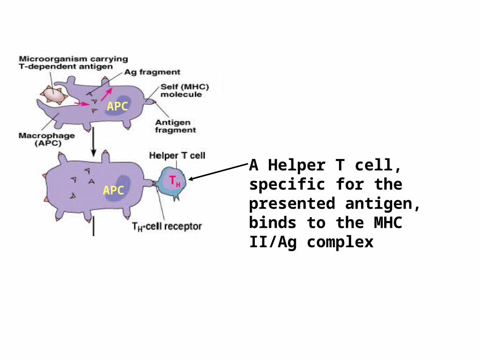
The Microbial antigen is ingested by an APC and partially digested. Fragments from microbe bind with the MHC II to form a MHC II /Ag complex on the surface of the APC
APC
APCTH
A Helper T cell, specific for the presented antigen, binds to the MHC II/Ag complex

APC
APCTH
APCTH
B
The helper T cell then activates an appropriate B cell by releasing IL-2 to it.
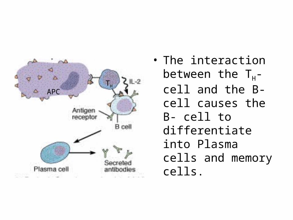
• The interaction between the TH-cell and the B-cell causes the B- cell to differentiate into Plasma cells and memory cells.
TH
APC
Memory cells
• Memory cells do not react right away but are held in reserve for later infections. The secondary response that is carried out by memory cells is different in 3 ways.
– Memory cells produce antibodies that bind with greater affinity to their antigens than the antibodies produced in the initial response.
– The response time is much vaster than the primary response
– A greater number of antibodies are produced.
Function of Antibodies

Function of Antibodies
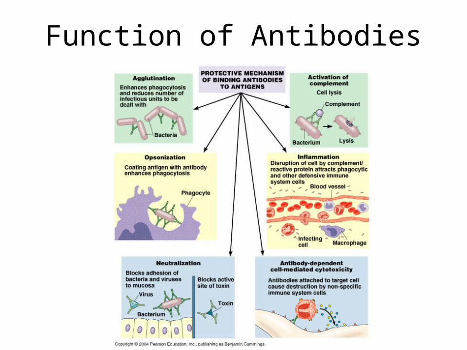
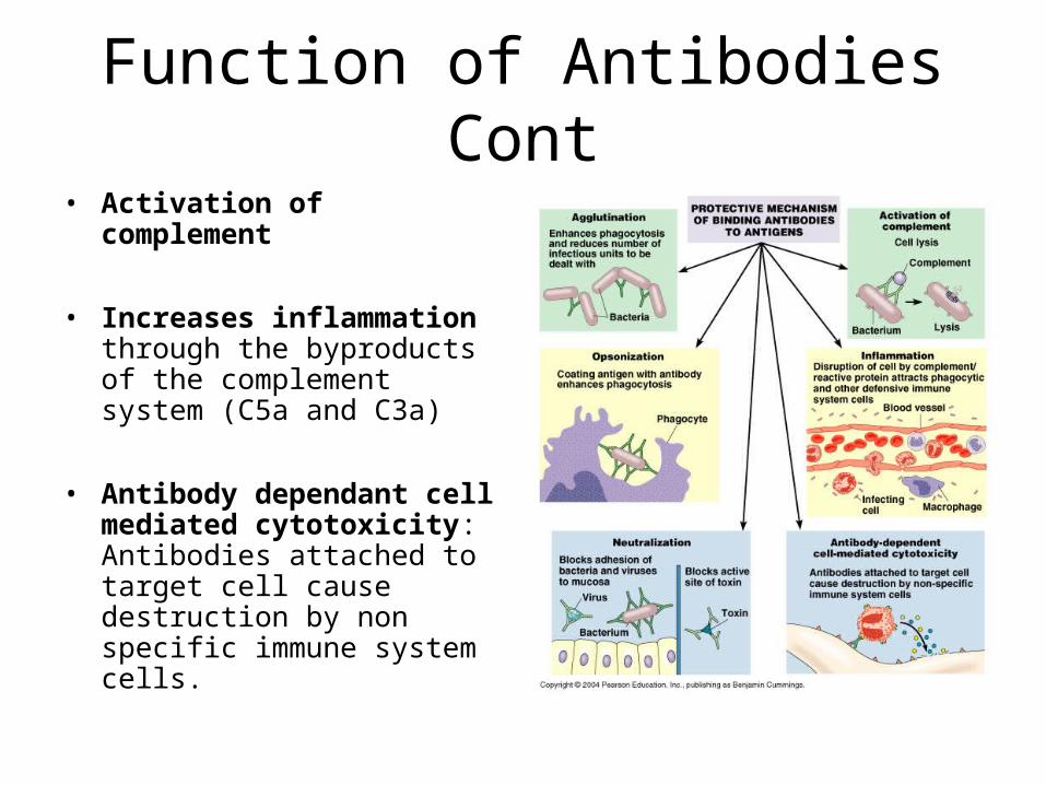
• Antibodies function in 6 ways to protect the body– Aggltination: Enhances
phagocytosis and reduces number of infectious units to be dealt with
– Opsonization: Coating antigen with antibody enhances phagocytosis
– Neutralization: blocks adhesion of bacteria and viruses to mucosa. Also blocks active site of toxin
Function of Antibodies Cont
• Activation of complement
• Increases inflammation through the byproducts of the complement system (C5a and C3a)
• Antibody dependant cell mediated cytotoxicity: Antibodies attached to target cell cause destruction by non specific immune system cells.
Structure of Antibodies
Structure of an Antibody
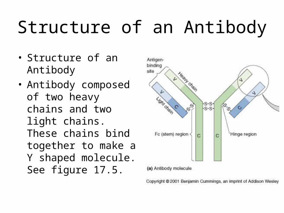
• Structure of an Antibody
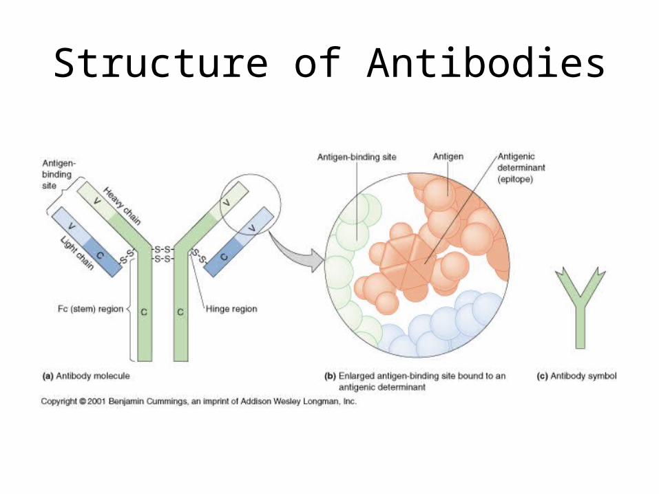
• Antibody composed of two heavy chains and two light chains. These chains bind together to make a Y shaped molecule. See figure 17.5.
Structure of Antibodies
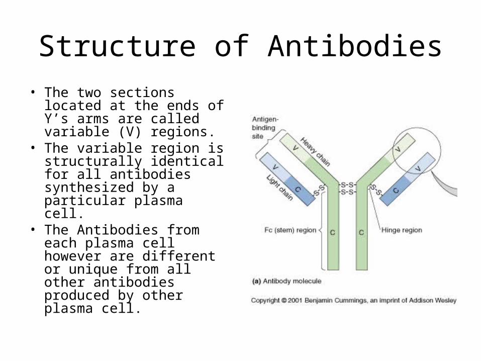
• The two sections located at the ends of Y’s arms are called variable (V) regions.
• The variable region is structurally identical for all antibodies synthesized by a particular plasma cell.
• The Antibodies from each plasma cell however are different or unique from all other antibodies produced by other plasma cell.
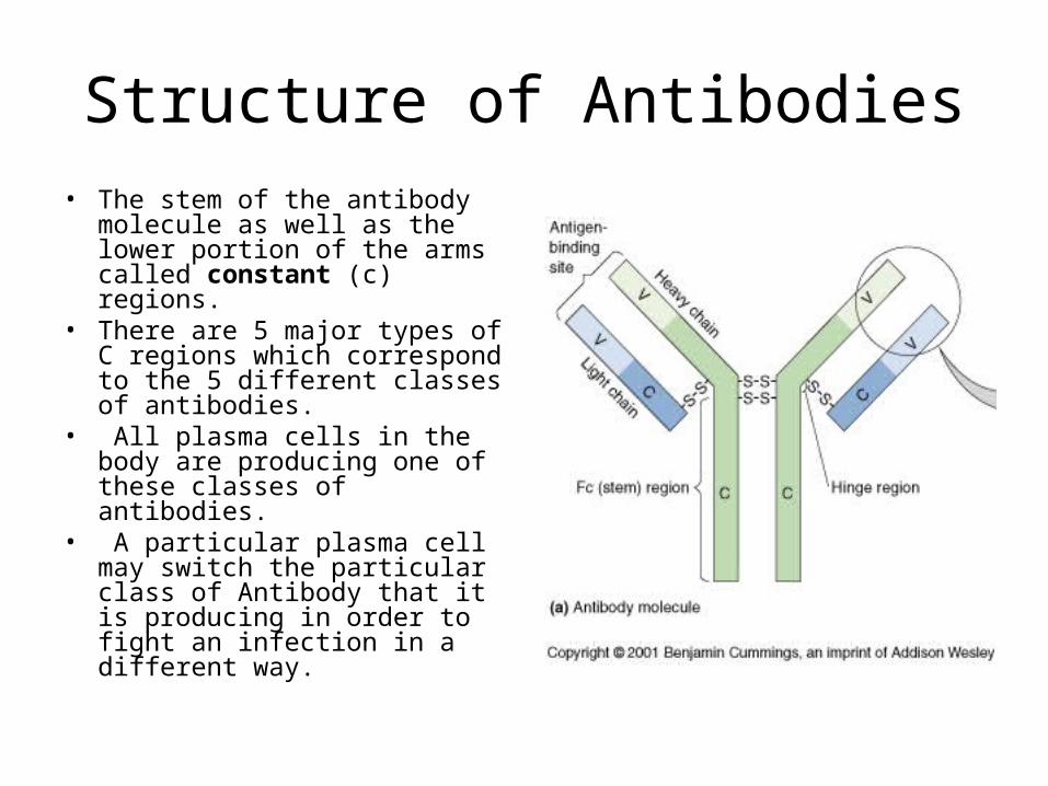
Structure of Antibodies• The stem of the antibody
molecule as well as the lower portion of the arms called constant (c) regions.
• There are 5 major types of C regions which correspond to the 5 different classes of antibodies.
• All plasma cells in the body are producing one of these classes of antibodies.
• A particular plasma cell may switch the particular class of Antibody that it is producing in order to fight an infection in a different way.

• The structure of Antibodies may be described by the way they are cut and digested by proteases.
• The stem portion is referred to as the FC region
• The Y portion with the top third of the stem is referred to as the Fab region.
• The FC region often acts as the receptor for phagocytes during opsonization or Antibody dependant cell mediated cytotoxicity.
• The FC region contains the antigen binding region

IgM• IgM expressed as membrane
bound anitbodies on B-cells • Pentamer
– 5 units held together by disulfide bonds
– J (Joining) chain functions in the polymerization of monomers
• First immunoglobulin class produced in a primary response to an antigen
• Has 10 anitgen binding sites• More effective at stimulating
complement• Large-size - does not diffuse well• The FC receptors on phagocytes
bind IgM (opsinization)

IgD
• Found on surface of mature B-cells.
• Biological function unknown (thought to function in activation of B-cells)

IgG
• Most abundant isotype in serum (80%)
• Cross placenta and play important role in protecting fetus– Provides passive immunity
to unborn fetus.– Placental cells bind the Fc
portion of IgG and transfer Ab across the placental membrane.
• Activate complement system
• Opsonin—phagocytosis

IgE
• Mediate the immediate hypersensitivity reactions (hayfever, asthma, hives, anaphylactic shock)– Mast cells and basophils
bind fc portion of IgE– Cross-linkage of receptor
bound IgE molecules by antigen, induces degranulaltion of the Mast and basophil cells
• Parasitic response– Eosinophils express
receptors for IgE
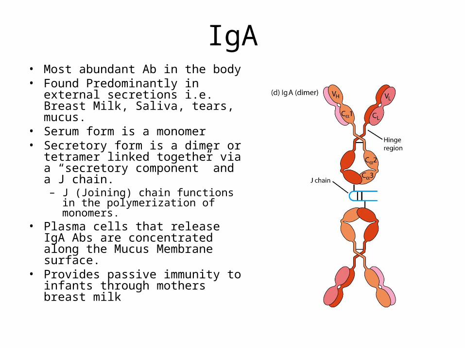
IgA• Most abundant Ab in the body• Found Predominantly in external
secretions i.e. Breast Milk, Saliva, tears, mucus.
• Serum form is a monomer• Secretory form is a dimer or
tetramer linked together via a “secretory component” and a J chain.– J (Joining) chain functions in the
polymerization of monomers.• Plasma cells that release IgA Abs
are concentrated along the Mucus Membrane surface.
• Provides passive immunity to infants through mothers breast milk