Embed Size (px)
Citation preview

GIT-2DR.DEEPAK N.KHEDEKAR
DEPARTMENT OF ANATOMYLTMMC
DEC- 2016

05/02/2023 DR.DEEPAK N.KHEDEKAR/LTMMC/2016
05/02/2023 DR.DEEPAK N.KHEDEKAR/LTMMC/2016
• Cardiac region (cardia)- part near the esophageal orifice, contains the cardiac glands
• Pyloric region (pylorus)- part proximal to the pyloric sphincter, which contains the pyloric glands
• Fundic region (fundus)- largest part , which is situated between the cardia and pylorus and contains the fundic or gastric glands
STOMACH-Divided histologically into three regions
05/02/2023 DR.DEEPAK N.KHEDEKAR/LTMMC/2016
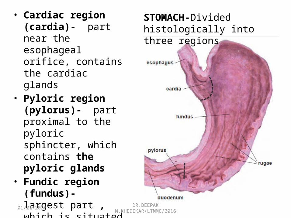
GASTRIC MUCOSARugae- • Longitudinal
submucosal foldsGastric pits, or
foveolae- • Numerous in the
mucosal surface. • Gastric glands open into
the bottom of the gastric pits.
05/02/2023 DR.DEEPAK N.KHEDEKAR/LTMMC/2016
GASTRIC MUCOSA
• Lining of the stomach-simple columnar E. • Columnar cells are -Surface mucous cellsSurface mucous cells :• Possesses a large, apical cup of mucinogen granules• Occupies most of the volume of the cell.• Appears empty in routine (H&E) sections because
the mucinogen is lost in fixation and dehydration. • When the mucinogen is preserved, the granules stain
intensely with toluidine blue and with the PAS

05/02/2023 DR.DEEPAK N.KHEDEKAR/LTMMC/2016
ESOPHAGOGASTRIC JUNCTION,• Stratified squamous epithelium of the esophagus ends abruptly, and the simple columnar epithelium of the stomach mucosa begins.
• Surface of the stomach show numerous and deep depressions
• Glands in the vicinity of the esophagus , the cardiac glands
• Extend from the bottom of these pits.
Fundic (gastric):• Glands arise at the base of the
gastric pits and are evident in the remaining part of the mucosa.
• Thick muscularis externa. .
05/02/2023 DR.DEEPAK N.KHEDEKAR/LTMMC/2016
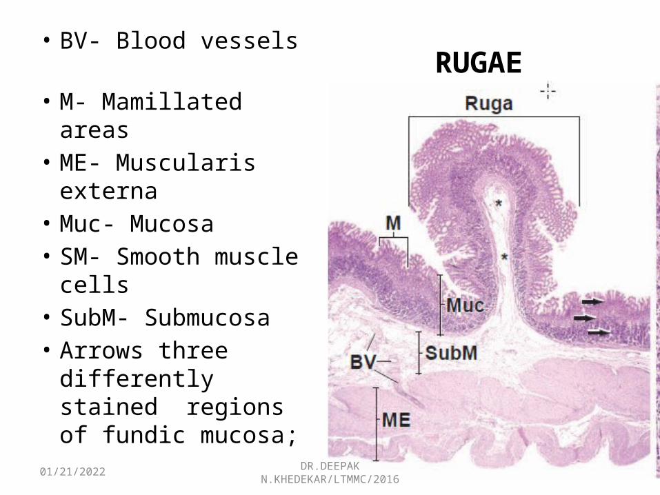
RUGAE• BV- Blood vessels • M- Mamillated areas • ME- Muscularis
externa • Muc- Mucosa• SM- Smooth muscle
cells • SubM- Submucosa • Arrows three
differently stained regions of fundic mucosa;

05/02/2023 DR.DEEPAK N.KHEDEKAR/LTMMC/2016
FUNDIC MUCOSA
• Alcian Blue/PAS.• One of the gastric pits
& Associated fundic gland
• Nucleus and Golgi apparatus are located below the mucous cup.
• Basal part of the cell contains small amounts of rER that impart a light basophilia to the cytoplasm.
05/02/2023 DR.DEEPAK N.KHEDEKAR/LTMMC/2016
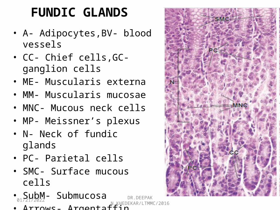
FUNDIC GLANDS• A- Adipocytes,BV- blood vessels• CC- Chief cells,GC- ganglion cells• ME- Muscularis externa• MM- Muscularis mucosae• MNC- Mucous neck cells• MP- Meissner’s plexus• N- Neck of fundic glands• PC- Parietal cells• SMC- Surface mucous cells• SubM- Submucosa• Arrows- Argentaffin cells• Arrowheads- nuclei of satellite
cells
05/02/2023 DR.DEEPAK N.KHEDEKAR/LTMMC/2016
FUNDIC GLANDS (GASTRIC GLANDS )
• Present throughout the entire gastric mucosa• Simple, branched, tubular glands • Extend from the bottom of the gastric pits to the
muscularis mucosae.• Has a narrow, long neck segment and a shorter and
wider base or fundic segment.
05/02/2023 DR.DEEPAK N.KHEDEKAR/LTMMC/2016
05/02/2023 DR.DEEPAK N.KHEDEKAR/LTMMC/2016
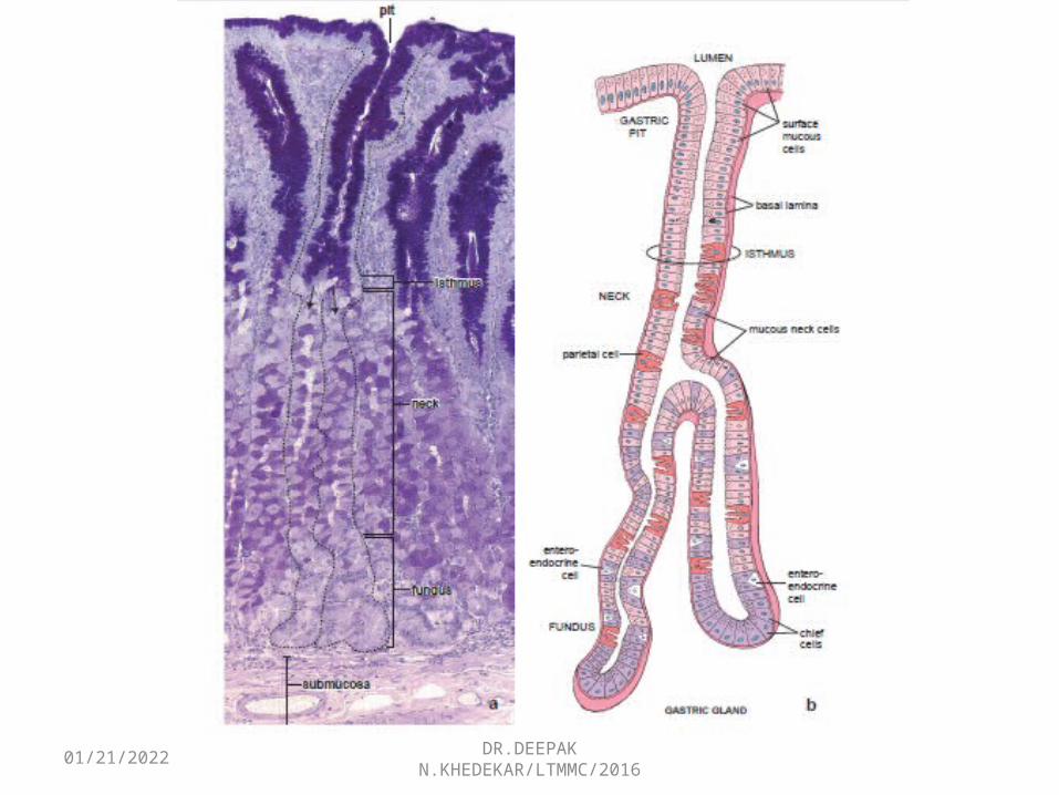
ISTHMUS OF THE FUNDIC GLAND - SITE OF STEM CELLS
• Short segment -Located between the gastric pit and the gland
• Cells destined to become mucous surface cells migrate upward in the gastric pits to surface.
• Other cells migrate downward, maintaining the population of the fundic gland epithelium.

05/02/2023 DR.DEEPAK N.KHEDEKAR/LTMMC/2016
FUNDIC GLANDS
Composed of four functionally differentcell types…
• Mucous neck cells• Chief cells• Parietal cells, also called oxyntic cells• Entero-endocrine cells• Undifferentiated adult stem cells
05/02/2023 DR.DEEPAK N.KHEDEKAR/LTMMC/2016
1.MUCOUS NECK CELLS
• Located in the neck region of the fundic gland. • Much shorter than the surface mucous cell and • Contains less mucinogen in the apical cytoplasm.• Do not exhibit a prominent mucous cup. • Nucleus tends to be spherical compared with the
more prominent, elongated nucleus of the surface mucous cell.
• Parietal cells are interspersed between these cells
05/02/2023 DR.DEEPAK N.KHEDEKAR/LTMMC/2016
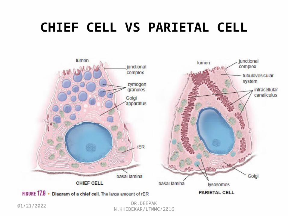
2.CHIEF CELLS
• Located in the deeper part of the fundic glands.• Typical protein-secreting. • Basal cytoplasm- basophilic due the abundant
rER , • Apical cytoplasm- eosinophilic due to presence
of the secretory vesicles, zymogen granules • Zymogen granules contain enzyme precursors. • Basophilia, in particular, allows easy
identification of these cells in H&E sections
05/02/2023 DR.DEEPAK N.KHEDEKAR/LTMMC/2016
3.PARIETAL CELLS (0XYNTIC CELLS)
• Found in the neck glands, among the mucous neck cells, and in the deeper part.
• More in no. in the upper & middle part. • Large cells, binucleate, and appear triangular in
sections, with the apex directed toward the lumen of the gland and the resting on the basal lamina.
• Nucleus-spherical• Cytoplasm- stains with eosin and other acidic dyes.
05/02/2023 DR.DEEPAK N.KHEDEKAR/LTMMC/2016
CHIEF CELL VS PARIETAL CELL
05/02/2023 DR.DEEPAK N.KHEDEKAR/LTMMC/2016
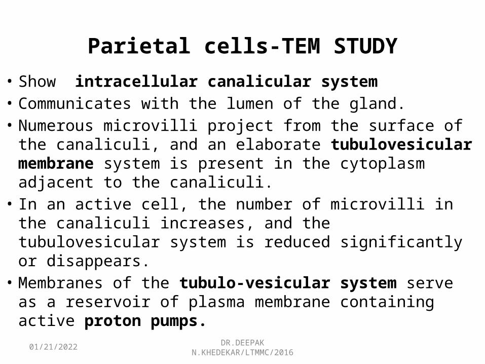
Parietal cells-TEM STUDY• Show intracellular canalicular system • Communicates with the lumen of the gland. • Numerous microvilli project from the surface of the canaliculi,
and an elaborate tubulovesicular membrane system is present in the cytoplasm adjacent to the canaliculi.
• In an active cell, the number of microvilli in the canaliculi increases, and the tubulovesicular system is reduced significantly or disappears.
• Membranes of the tubulo-vesicular system serve as a reservoir of plasma membrane containing active proton pumps.

05/02/2023 DR.DEEPAK N.KHEDEKAR/LTMMC/2016
TEM
05/02/2023 DR.DEEPAK N.KHEDEKAR/LTMMC/2016
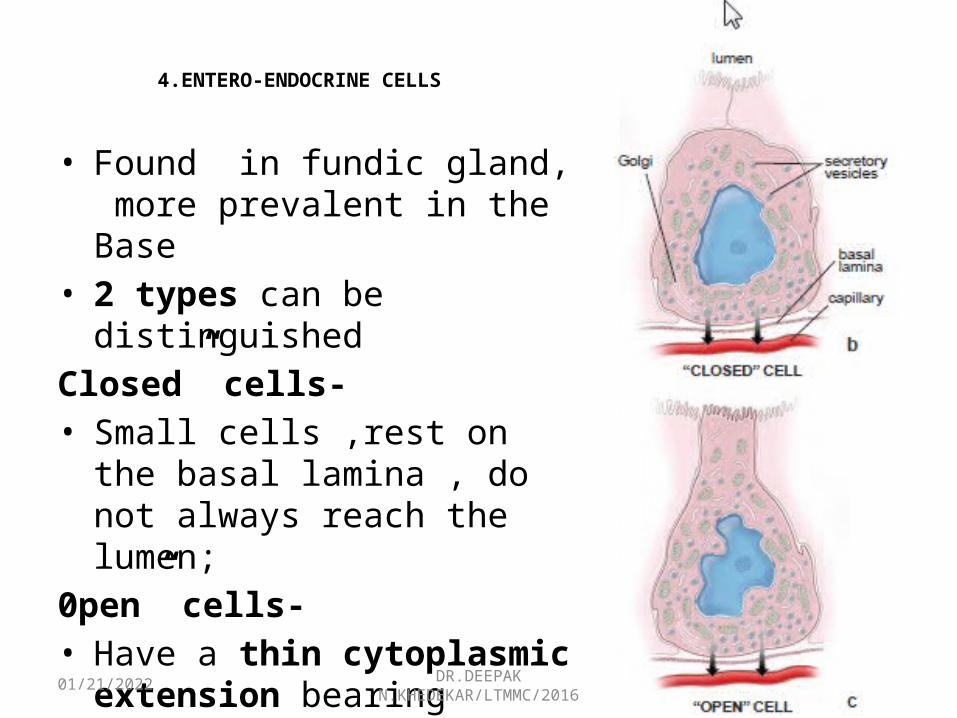
4.ENTERO-ENDOCRINE CELLS
• Found in fundic gland, more prevalent in the Base
• 2 types can be distinguishedClosed” cells- • Small cells ,rest on the basal
lamina , do not always reach the lumen;
0pen” cells-• Have a thin cytoplasmic extension
bearing microvilli that are exposed to the gland lumen
05/02/2023 DR.DEEPAK N.KHEDEKAR/LTMMC/2016
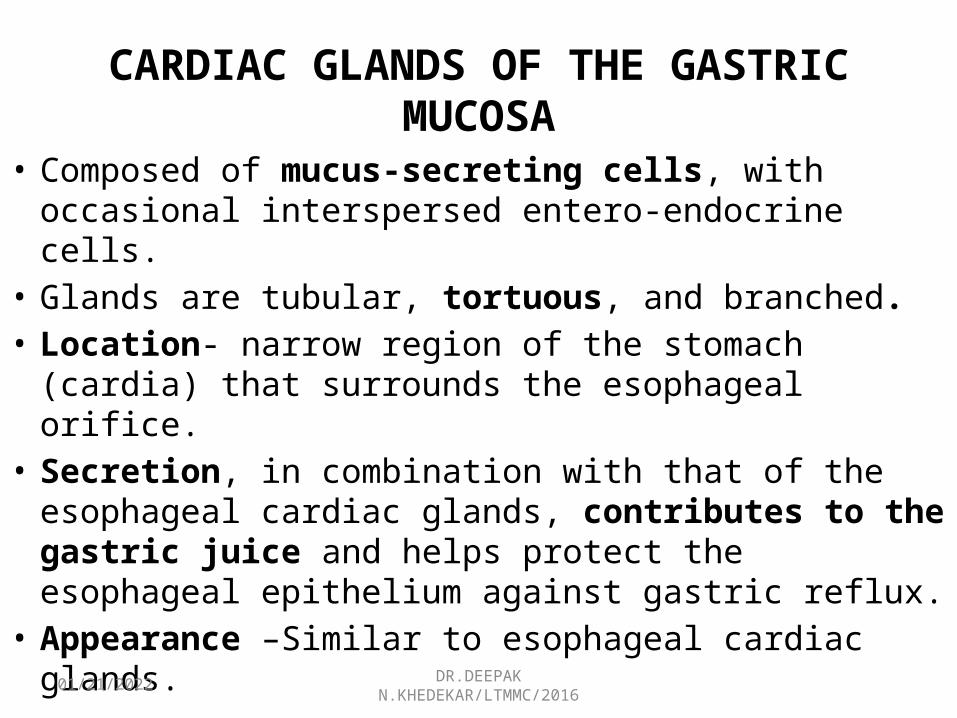
CARDIAC GLANDS OF THE GASTRIC MUCOSA
• Composed of mucus-secreting cells, with occasional interspersed entero-endocrine cells.
• Glands are tubular, tortuous, and branched.• Location- narrow region of the stomach (cardia) that
surrounds the esophageal orifice.• Secretion, in combination with that of the esophageal
cardiac glands, contributes to the gastric juice and helps protect the esophageal epithelium against gastric reflux.
• Appearance –Similar to esophageal cardiac glands.
05/02/2023 DR.DEEPAK N.KHEDEKAR/LTMMC/2016
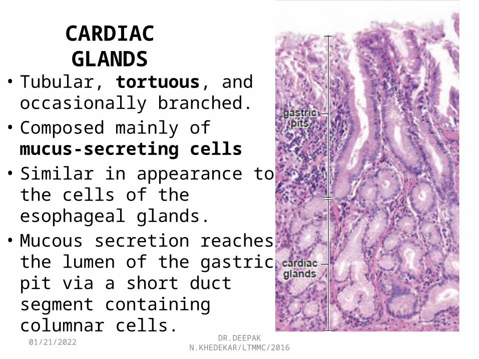
CARDIAC GLANDS
• Tubular, tortuous, and occasionally branched.
• Composed mainly of mucus-secreting cells
• Similar in appearance to the cells of the esophageal glands.
• Mucous secretion reaches the lumen of the gastric pit via a short duct segment containing columnar cells.
05/02/2023 DR.DEEPAK N.KHEDEKAR/LTMMC/2016
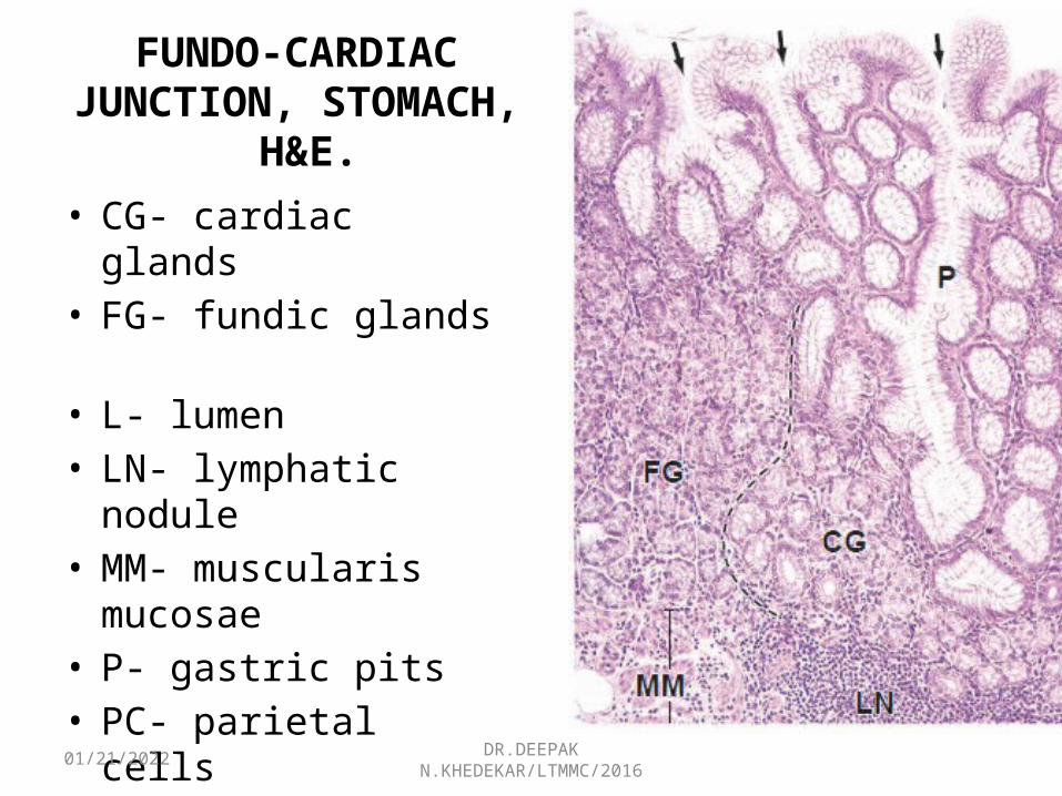
FUNDO-CARDIAC JUNCTION, STOMACH,
H&E.• CG- cardiac glands • FG- fundic glands • L- lumen • LN- lymphatic nodule• MM- muscularis
mucosae• P- gastric pits • PC- parietal cells
05/02/2023 DR.DEEPAK N.KHEDEKAR/LTMMC/2016
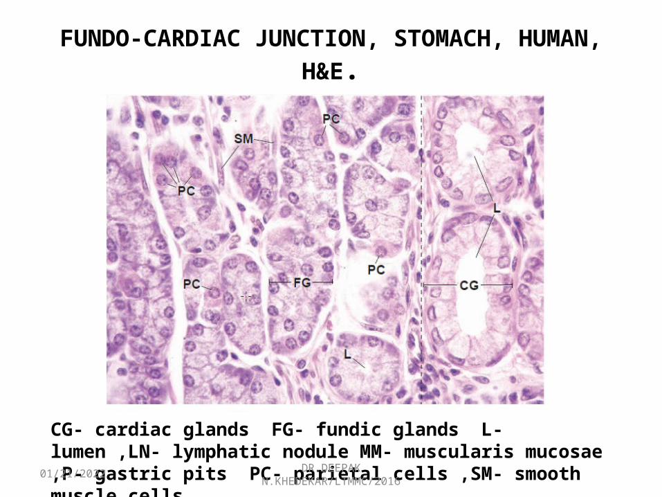
FUNDO-CARDIAC JUNCTION, STOMACH, HUMAN, H&E.
CG- cardiac glands FG- fundic glands L- lumen ,LN- lymphatic nodule MM- muscularis mucosae ,P- gastric pits PC- parietal cells ,SM- smooth muscle cells
05/02/2023 DR.DEEPAK N.KHEDEKAR/LTMMC/2016
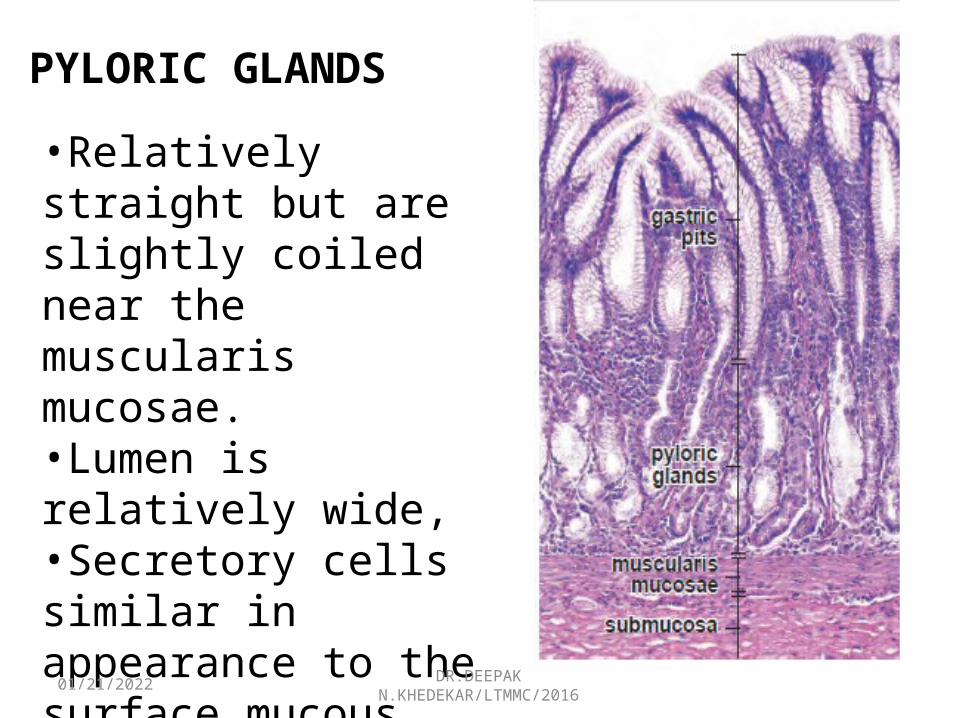
PYLORIC GLANDS
• Branched, coiled, tubular gland. • Location-Pyloric antrum • Lumen- wide • Appearance- cells similar to the surface mucous cells
(viscous secretion)• Enteroendocrine cells interspersed within the gland
epithelium along with occasional parietal cells. • Empty secretion into deep gastric pits• Gastric pits - Half the thickness of the mucosa.• Help protect pyloric mucosa.
05/02/2023 DR.DEEPAK N.KHEDEKAR/LTMMC/2016
PYLORIC GLANDS
•Relatively straight but are slightly coiled near the muscularis mucosae. •Lumen is relatively wide, •Secretory cells similar in appearance to the surface mucous cells. •Restricted to the mucosa and empty into the gastric pits.
05/02/2023 DR.DEEPAK N.KHEDEKAR/LTMMC/2016
LAMINA PROPRIA OF THE STOMACH• Relatively scant and restricted to the spaces
surrounding the gastric pits and glands. • Stroma - composed of reticular fibers with fibroblasts
and smooth muscle cells.• Other components- lymphocytes, plasma cells,
macrophages, and some eosinophils. • lymphatic nodules are also present, intruding into MM.
GASTRIC SUBMUCOSA• Composed of a dense connective tissue.• Contain adipose tissue,blood,nerve fibers and ganglion
cells that compose the submucosal (Meissner’s) plexus.

05/02/2023 DR.DEEPAK N.KHEDEKAR/LTMMC/2016
STOMACH -SUBMUCOSAA- adipocytesBV- blood vesselsCC- chief cellsGC- ganglion cellsME, muscularis externaMM- muscularis mucosaeMNC- mucous neck cellsMP- Meissner’s plexusN- neck of fundic glandsPC- parietal cellsSMC- surface mucous cellsSubM- submucosaarrows, argentaffin cellsarrowheads, nuclei of satellite cells

05/02/2023 DR.DEEPAK N.KHEDEKAR/LTMMC/2016
GASTRIC MUSCULARIS EXTERNA
• Outer longitudinal layer, a middle circular layer, and an inner oblique layer, Randomly oriented.
• longitudinal layer is absent from much of the anterior and posterior stomach surfaces
• Circular layer is poorly developed in the peri-esophageal region
• Myenteric (Auerbach’s) PLX- Ganglion cells and bundles of unmyelinated nerve fibers are present betwn the muscle layers.
SEROSA
05/02/2023 DR.DEEPAK N.KHEDEKAR/LTMMC/2016
SMALL INTESTINE
• Longest component of the GIT , measuring over 6 m, and is divided into three anatomic portions:
Duodenum (25 cm long) -first, shortest, and widest part of the small intestine. It begins at the pylorus of the stomach and ends at the duodenojejunal jn
Jejunum (2.5 m long)- begins at the duodenojejunal junction and constitutes the upper two fifths of the small intestine
Ileum (3.5 m long) is a continuation of the jejunum and constitutes the lower three fifths. It ends at the ileocecal junction.
05/02/2023 DR.DEEPAK N.KHEDEKAR/LTMMC/2016
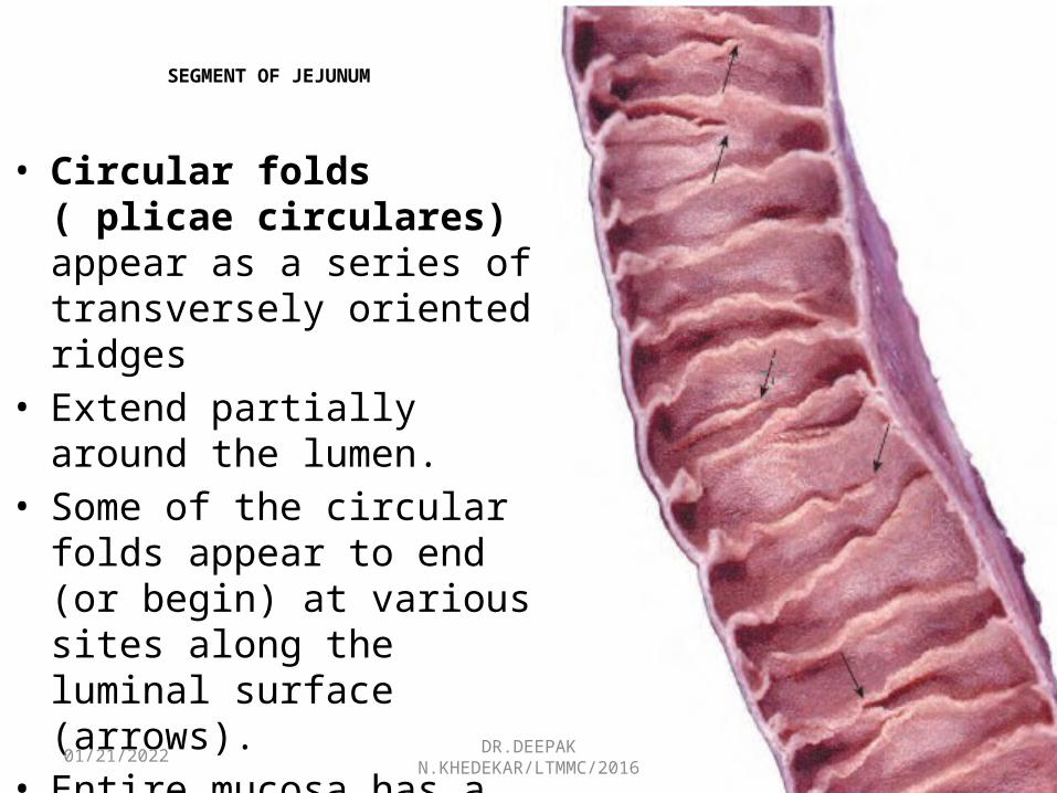
SEGMENT OF JEJUNUM
• Circular folds ( plicae circulares) appear as a series of transversely oriented ridges
• Extend partially around the lumen.
• Some of the circular folds appear to end (or begin) at various sites along the luminal surface (arrows).
• Entire mucosa has a velvety appearance because of the presence of villi.
05/02/2023 DR.DEEPAK N.KHEDEKAR/LTMMC/2016
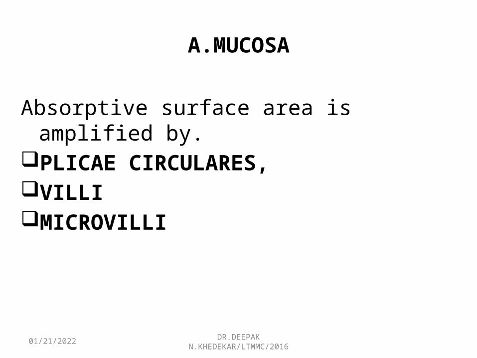
A.MUCOSA
Absorptive surface area is amplified by. PLICAE CIRCULARES, VILLIMICROVILLI
05/02/2023 DR.DEEPAK N.KHEDEKAR/LTMMC/2016
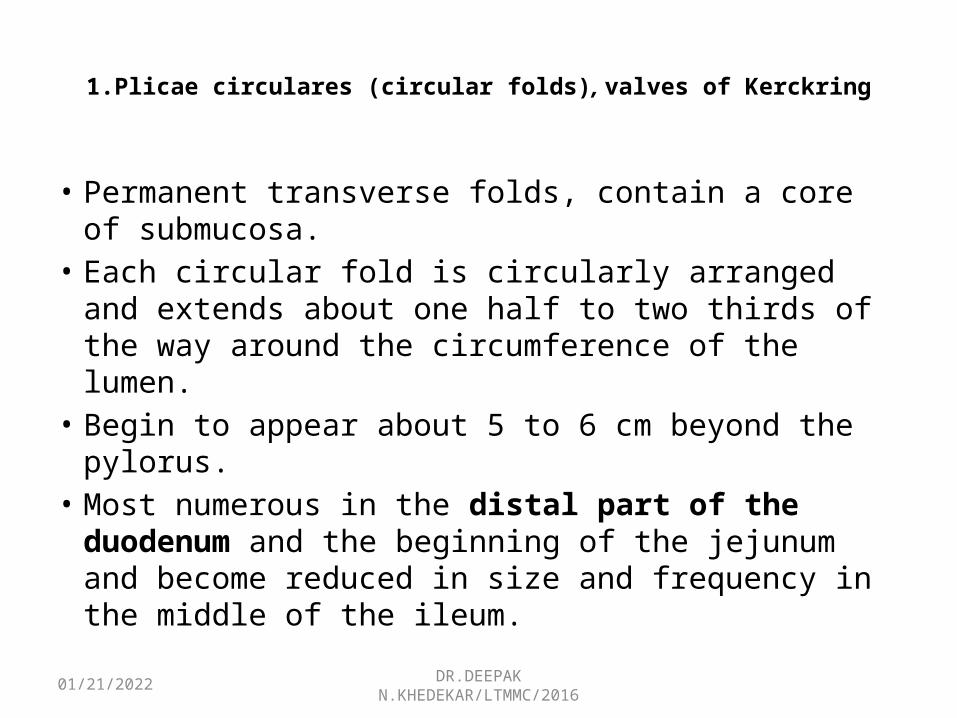
1.Plicae circulares (circular folds), valves of Kerckring
• Permanent transverse folds, contain a core of submucosa.
• Each circular fold is circularly arranged and extends about one half to two thirds of the way around the circumference of the lumen.
• Begin to appear about 5 to 6 cm beyond the pylorus. • Most numerous in the distal part of the duodenum
and the beginning of the jejunum and become reduced in size and frequency in the middle of the ileum.

05/02/2023 DR.DEEPAK N.KHEDEKAR/LTMMC/2016
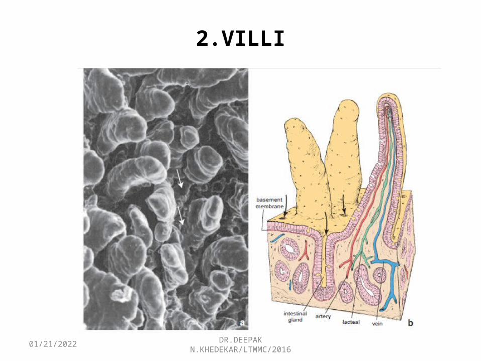
2.VILLI
• Unique, fingerlike and leaflike projections, Extend from the mucosal surface for 0.5 to 1.5 mm into the lumen
• Cover the surface , giving it a velvety appearance. • Consist of a core of loose CT covered by a Simple
columnar E.• Core of the villus - an extension of the lamina propria ,
which contains fibroblasts, smooth muscle cells, lymphocytes, plasma cells, eosinophils , macrophages, and a network of fenestrated blood capillaries and also lymphatic capillary, the lacteal.
05/02/2023 DR.DEEPAK N.KHEDEKAR/LTMMC/2016
2.VILLI
05/02/2023 DR.DEEPAK N.KHEDEKAR/LTMMC/2016
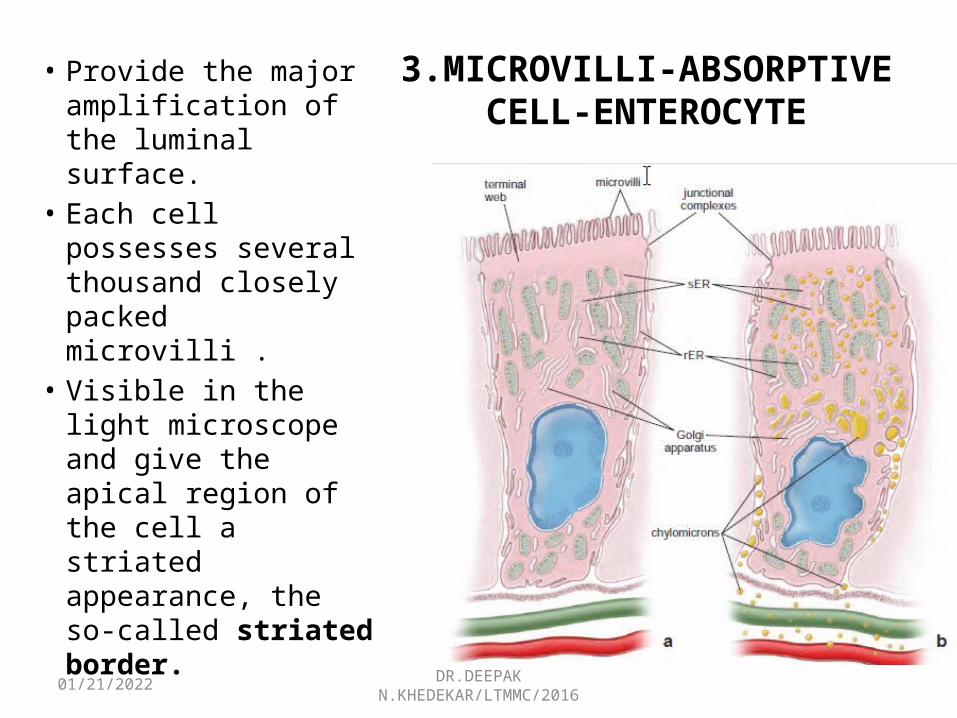
3.MICROVILLI-ABSORPTIVE CELL-ENTEROCYTE
• Provide the major amplification of the luminal surface.
• Each cell possesses several thousand closely packed microvilli .
• Visible in the light microscope and give the apical region of the cell a striated appearance, the so-called striated border.

05/02/2023 DR.DEEPAK N.KHEDEKAR/LTMMC/2016
A.1.LINING EPITHELIUM
• Simple Columnar EpitheliumINTESTINAL GLANDS
(CRYPTS OF LIEBERKÜHN)• Simple tubular structures • Extend from the MM through the thickness of
the lamina propria• Open onto the luminal surface of the intestine
at the base of the villi• Lined by simple columnar epithelium that is
continuous with the lining epithelium of the villi.

05/02/2023 DR.DEEPAK N.KHEDEKAR/LTMMC/2016
A.2.LAMINA PROPRIA
• Surrounds the intestinal glands• Contains numerous cells of the immune system
(lymphocytes, plasma cells, mast cells, macrophages, and eosinophils), particularly in the villi.
• Contains numerous nodules of lymphatic tissue that represent a major component of the GALT.
• Nodules are large and numerous in the ileum, where they are located on the side of the intestine opposite the mesenteric attachment .
• Nodular aggregations are known as aggregated nodules or Peyer’s patches.
05/02/2023 DR.DEEPAK N.KHEDEKAR/LTMMC/2016
A.3.MUSCULARIS MUCOSAE
• Consists of two thin layers of smooth muscle cells, an inner circular and an outer longitudinal layer.
• Strands of smooth muscle cells extend from the muscularis mucosae into the lamina Propria of the villi.
05/02/2023 DR.DEEPAK N.KHEDEKAR/LTMMC/2016
CELLS IN INTESTINAL MUCOSALEPITHELIUM.
They include the following:Enterocytes- whose primary function is absorptionGoblet cells- unicellular mucin-secreting glandsPaneth cells- whose primary function is to maintain
mucosal innate immunity by secreting antimicrobial Substances
Enteroendocrine cells- which produce various paracrine and endocrine hormones
M cells (microfold cells)- modified enterocytes that cover enlarged lymphatic nodules in the lamina propria
05/02/2023 DR.DEEPAK N.KHEDEKAR/LTMMC/2016
ENTEROCYTES
• Tall columnar cells with a basal Nucleus • Microvilli -increase the apical surface area as
much as 600 times;• form a striated border on the luminal surface• Lateral cell surface exhibits elaborate, flattened
cytoplasmic processes (plicae) that interdigitate with those of adjacent cells .
• Folds increase the lateral surface area of the cell, thus increasing the amount of plasma membrane containing transport enzymes
05/02/2023 DR.DEEPAK N.KHEDEKAR/LTMMC/2016
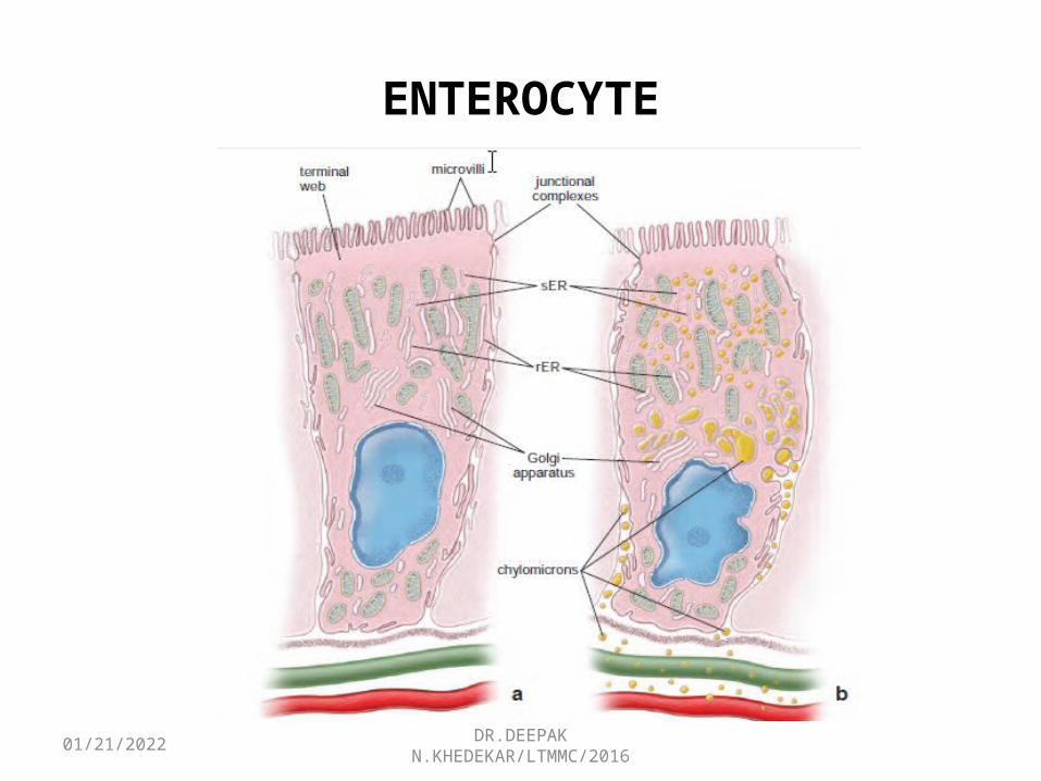
ENTEROCYTE
05/02/2023 DR.DEEPAK N.KHEDEKAR/LTMMC/2016
GOBLET CELLS
• Unicellular glands, produce mucus ,are interspersed among the other cells of the intestinal epithelium.
• Increase in number from the duodenum to the terminal part of the ileum.
• As water-soluble mucinogen is lost during preparation of routine H&E sections, the part of the cell that contains mucinogen granules appears empty.
• Examination with the TEM- reveals a large accumulation of mucinogen granules in the apical cytoplasm that distends the apex of the cell and distorts the shape of neighboring cells

05/02/2023 DR.DEEPAK N.KHEDEKAR/LTMMC/2016
GOBLET CELL• Basal portion of the cell contains the nucleus, rER, and mitochondria.
• Just apical to the nucleus are extensive profiles of Golgi apparatus. As the mucous product accumulates in the Golgi cisternae, they become enlarged.
• Mucous cup -Large mucinogen granules fill most of the apical portion of the cell.

05/02/2023 DR.DEEPAK N.KHEDEKAR/LTMMC/2016
PANETH CELLS
• Found in the bases of the intestinal glands• Occasionally found in the normal colon in small
numbers; No. increases in pathologic conditions. • Have a basophilic basal cytoplasm, a supranuclear
Golgi apparatus, and large, Intensely acidophilic, refractile apical secretory vesicles.
• Vesicles allow their easy identificationPaneth cells play a role in regulation of normal
bacterial flora of the small intestine.
05/02/2023 DR.DEEPAK N.KHEDEKAR/LTMMC/2016
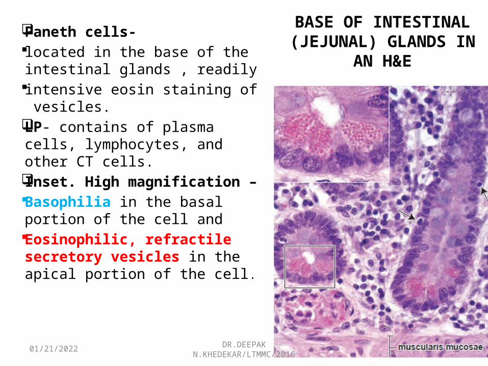
BASE OF INTESTINAL (JEJUNAL) GLANDS IN AN
H&E
Paneth cells- located in the base of the
intestinal glands , readily intensive eosin staining of
vesicles. LP- contains of plasma cells,
lymphocytes, and other CT cells.
Inset. High magnification – Basophilia in the basal portion
of the cell and Eosinophilic, refractile
secretory vesicles in the apical portion of the cell.
05/02/2023 DR.DEEPAK N.KHEDEKAR/LTMMC/2016
M CELLS
• Cells that overlie Peyer’s patches and other large lymphatic nodules;
• Differ significantly from the surrounding intestinal epithelial cells .
• Have microfolds rather than microvilli on their apical surface, and they take up Microorganisms and macromolecules from the lumen in endocytotic vesicles.
M cells convey microorganisms and other macromolecules from the intestinal lumen to Peyer’s
patches.

05/02/2023 DR.DEEPAK N.KHEDEKAR/LTMMC/2016
ENTERO-ENDOCRINE CELLS
• Resemble those that reside in the stomach . • “Closed cells”- concentrated in the lower portion of
the intestinal gland,whereas the • “Open cells” -found at all levels of each villus.• Activation of taste receptors -found on the apical
cell membrane of “open cells” activates G protein–signaling cascade, resulting in releasing of peptides that regulate a variety of gastrointestinal functions.
05/02/2023 DR.DEEPAK N.KHEDEKAR/LTMMC/2016
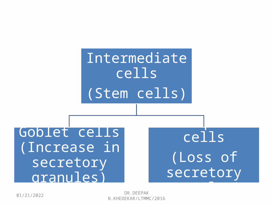
INTERMEDIATE CELLS• Constitute most of the cells found within the
intestinal stem cell niche• Located in the lower half of the intestinal gland. • Constitute the amplifying compartment of the cells• Capable of cell division and usually undergo one or
two divisions before they become committed to differentiation into either absorptive or goblet cells.
• Have short, irregular microvilli with long core filaments extending deep into the apical cytoplasm and numerous macular (desmosomal) junctions with adjacent cells.
05/02/2023 DR.DEEPAK N.KHEDEKAR/LTMMC/2016
Intermediate cells(Stem cells)
Goblet cells (Increase in
secretory granules)
Absorptive cells(Loss of secretory
granules)

05/02/2023 DR.DEEPAK N.KHEDEKAR/LTMMC/2016
GALT
• LP of the GI tract is heavily populated with elements of the immune system;
• Approximately one-fourth of the mucosa consists of a loosely organized layer of lymphatic nodules, lymphocytes, macrophages, plasma cells, and eosinophils in the lamina propria.
• Lymphocytes are also located between epithelial cells.
GALT serves as an immunologic barrier throughout the length of the gastrointestinal tract.
05/02/2023 DR.DEEPAK N.KHEDEKAR/LTMMC/2016
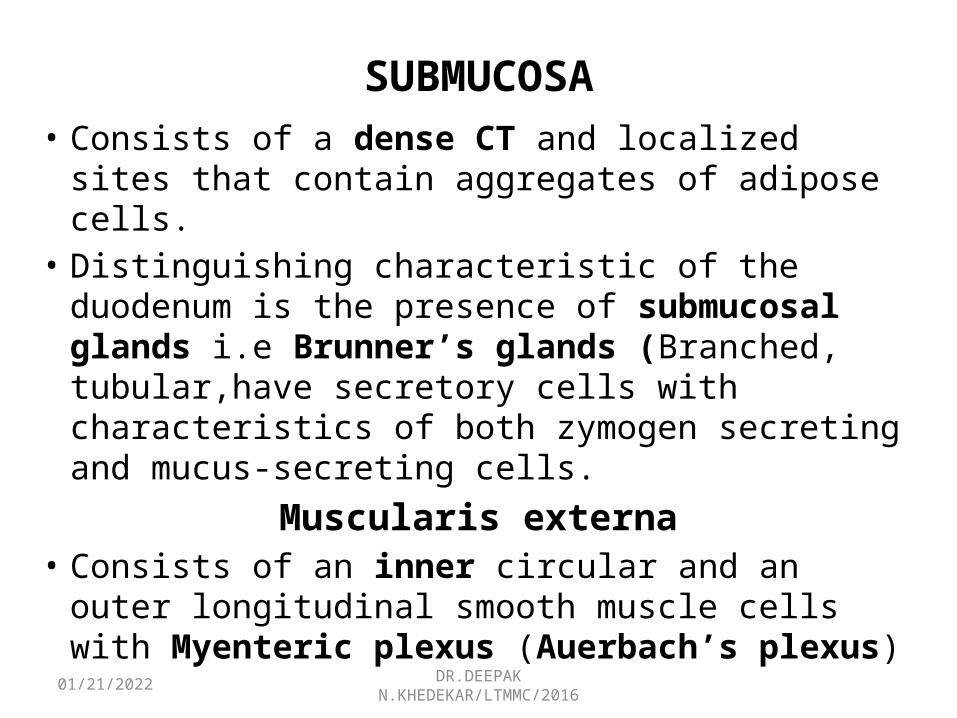
SUBMUCOSA• Consists of a dense CT and localized sites that
contain aggregates of adipose cells.• Distinguishing characteristic of the duodenum is the
presence of submucosal glands i.e Brunner’s glands (Branched, tubular,have secretory cells with characteristics of both zymogen secreting and mucus-secreting cells.
Muscularis externa• Consists of an inner circular and an outer
longitudinal smooth muscle cells with Myenteric plexus (Auerbach’s plexus)
05/02/2023 DR.DEEPAK N.KHEDEKAR/LTMMC/2016
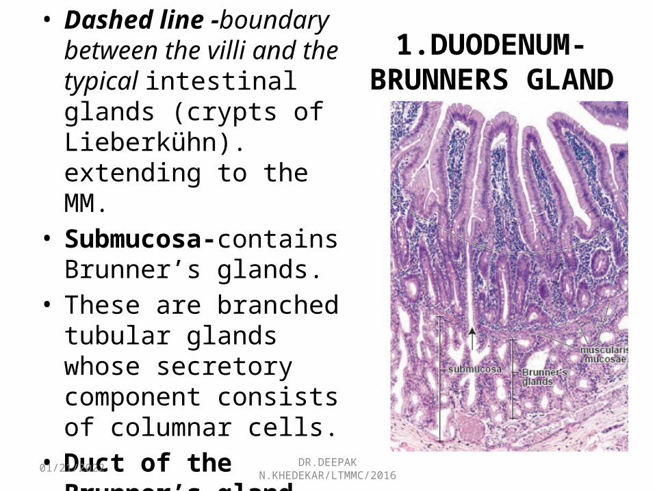
1.DUODENUM-BRUNNERS GLAND
• Dashed line -boundary between the villi and the typical intestinal glands (crypts of Lieberkühn). extending to the MM.
• Submucosa-contains Brunner’s glands.
• These are branched tubular glands whose secretory component consists of columnar cells.
• Duct of the Brunner’s gland -opens into the lumen of the intestinal gland (arrow). 120.
05/02/2023 DR.DEEPAK N.KHEDEKAR/LTMMC/2016
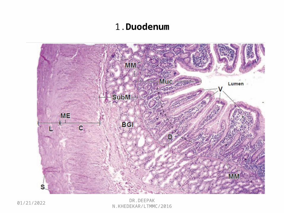
1.Duodenum
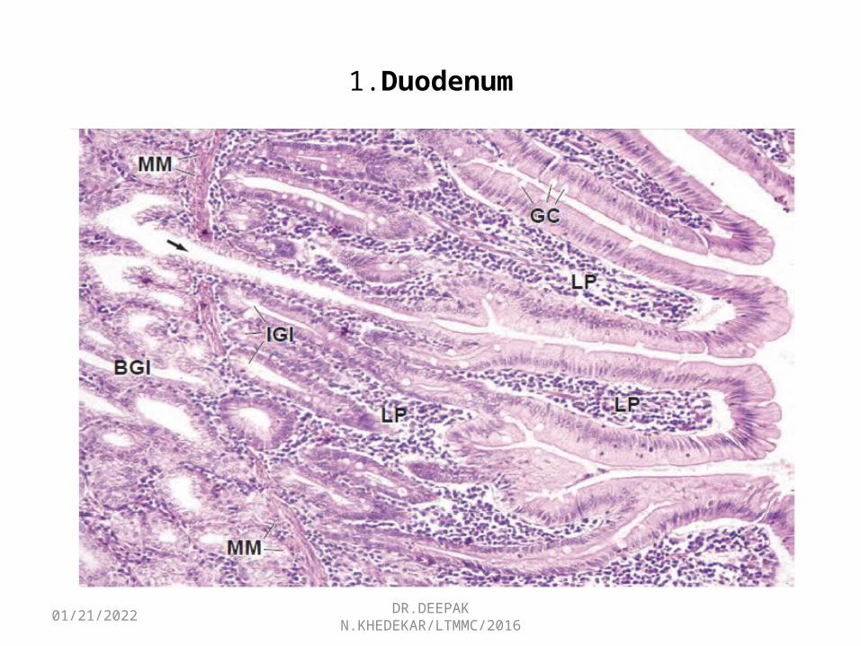
05/02/2023 DR.DEEPAK N.KHEDEKAR/LTMMC/2016
1.Duodenum
05/02/2023 DR.DEEPAK N.KHEDEKAR/LTMMC/2016
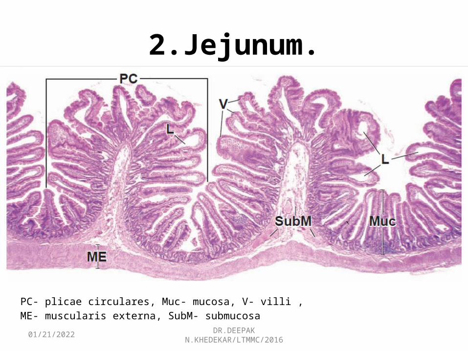
2.Jejunum.
PC- plicae circulares, Muc- mucosa, V- villi ,ME- muscularis externa, SubM- submucosa
05/02/2023 DR.DEEPAK N.KHEDEKAR/LTMMC/2016
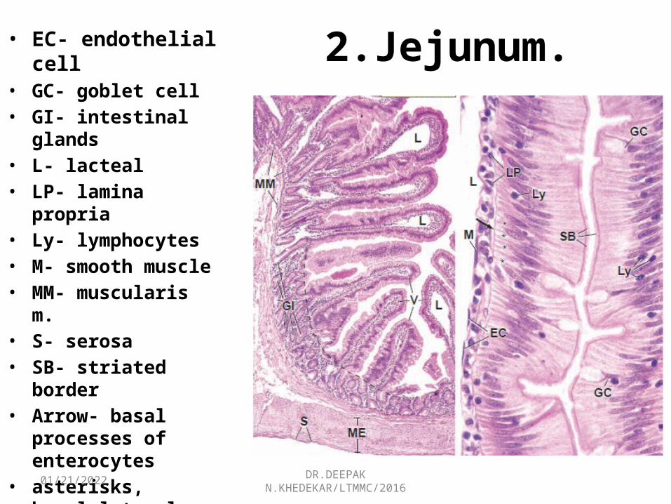
2.Jejunum.• EC- endothelial cell• GC- goblet cell• GI- intestinal glands• L- lacteal• LP- lamina propria• Ly- lymphocytes• M- smooth muscle• MM- muscularis m.• S- serosa• SB- striated border• Arrow- basal processes
of enterocytes• asterisks, basal-lateral
intercellular spaces• dashed line-boundary
between villi and intestinal glands
05/02/2023 DR.DEEPAK N.KHEDEKAR/LTMMC/2016
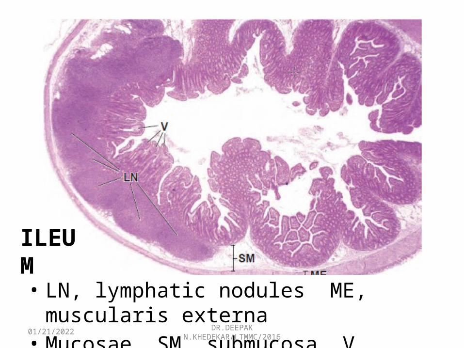
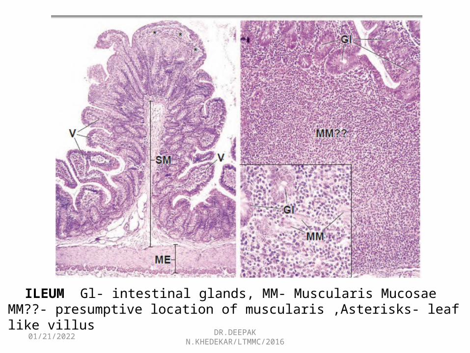
• LN, lymphatic nodules ME, muscularis externa• Mucosae SM, submucosa V, villi
ILEUM
05/02/2023 DR.DEEPAK N.KHEDEKAR/LTMMC/2016
ILEUM Gl- intestinal glands, MM- Muscularis Mucosae MM??- presumptive location of muscularis ,Asterisks- leaf like villus
05/02/2023 DR.DEEPAK N.KHEDEKAR/LTMMC/2016
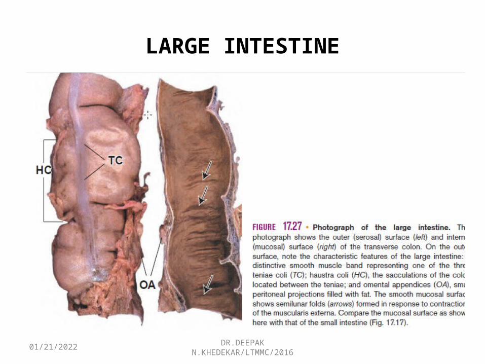
LARGE INTESTINE

05/02/2023 DR.DEEPAK N.KHEDEKAR/LTMMC/2016
COLON
BV- blood vessels , ME- muscularis externa ME(c), circular layer of muscularis externa ME(l)- longitudinal layer of muscularis externa Muc, mucosa S, serosa SubM, submucosa TC, tenia
05/02/2023 DR.DEEPAK N.KHEDEKAR/LTMMC/2016
Mucosa• Smooth surface, Neither plicae circulares nor villi are
present.• Contains numerous straight tubular intestinal glands• Glands extend through the full thickness of the mucosa • Glands consist of simple columnar epithelium, as does the
intestinal surface from which they invaginate. • O/E of luminal surface at the microscopic level reveals the
openings of the glands, which are arranged in an orderly pattern
• Morphology of absorptive cells is essentially identical to that of the Enterocytes of the small intestine

05/02/2023 DR.DEEPAK N.KHEDEKAR/LTMMC/2016
The mucosal epithelium of the large intestine contains the same cell types as the small intestine except Paneth cells, which are
normally absent in humans• Columnar absorptive cells predominate (4:1)
over goblet cells

05/02/2023 DR.DEEPAK N.KHEDEKAR/LTMMC/2016
05/02/2023 DR.DEEPAK N.KHEDEKAR/LTMMC/2016
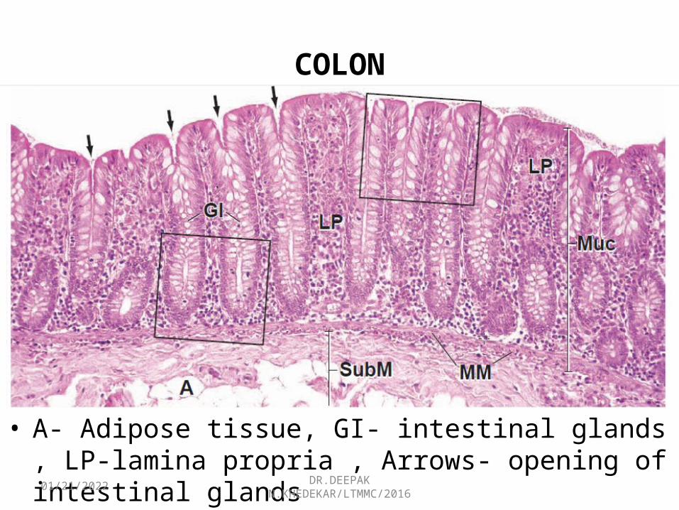
COLON
• A- Adipose tissue, GI- intestinal glands , LP-lamina propria , Arrows- opening of intestinal glands
05/02/2023 DR.DEEPAK N.KHEDEKAR/LTMMC/2016
LAMINA PROPRIA
• 1.Collagen table- thick layer of collagen and proteoglycans lies between the basal lamina of the epithelium and that of the fenestrated absorptive venous capillaries.
• 5 m thick in the normal human colon.• participates in regulation of water and
electrolyte• transport from the intercellular compartment of
the epithelium to the vascular compartment
05/02/2023 DR.DEEPAK N.KHEDEKAR/LTMMC/2016
LAMINA PROPRIA
Pericryptal fibroblast sheath- • constitutes a well developed fibroblast population
of regularly replicating cells. • Divide immediately beneath the base of the
intestinal gland, adjacent to the stem cells. • fibroblasts then differentiate and migrate upward
in parallel and synchrony with the epithelial cells. GALT LYMPHATIC VESSELS
05/02/2023 DR.DEEPAK N.KHEDEKAR/LTMMC/2016
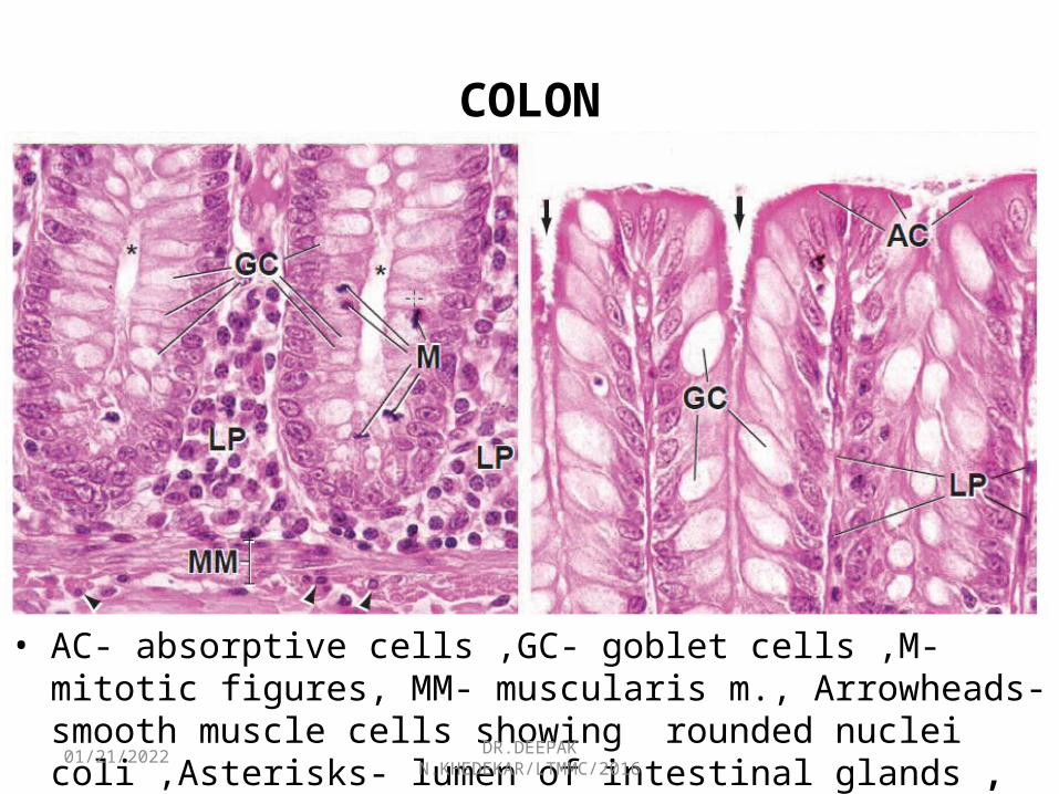
COLON
• AC- absorptive cells ,GC- goblet cells ,M-mitotic figures, MM- muscularis m., Arrowheads- smooth muscle cells showing rounded nuclei coli ,Asterisks- lumen of intestinal glands ,
05/02/2023 DR.DEEPAK N.KHEDEKAR/LTMMC/2016
MUSCULARIS EXTERNA• Outer layer, condensed into prominent
longitudinal bands of muscle, called teniae coli,• Inner, circular layer of muscle at irregular
intervals along the length and circumference of the colon.
SUBMUCOSA AND SEROSA

05/02/2023 DR.DEEPAK N.KHEDEKAR/LTMMC/2016
VERMIFORM APPENDIX
05/02/2023 DR.DEEPAK N.KHEDEKAR/LTMMC/2016
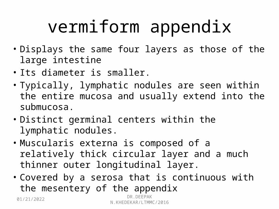
vermiform appendix• Displays the same four layers as those of the large
intestine • Its diameter is smaller.• Typically, lymphatic nodules are seen within the entire
mucosa and usually extend into the submucosa. • Distinct germinal centers within the lymphatic nodules. • Muscularis externa is composed of a relatively thick
circular layer and a much thinner outer longitudinal layer. • Covered by a serosa that is continuous with the
mesentery of the appendix

05/02/2023 DR.DEEPAK N.KHEDEKAR/LTMMC/2016
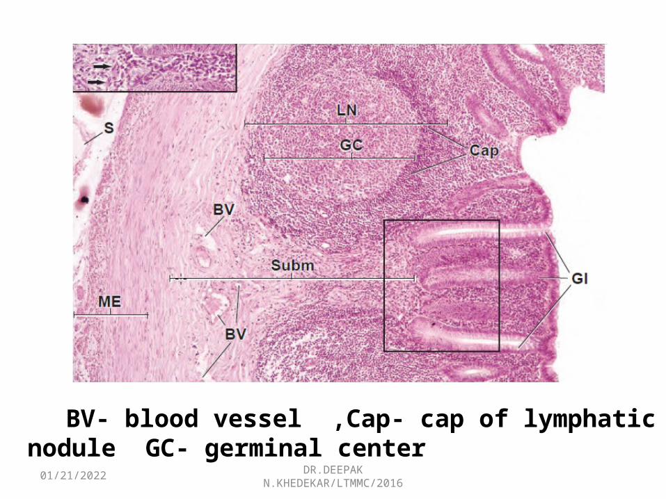
Cl- gland, L- lumen , LN- lymphatic nodule, ME- muscularis externa, Muc- mucosa S- serosa, Subm- submucosa
05/02/2023 DR.DEEPAK N.KHEDEKAR/LTMMC/2016
BV- blood vessel ,Cap- cap of lymphatic nodule GC- germinal center
05/02/2023 DR.DEEPAK N.KHEDEKAR/LTMMC/2016
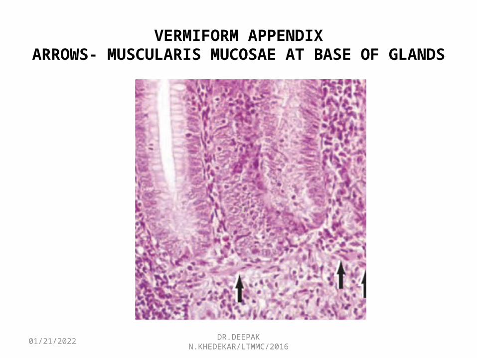
VERMIFORM APPENDIXARROWS- MUSCULARIS MUCOSAE AT BASE OF GLANDS

05/02/2023 DR.DEEPAK N.KHEDEKAR/LTMMC/2016
THE END