Embed Size (px)
Citation preview

HemangiosarcomaHemangiosarcoma
Brian Daubs, DVM, DACVS-SABrian Daubs, DVM, DACVS-SA
Animal Specialty and Emergency Animal Specialty and Emergency Hospital, Rockledge Fl.Hospital, Rockledge Fl.
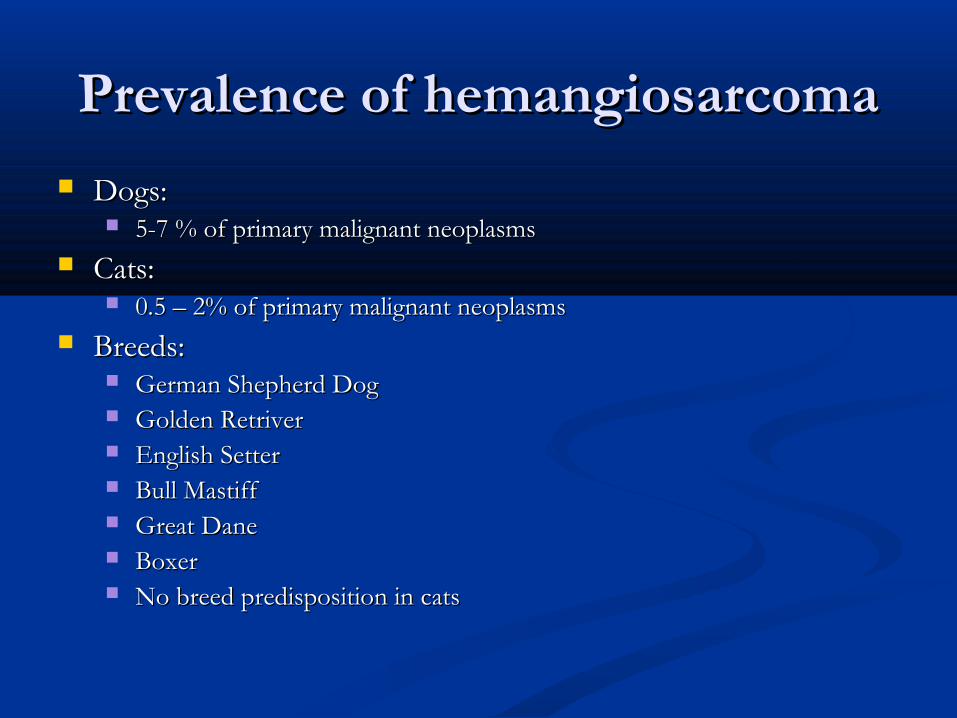
Prevalence of hemangiosarcomaPrevalence of hemangiosarcoma
Dogs:Dogs: 5-7 % of primary malignant neoplasms5-7 % of primary malignant neoplasms
Cats:Cats: 0.5 – 2% of primary malignant neoplasms0.5 – 2% of primary malignant neoplasms
Breeds:Breeds: German Shepherd DogGerman Shepherd Dog Golden RetriverGolden Retriver English SetterEnglish Setter Bull MastiffBull Mastiff Great DaneGreat Dane BoxerBoxer No breed predisposition in catsNo breed predisposition in cats
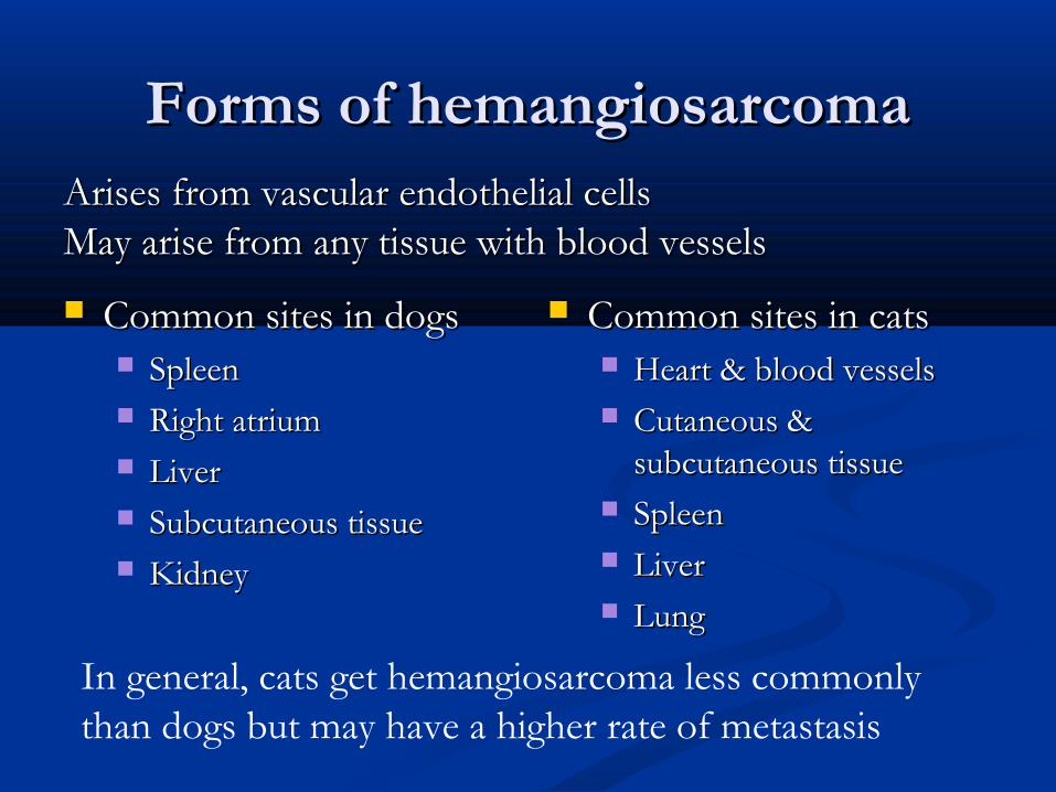
Forms of hemangiosarcomaForms of hemangiosarcoma
Common sites in dogsCommon sites in dogs SpleenSpleen Right atriumRight atrium LiverLiver Subcutaneous tissueSubcutaneous tissue KidneyKidney
Common sites in catsCommon sites in cats Heart & blood vesselsHeart & blood vessels Cutaneous & Cutaneous &
subcutaneous tissuesubcutaneous tissue SpleenSpleen LiverLiver LungLung
Arises from vascular endothelial cellsArises from vascular endothelial cellsMay arise from any tissue with blood vesselsMay arise from any tissue with blood vessels
In general, cats get hemangiosarcoma less commonly than dogs but may have a higher rate of metastasis
No known cause of hemangiosarcomaNo known cause of hemangiosarcoma Hemangiosarcoma is the diagnosis in 25-50% of Hemangiosarcoma is the diagnosis in 25-50% of
all splenic masses in dogsall splenic masses in dogs 25% of hemangiosarcoma cases in dogs also 25% of hemangiosarcoma cases in dogs also
have right atrium involvementhave right atrium involvement

Age of onsetAge of onset
Dog:Dog: 8 – 13 years8 – 13 years May be more common in male dogsMay be more common in male dogs
Cat:Cat: 8-10.5 years8-10.5 years No sex predilictionNo sex prediliction
Clinical signsClinical signs
Weakness and acute collapse most common sign Weakness and acute collapse most common sign in the dogin the dog Result of tumor rupture and acute intra-abdominal Result of tumor rupture and acute intra-abdominal
hemorrhagehemorrhage
Lethargy, anorexia, vomiting and abdominal Lethargy, anorexia, vomiting and abdominal distention most common in catsdistention most common in cats
Ascities may also be seenAscities may also be seen

TreatmentTreatment
Cutaneous Cutaneous Surgery plus radiation Surgery plus radiation
therapy preferredtherapy preferred Good prognosis with one Good prognosis with one
discrete, easily resectable discrete, easily resectable massmass
Dogs: 780 daysDogs: 780 days Cats: information Cats: information
unavailableunavailable
Non-cutaneousNon-cutaneous Surgery (often Surgery (often
splenectomy)splenectomy) ChemotherapyChemotherapy Many protocols in useMany protocols in use
VincristineVincristine AdriamycinAdriamycin CyclophosphamideCyclophosphamide

Survival timesSurvival times
DogsDogs Surgery alone Surgery alone
(splenectomy)(splenectomy) Survival times ranging Survival times ranging
from 19 days to 86 days from 19 days to 86 days depending on studydepending on study
Summarized median Summarized median survival time: 3 monthssurvival time: 3 months
Splenectomy and Splenectomy and chemotherpychemotherpy
91-492 days depending on 91-492 days depending on studystudy
Summarized median Summarized median survival time: 6 monthssurvival time: 6 months
CatsCats Surgery alone Surgery alone
(abdominal):(abdominal): 20 weeks20 weeks
MatildaMatilda
9 year old spayed female domestic short hair cat9 year old spayed female domestic short hair cat 6 month history of weight loss6 month history of weight loss 3 day history of lethargy and inappetance3 day history of lethargy and inappetance Bradycardic and hypothermic on presentationBradycardic and hypothermic on presentation Hemoabdomen and PCV of 12% Hemoabdomen and PCV of 12%
Hemothorax and hemoabomenHemothorax and hemoabomen Lungs:Lungs:
Multiple dark red, firm nodulesMultiple dark red, firm nodules 1-3 mm in diameter1-3 mm in diameter Covered > 50% of the serosal surfaceCovered > 50% of the serosal surface Serosanguinous fluid and cream colored foam in the Serosanguinous fluid and cream colored foam in the
distal trachea and lungsdistal trachea and lungs

Lung HistopathologyLung Histopathology
Focal area of hemorrhage in lung parenchymaFocal area of hemorrhage in lung parenchyma Some neutrophils and macrophages scattered Some neutrophils and macrophages scattered
within the hemorrhagewithin the hemorrhage Some hemosiderophages around the Some hemosiderophages around the
hemorrhagic areahemorrhagic area
Cardiovascular:Cardiovascular: Mediastinal hemorrhageMediastinal hemorrhage

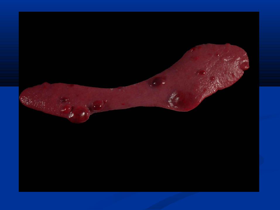
Spleen:Spleen: Dark red nodules ranging from 3-10 mm in diameterDark red nodules ranging from 3-10 mm in diameter Congested with blood on cut surfaceCongested with blood on cut surface
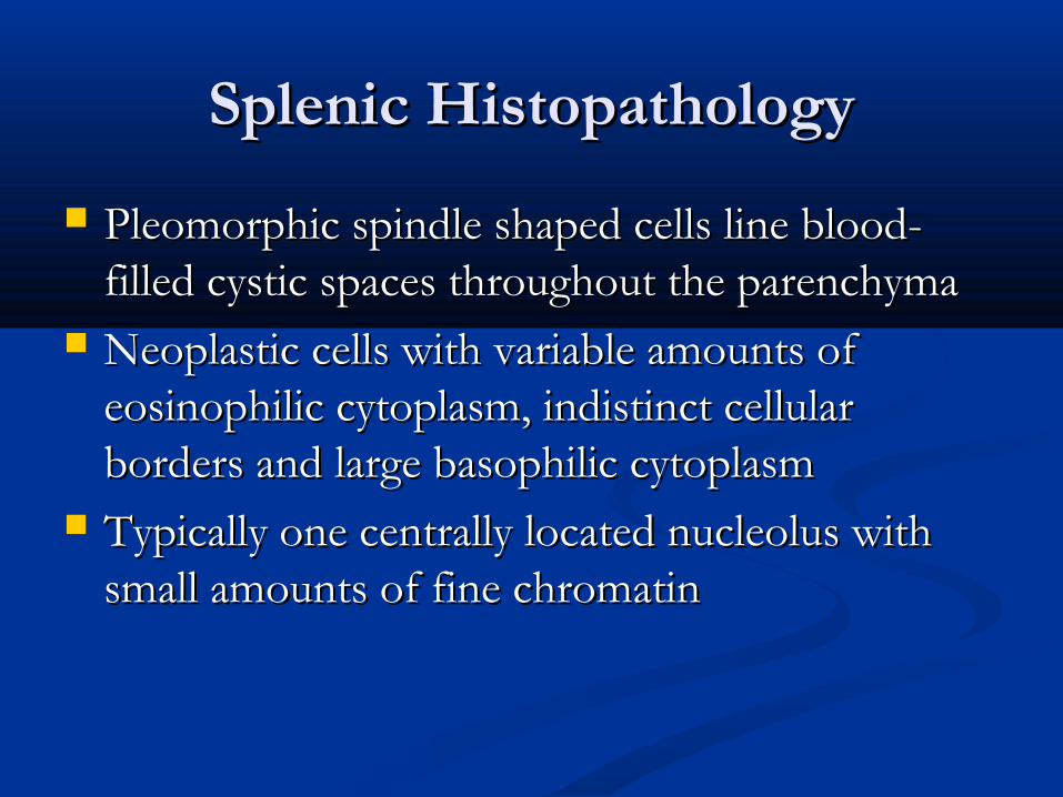
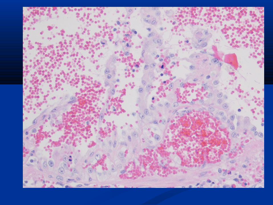
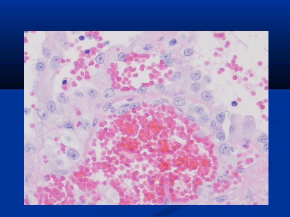
Splenic HistopathologySplenic Histopathology
Pleomorphic spindle shaped cells line blood-Pleomorphic spindle shaped cells line blood-filled cystic spaces throughout the parenchymafilled cystic spaces throughout the parenchyma
Neoplastic cells with variable amounts of Neoplastic cells with variable amounts of eosinophilic cytoplasm, indistinct cellular eosinophilic cytoplasm, indistinct cellular borders and large basophilic cytoplasmborders and large basophilic cytoplasm
Typically one centrally located nucleolus with Typically one centrally located nucleolus with small amounts of fine chromatinsmall amounts of fine chromatin

LiverLiver
Left lateral liver lobe was covered with diffuse, dark Left lateral liver lobe was covered with diffuse, dark red nodules over > 80% of the serosal surface.red nodules over > 80% of the serosal surface.
Pale pink area on the dorsal surface of the left lateral Pale pink area on the dorsal surface of the left lateral liver lobe measuring 6.4 x 3.0 cm.liver lobe measuring 6.4 x 3.0 cm.
Large nodule on ventral surface of the left middle Large nodule on ventral surface of the left middle liver lobe measuring 1.4 cm in diameterliver lobe measuring 1.4 cm in diameter
Multiple other small nodules throughout the liverMultiple other small nodules throughout the liver
Liver HistopathologyLiver Histopathology
A large cystic structure consisting of many A large cystic structure consisting of many neutrophils, fibrin, edema fluid and extravasated neutrophils, fibrin, edema fluid and extravasated red blood cellsred blood cells
Multifocal areas of mineral depositionMultifocal areas of mineral deposition Some spindle shaped neoplastic cells scattered Some spindle shaped neoplastic cells scattered
throughoutthroughout Some neutrophils presentSome neutrophils present
PancreasPancreas
Approximately 50% of the islets are replaced Approximately 50% of the islets are replaced with amyloid, a pale, eosinophilic homogeneous with amyloid, a pale, eosinophilic homogeneous materialmaterial
Pelvic Fat PadPelvic Fat Pad
Large, firm mass 9 x 5.2 x 5.0 cmLarge, firm mass 9 x 5.2 x 5.0 cm Dark redDark red Released 20 ml of bloody fluid when cutReleased 20 ml of bloody fluid when cut
Pelvic Fat Pad histopathologyPelvic Fat Pad histopathology
Neoplastic cells as seen in the spleen – Neoplastic cells as seen in the spleen – pleomorphic oval and irregular spindle shaped pleomorphic oval and irregular spindle shaped cells. The neoplastic cells have variable amounts cells. The neoplastic cells have variable amounts of eosinophilic cytoplasm, indistinct cellular of eosinophilic cytoplasm, indistinct cellular borders, a large basophilic nucleus and usually borders, a large basophilic nucleus and usually one centrally located nucleolus with small one centrally located nucleolus with small amounts of fine chromatin.amounts of fine chromatin.
The blood filled cyst contained many The blood filled cyst contained many neutrophils.neutrophils.


References:References: 1. Morrison, WB. 1. Morrison, WB. Cancer in Dogs and Cats: Medical Cancer in Dogs and Cats: Medical
and Surgical Management.and Surgical Management. Second ed. Teton Second ed. Teton NewMedia, Jackson Wyoming 2002. pgs. 480-481, 679-NewMedia, Jackson Wyoming 2002. pgs. 480-481, 679-685.685.
2. Rosenthall, RC. 2. Rosenthall, RC. Veterinary Oncology Secerts.Veterinary Oncology Secerts. Hanley & Belfus Inc. Philadelphia, 2001. Pgs. 199-204.Hanley & Belfus Inc. Philadelphia, 2001. Pgs. 199-204.
3. Smith AN. 3. Smith AN. Veterinary Clinics of North America: Veterinary Clinics of North America: Hemangiosarcoma in dogs and cats.Hemangiosarcoma in dogs and cats. Saunders, Philadelphia, Saunders, Philadelphia, 2003. pgs. 533-547.2003. pgs. 533-547.
4. MacEwen EG. 4. MacEwen EG. Small Animal Clinical Oncology: Small Animal Clinical Oncology: Miscellaneous tumors.Miscellaneous tumors. Saunders, Philadelphia, 2001. Saunders, Philadelphia, 2001. pgs. 639-644.pgs. 639-644.