Embed Size (px)
Citation preview

Head Injury

HEAD INJURY
DEFINITION
• Any trauma that leads to injury of the scalp, skull, or brain.
• The injuries can range from a minor scalp laceration to serious brain injury.

INCIDENCE
• Head injury is the number one Killer in trauma.
• 4 million people experience head trauma
annually(WHO).
• 25% of all trauma deaths.
• 50% of all deaths from MVA.

Males 15-24 years.
Males / females = 2/1
Infants & Young Children
Elderly
Risk Population

Motor vehicle accidents
Falls
Assaults
Sports-related injuries
Firearm-related injuries
Causes

MECHANISM
BLUNT INJURY
High Velocity
Low Velocity
PENETRATING INJURY
Gunshot
Sharp instruments

777
Acceleration
o Immobile head is struck by a moving object .
o Skull moves away from force
o Brain rapidly accelerates from stationary to in- motion state causing cellular damage
Acceleration
7

888
Deceleration
o Head is moving and hits an immobile object
o Brain continues movingin skull towards directionof impact, resulting insignificant forces thatdamage cells Deceleration

Coup/Contrecoup
• Coup
Injury at site
of impact
• Contrecoup
Injury on opposite
side from impact
Coup injury
Contrecoup injury
Injury resulting from rapid, violent movement of
brain

Basic Anatomy
Scalp
Skull
Meninges
Dura Mater
Arachnoid
Pia Mater
Brain Tissue
CSF and Blood

Anatomy

Scalp injuries
Skull fractures
Brain injuries
Morphological Classification

Scalp injury
(1) laceration or bruises (minor injury)
Scalp is highly vascular
(children may develop shock)
Major complication is infection
(2) Scalp hematoma include 3types:
Sub-cutaneous
Sub- galeal
Sub-periosteal
13

Sub-galeal Hematoma
Sub-periosteal Hematoma

SKULL FRACTURES
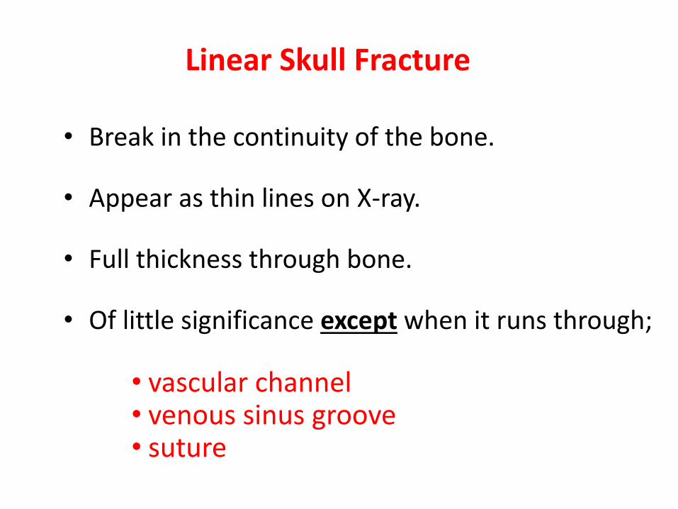
Linear Skull Fracture
• Break in the continuity of the bone.
• Appear as thin lines on X-ray.
• Full thickness through bone.
• Of little significance except when it runs through;
• vascular channel• venous sinus groove• suture


Depressed Skull Fracture
The broken piece of skull
bone is pressed towards
or embedded in the brain.
Neurological Deficit.
Dural Tear.
Hematoma .
Infection.
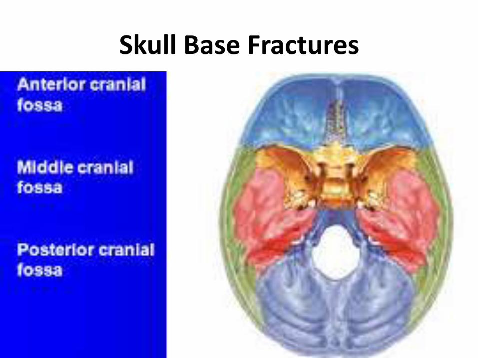
Skull Base Fractures

Anterior Skull Base fracture
Diagnosed clinically.
• CSF rhinorrhea.
• Epistaxis.
• Subconjunctival hemorrhage.
• Periorbital hematomas.
(raccoon eyes)
• Anosmia.

Middle Skull Base fracture
• CSF otorrhea.
• Hearing loss.
• Battle sign.
• Facial nerve palsy.
• Vertigo &nystagmus.
Posterior Skull Base fracture
• Bruising over occipital area.• cranial nerve injuries.

The double ring sign(Halo Sign)

Brain Injury
An insult to the brain, caused by an external
physical force.
Injuries
• Blunt
• Penetrating

• Primary Brain Injury :
- occurs at the time of impact
- Mechanical damage is irreversible
- Permanent mechanical cellular disruption
- Microvascular injury.
includes - cerebral contusions
- diffuse axonal injuries (DAI)
- cerebral lacerations
PATHOPHYSIOLOGY

• Secondary Brain Injury
Occurs at some time after the moment of impact
Preventable—>>>improved outcome.

TYPES OF BRAIN INJURIES
DIFFUSE
• Concussion
• Diffuse Axonal Injury
FOCAL
• Contusion
• Lacerations
• Epidural hge
• Subdural hge
• Subarachnoid hge
• Intracerebral hge

CONCUSSION
“Any trauma-induced alteration in mental status”
Temporary & brief interruption of neurological function without structural damage.
Cause
shearing / stretching of white matter fibres at the time of impact ==>> temporary neuronal dysfunction.

• Brief confusion, disorientation.
• Headache.
• Dizziness.
• Amnesia.
• Return of consciousness moments or minutes after impact (within 30 minutes).
• CT/MRI Normal
CONCUSSION

Post-concussive syndrome
Timing: 2 weeks to 2 months
c /o• Persistent headache• fatigue• Personality changes• Short attention span• Decreased short-term memory• sleep disturbances• depression, personality disorders

Severe widespread injury to axons in the cerebral hemispheres, corpus collosum, and brain stem.
Diffuse Axonal Injury

Clinical signs:
– LOC immediately
– ICP
– Decerebration or decortication.
– Cognitive impairment, spasticity .
– 90% pts with severe DAI will be vegetative.
– CT usually normal
Diffuse Axonal Injury

Epidural hematoma
Collection of blood between dura and bones of the skull.
Skull # present 75-95% of cases.
Source ; Arterial (MMA) , Venous.
Pathophysiology;• fracture of temporal bone ruptures branches of
the middle meningeal artery.
• stripping of dura from the calvarium --->> severe headache.
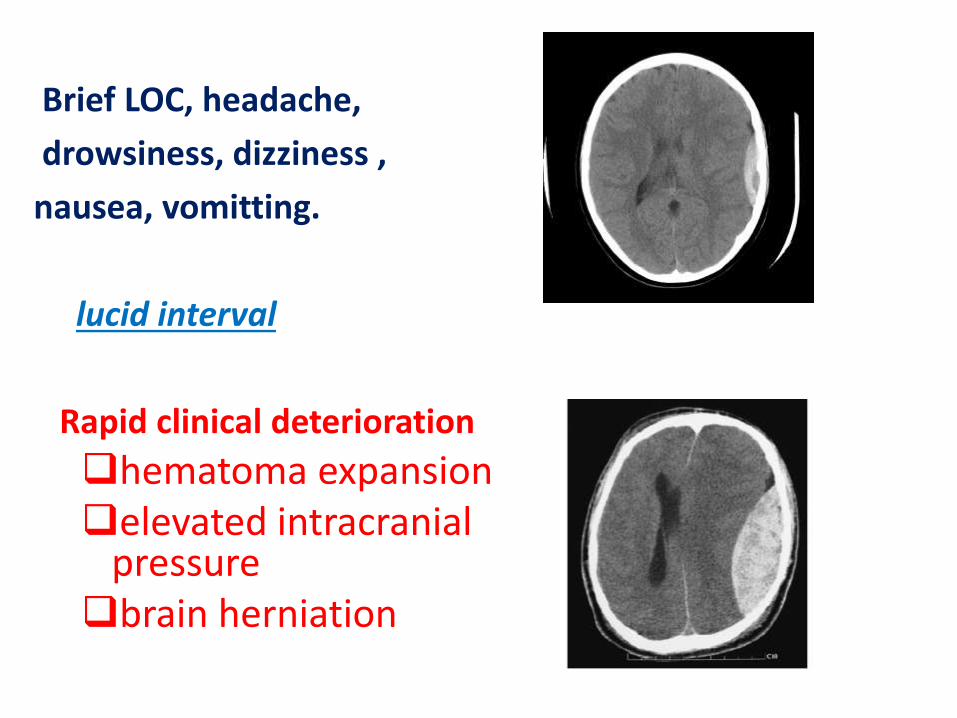
Brief LOC, headache,
drowsiness, dizziness ,
nausea, vomitting.
lucid interval
Rapid clinical deterioration
hematoma expansion elevated intracranial
pressurebrain herniation

Acute subdural Hematoma
• Accumulates in space between dura and arachnoid
• Source; - Disrupted cortical vessels (A,V)
- Brain laceration produces hematoma
• Signs within 48 hours of the injury.
• Associated with major trauma (Shearing Forces)
• Mortality rate as high as 40% in some series

CT scan
HyperdenseConcave Spreading across
brain
Midline shift disproportionateto size of lesion

Chronic subdural Hemorrhage
• Elderly on anti coagulant or anti platelet.
• History: minor head injury in weeks or months prior
to presentation
• Small bridging veins tear small, clinically silent
hematoma increases in sizemass effect.
• c/f: headache, cognitive impairment,
focal neurological deficit and seizures.

Chronic Subdural Hematoma
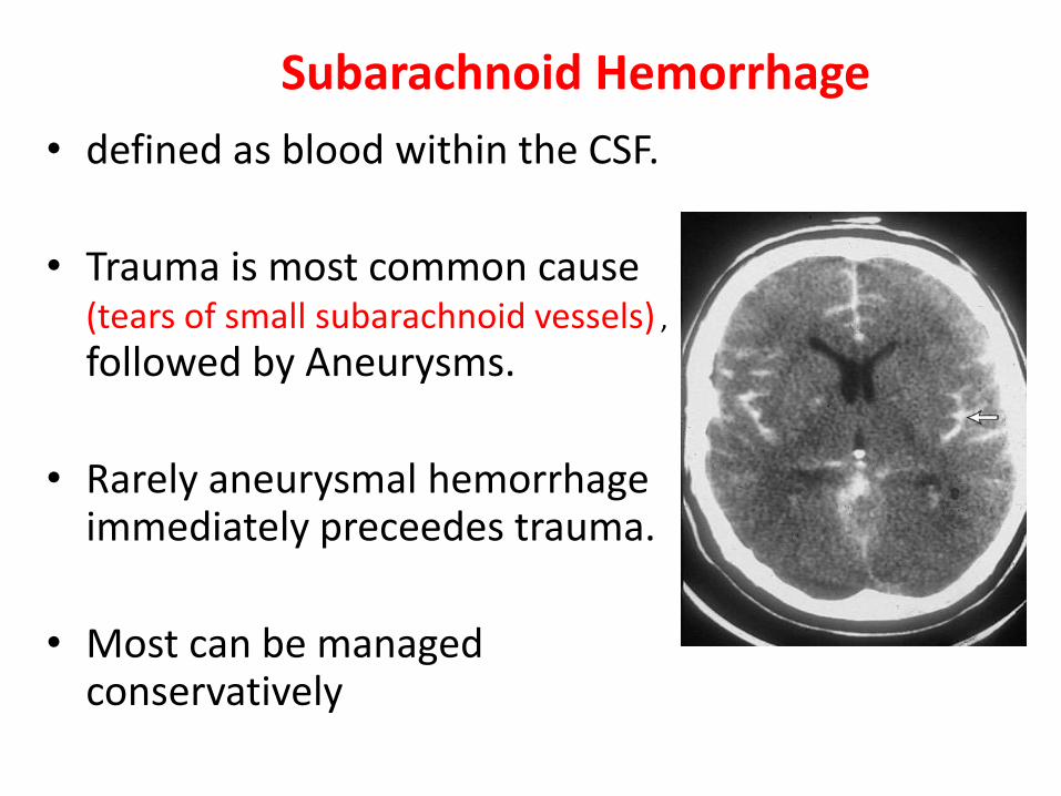
Subarachnoid Hemorrhage
• defined as blood within the CSF.
• Trauma is most common cause (tears of small subarachnoid vessels) ,
followed by Aneurysms.
• Rarely aneurysmal hemorrhage immediately preceedes trauma.
• Most can be managed conservatively

Cerebral contusions
• Coup and counter coup injuries.
• Most commonly affecting inf frontal fossa and temporal lobes
• CT appears heterogeneous with mixed areas of high and low density.
• Observation, Rarely require immediate surgery

Intracerebral Hematoma
Bleeding directly into the brain tissue.Two main types:
1) Intra-parencymal
2) Intra-ventricular
CausesTraumaHypertension (70-80%)Ruptured AVMCoagulopathy

Intra-parencymal hge Intra-ventricular hge

Clinical presentation
• Progressive severe headache (several minutes).
• Nausea and vomiting.
• Focal neurological deficits .
• Decreased level of consciousness.
•Brain herniation.