Embed Size (px)
Citation preview

Hematoxylin & Eosin Staining
Presented by
Dr. Shrikant Sonune
Guided by
Dr Ashok Patil,
Dr Shilpa Kandalgaonkar,
Dr Mayur Chaudhary,
Dr Suyog Tupsakhare,
Dr Mahesh Gabhane.
Hematoxylin and eosin technique
Principle
H and E are principle stain for demonstration of nucleus and cytoplasm.
Alum acts as a mordant and the hematoxylin containing alum stains the
nucleus light blue which turns red in the presence of acid.
The cell differentiation is achieved by treating the tissue with acid
solution. The counterstaining is performed using eosin which imparts
pink color to cytoplasm
Hematoxylin and eosin technique
Removal of paraffin wax (dewaxing)
Removed with xylene (impermeable to stains)
2-3min of xylene immersion sufficient for sections of 10 µ thickness
First facilitated by warming the slides at 60 degrees oven to melt
the wax
Removal of xylene
Xylene is not miscible with water or low grade alcohols, hence dipped in
two changes of absolute alcohol

Hematoxylin and eosin technique…..
Hydration – after removal from xylene sections are
transferred to absolute alcohol for 1-2min until it becomes
opaque
Sections rinsed in second bath of alcohol, drained and taken
to water
Any pigments or deposits should be removed at this stage

Hematoxylin and eosin technique
Staining Slides immersed in hematoxylin (Mayer s, Harris, Gills) If regressive stain is used longer time is used to overstained
the structures
Differentiation Sections are dipped in acid alcohol, agitated and washed in
tap water
Observed under microscope If underdifferentiated- returned to acid alcohol If overdifferentiaited – retured to hematoxylin and
differentiated again

Hematoxylin and eosin technique
Blueing
Slides after draining off hematoxylin is transferred to water
for 10min. Sections when removed from hematoxylin or
acid alcohol are pink in color
Washing turns them blue
Counterstain
Transfer the slides to 1% aqueous eosin for 2min. Wash in
running water
Dehydration
Slides are taken through 3 stages of acid alcohol

Hematoxylin and eosin technique
11. Clearing
Sections transferred to xylene and left until clear
Tested for clarity by being held against a dark background
12. Mounting
Surplous xylene wiped off from slide surface
This step completed quickly to avoid section drying
Whole operation takes 5-10 seconds
Hematoxylin and eosin technique
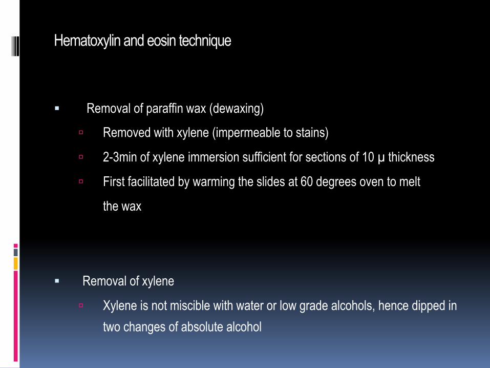
Results
Cell nuclei – blue
Muscle fibres – red
Collagen fibres – pink
RBC – bright red

Theory of H & E staining

Hematoxylin:
Most commonly used stain.
It is derived from core of the logwood tree, Haematoxylon
compechianum,
It is not a true dye until partially oxidized (ripened)
----by exposure to air
----by chemical means with an oxidizing agent such as sodium
iodate or mercuric oxide.

HAEMATOXYLIN AND EOSIN
The hematoxylin and eosin stain is the most widely used histological stain because……
Its comparative simplicity
Ability to demonstrate clearly an enormous number of different tissue structures.
The hematoxylin --cell nuclei blue / black,
Eosin stains cell cytoplasm and most connective tissue fibres

The hematoxylin may be used in the demonstration of:
Intracellular substances
Chromosomes
Keratohyaline granules
Extracellular substances – Elastin
Minerals – calcium, copper etc.
Central nervous system – myelin, neuroglia
fibers.

Hematoxylin
Dark red color
Extracted from the heartwood of the tree Hematoxylin camechianum
The hematoxylin is extracted from log wood with hot water and then precipitated out from the aqueous solution using urea.
It is sold commercially as a crude mixture of hematoxylin and other, unidentified substance.
It comes as a brownish tan powder which is poorly soluble in water and somewhat more soluble in ethyl alcohol.

Hematoxylin itself is not a stain.
On oxidation it produces haematin - a poor dye but
Metallic mordant, forms the most powerful stain.
When aluminum salts– will stain blue
When ferric salt– will stain blue-black.

Ripening
This process of oxidation is often referred to as
ripening or maturing.
This can be carried out in two ways
1. Natural oxidation
2. Chemical oxidation

Natural oxidation:
Carried out by exposure to light and air.
Slow process
Resultant solutions seem to retain its staining ability for a long time.
Advantage
Ones oxidation has reached an acceptable level, the staining solution may be used, and it last for longer,
Disadvantage
In the planning and organization required ensuring that usable solution is always available. For example: Ehrlich’s and Delafield’s hematoxylin.

Chemical oxidation:
It is achieved by the addition of the oxidizing agents
such as mercuric oxide, sodium iodate and
potassium permanganate.
The use of chemical oxidizing agents converts the
hematoxylin to haematin almost instantaneously, so
these hematoxylin solutions are ready for use after
preparation.

Properties of chemically oxidized
hematoxylin Have a shorter useful life than the naturally oxidized
haematoxylins .
However, the possibility of over – oxidation has been clearly established that the production of oxyhaematein inhibits successful staining.
To prevent these, glycerol has been incorporated in many formulas. Glycerol acts as stabilizer by preventing over oxidation and reducing evaporation.

Properties
Haematin is anionic, having poor affinity for tissue.
It is an inadequate stain without the presence of
mordant. (most useful mordant are salts of
aluminium,)
Hematoxylin solution using Lead as a mordant are
occasionally used for demonstration of argyrophil
cells.

Classification according to which mordant is
used.
Alum haematoxylins Ehrlich’s
Mayer’s
Harris
Cole’s
Delafield
Carazzi’s

Classification according to which mordant is
used. Iron haematoxylins
Weigert
Heidenhain’s
Loyez
Verhoeff
Tungesten
Molybdenum
Lead
Hematoxylin without mordant

Alum hematoxylin:
Routinely used in the hematoxylin and eosin stain,
and produce good nuclear staining.
The mordant is aluminium, in the form of ‘potash
alum’- aluminium ammonium sulphate.

The nuclei 1st become a red colored,
Differentiation is carried out
Then, Converted to familiar blue-black when the
section is washed in weak alkali.
The alum hematoxylin can be used regressively or
progressively.

The tomes for hematoxylin staining and for satisfactory
differentiation will vary according to:
The type and age of alum hematoxylin used.
The type of tissue.
The personal preference of the pathologist.
The most commonly used hematoxylin are
Ehrlich’s, Mayer’s, Harris’s, Cole’s and Delafield’s
haematoxylins.
Carazzi’s hematoxylin is occasionally used,
particularly for urgent frozen sections.

Harris’s hematoxylin: (1900)
This is an alum hematoxylin which is traditionally chemically ripened with mercuric oxide (sodium or potassium iodatemay be used as substitutes for oxidation.).
It is a powerful and selective nuclear stain giving clear nuclear staining.
It is used widely as a nuclear stain in exfoliative cytology.
In routine histology practice it is used regressively, but in exfoliative cytology it may be used as a progressive stain.

Harris’s hematoxylin: (1900)
Preparation of solution:
Hematoxylin - 1 g
Absolute alcohol - 10 ml
Ammonium or potassium alum - 20 g
Distilled water - 200 ml
Mercuric oxide - 0.5 g
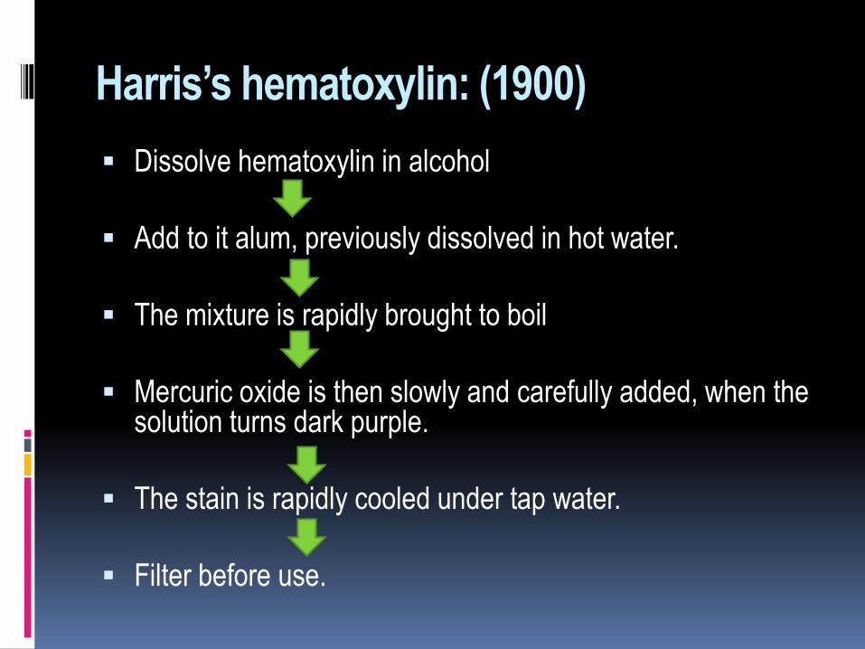
Harris’s hematoxylin: (1900)
Dissolve hematoxylin in alcohol
Add to it alum, previously dissolved in hot water.
The mixture is rapidly brought to boil
Mercuric oxide is then slowly and carefully added, when the solution turns dark purple.
The stain is rapidly cooled under tap water.
Filter before use.

Mayer’s hematoxylin: (1903)
A next widely used hematoxylin
Chemically ripened with sodium iodide.
It is more vigorous in action than Ehrlich’s
hematoxylin and gives little or no staining of muco-
polysaccharide material.

It is used as a nuclear counter stain in the
demonstration of glycogen (PAS, mucicarmine) in
various enzyme histological techniques.
The stain is applied for short period (progressive
stain usually 5-10 min.) until nuclei are stained, and
is then blued without any differentiation.
Differentiation might destroy or decolour the stained
cytoplasmic components. It can be used as a
regressive stain like any alum hematoxylin.
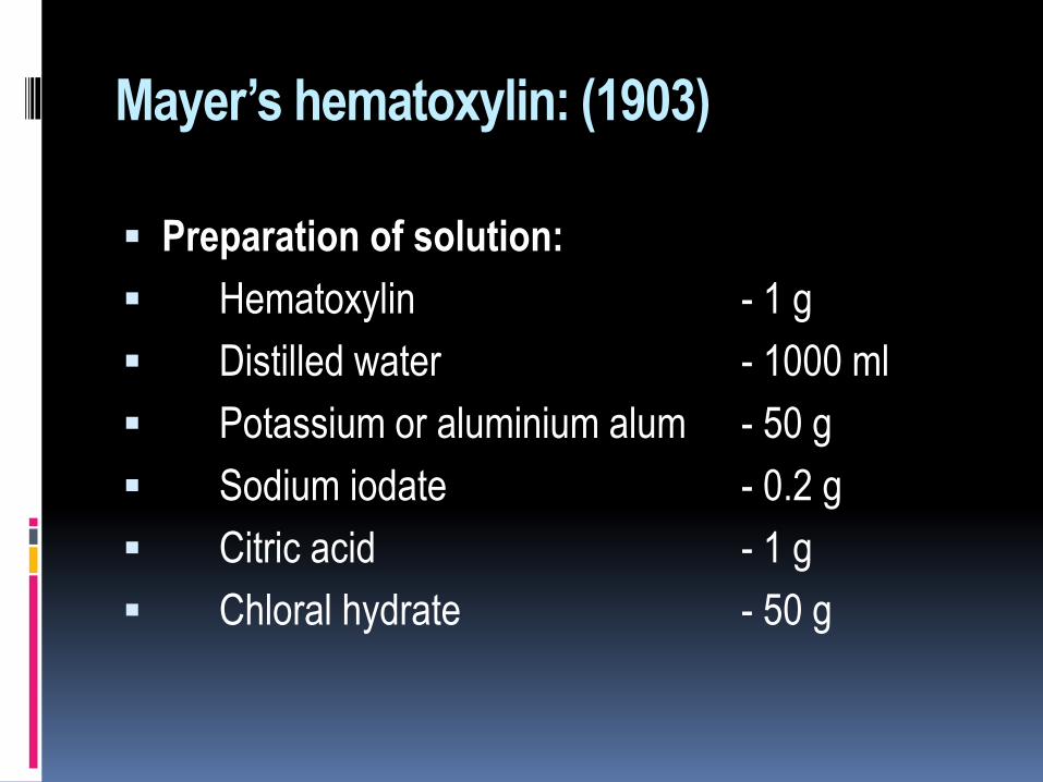
Mayer’s hematoxylin: (1903)
Preparation of solution:
Hematoxylin - 1 g
Distilled water - 1000 ml
Potassium or aluminium alum - 50 g
Sodium iodate - 0.2 g
Citric acid - 1 g
Chloral hydrate - 50 g

The hematoxylin, potassium alum, sodium iodate is
dissolved in distilled water by warming and stirring, or
by allowing to stand at room temperature overnight.
The chloral hydrate and citric acid are added, and the
mixture is boiled for 5 minutes, then cooled and
filtered.
Chloral hydrate acts as a preservative and citric acid
sharpens nuclear staining.

Ehrlich’s hematoxylin: (1886)
This is a naturally ripened alum hematoxylin, most commonly used in both normal and morbid histology.
Preparation of solution:
Hematoxylin - 2 g
Absolute alcohol - 100 ml
Glycerol - 100 ml
Distilled water - 100 ml
Glacial acetic acid - 10 ml
Potassium alum - 10 – 14 g
( in excess )

Dissolve the hematoxylin in the alcohol.
The incorporation of glycerol.
Finally, add potassium alum (till saturation).
The stain may be ripened naturally by allowing to stand in large flask, loosely stoppered with cotton wool.
Filter before use.

It may be partially oxidized
Stain used immediately by the addition of 0.3 g
sodium iodate to the above.
It also stains mucin in salivary glands, some muco-
polysaccharide substances such as cartilage, and
‘cement lines’ of bone etc. Ehrlich’s hematoxylin is
not ideal for frozen sections.

Delafield’s hematoxylin: (1885)
This is a naturally ripened alum hematoxylin, which has similar longevity to Ehrlich’s hematoxylin.
Preparation of solution:
Solution A:
Hematoxylin - 4 g
Absolute alcohol - 25 ml
Solution B:
Alumunium alum - 60 g
Distilled water - 400 ml
Solution C:
Glycerol - 100 ml
Absolute alcohol - 100 ml

The hematoxylin is dissolved in 25 ml of alcohol
added to solution ‘B’ (alum solution).
Mixture is allowed to stand in light and air for 5 days and is then filtered.
Added to solution ‘C’.
Allowed to stand exposed to light and air for about 3-4 months or until the stain is sufficiently dark in color,
Then filtered and stained.

Cole’s hematoxylin: (1943)
This is an alum hematoxylin, artificially ripened with an alcoholic iodine solution. It has good keeping qualities and is suitable for use in sequence with celestine blue, unlike Ehrlich’s hematoxylin.
Preparation of solution:
Hematoxylin - 1.5 g
1%iodine in absolute ethanol - 50 ml
Saturated aqueous potassium alum - 700 ml
Distilled water - 250 ml

The hematoxylin is dissolved in warm distilled water
and mixed with iodine solution.
The alum solution is added, and the mixture
brought to boil, then cooled quickly and filtered.
The solution is ready for immediate use,but may
need on occasion filtering after storage,

Carazzi’s hematoxylin: (1911)
This is an alum hematoxylin which is chemically
ripened using potassium iodate.
Preparation of solution:
Hematoxylin -5 gm
Glycerol -100 ml
Potassium alum - 25 gm
Distilled water - 400 ml
Potassium iodate - 0.1 g

Hematoxylin is dissolved in the glycerol
and the alum is dissolved in most of the water overnight.
The alum solution is added slowly to solutition. Potassium iodate is dissolved in the rest of the water with gentle warming and is then added to the haematoxylin-alum-glycerol mixture.
The final staining solution is mixed well and is then ready for immediate use, it remains usable for about six months.

Carazzi’s hematoxylin may be used as a
progressive nuclear counter stain using a short
staining time followed by blueing in tap water.
It is particularly suitable since its pale and precise
nuclear stain, does not stain any of the cytoplasmic
components.
It is particularly used for the frozen section (due to
rapid staining)

Gill’s hematoxylin: (1974)
Preparation of solution:-
Distilled water - 730 ml
Ethylene glycol - 250 ml
Hematoxylin - 2 g
Sodium iodate - 0.2 g
Aluminium sulphate - 17.6 g
Glacial acetic acid - 20 ml

The reagents are added in the order given and the
mixture stirred for 1 hour at room temperature.
The solution is ready for immediate use.
Double or triple concentrations of ethylene glycol
may be used as preferred
Gill’s 1 (normal),
Gill’s 2 (double conc of ethylene glycol),
Gill’s 3 (triple conc of ethylene glycol).

Advantages
They are fast in action,
Stable for at least 12 months,
Produce little or no surface precipitate,
Their preparation does not involve boiling the
solution.

Staining times with alum haematoxylins:
It will vary according to following factors:
Type of hematoxylin used:- e.g. Ehrlich’s hematoxylin 20-45 min, Mayer’s hematoxylin 10-20 min.
Age of stain: as the stain ages the staining time will need to be increased.
Intensity of use of stain: A heavily used hematoxylin will loose its staining power rapidly and longer staining times will be necessary.

Progressively or regressively method.
e.g. Mayer’s hematoxylin used progressively 5-10 min, used regressively 10-20 min.
Pretreatment of tissues or sections
e.g. length of time in fixative or acid decalcifying solution or whether paraffin or frozen section.
Post treatment of sections
e.g. subsequent acid stains such as Van Gieson.
Personal preference.

Disadvantages of alum hematoxylin
Sensitivity to any subsequently applied acidic
staining solutions
examples are in the Van Gieson and other trichrome
stains.
The application of the picric acid – acid fuchsin
mixture in Van Gieson’s stain removes most of the
hematoxylin so that the nuclei are barely
discernible.

Nuclear staining can be achieved by using an iron –
mordanted hematoxylin e.g.Weigert’s hematoxylin,
Alternative is the combination of celestine blue
staining solution with an alum hematoxylin.
Celestine blue is resistant to the effects of acid and
ferric salt.
Strengthens the bond between the nucleus and alum
hematoxylin.

Celestine blue- alum hematoxylin:
Preparation of solution:
Celestine blue solution
Celestine blue B -2.5 g
Ferric ammonium sulphate -25 g
Glycerol -70 ml
Distilled water - 500 ml

Ferric ammonium sulphate is dissolved in cold
distilled water with stirring.
The celestine blue B is added to this solution and
the mixture is boiled for few minutes.
Filtered
glycerin is added.
Filter before use.

Celestine blue B,
an oxazine dye,
Has little useful colouring property of its own.
It forms an additional strong mordant with certain
haematoxylins .
Celestine blue B is used as a preliminary to alum
hematoxylin staining.

Iron haematoxylin
Iron salts such as ferric chloride or ferric ammonium sulphate are used both as oxidizing agent and as mordant.
Demonstrating a much wider range of tissue structures than the alum haematoxylins,
The most common iron haematoxylins are
Heidenhain’s hematoxylin
Weigert’s hematoxylin
Verhoeff’s hematoxylin
Loyez hematoxylin

Over oxidation of the hematoxylin is a problem with these stains,
So it is usual to prepare separate mordant/ oxidant and hematoxylin solutions and mix them immediately before use
e.g. Weigert’s hematoxylin
To use them consecutively (e.g. Heidenhain’s and Loyez haematoxylins).
It may be used as
Differentiating fluid after hematoxylin staining,
Mordanting fluid before it.

Weigert’s hematoxylin (1904):
An iron hematoxylin used as a nuclear stain in techniques where acidic staining solutions are applied to the sections subsequently (e.g. Van Gieson stain).
In Van Gieson stain, picric acid is one of the constituents which have marked decolorizing action on nuclei stained with alum hematoxylin.
Weigert hematoxylin which is mordanted to iron salt (ferric chloride) has sufficient avidity to withstand this treatment.

Preparation:
The iron and hematoxylin solutions are prepared separately and are mixed immediately before use.
Solution A (stain) Solution B (mordant)
Hematoxylin – 1 g 30% aqueous ferric chloride
Absolute alcohol – 100 ml ( anhyd. ) -4 ml
Conc. HCl - 1 ml
Dist. Water - 95ml
The color of the mixture should be a violet black.
If muddy – brown, it must be discarded.

Heidenhain’s hematoxylin (1896):
Ferric ammonium sulphate as oxidant/ mordant.
The same solution is also used as a differentiating fluid.
The iron solution is used first
The section is treated with hematoxylin solution until it is over stained,
Then it is then differentiated with iron solution under microscopic control.
Heidenhain’s hematoxylin is a cytological stain.
It is used regressively
Requires careful differentiation, for which reason it is
only completely successful on thin sections.
It may be used to demonstrate
chromatin,
chromosomes,
nuclei,
centerosomes,
mitochondria,
muscle striations
myelin.

Heidenhain’s hematoxylin (1896):
Preparation:
Hematoxylin solution (stain)
Hematoxylin - 0.5g
Absolute alcohol- 10ml
Distilled water- 90ml
Iron solution (mordent and differentiator)
Ferric ammonium sulphate- 5g
Distilled water - 100ml

Dissolved hematoxylin in alcohol
add water.
Allow to ripen for a few weeks and store in a tightly
stoppered bottle.

Loyez hematoxylin (1910):
This is an iron hematoxylin in which ferric ammonium sulphate is used as the mordant.
The mordant and hematoxylin solution are used consecutively,
Differentiation is by Weigert’s differentiator (borax and potassium ferricyanide).
It is used to demonstrate myelin and can be applied to paraffin, frozen or nitrocellulose sections.

Verhoeff’s hematoxylin (1908):
Verhoeff’s hematoxylin is used to demonstrate
elastic fibers after all routine fixative.
Coarse fibres are intensely stained, but the staining
of fine fibers may be less than satisfactory.
The differentiation step is critical to the success of
this method.
Introduced by Mallory in 1897 for demonstration of neuroglia.
It is now widely used for other purposes (fibrin, muscle striation).
It is unique among hematoxylin stains in the number of structures that may be demonstrated, together with the two color staining (shades of blue and red) from the single solution.
Tungsten hematoxylin

Mallory first advocated a 1: 10 ratio of hematoxylin
to mordant, later a 1: 20 ratio should be used with
pure mordant due to earlier mordant was impure.
Later Turner et al proposed that this proportion is
too high and does not give the most satisfactory
staining.

Natural ripening of phosphotungstic acid
hematoxylin is slow and several chemical oxidizing
agents have been used to hasten the conversion of
hematoxylin to haematein. As an alternative to
chemical oxidation,
Haematein may be used initially, instead of
hematoxylin and provides a stain that is usable 24
hours after preparation.

Molybdenum hematoxylin:
Molybdenumic acid as the mordant are rare,
Technique which gained any acceptance was the
Thomas (1941) technique which was mentioned by
MacManus and Mowry (1964).
Demonstration of collagen and coarse reticulin,
More valuable and accepted for these connective
tissue fibre exist

Lead hematoxylin:
Hematoxylin solutions which incorporate Lead
salts have recently been used in the demonstration
of the granules in the endocrine cells of the
alimentary tract and other regions
Hematoxylin without a mordant
Freshly prepared hematoxylin solutions, used without a mordant,
Used to demonstrate various minerals in tissue sections – Mallory described a method for lead, iron and copper.
The basis of the Mallory methods is the ability of unripened hematoxylin to form blue black lakes with these metals.

Test for staining power of hematoxylin…
Adding few drops of hematoxylin to 50ml of tap
water will turn a bright, clear purple or blue violet
color.
Exhausted solutions will not be clear & bright & the
color will be rusty or green.

Hematoxylin Specific use
Harris;’s hematoxylin Exfoliative cytology
Mayers hematoxylin Nuclear counter stain in PAS
Ehrlichs hematoxylin Mucin & other mucopolysaccharide
Carazzis hematoxylin Progressive nuclear stain & frozen section
Celestine blue- alum hematoxylin Where subseqent acidic stains are to be used e.g. van Gieson stain
Weigerts hematoxylin Where subseqent acidic stains are to be used e.g. van Gieson stain
Heidenhains hematoxylin Differentiating cytological fluid stain
Loyes hematoxylin To demontrate myelin, frozen section
Tungsten hematoxylin Fibrin , & muscle straitions
Molybdenum hematoxylin Demonstration of collegen fibre
Lead hematoxylin Granules of endocrine cell
Hematoxylin without mordant Pigment lead , iron
Eosin
Eosin, a red dye,
Stains connective tissue and cytoplasm in varying intensity and shades (red to pink)
Eosin is derived from fluorescein and is available in following types:
Eosin Y (eosin yellowish, eosin water soluble)
Ethyl eosin (eosin S, eosin alcohol soluble)
Eosin B (eosin bluish, erythrosine B)

Eosin Y is most commonly used and is readily
soluble in water, less so in a alcohol thus it is
sometimes sold as ‘water and alcohol soluble’
Preparation
Eosin Y, water soluble 5gm.
Distilled water 1000ml.
Crystals of thymol added to inhibit the growth of fungi.

Alcohol soluble eosin is employed as a 0.5 % solution
in alcohol.
Eosin Y water &
Alcohol soluble 10gm
Distilled water 50ml
95%ethyl alcohol 940ml
In use, sections should be treated with 95% alcohol
before staining with alcoholic eosin, and the excess
stain washed out in the same solvent.

The addition of little acetic acid (0.5 ml to 1000 ml
stain) is said to sharpen the staining.
Ethyl eosin and eosin B are now rarely used,
although occasional old methods specify their use,
e.g. the Harris stain for Negri bodies.

Thank you……