Embed Size (px)
Citation preview

PERIPHERAL GIANT CELL GRANULOMA. NODULAR
REDDISH-PURPLE MASS OF THE MAXILLARY GINGIVA.

PERIPHERAL GIANT CELL GRANULOMA
ULCERATED MASS OF THE MANDIBULAR GINGIVA.
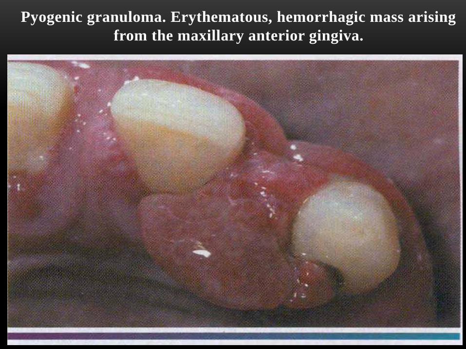
Pyogenic granuloma. Erythematous, hemorrhagic mass arising
from the maxillary anterior gingiva.

Pyogenic granuloma. Ulcerated and lobulated mass
on the dorsum of the tongue.
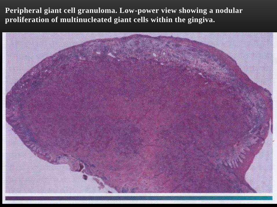
Peripheral giant cell granuloma. Low-power view showing a nodular
proliferation of multinucleated giant cells within the gingiva.
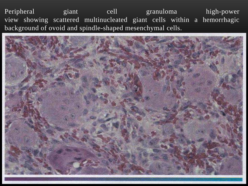
Peripheral giant cell granuloma high-power
view showing scattered multinucleated giant cells within a hemorrhagic
background of ovoid and spindle-shaped mesenchymal cells.

Central giant cell granuloma. the occlusal radiograph shows a radiolucent
lesion with cortical expansion.

Central giant cell granuloma. A bluish-purple mass is present on the
anterior alveolar ridge of this 4-year-old white boy.

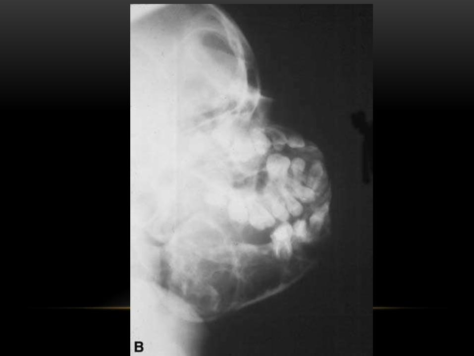
Central giant cell granuloma. Panoramic radio graph showing a
large,expansile radiolucent lesion in the anterior mandible.

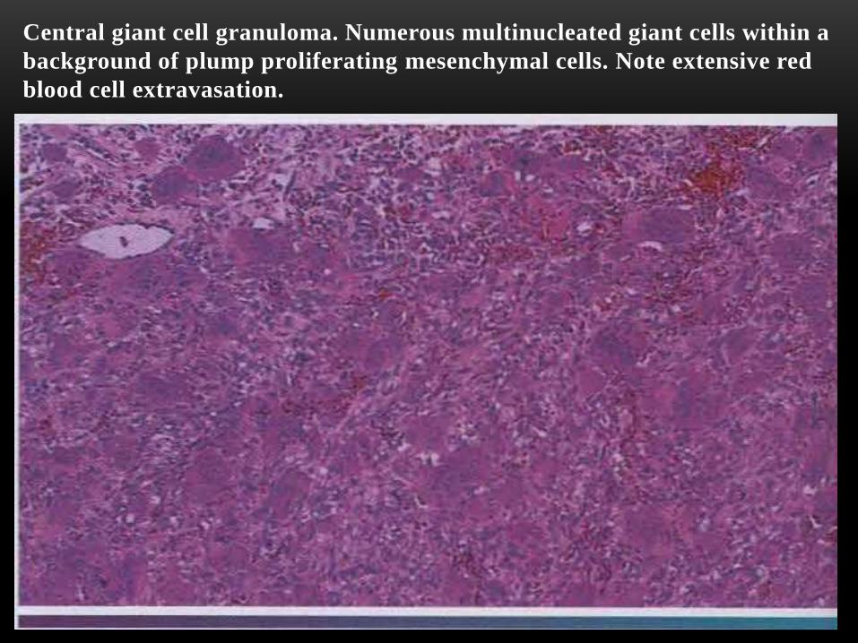
Central giant cell granuloma. Numerous multinucleated giant cells within a
background of plump proliferating mesenchymal cells. Note extensive red
blood cell extravasation.

Giant cell tumor. This photomicrograph shows large giant cells that are
distributed in a cellular mesenchymal tissue. This specimen was from an
aggressive lesion that had destroyed most of the maxilla.


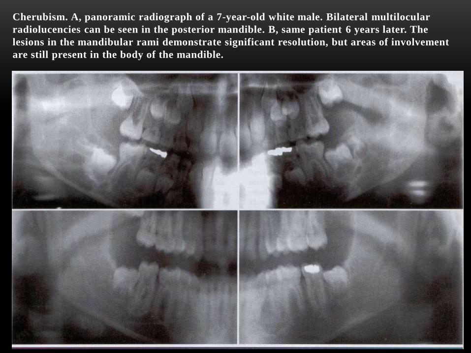
Cherubism. A, panoramic radiograph of a 7-year-old white male. Bilateral multilocular
radiolucencies can be seen in the posterior mandible. B, same patient 6 years later. The
lesions in the mandibular rami demonstrate significant resolution, but areas of involvement
are still present in the body of the mandible.

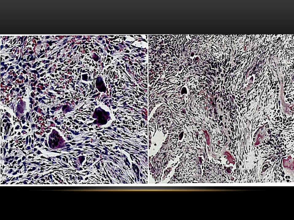
Cherubism. Photomicrograph showing scattered giant cells within a
background of cellular, hemorrhagic mesenchymal tissue. The inset
demonstrates perivascular eosinophilic cuffing.

Aneurysmal bone cyst . A large multilocular radiolucent lesion
involves most ofthe ascending ramus in a 5-yearold white boy.

Simple bone cyst. Panoramic film showing a large multilocular simple
bone cyst of the mandible in a 16-year-old white male.

Simple bone cyst. Panoramic film showing a large simple bone cyst of the mandible
in a 12-year-old girl. The scalloping superior aspect of the cyst between the roots of
the teeth is highly suggestive of, but not diagnostic for, a simple bone cyst.
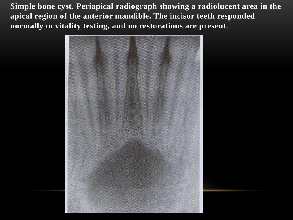
Simple bone cyst. Periapical radiograph showing a radiolucent area in the
apical region of the anterior mandible. The incisor teeth responded
normally to vitality testing, and no restorations are present.

Aneurysmal bone cyst. Photomicrograph showing a blood-filled space
surrounded by fibroblastic connective tissue. Scattered
multinucleated giant cells are seen adjacent to the vascular space.

Simple bone cyst. Photomicrograph of the bony wall of a
simple bone cyst. A thin. Vascular connective tissue membrane
is adjacent to the bone and no epithelia l lining is identified.

Hyperparathyroidism. This periapical radiograph reveals the "ground
glass" appearance of the trabeculae and loss of lamina dura in a patient
with secondary hyperparathyroidism.

Hyperparathyroidism. This occlusal radiograph of the edentulous
maxillary anterior region shows a multilocular radiolucency
characteristic of a brown tumor of primary hyperparathyroidism.

Hyperparathyroidism. Palatal enlargement is characteristic of the renal
osteodystrophy associated with secondary hyperparathyroidism.

Hyperparathyroidism. This high-power photomicrograph of a brown tumor of
hyperparathyroidism shows scattered multinucleated giant cells within a
vascular and proliferative fibroblastic background.

HYPERPARATHYROIDISM. THIS LOW-POWER PHOTOMICROGRAPH SHOWS
DELICATE, INTERCONNECTING TRABECULAE OF WOVEN BONE WITHIN A
BACKGROUND OF CELLULAR FIBROUS CONNECTIVE TISSUE.