Embed Size (px)
Citation preview

c cc
INTRODUCTION Red cell require energy in order to fulfill function
Only source of energy is in form of ATP derived from
1: GLYCOLYTIC PATHWAY
2: PENTOSE PHOSPHATE PATHWAY
3: GLUTATHIONE CYCLE
Reducing power is required:
To reduce metHb back to its functional state
To counteract the strong oxidative stress that cell carries

Processes that reduce metHb utilizes:
1:NADH reduced from NAD+by glycolytic pathway
2:Reduction of free oxygen radicals produce during reactions to infection is provided by NADPH catalyzed by G6PD
3:NADPH drives glutathione cycle, major reducing agent in the red cell

ATP provides energy for :
1:maintenance of red cell volume
2: red cell shape
3: flexibility
Sodium potassium ATPase pump
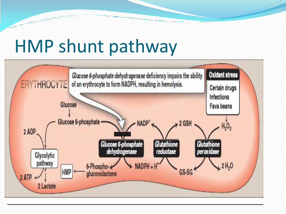
HMP shunt pathway

Gl-6-P dehydrogenase deficiency Most common erythrocyte enzyme disorder
Disease is sex linked carried by gene on X chromosome Xq28 therefore common in males
Age dependent, decrease slightly
First recognized during Korean world war when African soldiers develop hemolytic anemia when given anti-malarial drugs
Normal cells use 0.1% of their G6PD
Females heterozygote for G6PD deficiency always contain two population of cells, one normal and one G6PD deficient as compared to males

COMPOUNDS ASSOCIATED WITH G6PD DEFICIENCY
Drugs
Favism
infection

Oxidant drugs
Commonly used drugs that produce hemolytic anemia in patients with G6PD deficiency anemia
Antibiotics (for example, sulfamethoxazole and chloramphenicol),
Antimalarials (for example, primaquine but not quinine),
Antipyretics (for example, acetanilid but not aceta minophen).

Favism: Some forms of G6PD deficiency, for example the
Mediterr anean variant, are particularly susceptible to the hemolytic effect of the fava (broad) bean, a dietary staple in the Mediterranean region.
Favism, the hemolytic effect of ingesting fava beans, is not observed in all individuals with G6PD deficiency, but all patients with favism have G6PD deficiency

Infection: Infection is the most common precipitating factor of
hemolysis in G6PD deficiency.
The inflammatory response to infection results in the generation of free radicals in macro phages, which
can diffuse into the red blood cells and cause oxidative damage.


PATHOPHYSIOLOGY

PATHOPHYSIOLOGY
G6PD is necessary for maintaining adequate levels of GSH
In case of deficiency generation is impaired results in
accumulation of cellular oxidants (reactive oxygen species (ROS))
Buildup of cellular oxidant leads to RBC injury and hemolysis
Hb is oxidized to methemoglobin, which precipitate in form of
Heinz bodies
Heinz bodies attach to erythrocyte membrane cause increase
cell rigidity, membrane permeability

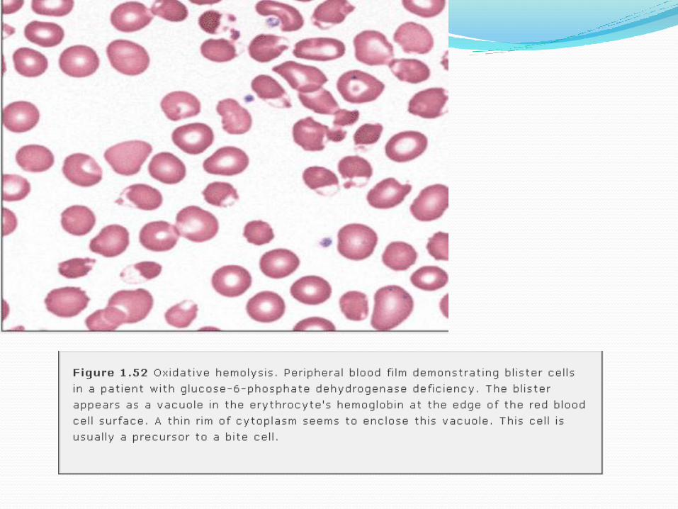
CONT… Heinz bodies are removed by splenic macrophages producing bite
cells and blister cells
Membrane loss leads to formation of spherocytes
These cells ultimately hemolyzed in spleen
Hemolysis is self limited
Important to remember that under stress of severe oxidants(drugs,
chemicals) normal cells may experience oxidant damage
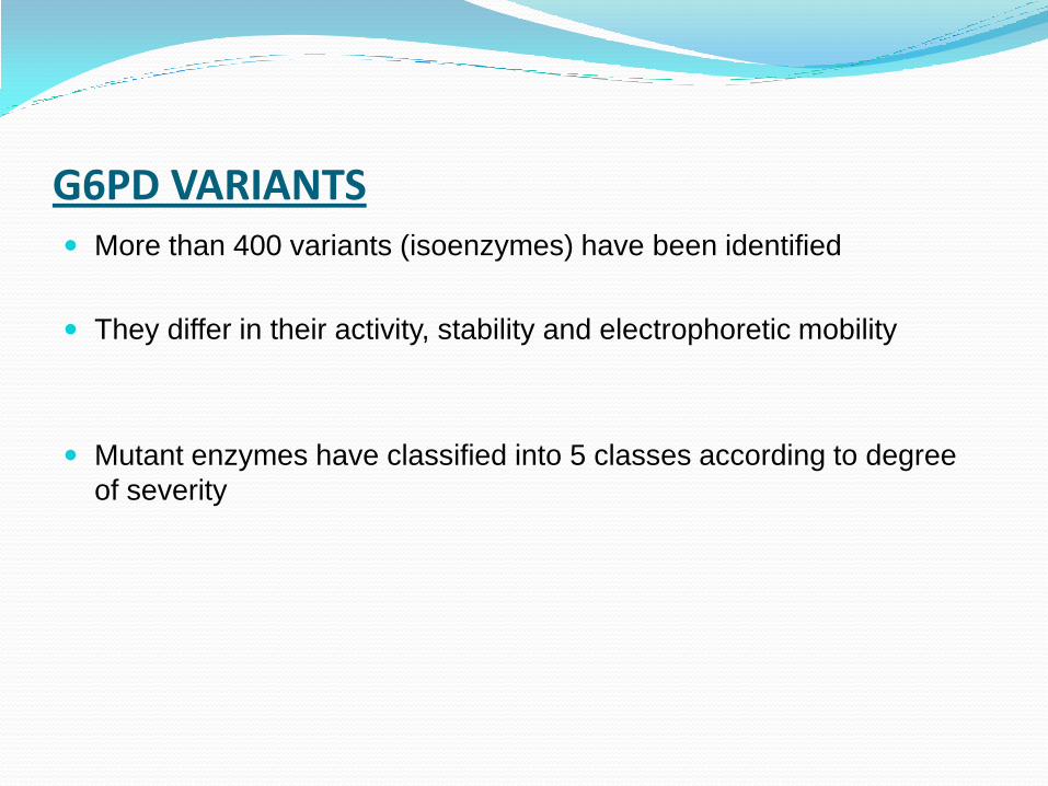
G6PD VARIANTS More than 400 variants (isoenzymes) have been identified
They differ in their activity, stability and electrophoretic mobility
Mutant enzymes have classified into 5 classes according to degree
of severity


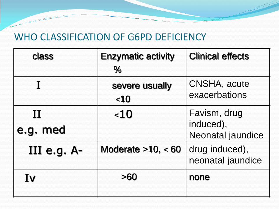
WHO CLASSIFICATION OF G6PD DEFICIENCY
class Enzymatic activity
%
Clinical effects
I severe usually
<10
CNSHA, acute
exacerbations
II
e.g. med
<10 Favism, drug
induced),
Neonatal jaundice
III e.g. A- Moderate >10, < 60 drug induced),
neonatal jaundice
Iν >60 none

CLINICAL FINDINGS Mostly deficient patients have no clinical symptoms
Hemolysis is variable dependent on degree of oxidant
stress, and sex of the patient
Symptoms are same as of acute intravascular hemolysis
FAVISM is disorder in which some individual with G6PD
deficiency develop hemolytic episode after ingestion of
fava beans?
isouramil, divicine
Signs of favism are malaise, nausea, vomiting, abdominal
pain

LAB FINDINGS
G6PD deficiency is easily detected in hemizygous(male),
or homozygous individual but it is difficult to detect in
heterozygous female
In female heterozygotes two population of cells exist, one
with normal G6PD activity and other deficient
It occurs due to inactivation of one of the two X
chromosome in individuals cells early in development of
embryo

LAB FINDINGS During hemolytic attack, polychromasia, spherocytes, erythrocyte
fragments and bite cells may be seen on blood smear
Bite cells are thought to be typical of G6PD deficiency
Reticulocytosis is characteristic following hemolytic attack
A Peculiar cells referred to by variety of descriptive terms (irregularly contracted cell, RBC hemi ghost) is present after oxidant related hemolysis. These cells are rigid, decreased volume, Hb is confined to one side while other side is transparent. Transparent site mostly contain Heinz bodies

6

Cont… Leukocyte count may be increased during hemolysis
Platelet count is normal
Indirect bilirubin and serum LDH may raised
Heptoglobin is decreased
Definitive diagnosis depends upon the demonstration of
decrease in RBC G6PD enzyme activity

FLUORESCENT SPOT TEST Reliable and sensitive screening test
Add 10µl of Whole blood either anticoagulated (EDTA,heparin or
added before clotting) is added to 100 µl mixture of G6P,NADP, and
saponin
Drop of mixture is placed on piece of filter paper
Examined it under UV light of fluorescence
G6PD enzyme present in RBCs metabolizes G6P,producing
NADPH.NADPH fluoresces but NADP not, lack of fluorescence
indicate deficiency
The test can be carried out on whole blood stored in ACD for up to 21
days at 4oC

False normal:
If there is reticulocytosis,fluorescence may
be seen with G6PD deficient blood sample
False deficient:
If patient is anemic, very little fluorescence
may be seen despite the sample is normal

DYE REDUCTION TEST
Hemolysate of patients blood+G6P+NADP and dye
brilliant cresyl blue are incubated together
If hemolysate contain G6PD, NADP reduced to NADPH
which in turn reduces blue dye to its colorless form
RESULT:
Time take for change to occur is inversely
proportional to amount of G6PD present

DETECTION OF HETEROZYGOTES FOR G6PD DEFICIENCY Elution test Individual cells retaining Hb02 in metHb elusion test are stained, remaining
appear as ghost cells Method: incubated the sample with Nile blue sulphate 1:Oxygenate the sample during incubation by bubbling with O2 with help of pipette 2:After 2-3 h add 20µl of KCN to 1 ml of reaction mixture 3:Make blood films 4:Dry, wash and stain it with hematoxylin, counter stain with erythrosin for
2 mins Examine cells under microscope and count the proportion of stained HbO2 and unstained cells


ENZYMATIC ASSAY
Erythrocyte hemolysate is incubated with G6P and NADP
Rate of reduction of NADP to NADPH is measured at
340nm in spectrophotometer

THANK YOU