Embed Size (px)
Citation preview

ENTERAL FEEDING TUBE FOR DRUG ADMINISTRATION
Surya AmalPresented For Pharmacy Department
University of Darussalam Gontor - Indonesia

Overview
1. The important reason to choice the route of enteral feeding (p2)
2. The possibility of drug delivery through this route (p5)
3. Choice of feeding tube for drug administration (p6)
4. Indications for enteral tube feeding (p7)
5. Complications of enteral tube feeding (p8)
6. Routes and types of feeding tubes (p9 – p19)
7. Characteristics of the tubing material (p20 – p21)
8. Technique of flushing (p22 – p23)
9. How to choice of medication formulation ? (p25)
10. Preparing medication for administration (p26 – p31)
11. Medications not suitable for administration via enteral tubes (p32)
12. Ethical issues of enteral tube feeding (p33)
The important reason to choice this route
“When patients are unable to be fed orally, enteral or parenteral
nutrition is recommended.”

Enteral Nutrition (EN)
“Enteral Nutrition (EN) offers some advantages over parenteral nutrition as the decrease in hospitalization
time, reduced clinical complications, greater convenience, improvement in bowel function,
maintenance of the structure and function of the gastrointestinal mucosa, and less possibility of bacteria
translocation.”
Presoti et al., J Gen Pract 2013, 1:2
Drug administration via enteral feeding tubes
The placement of a feeding tube in the gastrointestinal tract opens the possibility of drug delivery through this via, also reducing the risk of administration of injectable dosage forms.

Choice of feeding tube for drug administration
Use of enteral feeding tubes for drug
administration is increasing.
Sizes of feeding tubes are decreasing.
The range of healthcare professionals
involved in drug administration via enteral
feeding tubes is increasing.
Collation of all available information is
necessary.
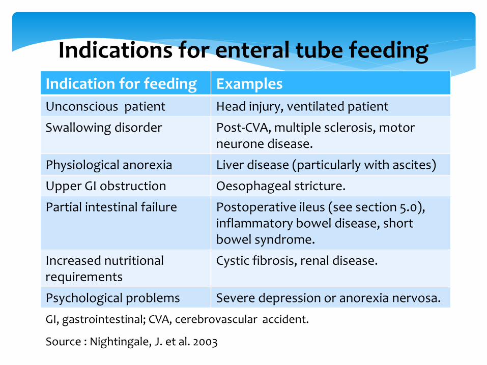
Indication for feeding Examples
Unconscious patient Head injury, ventilated patient
Swallowing disorder Post-CVA, multiple sclerosis, motor neurone disease.
Physiological anorexia Liver disease (particularly with ascites)
Upper GI obstruction Oesophageal stricture.
Partial intestinal failure Postoperative ileus (see section 5.0),inflammatory bowel disease, short bowel syndrome.
Increased nutritionalrequirements
Cystic fibrosis, renal disease.
Psychological problems Severe depression or anorexia nervosa.
Indications for enteral tube feeding
GI, gastrointestinal; CVA, cerebrovascular accident.
Source : Nightingale, J. et al. 2003
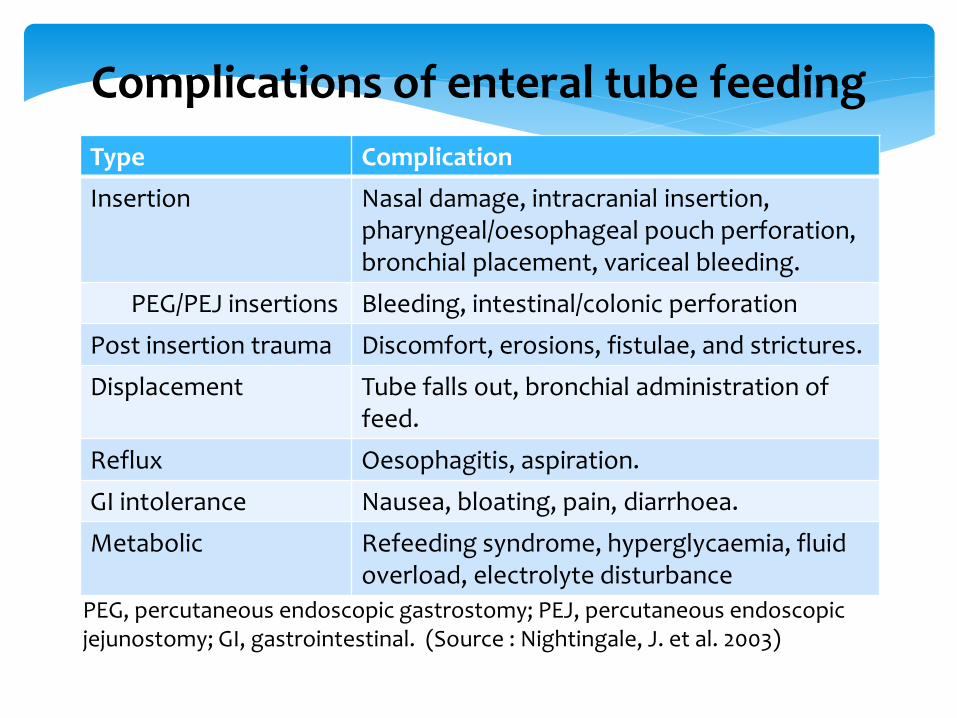
Type Complication
Insertion Nasal damage, intracranial insertion, pharyngeal/oesophageal pouch perforation,bronchial placement, variceal bleeding.
PEG/PEJ insertions Bleeding, intestinal/colonic perforation
Post insertion trauma Discomfort, erosions, fistulae, and strictures.
Displacement Tube falls out, bronchial administration of feed.
Reflux Oesophagitis, aspiration.
GI intolerance Nausea, bloating, pain, diarrhoea.
Metabolic Refeeding syndrome, hyperglycaemia, fluid overload, electrolyte disturbance
PEG, percutaneous endoscopic gastrostomy; PEJ, percutaneous endoscopic jejunostomy; GI, gastrointestinal. (Source : Nightingale, J. et al. 2003)
Complications of enteral tube feeding

Types of feeding tubes
Ensure that you know the type,
size and position of the enteral
feeding tube before administration
of medication via the tube.
The exit site of the tube may
affect drug pharmacokinetics or
side-effect profile.
Routes of Feeding
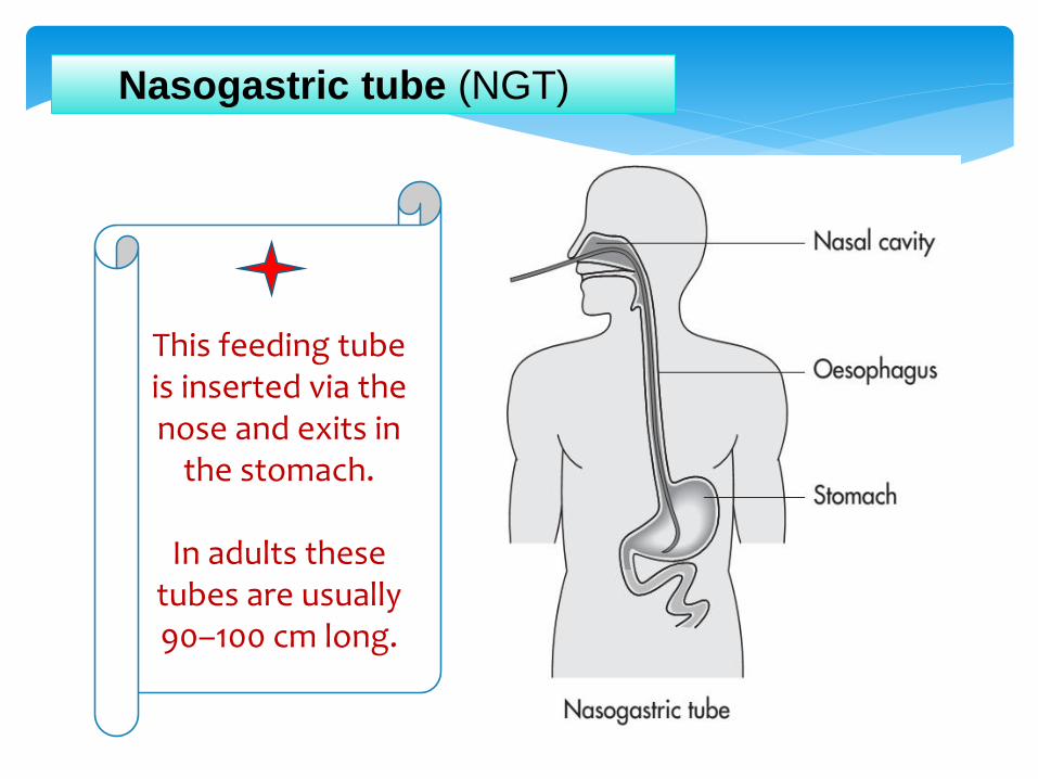
Nasogastric tube (NGT)
This feeding tube is inserted via the nose and exits in
the stomach.
In adults these tubes are usually 90–100 cm long.
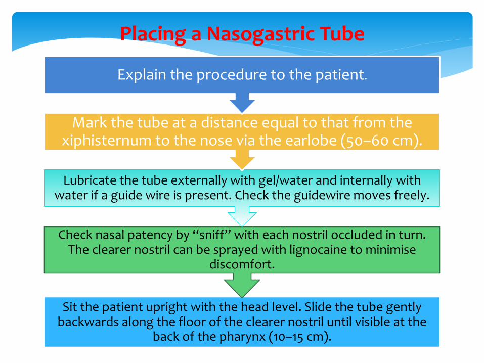
Sit the patient upright with the head level. Slide the tube gently backwards along the floor of the clearer nostril until visible at the
back of the pharynx (10–15 cm).
Check nasal patency by ‘‘sniff’’ with each nostril occluded in turn. The clearer nostril can be sprayed with lignocaine to minimise
discomfort.
Lubricate the tube externally with gel/water and internally with water if a guide wire is present. Check the guidewire moves freely.
Mark the tube at a distance equal to that from the xiphisternum to the nose via the earlobe (50–60 cm).
Explain the procedure to the patient.
Placing a Nasogastric Tube
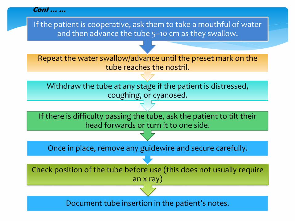
Document tube insertion in the patient’s notes.
Check position of the tube before use (this does not usually require an x ray)
Once in place, remove any guidewire and secure carefully.
If there is difficulty passing the tube, ask the patient to tilt their head forwards or turn it to one side.
Withdraw the tube at any stage if the patient is distressed, coughing, or cyanosed.
Repeat the water swallow/advance until the preset mark on the tube reaches the nostril.
If the patient is cooperative, ask them to take a mouthful of water and then advance the tube 5–10 cm as they swallow.
Cont … …
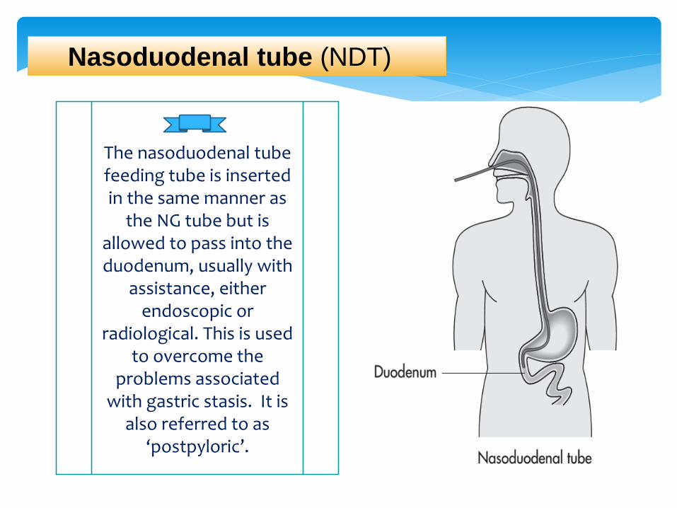
Nasoduodenal tube (NDT)
The nasoduodenal tube feeding tube is inserted in the same manner as
the NG tube but is allowed to pass into the duodenum, usually with
assistance, either endoscopic or
radiological. This is used to overcome the
problems associated with gastric stasis. It is
also referred to as ‘postpyloric’.
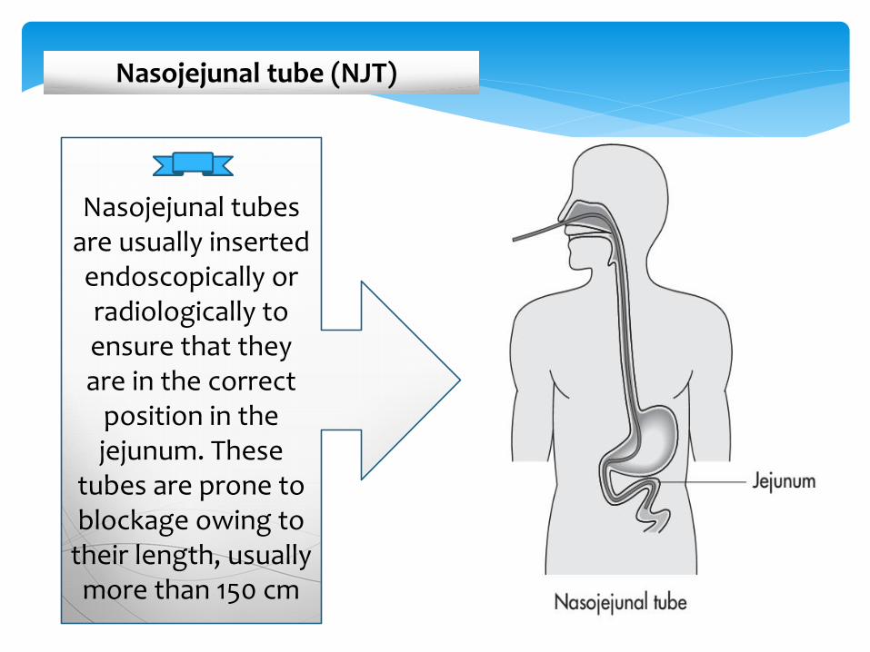
Nasojejunal tube (NJT)
Nasojejunal tubes are usually inserted endoscopically or radiologically to ensure that they are in the correct
position in the jejunum. These
tubes are prone to blockage owing to
their length, usually more than 150 cm
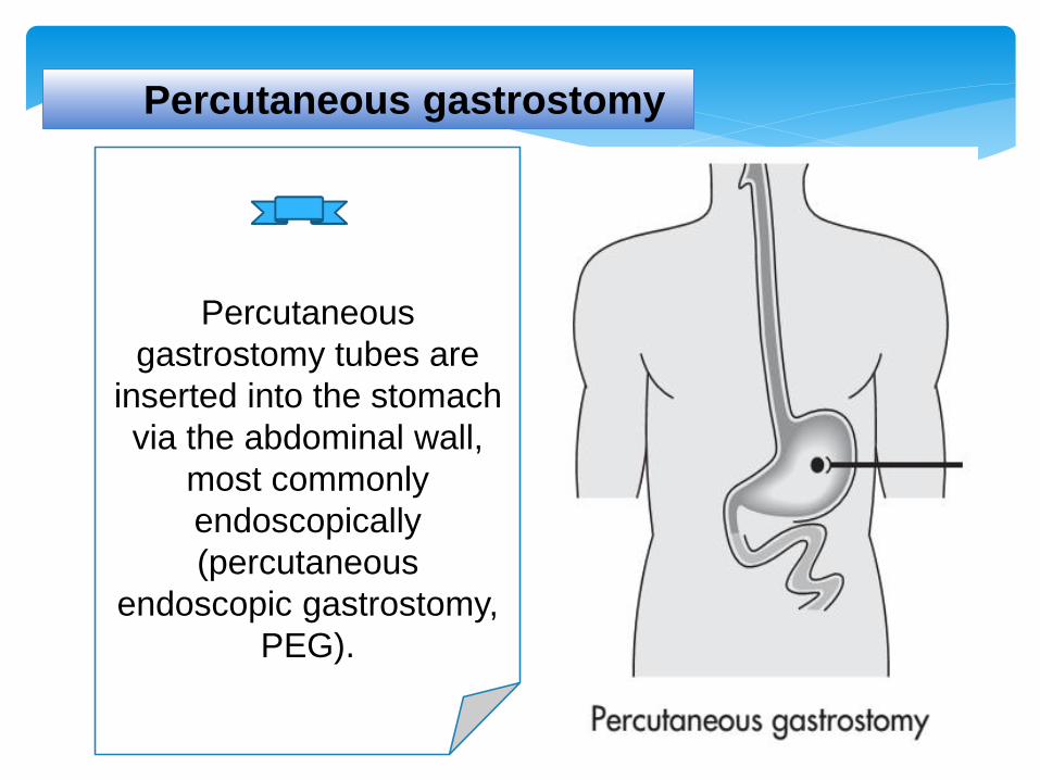
Percutaneous gastrostomy
Percutaneous
gastrostomy tubes are
inserted into the stomach
via the abdominal wall,
most commonly
endoscopically
(percutaneous
endoscopic gastrostomy,
PEG).
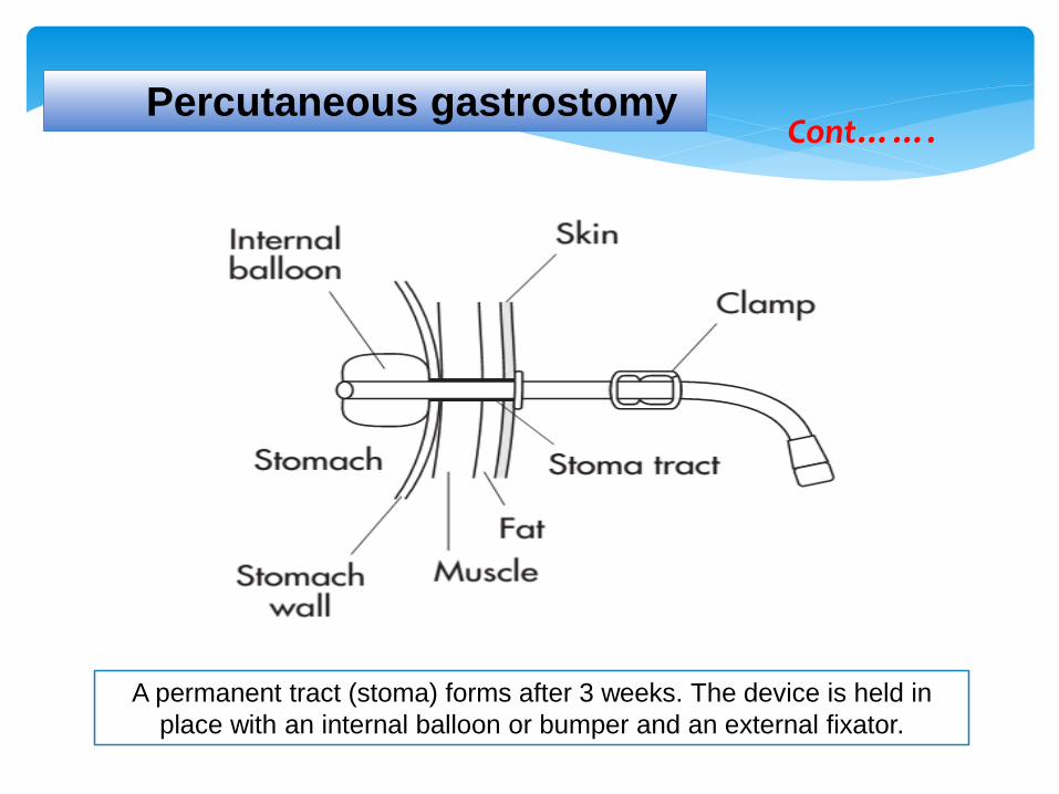
A permanent tract (stoma) forms after 3 weeks. The device is held in
place with an internal balloon or bumper and an external fixator.
Percutaneous gastrostomyCont…….
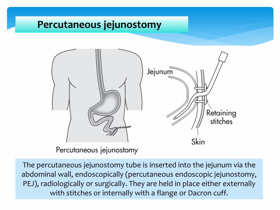
Percutaneous jejunostomy
The percutaneous jejunostomy tube is inserted into the jejunum via the abdominal wall, endoscopically (percutaneous endoscopic jejunostomy, PEJ), radiologically or surgically. They are held in place either externally
with stitches or internally with a flange or Dacron cuff.

Percutaneous gastrojejunostomy
The percutaneous gastrojejunostomy tube is inserted into the stomach via the abdominal wall and the exit of the feeding tube is
placed into the jejunum, most commonly endoscopically(percutaneous endoscopic gastrojejunostomy, PEGJ).

o Enteral feeding tubes are composed of polyvinylchloride (PVC), polyurethane (PUR), silicone or latex.
o The external diameter of the feeding tube is expressed using the French (Fr) unit where each ‘French’ is equivalent to 0.33 mm.
Nasoenteric tubes are used for short- to medium-term feeding (days to weeks).
Ostomy tubes are used for long-term feeding (months to years).
Characteristics of the tubing material
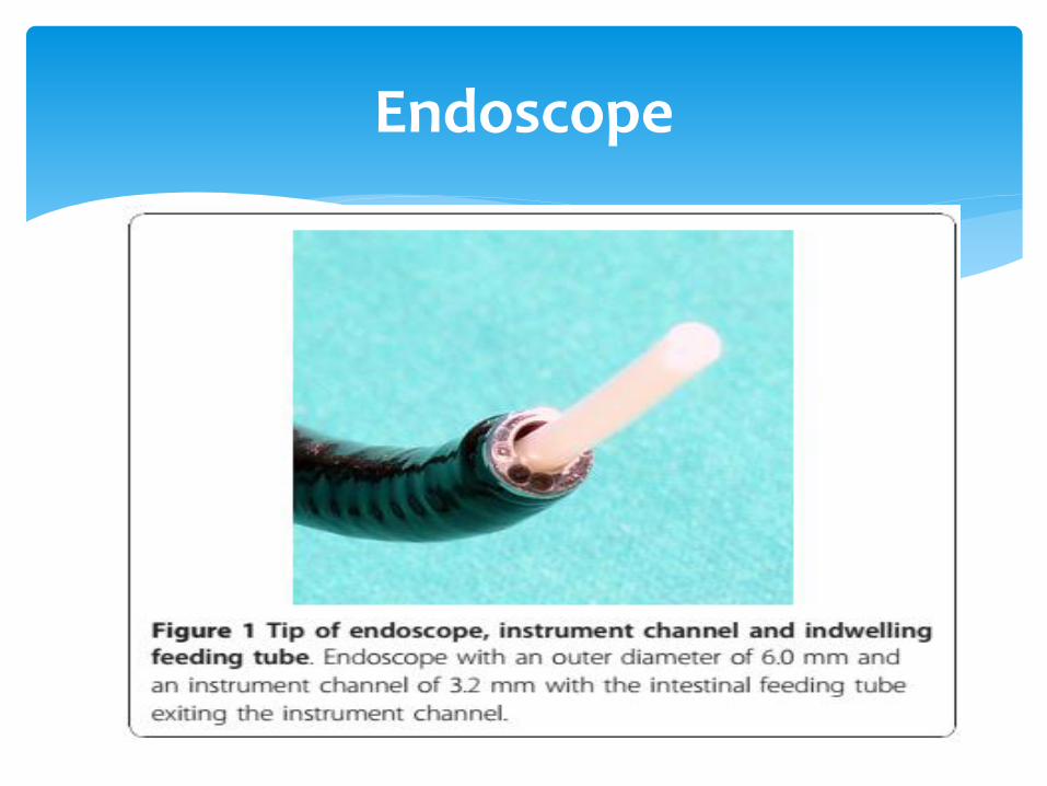
Endoscope
Flushing enteral feeding tubes
Tube flushing is the single most effective action in
prolonging the life of any enteral feeding tube.
Enteral feeding tubes require regular, effective
flushing to prevent tube blockage.

Technique of Air Flushing
1. Pre-fill a 50 mL syringe with 30 mL of air.2. Attach the syringe to the appropriate port of the patient’s
nasogastric feeding tube. 3. Ensure that any other ports are closed and airtight.4. Ensure that there is an airtight connection between the
syringe and the enteral tube and administer the flush.5. Listen for any evidence of the air venting into the mouth or
upper oesophagus; such venting may suggest misplacement of the tube tip in the upper oesophagus or rupture of the tube.
6. Attempt to aspirate with a 50 mL syringe. This will reduce the likelihood of the inner lumen of the enteral feeding tube collapsing under vacuum.

Technique Water flushing
1. Prepare a flush of water (according to local guidelines) in a 50 mL syringe and label if necessary. Place it in a clean tray.
2. Stop or suspend enteral feeding.3. Ensure that any other ports are closed and airtight.4. Attach the syringe to a port of the patient’s enteral feeding
tube. Ensure that there is an airtight connection between the syringe and the enteral tube.
5. Using a pulsatile flushing action, administer the flush.6. Positioning the patient in a semi-recumbent position can help
to prevent regurgitation and possible pulmonary aspiration from gastric flush and or drug residual.
7. Administer the drug and flush; cap off, or connect further enteral feeding depending on the patient’s requirements.
How to Choice of medication formulation ?
1. Solutions or soluble tablets are the formulations of choice.
2. Do not assume that liquid formulation will be suitable.
3. Do not crush tablets or open capsules unless an alternative formulation or drug is unavailable.
Preparing Medications for Administration
Soluble Tablets :
1. Dissolve the required number of tablets in a suitable volume of sterile potable water.
2. If the whole tablet dose is to be administered, rinse out the vessel in which the tablet was dissolved with sterile potable water, draw up into the same syringe used to administer the dose, and administer this residue to ensure the full dose is given.
3. If only a part dose is to be administered, ensure the resulting solution from the dissolved tablet is well suspended by continually agitating the solution. Administer the dose IMMEDIATELY.
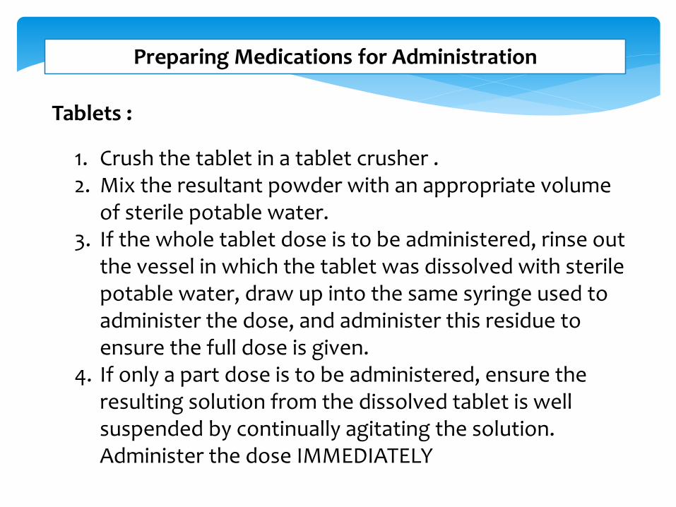
Preparing Medications for Administration
Tablets :
1. Crush the tablet in a tablet crusher . 2. Mix the resultant powder with an appropriate volume
of sterile potable water.3. If the whole tablet dose is to be administered, rinse out
the vessel in which the tablet was dissolved with sterile potable water, draw up into the same syringe used to administer the dose, and administer this residue to ensure the full dose is given.
4. If only a part dose is to be administered, ensure the resulting solution from the dissolved tablet is well suspended by continually agitating the solution. Administer the dose IMMEDIATELY

Preparing Medications for Administration
Capsules :
1. Carefully open the capsule and allow its contents to fall into a suitable container .
2. Disperse this powder with an appropriate volume of sterile potable water .
3. If the whole tablet dose is to be administered, rinse out the vessel in which the tablet was dissolved with sterile potable water, draw up into the same syringe used to administer the dose, and administer this residue to ensure the full dose is given.
4. If only a part dose is to be administered, ensure the resulting solution from the dissolved tablet is well suspended by continually agitating the solution. Administer the dose IMMEDIATELY
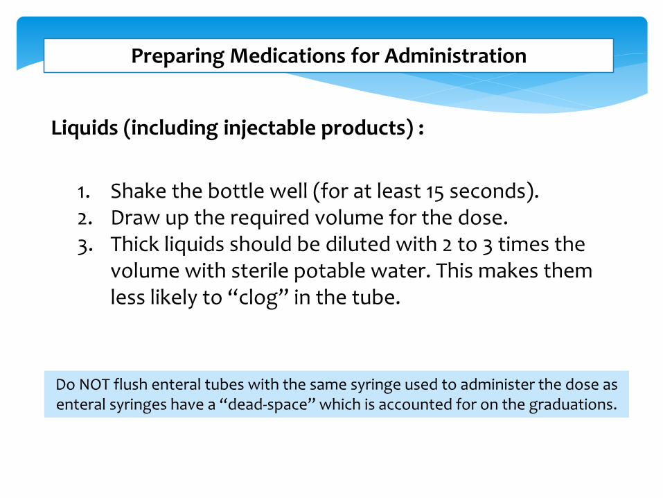
Preparing Medications for Administration
Liquids (including injectable products) :
1. Shake the bottle well (for at least 15 seconds).2. Draw up the required volume for the dose.3. Thick liquids should be diluted with 2 to 3 times the
volume with sterile potable water. This makes them less likely to “clog” in the tube.
Do NOT flush enteral tubes with the same syringe used to administer the dose as enteral syringes have a “dead-space” which is accounted for on the graduations.
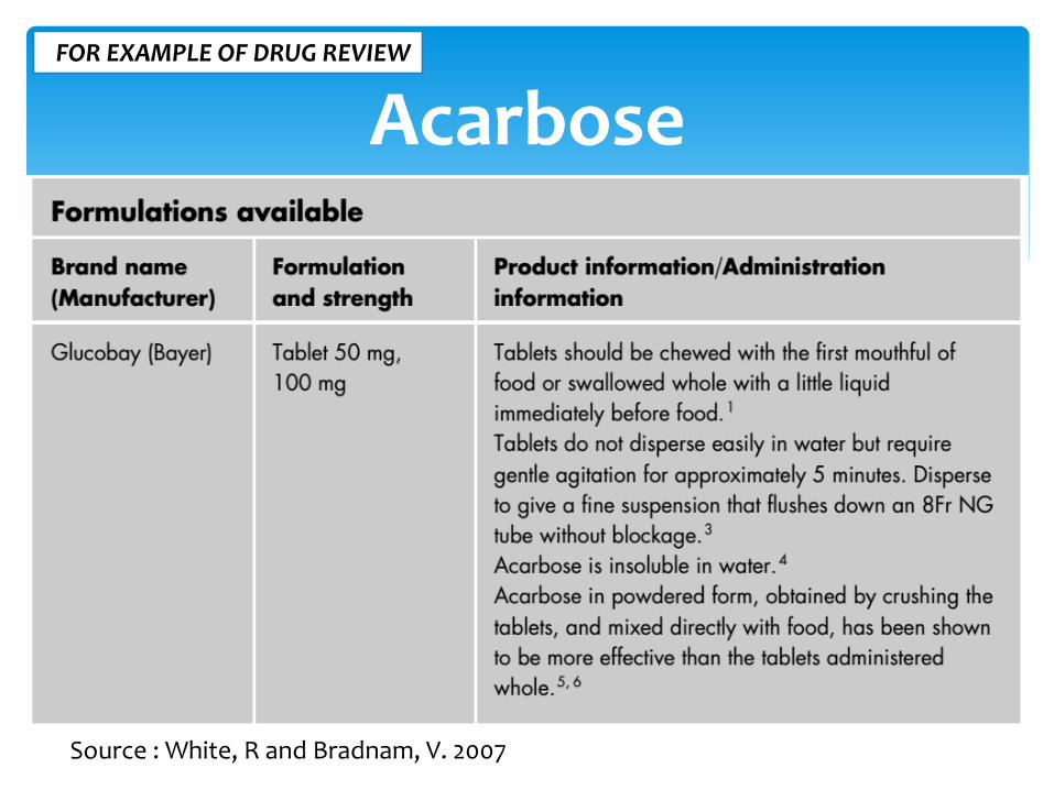
AcarboseFOR EXAMPLE OF DRUG REVIEW
Source : White, R and Bradnam, V. 2007
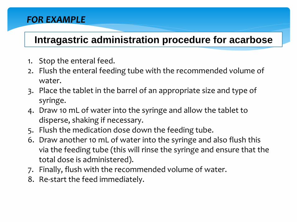
1. Stop the enteral feed.2. Flush the enteral feeding tube with the recommended volume of
water.3. Place the tablet in the barrel of an appropriate size and type of
syringe.4. Draw 10 mL of water into the syringe and allow the tablet to
disperse, shaking if necessary.5. Flush the medication dose down the feeding tube.6. Draw another 10 mL of water into the syringe and also flush this
via the feeding tube (this will rinse the syringe and ensure that the total dose is administered).
7. Finally, flush with the recommended volume of water.8. Re-start the feed immediately.
Intragastric administration procedure for acarbose
FOR EXAMPLE
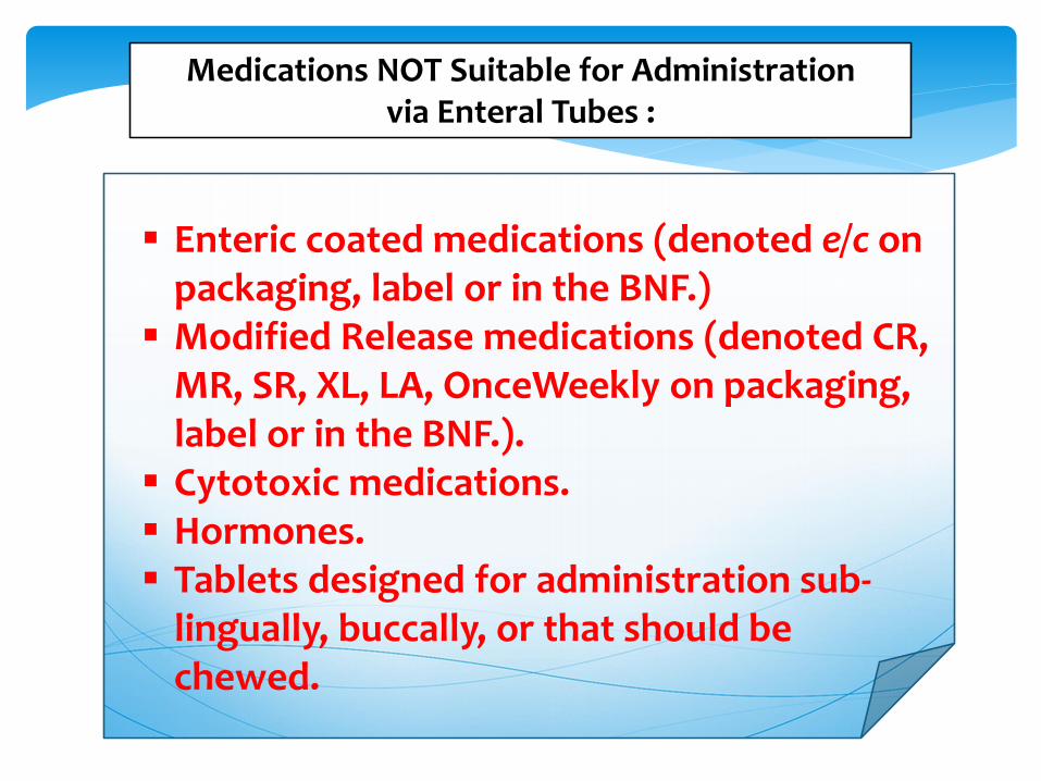
Medications NOT Suitable for Administration via Enteral Tubes :
Enteric coated medications (denoted e/c on packaging, label or in the BNF.)
Modified Release medications (denoted CR, MR, SR, XL, LA, OnceWeekly on packaging, label or in the BNF.).
Cytotoxic medications. Hormones. Tablets designed for administration sub-
lingually, buccally, or that should be chewed.
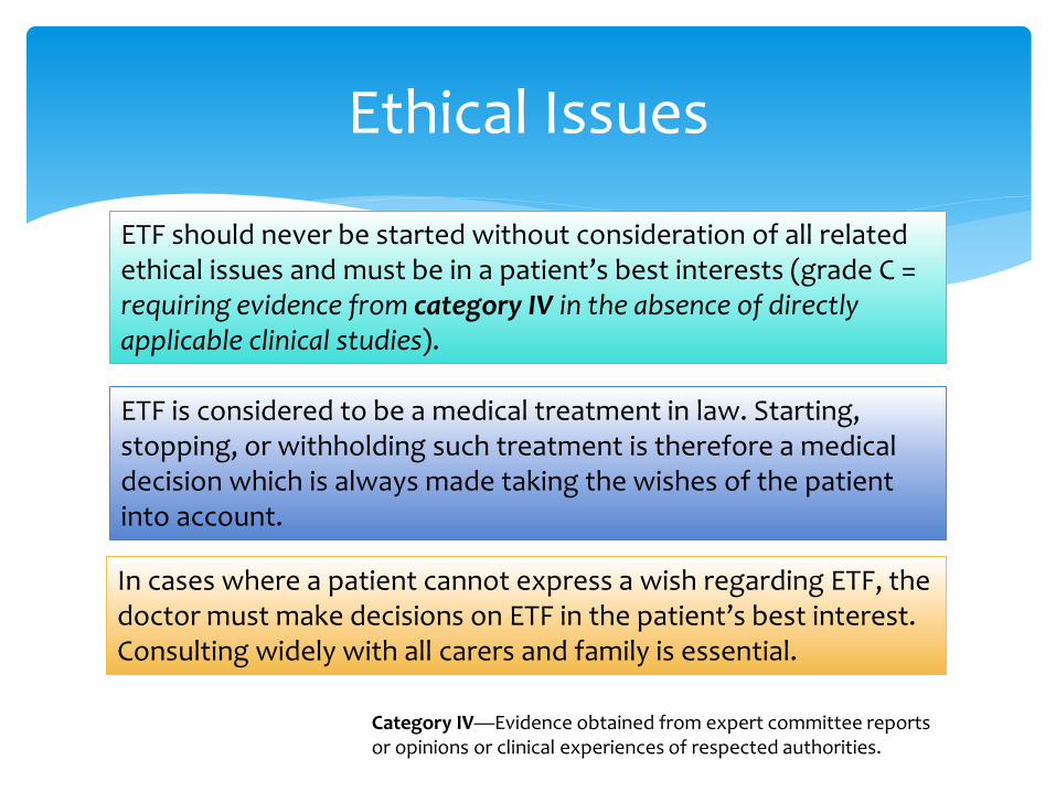
Ethical Issues
ETF should never be started without consideration of all related ethical issues and must be in a patient’s best interests (grade C = requiring evidence from category IV in the absence of directly applicable clinical studies).
ETF is considered to be a medical treatment in law. Starting, stopping, or withholding such treatment is therefore a medical decision which is always made taking the wishes of the patient into account.
In cases where a patient cannot express a wish regarding ETF, the doctor must make decisions on ETF in the patient’s best interest. Consulting widely with all carers and family is essential.
Category IV—Evidence obtained from expert committee reports or opinions or clinical experiences of respected authorities.

References
1. White, R and Bradnam, V. 2007. Handbook of Drug Administration via Enteral Feeding Tubes. Pharmaceutical Press, London –UK.
2. Nightingale, J. et al. 2003. Guidelines for enteral feeding in adult hospital patients. Gut 2003;52(Suppl VII):vii1–vii12
3. Presoti et al., 2013. Prescription of Drugs to be Administered through Feeding Tubes in a Brazilian Hospital: Profile and Qualification. J Gen Pract 2013, 1:2
4. Sutherland, A. 2009. Guideline On Administration of Medication Via Feeding Tubes. NHS. PICU Consultant Group.