Embed Size (px)
Citation preview

Outlines of development of
endocrine glands
the central nervous system
the eye
the ear and
skin and skin appendages

Development of endocrine glands
Endocrine system includes:
- endocrine glands - hypophysis, pineal, thyroid, parathyroid, adrenal, and islets of Langerhans
- endocrine components of glands with exocrine or other functions - pancreas, gonads, placenta, and kidneys
- cells with endocrine function that are scattered in nonglandular organs (as a gut, stomach, trachea, etc.) - e.g. GEP cells

Hypophysis

Epiphysis

Thyroid gland


Parathyroid gland


Adrenal gland

Development of suprarenal glands!
The cortex and medulla of the suprarenal glands have different origin. The cortex develops from mesoderm and the medulla from neural crest cells.
6th week aggregation of mesenchymal cells on each side.The cells that form the fetal cortex are derived from the mesothelial cells lining the posterior abdominal wall.
The cells that form the medulla are derived from the adjacent symphatetic ganglion. The cells lie first of the medial side of the cortex and as they become surrounded they start to develop into secretory cells of the suprarenal medulla.
Later more mesenchymal cells from the mesothelium enclose the cortex and they become the permanent cortex.
Zona reticularis develops in the end of 3rd year!!

Development of the pancreasDevelopment of the pancreas


Endocrine glands- summary
hypophysis: adenohypophysis - ectoderm of the stomodeum neurohypophysis - neuroectoderm of the
diencephalon (base)epiphysis - neuroectoderm of the diencephalon (roof)
thyroid gland - endoderm of the primitive pharynx
parathyroid glands - endoderm of pharyngeal pouches (3rd, 4th)
adrenal gland: cortex - coelomic mesoderm medulla - neural crest (crista neuralis)
Langerhans islets - endoderm of the foregut (duodenum)

Development the central nervous system
Development of neural tube
Histogenesis of neural tube
Overwiev of development of the brain and spinal cord
Development of cavitis in CNS
Development of the neural tubeCNS
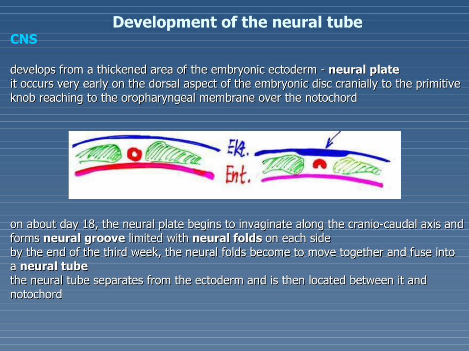
develops from a thickened area of the embryonic ectoderm - develops from a thickened area of the embryonic ectoderm - neural plateneural plateitit occurs very early on the dorsal aspect of the embryonic disc cranially to the primitive occurs very early on the dorsal aspect of the embryonic disc cranially to the primitive knob reaching to the oropharyngeal membrane over the notochordknob reaching to the oropharyngeal membrane over the notochord
onon about day 18, the neural plate begins to invaginate along the cranio-caudal axis and about day 18, the neural plate begins to invaginate along the cranio-caudal axis and forms forms neural grooveneural groove limited with limited with neural folds neural folds on each sideon each sidebyby the end of the third week, the neural folds become to move together and fuse the end of the third week, the neural folds become to move together and fuse into into a a neural tubeneural tube the neural tube separates from the ectoderm and is then located between it and the neural tube separates from the ectoderm and is then located between it and notochord notochord

..
aat the time when the neural folds fuse, some t the time when the neural folds fuse, some neuroectodermal cells separate from them and neuroectodermal cells separate from them and form along the dorsal aspect of the tube single form along the dorsal aspect of the tube single cord - called the cord - called the neural crestneural crest; it; it soon divides in soon divides in the left and right parts that migrate to the the left and right parts that migrate to the dorsolateral aspect of the neural tubedorsolateral aspect of the neural tube
nneural crest cells give rise to cells of the eural crest cells give rise to cells of the spinal spinal gangliaganglia and cells of the and cells of the autonomic gangliaautonomic ganglia

ffrom the beginning, the proximal segment of the neural tube is broadened and rom the beginning, the proximal segment of the neural tube is broadened and correspondcorresponds s to futureto future brain brainthe narrower caudal one developthe narrower caudal one developss in in the the spinal cordspinal cord

Histogenesis of the neural tubeHistogenesis of the neural tubetthe wall of the neural tube is initially composed he wall of the neural tube is initially composed of a thickof a thick pseudostratified columnar epitheliumpseudostratified columnar epithelium,,cells cells then then rapidly proliferate in entire thickness rapidly proliferate in entire thickness of the wallof the wall - - but later mitotic activity is reduced but later mitotic activity is reduced only on cells situated near the luminal aspect of only on cells situated near the luminal aspect of the neural tubethe neural tube; a; as a result of this process, the s a result of this process, the wall of neural tube differentiates into 2 zones: wall of neural tube differentiates into 2 zones: the inner the inner germinative germinative and the outer and the outer marginal onesmarginal ones iin the germinative zone the cells continue in n the germinative zone the cells continue in their mitotic activity and migrate peripherallytheir mitotic activity and migrate peripherally
finallyfinally, the wall of neural tube shows 3-layered , the wall of neural tube shows 3-layered structure:structure:
- the - the ependymalependymal layer = ependyma, layer = ependyma,- the - the intermediate intermediate or or mantle mantle layer= layer=
gray mattergray matter - - cells of mantle layer soon cells of mantle layer soon differentiate into primitive neurons - neuroblasts differentiate into primitive neurons - neuroblasts and spongioblasts (glioblasts),and spongioblasts (glioblasts),
-- the the marginalmarginal layer = layer = white matterwhite matter (contains no cells)(contains no cells)

DEVELOPMENT OF THE SPINAL CORDDEVELOPMENT OF THE SPINAL CORD
itit develops from the caudal portion of the neural tube develops from the caudal portion of the neural tubeinin contrast with lateral walls of the neural tube, contrast with lateral walls of the neural tube, wherewhere cells rapidly proliferate, the cells rapidly proliferate, the dorsal and ventral aspects remain thindorsal and ventral aspects remain thinllongitudinal groove - sulcus limitans - divides both lateral walls in the dorsal part - ongitudinal groove - sulcus limitans - divides both lateral walls in the dorsal part - alar platealar plate and ventral part - and ventral part - basal platebasal platecells of mantle layer rapidly cells of mantle layer rapidly proliferateproliferate and differentiate in the gray matter and differentiate in the gray matter Remember:Remember:The alar plate - The alar plate - givegivess rise to rise to dorsal horn, the basal plate - dorsal horn, the basal plate - to to ventral hornventral horn


Positional changes of the spinal cordPositional changes of the spinal cord Initially, the spinal cord extends the entire length of the vertebraInitially, the spinal cord extends the entire length of the vertebrall canal canaldduring further development, the vertebrauring further development, the vertebrall canal grows canal grows more more rapidly than spinal cord rapidly than spinal cord and its caudal end gradually comes to lie at relatively higher levelsand its caudal end gradually comes to lie at relatively higher levelsiin adults, it usually terminates at the inferior border of the first lumbar vertebran adults, it usually terminates at the inferior border of the first lumbar vertebra
DEVELOPMENT OF THE BRAIN
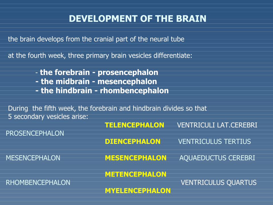
the brain develops from the cranial part of the neural tube
at the fourth week, three primary brain vesicles differentiate:
- the forebrain - prosencephalon- the midbrain - mesencephalon- the hindbrain - rhombencephalon
During the fifth week, the forebrain and hindbrain divides so that5 secondary vesicles arise:
TELENCEPHALON VENTRICULI LAT.CEREBRIPROSENCEPHALON
DIENCEPHALON VENTRICULUS TERTIUS
MESENCEPHALON MESENCEPHALON AQUAEDUCTUS CEREBRI
METENCEPHALONRHOMBENCEPHALON VENTRICULUS QUARTUS
MYELENCEPHALON


TELENCEPHALON VENTRICULI LAT. CEREBRIPROSENCEPHALON
DIENCEPHALON VENTRICULUS TERTIUS
MESENCEPHALON MESENCEPHALON AQUAEDUCTUS CEREBRI
METENCEPHALONRHOMBENCEPHALON VENTRICULUS QUARTUS
MYELENCEPHALON
Development of cavities in CNSDevelopment of cavities in CNS

Development the eye
Development of the retina
Development of the external and middle layer of the
eye
Development of the lens

tthe eye primordia appear very early (about on the 22nd day)he eye primordia appear very early (about on the 22nd day) asas optic optic groovesgrooves (optic sulci)(optic sulci) in the neural foldsin the neural folds at the site of forebrainat the site of forebrain
aas neural folds fuse, the optic s neural folds fuse, the optic grooves grooves evaginate to form pairedevaginate to form paired hollow hollow diverticula called diverticula called optic vesiclesoptic vesicles
tthe optic vesicle then grows laterally on each side and its connection withhe optic vesicle then grows laterally on each side and its connection withthe forebrain becomes to narrow and forms the forebrain becomes to narrow and forms optic stalkoptic stalk

llater both optic vesicles invaginate and become double-walled, cup-like structures - ater both optic vesicles invaginate and become double-walled, cup-like structures - optic optic ccupsupsththey reach up to the surface ectoderm that becomes thickened and form ey reach up to the surface ectoderm that becomes thickened and form lens placodelens placode
tthe central region of each lens placode invaginates and sinks below the surface, forming he central region of each lens placode invaginates and sinks below the surface, forming aa llens pitens pit
tthe edges of the lens pit gradually come together and fuse to form a spherical he edges of the lens pit gradually come together and fuse to form a spherical lens vesiclelens vesicle
Remember:Remember: the lens vesicle and optic cup derive from the ectoderm and are completely the lens vesicle and optic cup derive from the ectoderm and are completely surrounded with head mesenchymesurrounded with head mesenchyme

The The retinaretina
develops from the double-layered optic cupdevelops from the double-layered optic cup
ththe outer layer becomes the pigment epithelium, and the inner layer e outer layer becomes the pigment epithelium, and the inner layer differentiates into the remaining layers (rod and cone, bipolar, and differentiates into the remaining layers (rod and cone, bipolar, and ganglion cells)ganglion cells)
iintraretinal space, presented initially between the outer and inner layers ntraretinal space, presented initially between the outer and inner layers gradually disappears so that the pigment epithelium and remaining gradually disappears so that the pigment epithelium and remaining retinal layers fuseretinal layers fuse
the junction of definitive pigment layer with the layer of rods and cones is the junction of definitive pigment layer with the layer of rods and cones is not so firm as elsewhere so that not so firm as elsewhere so that detachmentdetachment of retina of retina may may occur (after occur (after traumatic injury of the eye)traumatic injury of the eye)
The edge of the optic cup gives rise to the ciliary epithelium and posterior The edge of the optic cup gives rise to the ciliary epithelium and posterior epithelium of the irisepithelium of the iris
isis identical with the identical with the not photosensitive portion of the retina not photosensitive portion of the retina


The The middle and external middle and external layers layers develop from thedevelop from the mesenchyma mesenchyma that that envelops the external surface of theenvelops the external surface of theoptic cup optic cup
The lensThe lens is is developdevelopeded from the from the lens vesiclelens vesicle thethe anterior wall of th anterior wall of the e vesicle gives rise to the anterior epithelium of the vesicle gives rise to the anterior epithelium of the lens, the cells of the posterior wall gradually lengthen and form lens fiberslens, the cells of the posterior wall gradually lengthen and form lens fibersthethe lens capsule is produced by the epithelial cells of both aspects of the lens capsule is produced by the epithelial cells of both aspects of thelens vesiclelens vesiclenunutrition of the lens during development is provided by the trition of the lens during development is provided by the hyaloid arteryhyaloid artery,,a branch of the ophthalmic arterya branch of the ophthalmic arteryrrests of the hyaloid artery found in vitreous body are known as hyaloid canalests of the hyaloid artery found in vitreous body are known as hyaloid canal(Cloqueti)(Cloqueti)
The The anterior eye chamberanterior eye chamber originates as cleft-like space that forms between originates as cleft-like space that forms betweenthe lens and the surface ectodermthe lens and the surface ectoderm
The The corneacornea develops from the surface ectoderm and mesenchyme adhering develops from the surface ectoderm and mesenchyme adheringto it after forming of the anterior eye chamberto it after forming of the anterior eye chamber the stalk of the optic cup becomes the optic nervethe stalk of the optic cup becomes the optic nerve


Development the ear
Development of the external ear
Development of the middle ear
Development of the external ear

THE EXTERNAL EARTHE EXTERNAL EAR
The external acoustic meatusThe external acoustic meatus develops from the dorsal end of the develops from the dorsal end of the 1st 1st branchial groovebranchial groove;; ectodermal cells at the bottom of th ectodermal cells at the bottom of thee groove groove proliferate and extend inward as a solid epithelial plate - proliferate and extend inward as a solid epithelial plate - meatal plugmeatal plug;; in in the fetal period, the central cells of this plug degenerate, forming cavity the fetal period, the central cells of this plug degenerate, forming cavity that becomes the inner part of the external acoustic meatusthat becomes the inner part of the external acoustic meatus

The auricleThe auricle develops fromdevelops from 6 6 swellings known as swellings known as auricle hillocksauricle hillocks that surround the margin of that surround the margin of the first branchial groovethe first branchial groove3 hillocks are on the first branchial (mandibular) arch and 3 on the second (hyoid) 3 hillocks are on the first branchial (mandibular) arch and 3 on the second (hyoid) branchial archbranchial archAt the end of the 2nd month all hillocks fuse to form the definitive At the end of the 2nd month all hillocks fuse to form the definitive pinnapinna
The tympanic membraneThe tympanic membrane (TM)(TM)derives from the branchial membrane separating the derives from the branchial membrane separating the 1st1st branchial groove and the branchial groove and the 1st 1st pharyngeal pouchpharyngeal pouchiinitially, the membrane is made up of only the ectoderm and endodermnitially, the membrane is made up of only the ectoderm and endoderm,, aas development proceeds, s development proceeds, mesenchyme grows between both germ layers and is differentiated into the fibrous stratum of the mesenchyme grows between both germ layers and is differentiated into the fibrous stratum of the (tm), t(tm), the he ectoderm gives rise to the epidermal and the endoderm to the mucous aspect of the definitive ectoderm gives rise to the epidermal and the endoderm to the mucous aspect of the definitive TMTM..

THE MIDDLE EARTHE MIDDLE EAR2 different embryonic anlagen2 different embryonic anlagen - - the the 1st 1st pharyngeal pouchpharyngeal pouch and and - - cartilages of the cartilages of the 1 st1 st and and 2nd2nd pharyngeal arches pharyngeal arches
TThe he tympanic cavitytympanic cavity - 1st- 1st pharyngeal pouch pharyngeal pouch - - its distal its distal end expands that then end expands that then envelopes auditory ossiclesenvelopes auditory ossiclestthe proximalhe proximal unexpanded portion becomes the unexpanded portion becomes the Eustachian tubEustachian tubee

THE INNER EARTHE INNER EARderivesderives from the external germ layer from the external germ layer -- the the ectodermectoderm
an anlage occurs earlyan anlage occurs early in the fourth week in the fourth week as as a thickened plate of the ectoderm a thickened plate of the ectoderm - - otic placodeotic placode - on each side of the head - on each side of the headbothboth placode placodess invaginate and sink below the surface ectoderm into the underlying invaginate and sink below the surface ectoderm into the underlying mesenchyme to form mesenchyme to form otic pitotic pitedges of the pit come together and fuse to form an edges of the pit come together and fuse to form an otic vesicle (otocyst)otic vesicle (otocyst) that lies that lieslaterallylaterally to the rhombencephalonto the rhombencephalon

tthe otocyst serves a primordium of future he otocyst serves a primordium of future membranous labyrinthmembranous labyrinth

two divisions two divisions are are early recognizable:early recognizable: a a dorsal dorsal oror utricular portion utricular portion, differentiating into , differentiating into the utricle, semicircular ducts and the utricle, semicircular ducts and endolymphatic duct and sacendolymphatic duct and sac and and
a a ventralventral or or saccular portion saccular portion that that gives rise to gives rise to the saccule and cochlear ductthe saccule and cochlear duct
Initially, the semicircular ducts form flat-like diverticula growing Initially, the semicircular ducts form flat-like diverticula growing out from the utricular portion; central parts of them then fuse out from the utricular portion; central parts of them then fuse and disappear and disappear
tthe peripheral infused portions of the diverticula become the he peripheral infused portions of the diverticula become the semicircular ducts semicircular ducts
ffrom the ventral saccular portion of the otocyst, the coiled rom the ventral saccular portion of the otocyst, the coiled cochlear diverticulum grows outcochlear diverticulum grows out
ststarting the arting the 4th month4th month, , differentiationdifferentiation of of maculae, cristaemaculae, cristae begins within the utricle, saccule, and semicircular ducts as begins within the utricle, saccule, and semicircular ducts as well as the well as the organ of Cortiorgan of Corti within the cochlear duct within the cochlear duct
tthe mesenchyme around the otic vesicle (later its parts) he mesenchyme around the otic vesicle (later its parts) condenses and differentiates into the condenses and differentiates into the bony labyrinthbony labyrinth
aa space separating the membranous labyrinth from the osseous space separating the membranous labyrinth from the osseous one soon fills the one soon fills the perilymphperilymph


Development the skin and skin appendages
Development of epidermis and dermis
Development of eccrine sweet glands
Development of hairs
Development of nails

EpidermisEpidermisiinitially, a single layer of ectodermal cells covers the embryonitially, a single layer of ectodermal cells covers the embryo
sstarting from the 2nd month, the ectodermal cells divide and form a superficial protective layer tarting from the 2nd month, the ectodermal cells divide and form a superficial protective layer ofof flattened cells, the flattened cells, the peridermperiderm or or epitrichium epitrichium
at the end of 4th month, the epidermis acquires its definitive arrangement aat the end of 4th month, the epidermis acquires its definitive arrangement andnd4 layers are distinguished: basal, spinous, granular and horny layer4 layers are distinguished: basal, spinous, granular and horny layer;; aall layers of the epidermisll layers of the epidermis-at birth-at birth
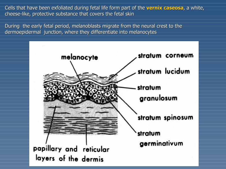
Cells that have been exfoliated during fetal life form part of the Cells that have been exfoliated during fetal life form part of the vernix caseosavernix caseosa, a white, , a white, cheese-like, protective substance that covers the fetal skincheese-like, protective substance that covers the fetal skin During the early fetal period, melanoblasts migrate from the neural crest to theDuring the early fetal period, melanoblasts migrate from the neural crest to thedermoepidermal dermoepidermal junction, where they differentiate into melanocytesjunction, where they differentiate into melanocytes

DermisDermis
tthe dermis is derived from the mesenchyme underlyinghe dermis is derived from the mesenchyme underlyingthe surface ectodermthe surface ectoderm tthe mesenchyme arises from 2 sources:he mesenchyme arises from 2 sources:
from the somatic layer of lateral mesoderm (most of the from the somatic layer of lateral mesoderm (most of the mesenchyme),mesenchyme),
from the dermomyotome regions of the somites (in lesser extent)from the dermomyotome regions of the somites (in lesser extent)
By 11 weeks, the mesenchymal cells have begun to produceBy 11 weeks, the mesenchymal cells have begun to producecollagenous and elastic connective tissue fiberscollagenous and elastic connective tissue fibers
As the epidermal ridges form, the dermis project upward intoAs the epidermal ridges form, the dermis project upward intothe epidermis and forms the epidermis and forms dermal papillaedermal papillae

Eccrine sweat glandsEccrine sweat glands develop as solid epidermal downgrowths that extend develop as solid epidermal downgrowths that extend into the underlying dermisinto the underlying dermisaas buds elongate, their ends become coiled, forming the primordia of future secretory s buds elongate, their ends become coiled, forming the primordia of future secretory portions of glandsportions of glands


Development of hairsDevelopment of hairs
begin to develop early in the fetal period, but they do not become readily visiblebegin to develop early in the fetal period, but they do not become readily visibleuntil about twentieth weekuntil about twentieth weekfirst recognizablefirst recognizable hairs occur hairs occur on the eyebrows, upper lip, and chinon the eyebrows, upper lip, and chin
A hair follicle begins as a solid downgrowth of the stratum A hair follicle begins as a solid downgrowth of the stratum germinativun of the epidermis and extends into the underlying germinativun of the epidermis and extends into the underlying dermisdermis
tthe deepest part of the he deepest part of the hair bud hair bud soon becomes club-shaped, forming asoon becomes club-shaped, forming a hair bulb. hair bulb. tthe epithelial cells of the hair bulb constitute the germinal matrixhe epithelial cells of the hair bulb constitute the germinal matrix- it- it gives rise to gives rise to hairhairThe hair bulb is then invaginated by a small mesenchymal The hair bulb is then invaginated by a small mesenchymal hair papillahair papilla
tthe peripheral cells of the developing hair follicle form the he peripheral cells of the developing hair follicle form the epithelial root sheathepithelial root sheathtthe surrounding mesenchymal cells differentiate into the he surrounding mesenchymal cells differentiate into the dermal (connective tissue) rootdermal (connective tissue) rootsheathsheath
first hairs are called first hairs are called lanugolanugo, , are fine a are fine andnd colourless colourlessthesethese hairs are replaced during the perinatal period by coarser hairs, called hairs are replaced during the perinatal period by coarser hairs, called vellusvellusthat that persist over most of the body, except in the axillary and pubic regionspersist over most of the body, except in the axillary and pubic regions
Hairs of these reagions are replaced during pubertyHairs of these reagions are replaced during puberty


apocrine sweat glands (axilla, pubic region, anal region, areolae) develop from the hair follicle similar as sebaceous glands

Development of mammary gland

The mammary glands develop during the sixth week as a solid The mammary glands develop during the sixth week as a solid downgrowth of the epidermis that extend into the underlying downgrowth of the epidermis that extend into the underlying mesenchymemesenchyme
tthese downgrowths occur along the hese downgrowths occur along the mammary ridgesmammary ridges, two , two thickened strips of ectoderm that extend from the axillary to thickened strips of ectoderm that extend from the axillary to the inguinal regionsthe inguinal regions
iin human embryos, these epithelial ridges occur during the n human embryos, these epithelial ridges occur during the fourth week, but except the pectoral area rapidly disappearfourth week, but except the pectoral area rapidly disappear
eeach ach primary mammary budprimary mammary bud soon gives rise to several soon gives rise to several secondary buds secondary buds that develop into lactiferous ducts and that develop into lactiferous ducts and their branches. The fibrous connective tissue and fat develop their branches. The fibrous connective tissue and fat develop from the surrounding mesenchymefrom the surrounding mesenchyme

NailsNailsToenails and fingernails begin to develop at the distal ends of Toenails and fingernails begin to develop at the distal ends of
the digits at about 10 weeksthe digits at about 10 weeks, d, development of fingernails evelopment of fingernails precedes that of the toenailsprecedes that of the toenails
The nails first appear as thickened areas of the developing The nails first appear as thickened areas of the developing epidermis on the dorsal aspect of each digitepidermis on the dorsal aspect of each digit
These These nail fieldsnail fields are surrounded laterally and proximally by are surrounded laterally and proximally by folds - folds - nail foldsnail folds
Cells from the proximal nail fold grow over the nail field and Cells from the proximal nail fold grow over the nail field and become keratinised to form the nail, become keratinised to form the nail, or nail plateor nail plate
At first, superficial layers of epidermis called the eponychium At first, superficial layers of epidermis called the eponychium cover the developing nail. This later degenerates, except at cover the developing nail. This later degenerates, except at the base of the nail, where it persists.the base of the nail, where it persists.