Embed Size (px)
Citation preview
Duhok UniversityFaculty of medical scienceSchool of health science
Dr. Azad Mustafa Ahmed
Lecturer, FIBMS (Path)University of Duhok, faculty of medical science
Cytology lecture series
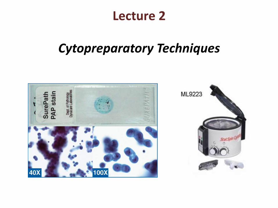
Lecture 2
Cytopreparatory Techniques
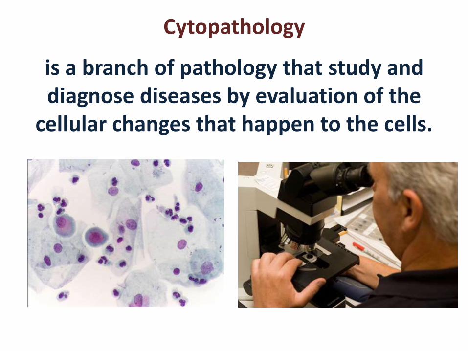
Cytopathology
is a branch of pathology that study and diagnose diseases by evaluation of the
cellular changes that happen to the cells.
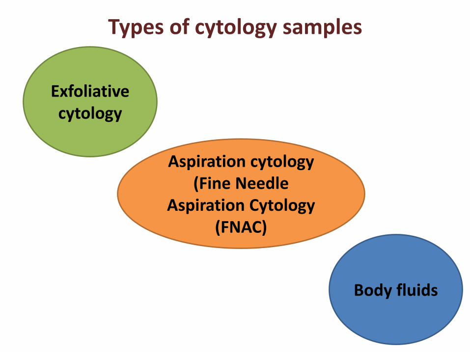
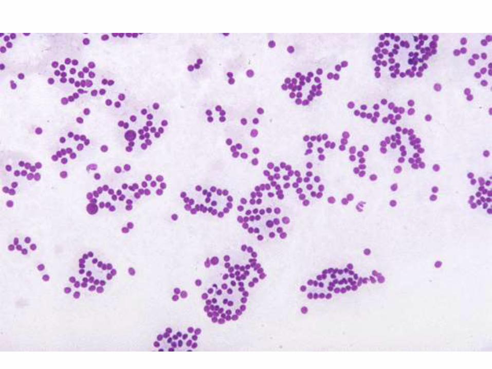
Types of cytology samples
Exfoliative cytology
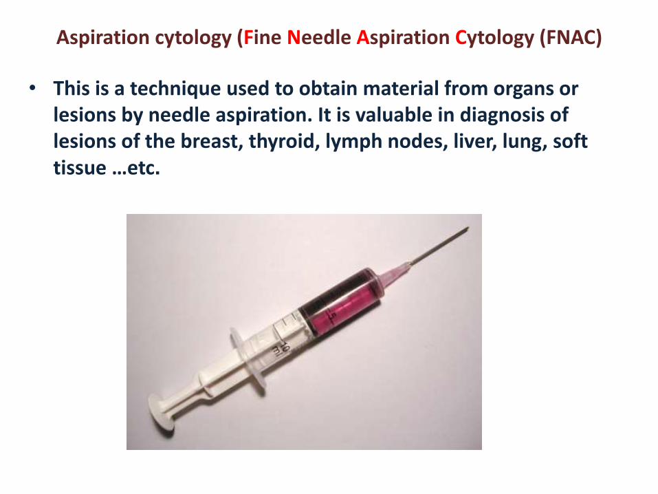
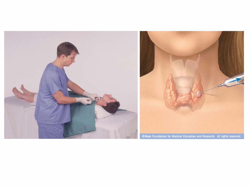
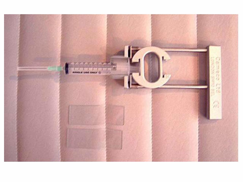
Aspiration cytology (Fine Needle
Aspiration Cytology (FNAC)
Body fluids
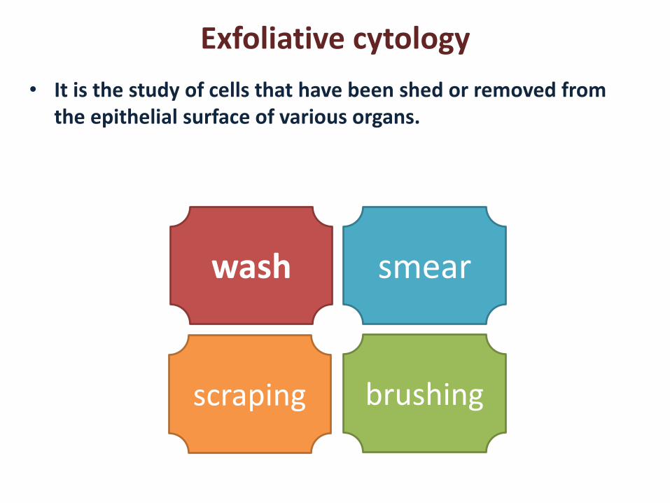
Exfoliative cytology
• It is the study of cells that have been shed or removed from the epithelial surface of various organs.
wash smear
scraping brushing
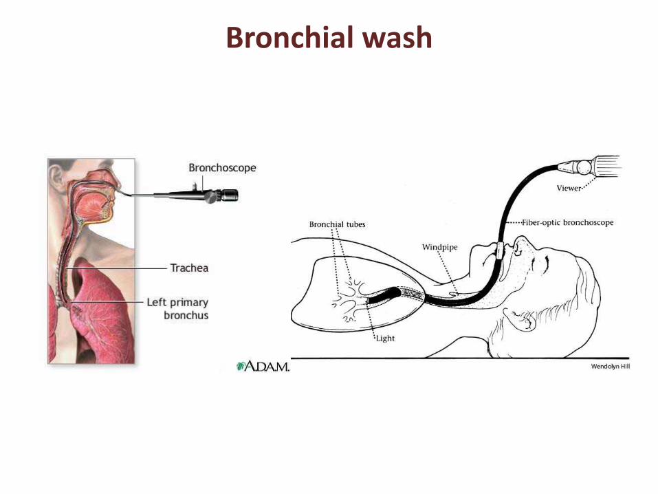
Bronchial wash
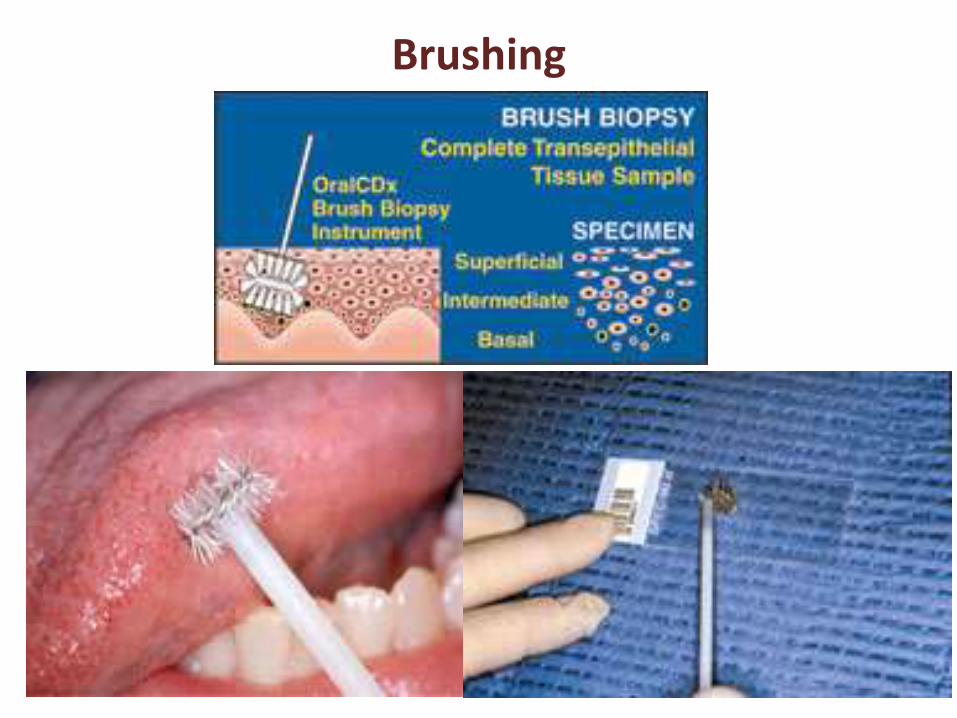
Brushing
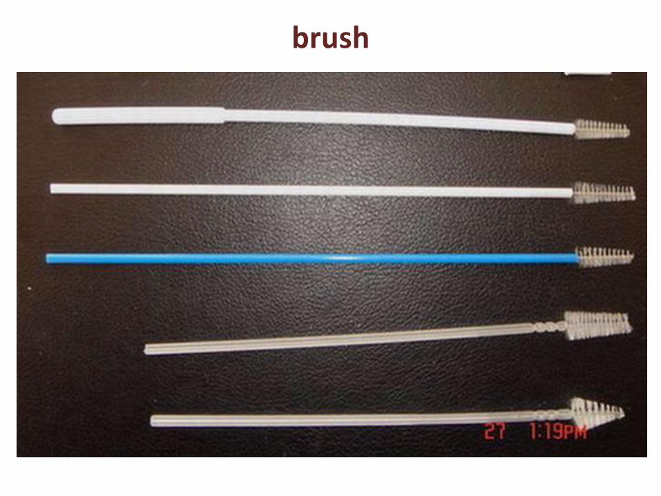
brush
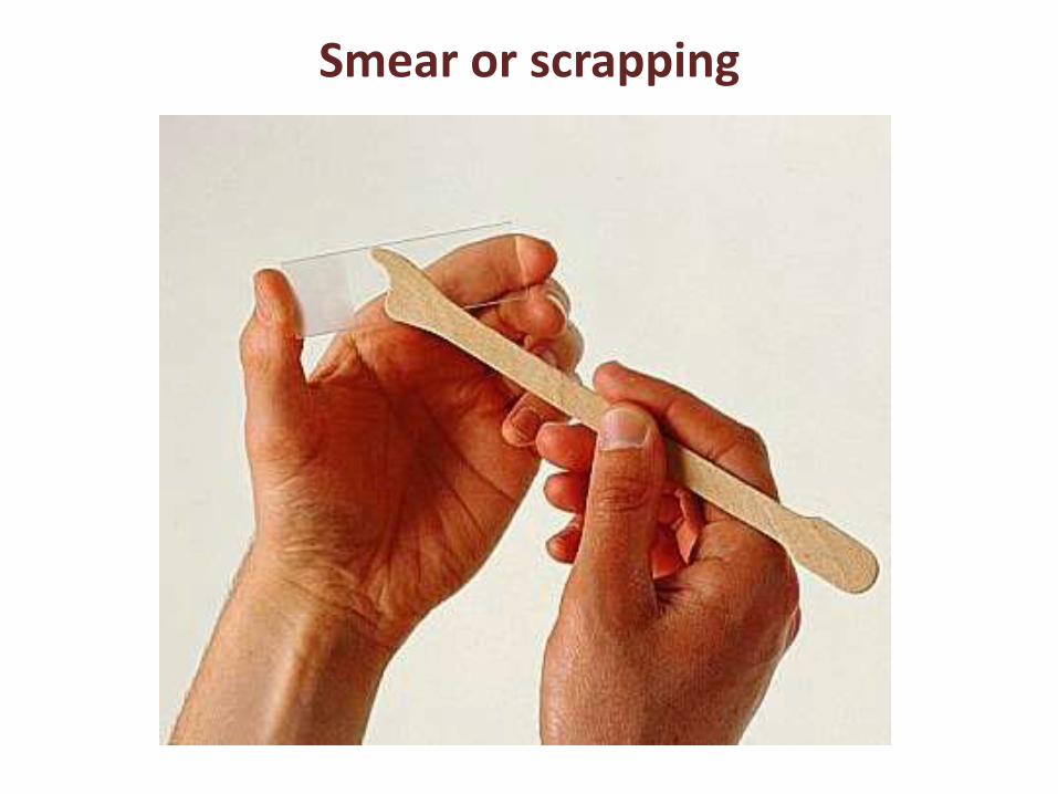
Smear or scrapping
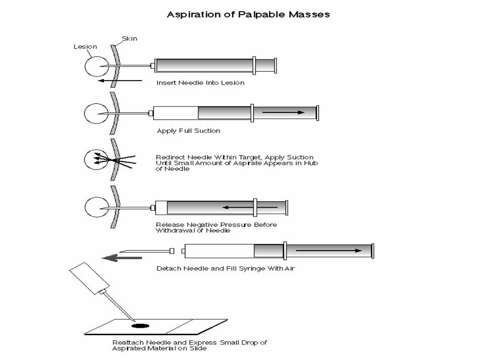
Aspiration cytology (Fine Needle Aspiration Cytology (FNAC)
• This is a technique used to obtain material from organs or lesions by needle aspiration. It is valuable in diagnosis of lesions of the breast, thyroid, lymph nodes, liver, lung, soft tissue …etc.
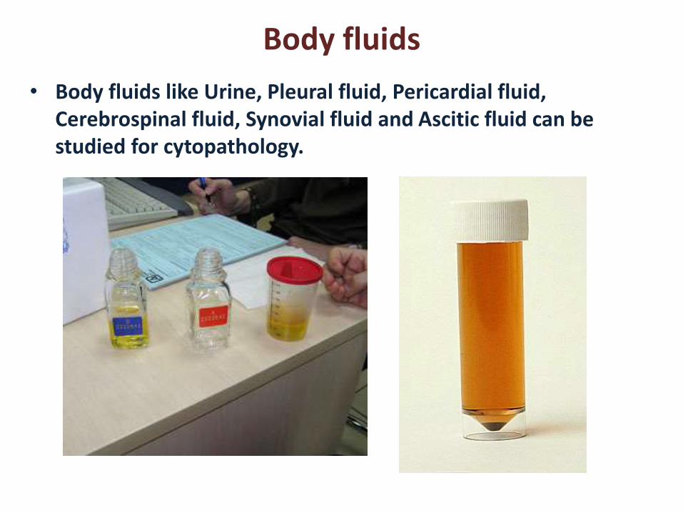
Body fluids
• Body fluids like Urine, Pleural fluid, Pericardial fluid, Cerebrospinal fluid, Synovial fluid and Ascitic fluid can be studied for cytopathology.
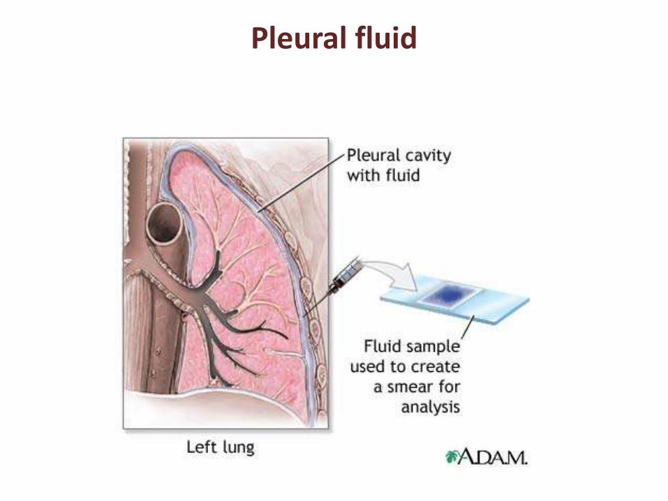
Pleural fluid
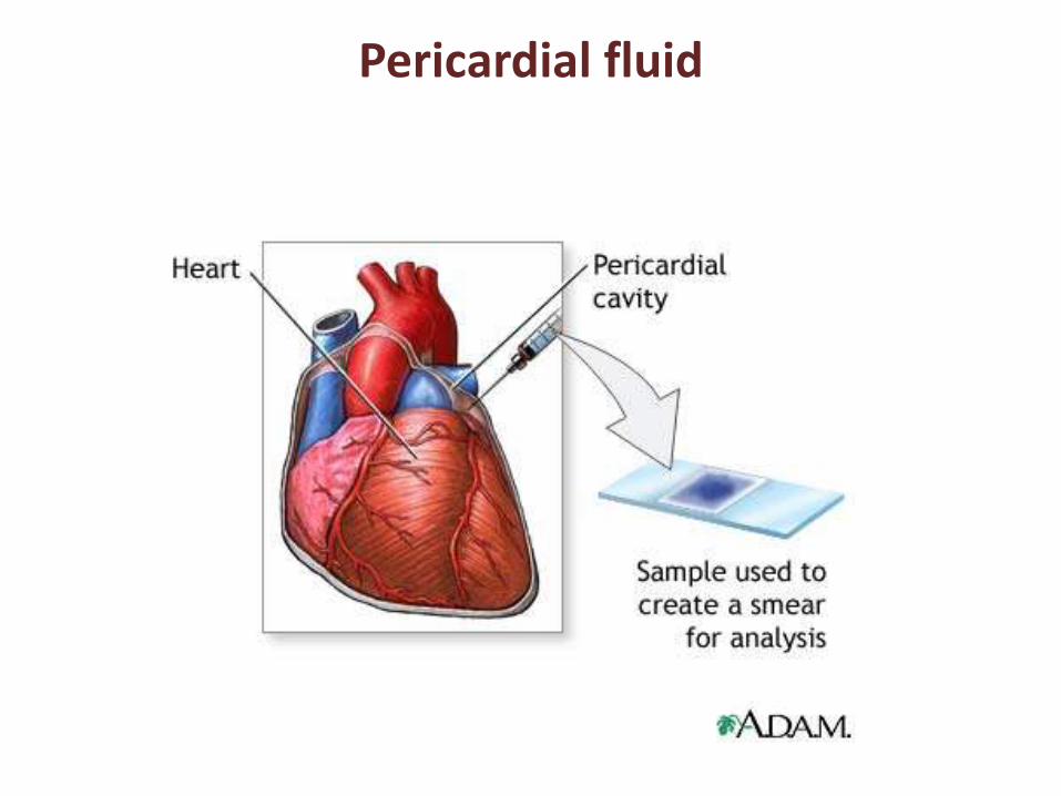
Pericardial fluid
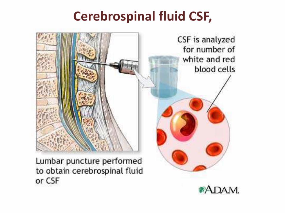
Cerebrospinal fluid CSF,
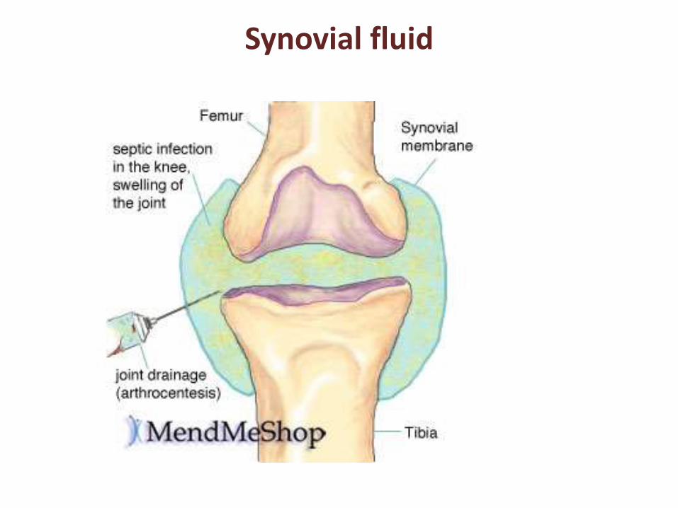
Synovial fluid
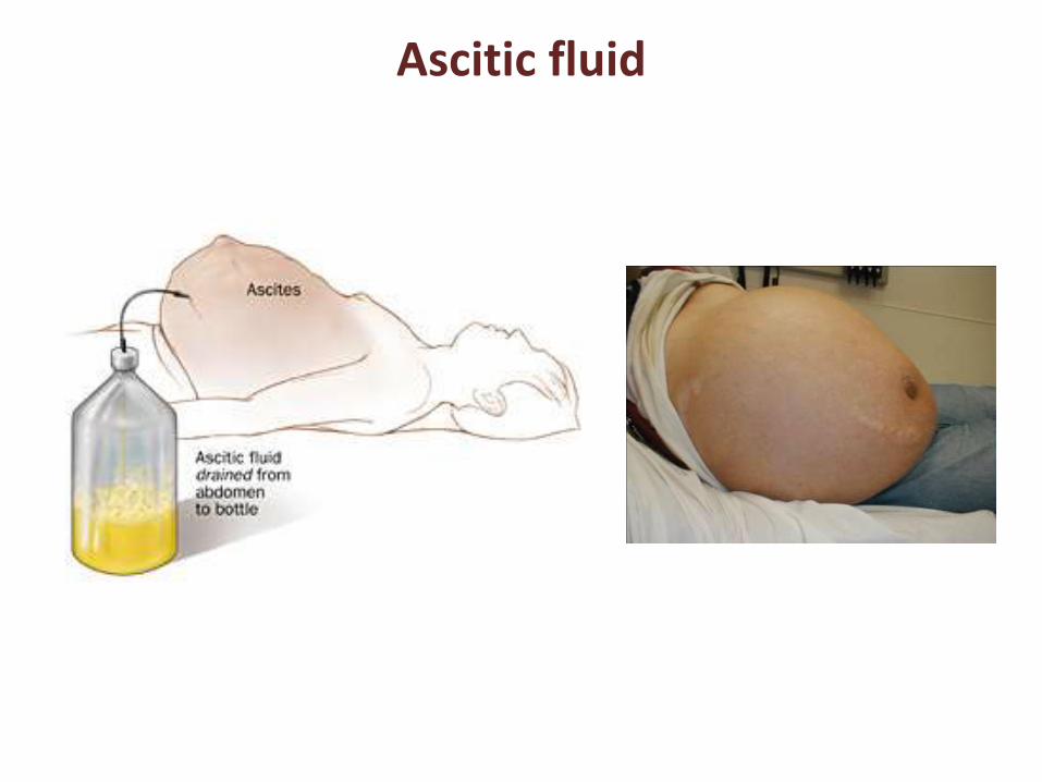
Ascitic fluid
Advantage of cytopathology:
1. a non-invasive procedure,
2. helps in faster reporting and guide the clinician
3. is relatively inexpensive
4. Has high population acceptance
5. Facilitates cancer screening.
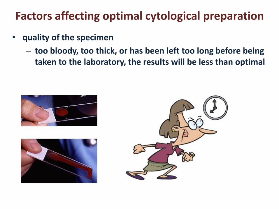
Factors affecting optimal cytological preparation
• quality of the specimen
– too bloody, too thick, or has been left too long before being taken to the laboratory, the results will be less than optimal
Factors affecting optimal cytological preparation
• fixative chosen to preserve the cellular details:
– If fixative not correct for the specimen or the fixative is not applied quickly, the results will again be less than optimal.
Factors affecting optimal cytological preparation
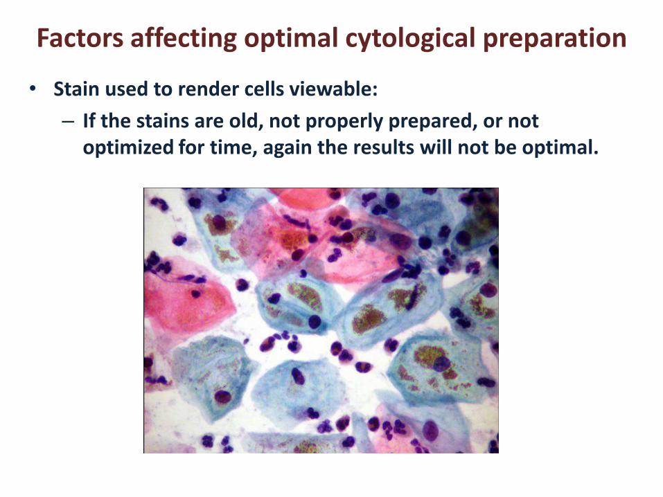
• Stain used to render cells viewable:
– If the stains are old, not properly prepared, or not optimized for time, again the results will not be optimal.
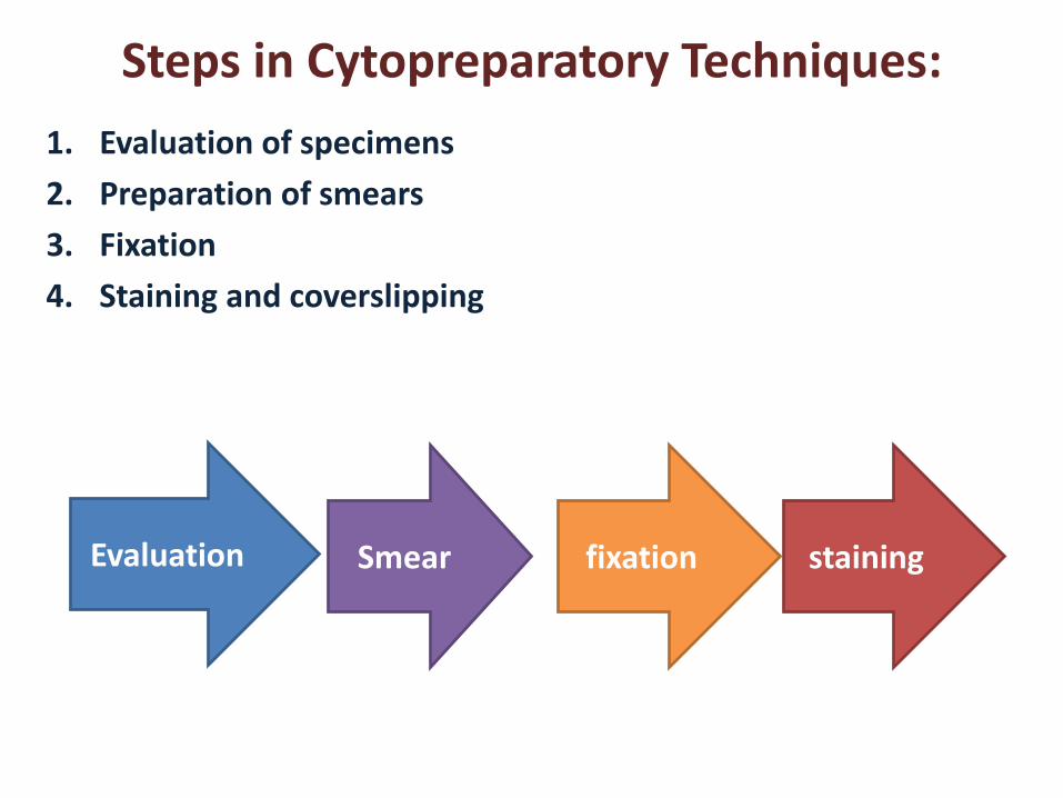
Steps in Cytopreparatory Techniques:
1. Evaluation of specimens
2. Preparation of smears
3. Fixation
4. Staining and coverslipping
Evaluation Smear fixation staining
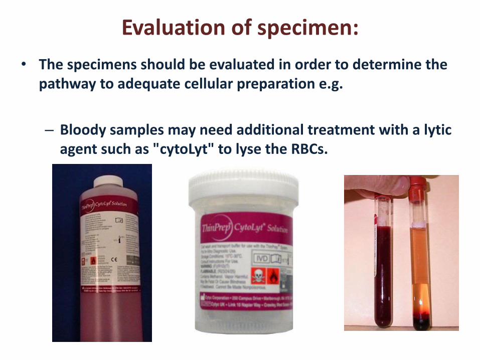
Evaluation of specimen:
• The specimens should be evaluated in order to determine the pathway to adequate cellular preparation e.g.
– Bloody samples may need additional treatment with a lytic agent such as "cytoLyt" to lyse the RBCs.
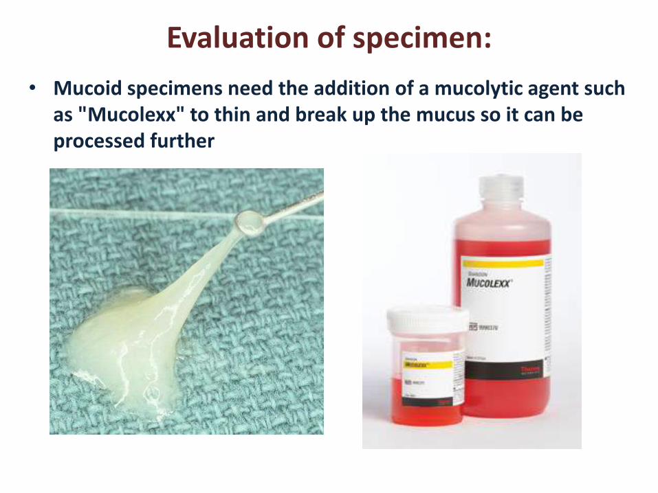
Evaluation of specimen:
• Mucoid specimens need the addition of a mucolytic agent such as "Mucolexx" to thin and break up the mucus so it can be processed further
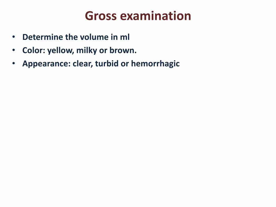
Gross examination
• Determine the volume in ml
• Color: yellow, milky or brown.
• Appearance: clear, turbid or hemorrhagic
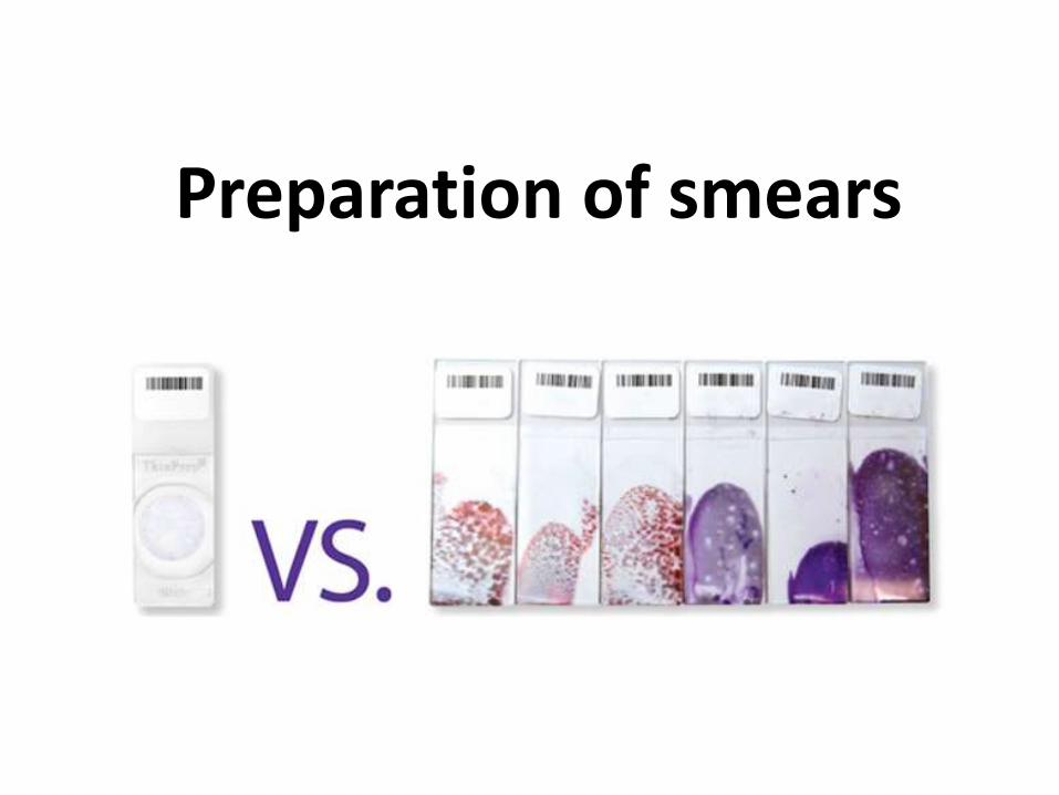
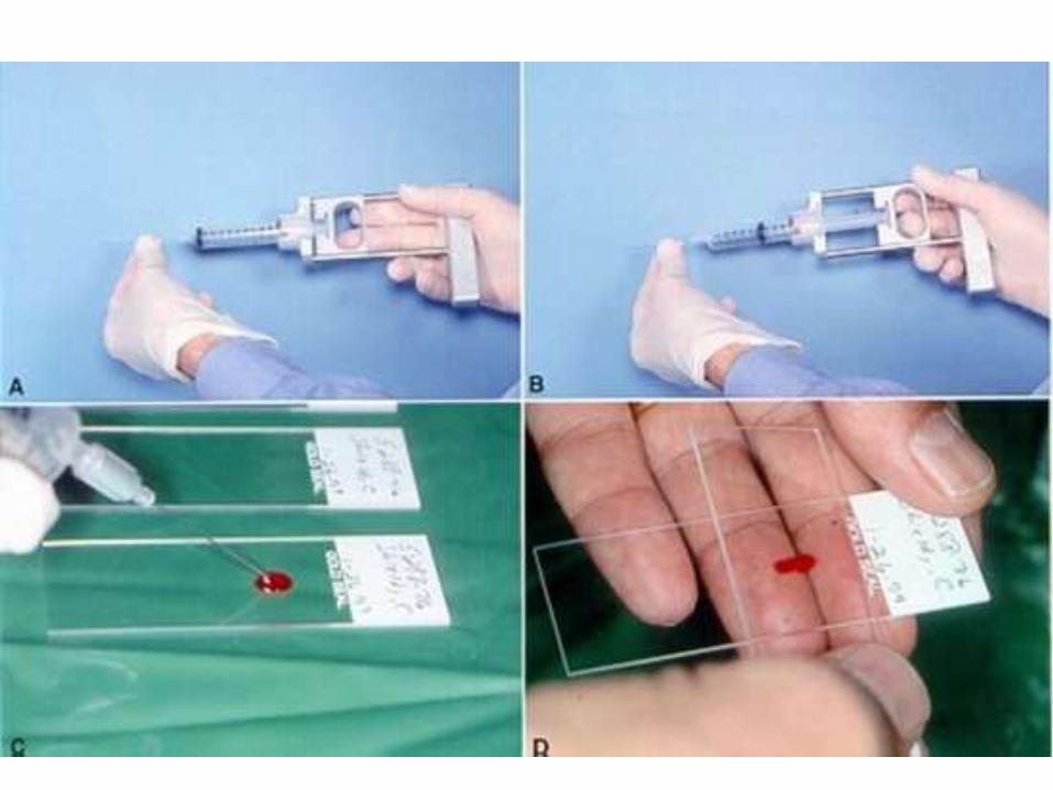
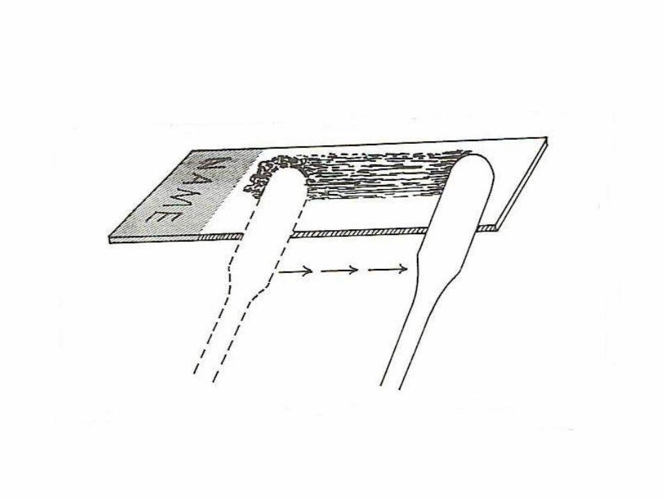
Preparation of smears
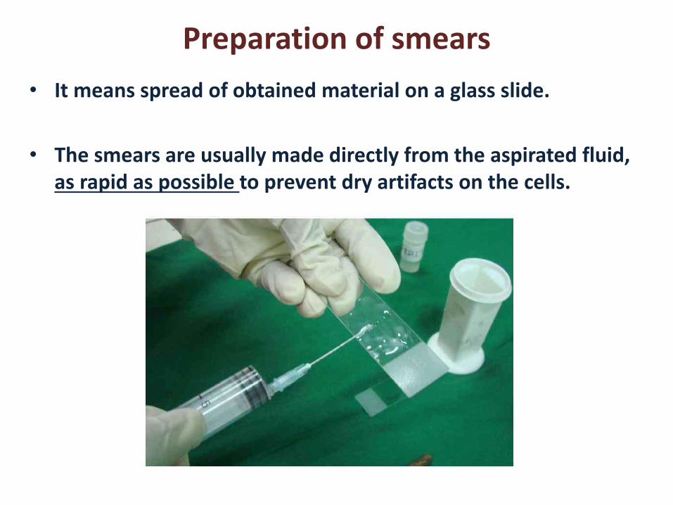
Preparation of smears
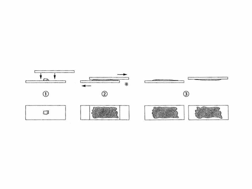
• It means spread of obtained material on a glass slide.
• The smears are usually made directly from the aspirated fluid, as rapid as possible to prevent dry artifacts on the cells.
Preparation of smears
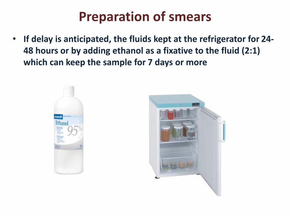
• If delay is anticipated, the fluids kept at the refrigerator for 24-48 hours or by adding ethanol as a fixative to the fluid (2:1) which can keep the sample for 7 days or more
Preparation of smears
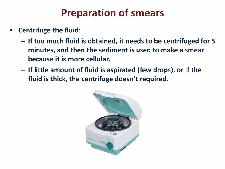
• Centrifuge the fluid:
– If too much fluid is obtained, it needs to be centrifuged for 5 minutes, and then the sediment is used to make a smear because it is more cellular.
– If little amount of fluid is aspirated (few drops), or if the fluid is thick, the centrifuge doesn’t required.

Preparation of smears
• Cytocentrifuge: It is a special machine that performs a centrifuge and collection of sediment on the center of the slides. This procedure is useful for hypocellular specimen like urine for cytology.
Preparation of smears
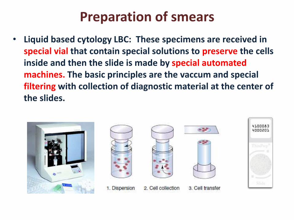
• Liquid based cytology LBC: These specimens are received in special vial that contain special solutions to preserve the cells inside and then the slide is made by special automated machines. The basic principles are the vaccum and special filtering with collection of diagnostic material at the center of the slides.
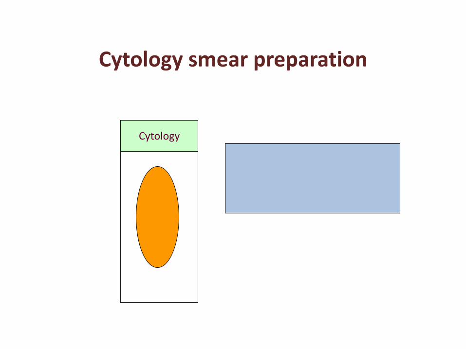
Cytology
Cytology smear preparation
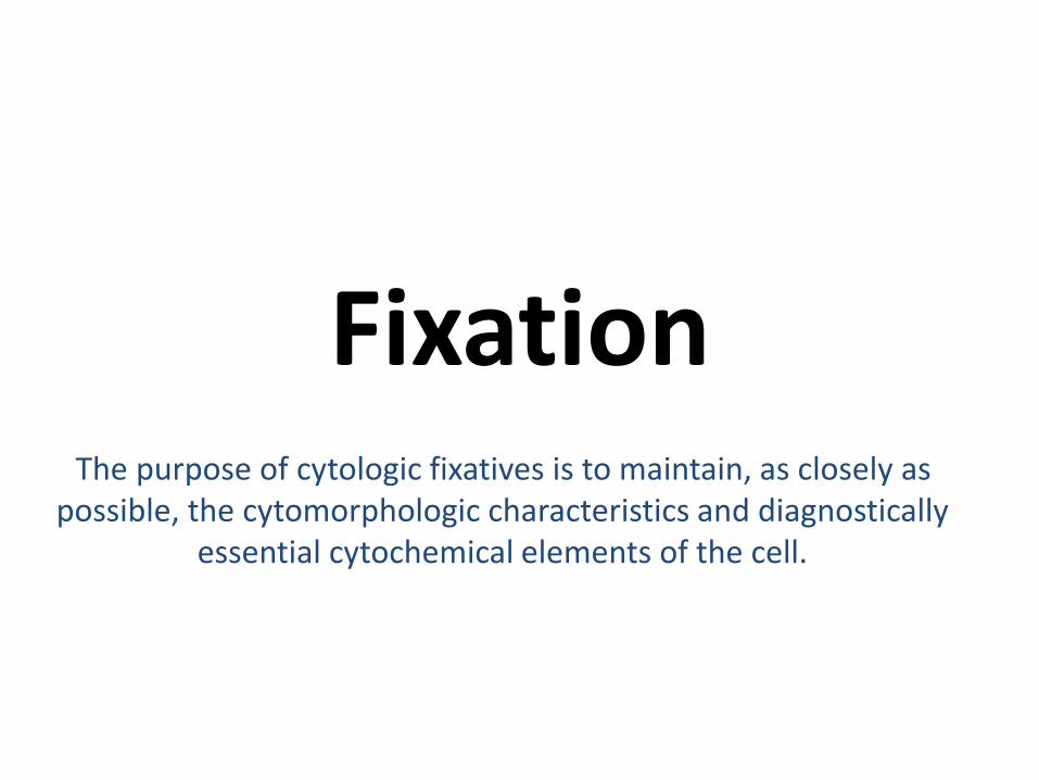
FixationThe purpose of cytologic fixatives is to maintain, as closely as
possible, the cytomorphologic characteristics and diagnostically essential cytochemical elements of the cell.
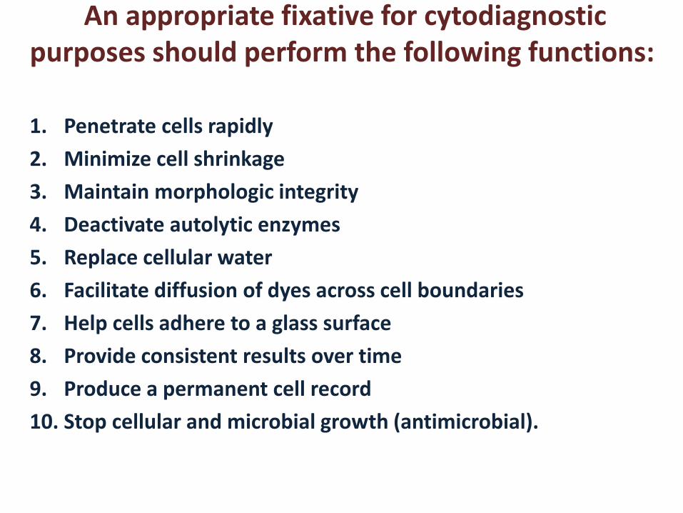
An appropriate fixative for cytodiagnostic purposes should perform the following functions:
1. Penetrate cells rapidly
2. Minimize cell shrinkage
3. Maintain morphologic integrity
4. Deactivate autolytic enzymes
5. Replace cellular water
6. Facilitate diffusion of dyes across cell boundaries
7. Help cells adhere to a glass surface
8. Provide consistent results over time
9. Produce a permanent cell record
10. Stop cellular and microbial growth (antimicrobial).
Types of fixative:
• Dry fixation:
• Wet fixation:
• Liquid-based Fixation:
• Lysing Fixation for Bloody Samples:
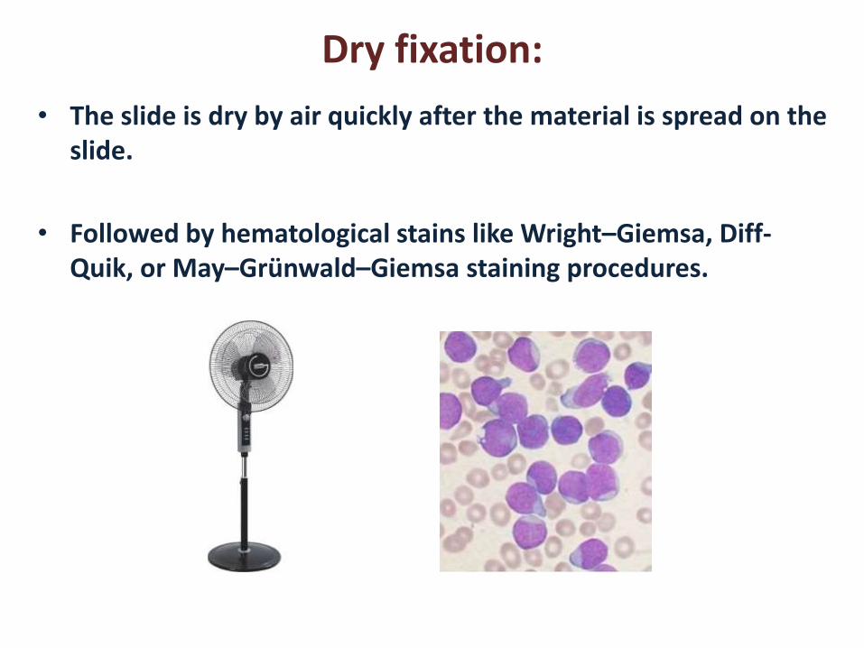
Dry fixation:
• The slide is dry by air quickly after the material is spread on the slide.
• Followed by hematological stains like Wright–Giemsa, Diff-Quik, or May–Grünwald–Giemsa staining procedures.
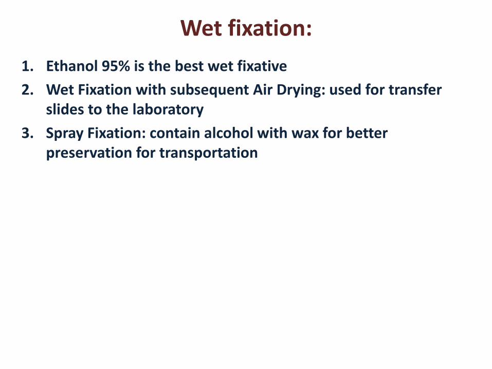
Wet fixation:
1. Ethanol 95% is the best wet fixative
2. Wet Fixation with subsequent Air Drying: used for transfer slides to the laboratory
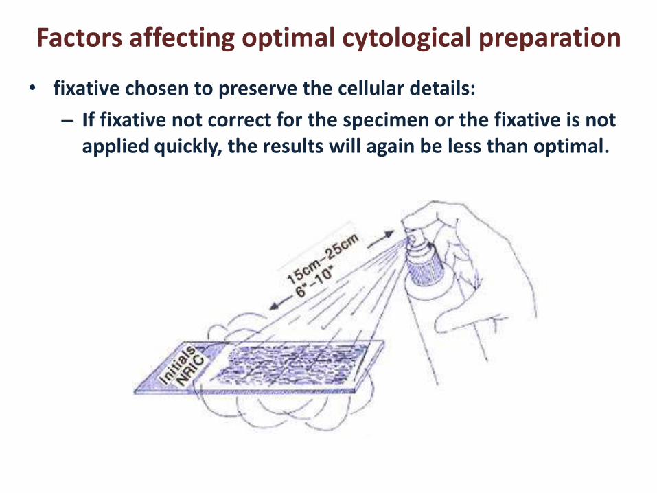
3. Spray Fixation: contain alcohol with wax for better preservation for transportation
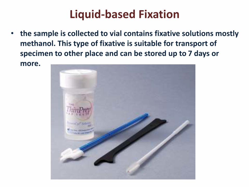
Liquid-based Fixation
• the sample is collected to vial contains fixative solutions mostly methanol. This type of fixative is suitable for transport of specimen to other place and can be stored up to 7 days or more.
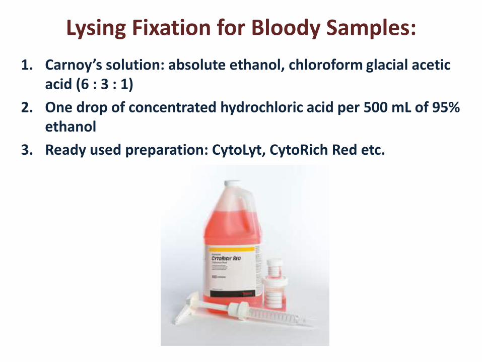
Lysing Fixation for Bloody Samples:
1. Carnoy’s solution: absolute ethanol, chloroform glacial acetic acid (6 : 3 : 1)
2. One drop of concentrated hydrochloric acid per 500 mL of 95% ethanol
3. Ready used preparation: CytoLyt, CytoRich Red etc.

`