Embed Size (px)
Citation preview

CT Diagnosis of Acute Mesenteric Ischemia from
Various Causes
AJR 2009
Akira Furukawa,Shuzo Kanasaki ,Naoaki Kono , Makoto, Wakamiya ,Toyohiko Tanaka ,Masashi Takahashi ,Kiyoshi
Murata

Acute mesenteric ischemia is a common but complex disorder with various primary cause and clinical presentation and high mortality.
Can be caused by various conditions such as arterial occlusion, venous occlusion, strangulating obstruction and hypoperfusion associated with nonocclusive vascular disease

Findings vary widely depending on the cause and underlying pathophysiology.
The severity of bowel ischemia (i.e. superficial mucosal or transmural bowel wall necrosis)
Location (i.e., small or large bowel)
Degree of hemorrhage subsequent super infections may affect the CT appearance.

CT ExaminationPreparation:- Oral and rectal administration of contrast material is
recommended for accurate CT and assessment of acute bowel ischemia.
Whether contrast material is indicated should be carefully considered for patients with bowel obstruction;
Materials containing barium are contraindicated in patients with bowel leak or perforation.
However, in the acute state, particularly life-threatening conditions, indication of transoral contrast may not be possible or may not be significant because of an adynamic ileus preventing contrast material from moving through the intestine.
Neutral contrast material should be used for the correct assessment of bowel enhancement after IV contrast administration.
The use of neutral contrast material is also beneficial in the formation of multiplanar images and in CT angiography because neutral contrast material does not interfere with image quality.
Positive contrast material used in assessing patients with ischemic colitis by showing thickened bowel wall and revealing the presence of bowel obstruction
Or in evaluating patients with a contraindication for IV contrast administration.

When contrast material is applied, oral administration of 600–750 mL of luminal contrast material 30–120 minutes before scanning and rectal administration of 400–800 mL of luminal
contrast material are used.

CT Technique
CT images are obtained from the dome of the liver to the level of the perineum to cover the entire course of the intestine.
With MDCT scanners,
Collimation of 0.5–2.5 mm
Detector pitch of 1.0–2.
Section thickness - 5- to 7-mm are usually constructed for image interpretation;

Acquisition of both unenhanced and contrast enhanced CT scans is always necessary.
Role of unenhanced CT is to identify vascular calcification, hyper attenuating intravascular clotting, and intramural hemorrhage.
CECT is to identify thrombi in the mesenteric arteries and veins, abnormal enhancement of the bowel wall, and the presence of embolism or infarction of other organs. 100–150 mL of iodinated contrast material Rate - 2–5 mL/s, scanning starts with delay times of 30 and 60
seconds for dual acquisition and 40 – 60 seconds for single acquisition.

Findings in Acute Bowel Ischemia• Variations depend on
- Pathogenesis of bowel ischemia - Acuteness,- Duration, site, and extent of the ischemic attack
- State of the collateral circulation- Superimposed bowel wall infection- Presence of perforation may also affect appearances of acute bowel ischemia.

BBowel wall thickness owel Wall
Normal ranges from 3 to 5 mm thick depending on the degree of bowel distention.
It is non specific but is the most frequently observed finding in mesenteric ischemia ,caused by mural edema, hemorrhage, or super infection of the ischemic bowel wall .
The degree of thickening is usually less than 1.5 cm, typically 8–9 mm.
Often observed in mesenteric venous occlusion, strangulation, ischemic colitis, and mesenteric arterial occlusion after reperfusion.

In arterial occlusive mesenteric ischemia ,the bowel wall becomes thinner rather than thicker
Because there is no arterial flow and neither mural edema nor hemorrhage occurs.
Thinning of the bowel wall or “paper-thin wall” is caused by volume loss of tissue and vessels in the bowel wall and by loss of intestinal muscular tone.
Bowel wall thickening is not a consistent finding in mesenteric ischemia, and the degree of thickening does not correlate with severity.

Fig. 1—Contrast-enhanced CT image of abdomen in 40-year-old woman withsuperior mesenteric vein and portal vein thrombosis. Wall thickening of ascending and transverse colon (arrowheads) is shown.
Fig 2ontrast-enhanced CT image of abdomen in 78-year-old man withembolism of superior mesenteric artery. Bowel loops are distended with air and their wall is “paper-thin.”

Bowel wall attenuation
Always be assessed on both unenhanced and contrast-enhanced CT images to avoid misinterpretation of high density of the bowel wall as normal positive enhancement on contrast-enhanced CT in cases of intramural hemorrhage.
On unenhanced CT images - low attenuation of the bowel wall indicates
bowel wall edema, occurs in mesenteric arterial occlusion after reperfusion, mesenteric venous occlusion, strangulation, and ischemic colitis.

High attenuation of the wall is caused by intramural hemorrhage and hemorrhagic infarction.
On CECT - a highly specific but not sensitive finding for acute
mesenteric ischemia is absent or diminished contrast enhancement of the bowel wall .
A halo or target appearance is also indicative of mesenteric ischemia, representing hyperemia and hyper perfusion associated with surrounding mural edema.
Can be seen in arterial occlusion after reperfusion, nonocclusive and veno-occlusive bowel ischemia, strangulation, and ischemic colitis.

Fig 1 CT image of lower abdomen in patient with strangulating obstruction.Bowel wall of involved segment is thickened and shows increased density (circle)indicating hemorrhagic infarction. Involved mesentery shows high attenuation
Fig 2 CECT image of pelvis in patient with strangulating obstruction. Bowel wall of involved loop shows target appearance

CECT image of lower abdomen obtained at equilibrium phase in patient with strangulating obstruction. Prominent mural enhancement of affected bowel segment (arrowhead) is shown, which indicates presence of bowel ischemia.

In the setting of mesenteric ischemia, Pneumatosis often indicates transmural infarction, particularly if it is associated with portomesenteric venous gas.

CECT images of upper (A), mid (B), and lower (C) abdomen show gas in portal venous branches (A), gas in mesenteric veins (B)

gas in bowel wall ( C).

Dilatation of the Bowel Lumen
Dilated because of interruption of normal bowel peristalsis (adynamic ileus).
Fluid distention of the bowel loops occurs by increased intestinal secretions, typically in veno-occlusive ischemia and strangulating bowel obstruction.
In exclusive arterial occlusion, the bowel seldom contains a large amount of luminal fluid.

Mesenteric Vessels
In most cases, emboli or thrombi in the mesenteric arteries and veins are clearly shown on contrast-enhanced CT images
Engorgement of the mesenteric veins caused by congestion of venous outflow is typically seen in venoocclusive bowel ischemia or strangulating bowel obstruction.

Mesentery
Mesenteric fat stranding and ascites appear with transudation of fluid in the mesentery or the peritoneal cavity caused by elevation of mesenteric venous pressure, which is commonly seen in strangulating bowel obstruction and venoocclusive bowel ischemia.
It is also frequently seen in ischemic colitis because of super infection of ischemic colonic segments.
mesenteric fat stranding in these conditions can appear without bowel infarction and has limited value in estimating the severity of bowel ischemia.
Patients with arterial occlusive mesenteric ischemia, because the CT finding of fat stranding is almost exclusively present with transmural infarction.

Bowel Ischemia and Infarction in
Various Conditions
Bowel ischemia and infarction can be caused by various conditions such as mesenteric arterial occlusion mesenteric venous occlusion strangulating bowel obstruction and hypo perfusion associated with nonocclusive
vascular disease.

Acute Mesenteric Arterial Occlusion
Caused by a Thromboembolism associated with cardiovascular problems followed by arterial thrombosis, which accounts for arterial embolism, 40–50%; arterial thrombosis, 20–30%.
Most emboli wedge at branching points around or distal to the middle colic artery, whereas thrombosis typically occurs at or near the origin of the mesenteric arteries.
Although the severity may vary, bowel ischemia is typically followed by infarction, perforation, and peritonitis unless reperfusion occurs.
On CECT images, emboli and thrombi can be seen as defects in the superior mesenteric artery and its branches.
The diameter of the superior mesenteric artery is often larger than that of the superior mesenteric vein.
The thickness of the bowel wall of the involved segments is the same as or thinner than that of the healthy segments unless reperfusion occurs.
The lumen of the bowel may be filled with fluid, gas, or both; however, the bowel seldom contains a large amount of fluid. Contrast enhancement of the involved bowel is absent or diminished.

Fig 1- defect (arrowhead) in superior mesenteric artery.
Fig 2 - 71-year-old woman with superior mesenteric artery embolism.A, Contrast-enhanced CT image obtained cephalad to B shows defect in superior mesenteric artery (arrowhead).B, Contrast-enhanced CT image shows that mural enhancement is absent at most intestinal loops.

Pneumatosis can typically be observed in cases with transmural infarction with or without associated portomesenteric venous gas.
In cases with reperfusion or rich collaterals, the involved bowel segments may thicken and show the halo or target pattern of contrast enhancement.

CECT image in 83-year-old woman with superiormesenteric artery embolism after reperfusion. Mural thickening of intestine(circle) is visible, showing target appearance.

Venous Occlusion
Thrombosis of the mesenteric vein can be primary or secondary to portal hypertension or infection or can be associated with various hypercoagulopathy states.
Mesenteric venous obstruction does not typically lead to severe bowel ischemia; however, thrombosis of the mesenteric vein, particularly at a distal level, may cause bowel infarction and
Accounts for 5–10% of acute bowel ischemia

Elevation of the hydrostatic pressure, which leads to extravascular leakage of plasma, RBCs, or both into the bowel wall, mesentery, and peritoneal cavity.
The bowel loops are typically prominently dilated.
Impairment of venous drainage may also compromise the arterial blood flow and cause bowel ischemia and infarction.
On CECT, thrombus in the mesenteric and portal veins is usually visible, and mesenteric venous obstruction can be confirmed by CT in more than 90% of cases.

Engorgement of the mesenteric veins is also observed.
Fat stranding in the mesentery and ascites are common findings.
The bowel wall is prominently thickened with absent or diminished enhancement, hyperenhancement, or a halo or target pattern of contrast .
Absent or diminished contrast enhancement of the bowel wall usually indicates transmural infarction,
Particularly when it is associated with pneumatosis and portomesenteric venous gas .

CECT image in 40-year-old manwith SMV thrombosis shows defect in SMV (arrowhead). Distal branches of vein are engorged. 46-y man with SMV thrombosis.A, Contrast-enhanced CT image obtained cephalad to B shows thrombi in SMV (arrowhead) and splenic vein (arrow).B, Contrast-enhanced CT image shows engorgement of mesenteric veins (arrowhead) and mural thickening of intestine. Ascites is present.

Strangulating Obstruction: Closed-Loop Bowel Obstruction
Mechanical bowel obstruction associated with bowel ischemia that is seen in approximately 10% of patients with small-bowel obstruction.
Strangulating obstruction is almost exclusively associated with a closed-loop obstruction.
Which is caused most often by an adhesive band and occasionally by an internal or external hernia. A closed-loop obstruction tends to involve the mesentery and mesenteric vessels and is prone to produce a volvulus.

Strangulation in a closed-loop bowel obstruction is caused initially by impairment of venous outflow
followed by arterial ischemia because the arterial pressure is higher than the venous pressure.
Congestion or hemorrhage in the bowel wall and mesentery occurs, and the affected bowel loops are distended and filled with fluid.

On CT, a closed-loop obstruction is identified by a unique configuration of C- or U shaped distended loops with the mesenteric vessels converging toward the site of obstruction.
The obstructed site of the closed loops can be located by following the course of the distended bowel loops.
The affected bowel is filled almost exclusively with only fluid and no gas.
The affected mesentery typically shows a fan shape.
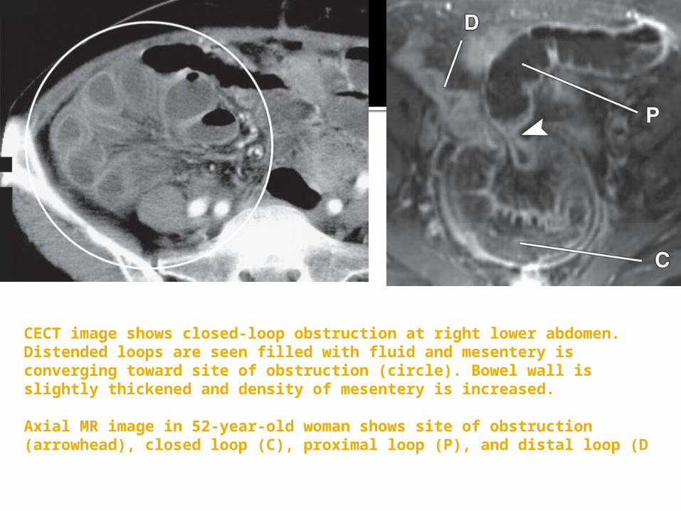
CECT image shows closed-loop obstruction at right lower abdomen. Distended loops are seen filled with fluid and mesentery is converging toward site of obstruction (circle). Bowel wall is slightly thickened and density of mesentery is increased.
Axial MR image in 52-year-old woman shows site of obstruction (arrowhead), closed loop (C), proximal loop (P), and distal loop (D

CECT SHOWS Distended loops are seen around their mesentery. Contrastenhancement is absent at bowel loops on right-hand side (arrowheads) andattached mesentery shows increased density, which indicates mesentericischemia.

absent or diminished bowel wall enhancement and infiltration of the affected mesentery are highly specific.
The “small-bowel feces sign” is also reported as a useful finding indicating the presence of strangulation.

Hypoperfusion Associated with Nonocclusive Vascular Disease
Bowel ischemia and infarction can occur with a reduction of mesenteric blood supply without vascular occlusion, which is called nonocclusive mesenteric ischemia or infarction.
This type of bowel ischemia accounts for 20–30% of all acute mesenteric ischemia or infarction cases, with mortality rates fromn30% to 93%
A reduction of the mesenteric blood supply is the result of mesenteric arterial vasoconstriction on reflex to hypotension or administration or abuse of digitalis, ergotamine, vasopressin or other vasoconstrictive agents, amphetamine, and cocaine.

Ischemic injury may range from reversible superficial damage localized to the watershed areas to a more severe form that extends to the entire bowel.
Hypoperfusion results in increased vascular permeability that leads to extravascular leakage of plasma, RBCs, or both into the bowel wall, mesentery, and peritoneal cavity.
Shock bowel is a variation of nonocclusive mesenteric ischemia caused by hypotensive shock induced by blunt abdominal trauma. Ischemic colitis and obstructive colitis are
considered similar disease entities.

On CT, the bowel wall of the involved segments may be normal or thickened.
The pattern of enhancement is variable as absent or diminished enhancement, increased enhancement, or halo or target type of enhancement.
Fat stranding of the mesentery and ascites are visible.
nonocclusive mesenteric ischemia is the most difficult condition to diagnose on CT, and angiography is often required for correct and confident diagnosis

CECT image (A) of pelvis in 69-year-old man with nonocclusive mesenteric ischemia. Contrast enhancement is prominently diminished or absent at distal ileal loops (arrowheads). Bowel wall thickening is not present.
B After reperfusion, bowel loops show prominent wall thickening with appearance of target sign

Thank you


![Challenges Encountered during the Treatment of Acute ...acute mesenteric ischemia is a great clinical challenge [1–6]. ... of acute mesenteric ischemia, occurring in 38 (92.68%)](https://img.dokumen.tips/doc/110x75/60f89ecd2be9754e8c1fff31/challenges-encountered-during-the-treatment-of-acute-acute-mesenteric-ischemia.jpg)