Embed Size (px)
Citation preview

DATE OF PRESENATION-:16-12-2014
PRESENTOR- DR. ANJU NAGAR

Normal extraocular muscle (EOM) innervation depends on-◦ normal cranial nuclear motoneuron formation from
neuronal precursors,
◦ normal axonal path finding from the cranial nuclei to developing EOMs, and
◦ the establishment and maintenance of normal connections between mature neurons and their target cells.

A number of well-defined syndromes characterized by congenital limitation of eye movements from abnormal innervation or miswiring of EOMs have recently been grouped as the “congenital
cranial dysinnervation disorders,” a term coined for
congenital disorders resulting from aberrant innervation of the ocular and facial musculature.

Assaf proposed the term of ‘CID syndrome’.
Features of this syndrome include:_ Congenital defect in the innervation EOMs.
_ Present since birth, and non-progressive.
_ Unilateral or bilateral
_ Findings are not explained by purely isolated oculomotor nerve palsy/palsies.
_ Anatomical muscle changes, including tight muscle
_ can be associated with synkinesis phenomena and/or co-contraction
_ abnormal head posture common

The term ‘congenital cranial dysinnervation disorders’ or CCDDs was derived in 2002 at a European Neuromuscular Centre (ENMC) international workshop by Gutowski et al for a group of congenital neuromuscular diseases reflecting the belief that these disorders resulted from developmental errors in innervation.
They define these disorders include-CFEOM, Congenital ptosis, Duane’s syndrome,Duane radial ray syndrome, Horizontal gaze palsy with progressive scoliosis (HGPPS), Mo¨bius syndrome.
The CCDDs also encompass developmental disorders of non-ocular cranial nerves, such as congenital familial facial weakness.

They summarized the features of the CCDDs as follows:
_ Congenital, non-progressive abnormalities of cranial musculature that result from developmental abnormalities of one or more cranial nerves with primary or secondary muscle dysinnervation
_ Primary may result from absence of normal muscle innervation. Secondary may occur from aberrant muscle innervation during development by branches of other nerves.
_ May be associated with secondary muscle pathology and/or other orbital and bony structural abnormalities.

_ Predominantly, vertical ocular motility defects are likely to result from abnormalities in development of oculomotor and trochlear nerves and/or nuclei (CFEOM variants and congenital ptosis).
_ Predominantly horizontal ocular motility defects are likely to result from abnormalities in the development of the abducens nerve and/or nucleus (Duane’s syndrome and HGPPS).
_ Predominately facial weakness is likely to result from abnormal development of facial nerve and/or nucleus, sometimes with associated ocular motor abnormalities(congenital facial weakness and Mo¨bius syndrome).
Recently, Kolling et al indicated congenital Brown’s syndrome is caused by missing fourth cranial nerve in some cases, which put it in the category of congenital dysinnervation.

The main CCDDs currently recognised and subdivided
1.Predominantly vertical disorder of ocular motility• Congenital fibrosis of the extraocular muscles (CFEOM)• Congenital ptosis
2.Predominantly horizontal disorder of ocular motility• Duane syndrome (DS)• DS + radial ray (DRRS)• Horizontal gaze palsy with progressive scoliosis (HGPPS)
3.Disorder of facial motility• Congenital facial palsy
4.Disorder of facial motility and ocular abduction
deficit• Möbius syndrome

Congenital fibrosis of the extraocular muscles (CFEOM) describes a group of rare congenital eye movement disorders that result from the dysfunction of all or part of the oculomotor (CN 3) and the trochlear (CN 4) nerves, and/or the muscles these nerves innervate.
CFEOMs are characterized by –◦ Variable impairment of horizontal and/or vertical eye
movements and ptosis. ◦ It can be unilateral or bilateral, with the bilateral form
being more common. ◦ Additional features include divergent strabismus and
abnormal head position, especially chin elevation
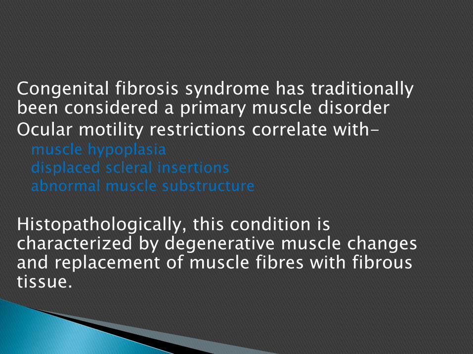
Congenital fibrosis syndrome has traditionally been considered a primary muscle disorderOcular motility restrictions correlate with-
muscle hypoplasiadisplaced scleral insertionsabnormal muscle substructure
Histopathologically, this condition is characterized by degenerative muscle changes and replacement of muscle fibres with fibrous tissue.

Recently, CFEOM has been accepted to be of neurogenic origin rather than primary muscle pathology.
α motoneurons of sup. division of III are present very early in development, but disappear early-?apoptosis, ?necrosis• Pulleys are normal-Demer
Engle EC., Leigh RJ.Genes, brainstem development and eye movements. Neurology 2002;59:304-305

1. Histopathology-◦ unreliable in distinguishing neurogenic from
myopathic ocular muscle weakness.
2. Co- contraction phenomenon-◦ Co-contraction phenomenon resulting in globe
retraction has also been described in patients with CFEOM.

4. Electro-myographic studies:
Patients with CFEOM commonly have variable angle strabismus.
Purely mechanical factors appear insufficient to explain this variability, which more likely reflects innervationaldisturbances.
Also, the degree of limitation of ocular movements in some patients do not always correlate with the degree of tightness of the agonist or antagonist muscle, as indicated by the intraoperative forced duction test.
Nystagmus is another indicator of central ocular motor disturbance in cases of CFEOM.

4. Neuro-imaging
Abnormalities such as cerebellar hypoplasia and asymmetrical ventricular size further hint at the possibility of brain malformation.
Hence, cases of CFEOM have neurogenic aetiologysimilar to that of DRS but involving the III cranial nerve complex.

CFEOM refers to at least seven genetically defined strabismus syndromes: ◦ CFEOM1A,
◦ CFEOM1B,
◦ CFEOM2,
◦ CFEOM3A,
◦ CFEOM3B,
◦ CFEOM3C, and
◦ Tukel syndrome

Affected individuals exhibit the following: Congenital non-progressive bilateral external
ophthalmoplegia Congenital non-progressive bilateral ptosis Primary vertical position of each eye: infraducted
(downward) Vertical eye movements: inability to elevate the eyes above
the horizontal midline Primary horizontal position of each eye: normal
(orthotropic), inward (esotropic), or outward (exotropic) Horizontal eye movements: normal to severely restricted Aberrant eye movements: common, especially both eyes
turning inward on attempted upgaze

Forced duction test positive for restriction
Binocular vision: usually absent
Refractive errors: frequently high astigmatism
Amblyopia: may be strabismic or refractive in nature
Pupils: normal
Family history: consistent with autosomal dominant inheritance; simplex cases (i.e., a single occurrence in a family) are observed. Parental germline mosaicism can mimic autosomal recessive inheritance.

CFEOM1 phenotype. A. Primary position showing marked bilateral ptosis. B. Primary position with lids held showing resting globe position. Both eyes remain infraducted below midline. C. Attempted up gaze showing inability to reach midline with convergence of the visual axis (synergistic convergence).

CFEOM- autosomal dominant inheritance with full penetrance and minimal variation in expression
CFEOM1 is divided into CFEOM1A and CFEOM1B based on genetic findings.
◦ CFEOM1A is associated with mutations in KIF21A gene; chr 12(centromeric region),
◦ CFEOM1B is associated with mutations in TUBB3 gene.

Affected individuals exhibit the following:
Congenital non-progressive bilateral external ophthalmoplegia
Congenital non-progressive bilateral ptosis
Primary vertical position of each eye: normal or positioned slightly above or below the midline
Vertical eye movements: severely restricted
Primary horizontal position of each eye: typically fixed outward (exotropic) or rarely fixed in a normal straight-ahead position (orthotropic)
Horizontal eye movements: severely restricted

Aberrant eye movements: small amplitude, if present
Forced duction test: positive for restriction
Binocular vision: absent
Refractive errors: frequent
Amblyopia: frequent
Pupils: often small and sluggishly reactive to light
Genetics:◦ autosomal recessive disorder
◦ mapped to chromosome 11g13.1.711
◦ result from mutations in the PHOX2A gene

CFEOM2 phenotype. A. Primary position showing bilateral ptosis and exotropia. B. Primaryposition with lids held showing resting globe position. C. Patient with right face turn andholding left upper lid to clear pupillary axis. D. Miotic irregular pupil.

Affected individuals may exhibit the following:
Lid position and movement: normal or congenital non-progressive bilateral or unilateral ptosis
Primary vertical position of each eye: downward (infraducted) or normal (primary position)
Vertical eye movements: variable restriction with presence or absence of upgaze above the midline
Primary horizontal position of each eye: normal (orthotropic) or outward (exotropic) may be more common than inward (esotropic)
Horizontal eye movements: normal to severely restricted

Aberrant eye movements: absent or present
Forced duction test: positive for restriction at least in attempted upgaze
Refractive errors: absent or present
Binocular vision: absent or present
Pupils: normal
Magnetic resonance imaging of cranial nerves and orbits: hypoplasia of the oculomotornerve and levator/superior rectus muscles

CFEOM3 is divided into CFEOM3A, CFEOM3B, and CFEOM3C based on a combination of clinical and genetic findings.

CFEOM3A refers to the CFEOM3 phenotype that results from mutations in TUBB3.
a subset of individuals may have associated findings, including: ◦ Intellectual disabilities◦ Social disabilities◦ Facial weakness◦ Progressive sensorimotor axonal polyneuropathy◦ Magnetic resonance imaging of the brain that reveals
dysgenesis of the corpus callosum, anterior commissure, corticospinal tracts, and basal ganglia
◦ Family history consistent with autosomal dominant inheritance

CFEOM3B refers to CFEOM3 when it results from mutations in KIF21A. ◦ Affected individuals exhibit CFEOM3 as described above. ◦ Family history is consistent with autosomal dominant
inheritance.
CFEOM3C refers to a single family that co-segregates CFEOM3 with a translocation. ◦ Affected family members harboring a balanced
translocation have the CFEOM3 phenotype.◦ One affected member with an unbalanced translocation also
had facial dysmorphisms, kyphosis, pectus excavatum, developmental delay, and motor regression.
◦ Family history is consistent with autosomal dominant inheritance

CFEOM3A phenotype. A. Primary position. B. Right gaze with absent adduction and slight down shoot OS. C. Left gaze with absent adduction, slight down shoot OD, and limited abduction OS. D. Attempted upgaze showing limitation OU with OS unable to reach midline and with development of an esotropia. E. Down gaze fixing OD with slight downward movement OD and only outward movement of non-fixing OS. F. Down gaze fixing OS with slight downward movement OS and only outward movement of non-fixing OD

CFEOM3B phenotype. All images are in primary position. A. Patient has unilateral ptosis and esotropia. B. Patient has severe bilateral ptosis in primary position despite with marked frontaliseffort. C. Same patient as B in primary position with lids held showing resting globe position marked by exotropia and bilateral infraduction

Tukel syndrome. Affected individuals exhibit the following: ◦ CFEOM3 phenotype
◦ Postaxial oligodactyly or oligosyndactyly of the hands
◦ Family history: consistent with autosomal recessive inheritance
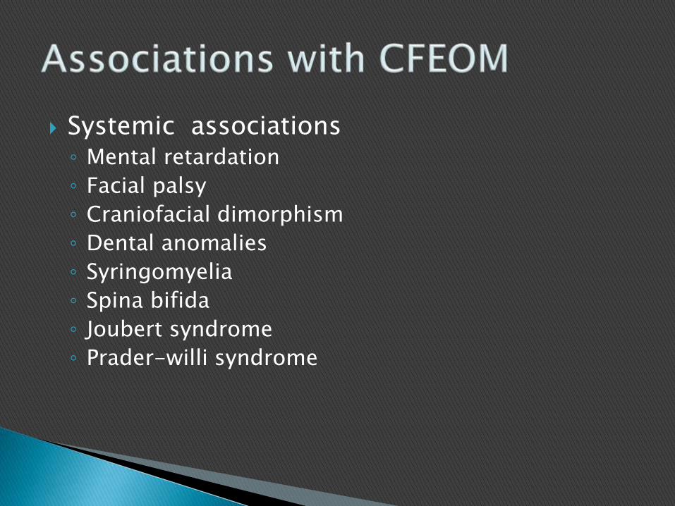
Systemic associations◦ Mental retardation
◦ Facial palsy
◦ Craniofacial dimorphism
◦ Dental anomalies
◦ Syringomyelia
◦ Spina bifida
◦ Joubert syndrome
◦ Prader-willi syndrome

Congenital non progressive ocular motility defect.
It was first described by Stilling and Turk.
The syndrome is characterized by limitation or absence of abduction and/or adduction of the eyes.
There is also retraction of the globe and narrowing of the palpebral fissure on adduction, which is often associated with elevation or depression of the globes.
When abduction is attempted, one often finds widening of the palpebral fissure

Pathological studies of DRS have provided clear evidence that innervational deficiencies can cause fibrotic muscle changes.
Most cases are due to innervational defects, which correlate with aplasia of the sixth nerve nucleus and the VI cranial nerve itself

As shown by electrophysiological studies, innervation of the lateral rectus is provided by the oculomotor nerve causing the pathognomonic co-contraction of the medial and lateral recti with globe retraction on adduction. (Synkinesis phenomenon)
Additionally, an association of DRS is seen with synkinesisphenomena such as Marcus Gunn jaw-winking (due to aberrant trigeminal nerve innervation of levator palpebraesuperioris)
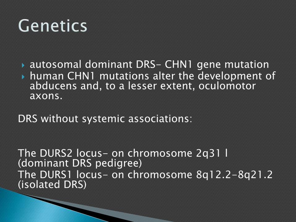
autosomal dominant DRS- CHN1 gene mutation human CHN1 mutations alter the development of
abducens and, to a lesser extent, oculomotoraxons.
DRS without systemic associations:
The DURS2 locus- on chromosome 2q31 l (dominant DRS pedigree)The DURS1 locus- on chromosome 8q12.2-8q21.2 (isolated DRS)
DRS with systemic associations
Of the various malformation syndromes, associated with DRS, Duane radial ray syndrome has been genetically mapped.
inherited as dominant and with incomplete penetrancemapped to chromosome 20
results from heterozygous nonsense frame shift, and deletion mutations in SALL4
The SALL4 gene has also been implicated in DRS associated with Holt–Oram and acro-renal-ocular syndromes.

Absence or hypoplasia of CN6 in the orbit or brainstem regions, often with mild hypoplasia and apparent misdirection of CN3 to the lateral rectus muscle. (The LR muscle in abducens palsy exhibits profound atrophy).

Type I◦ Limitation / absence of abduction◦ Norma/slightly defective adduction◦ Narrowing of palpebral fissure & retraction of globe on
adduction ◦ Widening of fissure on abduction.
Type II◦ Limitation / absence of adduction ◦ N/ slightly defective abduction◦ Narrowing of PF & retraction of globe on adduction
Type III◦ Limitation /absence of both abduction & adduction of
affected eye◦ Retraction of globe & narrowing of PF on attempted
adduction.

Modification to Huber classification based on primary position of gaze-◦ Subgroup A- affected eye esotropic
◦ Subgroup B- exo
◦ Subgroup C- eyes straight in primary position.

Globe retraction
Narrowing of palpebral fissure on attempted adduction
Abduction deficiency
Upshoot/downshoot on adduction
Small angle strabismus (usually <30 PD)
Face turn
Pattern: V more common than A.

Associated ocular anomalies-◦ Nystagmus, anisocoria, aniridia, epibulbar dermoid,
ptosis, optic nerve coloboma, marcus gunn jaw wink.
Non ocular anomalies-◦ Skeletal- limb hypoplasia, polydactyly,
hypoplastic/absent radius.
◦ Vertebral –scoliosis, spina bifida
◦ Genito-urinary defects- renal agenesis,VUR,
◦ Cardiac anomalies- PDA, Auricular septal defect.

heterogeneous clinical disorder, which includes congenital facial palsy with impairment of ocular abduction
developmental disorder of the brainstem rather than an isolated cranial nerve developmental disorder
The ocular motility disturbances in Mo¨bius syndrome arefrequently bizarre and asymmetrical, resembling more of acongenital fibrosis pattern than cranial nerve palsies.
In a study of 37 patients with facial paresis by Verzijl et al, 97% had bilateral and 3% had unilateral ocular abduction weakness.

Most cases are sporadic
Three Mo¨bius syndrome loci have been mapped: ◦ MBS1 to 13q12.2-q13,
◦ MBS2 to 3q21-q22
◦ MBS 3 to 10q21.3-q22.1

Neural Imaging:◦ Brain stem hypoplasia in the region of the sixth and
the seventh nerve complexes with or without other posterior fossa abnormalities
◦ Hypoplasia of extraocular muscles and intraorbitalmotor nerves

Some forms of congenital ptosis may result from aberrantdevelopment of the unpaired caudal central oculomotor sub-nucleus. Two congenital ptosis loci are reported.a. PTOS1 (1p32-p34.1) Variable degree congenital unilateral or bilateral ptosis.Inheritance is autosomal dominant with incomplete penetrance of 90%.
b. PTOS2 (Xp24-27.1)Congenital bilateral symmetrical and severe ptosis almost impinging on the visual axis in the primary position of gaze, with chin-up headposture. Inheritance is X-linked dominant (male and females are equally affected).

Horizontal gaze palsy is suggested to result from hypoplasia of the abducensnucleus with interneuron dysinnervation (medial longitudinal fasciculus and pontine paramedian reticular formation).
ROBO3 [HGPPS (Horizontal gaze palsy with progressive scoliosis), 11q23-25.
The ROBO3 gene encodes a transmembrane receptor required for hindbrain axon midline crossing.
HGPPS phenotype. There is congenital complete absence of conjugate horizontal gaze and childhood onset progressive scoliosis.
Inheritance is autosomal recessive.
The uncrossed corticospinal and dorsal column pathways are not associated with an obvious neurological deficit.

Treatment difficulties:◦ Varied genetic predisposition, penetrance &
expressivity
◦ Recalcitrant response to therapy
◦ Associated abnormal innervation & mechanical anomalies

1. Non surgical management-◦ A. correction of refractive error & anisometropia
◦ Treatment of amblyopia.
2.surgical management-◦ Indications of surgery-
Noticeable ocular deviation in primary position
Marked abnormal head posture
Marked globe retraction
Cosmetically unacceptaable upshoot or downshoot
Principles of surgery- No resection procedure should be attempted ( worsens
movement limitation, retraction & upshoot & downshoot)
Recession of overacting muscle is safest & most effective
Adjustable sutures may be used when indicated.
Surgical options-◦ For esotropia & face turn
Recession of I/L or B/L MR
Faden operation
Transposition of vertical recti to LR- risk of over & under correction, of inducing vertical tropias & anterior segement ischaemia.

◦ For exotropia
I/L or B/L LR resection.
◦ For retraction & upshoot & downshoot:
I/L MR & LR recession. Posterior fixation suture may be added.
Y- splitting with LR recession.

Non surgical management-◦ Detection & treatment of amblyopia, corneal
exposure & refractive errors
Surgical management-◦ No single surgery useful
◦ Transposition of vertical recti to insertion of LR described

Non surgical management◦ Correction of refractive error
◦ Treatment of amblyopia,
Surgical management-◦ Correction of hypotropia, exotropia followed by
ptosis correction