Embed Size (px)
DESCRIPTION
Citation preview

Biliary stricture
Guide : Dr M K ChouhanProfessor and HOD of surgeryDr SNMC,JODHPUR
Candidate-Dr Sumer

Biliary stricture

definition
A biliary stricture is an abnormal narrowing of the bile duct, the tube that moves bile (A substance that helps in digestion) from the liver to the small intestine

Anatomy of biliary tree
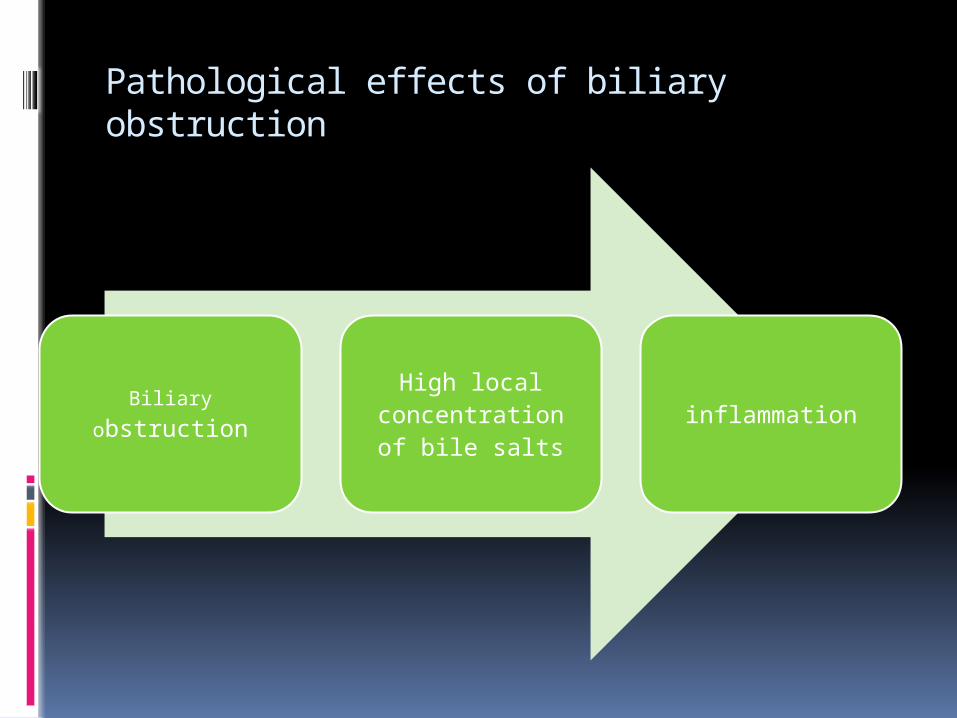
Pathological effects of biliary obstruction
Biliary obstructionHigh local
concentration of bile salts
inflammation

Pathological effects of biliary obstruction
Fibrosis and
scarring
Biliary fistula
Biliary stasis
Liver atrophy
Repeated cholangiti
s
Biliary cirrhosis
and PHTN

Causes of benign stricture
I. Congenital strictures
Biliary atresia
II. Bile duct injuries
A. Postoperative strictures
(1) Cholecystectomy or common bile duct
exploration (accounting 80% of nonmalignant stricture)
(2) Biliary-enteric anastomosis
(3) Hepatic resection
(4) Portocaval shunt
(5) Pancreatic surgery
(6) Gastrectomy
(7) Liver transplantation
B. Stricture after blunt or penetrating trauma

Causes of benign stricture
C. Strictures after endoscopic or percutaneous
biliary intubation
III. Inflammatory strictures
A. Cholelithiasis or choledocholithiasis
B. Chronic pancreatitis
C. Chronic duodenal ulceration
D. Abscess or inflammation of liver or subhepatic
space
E. Parasitic infection
F. Recurrent pyogenic cholangitis (Oriental
cholangiohepatitis)
IV. Primary sclerosing cholangitis
V. Radiation-induced stricture

Causes of malignant stricture
Primary tumors1. Cholangiocarcinoma
2. GB Cancer
3. Pancreatic adenocarcinoma
4. Ampullary carcinoma
5. Hepatoma
6. Gastric carcinoma
Metastatic tumors
1. pancreatic adenocarcinoma
2. Colon cancer
3. Breast cancer
4. Lung cancer
5. Melanoma
6. Ovarian cancer

Bile duct injury at cholecystectomy
Incidence 1.open cholecystectomy
0.1 -0.2% 2.lap cholecystectomy
0.4 -1.3% 80% of benign strictures occurs
following injury during a cholecystectomy.
A major factor is surgeons inexperience-learning curve effect

causes Anatomic variations Technical factors Pathologic factors

Anatomic variations(failure to recognize abnormal anatomy &anomalies)

Technical factors Experience of surgeon Improper assistance Extensive dissection Excess use of cautery Misplacement of clips Excess traction on gall bladder Subvesical duct of luschka in 1-2 %
patients CBD Exploration-use of metal bougies Attempts to achieve hemostasis

Pathologic factorsAcute cholecystitis inflammation leads to edema in
the porta hepatis and calots triangle—distortion of anatomy
Chronic cholecystitis chronic inflammation leads to
fibrosis, adherence, contracted fibrotic gall bladder, cholecystocholedochal fistula
(partial cholecystectomy, cholecystostomy, and cholecystocholedochoduodenostomy are options)

Laparoscopic specific- Classification of Causes of Laparoscopic Biliary
Injuries
1. Misidentification of the bile ducts as the cystic duct
a. Misidentification of the common bile duct as the cystic
duct
b. Misidentification of an aberrant right sectoral hepatic
duct as the cystic duct
2. Technical causes
a. Failure to occlude the cystic duct securely
b. Plane of dissection away from gallbladder wall into the
liver bed
c. Injudicious use of electrocautery for dissection or
bleeding control
d. Excessive traction on cystic duct with tenting upward of
common hepatic duct
e. Injudicious use of clips to control bleeding
f. Improper techniques of ductal exploration

Laparoscopic specificProper exposure –maximum cephalad traction on fundus with concomitant lateral traction on infundibulum

Location &classification
1. Bismuth`s classification—based on location of biliary stricture with respect to the hepatic duct confluence
2. Strasberg`s classification—is of laparoscopic biliary injuries, is applicable for acute injuries with bile leak, lateral injuries and transection.
3. Hannover classification—combine Bismuth and Strasberg classification and has also addressed the vascular injuries—most refined

Bismuth`s classification

Strasberg`s classification

Strasberg`s classification

Hannover`s classification

Clinical presentation

Clinical presentation

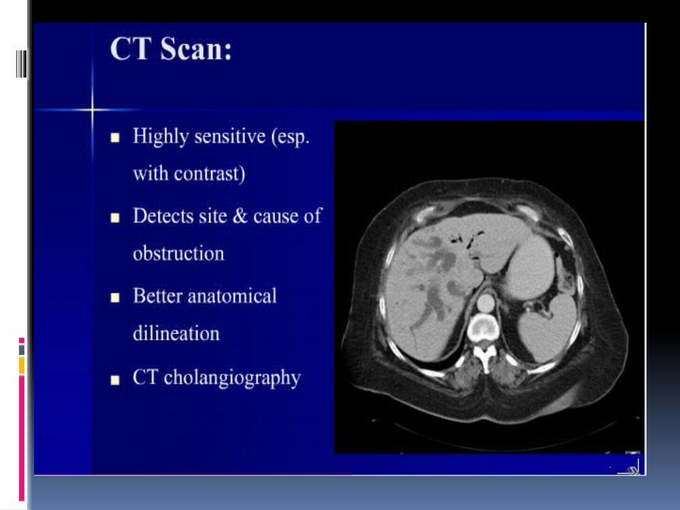
investigations

investigations


cholangioscopy
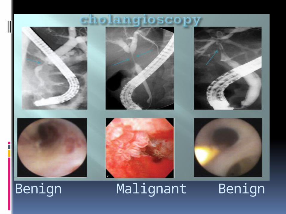
Benign Malignant Benign


Surgical treatment of BDI Recognized at operation
Immediate open conversion and repair by an experienced surgeon
If competent help unavailable, put a drain & should be referred to a specialist center
End to end repair over T- tubeRoux –en –Y hepaticojejunostomy(silk sutures should be avoided for all
biliary reconstructions, because they can act as nidus for stone formation)

Surgical treatment of BDI Recognized in immediate postoperative period
Avoid early reoperationBile leak from cystic duct, subvesical duct of
luschka or from noncircumferential laceration with no distal obstruction to bile flow may close spontaneously (1to 3 weeks)
Endoscopic sphincterotomy with stenting-hasten closure
For severe lacerations and complete transactions –delayed approach is best (timing of surgical intervention 4-10 weeks)

Surgical treatment of BDI injury presenting at an interval
Presented as late bile duct stenosis and stricture
Consider nonoperative biliary drainage procedures
Consider surgery if no resolution in 12 -24 months
Almost always requires Roux –en –Y hepaticojejunostomy


end t

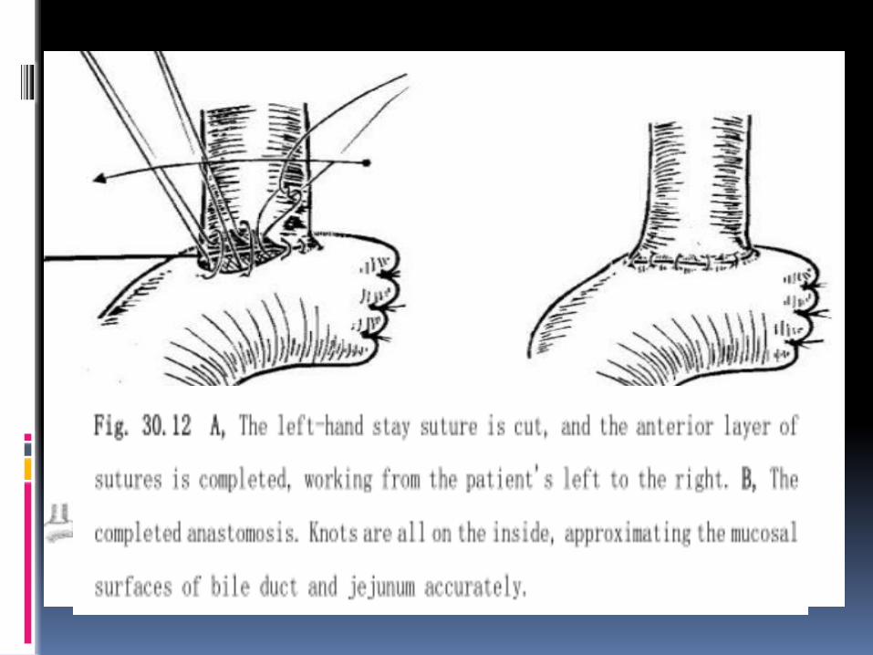
Roux-en-Y HepaticojejunostomyCommon method of repair of bile duct
injuryProper exposure of healthy ,well
vascularised proximal bile ductRoux- en –Y Limb of jejunum >60 cmMucosa to mucosa tension free
anastomosisSide to side or end to side
hepaticojejunostomy using left hepatic duct


• Factors associated with poor outcome after surgery
Proximal stricture (Bismuth type 3 and 4) Multiple prior attempts at repair Portal hypertension Hepatic parenchymal disease (cirrhosis or hepatic
fibrosis) End-to-end biliary anastomosis Surgeon inexperience Intrahepatic or multiple strictures Concurrent cholangitis or hepatic abscess Intrahepatic stones External or internal biliary fistula Intra-abdominal abscess or bile collection Hepatic lobar atrophy Advanced age or poor general health Many authors have advocated the use of anasto


Prevention is the best treatment of biliary strictures.

Thanks