Embed Size (px)
Citation preview

P1: PBU
0521870909pre CUFX091/Friedberg 0 521 87090 9 March 2, 2007 1:48
viii
This page intentionally left blank

P1: PBU
0521870909pre CUFX091/Friedberg 0 521 87090 9 March 2, 2007 1:48
ANESTHESIA IN COSMETIC SURGERY
One major by-product of the aging baby-boom generation has been a surg-
ing interest in cosmetic surgery. Outpatient cosmetic surgery clinics have
sprouted up in droves all over the United States, and the number of cosmetic
procedures performed in 2005 increased by more than 95% from the previ-
ous year. Although procedures like facelifts and abdominoplasties are consid-
ered minimally invasive, the anesthetic protocols and regimens involved are
often overly complex and unnecessarily toxic. Major complications involv-
ing anesthesia in this (and any other) surgical milieu can range from severe
postoperative nausea and vomiting (PONV) to postoperative pain to mor-
tality. Although mortality may be rare, there have been many cases in which
perfectly healthy cosmetic surgery patients require emergency intervention
due to a severe complication involving anesthesia. In recent years, many new
anesthetic protocols have been developed to reduce the incidence of PONV
and other complications, while ensuring that effective pain management and
level of “un-awareness” during surgery are always maintained.
Barry L. Friedberg, M.D., is a volunteer assistant professor at the Keck School
of Medicine, University of Southern California. Since 1992, he has practiced
exclusively in the subspecialty of office-based anesthesia for elective cosmetic
surgery. He founded the Society for Office Anesthesiologists (SOFA) in 1996
that he merged in 1998 with the Society for Office Based Anesthesia (SOBA),
another non-profit, international society dedicated to improving patient
safety through education. Dr. Friedberg is the developer of propofol ketamine
(PK) technique designed to maximize patient safety by minimizing the degree
to which patients need to be medicated to create the illusion of general
anesthesia, that is, “no hear, no feel, no recall.”
i

P1: PBU
0521870909pre CUFX091/Friedberg 0 521 87090 9 March 2, 2007 1:48
ii

P1: PBU
0521870909pre CUFX091/Friedberg 0 521 87090 9 March 2, 2007 1:48
Anesthesia in CosmeticSurgery
BARRY L. FRIEDBERG, M.D.
Assistant Professor in Clinical AnesthesiaVolunteer FacultyKeck School of MedicineUniversity of Southern CaliforniaLos Angeles, CA
iii

CAMBRIDGE UNIVERSITY PRESS
Cambridge, New York, Melbourne, Madrid, Cape Town, Singapore, São Paulo
Cambridge University PressThe Edinburgh Building, Cambridge CB2 8RU, UK
First published in print format
ISBN-13 978-0-521-87090-0
ISBN-13 978-0-511-28482-3
© Cambridge University Press 2007
Every effort has been made in preparing this book to provide accurate and up-to-date information that is in accord with accepted standards and practice at the time of publication. Nevertheless, the authors, editors, and publisher can make no warranties that the information contained herein is totally free from error, not least because clinical standards are constantly changing through research and regulation. The authors, editors, and publisher therefore disclaim all liability for direct or consequential damages resulting from the use of material contained in this book. Readers are strongly advised to pay carefulattention to information provided by the manufacturer of any drugs or equipment that they plan to use.
2007
Information on this title: www.cambridge.org/9780521870900
This publication is in copyright. Subject to statutory exception and to the provision of relevant collective licensing agreements, no reproduction of any part may take place without the written permission of Cambridge University Press.
ISBN-10 0-511-28635-X
ISBN-10 0-521-87090-9
Cambridge University Press has no responsibility for the persistence or accuracy of urls for external or third-party internet websites referred to in this publication, and does not guarantee that any content on such websites is, or will remain, accurate or appropriate.
Published in the United States of America by Cambridge University Press, New York
www.cambridge.org
hardback
eBook (NetLibrary)
eBook (NetLibrary)
hardback

P1: PBU
0521870909pre CUFX091/Friedberg 0 521 87090 9 March 2, 2007 1:48
Come mothers and fathers
Throughout the land
And don’t criticize
What you can’t understand
Your sons and your daughters
Are beyond your command
Your old road is
Rapidly agin’
Please get out of the new one
If you can’t lend your hand
For the times they are a-changin.’
– Robert “Bob Dylan” Zimmerman
“The Times They Are A-Changin,” 1963
v

P1: PBU
0521870909pre CUFX091/Friedberg 0 521 87090 9 March 2, 2007 1:48
vi

P1: PBU
0521870909pre CUFX091/Friedberg 0 521 87090 9 March 2, 2007 1:48
To my parents, my first teachers, who taught me it was acceptable to not be
like everyone else as long as I aspired to be the best I could be.
To Willy S. Dam, M.D., of Bispebjerg Hospital, Copenhagen, my first
anesthesia teacher, who encouraged me to become an anesthesiologist.
To all the patients who have suffered from previous anesthetics and who
may now be relieved of their PONV, postoperative pain, and prolonged
emergences.
vii

P1: PBU
0521870909pre CUFX091/Friedberg 0 521 87090 9 March 2, 2007 1:48
viii

P1: PBU
0521870909pre CUFX091/Friedberg 0 521 87090 9 March 2, 2007 1:48
Contents
Foreword Page xi
Adam Frederic Dorin, M.D., M.B.A.
Acknowledgments xiii
Introduction vx
C. Philip Larson, Jr., M.D., C.M., M.A.
Preface xvii
Barry L. Friedberg, M.D.
List of Contributors xix
PART I. MINIMALLY INVASIVE ANESTHESIA (MIA) R© FOR MINIMALLY
INVASIVE SURGERY
1 Propofol Ketamine with Bispectral Index (BIS) Monitoring 1Barry L. Friedberg, M.D.
2 Preoperative Instructions and Intraoperative Environment 14Barry L. Friedberg, M.D.
3 Level-of-Consciousness Monitoring 23Scott D. Kelley, M.D.
4 The Dissociative Effect and Preemptive Analgesia 39Barry L. Friedberg, M.D.
5 Special Needs of Cosmetic Dental Patients 47James A. Snyder, D.D.S.
6 Propofol Ketamine in the UK, Propofol Ketamine BeyondCosmetic Surgery 59Chris Pollock, M.B.
7 Propofol Ketamine Beyond Cosmetic Surgery: Implications forMilitary Medicine and Mass-Casualty Anesthesia 68Joel W. McMasters, M.D., M.A.J., M.C., U.S.A.
8 Lidocaine Use and Toxicity in Cosmetic Surgery 72Adam Frederic Dorin, M.D., M.B.A.
ix

P1: PBU
0521870909pre CUFX091/Friedberg 0 521 87090 9 March 2, 2007 1:48
x Contents
9 Local Anesthetic Blocks in Head and Neck Surgery 84Joseph Niamtu III, D.M.D.
10 Local Anesthetics and Surgical Considerationsfor Body Contouring 106Rodger Wade Pielet, M.D.
PART II. ALTERNATIVE ANESTHESIA APPROACHES
IN COSMETIC SURGERY
11 Intravenous Anesthesia for Cosmetic Surgery 112David Barinholtz, M.D.
12 Regional Anesthesia for Cosmetic Surgery 131Holly Evans, M.D., F.R.C.P., and Susan M. Steele, M.D.
13 General Inhalation Anesthesia for Cosmetic Surgery 155Meena Desai, M.D.
PART III. OTHER CONSIDERATIONS FOR ANESTHESIA
IN COSMETIC SURGERY
14 Preanesthetic Assessment of the Cosmetic Surgery Patient 171Norman Levin, M.D.
15 Psychological Aspects of Cosmetic Surgery 182David B. Sarwer, Ph.D., Canice E. Crerand, Ph.D., and Lauren M. Gibbons, B.A.
16 The Business of Office-Based Anesthesia for Cosmetic Surgery 199Marc E. Koch, M.D., M.B.A.
17 The Politics of Office-Based Anesthesia 206David Barinholtz, M.D.
18 Staying Out of Trouble: The Medicolegal Perspective 225Ann Lofsky, M.D.
APPENDIX A. A Guide to Perioperative Nutrition 241David Rahm, M.D.
APPENDIX B. Reflections on Thirty Years as an Expert Witness 248Norig Ellison, M.D.
Index 257

P1: PBU
0521870909pre CUFX091/Friedberg 0 521 87090 9 March 2, 2007 1:48
Foreword
Physicians, like all people, live in a world that is proscribed more by what wedo in rote fashion every day than by what we understand in any meaningfulway. Our modern lives have become so harried that most of us barely haveenough time to pause and reflect on what we have done and where we aregoing.
Dr. Barry L. Friedberg, at great personal effort and time, has put forththis pearl of a book: ideas, methods of practice, and salient knowledge onthe cutting edge of modern medical practice as they apply to the world ofminimally invasive anesthesia for cosmetic surgery. As many of our practicesprove every day in operating rooms across the United States and beyond, theinformation and anecdotes provided here apply equally well to a whole hostof different anesthetic and surgical settings.
Modern science is replete with heroic strides in improving patient care anddecreasing perioperative morbidity and mortality—and yet, today, we still donot understand the underlying mechanisms of general anesthesia on the brain,much less the construct of consciousness itself!
The field of anesthesiology and perioperative medicine achieved unprece-dented gains in patient outcomes through the advent of pulse oximetry decadesago. Since then, we have refined our techniques, implemented new airwaydevices, decreased postoperative nausea and vomiting, improved our times to“street readiness,” and done a better job of managing pain. Now is the time tomove to the next level of patient care.
Dr. Friedberg, through unrelenting drive and perseverance, has broughtto light the benefits of the age-old concept that “less is more.” Through theuse of minimally invasive anesthetic techniques, a resurgence in the prudentuse of ketamine via the propofol-ketamine (PK) technique, and the applicationof brain wave (level-of-consciousness) monitoring, Dr. Friedberg has broughtanesthesia care to a higher plane.
When Albert Einstein died, curious scientists autopsied his brain in thefutile quest to glean some insight into one of humanity’s greatest minds. Theywere desperately seeking answers to how this one man transformed Newtonianphysics into an advanced understanding of the universe itself. Today, physicistsstruggle with String Theory and other abstract mathematical concepts to solvethe ultimate riddle of bridging relativity theory with quantum mechanics inone grand unifying equation. But back in 1905, when Einstein’s first paperswere reaching the scientific print, he was greeted as a heretic. At one point, agroup of one hundred of the world’s most renowned scientists signed a doc-ument stating that Mr. Einstein was not correct in his radical departure from
xi

P1: PBU
0521870909pre CUFX091/Friedberg 0 521 87090 9 March 2, 2007 1:48
xii Foreword
conventional theory. Albert Einstein is reported to have replied, in paraphrase,“if they were so sure that they were right and I was wrong, then why does thisletter contain one hundred signatures—in that case, they should need onlyone signature!”
In this same vein, there have been those detractors who espouse oppositionto some of the elegant medical practices and insights put forth by Dr. Friedberg.To those voices, hiding in the shadow of inexperience, I say with a loud andconfident voice—come join us, read on, and enjoy this journey along the roadto greater insight and knowledge. Some have suggested that Dr. Friedberg is“redefining anesthesia”—and, in some contexts and practice paradigms, thismay be true. I like to think of his work, and this book, as a stepping-stone tothe next level of patient care.
Adam Frederic Dorin, M.D., M.B.A.
Medical Director
Grossmont Plaza Surgery Center
San Diego, CA

P1: PBU
0521870909pre CUFX091/Friedberg 0 521 87090 9 March 2, 2007 1:48
Acknowledgments
I wish to express my appreciation to the following individuals for their helpduring the creation of this book.
Raymond Hasel, M.D., an early propofol ketamine adopter, for his valuablesuggestions regarding my chapters.
The librarians at Hoag Hospital Medical Library, especially Cathy Drake,Michele Gordaon, and Barbara Garside for their generous support.
Marc Strauss, my editor and friend, who displayed extraordinary sagacityand forebearance in making this book a reality.
Ken Karpinski, my project manager, who guided me through the productionprocesses.
Brian Bowles for his help with the copyediting and Constance Burt for herassistance with the final proofing and corrections to the manuscript.
xiii

P1: PBU
0521870909pre CUFX091/Friedberg 0 521 87090 9 March 2, 2007 1:48
xiv
P1: PBU
0521870909pre CUFX091/Friedberg 0 521 87090 9 March 2, 2007 1:48
Introduction
Anesthesiology has undergone remarkable changes in recent years. Amongthem is the development of anesthesia subspecialties and of anesthesiologistswho focus most or all of their time in one area of anesthesia practice. Thischange has several advantages for patients, surgeons, and anesthesiologists.For one, the anesthesiologist learns the needs and expectations of the surgeon,which optimizes surgical outcome for patients. Furthermore, knowing whatto expect, the anesthesiologist is better able to adjust both the doses andtiming of drugs so that patients are adequately anesthetized for surgery butthen emerge from anesthesia in a timely and comfortable manner. Nowhere arethese issues more important than when surgery is performed in the ambulatoryor office-based setting. Expectations are that patients undergoing surgery inthese settings will go home the same day. Resources for extended care areusually nonexistent, as they should be.
Providing anesthesia for office- or clinic-based cosmetic surgery hasemerged as one subspecialty area for anesthesiologists. For patients, conve-nience is greatly enhanced and costs are greatly decreased in office- or clinic-based cosmetic surgery. To provide the best anesthetic care in this specializedsetting requires certain skills that are not emphasized in most anesthesia train-ing programs. Fortunately, we are blessed with a resource prepared by a highlyskilled and experienced anesthesiologist.
In this book, Dr. Barry L. Friedberg has assembled a compendium of hisfifteen years of providing anesthesia care in the office setting. Where scientificdocumentation is available, Dr. Friedberg provides it. Where it is lacking,he guides the reader with recommendations that represent both reasonedjudgment and innovative, effective results. He knows what works and whatdoesn’t and explains his views in text and illustrations that are concise andinformative.
Any anesthesiologist contemplating providing anesthesia care for cosmeticsurgery, regardless of the surgical setting, needs to read this book. For thoseproviding care in the office or clinic setting, it is virtually mandatory. Byreviewing this text, anesthesiologists will avoid the pitfalls that exist in thispractice and conclude their days with grateful patients and happy surgeons.
C. Philip Larson, Jr., M.D., C.M., M.A.
Professor Emeritus
Anesthesiology & Neurosurgery, Stanford University
Professor of Clinical Anesthesiology
David Geffen School of Medicine at UCLA
xv

P1: PBU
0521870909pre CUFX091/Friedberg 0 521 87090 9 March 2, 2007 1:48
xvi

P1: PBU
0521870909pre CUFX091/Friedberg 0 521 87090 9 March 2, 2007 1:48
Preface
The very essence of leadership is that you have a vision.
—Theodore Hesburgh
Caesar’s Gallic Wars begins with the observation that “All Gaul is divided intothree parts.” Anesthesia in Cosmetic Surgery is also divided into three parts.
Part I, Chapters 1–10, is devoted to minimally invasive anesthesia (MIA) r©
for minimally invasive surgery. (The United States Patent and TrademarkOffice [USPTO] granted trademark serial number 76/619,460, file number067202-0312946 to minimally invasive anesthesia [MIA] to Dr. Friedberg in2005.)
Part I advances the premise of a unitary anesthetic technique for all electivecosmetic surgery. Part I challenges the belief that only some types of electivecosmetic surgery are suitable for intravenous sedation. Many readers may besimilarly challenged by the description of abdominoplasty, an extraperitonealprocedure, as a minimally invasive surgery.
Inasmuch as the MIATM
technique is not universally applicable for every sur-gical personality, Part II, Chapters 11–13, is dedicated to providing a compre-hensive view of other anesthetic techniques administered by dedicated anes-thesia professionals. Deliberately omitted are those approaches of oral andintravenous sedation directed by the surgeon in the absence of a dedicatedanesthesia provider.
There is much about the practice of anesthesia in cosmetic surgery thatis not specifically related to anesthetic technique. Part III, Chapters 14–18,and Appendices A and B illustrate the chasm between the medically indicated(third-party reimbursed) anesthesia practice and that particular to anesthesiafor elective cosmetic surgery.
The reader who demands Level 1 study to accept new solutions to clinicalproblems is reminded that neither aspirin nor penicillin ever had a Level 1study to validate their efficacy. Nonetheless, both are well-accepted therapeuticagents. The efficacy of the MIA
TMtechnique will eventually make it a widely
accepted practice.“Insanity” is sometimes defined as performing the same act in the same way,
over and over, yet expecting a different outcome. Only by changing the “script”can outcomes be improved. MIA
TMfor minimally invasive surgery represents
a paradigm shift or change in the “script” for the anesthetic management ofthe patient intraoperative experience. MIA
TMtechnique is not only differ-
ent from anesthetic techniques described in Part II but also safer. Superiorpostoperative outcomes for postoperative nausea and vomiting (PONV) andpain management with MIA
TMtechnique are described in Part I.
xvii

P1: PBU
0521870909pre CUFX091/Friedberg 0 521 87090 9 March 2, 2007 1:48
xviii Preface
In 2007, American soldiers are dying in Afghanistan and Iraq. HIV/AIDSis still causing deaths throughout the world. Deaths from malnutrition, star-vation, and natural disasters still plague the third world. A nuclear disasterfrom weapons of the former Soviet Union in the hands of rogue nations orterrorists remains a threat. According to the National Highway Transportationand Safety Administration (NHTSA), on American highways in 2004, therewere 105 daily deaths (or 38,253 for the year) from motor vehicle accidents.Whereas death is a constant in life, the public has grown somewhat able toaccept these kinds of deaths. Death surrounding elective cosmetic surgery,surgery without medical indication, is never an acceptable outcome for thepatient, the patient’s family, the anesthesiologist, the surgeon, or the lay public.
There is a “perfect storm” of forces that have made this book not only pos-sible but necessary. The baby-boom or “me generation,” born 1946 to 1964,is beginning to age. Social forces creating the “sandwich” effect of simultane-ously caring for parents and children have created economic forces dictatingthat this generation will postpone retirement. The work force is a competitiveenvironment with a heavy emphasis on a youthful appearance. The combina-tion of narcissism and the need to remain competitive at work has created ahuge impetus for “boomers” to seek cosmetic relief of the aging process.
In the course of seeking cosmetic surgery, many patients receive generalanesthesia, opioid-based IV sedation, or regional anesthetics in hospital sur-gicenter (ASC) and office-based settings (see Part II). When death occurs inthe office-based setting, the public and media find it unacceptable. “Dying tobe beautiful,” read the headlines. States like Florida, California, New York, andothers have rushed to regulate the office surgical suite because it is frequentlythe site for elective cosmetic surgery.
Sadly, what remains is the absurd situation that it is acceptable to have adeath from a pulmonary embolism following an abdominoplasty in a hospitalor ASC setting but not the exact same outcome in an office-based setting.The emerging hypocrisy is that the hospital and ASC lobbies in Florida (andothers to follow) have persuaded the legislatures to mandate reporting of allmortalities from office-based cosmetic surgery while remaining exempt fromthe same requirement. This is clearly not in the interest of public safety. Alldeaths from elective cosmetic surgery should be subject to the same reportingand scrutiny as those in the office-based setting.
The old maxim that “while the surgeon can only maim, the anesthesiologistcan kill” rings true in the effort to affect the ultimate negative anesthesiaoutcome. How can tragic deaths in cosmetic surgery be avoided? Is the answersomewhere in the future with better drugs or better monitors? It is not possible toget the right answer by asking the wrong question. “Have we overlooked existingdrugs, techniques, and/or monitors that can provide for a safer anesthetic withbetter outcomes?” is, perhaps, the more insightful question. The answer to thisquestion is at the heart of the MIA
TMtechnique.
Barry L. Friedberg, M.D.
Corona del Mar
California

P1: PBU
0521870909pre CUFX091/Friedberg 0 521 87090 9 March 2, 2007 1:48
List of Contributors
David Barinholtz, M.D.
President and CEO
Mobile Anesthesiologists, LLC
Chicago, IL
Meena Desai, M.D.
Managing Partner
Nova Anesthesia Professsionals
Villanova, PA
Adam Frederic Dorin, M.D., M.B.A.
Medical Director
Grossmont Plaza Surgery Center
San Diego, CA
Norig Ellison, M.D.
Professor of Anesthesia
University of Pennsylvania
Philadelphia, PA
Holly Evans, M.D., F.R.C.P.
Associate Professor
Department of Anesthesiology
Division of Ambulatory Anesthesiology
Duke University Medical Center
Durham, NC
Barry L. Friedberg, M.D.
Assistant Professor in Clinical Anesthesia
Volunteer Faculty
Keck School of Medicine
University of Southern California
Los Angeles, CA
Scott D. Kelley, M.D.
Medical Director
Aspect Medical Systems, Inc.
Norwood, MA
Marc E. Koch, M.D., M.B.A.
Founder and CEO
Somnia Anesthesia Services, Inc.
New Rochelle, NY
C. Philip Larson, Jr., M.D., C.M., M.A.
Professor Emeritus
Anesthesiology & Neurosurgery
Stanford University
Palo Alto, CA
Professor of Clinical Anesthesiology
David Geffen School of Medicine at UCLA
Los Angeles, CA
Norman Levin, M.D.
Chief, Department of Anesthesiology
Century City Hospital
Los Angeles, CA
Ann Lofsky, M.D.
Staff Anesthesiologist
Saint John’s Hospital
Santa Monica, CA
Anesthesia Consultant and Governor Emeritus
The Doctors’ Company
Napa, CA
Joel McMasters, M.D., M.A.J., M.C., U.S.A.
Assistant Chief of Anesthesia
Director of Total Intravenous Anesthesia
Brooke Army Medical Center
San Antonio, TX
Joseph Niamtu III, D.M.D.
Private Practice
Cosmetic Facial Surgery
Richmond, VA
xix

P1: PBU
0521870909pre CUFX091/Friedberg 0 521 87090 9 March 2, 2007 1:48
xx List of Contributors
Rodger Wade Pielet, M.D.
Clinical Associate
Department of Surgery
University of Chicago
Chicago, IL
Chris Pollock, M.B.
Consultant in Pain Management and
Anaesthesia
Hull and East Yorkshire Hospital Trust
Hull, England
David Rahm, M.D.
President and CEO
Vitamedica
Manhattan Beach, CA
David B. Sarwer, Ph.D.
Departments of Psychiatry and Surgery
The Edwin and Fannie Gray Hall Center for
Human Appearance and the Weight
and Eating Disorders Program
University of Pennsylvania School of Medicine
Philadelphia, PA
James A. Snyder, D.D.S.
Founder and CEO
Center for Dental Anesthesiology
Alexandria, VA
Susan M. Steele, M.D.
Professor
Department of Anesthesiology
Division of Ambulatory Anesthesiology
Duke University Medical Center
Durham, NC

P1: PBU
cufx091-01 CUFX091/Friedberg 0 521 87090 9 Feb. 2, 2007 17:10
1 Propofol Ketamine with BispectralIndex (BIS) Monitoring
Barry L. Friedberg, M.D.
PART I MINIMALLY INVASIVE ANESTHESIA (MIA) R© FOR MINIMALLYINVASIVE SURGERY
INTRODUCTION
WHY IS MINIMALLY INVASIVE ANESTHESIAR©
IMPORTANT?
Postoperative Nausea and Vomiting (PONV)
How are PONV, preemptive analgesia, and postoperative painmanagement related?
Beware Laryngospasm
WHAT IS CLONIDINE-PREMEDICATED, BIS-MONITORED PK MAC, OR THEMIA™ TECHNIQUE?
Why Ketamine?
Making ketamine predictable
Premedication
Fluid management
Major confounding principle
BIS as fianchetto
Postoperative pain management
CONCLUSION
INTRODUCTION
Anesthesiologists are trained to administer anesthesia for
surgery. Elective cosmetic surgery is commonly performed
in an office-based facility with patients discharged to
home. However, elective cosmetic surgery differs from
elective or emergency surgery in many substantial aspects
(see Tables 1-1 and 1-2).
“Cosmetic surgery is almost always elective, and patients
are almost always in good health. The patient, however,
is willing to risk this good health (at least to a limited
extent) in order to experience improvements in physical
appearance, and perhaps more importantly, self-esteem,
body image, and quality of life.”1
There is no medical indication for elective cos-
metic procedures, excluding breast reconstruction post-
mastectomy. One may consider risk-benefit ratios of dif-
fering anesthetic regimens in medically indicated surgery.
However, surgery without medical indication should not
accept any avoidable risk. Halogenated inhalation anes-
thetics are triggering agents for malignant hyperthermia
(MH),2 carry an increased risk of deep venous thrombo-
sis with potential pulmonary embolism,3 and are eme-
togenic.4 If the patient is interested and the surgeon is
willing, all cosmetic procedures can be performed under
local only anesthesia. Therefore, any additional anesthetic
agents should be subject to the highest justification.
Most patients desire some alteration of their level of con-
sciousness from fully awake through completely asleep.
Given that all known risks should be avoided, when
possible, then which agents are best suited to the task,
what monitors should be employed, and to what level
1

P1: PBU
cufx091-01 CUFX091/Friedberg 0 521 87090 9 Feb. 2, 2007 17:10
2 Barry L. Friedberg
Table 1-1. Elective cosmetic procedures
Commonly performed cosmetic surgical procedures.All procedures have successfully been anesthetizedwith PK MAC/MIA™ technique in the office-basedsetting.
1. Rhinoplasty (closed or open)2. Liposuction or suction assisted lipoplasty (SAL)3. Blepharoplasty (open, transconjuctival, or
endoscopic)4. Rhytidectomy (open or endoscopic)5. Breast augmentation, subglandular, subpectoral
(via areaolar, inframammary, transaxilllary, ortransumbilical approach)
6. Hair transplantation with or without scalpreduction
7. Facial resurfacing (laser, chemical peel, ormechanical dermabrasion)
8. Brow lift (coronoplasty or endoscopic)9. Abdominoplasty (classical or simple skin)
10. Otoplasty11. Genioplasty (mandibular advancement or
recession)12. Facial implants (malar and mandibular with
silicone or autologous fat)13. Lip enlargement (autologous fat transfer,
radiated cadaver material [AllodermR©
],Gortex
R©extrusions, Restylane,
R©Juvaderm,
R©
etc.)14. Platsyma band plication15. Composite procedures; i.e., (a) endoscopic brow
lift and endoscopic rhytidectomy, with openplatysma band plication, (b) blepharoplasty,rhinoplasty, and rhytidectomy, or (c) breastaugmentation with abdominoplasty
of anesthesia should be administered (i.e., minimal seda-
tion [“anxiolyis”], moderate [“conscious”] sedation, deep
sedation, or general anesthesia [GA])? (See Appendix 1-1,
Defining Anesthesia Levels). If better outcomes are the
goal, doesn’t minimally invasive anesthesia for minimally
invasive surgery make sense?5 (See Table 1-3.)
WHY IS MINIMALLY INVASIVE
ANESTHESIAR©
IMPORTANT?
“Less is more” is a Mies Vanderohe principle applied to
the Bauhaus school of minimalist architecture. “Doing
more with less” is a Buckminster Fuller concept of housing
applied to his geodesic domes.
Table 1-2. Cosmetic procedures by type from PKMAC/MIA™ technique case log March 26,1992 – March 26, 2002 12
N %
Liposuction 663 (25)Breast augmentation 489 (18)Facial resurfacing mechanical
abrasion, chemical peel, orlaser resurfacing
389 (14)
Rhytidectomy 305 (11)Blepharoplasty 198 (7)Rhinoplasty 81 (3)Fat transfer 57 (2)Abdominoplasty 54 (2)Composite or misc. procedures 447 (18)Total 2,683 (100)
“We hold the basic premise that the less the involvement
of the patient’s critical organs and systems (i.e., the lower
the concentration of the agent, or the less ‘deep’ the anes-
thesia), the less will be the damage to the patient, whether
this be temporary or permanent.”6
“For the anesthetic itself, overall experiences indicate
that the least amount of anesthetic that can be used is the
best dose. Local and monitored anesthesia care (MAC) is
preferable to regional. Regional techniques are preferable
to general anesthesia.”7
Table 1-3. Minimally invasive surgeriesappropriate for BIS-monitored PK MAC, theMIA™ technique
1. All cosmetic procedures (see Table 1-1)2. Gyn: laparoscopy (tubal ligation, fulgeration
endometriosis)3. Ortho: arthoscopy4. Urology: lithotripsy5. Gen. surg.: herniorraphy & breast cancer
surgery6. Neuro: microdiscectomy, microlaminectomy,
carpal tunnel release7. sedation for morbidly obese8. peripheral injuries in U.S. Army field hospitals in
Iraq, Afghanistan
Cases being performed with PKRa TIVA1. U.S. Army neurosurgery in Iraq.aPropofol-Ketamine-Remifentanil

P1: PBU
cufx091-01 CUFX091/Friedberg 0 521 87090 9 Feb. 2, 2007 17:10
Propofol Ketamine with Bispectral Index (BIS) Monitoring 3
“When possible, procedures longer than three or four
hours should be performed with local anesthesia and intra-
venous sedation because general anesthesia is associated
with deep venous thrombosis at much higher rates under
prolonged operative conditions.”3
“Newer techniques for intravenous sedation that
include the use of propofol, often in combination
with other drugs, have made it possible to perform
lengthy or extensive procedures without general anesthe-
sia and without the loss of the patient’s airway protective
reflexes.”9
“When you can measure what you are speaking about,
and express it in numbers, you know something about it;
but when you cannot measure it, when you cannot express
it in numbers, your knowledge is of a meager and unsat-
isfactory kind; it may be the beginning of knowledge, but
you have scarcely, in your thoughts, advanced to the stage
of science.” (William Thompson, knighted Lord Kelvin.
Popular lectures and addresses [1891–1894])
The bispectral index (BIS) monitor facilitates a numer-
ical expression of the hypnotic component (anesthesia =hypnosis + analgesia) of the anesthetic state and may per-
mit a reasonable inference about the analgesic state. Heart
rate, blood pressure, and other clinical signs are noto-
riously unreliable indicators of anesthetic depth.10 BIS
provides new information about patients that is simply
unavailable from any other vital or clinical sign.11 BIS, as
an index, has no units. The scale is 0–100, with 100 repre-
senting awake and zero representing isoelectric (or zero)
brain activity. Hypnosis compatible with general anesthe-
sia (GA) occurs between BIS 45–60. BIS 45–60 with sys-
temic analgesia defines general anesthesia. BIS 60–75 with
adequate local analgesia is a major part of the MIA™ tech-
nique. Patients who received MIATM
neither hear, nor feel,
nor remember their surgical experience.12
Monk et al. published an associated 20% increase in the
one-year mortality risk associated with every hour of BIS
<45.13 Therefore, BIS <45 for cumulative periods greater
than one hour must be considered as overmedicating.
The routine practice of overmedicating for fear of under-
medicating is no longer a desirable or acceptable practice (see
Table 1-4).
Monk et al. postulated that the increase in one-year
anesthetic mortality might be related to an inflammatory
response from excessively deep anesthesia.13 A more recent
prospective, randomized controlled study demonstrated
Table 1-4. BIS levels and levels ofsedation/anesthesia
BIS Sedation/Anesthesia Level98–100 Awake78–85 Minimal Sedation (“Anxiolysis”)70–78 Moderate (“Conscious”) Sedationa
60–70 Deep Sedationb
45–60+ systemicanalgesia
General Anesthesiac
<45, >1 hr. Overanesthetized!13
aWith moderate sedation, passive maneuvers like extensionand rotation of the head or shoulder pillow may be all thatare necessary to maintain the airway.
b With deep sedation, active maneuvers, like nasal airway orLMA, may be required to maintain airway patency.
c See Appendix 1-1.
increased C-reactive protein levels with BIS <45 for more
than 50% of the cases.14
The BIS monitor does not replace traditional vital-sign
monitoring, that is, EKG, NIABP, SpO2, (or EtCO2 when
indicated). When measured, the EtCO2 typically runs
between 38–42 with the MIA™ technique. The EtCO2
offers the display of the waveform of the patient’s res-
piration. Many experienced anesthesiologists are capable
of assessing adequate respiratory movement without this
information. Over 3,000 PK MAC cases have been safely
anesthetized without EtCO2 monitoring.
Titrating anesthesia with BIS trend is limited by the
fact that the processing required for the BIS algorithm is
delayed 15–30 seconds behind real time. This delay has
given rise to the legitimate criticism that BIS does not
predict patient movement. BIS, a measure of the hypnotic
state, was not designed to predict patient movement (see
Chapter 3).
EMG is the instantaneous display of the frontalis muscle
activity if the XP software version of the BIS A2000, or later,
is used. Inadequate analgesia leading to patient movement
is predictable if the EMG is selected from the advanced
screen menu to trend as a secondary trace. A spike in
EMG (when BIS is 60–75, in spontaneously breathing
patients) nearly always predicts inadequate analgesia, pre-
ceding patient movement (see Fig. 1-1). The anesthesiol-
ogist should utilize the 15–30 second delay in the change
of the BIS value to simultaneously bolus propofol while
encouraging the surgeon to supplement the local analgesia.

P1: PBU
cufx091-01 CUFX091/Friedberg 0 521 87090 9 Feb. 2, 2007 17:10
4 Barry L. Friedberg
Figure 1-1. Incremental propofol induction began 08:45. Ketamine 50 mg IV administered 08:47, BIS = 63. In this particular case, BISincreases post-ketamine dose. However, the increase does not defeat the ability to titrate propofol to BIS 60–75!
Postoperative Nausea and Vomiting (PONV)
Macario et al. conducted a statistically validated survey of
a panel of expert anesthesiologists on what postoperative
anesthetic outcome they believed patients most wanted
to avoid.15 The anesthesiologists concluded that pain was
the number one anesthesia outcome patients most desired
to avoid. A follow-up, similarly statistically validated sur-
vey of patients’ anesthesia outcomes they most desired to
avoid was emesis!16 Clearly, a disconnect exists between
what anesthesiologists believe about their patients and
what the patients actually want most to avoid. A potential
explanation could be that patients who consent for elec-
tive surgery expect to have some postoperative discom-
fort but do not want their pain to be compounded by
emesis.
How are PONV, preemptive analgesia, and
postoperative pain management related?
There is a consensus among PONV authorities like Apfel,
Chung, Gan, Scuderi, and White, that both inhalational
anesthetics and opioids are emetogenic agents. “In the con-
text of [emetogenic] anesthesia, postoperative pain man-
agement and opioid related PONV remain problems.”17
In the context of emetogenic anesthesia, experts advise
“multimodal” prophylaxis in the highest risk group.18
Apfel’s recent NEJM article identifies the highest PONV
risk group of patients as nonsmoking females, with a his-
tory of previous PONV and/or motion sickness, having
emetogenic (i.e., elective cosmetic) surgery of two or more
hours.4 Apfel’s criterion of high risk applies exceptionally
well to Friedberg’s previously referenced series of 2,683
patients.12
Elective cosmetic surgery anesthesia for the “rich and
famous” of Beverly Hills and Newport Beach is the highest
risk PONV population! This conclusion reflects the south-
ern California geographic bias of the author. There are
many other such communities worldwide.
The MIA™ technique is not perfect but contextually
nonemetogenic. Without any antiemetic prophylaxis, this
highest risk group of patients experienced a total of thir-
teen PONV events for an unprecedented 0.5% PONV
rate!12 A 50 mg dissociative dose of ketamine at BIS <75
propofol levels eliminates the noxious input of the injec-
tion of local analgesia while avoiding emetogenic agents
like the halogenated inhalational vapors and intravenous
opioids.
Lidocaine provides intraoperative analgesia with
bupivicaine providing postoperative analgesia. In this con-
text, it has been extremely rare for patients to require (eme-
togenic) opioid relief of their postoperative discomfort.

P1: PBU
cufx091-01 CUFX091/Friedberg 0 521 87090 9 Feb. 2, 2007 17:10
Propofol Ketamine with Bispectral Index (BIS) Monitoring 5
Elimination of all emetogenic triggers defines nonopioid,
preemptive analgesia (NOPA). NOPA is the hallmark of
the MIA™ technique. In Friedberg’s fifteen-year experi-
ence, no patients have been admitted to the hospital fol-
lowing PK MAC/MIA™ technique for either PONV or
unmanageable pain.
Beware Laryngospasm
No technique is perfect. Classical laryngospasm can
be diagnosed by the characteristic “crowing” sound
generated by a small gap in the vocal cords owing to
their incomplete closure. With ketamine-associated laryn-
gospasm, the vocal cords most commonly close com-
pletely. Hence, only rarely will crowing noise alert the
anesthesiologist to impending desaturation. Additionally,
the usual remedy of positive pressure ventilation combined
with anterior jaw thrust is completely ineffective. The anes-
thesiologist must pay particular attention to sneezing or
coughing as the only prodrome warning him of impend-
ing laryngospasm.
The treatment of choice is a rapid IV bolus of lidocaine
1 mg · lb−1 or 2 mg · kg−1.
Concern about adding more lidocaine in patients receiv-
ing relatively large amounts of lidocaine local analgesia has
led other anesthesiologists to prefer to deepen the propofol
level by adding a 50 mg propofol bolus to break the laryn-
gospasm. However, when IV lidocaine has been admin-
istered for laryngospasm, no stigmata of lidocaine toxic-
ity have been observed. The BIS showed no decrease in
response to the IV lidocaine bolus. There was no transient
hypotension or widening of the EKG complex during the
case. No patient complained of tinnitus, tremulousness,
or metallic taste on the tongue after emergence.
Administering succinylcholine (SCH) to break the
spasm is suboptimal because SCH adds unnecessary (and
avoidable) risk as an MH triggering agent. (Neither propo-
fol nor ketamine are MH triggering agents.) Further, the
myalgias associated with SCH make the agent totally unac-
ceptable in the elective cosmetic surgery patient.
Waiting until desaturation occurs after the prodrome
will add a substantial amount of time until the lido-
caine can circulate to anesthetize (and open) the vocal
cords. Desaturation increases the physiologic stress to the
patient. The alarm of the pulse oximeter, accompanied
by the bluish discoloration of the patient, increases the
psychological stress to the anesthesiologist, surgeon, and
operating room nursing staff. This disturbing scenario is
best minimized by promptly giving IV lidocaine when the
patient coughs or sneezes.
WHAT IS CLONIDINE-PREMEDICATED,
BIS-MONITORED PK MAC, OR THE
MIA™ TECHNIQUE?
Something old (ketamine), something new (BIS-moni-
tored propofol hypnosis), something borrowed (diazepam
ketamine technique19), no one blue (SpO2 >90% on room
air).
Why Ketamine?
The brain cannot respond to stimuli it does not receive.
Critical concept: GA does not reliably block all incoming
noxious stimuli! The “wind-up” phenomenon,20 medi-
ated by the NMDA receptors, is often invoked to explain
acute postoperative pain after general anesthesia, as well
as the formation of chronic pain states.
“Dissociation” refers to a patient who, under the influ-
ence of ketamine, remains motionless in response to noxious
stimuli.
Based on clinical observation, the NMDA receptor
block from a 50 mg dissociative dose of ketamine reli-
ably blocks all incoming noxious stimuli to the cortex (the
so-called mid-brain spinal) for a period of 10–20 minutes.
After obtaining an equal dissociative effect with a 50 mg
ketamine dose in both 90-pound female and 250-pound
male patients, the author concluded that the number of
NMDA receptors does not vary with patient body weight in
adults.
Preemptive analgesia is most consistently observed
when the NMDA receptors are saturated prior to noxious
stimulation. Acetaminophen 1,000 mg po is adequate for
postoperative pain management (for the few patients who
request it) in the context of clonidine-premedicated, BIS-
monitored PK MAC patients.12 (See Table 1-5.)
Making Ketamine Predictable
In other contexts, ketamine has a well-deserved reputa-
tion for causing hypertension, tachycardia, and an unpre-
dictable 20% of patients experiencing hallucinations or
dsyphorias.21 Hypnotic doses of propofol block ketamine-
induced hallucinations as well as undesirable hemody-
namic sequellae.22 Being able to assign a numerical value

P1: PBU
cufx091-01 CUFX091/Friedberg 0 521 87090 9 Feb. 2, 2007 17:10
6 Barry L. Friedberg
Table 1-5. Ketamine tips
1. 80% patients achieve dissociative effect with25 mg ketamine, 98% with 50 mg ketamine.No “down side” to 50 mg dose as long as BIS<75. Wait 2–3 min. before injecting local. Waitan additional min. if patient is reactive beforeadministering more ketamine.
2. Preemptive analgesia effect is variable wheninadequate dissociative effect is obtained.Saturate NMDA receptors!
3. All adult patients, independent of body weight,require 50 mg ketamine initial dose tosaturate NMDA receptors.
4. Reinjection of previously injected field doesNOT require more ketamine.
5. Consider injecting both sides with initialketamine dose.
6. If prep. is cold, consider injecting 25 mgketamine 2–3 min. before prep. or considerwarming prep. solution!
7. With experience, less ketamine is administered.Friedberg’s case log of the last 500 cases (of2,683 patients) showed 80% performed witheither one or two 50 mg doses of ketamine.12
8. Mixing propofol with ketamine is TIVA23 notMAC.
9. Do not exceed an aggregate total of 200 mgketamine.
10. Do not give ketamine in the last 20–30 minutesof a case.
with BIS to the level of propofol hypnosis, prior to admin-
istering the ketamine, was an enormous breakthrough in
making ketamine a predictable agent. Not only could the
initial ketamine dose be administered without problems,
but also subsequent doses, when needed, could be given
with assurance.
First, create a stable level of propofol in the brain by
performing an incremental, not bolus, induction. The
incremental induction maintains spontaneous ventila-
tion, commonly maintains masseter tone, avoids propofol
waste, and is less apt to produce induction hypotension.
Incremental propofol induction provides hypnosis with
a minimal physiologic and pharmacologic trespass to the
patient. Lesser trespass increases patient safety.
Lesser trespass increases the probability of maintaining
the SpO2 >90% on room air (i.e., room air, spontaneous
ventilation, or RASV). Key concept: Titrate propofol to BIS
<75 before giving the ketamine! Do NOT give ketamine at
BIS >75.
Table 1-6. Clinical pathway for MIA™ technique
1. Clonidine 0.2 mg PO 30–60 min preop(Systolic >100, body weight >100 pounds).
2. Glycopyrrolate 0.2 mg IV with 2 ccs 1% lidocaineplain.
3. Incrementally titrate propofol to BIS <75 withmultiple, sequential 150 ug · kg−1 · 20 sec.mini-boluses. N.B. If pump does not have abolus feature, set initial rate to 450 ug · kg−1 ·min−1 and reduce the rate toward 50 as soonas the EMG begins to decrease.
4. Basal propofol infusion rate 50 ug · kg−1 · min−1.5. Ketamine 50 mg IVP @ BIS <75 2–3 minutes
prior to injection local anesthesia.6. Adjust basal propofol rate upward to maintain
BIS 60–75 if ketamine causes an increase.7. Inject adequate local analgesia.8. Administer more ketamine only after two
reinjections of the field fail to eliminate patientmovement.
9. Maintain propofol at BIS 60–75, EMG 0 on BISscale, 30 on EMG scale.
10. Bupivicaine in field before closure, especially forbrowlift, subpectoral breast augmentation,and abdominoplasty.
Because the elective cosmetic surgical patient tends to be
healthy, cardiac output and redistribution from the brain
tend not to be significant factors in altering established
brain levels of propofol. However, the nineteenfold inter-
patient variation in propofol hydroxylation may play a
significant role in the ability to maintain a stable level of
propofol in the brain.23 Measuring an individual patient’s
brain response to propofol with BIS would appear to be a
more effective strategy than employing target controlled
infusions (TCI) to achieve specific blood levels of propofol
(see Table 1-6).
Premedication
PK MAC was derived from diazepam ketamine MAC tech-
nique, which was first published in 1981.19 Vinnik clearly
enumerated that only after the patient was soundly asleep
from the diazepam was the ketamine to be administered.19
Diazepam hypnosis, followed by ketamine dissociation,
followed by local anesthetic injection was Vinnik’s clin-
ical pathway. Although Guit was the first to publish the
combination of propofol and ketamine, the technique was
described as a total intravenous anesthetic (TIVA).24 TIVA

P1: PBU
cufx091-01 CUFX091/Friedberg 0 521 87090 9 Feb. 2, 2007 17:10
Propofol Ketamine with Bispectral Index (BIS) Monitoring 7
strongly implies that the local analgesia injected by the
surgeon is not essential for the success of the TIVA
technique. In contradistinction, the surgeon’s local anal-
gesia is essential for the success of PK MAC.
Guit’s TIVA technique was unknown to Friedberg in
1992 when Friedberg embarked on replacing Vinnik’s
diazepam with propofol. The surgeons quickly com-
plained about the cost of the propofol and pleaded
for relief. Friedberg added midazolam in an effort to
reduce the amount of propofol. From March 26, 1992
through March 26, 1997, the case log Friedberg maintained
contained patient’s names, dates, surgeons, patient age,
gender, weight, surgical procedure(s) (see Table 1-2),
midazolam, propofol, ketamine, and anesthesia times.8
Propofol rates, mg · min−1 and ug · kg−1 · min−1, were
calculated retrospectively.
If 2 mg midazolam was good, perhaps 4 mg midazo-
lam could be better for propofol-sparing purposes. In the
aforementioned case log, a total of 354 patients received 0
mg midazolam, 316 patients received 2 mg, and another
303 patients received 4 mg midazolam premedication from
1992–97. No consistent, incremental relationship could be
established in propofol savings between the 0, 2, and 4 mg
midazolam groups.8 In June 1997, Friedberg eliminated
the midazolam from PK MAC.
In September 1997, Oxorn published a very elegant
Level I study confirming Friedberg’s uncontrolled, clinical
experience in 973 patients.25 Oxorn reported that there was
no statistical difference in either induction or maintenance
doses of propofol between those patients who received
2 mg midazolam premedication and those who received
none.25 However, the unexpected finding was that a statis-
tically significant threefold number of patients who received
midazolam required pain medication in the PACU.25
From July 7, 1997, through December 21, 1998, 268
patients received BIS-monitored PK MAC without pre-
medication, midazolam, or other benzodiazepine. During
BIS-monitored propofol hypnosis, there were no patients
who suffered from hallucinations or a lack of amnesia. This
experience led Friedberg to conclude that benzodiazepine
premedication was superfluous to provide amnesia or to
prevent hallucinations in the presence of BIS monitoring.
Some of these patients were included in a subsequent pub-
lication.26
Patients continued to request premedication to calm
them. After attending the New York Postgraduate Assem-
bly (PGA) in December 1998, Friedberg returned with the
renewed notion of adding po clonidine as a premedica-
tion. Like Vinnik’s concept of administering sleep doses of
diazepam to block ketamine hallucinations, clonidine for
premedication had also been previously reported in the
plastic surgery literature. 27,28
Inconsistent propofol sparing results were observed
with 0.1 mg po clonidine. A therapeutic clonidine dose
should be in a range between 2.5–5.0 ug · kg−1.29 Cloni-
dine 0.2 mg mg po achieves that range in patients weigh-
ing between 95–175 pounds. The higher dose of clonidine
provided consistent propofol sparing results and further
refinement of BIS-monitored PK MAC.30
From January 26, 2001 to September 2002, rofecoxib
50 mg po was added to the clonidine. When the drug
was voluntarily withdrawn from the market, rofecoxib was
deleted from the premedication. While the addition of the
rofecoxib appeared to benefit the patient, the deletion of
the agent did not appear to increase (the already few) post-
operative patient complaints of discomfort.
At the present time, only clonidine 0.2 mg po (30–60
minutes preoperatively) and glycopyrrolate 0.2 mg with
2 cc 1% lidocaine IV are given as premedication (see
Table 1-6).
Fluid Management
The long-standing teaching that patients who are NPO
after midnight are at least 500–1,000 ccs behind on their
fluids is not especially relevant for elective cosmetic surgery
patients. As stated earlier, these are by and large essentially
healthy patients who are far different from the debilitated
ward patients on whom most anesthesia trainees learn
about anesthesia. Elective cosmetic surgical patients are
not “dry.” Vasodilating anesthetics are no longer being
administered. Lastly, large fluid shifts and blood loss are
atypical experiences in most elective cosmetic surgery.
Other authors have analogized the insult produced by
liposuction to that of a burn injury. However, burn patients
do not have compression garments applied to obliterate
the “third space” created by the aspiration of subcutaneous
fat.
Fluid replacement regimens based on experience in burn
patients areinappropriate for liposuction patients.
Especially for cases up to 5,000 ccs of liposuction, fluid
replacement should remain modest, that is, not more
than 1,000 ccs. Otherwise, one may risk fluid overload,

P1: PBU
cufx091-01 CUFX091/Friedberg 0 521 87090 9 Feb. 2, 2007 17:10
8 Barry L. Friedberg
Table 1-7. MIA™ airway algorithm (assumesincremental propofol induction)
1. Extend and laterally rotate head, one side mayhave better gas exchange than the other.
2. Insert shoulder (not neck) pillow to increaseforce of extension.
3. Insert lubricated nasal airway (#28 FR mostcommonly).
4. Insert lubricated LMA (#4 most commonly).5. No ET required: >15 yrs, >3,000 patients;
no opioids, benzodiazepines, or musclerelaxants.
pulmonary edema, and dilution of platelets and other
coagulation factors.
Another unaesthetic consequence of 2,000–4,000 ccs
fluid replacement in this patient population is enuresis on
the operating room table. This will embarrass the patient
and annoy the nurse who had to clean it up. Catheterizing
the patient to compensate for inappropriate fluid admin-
istration exposes the patient to the risk of an unnecessary
bladder infection.
Patients who experience caffeine withdrawal headache
without their morning caffeine are encouraged to drink
their cup of coffee black or with non-dairy creamer, if
necessary. Apple juice or water is permitted up until an
hour before surgery. Patients who are hungry upon awak-
ening are encouraged to have toast and jam. Simple carbo-
hydrates and sugars are rapidly absorbed by the stomach
and pose no real threat to patient safety. It is far better to
have the patient arrive without hypoglycemia. Patients are
encouraged to void before getting on the operating table.
(See Table 1-7.)
Major Confounding Principle
A blanched surgical field does not guarantee adequate sur-
gical analgesia. More local analgesia resolves the patient
movement 99% of the time. Administer more ketamine
only after two reinjections of the field fail to eliminate
patient movement.
BIS becomes much more than a simple tool with which
to titrate propofol. BIS becomes a case management tool.
By being able to demonstrate adequate propofol levels
(i.e., BIS 60–75) during patient movement, the surgeon
Table 1-8. Local anesthesia tips
1. PDR limit of 500 mg lidocaine with epinephrine(7 mg · kg−1) is outdated and overlyconservative. Neither the 2005, 2006 nor the2007 (print or electronic) editions of PDR haveany entry for injectable lidocaine!
2. 200 ccs of 0.5% lidocaine (1,000 mg) withepinephrine is well tolerated and withoutsequellae of toxicity
3. Tumescent or “wetting” solution = 500 mglidocaine, 1 mg epinephrine in 1,000 ccs NSS(Klein) or LR (Hunstead)
4. 5,000 ccs of tumescent solution = 2,500 mglidocaine
5. 5,000 ccs of tumescent solution in a 60 kgfemale patient = 42 mg · kg−1
6. Avoid >50 cc 0.25% (125 mg) bupivicaine forpostoperative analgesia.
can be educated to inject more analgesia. In addition to
the initial injection of the local analgesia, the patient is
spared noxious, painful input during the surgery. The
brain cannot respond to stimuli it does not receive. Post-
operative pain management begins intraoperatively! Repro-
ducible preemptive analgesia occurs under conditions of
adequate dissociation secondary to the saturation of the
NMDA receptors. (See Table 1-5.)
BIS as Fianchetto
From Italian, fianchetto is a chess term meaning a “dou-
ble move.” In a “binary” system of anesthesia (hypnosis
+ analgesia = anesthesia), being able to measure hypno-
sis permits an inference about the adequacy of analgesia.
Adequate analgesia produces de facto muscle relaxation
for minimally invasive surgery. BIS 60–75 with EMG = 0
(on the BIS scale, 30 on the EMG scale) defines adequate
hypnosis for the MIA™ technique. Therefore, adequate
hypnosis in the presence of patient movement (usually
preceded by a spike in EMG) infers inadequate analgesia!
Postoperative Pain Management
In the context of clonidine-premedicated, BIS-monitored
PK MAC, now formally known as the MIA™ technique,
postoperative pain is minimal to nonexistent. Part of this
phenomenon may be explained by having patients emerge

P1: PBU
cufx091-01 CUFX091/Friedberg 0 521 87090 9 Feb. 2, 2007 17:10
Propofol Ketamine with Bispectral Index (BIS) Monitoring 9
from propofol with the clonidine still in effect. Patients
who have lower anxiety levels, secondary to lowered
catecholamines from the clonidine, tend to have less pain
complaints. In the diethyl ether era, “stormy induction,
stormy emergence” was the common rationale for pre-
medicating surgical patients. Preoperatively, a clonidine-
premedicated patient may not appear drowsy but, upon
questioning, usually admits to feeling “calmer.” A fur-
ther explanation for the remainder of the observation of
minimal-to-no postoperative pain appears to be the phe-
nomenon of preemptive analgesia.
With the dissociative effect of ketamine, no noxious
signals reach the cortex during the injection of local anes-
thesia. GA does NOT reliably block all incoming noxious
stimuli. Use the BIS to not only maintain hypnosis at
60–75 but also to assure inadequate local analgesia is dealt
with appropriately (i.e., more local) and not by subterfuge
(i.e., more ketamine, propofol, or opioids). Lastly, bupivi-
caine, especially for browlift, breast augmentation, and
abdominoplasty, provides long-lasting nonopioid relief.
Do not exceed a total of 125 mg bupivicaine (or 50 ccs
0.25%) for postoperative analgesia. Because the bupivi-
caine quickly binds to tissue, it is necessary only to splash
it into the operative field. Some surgeons prefer to close the
wound and inject the bupivicaine retrograde up the suc-
tion drainage tube(s). Both approaches with bupivicaine
are effective.
All of the anesthesiologists’ efforts to prevent PONV
and effect adequate pain management may be for naught
if the surgeon discharges the patient home with an opioid-
containing analgesic (i.e., Vicodinr©
or Tylenol #3r©
).
Darvocetr©
or other similar nonopioid analgesics may
provide an increment of relief greater than 1,000 mg
acetaminophen every six hours. Oral diazepam is espe-
cially effective for decreasing the muscle spasm associated
in subpectoral breast implant patients. N.B. This is also
a useful strategy for any other submuscular implants; i.e.,
gluteal.
The few patients who do complain of pain present a dif-
ferential diagnosis of “central” (or supratentorial) versus
“peripheral” (infratentorial) pain. Both complaints are real.
Some patients may complain of pain when they had been
predominantly immobile for the surgery. This pain is more
likely to be “central” in origin. This type of patient may
respond better if 50 mg po diphenhydramine (Benadrylr©
)
Table 1-9. Errors to avoid
1. Ketamine before propofol: NO2. Ketamine at BIS >75: NO3. Bolus propofol induction: NO4. Inadequate local analgesia: NO
BIS as fianchetto for adequate propofol andlidocaine
5. Opioids instead of more lidocaine: NO6. Ketamine instead of more lidocaine: NO7. >200 mg total ketamine or any in last 20 min. of
case: NO8. Tracheostomize patient for laryngospasm
instead of IV lidocaine: NO9. SCH instead of lidocaine for laryngospasm: NO
is added to the 1,000 mg acetaminophen (Tylenol P. M.r©
).
More experience with the MIA™ technique will elimi-
nate most of the patient movement seen with inadequate
local analgesia. These patients may require ketorolac 30–
60 mg IV to deal with “peripheral” pain issues. As the
surgeon becomes more willing to inject additional local
analgesia during the case when patient movement occurs
at BIS 60–75, fewer issues of “peripheral” pain will be
manifest. None of the more than 3,000 PK MAC patients
has ever required hospital admission for intractable pain.
(See Table 1-9.)
CONCLUSION
One must empathize with those who, understandably,
have difficulty believing that a subpectoral breast aug-
mentation in combination with a classical abdomino-
plasty can be performed as an office-based or day surgery
without PONV or postoperative pain management issues.
“Cognitive dissonance” is the psychological principle that
precludes individuals from believing what they observe
when it sharply contradicts what they have been taught to
believe.
The On-Qr©
pump may have some additional value;
but in the context described in this chapter, it offers little
pain management benefit to offset the additional $280 cost
(in 2005 dollars). While dexmedetomidine may possess 8
times the alpha2 agonist potency of clonidine, it is 400
times more expensive (2005 dollars) and more tedious to

P1: PBU
cufx091-01 CUFX091/Friedberg 0 521 87090 9 Feb. 2, 2007 17:10
10 Barry L. Friedberg
administer. There are no current plans to replace clonidine
with dexmedetomidine in the MIA™ technique.
The MIA™ technique reproducibly provides preemp-
tive analgesia and is not technically difficult to execute. It
does, however, require the active cooperation of the sur-
geon. Surgeons have become more interested in the use
of local anesthesia to diminish PONV and postoperative
pain management problems they perceive to be produced
by the emetogenic agents the anesthesiologist chooses to
administer.
Although initially developed for office-based, elective
cosmetic surgery, the MIA™ technique is by no means
limited to these types of cases (see Table 1-3). The MIA™
technique offers superior outcomes to alternative forms of
anesthesia (see Part II) for cosmetic surgery (i.e., essentially
zero PONV without the use of anti-emetics and minimal
postoperative pain management).
In the final analysis, the MIA™ technique provides
safety, simplicity, and satisfaction for all parties involved in
the surgical experience: patients, their at-home caregivers,
surgeons, nurses, and anesthesiologists.
REFERENCES
1. Goldwyn RM: Psychological aspects of plastic surgery: A sur-geon’s observations and reflections, in Sarwer DB, PruzinskyT, Cash TF, et al. (eds.), Psychological aspects of reconstruc-tive and cosmetic plastic surgery. Philadelphia, Lippincott,Williams & Wilkins, 2006; p13.
2. www.mhaus.org3. McDevitt NB: Deep venous thrombosis prophylaxis. Plast
Reconstr Surg 104:1923,1999.4. Apfel CC, Korttila K, Abdalla M, et al.: A factorial trial of
six interventions for the prevention of PONV. N Engl J Med350:2441,2004.
5. Friedberg BL: Minimally invasive anesthesia for minimallyinvasive surgery. Outpatient Surgery Magazine. Herrin Pub-lishing Partners LP, Paoli, PA. 2:57,2004.
6. Cullen SC, Larson CP: Essentials of Anesthetic Practice.Chicago, Year Book Medical Publishers, 1974; p82.
7. Laurito CE: Anesthesia provided at alternative sites, inBarasch PG, Cullen BF, Stoelting RK (eds.), Clinical Anes-thesia, 4th ed., Philadelphia, Lippincott, Williams & Wilkins,2001; p1343.
8. Friedberg BL: Propofol-ketamine technique, dissociativeanesthesia for office surgery: A five-year review of 1,264 cases.Aesth Plast Surg 23:70,1999.
9. Lofsky AS: Deep venous thrombosis and pulmonaryembolism in plastic surgery office procedures. The Doctors’Company Newsletter. Napa, CA, 2005. www.thedoctors.com/risk/specialty/anesthesiology/J4254.asp
10. Flaishon R, Windsor A, Sigl J, et al.: Recovery of con-sciousness after thiopental or propofol. Anesthesiol 86:613,1997.
11. www.aspectms.com/resources/bibliographies12. Friedberg BL: Propofol ketamine anesthesia for cosmetic
surgery in the office suite, chapter in Osborne I (ed.), Anes-thesia for Outside the Operating Room. International Anesthe-siology Clinics. Baltimore, Lippincott, Williams & Wilkins,41(2):39,2003.
13. Monk TG, Saini V, Weldon BC, et al.: Anesthetic man-agement and one-year mortality after non-cardiac surgery.Anesth Analg 100:4,2005.
14. Kersssens C, Sebel P: Relationship between hypnotic depthand post-operative C-reactive protein levels. Anesthesiol105:A578,2006.
15. Macario A, Weinger M, Truong P, et al.: Which clinical out-comes are both common and important to avoid? The per-spective of a panel of expert anesthesiologists. Anesth Analg88:1085,1999.
16. Macario A, Weinger M, Carney K, et al.: Which clinical anes-thesia outcomes are important to avoid? The perspective ofpatients. Anesth Analg 89:652,1999.
17. White PF: Prevention of postoperative nausea andvomiting—A multimodal solution to a persistent problem.N Engl J Med 350:2511,2004.
18. Scuderi PE, James RL, Harris L, et al.: Multimodal anti-emetic management prevents early postoperative vomit-ing after outpatient laparoscopy. Anesth Analg 91:1408,2000.
19. Vinnik CA: An intravenous dissociation technique for outpa-tient plastic surgery: Tranquility in the office surgical facility.Plast Reconstr Surg 67:199,1981.
20. Thompson SWN, King AE, Woolf CJ: Activity-dependentchanges in rat ventral horn neurons in vitro, summationof prolonged afferent evoked depolarizations produce a D-2-amino-5-phosphonovaleric acid sensitive windup. Eur JNeurosci 2:638,1990.
21. Corssen G, Domino EF: Dissociative anesthesia: furtherpharmacologic studies and first clinical experience withthe phencylcidine derivative CI-581. Anesth Analg 45:29,1968.
22. Friedberg BL: Hypnotic doses of propofol block ketamineinduced hallucinations. Plast Reconstr Surg 91:196,1993.
23. Court MH, Duan SX, Hesse LM, et al.: Cytochrome P-4502B6 is responsible for interindividual variability of propo-fol hydroxylation by human liver microsomes. Anesthesiol94:110,2001.
24. Guit JBM, Koning HM, Coster ML, et al.: Ketamine as anal-gesic with propofol for total intravenous anesthetic (TIVA).Anaesthesia 46:24,1991.
25. Oxorn DC, Ferris LE, Harrington E: The effects of midazo-lam on propofol-induced anesthesia: propofol dose require-ments, mood profiles and perioperative dreams. AnesthAnalg 85:553,1997.
26. Friedberg BL, Sigl JC: Bispectral (BIS) index monitoringdecreases propofol usage in propofol-ketamine office basedanesthesia. Anesth Analg 88:S54,1999.

P1: PBU
cufx091-01 CUFX091/Friedberg 0 521 87090 9 Feb. 2, 2007 17:10
Propofol Ketamine with Bispectral Index (BIS) Monitoring 11
27. Man D: Premedication with oral clonidine for facialrhytidectomy. Plast Reconstr Surg 94:214,1994.
28. Baker TM, Stuzin JM, Baker TJ, et al.: What’s new in aestheticsurgery? Clin Plast Surg 23:16,1996.
29. Goyagi T, Tanaka M, Nishikawa T: Oral clonidine pre-medication reduces awakening concentrations of isoflurane.Anesth Analg 86:410,1998.
30. Friedberg BL, Sigl JC: Clonidine premedication decreasespropofol consumption during bispectral (BIS) index moni-tored propofol-ketamine technique for office based surgery.Dermatol Surg 26:848,2000.
APPENDIX 1-1 DEFINING ANESTHESIA LEVELS:
THE TERMINOLOGY
Monitored anesthesia care (MAC) is a term created to
include all anesthesia services except general or regional
anesthesia. MAC is not especially useful to describe a par-
ticular anesthetic state or spectrum of states. MAC remains
a term of exclusion in that it specifically is NOT general or
regional anesthesia.
PK MAC connotes separately administering ketamine
after inducing the patient with a continuous infusion of
propofol.1 The MIA™ technique adds the layer of BIS
monitoring along with po clonidine premedication and
infusion pump administered propofol.2
BIS-monitored PK MAC or the MIA™ technique falls
well within the scope of the definition of IV sedation for an
AAAASF Class B facility, except in the (current) regulations
of the AAAASF and the state of Florida. The MIA™ tech-
nique provides a measure of the level of hypnosis achieved.
The MIA™ technique intensifies but does not depress the
laryngeal or “life-preserving” reflexes.
MINIMAL, MODERATE, DEEP SEDATION &
GENERAL ANESTHESIA∗
Minimal sedation (Anxiolysis)
Minimal sedation is a drug-induced state during which
patients respond normally to verbal commands. Although
cognitive function and coordination may be impaired,
ventilatory and cardiovascular functions are unaffected.
∗ Excerpted from ASA position on Monitored Anesthesia Care in ASAmanual for Anesthesia Departmental Organization and Management,2003–4. Reprinted with written permission of the American Societyof Anesthesiologists. A copy of the full text can be obtained from ASA,520 N. Northwest Highway, Park Ridge, Illinois 60068-2573.
Moderate Sedation/Analgesia(“Conscious Sedation”)
Moderate sedation/analgesia is a drug-induced depression
of consciousness during which patients respond purpose-
fully to verbal commands, either alone or accompanied by
light tactile stimulation. No interventions (Editor’s note:
“intervention” is undefined.—BLF ) are required to main-
tain a patent airway, and spontaneous ventilation is ade-
quate. Cardiovascular function is usually maintained.
N.B. A second physician is involved in: Deep sedation
analgesia.
Deep Sedation/Analgesia
Deep sedation/analgesia is a drug-induced depression of
consciousness during which patients cannot be easily
aroused but respond purposefully following repeated or
painful stimulation. The ability to independently main-
tain ventilatory function may be impaired. Patients may
require assistance (Editor’s note: “assistance” is undefined.
-BLF) in maintaining a patent airway, and spontaneous
ventilation may be inadequate. Cardiovascular function
is usually maintained. Reflex withdrawal from a painful
stimulus is NOT considered a purposeful response.
General Anesthesia (GA)
General anesthesia is a drug-induced loss of consciousness
during which patients are not arousable, even by painful
stimulation. The ability to independently maintain venti-
latory function is often impaired. Patients often require
assistance in maintaining a patent airway, and positive
pressure ventilation may be required because of depressed
spontaneous ventilation or drug-induced depression of
neuromuscular function. Cardiovascular function may be
impaired.
Because sedation is a continuum, it is not always possible
to predict how an individual patient will respond. Hence,
practitioners intending to produce a given level of seda-
tion should be able to rescue patients whose level of seda-
tion becomes deeper than initially intended. Individuals
administering moderate (“conscious”) sedation/analgesia
should be able to rescue patients who enter a state of deep
sedation/analgesia, while those administering deep seda-
tion/analgesia should be able to rescue patients who enter
a state of general anesthesia.

P1: PBU
cufx091-01 CUFX091/Friedberg 0 521 87090 9 Feb. 2, 2007 17:10
12 Barry L. Friedberg
COMMENT ON THE FOUR CLASSES
OF SEDATION/ANESTHESIA
Neither the term “intervention” (for “conscious” or mod-
erate sedation) nor “assistance” (for deep sedation) to
maintain an airway is defined in the preceding ASA posi-
tion paper.
“Intervention” for “conscious” or moderate sedation may
be any passive maneuver to maintain airway patency. “Inter-
ventions” include, but are not limited to, extending the
head with or without lateral rotation, and placement of a
one liter bag (or similar device) under the patient’s shoul-
ders. “Interventions” are designed to exert more force on
the genioglossus muscle, elevating the tongue off the back
of the oropharynx, and opening the airway. (The genioglos-
sus muscle is so named because it connects the “genu,” or
“knee,” of the mandible to the “glossus,” or tongue.)
An intermediate maneuver between “intervention” and
“assistance” is sometimes referred to as a “chinner” in the
dental and oral surgical community. A “chinner” is the
manual support of the chin to open the airway long enough
for drug levels to decrease enough to allow the patient
to regain an adequate SpO2. By definition, a “chinner” is
a transient maneuver as opposed to either a continuous
passive “intervention” or an active “assistance.”
“Assistance” for deep sedation may be any supraglottic
mechanical device actively inserted into the nose or mouth
to maintain airway patency. Examples of such devices are
nasal airways, oral airways, cuffed oropharyngeal airways
Figure 1-1. The patient is prepared for a rhinoplasty, is asleepat BIS 78, spontaneously breathing room air through an LMA.SpO2> 96%.
(COPAr©
), laryngeal mask airways (LMAr©
), and even
Combitube.r©
Propofol administered at an infusion rate sufficient to
produce a BIS 60–75 (moderate to deep sedation) will
depress the pharyngeal reflexes and inhibit swallowing (see
Table 4-2). The pharyngeal reflexes are not “life preserv-
ing” because they do not protect the glottic chink.
If the patient maintains a preinsertion BIS value of
60–75 after the insertion of a supraglottic device (mean-
ing that a deeper level of anesthesia was neither required
for the insertion nor maintenance), then the insertion of a
Figure 1-2. The BIS trace for the entire case. Note that at no time during the LMA insertion or the majority of the case does the patientrequire BIS 45–60 (hypnosis compatible with GA) to tolerate her LMA. Clearly, the insertion of an LMA per se does not transform PKMC/MIA™ technique from a sedation to general anesthesia!
P1: PBU
cufx091-01 CUFX091/Friedberg 0 521 87090 9 Feb. 2, 2007 17:10
Propofol Ketamine with Bispectral Index (BIS) Monitoring 13
supraglottic device, per se, does not transform a deep seda-
tion case into a general anesthetic! LMA does not equal GA! 3
See Figures 1-1 and 1-2.
Modification of the AAAASF classification to include
either a separate level or subsection of Level C should be
created to account for nontriggering anesthesia.
A Class C facility typically must have an anesthesia
machine, scavenging, and dantrolene to safely provide gen-
eral anesthesia. The MIA™ technique is a nontriggering
technique. Therefore, no increment in patient safety (i.e.,
substantial cost-zero benefit) will be achieved by require-
ments that ignore the value of measuring the patient’s level
of consciousness. Intravenous sedation can be minimal,
moderate, or deep sedation as well as general anesthesia
(vide supra).
In an attempt to bring a semblance of order into the
chaotic nomenclature of levels of sedation/anesthesia,
the ASA has defined four specific clinical levels. The
attempt to differentiate “conscious sedation” as being per-
formed by a single physician would appear to preclude
the possibility of “conscious sedation” being provided
by a second physician (i.e., an anesthesiologist or nurse
anesthetist). This is incompatible with current clinical
practice.
All of the first three levels of sedation may be described
MAC because they are neither general nor regional anesthe-
sia. One of the most cogent points contained in the ASA
position on MAC was the statement that it is not always
possible to predict how an individual patient will respond!
CORRELATING DEFINITIONS WITH
CLINICAL PRACTICE
Benzodiazepines may be used to provide minimal, moder-
ate, and deep states of sedation. Propofol can produce all
four levels of hypnosis for sedation/anesthesia. However,
benzodiazepines are not well measured by the BIS or other
currently commercially available level-of-consciousness
monitors. Propofol is well measured by BIS17,18 (see
Table 1-4). Propofol alone can provide minimal seda-
tion “anxiolysis” (BIS 78–85). Propofol in conjunction
with intermittent ketamine may be either moderate or
“conscious” sedation (BIS 70-78) or deep sedation (BIS
60–70) depending on whether passive “intervention” or
active “assistance” for airway maintenance is required (vide
supra).
The MIA™ technique may be classified as minimal (BIS
78–85), moderate “conscious” sedation (BIS 70–78), or
deep sedation (BIS 60–70), depending on whether a pas-
sive intervention (moderate sedation) or an active assis-
tance (deep sedation) is required to maintain the airway.
The insertion of an LMA without increasing the depth of
anesthesia below BIS 60–75 does not transform a sedation
case into a general anesthesia. The MIA™ technique is MAC,
not GA or TIVA. The MIA™ technique does not require
an anesthesia machine,21 scavenging, or dantrolene to be
safe, simple reproducible, and effective for patients having
ill elective office-based cosmetic surgeries.
Numerical terminology is more precise than verbal ter-
minology to describe levels of sedation and anesthesia.
Numerical terminology permits more precise and effec-
tive communication of the level of hypnosis and analgesia
between the anesthesiologist and his surgeons as well as
his fellow anesthesiologists.
REFERENCES
1. Friedberg BL: Propofol-ketamine technique. Aesth Plast Surg17:297,1993.
2. Friedberg BL: Minimally invasive anesthesia for minimallyinvasive surgery. Outpatient Surgery Magazine. Paoli, PA,Herrin Publishing Partners LP 2:57,2004.
3. Friedberg BL: Does LMA equal GA? (letter). OutpatientSurgery Magazine. Paoli, PA, Herrin Publishing Partners LP6:15,2003.
4. Friedberg BL: Brain is target organ for anesthesia. Anes-thesia Patient Safety Foundation Newsletter (www.apsf.org)20(3),Fall 2005.
5. Kearse LA, Rosow C, Zaslavsky A, et al.: Bispectral analysis ofthe electroencephalogram predicts conscious processing ofinformation during propofol hypnosis and sedation. Anes-thesiol 88:25,1998.
6. Twersky RS: Office-based anesthesia: Considerations foranesthesiologists in setting up and maintaining a safe officeanesthesia environment. Park Ridge, IL, American Societyof Anesthesiologists. 2000.

P1: PBU
cufx091-02 CUFX091/Friedberg 0 521 87090 9 Feb. 2, 2007 17:13
2 Preoperative Instructions and Intraoperative Environment
Barry L. Friedberg, M.D.
PREOPERATIVE INSTRUCTIONS
Adjusting Surgeon Expectations
Adjusting Patient Expectations
CONCLUSION
PREOPERATIVE INSTRUCTIONS
Nothing per os (NPO), or nothing by mouth, after mid-
night is the most commonly given preoperative instruction
to all surgical patients. This is not unreasonable given the
fact that the majority of surgical patients are exposed to
emetogenic inhalational vapors and/or emetogenic intra-
venous opioids. Both inhalational vapors and intravenous
opioids depress the laryngeal or “life-protecting” reflexes.
California Assembly Bill (AB)595 specifically mandated
office accreditation when sedatives and analgesics are used
in a manner that has the probability to depress the “life-
preserving” reflexes. The “Catch-22” is that neither the leg-
islature nor the anesthesia community ever defined what the
“life-preserving” reflexes are. In both the peer-reviewed lit-
erature1 and in unrebutted public testimony before the CA
Medical Board when Dr. Thomas Joas, a prominent anesthe-
siologist, was its presiding chairman, Friedberg has unequiv-
ocally defined the laryngeal reflexes as the “life-preserving”
reflexes.
Emetogenic inhalational vapors and/or emetogenic
intravenous opioids expose the surgical patient to an
increased likelihood of aspiration and death. If surgical
patients cannot reflexly protect their trachea, they cannot
preserve their lives. The lack of pharyngeal reflexes or swal-
lowing seen with propofol sedation/anesthesia does not
necessarily mean that the laryngeal reflexes are similarly
depressed. In fact, when ketamine is added to the regimen
of propofol sedation and opioids are scrupulously avoided
(i.e., PK MAC/MIA™ technique), laryngospasm has been
observed in about 1–2% of patients. Laryngospasm is the
antithesis of depressed laryngeal reflexes. Laryngospasm has
been observed as long as several hours after a single 50-mg
dose of ketamine! Laryngospasm is the ultimate in height-
ened laryngeal reflex activity. Because it does not depress
the “life-preserving” reflexes, PK MAC/MIA™ technique
is exempted from AB595 mandating office accreditation.
Notwithstanding the AB595 exemption, the medical lia-
bility carriers will still require that the ASA monitoring
standards (i.e., NIABP, SpO2, EKG) be followed in any
anesthetizing situation.
Temperature measurement is not especially relevant with
a nontriggering anesthetic technique. EtCO2 tends 38–42
with PK MAC or the MIA™ technique, when measured.
Being able to observe the waveform of the exhaled CO2 may
give additional reassurance to the anesthesiologist that the
patient is, in fact, breathing. This is potentially significant if
the patient is draped in a manner that precludes observation
of the rise and fall of the chest wall (Barinholtz D, personal
communication. 2005). EtCO2 monitoring, per se, does little
to enhance patient safety with an opioid avoidance technique
like the PK MAC/MIA™ technique.
Additionally, there must be a source of oxygen (i.e.,
an E tank), a means of positive pressure ventilation (i.e.,
an Ambu r© bag), and suction readily available in any
anesthetizing situation. Lastly, insurers will defer to the
state authorities for any requirement for a crash cart and a
defibrillator (see Chapter 18). Friedberg has never discour-
aged offices from seeking accreditation despite the fact
14

P1: PBU
cufx091-02 CUFX091/Friedberg 0 521 87090 9 Feb. 2, 2007 17:13
Preoperative Instructions and Intraoperative Environment 15
that PK MAC/MIA™ technique is exempted from AB595.
However, he has safely administered PK MAC/MIA™
technique for offices either in the process of accreditation
or unaccredited ones that have met the prerequisites for the
safe administration of PK MAC/MIA™ technique.
In every state except Florida, PK MAC/MIA™ technique
is recognized as IV sedation. Florida Medical Board
regulations follow those of the American Association for
the Accreditation of Surgical Facilities (AAAASF). The
Medical Board of Florida has arbitrarily classified PK
MAC/MIA™ technique as GA. The net effect of this ruling is
to require every office-based surgery suite in Florida desiring
to have the safer, superior outcomes of PK MAC/MIA™
technique to be required to increase their classification from
a “B” to a “C” facility. All “C” facilities must have (1) an
anesthesia machine, (2) scavenging, (3) dantrolene. PK
MAC/MIA™ technique is a nontriggering IV technique.
Florida’s requirements add substantial costs without adding
a scintilla of patient safety. Both the AAAASF and the
Florida Medical Board have rebuffed numerous phone calls
and e-mail entreaties to calendar this item on their meeting
agenda to even permit a discussion of the factual definition
of sedation versus general anesthesia (see Chapter 1,
Appendix 1-1).
Friedberg’s preoperative instructions have evolved
after lengthy experience with a nonemetogenic anesthetic
regimen (see Table 2-1). In general, patients who are stable
Table 2-1. Preoperative instructions
1. Patients taking antihypertensives, antidepressants,beta-blockers, asthma medications, or oralhypoglycemic agents should maintain their usualmorning dosage with enough water to comfortablyget their medications down. Asthmatics should bringtheir inhalers with them to surgery.
2. Patients who regularly consume caffeinatedbeverages and who experience headache withoutthe usual morning caffeine dose are encouraged tohave their usual morning dose of caffeine WITHOUTany dairy product. Nondairy creamers areacceptable, if needed.
3. Patients who are very hungry upon awakening mayhave toast and jam and/or apple juice if so desired.
4. Patients who are scheduled for afternoon surgerymay have a light breakfast not closer than four hoursprior to their surgery. Again, “light” means NODAIRY PRODUCTS (i.e., milk, cream, butter, yogurt,or cheese).
on their preoperative regimen of medications should
continue taking those medications with the following
exceptions. Hypertensive patients on diuretics like
furosemide or hydrochlorothiazide are instructed not to
take their a.m. dose as this will tend to cause their bladder
to become full under anesthesia. A full bladder can cause
patients to squirm about the OR table, elevate their blood
pressures, or void spontaneously (enuresis). Owing to the
higher probability of blood loss and fluid replacement,
a three-to-four–hour hospital-type noncosmetic surgery
case is often begun with a Foley bladder catheter. PK
MAC/MIA™ technique does not routinely require blad-
der catheterization. The exception to this caveat is for a case
scheduled for at least five to six hours. For many patients,
eliminating the catheterization eliminates the risk of an
iatrogenic bladder infection, a decidedly undesirable out-
come in an office-based, elective cosmetic surgical patient.
Elective cosmetic surgical patients fasted overnight are
not generally 500–1,000 ccs “behind” on fluid volume,
as is traditionally taught. Blood loss and replacement
are not contemplated. Fluid shifts do not occur. The
physiologic insult of liposuction is not analogous to burn
cases! Even with a tumescent or “super wet” liposuction
procedure, the “third space” created by the aspiration of
fat is functionally obliterated by the use of compression
garments! A recent article in the plastic surgery literature
was disingenuous when the authors suggested that
liposuction was not for the treatment of obesity.2 The
article subsequently described the means to safely extract
more than 5,000 ccs per operative visit! Rebuttal to the
liposuction advisory panel was subsequently published.3
Liposuction is clearly safer when 5,000 ccs or less are
aspirated. Friedberg supports both the Florida and
California medical boards’ limitations (4,000 and 5,000
ccs of fat, respectively) on the amount of liposuction that
may be safely performed in a single office-based surgery.
Florida’s board mandates the reporting of hospital
admissions and deaths from office-based cosmetic surgery.
Public safety also demands the same mandatory reporting
requirements for elective cosmetic surgery deaths in hos-
pital and ASC settings! An eight-hour limitation in Florida
on office-based surgical procedures is reasonable and
likely to improve patient safety. Both mandatory reporting
and surgery time limitations are supported by Friedberg.
Patients who are very hungry upon awakening may
have toast and jam and/or apple juice if so desired. Simple

P1: PBU
cufx091-02 CUFX091/Friedberg 0 521 87090 9 Feb. 2, 2007 17:13
16 Barry L. Friedberg
sugars and carbohydrates are readily absorbed from the
stomach. The stomach will be empty without having the
patient present hypoglycemic (or, at the least, unhappily
hungry) before surgery. Management of the patients’
blood sugar has been greatly facilitated by the advent of
the battery-operated glucose meter, that is, Accu-Check r©
or One Touch. r© Nonetheless, it is still incumbent on
the anesthesiologist to assure that hypoglycemia under
anesthesia does not occur. Juvenile, Type I, or insulin-
dependent diabetic patients should avoid their full a.m.
dose of insulin. However, they should have some insulin!
One half to one third of their usual morning insulin dose
should be satisfactory for control without substantially
risking hypoglycemia for two-to-four–hour morning
cases. For these patients, an hourly check of their blood
sugars during anesthesia is strongly recommended.
Insulin-dependent diabetics should not be scheduled as
afternoon elective cosmetic surgery cases!
Diabetics brittle enough to require an insulin infusion
are not suitable candidates for office-based cosmetic
surgery. Postoperative nausea may sometimes be an
expression of hypoglycemia. Nausea of this etiology is
more appropriately treated with oral or IV glucose, not
antiemetic medication. Whether or not the patient is
diabetic, it is useful for patient comfort to offer apple
juice or a glucose-containing sports drink like Gatorade r©
at the conclusion of any case, especially those that run
longer than two hours. By eliminating the root causes
of PONV (i.e., inhalational anesthetics and opioids),
patients rapidly emerge PONV free and able to resume PO
fluid intake after the propofol is discontinued. Therefore,
resumption of PO fluids is an irrelevant discharge criterion
for patients anesthetized with PK MAC/ MIA™ technique.
Given the enormous commercial success of the
Starbucks r© coffee company, no account of preoperative
instructions would be complete without some discussion
of the issue of PO intake of caffeinated beverages in
the morning of surgery. From 1992 to 1994, Friedberg
noted a number of patients complaining of postoperative
headache after PK MAC. He asked the patients with
headache complaints if they were regular consumers of
caffeinated beverages, coffee, tea, or so-called “energy”
drinks like Red Bull. r© Most patients experiencing
headaches answered in the affirmative. Patients who
regularly consume caffeinated beverages should be asked
preoperatively if they experience headache if they miss
their morning drink. For those who answered affirma-
tively (not all caffeine drinkers will answer so), allowing
them to have some caffeine preoperatively will avoid the
postoperative complaint of headache.
Taken without dairy products, a cup of caffeinated coffee
or tea will have no greater effect on gastric content than
water in this author’s clinical experience.
For patients who desire some whitening of their coffee,
a nondairy “creamer” is acceptable. For anesthesiologists
who have difficulty allowing patients to have their coffee,
one tablet of No Doze, r© an over-the-counter caffeine
tablet, with sips of water may be a suitable alterna-
tive. Caffeine maintenance has the same logic as does
maintenance of preexisting prescription drug therapy.
Patients who are stable on their preoperative prescription
medications are best left stable.
Do not abruptly withdraw these agents (caffeine or
prescription drugs) unless there is a pressing reason to
do so. If any doubt exists, consult with the prescribing
physician. Lastly, patients who are scheduled for afternoon
surgery may have a light breakfast not closer than four
hours prior to their surgery. “Light” means NO DAIRY
PRODUCTS, that is, milk, cream, butter, yogurt, or
cheese. Water or apple juice may be consumed up to one
hour preoperatively if so desired.
Table 2-2 summarizes the elements of the preoperative
information routinely elicited from patients (see Chapter
14 for an in-depth discussion of preoperative assessment).
Middle-aged (i.e., 35–60), sedentary adults, both men
and women, may have significant coronary artery disease
(CAD) without symptoms or taking medications like
Table 2-2. Preoperative patient information
1. Age and weight2. Current medications, including herbal supplements
like Ginko Biloba, garlic, or St. John’s Wort3. Smoking status, pack-year history, time from last
cigarette4. Pregnancy status, “Do you believe that you may be
pregnant at this time?”5. Allergies to medication and the specific reaction, i.e.,
urticaria (hives), problem breathing6. History of asthma or hepatitis7. Previous anesthetic experience, i.e., prolonged
emergence or PONV8. History of motion sickness

P1: PBU
cufx091-02 CUFX091/Friedberg 0 521 87090 9 Feb. 2, 2007 17:13
Preoperative Instructions and Intraoperative Environment 17
nitroglycerin to alert the anesthesiologist. These patients
are particularly at risk for destabilization of their asymp-
tomatic, underlying CAD. The stress of the injection of
epinephrine-containing local anesthetic at the beginning
of all cosmetic surgical procedures can potentially produce
a “chemical” stress treadmill.
Much confusion continues over whether ketamine
produces tachycardia and hypertension. The answer is
both “yes” and “no” depending on the context in which
ketamine is administered!
Ketamine is often given in close temporal sequence
to the injection of local anesthesia by the surgeon.
When a tachycardia occurs in this context, it is clearly
impossible to differentiate a ketamine effect from an
epinephrine effect. When given as a single anesthetic
agent (i.e., 1–2 mg · kg−1), ketamine will produce hyper-
tension and tachycardia, just as the original investigators
described. (Using doses of 2.0–4.0 mg · kg−1 in 1968,
Corssen reported an 8.1% incidence of hypertension
[25% above resting baseline] and a 4.1% incidence of
tachycardia.)4
However, the context in which ketamine is given in PK
MAC/MIA™ technique is entirely different. The incre-
mental induction technique with propofol is designed to
create a stable level of propofol in the brain. By titrating the
propofol to a quantitative level with the BIS (i.e., 70–75),
the context in which the ketamine interacts with the brain
is precisely and reproducibly defined. Friedberg has admin-
istered the ketamine on many occasions in the course of
administering PK MAC/MIA™ technique with ten to
twenty minutes elapsing before the surgeon was ready
to inject the local. In none of those “ketamine-without-
local-anesthetic-injection” contexts did tachycardia or
hypertension occur.
Fifty mg ketamine, in the context of a stable brain level of
propofol, produces neither tachycardia nor hypertension.
The epinephrine in the surgeon’s local anesthetic
may cause the heart rate to increase. Diastolic filling
time shortens as heart rate increases. Normal coronary
arteries dilate to compensate for the shortened filling
time. Plaque-filled or atherosclerotic coronaries are
unable to dilate in response to the demand for increased
oxygen with tachycardia. When oxygen demand exceeds
the diseased coronaries’ ability to supply it to the
myocardium, the patient’s heart will most likely become
destabilized.
Those anesthesiologists unwilling to prevent tachy-
cardia may find the ACLS algorithm for ventricular
tachycardia/fibrillation useful.
Cardiac destabilization does not require a full thick-
ness myocardial infarction. Destabilization with a low
probability of resuscitation may occur just as easily with
a subendocardial infarction.
To anticipate destabilization, it may be useful to
routinely monitor a modified V5 (MV5) EKG. This may
be accomplished by first selecting lead I on the monitor.
Then, place the left arm (black) lead over the point of
maximum impulse or apex of the left ventricle, and
use the third lead (green or red) on the left shoulder
to observe the ST-T waves as an indicator of coronary
ischemia. Obviously, monitoring an MV5 during a breast
augmentation or mastoplexy case will not be practical.
Even more useful than monitoring MV5 is preventing
tachycardia in all patients over the age of 35 with the
judicious use of beta-blockers. Friedberg advocates 10 mg
labetolol (Trandate r© or Normodyne r©) IV push but rec-
ognizes propranolol (Inderal r©) or esmolol (Brevibloc r©)
may be acceptable alternatives. In the context of opioid
administration, it may be unwise to administer more than
5 mg labetolol at a time. However, dividing the labetolol
doses may be ineffective in preventing the “chemical”
stress treadmill in a timely fashion.
Another advantage of opioid avoidance PK MAC/MIA™
technique is that one may administer labetolol as a 10 mg
bolus without creating a severe bradycardia.
Elicitation of the patient’s body weight will facilitate
using any syringe-pump-type device for the administra-
tion of propofol. The utility of body weight based dosing of
propofol is limited by the fact that there may be as great as
a nineteenfold variability in propofol hydroxylation.5 This
variability was most closely correlated with cytochrome
P450 P2B6.5 This interindividual variability confounds
the best pharmacokinetic or pharmacodynamic mod-
eling. Interindividual variability is another foundation
for using a level-of-consciousness monitor, like BIS, to
titrate propofol to produce PK MAC/ MIA™ technique
(see Chapter 3).
See Chapter 14 and Appendix A for a discussion of
the potential impact current medications and herbal
supplements may have on anesthetic management.
Patient’s smoking status is a concern because smokers
often do not tolerate oral airways as well as nasal airways.

P1: PBU
cufx091-02 CUFX091/Friedberg 0 521 87090 9 Feb. 2, 2007 17:13
18 Barry L. Friedberg
Table 2-3. Airway management algorithm for theMIA R© technique (assumes incremental propofolinduction)
1. Head extended, rotated laterally (facelift position)2. 1,000 cc IV bag (or shoulder roll) under shoulders3. Nasal airway (#28, most commonly), lubricated4. LMA (#4, most commonly), lubricated. Patients will
breath spontaneously throughout the case.Supplemental oxygen will be applied to maintainSpO2 > 90% and the following sequence of airwayinterventions will be started until saturation issatisfactory.
Coughing is more likely to result if the anesthesiologist
tries to maintain a patent airway with an oral device instead
of the recommended PK MAC/MIA™ airway management
algorithm.
All MIA™ patients are managed with the same airway
algorithm, namely, whatever level of intervention is
required to maintain a patent airway (see Table 2-3).
In Friedberg’s experience, smokers tend to have heightened
sensitivity of their glottic chink. They are more susceptible
to laryngospasm, especially if there is a history of a recent
upper respiratory infection (URI).
Wide variability among cosmetic surgery practices
exists on how to deal with the sensitive issue of the patient’s
pregnancy status. At one end of the spectrum of contem-
porary practice is a patient disclosure: “I do not believe
that I am pregnant at this time.” An intermediate position
would be to use an over-the-counter pregnancy urine spot
test to rule out pregnancy. At the other extreme of practice
is demanding a human chorionic gonadrotropin (HCG)
assay on every female patient prior to giving potentially
teratogenic anesthetic agents. In many private practice set-
tings, an HCG test imposes an additional financial burden
on the cash-paying cosmetic surgery patient. An HCG test
is not as relevant in the menopausal rhytidectomy patient,
despite reproductive technology having pushed the typical
age boundaries for pregnancy. The HCG test is more
relevant to the younger and more fertile breast augmen-
tation or liposuction patient who tends to be more cost-
conscious. Increasing the financial burden exacted on
these patients preoperatively may increase their motiva-
tion to find a different cosmetic surgeon who may be will-
ing to forego this testing. The issue is further compounded
by considerations raised by the new federal privacy statute
(HIPAA). HIPAA is principally applicable for medically
indicated, third-party, or insurance cases. Elective cos-
metic patients are not covered by this statute. A binding
arbitration agreement is currently being utilized, placing
one more step in the patient’s process of filing a lawsuit.
Most allergy histories, if carefully taken, involve known
side effects from drugs rather than true allergic reactions.
Examples are, “my heart races every time my dentist
injects my teeth,” or “I vomit every time I take codeine.”
One must take cognizance of true allergic phenomenon
like urticaria, rash, and anaphylaxis. By avoiding neuro-
muscular blocking agents, especially succinylcholine, PK
MAC/MIA™ technique eliminates many of the offending
agents. Avoiding morphine will eliminate histamine-type
reactions. Avoiding meperidine adds more safety to
PK MAC/MIA™ technique, especially if patients are
taking the monamine oxidase inhibitors (MAOI) like
phenelzine (Nardil r©) or tranylcypromine (Parnate r©).
The hypertensive crisis precipitated by the administration
of meperidine to patients on MAOI, although not an
allergic reaction, will nevertheless cause considerable but
avoidable stress in the office-based surgical suite along
with the significant potential of the loss of patient life.
When one elicits a history of asthma, inquire about the
most recent attack and what measures were taken to break
it. A common response to the question about asthma has
been, “I had it as a child but haven’t had any problems
in years.” For the patient who has an active asthmatic
history, it is imperative that they bring whatever inhalers
they typically use to the office prior to having anesthesia
for cosmetic surgery. It is also important to inquire about
how well the patient feels they are breathing on admission
to the office surgery suite. It is not unreasonable to ask the
patient to take a few puffs of their inhaler of choice before
inducing anesthesia. Avoiding both inhalational agents
as well as endotracheal intubation with PK MAC/MIA™
technique are significant advantages for the asthmatic
patient. Ketamine has some bronchodilating properties
that may also be advantageous for the asthmatic patient.
An asthmatic attack may be triggered by administering
beta-blocking drugs to treat tachycardia. Fortunately,
actively asthmatic patients tend to be more tolerant
of tachycardia than nonasthmatics. Be judicious when
deciding to treat tachycardia with beta-blocking agents in
this group of patients.

P1: PBU
cufx091-02 CUFX091/Friedberg 0 521 87090 9 Feb. 2, 2007 17:13
Preoperative Instructions and Intraoperative Environment 19
Eliciting a history of hepatitis will not change the dosing
of propofol or ketamine but should alert the anesthesiolo-
gist to take greater care with blood and bodily fluid expo-
sure. In the era of HIV/AIDS, anesthesiologists are obliged to
use “universal precautions” for all patients. Nonetheless, one
tends to exercise even more caution when dealing with a
patient whose lifestyle suggests a greater likelihood of being
positive (i.e., intravenous drug using, homosexual males).
Acutely jaundiced patients are not suitable candidates for
office-based elective cosmetic surgery.
Patients with previous hepatic injury would probably
be better served by avoidance of halogenated inhalational
anesthetic agents for elective cosmetic surgery, especially
since a suitable alternative in PK MAC/MIA™ technique
exists.
During the ten years Friedberg logged his PK
MAC/MIA™ technique cases, approximately 35% of
all patients admitted to having PONV with previous
anesthetic experiences. Few patients answered positively
to the open-ended question “Have you had any problems
with previous anesthesia?” Only by asking the close-
ended question “Did you throw up after your previous
anesthesia?” will one discover the surprisingly high
levels of previous PONV in the cosmetic surgical patient
population. So many patients have experienced PONV
that they believe it is a normal part of the anesthetic
experience! Both Gan6 and Apfel7 agree that a positive
history of previous PONV is only one marker to determine
high risk of repeat PONV. Other risk markers include
female gender, nonsmoking status, history of motion
sickness and plastic surgery. By all standards, Friedberg’s
practice would qualify as high risk for PONV. Despite the
risk of PONV, antiemetics are not used preemptively for
patients receiving the MIA™ technique, even those with
a positive history of PONV. Only 13 patients experienced
PONV out of a total 2,680 patients, an astounding and
unprecedented 0.5% PONV rate, anesthetized with PK
MAC/MIA™ technique from March 26, 1992 through
March 26, 2002.8 All thirteen patients who experienced
PONV stated they would still prefer PK MAC/MIA™
technique to their previous anesthetic. The patients stated
that their emesis was a “once-and-done” experience
compared to the hours and, sometimes, even days of
PONV.
Patients repeatedly confirmed Macario’s scientifically
validated survey9 that it was preferable to avoid vomiting
before avoiding pain. Essentially, all patients who sign up
for elective surgery know, at some level (consciously or
subconsciously), that they are going to experience some
level of discomfort or pain on emergence. What they are
trying to ask of their anesthesiologist is not to “rub salt
in their wounds” by making them throw up in addition
to their pain! For many reasons, not the least of which
is production pressure, anesthesiologists tend not to take
cognizance of their patients’ concerns on this issue. It is
easier to give ondansetron, et cetera, than to modify one’s
anesthetic “game plan” of nearly always administering
opioids.
Once the hospital or ASC patient is deposited in
the PAR, PONV is often no longer the problem of the
anesthesiologist. Small wonder why some surgeons regard
their anesthesia colleagues as indifferent to the surgeon’s
problems. Many surgeons (and patients) rightly perceive
the anesthesia as the cause. The hostility generated by
the anesthesiologists’ apparent indifference to PONV is
a significant impediment to securing the cooperation of
the surgeon to implement the paradigm shift involved in
performing PK MAC/MIA™ technique.
The office-based anesthesiologist, in comparison to his
institutionally based colleague, does not require a Level
I study to learn if his patient is experiencing PONV on
emergence. The office-based anesthesiologist is typically
physically present with the patient during emergence
and can readily observe with his own eyes the results
of his anesthetic regimen. There is no hiding from the
outcomes. They may be quite literally in his face!
Adjusting Surgeon Expectations
There is little likelihood of successfully implementing PK
MAC/MIA™ technique without eliciting the active coop-
eration of the surgeon. It is incumbent on the anesthesiol-
ogist attempting to introduce the MIA™ technique to seek
to educate his surgeons about the need for adequate local
analgesia. A blanched field does not always mean adequate
analgesia. Patient movement in the presence of BIS 60–75
is an indication for the reinjection of the field on which
the surgeon is working.
Specifically, the advantages of the MIA™ technique are
nonopioid, preemptive analgesia (NOPA), the subsequent
elimination of PONV and the overwhelming majority of
postoperative pain. One cannot hope to effect this educa-
tion in the heat of an incipient surgery. Specifically, do not

P1: PBU
cufx091-02 CUFX091/Friedberg 0 521 87090 9 Feb. 2, 2007 17:13
20 Barry L. Friedberg
attempt to introduce the MIA™ technique while looking
over the ether screen or in the surgical lounge immediately
preceding the case. The surgeon is typically distracted by
the anticipation of the operation and is rarely receptive to
instituting profound changes on the spot. Much preferable
to the lounge or the OR is to schedule an outside meeting
(i.e., the hospital cafeteria or medical library) to begin a
dialogue with the surgeon. Even better, a meeting outside
the hospital, ASC, or surgical office is much more likely to
produce a more receptive audience and the desired effect
of cooperation.
The greater the distance is from the stressor environment
(i.e., the OR), the greater the likelihood is of gaining the
surgeon’s undivided attention.
In this unfettered setting, try to explain to the sur-
geon that the BIS monitor helps to tell the difference
between patient movement that originates from the spinal
cord (meaning no issue of awareness or recall) from that
originating from the brain. If he appears not to under-
stand, consider using the example of the chicken mov-
ing after its head has been severed. Still, no guarantee of
success can be offered, even under these circumstances.
All people resist change: lay people, surgeons, and even
anesthesiologists.
Having the anesthesiologist express his genuine desire
to eliminate PONV for his patients and their surgeons is
an enormous act of kindness. “A single act of kindness
throws out roots in all directions, and the roots spring up
and make new trees. The greatest work that kindness does
to others is that it makes them kind themselves” (Amelia
Earhart, missing pioneering female aviatrix).
The MIA™ technique is not universally acceptable be-
cause not all surgeons take their own postoperative patient
calls. Therefore, the advantage of decreasing the number
of those calls for PONV and pain management is not offset
by the minor inconvenience of having to stop operating
for ten to fifteen seconds to reinject a field. Most surgeons’
eyebrows arch upward when approached with the ques-
tion, “Are you interested in working with me to eliminate
PONV and postoperative pain in our patients?”
Many surgeons have unpleasant memories of perform-
ing surgery on patients under IV sedation. (The modern
practice of TIVA has helped to erase many of those mem-
ories. See Chapter 11.) Those memories usually consist
of intolerable patient movement, patients being too asleep
and then too awake, generally distracting and annoying
Table 2-4. The surgeon’s “golden” rules for theMIA™ technique
1. BIS 60–75 means the patient is adequately asleep.2. A blanched surgical field does not guarantee
complete analgesia.3. Reinject the field if the patient moves at BS 60–75.
the surgeon from his work. In the context of the MIA™
technique, it is almost always difficult to discern from the
conduct of a smooth general anesthetic. It is so if the sur-
geon can learn to follow the “golden” rules of the MIA™
technique (see Table 2-4).
Adjusting Patient Expectations
Elective cosmetic surgery patients complain after anesthe-
sia for cosmetic surgery for a variety of reasons. Sometimes
the anesthesiologist wasn’t nice to them, didn’t appear to
take their concerns to heart, or was too busy to listen. This
type of complaint illustrates why many otherwise com-
petent anesthesiologists may be unsuited for office-based
cosmetic surgery anesthesia. Some anesthesiologists may
feel that “frivolous” cosmetic surgery patients are wasting
the anesthesiologist’s time when there are medically indi-
cated surgeries that need his expertise. Worse still, some
anesthesiologists may offer their unsolicited and unwel-
come opinion that they do not see anything wrong with
the cosmetic surgery patients’ appearance. Sometimes the
complaint is only that the anesthesiologist was rough or
hurt them starting the IV.
Other complaints that can be avoided are those neces-
sary to disclose because they are known outcomes or side
effects. If patients know what to expect, then it is less likely
that they will complain.
Glycopyrrolate causes dry mouth. Complaints about
dry mouth can usually be circumvented by explaining that
dryness is expected for six to eight hours after surgery. It is
also useful to apologize, in advance, for the inconvenience
of the dry mouth in front of a witness (when available)
who will be with the patient after surgery. That the patient
may drink and eat freely but their mouth will remain dry
until the medicine wears off satisfies most patients. For
those still unsatisfied, it may be helpful to explain that the
alternative to the dry mouth could be a sore throat from
suctioning the excess secretions caused by the ketamine.

P1: PBU
cufx091-02 CUFX091/Friedberg 0 521 87090 9 Feb. 2, 2007 17:13
Preoperative Instructions and Intraoperative Environment 21
Table 2-5. Preoperative admonitions
1. Dry mouth for six to eight hours postoperatively2. Blurry vision3. Slight discomfort on application of BIS sensor4. One out of twenty, or 5%, experience pleasant,
colorful dreams
To date, given a choice, no cosmetic surgery patient has
preferred the pain of a sore throat to the annoyance of a
dry mouth. All inconveniences are relative.
Some patients will complain about blurry vision upon
emergence unless they are told that ointment intended to
protect their corneas (or eyes) will be put in after they are
asleep. To date, no cosmetic surgery patient has chosen
dry, burning eyes to transient blurry vision. see Table 2-5
If a level-of-consciousness monitor like the BIS is used,
it may be helpful to permit patients to touch the back of the
sensor where it will meet their skin. The anesthesiologist
can reassure the patient that the sensor will not puncture
Figure 2-1. Dr. Friedberg holding the left hand of a consentingpatient prior to induction. Witnesses are in the OR but out of thepicture field.
the skin, even though it is slightly uncomfortable when
applied. Many patients have stated that the sensor feels
like Velcro r© being stuck to their head. Explaining that BIS
empowers patients to be in “control” while they are asleep
is very comforting for some anxious patients. During the
preoperative patient interview, Friedberg writes a series of
four numbers with accompanying phrases on the BIS sen-
sor package: 0–100 (range), 98–100 (awake), 60–75 (MIA),
and 45–60 (GA). The explanation goes as follows: “For the
first twenty years of my practice, I, like most anesthesiolo-
gists, would guess how asleep you were based on your heart
rate and blood pressure. We anesthesiologists formerly
believed that any changes in your level of anesthesia in your
brain would be reflected by changes in your heart rate and
blood pressure. For fear of giving you too little medicine,
I would routinely overmedicate by a factor of 20–30% for
fear of administering too little. By giving me a number
by which I may help judge how asleep you are, BIS has
revolutionized the delivery of anesthesia care for you, the
patient.
BIS is an index with no units like pounds or inches.
The range is from 0–100. Right now, all of us in the
preoperative room are between 98–100. The deeper
your anesthetic becomes, the lower your BIS value will
become. By gradually getting you off to sleep with a
series of minidoses of propofol, I am able to carefully
custom fit your anesthetic to you, the individual, at this
moment in time, rather than relying on an off-the-rack,
one-size-fits-most approach. By medicating you only to
the level of 60–75, we are able to give you the illusion of
general anesthesia in that you neither hear, nor feel, nor
remember the surgery. I refer to this state as the minimally
invasive anesthesia (MIA)™ technique. Giving still more
medication will take you down to the level recognized
as ‘general anesthesia’ at 45–60. Using this numerical
scale helps to avoid the unnecessary medication we were
formerly obliged to administer for fear of giving you too
little medication. Why would I not want to know how
asleep you are? Isn’t measuring better than guessing? The
medication I am giving you is not designed to medicate
your heart rate or blood pressure. I am trying to medicate
your brain! Hopefully, my presentation was simple and
easy to understand. Do you have any questions for me?”
If one was initially seated for the interview, try to remain
seated when asking if the patients have any questions.
Remaining seated conveys a more sincere interest on the

P1: PBU
cufx091-02 CUFX091/Friedberg 0 521 87090 9 Feb. 2, 2007 17:13
22 Barry L. Friedberg
part of the anesthesiologist than standing up and
beginning to exit the room.
As long as the patient is asked for permission in advance,
it is a very supportive gesture to hold the patient’s hand
during induction see Fig 2-1. Never induce the patient
without another person present in the room. “No adminis-
trator of an anesthetic is safe from having such a charge
preferred against him, and if he and his supposed victim
are alone, it is simply a case of word against word.”10 Lia-
bility suits have been filed when the patient has fantasized
and subsequently alleged inappropriate, unprofessional
behavior on the part of the anesthesiologist.11 The
anesthesiologist is defenseless without a witness.
CONCLUSION
The office-based nurses who are full-time employees (as
opposed to leased solely for the surgical schedule) are more
likely to have personal contact with the patients on preop-
erative as well as postoperative visits in addition to the sur-
gical experience. In contradistinction to the nursing staff
in the hospital or ASC (who are more like the leased office
employee), the office-based nurses tend to take their work
more personally and “bond” with the patients because of
the extended contact. This continuity of care promotes the
personalization of medical care that is a reason, beyond
the welcome financial advantage, that growing numbers
of patients prefer to have cosmetic surgery in the office
setting. There is a greater perception of “caring” in their
care!
REFERENCES
1. Friedberg BL: Propofol-ketamine technique, dissociativeanesthesia for office surgery: A five-year review of 1,264 cases.Aesth Plast Surg 23:70,1999.
2. Iverson, R. E., Lynch, D. J., and the ASPS Committee onPatient Safety: Practice advisory on liposuction. Plast Recon-str Surg 100:1478,2004.
3. Friedberg BL: Inaccuracies and omissions with the report ofthe ASPS Committee on Patient Safety Practice Advisory onLiposuction. Plast Reconstr Surg 117:2142,2005.
4. Corssen G, Miyasaka M, Domino EF: Changing conceptsin pain control during surgery: Dissociative anesthesia withCI-581. Anesth Analg Curr Res 47:746,1968.
5. Court MH, Duan SX, Hesse LM, et al.: Cytochrome P-4502B6 is responsible for interindividual variability of propo-fol hydroxylation by human liver microsomes. Anesthesiol94:110,2001.
6. Gan TJ, Meyer T, Apfel CC, et. al.: Consensus guidelines formanaging postoperative nausea and vomiting. Anesth Analg97:62,2003.
7. Apfel CC, Korttila K, Abdalla M, et al.: A factorial trial ofsix interventions for the prevention of PONV. N Engl J Med350:2441,2004.
8. Friedberg BL: Propofol ketamine anesthesia for cosmeticsurgery in the office suite, in Osborne I (ed.), Anesthe-sia for Outside the Operating Room. International Anesthe-siology Clinics. Baltimore, Lippincott, Williams & Wilkins,41:39,2003.
9. Macario A, Weinger M, Carney K, et al.: Which clinical anes-thesia outcomes are important to avoid ? The perspective ofpatients. Anesth Analg 89:652,1999.
10. Buxton D: Anaesthetics. London, H.K. Lewis, 1888;p 145.
11. Balasubramaniam B, Park GR: Sexual hallucinations dur-ing and after sedation and anaesthesia. Anaesthesia 58:1149,2003.

P1: PBU
cufx091-03 CUFX091/Friedberg 0 521 87090 9 Feb. 2, 2007 17:30
3 Level-of-Consciousness Monitoring
Scott D. Kelley, M.D.
INTRODUCTION
EVOLUTION OF PATIENT MONITORING
LEVEL-OF-CONSCIOUSNESS MONITORING
BISPECTRAL INDEX: THE PROTOTYPE LEVEL-OF-CONSCIOUSNESSMONITOR
Bispectral Analysis
BIS Algorithm
BIS Index
VALIDATION OF THE BIS INDEX
ASSESSING THE BIS INDEX IN RECOVERY OF CONSCIOUSNESS
ASSESSING CONSISTENCY OF BIS INDEX PERFORMANCE
FACTORS AFFECTING THE CONSCIOUSNESS-MONITOR VALUES
USING CONSCIOUSNESS MONITORS IN ANESTHESIA PRACTICE
CONSCIOUSNESS MONITORING DURING TYPICAL GENERAL ANESTHESIA
Consciousness Monitoring During the Induction of Anesthesia
Consciousness Monitoring During Endotracheal Intubation
Consciousness Monitoring During the Maintenance of Anesthesia
Maintenance strategies using BIS monitoring
Consciousness Monitoring During Emergence from Anesthesia
Responding to Consciousness-Monitoring Changes During Anesthesia
CONSCIOUSNESS MONITORING AND GLOBAL CNS FUNCTION
EEG Variants and EEG Signal Analysis
Paradoxical delta phenomenon
Small-amplitude EEG
Epileptiform activity
High-frequency artifacts
Pacemakers
Medical/surgical devices
Electrocautery devices
CONSCIOUSNESS MONITORING AND THE “ABNORMAL” BRAIN
BIS AND PHARMACOLOGIC RESPONSES
Nitrous oxide N2O
Ketamine
Etomidate
CLINICAL MANAGEMENT
SUMMARY: CLINICAL BENEFITS OF CONSCIOUSNESS MONITORING
23

P1: PBU
cufx091-03 CUFX091/Friedberg 0 521 87090 9 Feb. 2, 2007 17:30
24 Scott D. Kelley
INTRODUCTION
Level-of-consciousness monitoring allows anesthesia clin-
icians to measure the effects of anesthesia and sedation
on the brain, allowing them to deliver anesthesia with
more precision. With the variety of anesthetic techniques,
agents, and approaches utilized during anesthesia for cos-
metic surgery, a consciousness monitor is one of the
important tools that aid in the goal of improving patient
care and achieving excellent outcomes.
EVOLUTION OF PATIENT MONITORING
Patient assessment and intraoperative monitoring during
anesthesia has undergone gradual change and refinement.
Observations of clinical signs such as pupil response, pat-
terns of respiration, quality of the pulse, and movement
were first augmented by direct measurement of physiologic
endpoints including blood pressure, heart rate, and respi-
ratory rate and volume. With the development of pulse
oximetry and capnography, precise assessments of venti-
latory management could be made. The use of end-tidal
agent analysis and peripheral nerve stimulation provided
anesthesia clinicians the ability to measure pharmacologic
agent concentration and effect, respectively. Although not
used during cosmetic surgery, cardiac function can be
evaluated using technologies that range from pulmonary
artery catheters and transesophageal echocardiography to
new methods of continuous blood pressure and cardiac
output monitoring.
Despite the remarkable improvements in assessment of
the cardiovascular and pulmonary function during anes-
thesia, direct determination of the effect of the anesthetic
and sedative agents on the central nervous system has
remained limited. Careful clinical investigations demon-
strated that hemodynamic responses do not necessarily
provide an accurate representation of the central ner-
vous system responsiveness to anesthetic agents. Therefore,
hemodynamic responses were unreliable indicators of brain
status.1
In contrast, technologies that permit independent
neurophysiological monitoring of the central nervous
system provide a direct measure of brain response
during anesthesia and sedation, allowing clinicians to
fine-tune the perioperative management of each patient.
Accurate monitoring and targeting of brain effect, in
combination with assessment of clinical signs and tradi-
tional monitoring, permit a more complete approach to
adjusting the dosing and mixture of anesthetic, sedative,
and analgesic agents.
LEVEL-OF-CONSCIOUSNESS MONITORING
Consciousness-monitoring technologies provide mea-
surement of the hypnotic effect of anesthesia. They have
proven to be accurate and reliable in nearly all patients
and clinical settings and are robust in the presence of the
most commonly used anesthetic and sedative agents. At
the core of most consciousness-monitoring technologies
is the surface electroencephalogram (EEG). This complex
physiologic signal is a waveform that represents the sum
of all brain activity produced by the cerebral cortex. The
EEG changes in response to the effects of anesthetic and
sedative/hypnotic agents.2 Although individual drugs can
induce some unique effects on the EEG, the overall pattern
of changes is quite similar for many of these agents. During
general anesthesia, typical EEG-changes responses include
an increase in average amplitude (power) and a decrease
in average frequency. These changes become more evident
as the EEG waveform frequency patterns move from beta
to delta—the pattern consistent with deep anesthesia.
The complex EEG waveform can be broken down into
its individual components, analyzed using a mathematical
technique called power spectral analysis and displayed as
power per frequency component in a “power spectrum.”
Power spectral analysis results in one or more numeric
descriptors known as processed EEG parameters.3
Many attempts have been made to utilize power spec-
tral analysis and processed EEG parameters to gauge the
effect of anesthesia on the brain. Processed EEG parame-
ters that have been investigated as indicators of anesthetic
effects include the 95% spectral edge frequency as well as
the median frequency. These parameters are various char-
acteristics that describe the EEG power spectrum. Median
frequency and 95% spectral edge frequency indicate the
spectral frequency below that contains either 50% or 95%
of the power in the EEG.
Unfortunately, for most anesthetic drugs, the relation-
ship between dosage and changes in EEG power and fre-
quency is not straightforward, so it has been difficult to use
traditional processed EEG parameters in a clinically reli-
able way. A clear challenge for further adoption of the EEG

P1: PBU
cufx091-03 CUFX091/Friedberg 0 521 87090 9 Feb. 2, 2007 17:30
Level-of-Consciousness Monitoring 25
as a reliable indicator of anesthetic effect was to overcome
the lack of adequate correlation between anesthetic dose
and processed EEG parameters derived from power spec-
tral analysis. New waveform analysis techniques have been
developed. Algorithms have been introduced to translate
EEG waveform analysis into clinical monitoring platforms.
The challenge of using processed EEG parameters to suc-
cessfully monitor hypnotic level during anesthesia and
sedation has been met.
BISPECTRAL INDEX: THE PROTOTYPE
LEVEL-OF-CONSCIOUSNESS MONITOR
The Bispectral Index (BIS Index) is the most widely utilized
level-of-consciousness monitor. Because of its wide avail-
ability and extensive investigation, BIS provides the best
method to examine this class of anesthesia-effect monitors.
BIS monitoring provides a direct and accurate method for
continuous level-of-consciousness monitoring through-
out the course of anesthetic or sedative administration in
the setting of cosmetic surgery.
The BIS Index is a numerically processed, clinically val-
idated EEG parameter. Unlike traditional processed EEG
parameters derived from power spectral analysis, the BIS
Index is derived utilizing a composite of multiple advanced
EEG signal-processing techniques—including bispectral
analysis, power spectral analysis, and time domain analy-
sis.4 As seen in Figure 3-1, these components are combined
to optimize the correlation between the EEG and the clin-
ical effects of anesthesia.
The U.S. Food and Drug Administration cleared the BIS
Index (Aspect Medical Systems) as an aid in monitoring
BIS=40
EEG
Near Suppressionand Suppression
Measurement
Power Spectrum Bispectrum
Frequency (Hz)
Pu
[IV2
nd3]
r1 (Hz)r2 (Hz)
log-
v2
Figure 3-1. Components of Bispectral (BIS) Index.

P1: PBU
cufx091-03 CUFX091/Friedberg 0 521 87090 9 Feb. 2, 2007 17:30
26 Scott D. Kelley
the hypnotic effect of anesthetics and sedatives in 1996,
allowing its introduction into routine clinical practice.
Subsequent to BIS, other level-of-consciousness moni-
tors now cleared to measure anesthetic effect in surgi-
cal patients include Patient State Index (Physiometrix);
Entropy (GE Healthcare), SNAP II (Everest Biomedi-
cal), Cerebral State Monitor (Danmeter), and Narcotrend
(Schiller Medical).
Focusing on the BIS technology as the prototype con-
sciousness monitor, there are three key elements integral
to the system as it functions as a consciousness monitor
during anesthesia:
1. EEG Signal Analysis: Bispectral Analysis
2. BIS Algorithm
3. BIS Index
Bispectral Analysis
A portion of the cortical EEG reflects changes attributable
to harmonic and phase relationships between cortical
and subcortical neural generators. These relationships are
altered during hypnosis, producing characteristic patterns
in the EEG. Bispectral analysis—and its mathematical
results, for example, bicoherence, bispectrum, real triple
product—is a sophisticated signal-processing methodol-
ogy that assesses relationships among signal components
and captures synchronization within signals like the EEG.
By quantifying the correlation between all the frequencies
within the signal, bispectral analysis (together with power
spectral and cortical EEG analysis) yields an additional
EEG descriptor of brain activity during hypnosis.4
BIS Algorithm
A key milestone in the development of a consciousness
monitor was to identify EEG features or “descriptors”—
bispectral or otherwise—that were highly correlated with
sedation/hypnosis induced by the most commonly used
anesthetic agents. During development of the BIS Index,
several EEG features were identified by analyzing a
database of EEGs from more than 5,000 subjects who had
received one or more of the most commonly used hyp-
notic agents and who had been evaluated with simultane-
ous sedation assessment.5 Multivariate statistical models
were used to derive the optimum combination of EEG fea-
tures to correlate with clinical endpoints of sedation. From
this iterative process, the BIS algorithm that would yield
a clinically tuned, validly processed EEG parameter was
developed.
BIS Index
The BIS Index is a number between 0 and 100 scaled to
correlate with important clinical endpoints during admin-
istration of anesthetic agent (see Fig. 3-2). BIS values near
100 represent an “awake” clinical state while 0 denotes the
maximal EEG effect possible (i.e., an isoelectric EEG).
As the BIS Index value decreases <70, the probability
of explicit recall decreases dramatically. At a BIS Index
value of <60, a patient has an extremely low probability of
consciousness.
BIS Index values <40 signify a greater effect of the
anesthetic on the EEG. At low BIS values, the degree of
EEG suppression is the primary determinant of the BIS
value.6 Prospective clinical trials have demonstrated that
maintaining BIS Index values in the range of 40–60
improves the perioperative period following general anes-
thesia and reduces the risk of intraoperative awareness.7
During sedation care, BIS Index values >70 may be
observed during adequate levels of sedation, but patients
may have a greater probability of consciousness and poten-
tial for recall.
VALIDATION OF THE BIS INDEX
A number of studies have validated the accuracy of the BIS
Index in assessing hypnotic drug effect on level of con-
sciousness. In one investigation utilizing common anes-
thetic agents and combinations (propofol, midazolam,
isoflurane, midazolam-alfentanil, propofol-alfentanil, and
propofol-nitrous oxide), simultaneous measurements of
the BIS Index and sedation state were obtained.8 In
Figure 3-3, logistic regression curves display the proba-
bility of response to voice and the probability of free recall
as a function of BIS Index for all agents tested. The overall
sigmoid shape of the curve indicates that the BIS Index
proved to be a good indicator of hypnotic state. The BIS
Index performed as well as (or better than) measured or
targeted drug concentration as an indicator of the hyp-
notic state. Free recall of word or picture cues is lost when
the BIS Index decreases to the 70–75 range, indicating that
memory impairment occurs at higher BIS Index values than
loss of consciousness.

P1: PBU
cufx091-03 CUFX091/Friedberg 0 521 87090 9 Feb. 2, 2007 17:30
Level-of-Consciousness Monitoring 27
Deepanesthesia
BIS=10
BIS=30
BIS=50
BIS=70
BIS=90
100
BIS
IND
EX
RA
NG
E
80
60
40
20
0
Generalanesthesia
Moderatesedation
Awake
Figure 3-2. BIS index range.
Further investigation has suggested that some memory
function—that is, “learning” memory formation without
conscious recall—may occur at lower BIS Index values.9
This early data has been supported by subsequent inves-
tigations that tested the ability of the BIS Index to accu-
rately predict the state of consciousness. In one study, the
BIS Index had significantly higher prediction probability
for level of consciousness when compared to the tradi-
tional hemodynamic variables (blood pressure and heart
rate).10 More importantly, in this study of volunteers dur-
ing propofol anesthesia, a BIS Index threshold value of
60 achieved a sensitivity of 99% and a specificity of 81%
to predict responsiveness to verbal command, indicative
of the accuracy of the BIS Index in the assessment of
unconsciousness.
ASSESSING THE BIS INDEX IN RECOVERY
OF CONSCIOUSNESS
Taken together, these studies support the accuracy of the
BIS Index as a measure of hypnotic state. In particular, they
validate the ability of the BIS Index to determine the tran-
sition into unconsciousness. During general anesthesia
care, a key imperative is the maintenance of unconscious-
ness. Using the isolated forearm technique, accuracy of BIS

P1: PBU
cufx091-03 CUFX091/Friedberg 0 521 87090 9 Feb. 2, 2007 17:30
28 Scott D. Kelley
BIS
Pro
babi
lity
of R
espo
nse
00.00
0.20
0.40
0.60
0.80
1.00
20 40 60 80 100
Explicit Recall
Responsiveness
Figure 3-3. Probability of response at differing BIS levels.
Index monitoring to predict the return to consciousness
following induction of anesthesia was investigated.1 After
a single bolus dose of propofol or thiopental, patients were
assessed for consciousness at intervals by asking them to
squeeze the investigator’s fingers while the BIS Index was
monitored continuously. Although the intensity and dura-
tion of hypnotic effect varied considerably among patients,
the recovery of consciousness occurred consistently at a
BIS Index value above 60. A BIS Index value <65 indicated
a probability of <5% that consciousness would return
within fifty seconds. Changes in blood pressure and heart
rate, in contrast, were poor predictors for the recovery of
consciousness.
ASSESSING CONSISTENCY OF BIS
INDEX PERFORMANCE
Crucial to the value of level-of-consciousness monitor-
ing is consistency of performance with different anesthetic
agents and different patient populations. This consistency
is extremely important to anesthesia providers because of
the broad range of agents utilized and intrinsic patient
variability.
The consistency of the relationship between the BIS
Index value and brain status has been tested across dif-
ferent hypnotic agents (intravenous and inhalation anes-
thetics) as well as different patient types. In the validation
studies mentioned earlier, the relationship between the BIS
Index and level of clinical response was nearly identical for
all hypnotic agents tested or when two anesthetic drugs are
combined.8 Furthermore, during steady-state conditions
of anesthesia or sedation, the BIS Index is a stable mea-
surement of hypnotic effect and does not vary significantly
over time.
Patient age is a strong determinant of the anesthetic
dose required to produce a clinical effect. For example,
the MAC values for inhalation agents decrease as patient
age increases. In a study of the influence of age on hyp-
notic dose requirements, the dose of sevoflurane required
to achieve hypnotic effect differed markedly among dif-
ferent age groups and showed the expected decrease in
dose required with increasing age. The BIS Index value
displayed a consistent relationship to the sedative effects
of sevoflurane across this wide range of age groups – unre-
lated to dosage.11 Thus, the BIS Index offers a distinct
advantage over anesthetic-dose monitoring as a tool to
measure and manage depth of sedation.
FACTORS AFFECTING THE
CONSCIOUSNESS-MONITOR VALUES
Displayed values on consciousness monitors, such at the
BIS monitor, are derived from the preceding ten to thirty
seconds of EEG data. As such, the displayed value of a brain
monitor is a measure of the state immediately prior to the
calculation. A similar analogy would be the data provided
by pulse oximetry during management of a difficult air-
way. There is an expected delay in oxygen saturation that
results from physiologic processes, and airway difficulty
may be clearly evident prior to any changes in saturation.
Similarly, increases in saturation will lag behind the
restitution of adequate ventilation and oxygenation of the
lungs.
Under steady-state conditions (e.g., in a controlled
research trial), most consciousness monitors predict sub-
sequent responses to voice command or memory for
words. However, the clinical situation during surgery is
notably different because of the lack of steady-state condi-
tions. Intraoperative consciousness-monitor values will be
influenced by a number of variables including brain con-
centration of anesthetic, level of analgesia (via infiltrated
local anesthetic or systemic analgesic administration), and
degree of surgical stimulation.
It must be recognized that the net brain state, as mea-
sured by the BIS Index or another consciousness-monitor
parameter, changes as a result of these dynamic variables.
Nevertheless, consciousness monitors provide an accurate

P1: PBU
cufx091-03 CUFX091/Friedberg 0 521 87090 9 Feb. 2, 2007 17:30
Level-of-Consciousness Monitoring 29
measure of the net effect and responses of the brain to new
conditions. They are unable, however, to predict future
changes.
USING CONSCIOUSNESS MONITORS IN
ANESTHESIA PRACTICE
The addition of BIS or other forms of level-of-conscious-
ness monitoring adds a new dimension to patient man-
agement during anesthesia and sedation. These monitors
complement other forms of monitoring and potentially
impact all phases of anesthesia care.
Despite significant advances in patient monitoring tech-
niques, most clinicians would agree that patient responses
to anesthesia are frequently unpredictable and precise dos-
ing of anesthetics and sedatives remains a challenge. Much
of the uncertainty in anesthesia centers on the fundamen-
tal concern of “adequacy of anesthesia.” Traditionally, this
concern is justified since the hemodynamic parameters
used to infer adequacy of anesthetic effect have been
shown to be unreliable indicators of brain status. Clin-
ical judgment and experience remain the cornerstones
for managing uncertainty, and consciousness monitor-
ing provides valuable additional data that enhances such
judgment. Direct measurement of the hypnotic effect of
the agent, through continuous consciousness monitor-
ing, substantially facilitates intraoperative patient assess-
ment, titrated dosing and balance of anesthetic agents, and
patient recovery process.
Some of the challenges and clinical considerations asso-
ciated with hemodynamic monitoring are outlined in
Table 3-1. Because heart rate and blood pressure are not
exquisitely sensitive to changing levels of consciousness, the
patient’s hypnotic state cannot be accurately inferred from
changes in these vital signs.12 For example, administration
of cardiovascular agents will change blood pressure and/or
heart rate, typically without affecting anesthetic depth.
A variety of anesthetic adjuvants, such as neuromuscular
blocking agents, reversal agents, and local anesthetic solu-
tions with vasoconstrictors, may have an effect on both
cardiovascular reactivity and the anesthetic state but with-
out a direct correlation between the two. Finally, changing
levels of surgical stimulation may impact hemodynamics
or level of consciousness independently.
Table 3-1. Considerations in hemodynamicmonitoring
Average hours at work monitoring traditional vital signsprovide a measure of cardiovascular responses toanesthesia administration and surgical stimulation.Although changes in blood pressure and heart rate maycorrelate with the anesthetic effect in some instances,many factors can interfere with this relationship,including the following:1. Vasoconstrictor additives to local anesthetic
solutions2. Interaction of multiple anesthetic agents3. Unexpected synergistic drug effects4. Patient cardiovascular status5. Medications that attenuate cardiovascular
responses (e.g., clonidine premedication orother antihypertensives)
Anesthetic dosing that ensures adequacy of anesthesia
may produce hemodynamic changes close to acceptable
limits of cardiovascular response. This approach is fur-
ther complicated by the difficulty of measuring the thera-
peutic window in some patients. In these cases, the anes-
thesia provider may be unable to discriminate between
the dose required to achieve the therapeutic effect (i.e.,
unconsciousness) and the dose producing undesired car-
diovascular effects.
Anesthetic agent monitoring (e.g., end-tidal agent anal-
ysis), although common in hospital and surgery center
locations, is not routinely available in many of the loca-
tions where cosmetic anesthesia is provided. Some of the
clinical considerations important in utilizing anesthetic
agent analysis are highlighted in Table 3-2. End-tidal agent
analysis, although very accurate, measures only anesthetic
dose, not the anesthetic effect on the target organ, the brain.
Thus, agent-analysis measurements cannot identify alter-
ations in expected levels of hypnosis due to pharmaco-
dynamic variability among patients. Rather, the existence
of this variability means that identical drug concentra-
tions commonly produce considerably different hypnotic
responses among individuals or within the same person at
different times.
Consciousness monitoring continually measures the
hypnotic effects of administered anesthetic doses, regard-
less of pharmacokinetic or pharmacodynamic variability.

P1: PBU
cufx091-03 CUFX091/Friedberg 0 521 87090 9 Feb. 2, 2007 17:30
30 Scott D. Kelley
Table 3-2. Considerations in anesthetic agentmeasurement
Measurement of end-tidal inhalation agentconcentration is an effective method of confirmingagent delivery and assessing anesthetic uptake anddistribution. Empiric-dosing regimens to maintainend-tidal agent concentration at certain values (e.g.,greater than MACawake) provide useful guidelinesduring anesthesia care. However, anesthetic dosingusing only this measurement does not necessarilyconsider the following:1. Changing requirement due to varying levels of
surgical stimulation or adequacy of localanesthetic effect
2. Individual responses and sensitivity to an agent3. Impact of age, gender, or metabolic rate on
anesthetic requirement4. Synergistic interactions among multiple anesthetic
agents5. Impact of coexisting disease, or preexisting
alcohol/drug dependence
For example, use of BIS values and responses as a guide
allows the anesthesia provider to administer a particular
anesthetic agent at the dose required to achieve the desired
hypnotic effect in the individual patient.
CONSCIOUSNESS MONITORING DURING
TYPICAL GENERAL ANESTHESIA
A “typical” general anesthetic case involves three phases:
1. induction of anesthesia (and frequently airway
management)
2. maintenance of anesthesia
3. emergence from anesthesia
Monitoring level of consciousness during all phases of
anesthesia can assist in evaluating a patient’s current status
and will provide a continuous indicator of the hypnotic
state.
Most consciousness-monitoring systems display a
graphical trend that represents the ongoing calculations of
consciousness index during the case. A typical BIS trend
obtained during general endotracheal anesthesia is dis-
played in Figure 3-4. The BIS value itself is displayed as a
single value that is calculated from data gathered over the
last fifteen to thirty seconds of EEG recording and updated
every second. Deriving the BIS Index value from several
seconds of EEG data effectively “smooths” the data to pre-
vent excessive fluctuations in BIS values and allows a value
to be determined even if the EEG signal is briefly inter-
rupted. When abrupt changes occur in hypnotic state—
for example, during induction or rapid emergence—the
BIS value may lag behind the observed clinical change by
approximately five to ten seconds. A BIS value, although
extremely responsive, is not instantaneously altered by
changes in clinical status.
Monitoring the BIS trend is particularly useful during
surgery. Changing anesthetic dosing to lighten or deepen
anesthesia will usually manifest as a slow upward or down-
ward trend, respectively. As seen in Figure 3-4, for exam-
ple, a small bolus of propofol will be displayed as a short-
lived downward dip in the BIS trend. In contrast, a cortical
response caused by intense surgical stimulation is often sig-
naled by large, abrupt increases in the BIS trend. This latter
trend change is most likely to occur when the anesthetic
technique relies heavily on hypnotic agents but includes
little or no analgesic component (local analgesia, opioids,
or other analgesic agent).
08.450
20
40
60
80
100
08.55
Induction Maintenance Emergency
09.05
Time
BIS
09.15 09.25 09.35
Figure 3-4. BIS trend during a typical gen-eral anesthesia procedure.

P1: PBU
cufx091-03 CUFX091/Friedberg 0 521 87090 9 Feb. 2, 2007 17:30
Level-of-Consciousness Monitoring 31
Clinicians should be cautious about using a particular
consciousness-monitor value in isolation as a predictor of
patient responsiveness, because arousal responses to pain
are not well correlated with absolute hypnotic effect. How-
ever, BIS monitoring will document the cortical EEG reac-
tivity responses associated with stimulation.
Further, even allowing for the delay associated with sig-
nal processing, surgical stimulation can sometimes pro-
duce a rapid increase in BIS values prior to the appearance
of other clinical signs such as hypertension or movement,
facilitating timelier anesthesia management.
Cyclic oscillation in BIS under steady-state conditions
may provide an indication of the shifting balance between
sensory suppression and sensory stimulation. In volunteer
studies, greater BIS variability was observed when seda-
tives were used alone compared to when an analgesic agent
(i.e., alfentanil) was used concurrently.13
Consciousness Monitoring Duringthe Induction of Anesthesia
Induction of anesthesia may have individualized goals that
are case-specific or patient-specific. The overall goal of
induction is to produce unconsciousness rapidly.
Another induction goal may be to potentially manage
the airway in the unconscious patient and establish ade-
quate anesthesia conditions for surgery. The most com-
mon forms of controlling the airway during general anes-
thesia are either via endotracheal intubation or insertion
of a laryngeal mask airway (LMA). In each of these situa-
tions, production of certain anesthetic conditions related
to the airway need to be ideal. Consciousness monitor-
ing can assist the anesthesia professional to achieve those
conditions.
As seen in Figure 3-4, the BIS Index trend clearly
displays the effects of anesthesia induction in a patient
undergoing hernia repair. Close inspection of the left
portion of the trend demonstrates rapid decrease of the
BIS Index during induction using bolus administration
of intravenous hypnotic in preparation for endotracheal
intubation.
Consciousness Monitoring DuringEndotracheal Intubation
During endotracheal intubation, one general goal of the
anesthesia provider is to minimize cardiovascular stimu-
lation, thus preventing resultant hypertension and tachy-
cardia. Several strategies are commonly used to blunt the
blood pressure and heart rate response, including the
following:
1. Sufficient dosing of intravenous induction agent
(e.g., propofol, thiopental)
2. Opioid supplementation (e.g., fentanyl)
3. Administration of intravenous or endotracheal
lidocaine
4. Administration of antihypertensives (e.g., esmolol)
5. Alternative intubation methods (e.g., fiberoptic
intubation)
With the use of these concomitant medications, how-
ever, the potential for hypotension during the induction
period may also increase (vide supra).
BIS responses to stimulation associated with laryn-
goscopy and intubation can be markedly attenuated in
a dose-dependent fashion with opioid administration, for
example, fentanyl or remifentanil. It should be noted that
a single BIS value during the induction period is unable to
predict subsequent BIS responses to significant stimula-
tion. For example, in one study examining hemodynamic,
BIS, and awareness responses, BIS values less than 60 prior
to intubation did not guarantee a lack of arousal responses
following laryngoscopy and intubation. The utility of the
BIS monitor is greatly enhanced by trending the EMG as
a secondary trace. BIS values lag 30 seconds behind real
time. Typically, arousal responses are preceded by a spike
in (real time) EMG. This study did note that BIS Index
was an accurate indicator of current clinical state: arousal
responses were observed only in patients with high BIS
values.14
In other settings, particularly in elderly patients or
patients with significant coexisting illness, a gentle induc-
tion technique is sometimes used to minimize perturba-
tion of blood pressure and heart rate.
This can be achieved with smaller and/or divided
dose administration of induction agent or with low-dose
administration of an inhalation agent. During this method
of induction, BIS monitoring can measure achievement
of the desired hypnotic effect from the various induction
protocols.

P1: PBU
cufx091-03 CUFX091/Friedberg 0 521 87090 9 Feb. 2, 2007 17:30
32 Scott D. Kelley
Consciousness Monitoring Duringthe Maintenance of Anesthesia
In most surgical cases, the “maintenance” phase of anes-
thesia care is the longest. During this intraoperative period,
anesthesia care focuses on the following:
1. Maintenance of an adequate anesthetic state
2. Maintenance of physiologic homeostasis during
surgical events
3. Avoidance of potential adverse events
4. Preparation for smooth, rapid emergence
Consciousness monitoring during anesthesia mainte-
nance can help meet these goals of intraoperative care by
providing continuous confirmation of hypnotic effect—
for all classes of anesthetics, under most operative condi-
tions, and for nearly all types of patients.
Level-of-consciousness monitoring can improve intra-
operative decision making. For example, observation of
the BIS trend can facilitate diagnostic evaluation of unex-
pected changes in cardiovascular system reactivity, per-
mitting rapid restoration of homeostasis. Consciousness
monitoring can also guide adjustments in anesthesia
care—for example, an increase in administered anesthetic
dose, analgesic supplementation, or the addition of an
antihypertensive. With the addition of consciousness-
monitoring information, the anesthesia provider can
monitor not only cardiovascular responses but central ner-
vous system—specifically, cortical—responses as well.
Maintenance strategies using BIS monitoring
Two important clinical trials have demonstrated that the
adjustment of anesthetic delivery to maintain the BIS
Index within a bracketed target range during mainte-
nance of general anesthesia results in improved periop-
erative recovery patterns as compared to standard anes-
thesia care.15,16 These studies and several others have
highlighted the positive patient outcomes realized when
consciousness monitoring is combined with assess-
ment of intraoperative hemodynamic data and clinical
observations of movement and autonomic response to
generate patient-management strategies. Consciousness-
monitoring data can ensure that the key anesthetic goals
of hypnosis and analgesia are met throughout the main-
tenance phase.
The integration of consciousness monitoring with
other traditional monitoring creates new insight regarding
patient management. Table 3-1 outlines conceptual man-
agement strategies based on integration of clinical profile
with BIS data for “balanced” anesthesia techniques uti-
lizing hypnotic and analgesic components. Using the BIS
value in combination with hemodynamic data improves
the rational selection of sedatives, analgesics, and auto-
nomic blockers in what can otherwise be very confusing
clinical situations.
Although a BIS value of 45–60 is a typical target dur-
ing the maintenance phase, the BIS value target range
needs to be tailored to the anesthetic technique. For
example, in cases of balanced anesthesia involving suf-
ficient analgesia administration or other agents to assure
adequate analgesia, the typical target range of 45–60 is
most appropriate for general anesthesia. However, for
anesthesia techniques that utilize little or no opioid or
analgesic supplementation, increased dosing of the hyp-
notic agent—typically, an inhalation anesthetic—to pro-
duce acceptable suppression of noxious stimulation will
result in lower BIS values, frequently in the 25–35 range.
BIS values in the range of 60–75 may produce excel-
lent results in cosmetic procedures performed with local
anesthetic infiltration and intravenous sedation/analgesia
administration.17
It is again important to note that reliance on
consciousness-monitoring information alone for intra-
operative anesthetic management is not appropriate.
Clinical judgment is crucial when interpreting this data.
Patient assessment should include evaluation and corre-
lation of level-of-consciousness data with hemodynamic
and other monitoring data as well as observation of clinical
signs. Consciousness-monitoring data should be thought
of as an additional piece of information that must be inter-
preted in the context of all other information available for
patient assessment.
Consciousness Monitoring DuringEmergence from Anesthesia
Level-of-consciousness monitors document the decreas-
ing effect of anesthesia when agent delivery is reduced
or stopped and the patient enters the emergence phase.
Because these monitors provide a real-time measure of
level of consciousness, it allows the anesthesia provider
to fine-tune titration downward according to individ-
ual patient response. Consciousness monitoring permits
reduction in anesthesia dosing in tandem with the decrease
P1: PBU
cufx091-03 CUFX091/Friedberg 0 521 87090 9 Feb. 2, 2007 17:30
Level-of-Consciousness Monitoring 33
in surgical stimulation, promoting a rapid emergence that
avoids premature recovery of consciousness as well as
delayed emergence from anesthesia.
As seen in Figure 3-4, in the patient undergoing hernia
repair, emergence was heralded by the rapid increase in
the BIS Index. BIS monitoring of brain status documented
the decreasing anesthetic effect and the increased level of
consciousness that correlated with patient eye opening in
response to voice command.
Responding to Consciousness-MonitoringChanges During Anesthesia
When consciousness monitoring is used during anesthe-
sia care, it is necessary to note fluctuations in calculated
level-of-consciousness values. However, many such fluc-
tuations, like a single fluctuation in blood pressure, are
not necessarily clinically significant. However, in some sit-
uations, additional assessment is required in response to
changes in these values.
Changes in the hypnotic state due to changes in dose
and/or patterns of agent delivery will produce changes
in the consciousness-monitoring value. Normally, if the
change in anesthetic dosing was incremental—for exam-
ple, slight adjustment in the vaporizer setting or modest
changes in intravenous anesthetic infusion dosing—these
changes in BIS values are gradual. In contrast, sudden
changes would not be expected and would require con-
firmation and assessment.
CONSCIOUSNESS MONITORING
AND GLOBAL CNS FUNCTION
Since the introduction of more routine cortical EEG mon-
itoring using BIS and other consciousness-monitoring
technologies, a variety of clinical reports have noted anec-
dotal benefits offered by this form of brain monitoring.18
Although consciousness monitoring is not intended to
be used for regional ischemia monitoring—for exam-
ple, during carotid endarterectomy procedures—the rela-
tionship of the EEG and to global CNS function does
provide an indication of patient response and tolerance
to intraoperative conditions. As such, acute variations
may alert the anesthesia and surgical teams to changes
in the patient condition that indicate the need for addi-
tional evaluation of brain status, including adequacy of
perfusion.19
CONSCIOUSNESS MONITORING AND GLOBAL
CNS FUNCTION
Consciousness-monitoring technologies aim to provide a
consistent and reliable measure of level of consciousness
across a wide spectrum of patients and anesthetic agents.
Nevertheless, in certain circumstances, displayed values on
consciousness monitors may not be an accurate reflection
of the hypnotic state of the patient. As noted earlier, con-
sciousness monitoring is an adjunct to clinical judgment,
not a substitute for it. This section presents certain unusual
circumstances that may produce inaccurate readings and
the management of those situations.
Most signal artifact in waveforms such as the ECG,
SpO2, or arterial blood pressure is easier to detect than
artifact within the EEG waveform. Indeed, with the vari-
able frequency and amplitude of the EEG waveform, the
presence of certain types of artifact may be extremely diffi-
cult to recognize visually. Most consciousness-monitoring
systems utilize a variety of signal analysis methods to detect
and reduce extraneous artifacts that contaminate the EEG.
For example, many of the improvements in the BIS sys-
tem over the past decade have been in the area of artifact
processing. Despite these improvements, however, artifact
produced by some non-EEG signals can potentially inter-
fere with the ability of these systems to render an accurate
value. Given this potential for artifact contamination, the
clinician must identify situations where the underlying
EEG signals—and hence the calculated value—may not
accurately reflect the clinical endpoints of sedation and
hypnosis.
Inaccurate calculation of level-of-consciousness values
due to artifact contamination of the EEG signal may result
from electromyogram (muscle) activity, high-frequency
artifacts (e.g., from medical devices), EEG variants and
signal analysis, and unique pharmacologic responses (elec-
tromyogram [EMG]).
The most frequent source of EEG contamination in
sedated and lightly anesthetized patients is the EMG.
This contamination results from increased tone of
the frontalis muscle of the forehead that lies beneath
the BIS sensor. Typically, significant EMG activity is
present during awake states and during emergence from
anesthesia.
The frequency spectrum of endogenous EMG activ-
ity partially overlaps with the frequency spectrum of the

P1: PBU
cufx091-03 CUFX091/Friedberg 0 521 87090 9 Feb. 2, 2007 17:30
34 Scott D. Kelley
awake EEG. In order to maximize the sensitivity of BIS to
detect wakefulness, high frequency signals are analyzed by
the BIS processing system. As a result, in the presence of
significant EMG activity, calculated BIS values may tend
to be higher—in a range that normally may indicate the
potential for inadequate anesthesia—than would actually
reflect the true hypnotic state of the patient.20 As in all clin-
ical situations, patient-care decisions should not be based
solely upon the displayed BIS value but rather upon com-
plete clinical assessment of the patient. During intraoper-
ative anesthesia situations where EMG is biasing BIS to a
higher value, administration of either increased anesthetic
or a muscle relaxant can produce a significant decrease in
EMG.
As noted, appearance of high-frequency facial EMG
activity commonly occurs during awakening and, in fact,
has been incorporated into other consciousness moni-
tors. During emergence from anesthesia, BIS frequently
increases in conjunction with this increased EMG activity,
although the presence of EMG is not required for BIS to
track the return of consciousness.
Although EMG activity can sometimes be seen in the
raw EEG trace, typically it is more difficult to discern.
Therefore, in situations with the potential for EMG con-
tamination, it is important to note the amount of activ-
ity generated by EMG. BIS systems display an EMG
parameter that shows total power of electrical activity
seen in the frequency bandwidth of 70–110 Hz. When
the EMG power exceeds 50 dB, there is greater poten-
tial for EMG contamination of the underlying EEG
signal.
To further address the problem of EMG contamination,
Aspect Medical Systems developed the BIS-XP platform.
This system uses dual-channel EEG processing, making it
more resistant to the effects of EMG. The potential for spu-
rious BIS values is reduced when using the XP platform;
however, it is not eliminated entirely.
EEG Variants and EEG Signal Analysis
Two challenges to any EEG-based assessment of the level
of consciousness are the presence of EEG variant activity
and the recognition of anesthesia-induced EEG effects.
Specifically, these challenges are presented by:
1. Paradoxical delta phenomenon
2. Small-amplitude EEG
3. Epileptiform activity
4. Missed near-suppression
Paradoxical delta phenomenon
In a small percentage of patients, a paradoxical response
develops in the EEG during a lightening of anesthesia
effect or in response to surgical stimulation. This phe-
nomenon, known as “paradoxical arousal” or “paradox-
ical delta,” is characterized by a slowing of the EEG,
with large delta waves.21 In response to this unusual EEG
slowing, the level-of-consciousness value may decrease
suddenly.
Small-amplitude EEG
In one case report, an awake individual had a very low BIS
value. This was presumed to be the result of EEG vari-
ant activity—specifically, a congenital, extremely small-
amplitude EEG.22
Epileptiform activity
The occurrence of epileptiform activity, for example, dur-
ing the administration of high concentrations of sevoflu-
rane anesthesia, can also lead to temporal increases in BIS
values. In one report, BIS values were appropriately low
during administration of high concentrations of sevoflu-
rane. However, with the development of epileptiform
activity, BIS values increased abruptly during the epilep-
tic discharge, corresponding to increases in cerebral blood
flow (and presumably glucose metabolism) measured with
PET scanning.23 Also of note is the transient dip in BIS
following discontinuation of sevoflurane and cessation of
seizure activity. In situations of concern regarding sevoflu-
rane administration or potentially local anesthetic toxic-
ity, unexpected increases in BIS, particularly following an
increase in the administered dose, should prompt a rapid
inspection of the raw EEG to assess for the presence of
epileptiform activity.
High-frequency artifacts
A variety of medical devices generate high-frequency sig-
nals that can contaminate the EEG signal. If this extraneous
artifact is not detected, the inclusion of the high-frequency
signal could lead to errors in the calculation of BIS. Some
of the devices that have been reported, in rare settings, to
produce artifact and resultant inaccuracy of the BIS are
listed in Table 3-3.

P1: PBU
cufx091-03 CUFX091/Friedberg 0 521 87090 9 Feb. 2, 2007 17:30
Level-of-Consciousness Monitoring 35
Table 3-3. External sources of electrical/mechanical artifact
1. Warming systems (fluid and forced-air warmingsystems)
2. Circulatory-assist systems (CPB, VAD, ECMO)3. High-frequency ventilators4. Suctioning systems5. Surgical instruments (shavers, drills, radiofrequency
devices)6. Cardiac-pacing devices (pacemakers, defibrillators)
For an external device with the potential to generate
artifact, proximity to the EEG sensor increases the risk
of EEG signal contamination and effects on calculated
values. Therefore, it is critical to consider the physical loca-
tion of such devices in relation to level-of-consciousness–
monitoring system components. To confirm artifact in sit-
uations where an external device may be interfering with
the level-of-consciousness monitor, temporary cessation
of the device usage (if appropriate) may reveal a charac-
teristic pattern of interference.
A few important external sources of artifact noted in
Table 3-1 include the following:
1. Pacemakers
2. Medical/surgical devices
3. Electrocautery device
PACEMAKERS. Typically, signals emitted from pacemakers
have a high amplitude and regular pattern. As a result, they
are readily identified as artifact by most consciousness-
monitoring systems and are not processed as EEG. How-
ever, in some situations, the programmed pacing rate and
current profile causes the extraneous paced signal to be
interpreted as an EEG signal. The presence of this artifact
influences the BIS value.
MEDICAL/SURGICAL DEVICES. A variety of medical/surgical
devices generate high-frequency electrical or mechanical
signals and may produce artifacts within the measured
EEG. Such devices include fluid- and forced-air–warming
devices,24 intravenous administration devices, mechan-
ical surgical instruments, and cardiopulmonary bypass
machine.
ELECTROCAUTERY DEVICES. In many situations, the elec-
trical signature of an electrocautery device is recognized
as nonphysiological and is not processed with the EEG
data. However, these devices can generate a variety of
electrical artifacts that may affect level-of-consciousness
monitors as well as other patient-monitoring systems used
in the operating room. In situations of prolonged elec-
trocautery, there may be a reduction in the amount of
artifact-free EEG available for analysis and calculation of
level-of-consciousness value. Most level-of-consciousness
monitors include significant mechanisms designed to filter
out electrical artifact produced by electrocautery use.25
CONSCIOUSNESS MONITORING
AND THE “ABNORMAL” BRAIN
Some anesthesia providers have appropriately expressed
concern about the accuracy and reliability of the con-
sciousness monitors in patients who have abnormal brain
structure or function as the result of injury or disease.
This would include patients with clear evidence of CNS
disease such as prior cerebral vascular accident with resid-
ual neurologic impairment. It would also include patients
with systemic illness who may have neurologic implica-
tions, for example, those with encephalopathy compli-
cating hepatic or renal disease. Because of limited clini-
cal experience with such patients, level-of-consciousness
values should be interpreted cautiously in patients with
known neurological disorders. For example, one case
series reported response to command at lower BIS values
(50–70) in patients while undergoing tumor resection dur-
ing awake craniotomy.26 All of these patients were taking
anticonvulsant medication. One approach advocated by
two authors is to obtain a baseline BIS value prior to
induction of anesthesia to determine whether abnormal
CNS status may impact the reliability of the consciousness
monitor.27
BIS and Pharmacologic Responses
Nitrous oxide (N2O)
The BIS Index is sensitive to the clinical pattern of adminis-
tration and the relative dosing of N2O and other anesthetic
agents. For example, as a sole agent administered for seda-
tion, N2O appears to have little sedative effect at concen-
trations of up to 50%, and the BIS value similarly is unaf-
fected.28 In one volunteer study, however, administration

P1: PBU
cufx091-03 CUFX091/Friedberg 0 521 87090 9 Feb. 2, 2007 17:30
36 Scott D. Kelley
of 70% N2O did produce unconsciousness but without a
change in BIS value. A recent paper demonstrated a lack of
correlation with OAAS scores during bupivicaine epidural
anesthesia with 33%, 50%, and 67% N2O sedation.29
The intraoperative addition of N2O to inhalation anes-
thesia has had variable effect on measured BIS values. One
study reported a dose-dependent decrease in BIS when
20–60% N2O was administered, whereas another found no
change in BIS with addition of 50% N2O.30 In studies with
intravenous balanced techniques (propofol/remifentanil
or midazolam/fentanyl), the addition of 70% N2O did
not alter BIS with or without surgical stimulation. In
a study focusing on the response to laryngoscopy, N2O
administration prevented the movement response but not
a hemodynamic response, without changing BIS.31 Thus,
the effect of N2O administration prevented the movement
response but does not per se seem to be nonlinear with
respect to hypnosis, and the contribution to the anesthetic
state may be via its potent analgesic effects.
Ketamine
Ketamine, an intravenous anesthetic of unique chemical
and pharmacodynamic characteristics, plays an important
role in office-based practices as well as cosmetic anesthe-
sia. One of the observed physiologic effects of ketamine
is the dose-dependent activation of the EEG (increase in
high-frequency activity). Thus, frequently following bolus
administration of a clinically effective dose of ketamine
(e.g., 0.4–0.5 mg · kg−1), both BIS and Entropy values
tend to increase transiently, and bolus administration of
ketamine presumably modifies the relationship between
these parameters and the level of consciousness.32
The timing of administration of ketamine is also impor-
tant. When smaller dissociative doses of ketamine are
administered in the setting of propofol-induced sedation,
it has no acute effect on BIS but minimizes the increase
in BIS in response to profound stimulation.33 Following
bolus administration of ketamine, BIS values decreased to
baseline values during low-dose propofol-infusion admin-
istration. (1 mg · kg−1 · hr−1).33
Several reports have described the successful use and
clinical utility of BIS during intravenous techniques
involving ketamine administration (i.e., dose administra-
tion <1 mg · kg−1) with simultaneous propofol admin-
istration.34,35 The clinical application of this technique,
including the use of BIS monitoring in the setting of
cosmetic surgery, is more fully described in Chapters 1
and 4.
Etomidate
Etomidate, another intravenous anesthetic agent, also has
a unique pharmacodynamic profile. Anesthesia induction
with etomidate frequently results in skeletal muscle exci-
tation (i.e., myoclonus, tremor, fasciculations). This clin-
ical effect may result in the presence of high EMG activity
and thus increased BIS during the period of musculoskele-
tal excitement. However, following induction (or with the
onset of neuromuscular blocking agent activity), BIS will
reflect the hypnotic state of most patients. The ability of
the BIS Index to reflect the sedative effect of etomidate dur-
ing induction and allow effective titration of an etomidate
infusion has been reported.36
CLINICAL MANAGEMENT
In clinical situations where artifact seems likely to have
influenced the consciousness-monitor value, the anesthe-
sia provider should review all of the available data col-
lected by the monitoring system. For example, additional
data provided by the EMG trend display can be used to
evaluate increasing level-of-consciousness values. Where
there is concern regarding the accuracy of the currently
displayed consciousness value, a simple strategy facilitates
a rapid determination of the potential for artifact, includ-
ing assessment of the signal quality index (SQI), the EMG
activity, and the real-time EEG signal. The BIS system con-
tinuously calculates a signal-quality index to reflect the
amount of quality EEG data entering the BIS system over
the previous minute and provides that data on the dis-
play monitor. In situations of extraneous artifact correctly
detected by the BIS processing system, the SQI will decline
rapidly.
Many, but not all, extraneous artifacts have been
reported to be associated with increased “EMG” activity
as measured by the BIS system. Because the EMG param-
eter displayed in BIS systems utilizes a high-frequency
spectral window (70–110 Hz), many electromechanical
devices may generate an artifact that is apparent within
the EMG parameter. In addition to assessment of the SQI
and EMG parameters, inspection of the current, real-time
EEG directly recorded (and displayed on the monitor)
may assist in the assessment of the patient and current
P1: PBU
cufx091-03 CUFX091/Friedberg 0 521 87090 9 Feb. 2, 2007 17:30
Level-of-Consciousness Monitoring 37
anesthetic effect. The EEG tracing may reveal a clearly
contaminated appearance, thus facilitating the determi-
nation that artifact may be affecting the calculation of the
BIS. However, some subtle artifacts may not be apparent
in the assessment of the EEG recording from the monitor
screen.
In situations where the consciousness-monitor value
seems discordant with another clinical parameter, EEG
assessment can facilitate clinical assessment of the ade-
quacy of anesthetic effect. Typical EEG waveform patterns
will be seen frequently and, with experience, are easily rec-
ognized (see Fig. 3-2). It is important to note that no single
pattern of EEG waveform will always be observed at each
BIS value.
The varieties of special situations discussed are impor-
tant reminders to anesthesia clinicians about the need
to always consider level-of-consciousness monitoring an
additional parameter used in their assessment and man-
agement of patients under their care. No single monitoring
parameter (whether the consciousness-monitor value or
another vital sign) should be used alone or in isolation to
determine patient care.
SUMMARY: CLINICAL BENEFITS OF
CONSCIOUSNESS MONITORING
The use of level-of-consciousness monitoring supports
three primary components of anesthesia care: vigilance,
intraoperative patient assessment, and anesthetic agent
management.
Vigilance is a cornerstone of anesthesia care. Level-of-
consciousness–monitoring technologies provide continu-
ous documentation of central nervous system status dur-
ing anesthesia care. As such, these monitors provide early
indicators of changes in brain effect due to anesthetic dos-
ing and delivery. Level-of-consciousness monitoring can
help answer the question: “Is my patient adequately anes-
thetized?” In the operating room, dramatic changes in
blood pressure and heart rate may occur and require the
anesthesia professional to make rapid diagnostic assess-
ments and timely interventions. Level-of-consciousness
monitoring provides data that can facilitate decision-
making and management approaches in many of these
situations. Consciousness monitors are not a substitute
for keen clinical judgment. However, using consciousness
monitoring information as part of their assessment, anes-
thesia providers can make more informed decisions about
the dosing and balance of anesthetic agents and other adju-
vant therapies such as analgesics, local anesthesia infiltra-
tion, and cardioactive agents.
Improved anesthetic agent administration is the great-
est benefit that results from level-of-consciousness mon-
itoring. Using these new parameters, the clinician can
manage patients within the optimal plane of anesthesia
effect and reduce the unwanted occurrence of excessive
or inadequate anesthetic effect. Clinical investigations of
consciousness monitoring during general anesthesia con-
sistently demonstrate approximately a 20% reduction in
intraoperative anesthetic use and a consistent reduction
in the time for emergence from general anesthesia.37 With
consciousness-monitoring technology, the question—
“Am I overdosing my patient?”—is often quite easy to
answer.
Emerging data suggests that subtle differences in anes-
thetic effect may be associated with patient outcomes days,
weeks, and even months after surgery.38 This type of long-
term perspective, assessing the impact of anesthesia man-
agement, may broaden the scope of positive patient out-
comes associated with consciousness monitoring beyond
the immediate perioperative period.
Level-of-consciousness monitors allow the anesthesia
clinician to trend and manage changes in the hypnotic state
during a case. Throughout the period of anesthesia care—
during induction, maintenance, and emergence—it high-
lights the important transitions in level of consciousness
and provides valuable patient-management data. Level-of-
consciousness monitoring is an important tool to enable
the best intraoperative care and postoperative outcomes
important in the patient undergoing cosmetic surgery.
REFERENCES
1. Flaishon R, Windsor A, Sigl J, et al.: Recovery of conscious-ness after thiopental or propofol. Anesthesiol 86:613,1997.
2. Gibbs FA, Gibbs EL, Lennox WG: Effect on the electroen-cephalogram of certain drugs which influence nervous activ-ity. Arch Intern Med 60:154,1937.
3. Rampil IJ: A primer for EEG signal processing in anesthesia.Anesthesiol 89:980,1998.
4. Sigl JC, Chamoun NC: An introduction to bispectralindex analysis for the electroencephalogram. J Clin Monit10:392,1994.
5. Johansen JW, Sebel PS: Development and clinical appli-cation of electroencephalographic bispectrum monitoring.Anesthesiol 93:1336,2000.
P1: PBU
cufx091-03 CUFX091/Friedberg 0 521 87090 9 Feb. 2, 2007 17:30
38 Scott D. Kelley
6. Bruhn J, Bouillon TW, Shafer SL: Bispectral index (BIS) andburst suppression: Revealing a part of the BIS algorithm.J Clin Monit Comput 16:593,2000.
7. Myles PS, Leslie K, McNeil J, et al.: Bispectral index moni-toring to prevent awareness during anaesthesia: The B-awarerandomised controlled trial. Lancet 363:1757,2004.
8. Glass PS, Bloom M, Kearse L, et al.: Bispectral analysis mea-sures sedation and memory effects of propofol, midazolam,isoflurane, and alfentanil in healthy volunteers. Anesthesiol86:836,1997.
9. Lubke G, Kerssens C, Phaf H, et al.: Dependence of explicitand implicit memory on hypnotic state in trauma patients.Anesthesiol 90:1,1999.
10. Struys M, Jensen EW, Smith W, et al.: Performance of theARX-derived auditory evoked potential index as an indica-tor of anesthetic depth: A comparison with bispectral indexand hemodynamic measures during propofol administra-tion. Anesthesiol 96:803,2002.
11. Katoh T, Bito H, Sato S: Influence of age on hypnoticrequirement, bispectral index, and 95% spectral edge fre-quency associated with sedation induced by sevoflurane.Anesthesiol 92:55,2000.
12. Nakayama M, Hayashi M, Ichinose H, et al.: Values of the bis-pectral index do not parallel the hemodynamic response tothe Rapid Increase in isoflurane concentration. Can J Anesth48:958, 2001.
13. Bloom M, Greenwald S, Day R: Analgesics decrease arousalresponse to stimulation as measured by changes in bispectralindex (BIS). Anesthesiol 85:A481,1999.
14. Schneider G, Wagner K, Reeker W, et al.: Bispectral index(BIS) may not predict awareness reaction to intubation insurgical patients. J Neurosurg Anesthesiol 14:7,2002.
15. Gan TJ, Glass PS, Windsor A, et al.: Bispectral indexmonitoring allows faster emergence and improved recov-ery from propofol, alfentanil, and nitrous oxide anesthesia.Anesthesiol 87:808,1997.
16. Song D, Joshi G, White PF: Titration of volatile anestheticsusing bispectral index facilitates recovery after ambulatoryanesthesia. Anesthesiol 87:842,1997.
17. Friedberg BL: Propofol ketamine anesthesia for cosmeticsurgery in the office suite, chapter in Osborne I (ed.), Anes-thesia for Outside the Operating Room. International Anesthe-siology Clinics. Baltimore, Lippincott, Williams & Wilkins,41:39,2003.
18. England MR: The changes in bispectral index during a hypo-volemic cardiac arrest. Anesthesiol 91:1947,1999.
19. Billard V: Brain injury under general anesthesia: Is monitor-ing of the EEG helpful? Can J Anesth 48:1055,2001.
20. Bruhn J, Bouillon TW, Shafer SL: Electromyographic activ-ity falsely elevates the bispectral index. Anesthesiol 92:1485,2000.
21. Schultz B, Schultz A, Plein S, et al.: Slowing down of theEEG during hypoventilation in emergence from anesthesia.Anaesthetist 40:672,1991.
22. Schnider TW, Luginbuhl M, Petersen-Felix S, et al.: Unrea-sonably low bispectral index values in a volunteer with genet-
ically determined low-voltage electroencephalographic sig-nal. Anesthesiol 89:1607,1998.
23. Kaisti KK, Jaaskelainen SK, Rinne JO, et al.: Epileptiform dis-charges during 2 MAC sevoflurane anesthesia in two healthyvolunteers. Anesthesiol 91:1952,1999.
24. Hemmerling T, Fortier JD: Falsely increased bispectral indexvalues in a series of patients undergoing cardiac surgeryusing forced-air-warming therapy of the head. Anesth Analg95:322,2002.
25. England MD, Mosca S, Wong G, et al.: BIS XP platform per-formance during electrocautery in cardiac surgery. AnesthAnalg 94:SCA79,2002.
26. Pemberton PL, Dinsmore J: Bispectral index monitoringduring awake craniotomy surgery. Anaesthesia 57:1244,2002.
27. Renna M, Handy J, Shah A, et al.: Does dementia affect thebispectral index? Anesthesiol 95:A286,2001.
28. Park KS, Hur EJ, Han KW, et al.: Bipectral index does notcorrelate with observer assessment of alertness and seda-tion scores during 0.5% bupivicaine epidural anesthesia withnitrous oxide sedation. Anesth Analg 103:385,2006.
29. Rampil IJ, Kim JS, Lenhardt R, et al.: Bispectral EEG indexduring nitrous oxide administration. Anesthesiol 89:671,1998.
30. Coste C, Guignard, B, Menigaux C, et al.: Nitrous oxideprevents movement during orotracheal intubation withoutaffecting BIS value. Anesth Analg 91:130,2000.
31. Hans P, Dewandre PY, Brichant JF, et al.: Comparative effectsof ketamine on bispectral index and spectral entropy of theelectroencephalogram under sevoflurane anaesthesia. Br JAnaesth 94:336,2005.
32. Vereecke HE, Struys MM, Mortier EP: A comparison ofbispectral index and ARX-derived auditory evoked poten-tial index in measuring the clinical interaction betweenketamine and propofol anaesthesia. Anaesthesia 58:957,2003.
33. Friedberg BL: The effect of a dissociative dose of ketamine onthe bispectral index (BIS) during propofol hypnosis. J ClinAnesth 11:4,1999.
34. Friedberg BL, Sigl JC: Clonidine premedication decreasespropofol consumption during bispectral index (BIS) moni-tored propofol-ketamine technique for office-based surgery.Dermatol Surg 26:848,2000.
35. Friedberg BL, Sigl JC: Bispectral index (BIS) monitoringdecreases propofol usage in propofol-ketamine office-basedanesthesia. Anesth Analg 88:S54,1999.
36. Doenicke AW, Roizen MF, Hoernecke R, et al.: TIVA withetomidate or propofol in day-case surgery: Is the bispectralindex a useful parameter to lower the maintenance dose?Anesth Analg 88:S470,1999.
37. Liu SS: Effects of bispectral index monitoring on ambulatoryanesthesia: A meta analysis of randomized controlled trialsand a cost analysis. Anesthesiol 101:311,2004.
38. Monk TG, Saini V, Weldon BC, et al.: Anesthetic man-agement and one-year mortality after noncardiac surgery.Anesth Analg 100:4,2005.

P1: PBU
cufx091-04 CUFX091/Friedberg 0 521 87090 9 Feb. 2, 2007 16:25
4 The Dissociative Effect and Preemptive Analgesia
Barry L. Friedberg, M.D.
INTRODUCTION
THE DISSOCIATIVE EFFECT
PREEMPTIVE ANALGESIA
CONCLUSION
INTRODUCTION
A Medline search for the number of articles that con-
tain “ketamine” yielded 8,553, with 6,905 in English. A
Melvyl (University of California system catalog) search for
the number of books containing “ketamine” as a word
in the title or keyword yielded thirty two, with twenty
in English (many are dissertations). A “Google” search
for “ketamine” yielded over 250,000 sites. As reported on
the World Wide Web, a 50 mg dose of ketamine (route
of administration unclear) taken by itself produces an
“NDE” or “near death experience,” whereas a 100 mg
dose of ketamine produces an “out of body” experience.
In the world of the street drug user, ketamine is a recre-
ational drug with the reputation of unpredictable, nasty
side effects. Uncontrolled “street” experimentation pro-
duced a “solution” for the nasty side effects commonly
referred to as “kitty flipping.” “Kitty flipping” is the inges-
tion of methylenedioxymethamphetamine (MDMA), or
“Ecstasy,” to produce a positive state of mind before “flip-
ping” to “kitty,” a code name for the “k” word ketamine.
MDMA is not to be confused with NMDA, N-methyl
D-aspartate, the receptors blocked by ketamine.
For all that has been written about the drug ketamine,
the reader could assume that much is understood. Despite
the prodigious output of articles, books, and web sites,
ketamine remains maligned, feared, and misunderstood
as an anesthetic agent.
Ketamine was originally introduced to the anesthe-
sia profession as a complete, total intravenous anesthetic
agent. Ketamine was supposed to be the magic “silver
bullet,” for which no other drugs were required. Negative
psychotomimetic experiences (in the form of hallucina-
tions or dysphorias) were visited not only on unsuspect-
ing patients but also on unsuspecting PACU personnel.
Unhappy PACU staff had to try to manage these wild and
unpleasant emergence issues. Quickly, ketamine gained a
reputation as an unpredictable agent. At least two gen-
erations of anesthesiologists have given ketamine a wide
berth.
However, anesthesiologists involved in pediatric burn
units find IM ketamine, mixed with atropine, very useful
for painless dressing changes in children under the age of
nine or ten.
Older children require the concomitant use of ben-
zodiazepines to mitigate the tendency for negative psy-
chotomimetic side effects of the drug. Shortly after
ketamine’s introduction, the veterinary anesthesia com-
munity adapted it. Animals did not complain of hallu-
cinations. It was also virtually impossible to destroy an
animal for lack of the correct body weight in the dosage
calculation.
Even in the 21st century, anesthesia trainees con-
tinue to express concerns about the psychotomimetic side
effects of the drug. Ketamine is a protean drug, changing
faces within different contexts! Ketamine’s most beneficial
usage is not as a solo anesthetic agent but as an adjuvant.
In Vinnik’s hands, it has been used as a separate bolus after
a hypnotic level of diazepam had been titrated.1 Vinnik,
a plastic surgeon, described the dissociative phenomenon
39

P1: PBU
cufx091-04 CUFX091/Friedberg 0 521 87090 9 Feb. 2, 2007 16:25
40 Barry L. Friedberg
as “all or nothing” without reference to NMDA receptors.
The patient either is immobile for the injection of the local
analgesia or is not. Movement with injection implies that
more ketamine would be required. After a test dose of 5 mg
diazepam, 15 mg diazepam followed to induce a state of
hypnosis. Vinnik administered diazepam through an exter-
nal jugular IV to minimize the phlebitis seen when using
peripheral veins.
More diazepam in 5–10 mg increments, up to 40 mg
total, is given until the desired hypnotic level was achieved.
Vinnik’s initial dose of ketamine was 75 mg, based on adult
brain weights being essentially equal. Ketamine’s dissocia-
tive effect is independent of adult body weight. Adminis-
tering the 75 mg ketamine dose, Friedberg observed sev-
eral patients in his first fifty cases emerging with horizontal
nystagmus. This undesirable side effect led Friedberg to try
a 50 mg dose of ketamine. Horizontal nystagmus may also
induce PONV in patients with a positive history of motion
sickness.
Vinnik further asserted that there was no upper limit
on the amount of ketamine one could use. More ketamine
simply meant a longer emergence. This caveat turns out
to be true only if one is using a long half-life drug like
diazepam. More ketamine does prolong emergence. How-
ever, with propofol, the upper useful, aggregate dose of
ketamine is 200 mg.2 In Vinnik’s practice, patients were
discharged to the care of a nurse for twenty-four hours.
Aldrete scores in Vinnik’s patients that Friedberg observed
would have precluded discharge by most anesthesiologists.
When asked if he was concerned about the state of postop-
erative grogginess of his patients, Vinnik responded that
he wanted them sleepy for two to three days to prevent
them from undoing his surgery (Vinnik CA, Personal com-
munication, March 1992). Vinnik felt if the patients were
too awake after surgery, they would be less inclined to fol-
low their postoperative instructions.
In case #51 (8/24/92), before Friedberg understood
the difference between diazepam and propofol protection
from ketamine-induced hallucinations, a total of 650 mg
ketamine was administered over five hours. During the
case, the surgeon refused to give additional local analgesia
after the initial injection (see Chapter 1, Table 9). Fried-
berg was forced to administer progressively more ketamine
to compensate for inadequate local analgesia. The patient
emerged three hours postoperatively. She had severe per-
sistent nystagmus and ultimately experienced emesis. This
negative experience was the last time Friedberg attempted
to use his PK MAC technique for that surgeon. Fried-
berg was forced to reevaluate Vinnik’s claim that there
was no upper limit on the amount of ketamine one
could use, especially when using a short-acting hypnotic
propofol.
A forty-two-year-old, Caucasian female patient with a
positive history of previous PONV and motion sickness
was anesthetized for a rhinoplasty with the MIA™ tech-
nique on March 1, 2005. The initial 50 mg of ketamine
failed to produce a complete dissociative effect within
two to three minutes. A second 50 mg dose of ketamine
was administered and an additional two minutes elapsed
before the surgeon attempted to inject the local anesthetic.
The patient continued to display movement in response to
the needle. A third 50 mg dose of ketamine was adminis-
tered and an additional minute was allowed to elapse. After
a total of 150 mg ketamine, the patient was completely
immobile for the injection of her breasts. The BIS dur-
ing the entire sequence of securing the dissociative effect
remained below 75. Unfortunately, the surgery was com-
pleted an hour after the last dose of ketamine had been
administered. The patient emerged promptly but experi-
enced persistent horizontal nystagmus. She experienced
hours of PONV at the office, delaying her discharge to
home. The patient experienced PONV for approximately
six hours total postoperatively. At that time, the PONV
spontaneously resolved.
Consider the case duration when pursuing a complete dis-
sociative effect.
In retrospect, this patient probably would have been
better served with a less-than-complete dissociative effect.
The patient did not experience hallucinations, hyperten-
sion, or tachycardia from the ketamine. She had no com-
plaint of postoperative pain. The persistent horizontal nys-
tagmus that produced the PONV could probably have
been avoided by accepting a less-than-total dissociative
effect for a relatively brief case. Had the case been a four-
hour abdominoplasty or a four- to six-hour rhytidectomy,
there would likely have been sufficient time for the patient
to redistribute and metabolize the ketamine. Fortunately,
this patient suffered no negative effect on her surgical out-
come. On the first-postoperative-day interview, her sis-
ter admitted that they were both so motion sensitive that
they had to sit in the front of the bus and use scopo-
lamine (Transderm r©) patches wherever they traveled. The

P1: PBU
cufx091-04 CUFX091/Friedberg 0 521 87090 9 Feb. 2, 2007 16:25
The Dissociative Effect and Preemptive Analgesia 41
Table 4-1. Essential dissociative concepts
1. Neither the size of the brain nor the number ofNMDA receptors varies appreciably in adults.
2. The number of NMDA receptors does NOT varywith body weight or skeletal muscle mass inadults.
3. Ketamine’s dissociative effect is independent ofadult body weight.
4. Visualize the dissociative effect of ketamine as a“midbrain spinal” (i.e., no afferent signalsreach the cortex).
5. Hypnosis first, then dissociation.
patient accepted the author’s explanation of her postoper-
ative experience and his apology.
This patient is only the third patient in the author’s
fifteen-year experience that required more than 100 mg
of ketamine to achieve a complete dissociative effect.
Curiously, both of the other two cases were also rhino-
plasties. The author’s series includes more than 3,000 PK
MAC cases (including 1,500 MIA™ technique). Learning
is an ongoing process. Our activities are rightly described
as the “practice” of medicine, not the “perfection.” Patients
can continue to teach us, if we listen. Patients will accept
less-than-perfect outcomes if some humility is exhibited
(see Table 4-1).
Building on Vinnik’s work with ketamine, Friedberg
substituted propofol for diazepam. Friedberg described
the use of a ketamine bolus after a stable level of hypnosis
from propofol had been achieved with a gradual propo-
fol induction.3 Friedberg’s PK technique is a MAC or the
MIA™ technique when patients are premedicated with po
clonidine, the propofol is titrated with a quantitative infu-
sion pump, and the BIS monitors the effect of the propofol
(see Appendix in Chapter 1).
THE DISSOCIATIVE EFFECT
The primary site of CNS action of ketamine appears to
be the thalamoneocortical projection system.4 Ketamine
selectively depresses neuronal function in parts of the
cortex (especially association areas) and thalamus while
simultaneously stimulating parts of the limbic system,
including the hippocampus. This process creates what is
termed functional disorganization5 of nonspecific path-
ways in the midbrain and thalamic areas.6,7 How does the
preceding description translate into clinical terms for the
anesthesiologist seeking to utilize ketamine?
Visualize the dissociative effect of ketamine as a “mid-
brain spinal.” The ketamine “midbrain spinal” sets the
stage for preemptive analgesia (vide infra). The cortex can-
not respond to (noxious) signals it does not receive! No
“wind-up” phenomenon is likely without afferent stimu-
lation.
After obtaining identical dissociative effects with a
50 mg dose of ketamine in 90-pound women and muscu-
lar 250-pound men, Friedberg concluded that the number
of NMDA receptors does NOT vary with body weight or
skeletal muscle mass in adults. Investigators attempting
to reliably reproduce preemptive analgesia with ketamine
doses based on body weight that fall short of saturating the
number of NMDA receptors (i.e., the dissociative effect)
are doomed to, at best, variably positive8−13 and, at worst,
negative results.14−19
A recent negative outcome paper by Vallejo et al.19 was
received for publication in January 2002. Although Fried-
berg’s 1993 paper was cited as a reference in Vallejo et al.,19
none of his more recent papers were.2,20 Vallejo et al.19
were unable to demonstrate a difference in PONV rates.
Their negative outcome was not surprising. Both groups
received emetogenic isoflurane 0.5–1.5%. Vital signs and
visual analog scale scores were utilized as indices of anes-
thetic depth, not a level-of-consciousness monitor, like the
BIS. Vallejo et al.19 administered ketamine 1–1.5 mg · kg−1
or doses between 70–120 mg without subsequent local
analgesia. Friedberg advocates doses of 50 mg ketamine
(independent of body weight) followed by local anesthe-
sia. Vallejo et al.19 had no specific endpoint of hypno-
sis before giving a greater-than-50-mg dose. To no sur-
prise, they reported an elevated heart rate and increased
incidence of dreaming in the ketamine group. Friedberg’s
endpoint of propofol for ketamine administration is pre-
cise and reproducible, that is, BIS 70–75. The outcome of
Friedberg’s approach is the lack of historically described
ketamine side effects. Table 4-2 contrasts the number of
medications administered by Vallejo et al.19 with Fried-
berg’s MIA™ technique. By failing to follow the “hypno-
sis first, then dissociation” rule, followed by adequate local
anesthesia, Vallejo et al.19 violated the entire algorithm for
the successes published by Friedberg.2,3,20

P1: PBU
cufx091-04 CUFX091/Friedberg 0 521 87090 9 Feb. 2, 2007 16:25
42 Barry L. Friedberg
Table 4-2. Drugs in Vallejo et al.19 compared withMIA™ technique2
Vallejo et al.19
Friedberg MIA R©
technique2
1. Midazolam (1–2 mg) IV 1. Clonidine (0.2 mg) PO2. Glycopyrrolate (0.2 mg) 2. Glycoprryolate (0.2 mg)3. Propofol (2 mg · kg−1)
monitored by vitalsigns & visual analogscores
3. Propofol (150 ug · kg−1
q 20 seconds –minibolus) monitoredby BIS titrated to <75
4. Ketamine (0.5 mg ·kg−1)
4. Ketamine 50 mg IVpush
5. d-Tubocuarare 5. Lidocaine withepinephrine (injectedto adequate analgesia)
6. SCH7. Ketorolac8. Isoflurane9. Oxygen
10. Nitrous oxide11. Rocuronium12. Neostigmine
Pavlin et al.14 used a multimodal approach to diminish
postoperative pain. Like Friedberg, they used rofecoxib as
a preemptive anti-inflammatory drug. Rofecoxib was with-
drawn from the market by the F. D. A. in 2004. The dose of
ketamine Pavlin et al.14 administered was 0.2 mg · kg−1.
Ketamine was given prior to the skin incision but after
the afferent stimulus of the local analgesic! This feature of
their study design demonstrates their probable belief that
a sevoflurane/fentanyl anesthetic would block incoming
stimuli to the cortex from the local injection. GA does not
reliably block the entry of all noxious stimuli to the cortex.
Injection of local anesthesia under GA alone will not reli-
ably produce preemptive analgesia.
At 0.2 mg · kg−1, the average dose of ketamine in the
Pavlin et al.’s14 82.5 kg patients was 16.5 mg. This dose of
ketamine is highly unlikely to saturate the NMDA recep-
tors in the adult brain. At best, only 80% of patients will
achieve a dissociative effect at 25 mg; 98% of patients obtain
a dissociative effect with a 50 mg dose of ketamine. Because
Pavlin et al.14 were titrating their anesthetic to BIS 44 (com-
pared with Friedberg’s reports of 70–75), they could have
safely given a 50 mg dissociative dose of ketamine without
producing hallucinations, tachycardia, or hypertension.
Assuming they had given the ketamine prior to the injec-
tion of the local, paralyzing their patients would have made
it impossible for Pavlin et al.14 to determine whether their
patients were immobile for the injection. Using dissocia-
tion for the injection of the local anesthetic would have
obviated the need for opioids, sevoflurane, endotracheal
intubation, and neuromuscular blockade.
A 0.6% PONV rate has been reported in Friedberg’s
high-risk practice,2 compared with Pavlin et al.14 reporting
a 33% PONV rate. “High risk” has been defined as non-
smoking, female patients with a previous PONV/motion-
sickness history having elective cosmetic surgical proce-
dures greater than two hours in duration. Patients perceive
PONV on emergence as caused by the anesthetic. In
contrast, they do not blame the anesthesiologist for
PONV after taking postoperative pain medication! Opi-
oid avoidance defines a nonopioid, preemptive analge-
sia (NOPA) that is essential to the elimination of PONV
with the MIA™ technique. Opioid avoidance facilitates
room air, spontaneous ventilation (RASV) in the major-
ity of healthy patients. Elimination of routine adminis-
tration of oxygen, a drug that supports combustion or
fire, is an obvious safety advantage for laser resurfacing
cases.20
PREEMPTIVE ANALGESIA
Adequate local analgesia is mandatory to facilitate the
NOPA of the MIA™ technique. Without a level-of-con-
scious monitor, like BIS, it is impossible to differentiate
between patient movement from inadequate local analge-
sia and patient movement from inadequate propofol.
Patients receiving Friedberg’s MIA™ technique (i.e., BIS
60–75) experienced the lesser trespass than general anes-
thesia (i.e., BIS 45–60 with systemic analgesia). Propofol
was titrated to BIS 60–75 prior to the dissociative effect
from the 50 mg ketamine dose. Over 1,500 MIA™ tech-
nique patients have experienced no recall of their surgical
experience without concomitant benzodiazepine admin-
istration (see Table 4-3).
Without the ability to differentiate brain-based (imply-
ing inadequate propofol) versus spinal-cord–based move-
ment, the anesthesiologist may be hard-pressed to con-
vince the surgeon to inject more local analgesia. Too often,
patient movement engenders comments from the surgeon
that the patient is “too light.” The anesthesiologist’s invari-
able response is “needs more local.” Without the ability
to measure either component, there can be no resolution
of this familiar argument. The “too light” versus “needs

P1: PBU
cufx091-04 CUFX091/Friedberg 0 521 87090 9 Feb. 2, 2007 16:25
The Dissociative Effect and Preemptive Analgesia 43
Table 4-3. Essential preemptive analgesiaconcepts
1. Reproducible preemptive analgesia occursadministering the dissociative concepts.
2. GA does not reliably block the entry of allnoxious stimuli to the cortex.
3. Injection of local anesthesia under GA alone willnot reliably produce preemptive analgesia.
4. Adequate local analgesia is mandatory tofacilitate the nonopioid, preemptive analgesia(NOPA) of the MIA™ technique.
5. Without a level-of-consciousness monitor, likeBIS, it is impossible to differentiate betweenpatient movement from inadequate localanalgesia and patient movement frominadequate propofol.
6. Insisting on adequate local analgesia during thecase minimizes most of postoperative pain-management issues.
7. When BIS is 60–75 with a “zero” EMG andpatient movement, the surgeon must beeducated to inject more local!
8. Pain signals can be transmitted to the patient’scortex, even in the presence of avasoconstricted field.
more local” argument becomes circular and unresolvable.
Everyone becomes frustrated and the patient suffers
needlessly. Demonstrating a BIS 60–75 (with an iso-
electric EMG) to the surgeon who may complain about
patient movement should provide ample information to
defuse the “too light” complaint. The surgeon is correct
that the patient is “too light” but incorrect about the
etiology. The correct solution 99% of the time is more
local analgesia!
The surgical field can be blanched from the epinephrine
effect, yet the patient may still experience pain.
This can be very confusing to the surgeon, who observes
the epinephrine effect and believes there must be concomi-
tant lidocaine effect. Only after three total injections (the
initial and two subsequent ones) of local to an area, con-
tinued patient movement is managed with an additional
25–50 mg ketamine.
Assuring on adequate local analgesia during the case min-
imizes most postoperative pain-management issues.
BIS can serve as a “case management” tool. It is criti-
cally important for the anesthesiologist trying to provide
a value-based service to the cosmetic surgeon to appreci-
ate that EVERY cosmetic procedure, including abdomino-
plasty, can be performed successfully solely under local
anesthesia! This is true only if the patient is so motivated
and the surgeon is very skilled with local anesthesia. Local
analgesia with an awake, alert patient is not the preferred
technique for most surgeons and patients. Despite his ser-
vice being requested, the anesthesiologist should maintain
some perspective and humility when approaching the cos-
metic surgical patient.
Commonly, anesthesia services provided in residency-
training situations tend to be GA with neuromuscular
blockade or major neuraxial anesthesia. The young sur-
gical trainee may be deprived of the feedback to know
how well his local analgesia is, or is not, working in these
circumstances. Further, the surgical trainee, as well as
his anesthesia counterpart, may also be denied the abil-
ity to see how few postoperative pain-management issues
remain when adequate intraoperative local analgesia is
employed under dissociative anesthesia. (See Chapters 9
and 10 for specifics in providing adequate local analgesia
for cosmetic surgery.) More importantly, if every proce-
dure can be performed solely under local, the anesthesiol-
ogist must be able to justify every medication administered
to the cosmetic surgical patient.
Patient movement occurring at BIS 60–75 with EMG
at “zero” (more accurately, 30 on the EMG scale [right
side] but reads zero on the BIS scale [left side]) is most
likely being generated from the spinal cord level. When
trending the EMG as a secondary trace, it is visually easier to
recognize a vertical spike compared to the horizontal factory
default setting. To trend EMG as a secondary trace, go to
the advanced screen setup page. Once there, set the “save
setting” button after selecting EMG as a secondary trace.
Patient movement with BIS 60–75 and a “zero” EMG
defines an adequate level of propofol hypnosis for the
MIA™ technique. When patient movement occurs while
the BIS is 60–75 and the EMG remains “zero,” more local
analgesia is the most effective means to preserve the out-
come advantages of the MIA™ technique. Spinal-cord–
generated movement is devoid of the implication of aware-
ness and recall. While spinal-cord–generated movement
may be annoying to the surgeon, it generally does not dis-
rupt the surgical field. Passive restraints of the patient’s arms
and legs are recommended with the MIA™ technique. When
restraining the arm, pronate it so the wrist is facing the arm
board.

P1: PBU
cufx091-04 CUFX091/Friedberg 0 521 87090 9 Feb. 2, 2007 16:25
44 Barry L. Friedberg
Spinal-cord–generated movement is merely annoying.
The same degree of movement could be extremely dele-
terious for a patient having cerebral aneurysm clipping.
Attempts to suppress patient movement with more propo-
fol may be successful but are not productive. Propofol does
not block transmission of noxious stimuli to the cortex.
Propofol suppression of patient movement will only add
to the problems of postoperative pain management. More
propofol is only a Band-Aid r© when the patient needs a
“suture.” Attempts to suppress patient movement with
more ketamine will often result in exceeding an aggre-
gate dose of 200 mg. Whether ketamine is mixed with
the propofol or administered separately, once the 200 mg
aggregate dose is exceeded, patients do not emerge quickly
or without side effects like horizontal nystagmus. N. B. This
caveat does not apply to cases less than sixty to ninety minutes
without superior outcomes. The anesthesiologist will not
be successful in convincing the surgeon or his nurses that
the MIA™ technique is a technique to be preferred over
opioid-based IV sedation, regional anesthesia, or GA.
The skin is the largest organ in the body. The skin
has the largest number of nerve endings. Failure to block
negative afferent signals from the skin is a common fea-
ture of the negative studies showing failure to obtain pre-
emptive effect with ketamine. GA primarily depresses the
CNS’s ability to respond to afferent traffic. Opioid-based
mu receptor blockade also fails to prevent the entry of
noxious, painful stimuli from reaching the cortex. Opioid-
medicated, awake patients still acknowledge that they have
pain. They state it just doesn’t “bother them.” Opioids
modify only the affective response but not the perception
of pain. General anesthesia does not reliably block all afferent
stimuli from reaching the brain. The brain responds to these
noxious stimuli by secreting several different polypeptides
that are responsible for the “wind-up” phenomenon.21
After patients emerge from GA, it is apparent that neg-
ative signals have reached the cortex. Postoperative pain
management continues to be an issue for many patients.22
Observing patients emerge essentially pain free is one of
the hallmarks of the MIA™ technique.
Failure to dissociate prior to stimulation (injection
and/or incision) is why a scholarly review of eighty ran-
domized clinical trials, including eight with systemic
NMDA receptor antagonists, by Moiniche et al.23 con-
cluded a negative potential benefit of preemptive analgesia
on postoperative pain. The power of this negative review
Table 4-4. Dissociation and preemptive analgesia
1. Variable preemptive analgesic effect with lessthan a dissociative effect.
2. Time to dissociative effect typically 2–3 min.s/p ketamine injection.
3. Prevent hallucinations from ketamine by havinga stable level of propofol in the brain (BIS 70–75)BEFORE giving the ketamine.
4. Create stable level of propofol with incremental,not bolus, induction.
5. No ketamine in the last twenty minutes of a case.6. 200 mg,a in divided doses, is the upper limit of
ketamine when using a short-acting agent likepropofol.
aEspecially in motion sickness sensitive patients, con-sider accepting a less-than-perfect dissociative effect byrestricting ketamine dose to 50 mg for cases less thanninety minutes.
in Anesthesiology was amplified by an insouciant, accom-
panying editorial. “It is not clear to me that the failure of
preemptive analgesia is a great loss in the pragmatic clinical
setting.”24 Other studies in the peer-reviewed anesthesia
literature beyond the academic purview of Anesthesiology
have confirmed what many clinicians have known for some
time.25−28
Table 4-4 summarizes the salient points of dissociation
and preemptive analgesia with the MIA™ technique. Table
4-5 is included to help avoid known, possible errors per-
forming the MIA™ technique. The worst possible error
is to administer ketamine before the propofol.
A stable, hypnotic level of propofol must precede the
ketamine if hallucinations are to be avoided. While teach-
ing a resident, Friedberg noted that the resident had
administered the ketamine when the BIS was 87. Fried-
berg moved to rapidly administer propofol to lower the
BIS level. Subhypnotic doses of propofol (i.e., BIS >75) fail
to block ketamine hallucinations. Predictably, the patient
emerged hallucinating. The hallucinations were promptly
eliminated with 2 mg IV midazolam prior to PACU trans-
port. A calmed patient was then delivered to the PACU.
The PACU nurses were spared the emotional trauma of
dealing with a hallucinating patient.
The anesthesiologist wishing to perform the MIA™ tech-
nique, especially in an institutionally based environment,
must avoid traumatizing his supporting PACU staff.

P1: PBU
cufx091-04 CUFX091/Friedberg 0 521 87090 9 Feb. 2, 2007 16:25
The Dissociative Effect and Preemptive Analgesia 45
Table 4-5. Errors to Avoid with the MIA™Technique
1. Ketamine before the propofol: NO2. Ketamine before LLR/LVR or BIS >75 (i.e., PK
MAC without BIS): NO3. Ketamine at BIS >75: NO4. Bolus propofol induction: NO5. Inadequate local analgesia: NO, use the power
of BIS/EMG6. Opioids instead of more local analgesia: NO7. Ketamine instead of more local analgesia: NO8. Give >200 mg total ketamine or in last 20 min:
NO9. Tracheostomize patient instead of IV lidocaine:
NO10. SCH instead of IV lidocaine: NO11. Persist trying to obtain complete dissociative
effect by administering more than a single 50-mgdose of ketamine for brief cases (i.e., 60–90min.), especially in motion sensitive patients: NO
Do not permit hallucinations to occur or promptly treat
them if they do. Hallucinating patients will only confirm
the well-earned, anti-ketamine bias among the nursing
staff. Do NOT administer ketamine until the BIS is <75 in
the context of a gradual propofol induction.
If one is performing PK MAC without a BIS monitor, do
not administer ketamine until both the loss of lid reflex [LLR]
and loss of verbal response [LVR].
Another error is to hasten the induction with a 1 mg ·kg−1 bolus of propofol. The brain level of propofol will
peak and then rapidly decline about the same time the
ketamine level is peaking. Ketamine concentration peak-
ing in the brain while propofol concentration is declining
from a hypnotic level is functionally the same error as
giving the ketamine before the propofol. Both error types
set the stage for (avoidable) emergence hallucinations or
dysphorias.
As the patient’s advocate in the OR, the anesthesiologist
must insist on adequate local analgesia, either by provid-
ing it himself or helping the surgeon understand the need
for adequate local. During patient movement, the anes-
thesiologist should demonstrate the BIS level of 60–75 to
the surgeon, who will likely complain about the patient
being “too light.”
When BIS is 60–75 with a “zero” EMG and patient move-
ment, the surgeon must be educated to inject more local.
The BIS monitor is indifferent to the surgeon’s (and the
anesthesiologist’s) ego. Without the BIS, it may be very
difficult to persuade the surgeon to inject more local after
the initial injection. It is not uncommon for the surgeon
to comment (on the request for more local) that the field
is already blanched. The surgeon’s (not entirely unrea-
sonable) assumption is that, if the field is blanched from
the epinephrine, the patient must also be numb from the
lidocaine.
Pain signals can be transmitted to the patient’s cortex even
in the presence of a vasoconstricted field.
There is no more efficacious IV agent to remedy inad-
equate local analgesia than more local analgesia injected
into the surgical field. Only after two subsequent lido-
caine injections after the initial one (three total injections)
should one administer an additional 25–50 mg ketamine
to suppress patient movement.
One of Friedberg’s surgeons was not very gifted admin-
istering local analgesia. To compensate, this surgeon would
inject 1,000 mg lidocaine with epinephrine in each breast
(as 50 cc of 2%). The total lidocaine dose was 2,000 mg
with epinephrine! Most anesthesiologists would be very
uncomfortable, at the least, with a surgeon injecting this
amount of lidocaine. Working with the MIA™ technique,
this surgeon performed over 100 breast augmentation
surgeries without any stigmata of lidocaine toxicity. As
long as SpO2 >95% and not more than 2,000 mg total
lidocaine (typically as 400 cc 0.5%, 200 cc 1%, or 100 cc
2%) is used in the surgical field(s), none of the stigmata of
lidocaine toxicity has been observed in Friedberg’s expe-
rience, either intra- or postoperatively.
Insisting on adequate intraoperative local analgesia
for the patient is a critical step in creating preemptive
analgesia and dramatically reducing postoperative pain-
management issues!
CONCLUSION
Preemptive analgesia does exist but only under specific and
clearly defined, reproducible conditions. The dissociative
effect is regularly observed when the NMDA receptors are
saturated. The dissociative effect sets the stage for reliable
preemptive analgesia. There are a finite number of NMDA
receptors in the spinal cord and mid-brain. This number
does not appear to vary with body weight in adults. A
50 mg IV ketamine bolus will effectively saturate the fixed

P1: PBU
cufx091-04 CUFX091/Friedberg 0 521 87090 9 Feb. 2, 2007 16:25
46 Barry L. Friedberg
number of NMDA receptors in the adult brain of approx-
imately 98% of patients.
Very few exceptions will occur (vide supra). Completely
blocking incoming noxious signals to the cortex using the
dissociative effect (the so-called mid-brain spinal) is most
likely responsible for the observed preemptive analgesia.
“Hypnosis (propofol to BIS <75) first, then dissociation”
(50 mg ketamine) eliminates the historically reported
undesirable side effects of ketamine.29
REFERENCES
1. Vinnik CA: An intravenous dissociation technique for out-patient plastic surgery: Tranquility in the office facility. PlastReconstr Surg 67:799,1981.
2. Friedberg BL: Propofol ketamine anesthesia for cosmeticsurgery in the office suite, chapter in Osborne I (ed.), Anes-thesia for Outside the Operating Room. International Anesthe-siology Clinics. Baltimore, Lippincott, Williams & Wilkins,41:39,2003.
3. Friedberg BL: Propofol-ketamine technique. Aesth Plast Surg17:297,1993.
4. Miyasaka M, Domino E: Neural mechanisms of ketamine-induced anesthesia. Int J Neuropharmacol 7:557,1968.
5. Corssen G, Domino EF: Dissociative anesthesia: Furtherpharmacologic studies and first clinical experience with thephencylcidine derivative CI-581. Anesth Analg 45:29,1968.
6. Massopust LC Jr, Wolin JR, Albin MS: Electrophysiologicand behavioral responses to ketamine hydrochloride in theRhesus monkey. Anesth Analg 51:329,1972.
7. Sparks DL, Corssen G, Aizenman B, et al.: Further studieson the neural mechanisms of ketamine induced anesthesiain the Rhesus monkey. Anesth Analg 54:1889,1975.
8. Kwok RFK, Lim J, Chan MTV, et al.: Preoperative ketamineimproves postoperative analgesia after gynecologic laparo-scopic surgery. Anesth Analg 98:1044,2004.
9. Menigauz C, Fletcher D, Dupont X, et al.: The benefitsof intraoperative small dose ketamine on postoperativepain after anterior cruciate ligament repair. Anesth Analg90:1129,2000.
10. Royblat L, Korotkurutchko A, Katz J: Post-operative pain:the effect of low dose ketamine on general anesthesia. AnesthAnalg 77:1161,1993.
11. Fu ES, Miguel R, Scharf JE: Preemptive ketamine decreasespost-operative narcotic requirements in patients undergoingabdominal surgery. Anesth Analg 84:1086,1997.
12. Suzuki M, Tseuda K, Lansing PS, et al.: Small-doseketamine enhances morphine-induced analgesia after out-patient surgery. Anesth Analg 89:98,1999.
13. Pavlin DJ, Horvath K, Pavlin EG, et al.: Preincisional treat-ment to prevent pain after ambulatory hernia surgery. AnesthAnalg 97:1627,2003.
14. Adam F, Libier M, Oszustowicz T, et al.: Preopera-tive small-dose ketamine has no preemptive effect inpatients undergoing total mastectomy. Anesth Analg 89:444,1999.
15. Dahl V, Ernoe PE, Steen T, et al.: Does ketamine have pre-emptive effects in women undergoing abdominal hysterec-tomy procedures? Anesth Analg 90:1419–22,2000.
16. Mathisen LC, Aasbo V, Raeder J: Lack of preemptive anal-gesic effect of (R)-ketamine in laparoscopic cholecystectomy.Acta Anaesthesiol Scand 43:220,1999.
17. Yaksch W, Lang S, Reichhalter R, et al.: Perioperative small-dose S(+)-ketamine has no incremental beneficial effects onpost-operative pain when standard-practice opioid infusionsare used. Anesth Analg 94:981,2002.
18. Van Elstraete AC, Lebrun T, Sanfedo I, et al.: Ketamine doesnot decrease post-operative pain after remifentanil-basedanesthesia for tonsillectomy in adults. Acta Anesthesiol Scand48:756,2004.
19. Vallejo MC, Romeo RC, Davis DJ, et al.: Propofol-ketamineversus propofol-fentanyl for outpatient laparoscopy: Com-parison of postoperative nausea, emesis, analgesia and recov-ery. J Clin Anes 14:426,2002.
20. Friedberg BL: Facial laser resurfacing with propofol-ketamine technique: Room air, spontaneous ventilation(RASV) anesthesia. Dermatol Surg 25:569,1999.
21. Thompson SWN, King AE, Woolf CJ: Activity-dependentchanges in rat ventral horn neurons in vitro, summation ofprolonged afferent evoked depolarizations produce a D-2-amino-5-phosphonovaleric acid sensitive windup. Eur JNeurosci 2:638,1990.
22. Cousins MJ, Power J, Smith G: Pain – A persistent problem(Labat lecture). Reg Anesth Pain Med 25:6,2002.
23. Moinche S, Kehlet H, Berg J: A qualitative and quantitativesystemic review of preemptive analgesia for postoperativepain relief. Anesthesiol 96:725,2002.
24. Hogan Q: No preemptive analgesia. Is that so bad? Anesthesiol96:526,2002.
25. Woolf CJ, Chong MS: Preemptive analgesia: Treating post-operative pain by preventing the establishment of centralsensitization. Anesth Analg 77:362,1993.
26. Ong KS, Lirk P, Seymour RA: The efficacy of preemptiveanalgesia for acute postoperative pain management: A meta-analysis. Anesth Analg 100:757,2005.
27. McQuay HJ: Pre-emptive analgesia: A systematic review ofclinical studies. Ann Med 27:249,1995.
28. McQuay HJ: Pre-emptive analgesia. Br J Anaesth 69:1,1992.29. Friedberg BL: Hypnosis first, then dissociation. Anesth Analg
96:911,2003.

P1: PBU
cufx091-05 CUFX091/Friedberg 0 521 87090 9 Feb. 2, 2007 18:2
5 Special Needs of Cosmetic Dental Patients
James A. Snyder, D.D.S.
INTRODUCTION
FINDING AN ANESTHESIA PROVIDER
DENTISTRY IN THE HEALTHCARE UNIVERSE
UNIQUE REQUIREMENTS OF DENTAL PROCEDURES
AIRWAY MANAGEMENT
Endotracheal Anesthesia
Nonintubated Anesthesia
MONITORING
ANESTHESIA TECHNIQUE
ANESTHESIA AND PAIN CONTROL OPTIONS
POSTOPERATIVE NAUSEA AND VOMITING (PONV)
VENUE SELECTION
CONSUMER DUE DILIGENCE
INTRODUCTION
Cosmetic dental procedures are increasing.1 Americans
have demonstrated their interest in cosmetic medical pro-
cedures through their interest in television features, doc-
umentaries, and “reality” programming. This program-
ming often features dental procedures ranging from recon-
structive maxillofacial surgery through complex dental
restorative procedures to simple cosmetic teeth bleaching.
Modern dental practice offers many treatment solutions
to almost any patient need or desire. Some of these proce-
dures require highly technical and precise care to produce
a desirable outcome. The dentist must obtain near-perfect
conditions in an otherwise hostile environment.
The primary operating area, a person’s mouth, is a small,
dark, wet hole with plenty of very delicate, moving parts
(see Fig. 5-1). An ill-timed twitch or swallow, a hiccup or
sigh, a startled wince and a big problem has been created.
Further, in all cases, the treatment objects—tooth, peri-
odontal tissue, dentoalveolar ridge—are tiny. For exam-
ple, a coronary artery being grafted during coronary artery
bypass surgery (CABG) is usually more than 5 millimeters
in diameter whereas the distance from the outside of a
front tooth to the pulp (nerve) is less than half that.
Modern dentistry is truly powerful. Many cosmetic and
restorative treatment options are available from dentists
in all parts of the country. Ceramic or polymer laminates
(veneers) and crowns can be attached to teeth or titanium
implants placed in the bone allowing a fixed, permanent
correction for lost, damaged, or irregularly placed teeth. If
one does not have enough bone for placement of implants,
arrays of bone-grafting procedures are available to replace
the lost bone so that dental implants can be placed. Skele-
tal deformities can be corrected with maxillofacial surgery.
Most of these processes can be tedious, and dental treat-
ment is often anxiety-provoking for many people.
FINDING AN ANESTHESIA PROVIDER
There is no American Dental Association (ADA) accred-
ited specialty for anesthesiology! Whereas medicine,
nursing, and even veterinary medicine have approved
47

P1: PBU
cufx091-05 CUFX091/Friedberg 0 521 87090 9 Feb. 2, 2007 18:2
48 James A. Snyder
Figure 5-1. The crowded environment of dental work. Note thesecuring of the endotracheal tube and head wrap.
training and certification programs in anesthesiology,
the ADA does not. The ADA is currently the only
Department of Education–approved accrediting body for
dentists. Although it does not accredit a specialty in anes-
thesiology, the ADA does have extensive guidelines for the
educational requirements expected of providers for all lev-
els of pain and anxiety control in dentistry. The ADA stip-
ulates two years of anesthesia training in Part 2 of the
Guidelines for the Teaching the Comprehensive Control of
Anxiety and Pain in Dentistry. Part 2 is entitled “Teach-
ing the Comprehensive Control of Anxiety and Pain at the
Advanced Education Level.”2
Although the ADA provides guidelines for dental anes-
thesia training, the ADA has refused to recognize those
who have successfully completed that training as special-
ists in anesthesia. Also, the ADA has not requested that
the Commission (CODA) accredit programs that state
they meet or surpass those standards. Despite these cir-
cumstances, there are trained dentists (and physicians and
nurses) who can provide reliable anesthesia care. Further,
there are credentials offered that identify dentists with a
proper anesthesia background.
The American Society of Anesthesiologists (ASA)
requires two or more years (at least twenty-four months)
of training solely in anesthesiology for membership.
The American Dental Board of Anesthesiology (ADBA)
reviews curricula in the dentist anesthesia residency pro-
grams. Further, the ADBA examines those completing two
years of anesthesia training with a rigorous day-long, vali-
dated oral and written test similar to the one administered
by the American Board of Anesthesiology.3 Success leads
to Diplomate status—Board Certification. These persons
are Dentist Anesthesiologists (DAs). This is currently the
most reliable credential for dentist anesthesia providers.
Board certification as an Oral and Maxillofacial Surgeon
(OMS) requires only a four-month rotation in anesthesia-
related areas. One hundred deep-sedation cases or general
anesthetics must be documented.4
Some dental graduate programs contain a sedation
module or rotation. These vary widely in intensity and
duration. Some of the ADA-recognized specialties and
many general practice residencies have sedation require-
ments varying from exposure to competence. A common
concern lies in whether a dentist (or nonanesthesiologist
physician) trained for a light level of sedation can res-
cue a patient who inadvertently goes into the next deeper
level. These programs vary from a single-day didactic
(lecture) to several-days didactic with clinical exposures.
The programs are continuing education (CE), not part of
a graduate or undergraduate curriculum. The ADA pro-
vides detailed guidelines for each level of instruction but
has no oversight mechanism.2 Further, examination of
participants is spotty or nonexistent. In-depth exposure
to anesthesia and anesthetized patients is very minimal in
undergraduate dental education and only slightly greater
in most postgraduate dental curricula.5
State regulations play an enormous role in the anesthe-
sia strategy for dental patients. Because of the very high risk
of patient harm and several high-profile media events fol-
lowing dental-office misadventures, states have sought to
regulate anesthesia safety for dental patients. The absence
of a professional standard-bearer (i.e., a specialty group
such as the American Society of Anesthesiologists [ASA]
provides for medicine) and a hodgepodge of legacy anes-
thesia techniques have made some state laws and regula-
tions highly specific. Providing modern, advanced forms
of anesthesia care for dental patients will have to begin with
an analysis of one’s respective state’s laws, regulations, and
customs.
Although an increasing number of anesthesiologists
and nurse anesthetists have offered their services to
dental patients in recent years, the total number of quali-
fied providers to this group of patients is very small. Dentist
anesthesiologists have proliferated in states where regula-
tions thoughtfully provide for this higher standard of care

P1: PBU
cufx091-05 CUFX091/Friedberg 0 521 87090 9 Feb. 2, 2007 18:2
Special Needs of Cosmetic Dental Patients 49
but are absent from large blocks of the country. Anesthe-
siologists and nurse anesthetists may be excluded from
conventional practice for dental patients by poorly crafted
regulations.6
One thing remains constant, regardless of the venue or
treatment: general anesthesia or deep sedation (which can
quickly change to general anesthesia) should be provided
by a specialist in anesthesia, that is, an anesthesiologist or
dentist anesthesiologist or nurse anesthetist.7
DENTISTRY IN THE HEALTHCARE UNIVERSE
What are “Legacy” anesthesia techniques and why do they
affect anesthesia practice for dental patients? The Institute
of Medicine (IOM) published its study of dental educa-
tion in 1995 with a 345-page bound volume. Some of its
conclusions are instructive in seeking perspective on the
differences between dental and medical practices.
On the surface, it would seem there should be little dif-
ference in the need and application of anesthesia services.
After all, it’s only a centimeter from a cosmetic labial pro-
cedure to a dental one. This is sometimes a pretty long
centimeter. Why would there be such widely different reg-
ulations and techniques? One important explanation can
be found in some of the IOM conclusions.
“The mission of education is undermined by curric-
ula and faculty that have become out of touch with the
needs of students and prospective practitioners, patients,
or communities. . . . Further, each mission is weakened by
dental schools’ isolation from the intellectual and organi-
zational life of the university, from the broader research
community, and from the larger health care system. . . .
Dentistry has been relatively slow to support outcomes
research, to investigate the rationale for practice varia-
tions, and to demand proof of cost-effectiveness for new
technologies.” 5 “Dental accreditation has been criticized
for being inflexible, overly prescriptive, insufficiently inde-
pendent of dental society leadership, and too focused on
process and structure. It is said, thus, to stifle innovation.
Further, the current system is criticized as being too little
concerned with outcomes.”5 Since the 1995 IOM report,
“There is simply no evidence that I know of showing that
dental education is following the prescient recommenda-
tions of the IOM,” former Dean Nash opined in AGD
Impact, May 2000.8 Given this background, it may be easier
to visualize why operator-anesthetist strategies, or strate-
gies relying on methohexital, could be still taught through
the 1990s. It may also be easier to understand how these
practice modes could find their way into a state’s legal
code.
UNIQUE REQUIREMENTS OF
DENTAL PROCEDURES
All dental treatment is in the airway. Aspiration risk is high.
Most dental procedures must be accompanied by copi-
ous irrigation to cool the rotary instruments (drills) used.
The result of all that drilling often causes chips, pieces,
and fragments of hard tissue to fly around. Dentists use a
lot of very small instruments that can get slippery when
wet. Persons with oral guarding, including hyperactive gag
reflex, prevent the simplest treatments without a profound
level of anesthesia. A small mandibular range of motion
or large tongue size may necessitate general anesthetic to
gain adequate access to the treatment area. Treatment in
close proximity to the airway requires opening the mouth
and may require manipulating the head. Either (or both)
maneuver(s) may produce a loss of airway patency. These
circumstances are not usually issues for other cosmetic
procedures, and each has a priority in anesthesia planning.
Sensory perception of the oral and pharyngeal areas is
extremely high. Protective reflexes produce very reactive
responses to intrusions in the oropharynx. For this reason,
it is usually necessary to have a deeper, more complex anes-
thesia plan than for treatments of seemingly similar acuity
on other parts of the body. For instance, a person may
permit traction of a skin pedicle after light sedation and
local anesthesia infiltrated into the area but react sharply
after similar traction of the tongue. Complex muscle and
nerve interrelations abound and the autonomic overlay
is very present in the head and neck. Many individuals
have to compete for a confined space around the patient’s
head. Current practice puts at least the dentist, a chair-
side assistant, the anesthesiologist, and everybody’s gear,
equipment, and instruments in a pretty small space. Some
pretreatment choreography is a prerequisite!
Looking at treatment acuity or plan alone can’t always
determine anesthetic strategy. For instance, a person with
hyperactive gag reflex (or Parkinson’s tremors) may intend
fully to cooperate, require minimal invasive treatment, but
require considerable anesthesia management to prevent a
tremor or wretch at an inconvenient time (e.g., while the

P1: PBU
cufx091-05 CUFX091/Friedberg 0 521 87090 9 Feb. 2, 2007 18:2
50 James A. Snyder
dental drill is spinning at 400,000 RPM near the tongue).
Patients manifesting presenile dementia often seem docile
but react violently when an oral trespass is attempted.
Deeper anesthesia than expected is often needed to obtund
this reaction for even the simplest oral examination or
treatment.
Dental phobia, “White coat syndrome,” fear of nee-
dles, fear of pain or the unknown are pretty common in
healthcare. Dentistry has its own level of phobia. Dental
Phobia is in the DSM,9 a kind of pantheon of fear. Fear
of dental treatment is so universal that it is a common
usage metaphor. It is a common plot device for authors
and movies (e.g., the 1976 movie “Marathon Man,” with
Dustin Hoffman and Sir Laurence Olivier). The fear is
really, really deep for many people. From where does it
come? While the impact of any treatment in the mouth is
probably more likely to have negative responses, the answer
is most likely that it actually is unpleasant. A majority of the
population goes for dental care and discovers that some-
times it sounds bad, tastes worse, and sometimes hurts.
When it hurts, it hurts in a sharp, ugly, startling way that
leaves an indelible memory, often crossing generations.
Regrettably, only a very small percentage of dentists uti-
lize even the most rudimentary pain- and anxiety-control
techniques (other than local anesthetic injection). For this
reason, patients and their dentists have few options to
reduce the likelihood of a bad experience or prevent the
repeat of one that has already happened.
Studies show large numbers of the American population
do not seek regular dental care.10−11 These studies usually
show that if advanced forms of anesthesia where available,
they would seek treatment.13−15 The offer of competent
anesthesia care would likely increase interest in cosmetic
dental procedures.
AIRWAY MANAGEMENT
Endotracheal Anesthesia
Anesthesia management for dental procedures requires
the usual consideration of operator or surgeon work-
ing conditions and patient safety and comfort with the
additional concerns highlighted herein, and in addition
usually requires managing the airway in a dental office.
The concerns outlined would suggest to most anesthe-
siologists that an endotracheal tube is required—period.
Interestingly, most sedation and anesthesia provided for
cosmetic dental procedures do not employ an endotra-
cheal tube (vide infra). Endotracheal intubation is typi-
cally performed with general anesthesia (BIS 45–60 with
systemic analgesia), not MAC or “deep sedation.” An anes-
thesia machine and all the mandated monitoring are also
employed.
Hospitals and stand-alone surgicenters are always
equipped with anesthesia machines and ASA-specified
monitoring.7 Increasingly, busy offices have installed them
for their anesthesia provider. Further, mobile anesthesia
providers have found transportable anesthesia machines
and monitors allowing safe, fully compliant anesthetics to
be provided almost anywhere. As most who have worked
in military field hospitals or third world medical facili-
ties know, what’s critical isn’t the place but the profes-
sionals with the necessary equipment. Modern technique
including BIS monitoring, simple end-tidal CO2 measure-
ment, ultra-short acting agents, and computerized infu-
sion pumps make intubated general anesthesia a practical
option.
An obstacle to endotracheal intubation for dental pro-
cedures has been the requirement for a nasal approach.
Although armored tubes placed orally can be manipulated
inside the mouth to allow access for most dental proce-
dures, the constant manipulation increases both intraop-
erative and postoperative risk. Additionally, tube manip-
ulation increases mouth, throat, and larynx soreness,
leading to lower satisfaction levels for patients. Some den-
tal cosmetic procedures may be compromised by the pres-
ence of an oral tube. For instance, precise occlusal registra-
tion may not be possible and exact lip position cannot be
determined. Actually, occlusal registration is the relation-
ship of the teeth in various jaw positions such as protruding
or moved laterally. The exact position of tooth cusps and
ridges determines function and comfort. N. B. “Occlusal
registration” is the indexing of the relationship of the teeth.
It is sometimes called the “bite.” An accurate occlusal regis-
tration can have profound influence on the final cosmetic
outcome.
DAs and others who have extensive experience provid-
ing anesthesia for dental procedures use nearly exclusively
a nasal approach. Like most things, experience improves
outcomes. A common strategy is to dilate the nasal passage
with lidocaine gel or Surgilube™–lubricated straight nasal
airways over about five minutes. Three to six increasing
sizes are used in the side with the least resistance. Selecting

P1: PBU
cufx091-05 CUFX091/Friedberg 0 521 87090 9 Feb. 2, 2007 18:2
Special Needs of Cosmetic Dental Patients 51
the nasal passage is aided by obtaining a panoramic den-
tal x-ray. The final airway should be one size larger than
the planned ET tube. Heating the ET tube in hot water or
in the folds of a hot towel is very important in minimiz-
ing trauma. Studies of examined optimum time of heating
and the minimum temperature have been done.17 About
45◦–50◦ C is fairly reliable.
ET tube will resist boiling for a short time. However, the
patient will be burned if the tube is heated to temperatures
in excess of 60◦C. This combination of nasal passage selec-
tion, dilation, lubrication, and heated tube nearly elim-
inates the other nasal intubation issue—epistaxis (nose-
bleeds). Because the entire treatment is around the head
and the head is going to be moved during treatment, the
properly positioned tube must be secured (see Fig. 5-1).
Nasal tubes need to be secure for length (to prevent one-
lung ventilation), but they also need to be free from twist-
ing, torquing, rotating, or otherwise spindling or stapling.
Tube movement during the procedure can result in nose-
bleeds after extubation as well.
Patient eye protection is required for dental procedures
for all anesthetic levels. Goggles or safety glasses are fine for
awake or conscious sedation strategies, but tape, drapes,
and/or a specific protective device are needed for deeper
levels. There is just too much going on around the head not
to protect the patient’s eyes. An operator’s finger, a flying
tooth fragment, a dropped instrument (remember, these
things are tiny and slippery), or a drip of some dental mate-
rial could all easily end up in an unprotected eye. The risk
of microbe inoculation of the eye in the dental treatment
environment is very high. Ultra high-speed dental hand-
pieces (drills) with copious irrigation to protect the tooth
and bone from its heat create a large aerosol cloud filled
with everything that could be found in the mouth—virus,
bacteria, fungus—a real microbiology workshop. Eye pro-
tection has to be considered in securing the nasal tube.
After all, there is only so much room and this is also going
to be the operating area. Luckily, dental treatment teams
usually work at roughly the three and nine o’clock posi-
tions around the head, leaving the twelve o’clock position
for the anesthesiologist.
Who would have thought the simple endotracheal tube
(ETT) could be improved on? Disposable, safer materials,
pneumatic cuffs, and size and pressure options for the cuffs
all evolved. The RAE™ nasal and then oral curved tubes
are a nice convenience, especially for dental and other head
and neck procedures. Although there is a massive array of
rigid and flexible anesthesia standard connectors allowing
almost any conceivable positioning, RAE™ can usually
be used without additional connectors to allow improved
access to the treatment area. A recent modification in the
tip design of the ETT, the Parker,™ reduces trauma during
insertion.18 Tubes shielded from laser are available and
are necessary if laser is used in the mouth with an oral
intubation.
Muscle-relaxant use is not necessary for intubation.
While 50% of DAs routinely intubate, none use muscle
relaxants to achieve it.18 How is this done? Current litera-
ture contains numerous successful techniques using high
doses of opioids or ketamine. However, the use of topi-
cal local anesthesia for the airway has been the favorite
technique for DAs.19 Delivery of lidocaine spray by LTA,™
atomizer, or transcrichoid injection renders the cords non-
reactive to the tube passing. A topicalized airway low-
ers the overall drug requirement for the general anesthe-
sia. Cheaper, faster, better anesthesia and recovery are the
desired outcome goals. LTA™ delivery is the most popu-
lar route of administration. Delivery of topical lidocaine
to the airway requires about five minutes to achieve effect.
For those persons with reactive airway, good results have
been achieved with 2% lidocaine, 100 mg, .
Even if a muscle relaxant is necessary to the anesthesia
plan, recent drug introductions have reduced the concerns.
Several rapid-onset, short-acting nondepolarizing muscle
relaxants are available. Depending on the procedure dura-
tion, it may not be necessary to use a reversal agent if the
muscle relaxant was used only to facilitate intubation. The
presence of nondepolarizers in the anesthesia technique
requires the availability of a reversal agent and a periph-
eral nerve stimulator. Further, an unexpected incomplete
reversal could mean a transfer to a nearby intensive care
unit.
A common concern of operating dentists is access to the
posterior teeth when patients have decreased mandibular
range of motion (ROM). Normal adult opening at the
incisors is usually about 45–55 mm. However, a massetter
muscle spasm or intracapsular temporomandibular joint
dysfunctions such as an anteriorly trapped meniscus can
dramatically reduce this opening. Muscle relaxants can
also improve the ROM. A fully secure airway not only
allows a safe and precise anesthetic but also gives the oper-
ator the fullest possible treatment technique latitude: exact

P1: PBU
cufx091-05 CUFX091/Friedberg 0 521 87090 9 Feb. 2, 2007 18:2
52 James A. Snyder
patient positioning for optimum access, protection from
treatment detritus, limitless irrigation, and no treatment
interference. A helpful additional benefit of the intubated
dental patient is that auxiliary staff need little additional
skills to assist for these procedures. Loss of airway due to
positioning or contamination is very difficult when com-
pared to other airway techniques.
A common criticism of intubation for dental proce-
dures is that it takes too long. A careful induction of the
type described herein takes about fifteen to twenty min-
utes from entering the operating room to ready for treat-
ment. Although this is probably twice the time needed
for a deep sedation, the time is quickly recovered with
interest during the treatment time in which interruptions
for throat packing, coughing, gagging, or hypoventilation
don’t occur. Additionally, time is saved due to complete
freedom in head position and mouth opening.
Nonintubated Anesthesia
Since most dental anesthesia does not involve endotracheal
intubation, how is it done? Although a laryngeal mask air-
way (LMA)™ —or one of the variations—may be used
in a carefully prepared patient while staying short of gen-
eral anesthesia (Fig. 5-4), the rostral tongue posturing and
large oral tube makes most dental treatment more diffi-
cult. In limited situations, the LMA™ can be useful. When
treatment considerations allow for it, the LMA™ can be
used as a “super” throat pack allowing high-volume irri-
gation or higher protection from bleeding with very small
aspiration risk. With the LMA™ properly placed, a small-
bore suction can be placed through the nose to the top of
the LMA™ , then a gauze throat pack placed. This strategy
also greatly reduces swallowed blood, thereby decreasing
the PONV risk.
Oral pharyngeal airways (and variants like COPA™ ) are
completely in the way during dental treatment. Nasopha-
ryngeal airways lend themselves nicely to MAC or general
anesthesia. Dilation with increasing sizes lubricated with
lidocaine gel works even if an endotracheal tube is not uti-
lized. Length is also critical when less than general anesthe-
sia is the goal. A too short nasal airway is useless, whereas
one that is too long can prevent a working environment if
a restless patient coughs or resists.
Topical anesthetics (or local anesthetics) can help
with patient tolerance, but they have short action dura-
tions. Additionally, the dental treatment requires an open
Figure 5-2. Dr. Synder with electronic stethoscope, with bothmonitors and patient within his eye scan. BIS is 44.
mouth. Occasionally, a perfectly positioned nasopharyn-
geal airway becomes useless with the mouth open. A big
tongue can displace or compress the orifice of the airway. A
useful strategy in these cases is to try using a low-pressure
endotracheal tube. Position it as if it was a nasopharyngeal
airway, inflate the cuff, and cut off the excess. It is possible
even to use two nasal airways, connect them with a com-
mercially available adaptor to an anesthesia circuit, and
proceed semiclosed. Sometimes there is no exact nasal air-
way position possible. In those cases, a “go”/”no go” deci-
sion is necessary. Cancel, lighten to conscious sedation, or
deepen to general anesthesia.
Everything about (dental) anesthesia starts with airway.
Because the “prime directive” (protect the airway) and the
treatment area are in competition, extra and even extraor-
dinary planning is needed for dental cases (see Fig 5-2).
Millions of MAC and general anesthetics are provided for
dental patients every year. Most are done without a fully
protected airway! The public and the profession believe
these are safely done. Unfortunately, there are no unim-
peachable studies verifying this. Further, dentistry has no
institutional review of mortality and morbidity, so accu-
rate statistics do not exist. The absence of professional
introspection perpetuates empirical practices. Empirical
evidence is sometimes dead on. For instance, legend has it
that the first defibrillation of a human was done with the
energy levels “guessed” based on humans being between
mice and horses in size. The “guess,” 350 joules, is about
what studies verified over time. Good guess! Sometimes,
the empirical observation is off the mark. G. V. Black, the

P1: PBU
cufx091-05 CUFX091/Friedberg 0 521 87090 9 Feb. 2, 2007 18:2
Special Needs of Cosmetic Dental Patients 53
“Father of Modern Dentistry,” is reported to have taught
that tooth brushing with a hard toothbrush, back and
forth, after every instance of eating, leads to good dental
health. Black based this observation on those who prac-
ticed this ritual. The hard toothbrushers had less dental
disease than those who didn’t. He failed to notice those
who didn’t brush at all. . . ever! No control existed for
those that brushed occasionally, brushed up and down, or
brushed with implements other than a “brush.” The germ
theory came later. Food proved much less a problem than
the microbes. Hindsight can sometimes be amusing, as the
Black anecdote illustrates.
There are some published surveys available on
anesthesia-related mortality and morbidity for dental
patients.20 In addition to being uncontrolled studies, they
contain inconsistencies that the trained observer finds
curious. One such survey, the Perrott survey21 of oral sur-
geons published in 2003, has been criticized for method-
ology and integrity. Also worrisome are some of the data
reported. Of 24,737 general anesthesia (GA) or deep seda-
tions (DS), 11,138 (or 45%) were done without contin-
uous flow (9,208 with no at all).21 Also interesting is
that 95.5% of the GA/DS were provided by an operator-
anesthetist oral surgeon anesthesia “team.” The “team”
concept is described in the AAOMS guidelines22 as con-
sisting of three people for GA/DS. However, the Perrott
et al. study reports a mean size anesthesia team of 2.7.21
Another curious finding was that 7.9% of GA/DS were
“awake.” If 7.9% of these anesthetics clearly failed, why
was the dissatisfaction rate only 1.1%? Most interesting was
the Perrott et al.21 conclusion that a 1999 study reporting
a 1:1 million mortality rate and a later 2001 study report-
ing a 1.28:1 million mortality rate proved that mortality
provided by oral-maxillofacial surgeons (OMS) has been
decreasing.20
MONITORING
Although there are many monitoring guidelines for anes-
thesia, the very highest level should always be employed
for each level of anesthesia care for all patients—including
dental. There is no reason to provide less than the best.
(See Chapter 18.) Modern electronics have made it pos-
sible to monitor every parameter in practically any situa-
tion. Monitoring can be portable, accurate, noninvasive,
and inexpensive.
Monitors (and anesthesia equipment) are another area
where state regulations play a large role for dental cases.
All states currently have laws and/or regulations regard-
ing sedation and anesthesia for dental patients. As dis-
cussed herein, these regulations are often very specific.
They often have lists of drugs, instruments, and electronic
monitors that must be present for various levels of anesthe-
sia. Sometimes the anesthesia levels are defined; however,
the definitions may not be clear in the context of current
therapeutics (see Appendix 1-1). Some states require mon-
itors to be fixed at the treatment site. Portable monitors,
regardless of quality, do not qualify! Providing anesthesia
for dental patients has to start with individual state regu-
lations. Be prepared to be surprised at what one may find
there.
Most parameters are monitored exactly as they would be
for any type procedure: pulse oximeter sensor on finger or
ear, NIABP cuff on arm, cardioscope electrodes on chest,
BIS electrode on forehead, end-tidal CO2 in the circuit,
gas analyzer in circuit, and so forth. What if there isn’t a
closed system? Many dental cases are done with open or
semi-open systems. How can one monitor gases? A mon-
itor with a pump (“side streaming”) for its CO2 and gas
bench only needs access to the patient to function. When
the circuit is not completely closed, the data is subject to
atmospheric corruption, but the “relative” value is still
worthwhile. Further, a waveform is created and the alarms
can be used.
Creative connections abound, but the use of an 18 ga
catheter (sans needle) placed in the nose or nasal air-
way and attached to the luer end of the sample tubing
gives a reasonably good result. Once placed, a dental office
nitrous-oxide/oxygen hood can be used over it. The result
is comprehensive electronic monitoring without an ETT
(see Figs. 5-2 and 5-3).
Every monitoring discussion requires a mention of the
precordial stethoscope. A heavy metal bell placed on the
neck in the sternal notch and attached to tubing and an ear-
piece or an electronic microphone provides real-time heart
and breath sounds. Commercially available radio trans-
mitter and receiver devices provide high-quality audio
and freedom of movement.23 In addition to the informa-
tion, it provides redundancy because it is separate from
a vital-signs or anesthesia monitor. A highly experienced
person can detect airway nuance and make a coarse anes-
thesia depth determination. However, no special training

P1: PBU
cufx091-05 CUFX091/Friedberg 0 521 87090 9 Feb. 2, 2007 18:2
54 James A. Snyder
Figure 5-3. Dr. Snyder maintaining a watchful eye on his patient.
is needed to obtain a benefit from the sounds. Coarsely
put: sounds are good—silence is bad.
Bispectral (BIS) monitoring24 is a recent development.
(See Chapter 3.) Monitoring the level of hypnosis is highly
valuable in anesthesia care. BIS is particularly useful for
office-based anesthesia. It makes it possible to achieve
amnesia reliably with less than general anesthetic tech-
nique. Careful skin preparation and placement of the
forehead electrode provides reliable values during any
anesthetic for all dental procedures.25,26 Most patient
monitoring results in safer, more precise anesthetics. Once
the learning curve is passed, BIS will do this. BIS pro-
vides information not available from any other monitor-
ing device. Thoughtful use will result in decreased drug
use and faster wakeups and discharges.27 Best of all, it will
allow certainty of amnesia during the procedure. In the
end, that is the one thing patients are absolutely, positively
expecting.
Figure 5-4. Dr. Snyder charting the anesthesia record.
Figure 5-5. Dr. Snyder scans his monitors and anesthesiamachine.
ANESTHESIA TECHNIQUE
Any modern technique will do nicely for dental patients.
The basic formula (recipe) for anesthesia is as follows:
1. hypnosis + analgesia + muscle relaxation
2. add modern drugs to balance the formula
3. monitor all necessary parameters
4. use local anesthesia liberally
5. supervise 1–4 with anesthesia professional
The majority of anesthesia for dental procedures is deep
intravenous sedation without airway protection or venti-
latory support. Benzodiazepines in conjunction with opi-
oids have been favored by oral surgeons.28 Dentist anes-
thesiologists favor propofol with fentanyl, remifentanil,
or ketamine. Benzodiazepines are sometimes used supple-
mentally. Remifentanil is more safely delivered by infusion
pump.
In those instances where general anesthesia is planned,
after securing the airway, standard inhalation agents
are used for maintenance. Desflurane offers very rapid

P1: PBU
cufx091-05 CUFX091/Friedberg 0 521 87090 9 Feb. 2, 2007 18:2
Special Needs of Cosmetic Dental Patients 55
emergence to eye opening, but time to discharge may not
be similarly shortened.29
ANESTHESIA AND PAIN CONTROL OPTIONS
Years of experience and studies too numerous to count
have concluded the same thing: pain is bad. Pain slows
recovery.30 Absence of pain is nearly always the highest
criterion (or lowest depending on one’s point of view)
for patient satisfaction. Every patient believes the fee is
too high regardless of what it is. However, a pain-free (or
much less pain than anticipated) experience usually yields
a satisfied patient. The converse is also true: a near-perfect
procedure can be highly underappreciated by a patient
having greater than anticipated pain. Patients seeking cos-
metic procedures seem particularly demanding of a pain-
free experience. Success in this practice requires mastery
of pain control.
A thoughtful anesthesia plan can considerably decrease
postoperative pain. Dentist researchers pioneered early
study of this area. The primary pharmacology tool for
studying oral analgesics is the “Cooper—Dionne third
molar extraction” model. Dionne31 demonstrated that the
use of pretreatment ibuprofen coupled with long-duration
local analgesia nearly eliminated postoperative pain med-
ication needs. Including local anesthetics during and at
the end of surgical procedures minimizes neuronal “wind
up”32 and is now accepted practice.
Studies have shown that preoperative acetaminophen,
intraoperative low-dose ketamine, and intraoperative low-
dose fentanyl have all reduced the need for postoperative
analgesia. The use of clonidine as an anesthesia drug is not
new among plastic surgeons,33,34 but it has been getting
more use by anesthesiologists recently.35 Alpha2 adrener-
gic agonists may open an entirely new pathway for anes-
thesiologists. For instance, infusion of dexmedotomidine
has been used as a sole agent for sedation.36
Clonidine has been added to local anesthetics to
improve results of regional and epidural blocks for chronic
pain or postoperative pain relief.37,38 Oral clonidine 0.4–
0.5 μg · kg−1 taken ninety minutes prior to induc-
tion reduces drug needs for sedation or general anes-
thesia35,39 and reduces postoperative pain medication
requirements.39 A recent study demonstrated low pain,
low nausea and vomiting, and high satisfaction with
general anesthetics using clonidine, low-dose ketamine,
and ketorolac for analgesia.40 Other studies35,40 with po
clonidine premedication consistently demonstrate a 20%
reduction in propofol requirements.
The movement away from opioids, both for intraoper-
ative analgesia and postoperative pain management, has
recently hit a snag. The safety of COX-2 inhibitors has
been called into question, and some drugs in this class have
been withdrawn from the market. Inspection of the mech-
anism leading to this action suggests that more periph-
erally acting analgesics such as NSAIDs will also come
under scrutiny. Managing postoperative pain with pre-
and intraoperative anesthesia strategies may become crit-
ical to patient comfort.
The use of opioid analgesics is alive and well in dental
practice. However, Moore’s recent survey of oral surgery
practice showed nearly 93% always or nearly always using
opioids as standard postoperative medication.28
Pain complaints from lengthy dental procedures origi-
nate more often from temporo-mandibular joint and mas-
ticatory muscles than from the treatment area. Inflam-
mation after prolonged near-maximum opening of the
normally closed mouth is definitely unpleasant. Mouth
and face pain is tenacious. There is no way to rest or support
a painful TMJ or massetter. Swallowing, speaking, eating,
drinking, even lifting, all result in jaw movement. Dex-
amethasone 8–10 mg IV immediately after induction, is
useful in reducing this inflammation and thereby the pain
associated with it. Other steroid preparations, adjusted
for dose equivalency, may also be useful, but dexametha-
sone has favorable pharmacokinetics, phamacodynamics,
and cost index. Dexamethasone, given early in the anes-
thesia, also reduces PONV.41,42 Two patient-satisfaction
issues addressed with one drug provides a win-win
situation.
POSTOPERATIVE NAUSEA
AND VOMITING (PONV)
If pain isn’t a patient’s highest (or lowest) satisfaction
criterion, PONV certainly is. Pain or PONV can make
everybody unhappy with the postoperative period. Mis-
ery and low satisfaction aren’t the only issue with PONV.
It can reduce the outcome of many cosmetic procedures,
including dental ones. Because the mouth is the primary
exit portal in vomiting, dental procedures are especially
vulnerable to the damages caused by vomiting. Implants,
P1: PBU
cufx091-05 CUFX091/Friedberg 0 521 87090 9 Feb. 2, 2007 18:2
56 James A. Snyder
bone grafts, periodontal plastic surgery, jaw surgery, and
extractions will all be adversely affected by vomitus. Out-
come is reduced, pain increased, and the misery factor
multiplied—a lot of badness.
Another unique requirement of dental procedure anes-
thesia is that treatment in the mouth increases the like-
lihood of PONV. Treatment site bleeding or oozing is
swallowed, detritus from treatment including organic and
inorganic dust is swallowed (or possibly aspirated), treat-
ment often produces edema, prolonged oral opening and
manipulation can result in tongue and pharynx edema,
and dental treatment involves really bad (or worse) tasting
materials. These are all PONV provocateurs.
With all treatment occurring in close proximity to the
airway, periods of hypoventilation are likely unless a closed
airway system is employed. Periods of elevated PaCO2 can
lead to PONV. Most dental anesthetics do not employ ETT
or other closed-airway-system devices. For instance, oral
surgeons provide the majority of dental anesthetics and
rarely use ETT.20
Underhydration, intraoperative opioid use, oxygen
desaturation, prolonged high nitrous oxide concentra-
tion, early ambulation, hypoglycemia, and postopera-
tive opioids have all been implicated in PONV. All of
these factors can be common, if unintentional, occur-
rences in dental cases.43 In preventing pain and PONV
for cosmetic procedures, an ounce of prevention truly
is worth a pound (or with inflation these days, a ton)
of cure. A thoughtfully crafted anesthesia plan begins
prior to induction and extends for twenty-four hours.
For instance, techniques using minimal opioids, good
hydration, adequate alveolar ventilation, and thought-
ful recovery techniques will minimize the first nausea
episode. Reducing or eliminating opioids from the anes-
thesia technique looks like a good idea when considering
the PONV concern. Remifentanil is the only exception at
this time. However, its rapid elimination and favorable dis-
tribution characteristics provide negligible postoperative
analgesia.
All antiemetics have vastly superior success rates when
administered before PONV as opposed to their use as a
“rescue” therapy. This includes the newer 5-HT3 inhibitors
as well as older drugs. The ReliefBand r© electric stimulator
device works well when used in advance of an episode. Even
the lowly OTC drug, meclizine, is effective if given well in
advance of the potential triggering events.
VENUE SELECTION
Treatment outside a hospital has always been popular for
cosmetic procedures, and the office is the primary location
for dental care. The location where a procedure is done has
little to do with successful outcomes. Complex surgeries
are successfully done on battlefields, makeshift operating
rooms, and in many unusual locations. Good cosmetic and
dental results have been achieved for millions outside hos-
pitals. The critical ingredient is the training, experience,
and ethics of the professionals involved in the treatment.
Dental procedures require special and unique instru-
ments, materials, and procedures. Few hospitals or stand-
alone surgery centers are properly equipped. There are
many obstacles. Hospital operating rooms are typically
arranged for the surgeons and their assistants to be stand-
ing. Dental treatment is almost universally provided while
sitting.
Dental treatment requires a lot of little x-rays that can-
not be exposed accurately with hospital x-ray units. Even
a “dental friendly” operating center can stock only a very
limited number of dental supplies and materials.
There are many, many materials suitable for almost any
dental procedures. The technical nuances of using the
materials differ from product to product. Suboptimum
outcomes are more likely with dentists using unfamiliar
or inexactly selected materials.
There are some devices so unique they are unlikely to
be found outside a dental office, for example, a ceramic
glazing oven, impression mixing machines, and den-
tal lasers. The latest cosmetic materials and techniques
often require unique equipment and materials. Treatment
options shouldn’t be “square pegged” by a facility’s “round
hole.” Finally, dentists, like most professionals, are most
comfortable where they are most familiar. If they do not
regularly visit a treatment site outside their own offices,
they may not be as efficient.
CONSUMER DUE DILIGENCE
Most cosmetic procedures require a considerable finan-
cial commitment from the patient. Patients usually invest
considerable time and resources selecting a dentist to give
them the very best outcome possible. Comfort is usually
a part of the calculation. Finding a compatible anesthesia
provider to complement the procedure must be part of the
equation.

P1: PBU
cufx091-05 CUFX091/Friedberg 0 521 87090 9 Feb. 2, 2007 18:2
Special Needs of Cosmetic Dental Patients 57
Questions to ask of the anesthesia provider are the
following:
1. Is this true “conscious” sedation? (Which means I
will be lightly sedated, still respond to verbal com-
mands, and be able to breathe normally without
snoring.)
2. Is this a deep sedation technique? (I will not respond
to verbal commands and I may need some airway
support, e.g., chin lift, mechanical airways.)
3. Will I be given general anesthesia? (Will I be com-
pletely asleep? The intravenous medications may be
supplemented with an inhalation anesthetic, and a
type of artificial airway will probably be used, e.g.,
endotracheal tube [breathing tube], LMA.)
4. Who will monitor me during the procedure?
5. Will this person have any other responsibilities
beside the anesthetic? (The answer should be “no.”)
6. How much training and experience with this type
of anesthetic does he/she have?
7. What is the rate of successful completion of planned
treatment with this type of anesthetic?
8. What type of monitors will be used? (During con-
scious sedation, your blood pressure, oxygen satura-
tion, heart rate, and respirations should be continu-
ously monitored. Additional monitors are needed if
deep sedation or general anesthesia is planned. State
regulations may mandate monitoring for different
levels. Call or click one’s state health professions
agency.)
9. How many drugs will be used during the procedure?
(When two or three drugs are used together, it
increases the likelihood that your sedation may
become deep sedation rather than conscious
sedation.)
10. If there were a problem, where would I be trans-
ferred? How often do problems happen?
11. Who in the office is CPR and/or ACLS certified?
12. How can I reach the anesthesia provider after
hours?43
REFERENCES
1. Skidmore S: Toothsome treatments; Smile-conscious con-sumers are boosting the popularity—and profitability—ofcosmetic dentistry procedures. San Diego Union-Tribune.May 8, 2005; H-1.
2. American Dental Association: ADA Council on Dental Edu-cation. Part Two: Guidelines for teaching the comprehensivecontrol of pain and anxiety at the advanced education level.Chicago: First adopted May 1971; reviewed October 2003.
3. American Dental Board of Anesthesiology: The ADBAMission. Available at www.adba.org/mission.html. AccessedJune 14, 2005.
4. American Dental Association: ADA Council on Dental Edu-cation. Accreditation standards for advanced specialty edu-cation programs in oral and maxillofacial surgery. Chicago:Adopted October 1990; reviewed 1998.
5. Field M, ed.: Dental education at the crossroads: Challengesand changes. Washington, DC: National Academy Press;1995.
6. Florida Statue Title , Chapter 466, Section 0285.7. American Society of Anesthesiologists: Guidelines for office
based anesthesia. Reviewed 2000. Available at www.asahq.org/publicationsAndServices/office.pdf. Accessed June 21,2005.
8. Diogo S: Reconcilable differences?: Dental, medical educa-tion remain at odds. AGD Impact 28:14,2000.
9. American Psychiatric Association: Diagnostic and Statisti-cal Manual of Mental Disorders, 4th ed., text revision. Sec-tion 300.29 Specific Phobia of blood-injection-injury type.(443–450) Washington, DC: American Psychiatric Associa-tion, 2000.
10. Milgrom P, Garcia RI, Ismail A, et al.: Improving Amer-ica’s access to care: The national institute of dental andcraniofacial research addresses oral health disparities. JADA135:1389,2004.
11. Doyle N, Thompson L, Anderson D, et al.: The use of generalanesthesia to facilitate dental treatment. Gen Dent 51:464,2003.
12. Newman JF, Gift HC: Regular pattern of preventative dentalservices: A measure of access. Soc Sci Med 35:997,1992.
13. Dionne RA, Gordon SM, McCullagh LM, et al.: Assessing theneed for anesthesia and sedation in the general population.JADA 129:167,1998.
14. Yagiela JA: Office-based anesthesia in dentistry. Past, presentand future trends. Dent Clin North Am 43:201,1999.
15. Gordon SM, Dionne RA, Snyder J: Dental fear and anxietyas a barrier to accessing oral health care among patients withspecial health care needs. Spec Care Dentist 18:88,1998.
16. Monk TG, Saini V, Weldon BC, et al.: Anesthetic man-agement and one-year mortality after non-cardiac surgery.Anesth Analg 100:4,2005.
17. Kim YC, Lee SH, Noh GJ, et al.: Thermosoftening treatmentof the nasotracheal tube before intubation can reduce epis-taxis and nasal damage. Anesth Analg 91:698,2000.
18. Kristensen MS: The Parker flex-tip tube versus a standardtube for fiberoptic orotracheal intubation: A randomizeddouble-blind study. Anesthesiol 98:354,2003.
19. ASDA membership survey, 2004.20. Oral and Maxillofacial Surgery National Insurance
Company: Anesthesia Morbidity and Mortality: 1988–2001.Rosemont, IL: OMSNIC, 2002.
21. Perrott DH, Yuen JP, Anderson RV, Dodson TB: Office-based ambulatory anesthesia: Outcomes of clinical practice

P1: PBU
cufx091-05 CUFX091/Friedberg 0 521 87090 9 Feb. 2, 2007 18:2
58 James A. Snyder
of oral and maxillofacial surgeons. J Oral Maxillofac Surg61:983,2003.
22. Parameters of care for oral and maxillofacial surgery: A guidefor practice, monitoring and evaluation. J Oral MaxillofacSurg 53:5S,1995.
23. NovaMed: Lifesound heart and breath sound monitor-ing system: The gold standard in telemetric auscultation.Available at http://www.novamed-usa.com/LifeSound%20Heart%20&%20Breath%20Monitor.html. Accessed July7, 2005.
24. Aspect Medical Systems: An important refinement in brainmonitoring. Available at www.aspectmedical.com/assets/documents/pdf/or brochure.pdf. Accessed June 20, 2005.
25. Mak S, Crowley J: The utility of the Bispectral Index vs. stan-dard practice anesthetic care: A meta analysis of randomizedtrials comparing drug reduction and recovery times. SAMBANewsletter Park Ridge, IL: American Society of Anesthesiol-ogists, 2002.
26. Boodman SG: It knows when you’re awake. Washington Post,Health Section. November 23, 2004.
27. Song D, Joshi GP, White PF: Titration of volatile anestheticsusing bispectral index facilitates emergence after ambulatoryanesthesia. Anesthesiol 87:842,1997.
28. Moore P: Data presented at ASDA, April 2005.29. Neumann MA, Weiskopf RB, Gong DH, et al.: Changing
from isoflurane to desflurane toward the end of anesthesiadoes not accelerate recovery in humans. Anesthesiol 88:914,1998.
30. Pavlin DJ, Rapp SE, Polissar NL, et al.: Factors affecting dis-charge time in adult outpatients. Anesth Analg 87:816,1999.
31. Dionne RA, Wirdzek PR, Fox PC, et al.: Anesthesiology sup-pression of postoperative pain by the combination of a non-steroidal antidrug, flurbiprofen, and a long-acting local anes-thetic, etidocaine. JADA 108:598,1985.
32. Jinks SL, Antognini JF, Dutton RC, et al.: Isoflurane depresseswindup of C fiber-evoked limb withdrawal with variable
effects on nociceptive lumbar spinal neurons in rats. AnesthAnalg 99:1413,2004.
33. Man D: Premedication with oral clonidine for facialrhytidectomy. Plast Reconstr Surg 94:214,1994.
34. Baker TM, Stuzin JM, Baker TJ, et al.: What’s new in aestheticsurgery? Clin Plast Surg 23:16,1996.
35. Friedberg BL, Sigl JC: Clonidine premedication decreasespropofol consumption during bispectral (BIS) index moni-tored propofol-ketamine technique for office based surgery.Dermatol Surg 26:848,2000.
36. Hall JE, Uhrich TD, Barney JA, et al.: Sedative, amnesic andanalgesic properties of small-dose dexmedetomidine infu-sions. Anesth Analg 90:699,2000.
37. Gentili M, Bernard JM, Bonnet F: Adding clonidine to lido-caine for intravenous regional anesthesia prevents tourni-quet pain. Anesth Analg 88:1327,1999.
38. Reuben SS, Steinberg RB, Klatt JL, et al.: Intravenous regionalanesthesia using lidocaine and clonidine. Anesthesiol 91:654,1999.
39. Goyagi T, Tanaka M, Nishikawa T: Oral clonidine premedi-cation reduces induction dose and prolongs awakening timefrom propofol-nitrous oxide anesthesia. Can J Anaesth 46:894,1999.
40. Dalsasso M, Tresin P, Innocente F, et al.: Low-dose ketaminewith clonidine and midazoalm for adult day care surgery.Eur J Anesthesiol 22:67,2005.
41. Wang J, Ho S, Tzeng J, et al.: The effect of timing of dex-amethasone administration on its efficacy as prophylacticantiemetic for nausea and vomiting. Anesth Analg 91:136,2000.
42. Henzi, I, Walder B, Tramer MR: Dexamethasone for the pre-vention of postoperative nausea and vomiting: A quantitativesystematic review. Anesth Analg 90:186,2000.
43. Snyder JA: Communicating with patients about OBA:SOBA’s patient assistance sheet. Am J Anesthesiol 591,2000.

P1: PBU
cufx091-06 CUFX091/Friedberg 0 521 87090 9 Feb. 23, 2007 20:35
6 Propofol Ketamine (PK) in the UK, Propofol KetamineBeyond Cosmetic Surgery
Chris Pollock, M.B.
INTRODUCTION
PROPOFOL KETAMINE (PK) ANESTHESIA
REVIEW OF BREAST SURGERY USING PK FROM AUGUST 2000 TO AUGUST2004 (POLLOCK’S SERIES)
Conduct of the Anesthetic
Patient Outcomes
REVIEW OF PK FOR MINOR GYNECOLOGICAL PROCEDURES
Conduct of the Anesthetic
Patient Outcomes
REVIEW OF PK IN OTOPLASTY IN CHILDREN
Conduct of the Anesthetic
Patient Outcomes
OTHER PK PEDIATRIC EXPERIENCE
CONCLUSION
INTRODUCTION
“Nothing can be said to be certain except death and taxes.”1
Had Benjamin Franklin lived two centuries later, he might
have cared to add a third—anesthesia with nausea. It is
almost an expectation by both patients with health pro-
fessionals alike that nausea and vomiting follow general
anesthesia: it is the “big little problem,” it “goes with the
territory.” Can anesthesiologists honestly conceive of a day
when the emesis basin in the PACU is relegated to the
museum? Perhaps not, but how far are anesthesiologists
moving toward that halcyon day, and exactly just how seri-
ous are anesthesiologists about preventing or even treat-
ing postoperative nausea and vomiting (PONV)? Just how
close are anesthesiologists to “zero tolerance” using what
is currently available? Just how often is the full panoply of
the available antiemetics prescribed, and just how often are
those that are prescribed administered in a timely fashion
and by an appropriate route? Just exactly how seriously are
anesthesiologists interested in eliminating PONV?
The risk of PONV is generally conceived as an attribute
of the patient with subsidiary risks attached to the context,
that is, the drugs and operation. However, drugs them-
selves have no executive power. Additionally, it is unusual
for a patient scheduled for elective surgery to enter the
theater suite with preoperative nausea and vomiting. The
uncomfortable reality surely must be that the major risk
factor for the presence or absence of PONV is the choice
of the anesthesiologist. For it is, indeed, he or she who
chooses which drugs and which techniques to employ and
which to avoid in the perioperative management of that
patient. Lastly, it is the anesthesiologist’s responsibility to
ensure that what has been prescribed for the postoperative
management is, in fact, given.
The commonest approach to the PONV problem
is to continue giving the standard emetogenic anes-
thetic and bolt on antiemetic drugs,2 either as pro-
phylaxis, as rescue medication, or as both. This may
lessen the immediate PONV burden but does not abol-
ish it. Reliably continuing this type of strategy into the
59

P1: PBU
cufx091-06 CUFX091/Friedberg 0 521 87090 9 Feb. 23, 2007 20:35
60 Chris Pollock
postoperative period on the ward or at home is proble-
matic.
The other approach is to minimize or eliminate the use
of drugs that are known to contribute to PONV, to reduce
the baseline, as it were. For this strategy some changes
are relatively easy to make, for example, the avoidance of
nitrous oxide (N2O). Thus, in using an inhalational anes-
thetic technique, adjustments can be made to compensate
for the absence of the N2O without a major alteration in
the technique itself. Nowadays, especially with the avail-
ability of desflurane and sevoflurane, vapors with relatively
low blood gas solubility coefficients, the rate of emergence
from anesthesia need not necessarily be prolonged in the
absence of N2O. It is probably this ease of elimination
from the anesthetic technique that has focused attention
on nitrous oxide as the bete noir of PONV, but it is perhaps
less of a key player in PONV than has been supposed.2
Eliminating anesthetic vapors, by contrast, requires a
significant change in both technique and hardware. How-
ever, it is the use of opioids that is probably the major
contributor to PONV. It is the minimization of their use
in the whole perioperative period that is the real challenge.
The whole perioperative period is emphasized as there is
little to be achieved by avoiding emetogenic agents during
the anesthetic and then relying on postoperative opioids as
the main means of pain control.3
John Snow is widely regarded as the father of British
anesthesia, but in his own lifetime he was more noted for
his epidemiological aptitude. He traced the source of a
major outbreak of cholera in London in 1854 to a well.
Snow famously interrupted this epidemic by the simple
expedient of removing the handle from the water pump in
Broad Street, Golden Square, London. Were he alive today
how might he have combined his epidemiological and his
anesthetic interests in approaching the PONV problem?
Would some of the handles be removed from certain drug
cupboards in the anesthetic room? Perhaps.
In the United Kingdom, almost all general anesthet-
ics are given within a hospital setting. There is neither
an administrative nor a financial incentive to the indi-
vidual anesthesiologist to eliminate PONV. For a patient
operated on in a Day Surgery Unit who has “intractable”
PONV, an overnight bed can always be found. Manag-
ing the PONV subsequently becomes somebody else’s prob-
lem. It is therefore worthwhile to look to the office-based
practitioners for innovative ways around that “big little
problem.” Early discharge and fully managed PONV is a
prerequisite for the office-based practice and commercial
survival.
PROPOFOL KETAMINE (PK) ANESTHESIA
Friedberg has reported a twenty-four-hour incidence of
PONV of 0.6% in elective cosmetic surgical procedures,4 a
whole order below the 7% result quoted by Eberhart using
a total intravenous anesthetic technique along with three
prophylactic antiemetics.5 PK is the immediate heir to
the diazepam and ketamine technique devised by Vinnik,6
which retained the desirable properties of ketamine while
abolishing the undesirable dysphoria. Friedberg replaced
the diazepam with propofol, which has a far superior phar-
macokinetic profile, while still maintaining the taming of
ketamine. PK is perhaps more usefully paraphrased as “PK
LA,” which emphasizes the key part that the local anesthe-
sia plays in providing the analgesic component.
Ketamine is a dissociative agent with analgesic activ-
ity, but its predominant role in PK is to minimize patient
movement in response to the local anesthetic injections
rather than to the surgery itself. Ketamine certainly should
not be viewed as an opioid replacement. Sleep doses of
propofol reliably block the unpleasant hallucinogenic haz-
ard of ketamine.4
PK was originally conceived for use in office-based elec-
tive cosmetic surgery practice, in part to reduce the capi-
tal and running costs of providing anesthesia. The poten-
tial for PK to substantially improve patient outcomes also
makes it highly relevant for hospital-based practitioners.
The PK technique as described is strictly appropriate
only for noncavity surgery, with the intraoperative anal-
gesia being provided either by nerve conduction blockade
or field block (see Chapters 9 and 10). PK can, however,
also be used to considerable advantage in minor gyneco-
logical and urological surgery without the additional use
of local anesthesia. PK anesthesia has been well evaluated
for surgery in adults, but the experience in its use in pedi-
atric practice is more limited. PK does appear to compare
favorably with more standard anesthetic approaches in
this group. Over the last five years, Pollock has had the
opportunity to use PK anesthesia for a variety of surgi-
cal procedures, both adult and pediatric. The response by
patients, surgeons, PACU, and ward nurses has been very
positive.

P1: PBU
cufx091-06 CUFX091/Friedberg 0 521 87090 9 Feb. 23, 2007 20:35
Propofol Ketamine (PK) in the UK, Propofol Ketamine Beyond Cosmetic Surgery 61
Pollock’s English surgeons are more accustomed to
operating on patients under general anesthesia rather than
under local infiltration anesthesia. So it is prudent for the
anesthesiologist to take the responsibility for the infiltra-
tion of local anesthetic or nerve block prior to the surgery
and to present the surgeon with a patient “as if under
GA.” Although it may seem obvious, it is imperative that
the anesthesiologist discuss with the surgeon beforehand
exactly where the initial incision and subsequent dissec-
tion is going to be to ensure the correct placement of local
anesthetic. If the local anesthetic doesn’t cover the whole
field of the surgery, no amount of rhetoric about improved
postoperative outcomes is going to enamor the surgeon to
a technique that does not emulate the conditions of a more
standard general anesthetic, namely, a relatively still oper-
ating field.
In the United Kingdom and most of Europe, Target Con-
trolled Infusion (TCI) pumps incorporating Diprifuser™
are available to administer propofol from prefilled 50-ml
syringes to adults. The Diprifuser™ subsystem and soft-
ware uses a readable tab on the syringe to identify the
propofol concentration as either 1% or 2%. The software
includes pharmacokinetic parameters based on a three-
compartment model. The variables, which can be keyed
in at the beginning of anesthesia, are the patient’s age
(16–100 yrs.), weight (30–150 kg), and the target blood
concentration to be achieved. The pumps offer a choice
of rate of induction between 30 seconds and 10 minutes.
In these pumps, the typical default settings are 40 years,
70 kg, and 4 ug · ml−1. Hence, for a more elderly or physi-
ologically brittle patient, a lower blood concentration and
a slower speed of induction might be chosen. Although
the Diprifusor™ takes care of the time-dependent aspects
of the infusion, the anesthesiologist needs to adjust the
desired target level of blood propofol in light of the indi-
vidual patient response to noxious or indeed nonnoxious
stimulation. It is analogous to adjusting the dial setting on
a plenum vaporizer for an inhalational anesthetic. Biolog-
ical variability certainly rules out a “one target concentra-
tion suits all” approach. The correct concentration for any
individual is empirical rather than predictable. Whereas
the default setting of 4 ug · ml−1 propofol manages the
majority of adult patients, some will require a 10 ug · ml−1
setting (or even higher) to prevent intraoperative move-
ment. Without a level-of-consciousness monitor, like the
BIS, it is impossible to differentiate spinal-cord–generated
patient movement from brain-generated patient move-
ment. Suppressing movement with additional propofol is
not as optimal a strategy as empirically injecting more
local to the operative field. After completion of the surgery,
patients may well then wake up very rapidly after discon-
tinuation of the infusion and be completely pain free. The
high concentration requirement may not be a result of
noxious stimulation within an area uncovered by the local
anesthetic block.
REVIEW OF BREAST SURGERY USING PK
FROM AUGUST 2000 TO AUGUST 2004
(POLLOCK’S SERIES)
Conduct of the Anesthetic
The majority of the breast procedures were for tumor
management both benign and malignant. Without the BIS
monitor to guide therapy, one may be hard-pressed to gain
the surgeon’s cooperation. One suppresses movement at
the expense of adding incoming noxious afferent stimula-
tion, which may add to postoperative pain management.
In the anesthetic room, a 22G cannula was placed ide-
ally in the nondominant antecubital fossa. Patients were
then given 40 mg bolus of propofol (or titrated up to 40
mg in the elderly or frail) and transferred to the oper-
ating theater. There, standard monitoring was attached,
oxygen via a Hudson mask at 1 L · min−1 administered,
and the patient turned into the lateral position if inter-
costal nerve blocks were planned (see Chapter 10). The
initial dose of propofol provides significant relief of anx-
iety and acts as an indicator of the likely sensitivity (or
resistance) to the drug. Propofol was then infused using
a Diprifusor™ infusion pump set to default values for
age, weight, and blood concentration and a 30-seconds
induction time. After loss of consciousness, ketamine
50 mg was administered. Local anesthetic was adminis-
tered either by infiltrating the proposed surgical area or
by intercostal blocks performed posteriorly at the angle
of the ribs from T6 to T1, with 5 ml injected at each
level after negative aspiration for air and blood. Although
additional patient positioning is required to perform the
intercostal blocks using a posterior approach,7 it allows
access to T1–3. Inasmuch as the depth of the rib is great-
est at the posterior angle (about 8 mm), the posterior
approach also minimizes the risk of pneumothorax in
comparison to the lateral approach.8 If the surgery was

P1: PBU
cufx091-06 CUFX091/Friedberg 0 521 87090 9 Feb. 23, 2007 20:35
62 Chris Pollock
likely to extend to T3 or into the axilla, a supraclavicular
block was added. The standard local anesthetic used was
a 1:1 mixture of 1% lidocaine with 1:200,000 epinephrine
and 0.25% L-bupivacaine.9 For procedures where large
volumes of local anesthetic were necessary (e.g., latis-
simus dorsi flaps), the lidocaine was omitted and 0.25%
L-bupivicaine with 1:200,000 epinephrine was used on its
own.
If there was no contraindication, an NSAID was
included at some time during the procedure either as rofe-
coxib 50 mg oral preoperatively or ketorolac 10 mg intra-
operatively. With the withdrawal of rofecoxib from the
market, parecoxib 40 mg or etoricoxib 90 mg PO are Pol-
lock’s current choice of agents.
For patients who would be sat up at some stage during
the operation (e.g., breast-reduction patients or patients
undergoing latissimus dorsi flap [LDF] breast reconstruc-
tions), glycopyrrolate 0.2 mg was used at induction as an
antisialogogue to reduce the likelihood of coughing. Its
utility in patients who remained supine was less apparent,
and thus was not routinely given (see Tables 6-1 and
6-2). Details of the patients’ age, weight, propofol usage,
and duration of anesthetic are given in Table 6-3. All the
patients were ASA 1 or 2, apart from twelve who were
ASA 3.
Ketamine 50 mg bolus was adequate for 98% of the
patients, the remaining 2% requiring up to 100 mg to min-
imize movement while injecting the local anesthetic. Intra-
operative coughing was a problem in twenty-four patients
requiring bolus doses of lidocaine 30–50 mg and increas-
ing the propofol infusion rate to settle. In Pollock’s experi-
ence, coughing tended to occur early in the anesthetic and
did not appear to be more common in smokers compared
with nonsmokers. As previously mentioned, the adminis-
tration of glycopyrrolate did not have an obvious effect in
reducing the incidence of coughing. All patients were effec-
Table 6-1. Procedures using PK and localanesthetic infiltration
1. Fibroadenomas 1092. Major duct excision 1233. Excision male gynecomastia 104. Scar revision 17Total 259
Table 6-2. Procedures using PK andintercostals blocks
1. Wide local excisions 3322. Wide local excisions plus axillary gland
sampling330
3. Mastectomy +/−Prosthesis or tissueexpander +/− axillary gland sample
267
4. Mastectomy with latissimus dorsi flap 95. Breast reductions (25 bilateral) 326. Axillary clearance/sampling 277. Breast prosthesis insertion/replacement 1498. Breast augmentation 109. Miscellaneous 24
Total breast surgical procedures 1,439
tively managed by the lidocaine and a transient increase
in the propofol infusion rate. None required opioids or
paralyzing agents to control it.
At the beginning of the anesthesia, the Diprifuser™ is
set for a slow induction over thirty seconds. This typically
gives rise to a loss of consciousness after thirty to sixty
seconds. Nevertheless, apnea often occurred, but its dura-
tion was very brief (less than fifteen seconds), and blood
oxygen saturation was well maintained by the prior use of
supplemental oxygen through a Hudson mask. No patient
required ventilation on account of prolonged apnea. The
majority of airways could be maintained by careful posi-
tioning of the head. In a few patients (26 of 1,439, or
1.7%), a nasopharyngeal airway had to be inserted, par-
ticularly in those procedures where the patient was sat
upright.
Although patient movement is not uncommon with PK,
its incidence and severity decreased with experience in
using the technique. Rarely did patient movement interfere
with the conduct of the surgery. Five patients required
supplemental opioids (nalbuphine or tramadol along with
two antiemetics) during the operation. Only one patient,
an LDF procedure, required conversion to a more standard
general anesthetic with a laryngeal mask, opiate, paralysis,
and ventilation.
Patient Outcomes
Pneumothorax as detected by aspiration of air prior to
injecting the local anesthetic occurred in three patients
(0.36% of patients, 0.06% per space injected). None of

P1: PBU
cufx091-06 CUFX091/Friedberg 0 521 87090 9 Feb. 23, 2007 20:35
Propofol Ketamine (PK) in the UK, Propofol Ketamine Beyond Cosmetic Surgery 63
Table 6-3. Surgical procedures
Age Wt Prop T
Procedure yrs kg mg min
1. Excision fibroademonaand similar
32 63 373 19(16–60) (40–109) (174–970) (8–54)
2. Wide local excision caxillary sample
58 69 601 40(31–86) (41–117) (274–2612) (20–110)
3. Mastectomy +/− 59 70 775 76axillary sample +/−implant
(28–92) (45–130) (240–2653) (22–215)
4. Latissimus dorsi flap+/− mastectomy
48 77 3656 335(36–68) (61–107) (2400–5132) (180–403)
the patients became symptomatic and were treated expec-
tantly. In other series, the incidence of pneumothorax
was as low as 0.073%,10 but typically they are closer
to 2%.11−13
One eighty-year-old ASA 3 patient died on the third
postoperative day from a stroke. Preoperatively she was in
intermittent atrial fibrillation (AF) and heart failure. The
procedure was a mastectomy and axillary gland clearance.
There was one instance of awareness during the opera-
tion (a mammotome excision of gynecomastia) in a 100 kg
male, but without intraoperative discomfort. This proba-
bly resulted from the inappropriate reduction in the rate of
propofol infusion near the beginning of the anesthetic after
75 mg of ketamine had been administered. There were no
reports of unpleasant dreams or feelings on questioning
in the PACU. Indeed, most patients experienced decid-
edly pleasant dreams, although the content was typically
mundane. The recall of the dream was rapidly lost soon
after awakening. Thus, the timing of questions regarding
dreaming will be critical to the response. This may explain
the marked discrepancy between this and other reports of
an incidence of dreaming in only 1% of patients.4 Never-
theless, the important observation is that, given the bad
reputation of ketamine for emergence dysphoria, there
were no such cases in this series. Pollock has related his
patients’ dreams as being pleasant. It is probably more
accurate to state that the patients were in a rather euphoric
state14 and that they were dreaming; the two were not nec-
essarily linked.
Postoperative strong opioids were required in 21%
of the patients in the PACU. Using titrated morphine,
the median dose was 6 mg, the max-
imum 20 mg. All patients receiving
morphine were given ondansetron 4
mg and cyclizine (Marazine) 10 mg
at the same time. Only two patients
required additional morphine
during their stay on the ward. Oral
codeine or tramadol was available
on the ward along with paracetamol
(acetaminophen) and an NSAID.
Two-thirds of the patients were inpa-
tients and were reviewed the follow-
ing day (a minimum of fifteen hours
postoperatively). The incidence
of nausea (requiring treatment
with antiemetics) was 2.2% and of vomiting 0.27%. The
majority of these episodes appeared to be in associa-
tion with the use of oral codeine or tramadol post-
operatively. This rate is approaching Friedberg’s inci-
dence of 0.6%.4 The comparable incidence of PONV in
breast-surgery patients under general anesthesia (GA)
and single-level paravertebral block was 33%.15 With
paravertebral block and conscious sedation (CS), the
PONV rate was 20%.16,17 This rate may relate to
the use of opioids as part of the CS technique. The
twenty-four-hour PONV incidence after a GA using
sevoflurane, fentanyl, and prophylactic ondansetron was
17%.18
In the United Kingdom, there is no regulatory advan-
tage in defining a procedure as “deep sedation” as opposed
to “general anesthesia” (GA). The two significant distinc-
tions are between conscious sedation, which can be given
by a nonanesthesiologist, and general anesthesia, which
need be administered by a physician trained in anesthe-
sia. Deep sedation is regarded as a GA. Nonetheless, deep
sedation using PK appears to be an optimum mode of
management in breast surgery in (a) avoiding the fail-
ure rate of 9% using paravertebral block with CS,17 and
(b) the PONV morbidity associated with a more standard
GA, or a CS that includes the use of opioids. An additional
advantage is the absence of delay between completing the
blocks and commencement of surgery. Either the inclu-
sion of lidocaine in the block or the analgesic effect of
the ketamine allowed satisfactory operating conditions to
be achieved almost straight away after performance of the
local anesthetic block, avoiding the twenty-four-minute

P1: PBU
cufx091-06 CUFX091/Friedberg 0 521 87090 9 Feb. 23, 2007 20:35
64 Chris Pollock
set up time for CS plus paravertebral block.19 The time
to complete a set unilateral intercostals blocks was typi-
cally between 90 and 120 seconds. In using PK and inter-
costal nerve block, there is a limited increase in setup time
compared to general anesthesia, but there is a substantial
improvement in the quality and duration of analgesia. The
outcome from this approach is a major reduction in the
use of perioperative opioids. This minimization of opioid
use combined with the avoidance of vapors and nitrous
oxide lurches the benchmark for PONV decidedly far to
the left.
Intercostal block is neither a widely utilized nor a widely
taught procedure in the United Kingdom. The literature
enthuses about its use in (a) open cholecystectomy where
it reduces opioid requirement (but does not abolish it),
(b) in rib fracture management—which rarely comes to
the attention of the practicing anesthesiologist, and (c) in
thoracic surgery, where its benefit is limited as the pos-
terior branch of the intercostal nerve is better picked up
by either an epidural or paravertebral block. In addition,
pneumothorax is perceived as a risk better avoided than
managed. In reality, the posterior approach to intercostal
blockade is surprisingly safe—even in moderately obese
patients. Intercostal blockade (a) has a prolonged dura-
tion of action, (b) is quick to perform, (c) can be used
with a reasonable regard to the infiltration of LA onto the
rib, acceptable to an unsedated patient, and (d) has high
degree of efficacy. The incidence of pneumothorax is about
1%. The vast majority of patients is asymptomatic and can
be managed expectantly.
REVIEW OF PK FOR MINOR
GYNECOLOGICAL PROCEDURES
Conduct of the Anesthetic
PK anesthesia as described4 requires local anesthetic infil-
tration or nerve block to provide the analgesic platform for
the procedure. Nonetheless, for short gynecological pro-
cedures with a minimal incisional stimulus, the same tech-
nique can be successfully employed without the necessity
of presurgical infiltration with local anesthetic. In proce-
dures such as suction termination of pregnancy (STOP),
acceptable operating conditions can be achieved, along
with a good recovery profile particularly in relation to
PONV by using PK. Even for laparoscopic clip steriliza-
tion, it is feasible to use PK if the intraperitoneal insuffla-
tion of carbon dioxide is not excessive and the surgeon is
well skilled.
Patients were given an NSAID if there was no con-
traindication either as an oral premed (rofecoxib 50 mg)
or at induction ( ketorolac 10 mg). For the patients who
did not receive an NSAID, tramadol 100 mg was given
at induction along with 10 mg cyclizine (Marazine) as an
antiemetic. Cervical misoprostal was applied two hours
preoperatively in patients for STOP. A sedative dose of
propofol (40 mg) was given in the anesthetic room, and
then the patient was transferred to the operating the-
ater. There standard monitoring was attached, and oxygen
1 L · min−1 administered through a Hudson mask with gas
sampling for the display of expired CO2. In these relatively
short cases, the propofol was titrated manually instead
of by Diprifusor.™ Further increments of propofol were
administered until loss of consciousness, then ketamine
30–50 mg was given. Further increments of propofol were
administered during the procedure to maintain loss of con-
sciousness and in response to excessive movement to the
surgical stimulus. It is important not to give large boli
of propofol in response to movement but to titrate it in
order to avoid apneic episodes. There is often some patient
movement (adduction of legs in response to cervical dilata-
tion), but this is rarely of such severity to interfere with
the conduct of the procedure.
The anesthetic management for the laparoscopies was
similar, but with the addition of 0.5% bupivicaine infil-
tration of the abdominal wall at the sites of instrumenta-
tion. These patients were positioned with a thirty-degree
head down tilt (see Table 6-2). All the procedures were
performed as day cases, and postoperative assessment was
limited to six hours after the procedure (see Table 6-4).
Patient Outcomes
Two patients (1.4%) vomited in the postoperative period,
one patient in the STOP group, and the other patient after
a laparoscopy and tubal clip who had received morphine
without antiemetic prophylaxis in the PACU. There were
no additional patients with significant nausea requiring
medication. No patient experienced dysphoria. This low
incidence of PONV in the absence of routine antiemetic
prophylaxis is encouraging in a group of procedures
that are regarded as being of high risk. As in the breast
procedures, the use of ketamine did not precipitate any
instances of unpleasant dreams or feelings.

P1: PBU
cufx091-06 CUFX091/Friedberg 0 521 87090 9 Feb. 23, 2007 20:35
Propofol Ketamine (PK) in the UK, Propofol Ketamine Beyond Cosmetic Surgery 65
Table 6-4. Details of minor gynecological procedures under PK anesthesia
N Age Wt Prop K T
Procedure yrs kg mg mg min
1. STOP,a
ERPC 117 24 65 187 45 10(14–43) (40–104) (100–375) (35–50) (5–15)
2. Lapb 22 31 65 302 59 15(18–49) (54–95) (227–480) (50–75) (10–24)
3. Hscopyc 5 49 78 234 48 12(44–52) (72–90) (150–250) (40–50) (10–15)
4. Miscd 3 37 86 233 50 13(27–48) (73–94) (200–350) (50) (5–20)
aSTOP—suction termination of pregnancybLaparoscopy with and without tubal clipscHscopy—hysteroscopydMisc.—insertion of coil, balloon endometrial ablation, excision Bartholin’s cyst
In principle, any operation that could be performed
using local anesthesia in a fully cooperative awake child
would be a candidate for PK plus local anesthetic in the
more typical, uncooperative child. The chief issues are (a)
does it work in children, (b) how well is the airway main-
tained, and (c) is it worth the additional effort?
Does it work in children? Yes, but not as smoothly as
in adults. In an adult, ketamine 50 mg (less than 1 mg ·kg−1) is usually sufficient to prevent significant movement
in response to LA infiltration or other moderate noxious
stimulus. In children, even with a dose of ketamine of 2 mg
· kg−1, there can often be substantial movement. Increas-
ing the dose above 2 mg · kg−1 compromises the speed
of recovery. Reduce the stimulus of the local anesthetic
injection by (a) using of a fine gauge (30 ga) needle, (b)
buffering the LA with sodium bicarbonate, and (c) inject-
ing into distensible tissue planes slowly. Airway integrity?
Yes, again. As with adults, the key is to titrate the propofol
and to eschew large boli in order to minimize the occur-
rence of central apnea and of glossopharyngeal relaxation.
Active laryngeal reflexes as evinced by coughing are present
(although not guaranteed). In the few children who devel-
oped upper-airway obstruction, a chin lift was all that was
required to recover a patent airway. Is it worth the addi-
tional effort? There are obviously issues around the debate
between an intubated or laryngeal masked airway versus
the unintubated airway that only a large comparative study
can address. The avoidance of triggering agents for malig-
nant hyperpyrexia (MH) and the likely greatly reduced
PONV are certainly in favor of PK.
REVIEW OF PK IN OTOPLASTY IN CHILDREN
Conduct of the Anesthetic
All the children were admitted to the hospital ward and
most were discharged home the same evening. Those that
weren’t discharged were kept overnight for domestic or
transport reasons. Children were unpremedicated but had
topical Ametop gel applied over a vein on the dorsum of the
hand or in the antecubital fossa. Anesthesia was induced
with increments of propofol, and then either a pediatric
Hudson mask or nasal prongs were positioned and oxygen
1 L · min−1 administered. Ketamine 1 mg · kg−1 was given,
increasing to 2 mg · kg−1 if there was significant move-
ment while the local anesthetic was being infiltrated. The
surgeons routinely inject 0.5% lidocaine with epinephrine
into the auricle to separate the tissue planes and to improve
hemostasis. The injection is usually very stimulating and
would require a relatively high dose of ketamine to min-
imize movement. An additional ketamine bolus would
need to be given prior to injecting the second ear twenty
minutes or so later. Additional ketamine can be avoided if
bilateral auriculotemporal and posterior auricular blocks
are performed after induction. These nerve blocks allow

P1: PBU
cufx091-06 CUFX091/Friedberg 0 521 87090 9 Feb. 23, 2007 20:35
66 Chris Pollock
Table 6-5. PK Anesthesia for pediatric procedures 3 unilateralotoplasties
N Age Wt Prop K T
Procedure yrs kg mg mg min
1. Otoa 85 9.6 37 366 50 52(2–15) (13–66) (100–690) (10–10) (22–100)
2. Otherb 170 8 34 214 37 23(1–15) (8–103) (30–802) (10–100) (5–90)
aOto—OtoplastybSee text for details of “Other”
both sides to be injected in quick succession under a sin-
gle dose of ketamine. Inasmuch as these blocks are less
stimulating than the intra-auricular infiltration, the total
dose of ketamine can be substantially reduced. The nerve
blocks are performed with a 1:1 mixture of 1% lidocaine
with epinephrine and 0.25% L-bupivicaine, typically 2–
3 ml per side. The subsequent surgical infiltration of the
ears with 0.5% lidocaine and epinephrine evokes min-
imal movement. In addition, bupivicaine provides pro-
longed postoperative analgesia, minimizing the need for
postoperative, emetogenic opioids. The current Diprifu-
sor™ pumps are not suitable for use in children. Hypnosis
was maintained by incremental injections throughout the
surgery. There were eighty-five children, of whom three
had unilateral surgery.
Patient movement during local-anesthetic intra-
auricular infiltration was a problem in the earlier patients,
but this was largely resolved by the use of the nerve blocks,
and thus the ketamine requirements and wake-up time
became progressively less. Three children coughed during
the early stages of the anesthetic, requiring suction of the
oropharynx and cautious deepening with propofol. One
child briefly lost his upper airway, which was regained by
a chin lift under the surgical drapes. Neither supplemental
opioids nor conversion to a GA was required for any of
the cases.
Patient Outcomes
Awakening time was highly variable. Some children were
chatting while the bandages were being put on, but oth-
ers took up to fifteen minutes to emerge. The PACU
nursing staff tended to allow chil-
dren to awaken in their own time
without exogenous stimulation. A
common spontaneous complaint by
children on awakening was of dou-
ble vision and a feeling of dizzi-
ness. Horizontal nystagmus was fre-
quently observable, but there were
no incidences of vomiting in the
PACU. One child said he had had
an unpleasant dream. Follow up
the following day by phone to
the ward or to the home elicited
one patient with vomiting (1.2%).
No child required oral opioids postoperatively, and all
were managed on oral paracetamol (acetaminophen) and
ibuprofen. The type of follow up was not adequate to elicit
the presence or absence of dysphoric symptoms.
OTHER PK PEDIATRIC EXPERIENCE
PK anesthesia was used in a further 170 children between
one and fifteen years of age for a variety of superficial
procedures, including (a) toe straightening surgery, (b)
hypospadias repair, (c) excision of skin nevi, (d) scar revi-
sion, and (e) the removal of wires or screws after resolution
of bone fractures. These were mainly performed as day
procedures. There were no management problems during
the anesthetic. Again, no patients required oral opioids in
the postoperative period, and there were no episodes of
vomiting prior to discharge.
CONCLUSION
PK anesthesia is feasible in adult patients and children
over the age of one year having superficial surgery, with-
out producing problems relating to airway management or
respiratory depression. In all the cases where the upper air-
way was lost, a simple chin lift was all that was required to
restore ventilation. The reliance on long-acting local anes-
thetics to provide the analgesic platform for the surgery
to take place allows the elimination (or minimization) of
opioids in the perioperative period and thus avoids signif-
icant respiratory depression and unnecessary vomiting.
The elimination of triggering agents for MH was certainly
reassuring. Some wake-up times in the PACU were perhaps

P1: PBU
cufx091-06 CUFX091/Friedberg 0 521 87090 9 Feb. 23, 2007 20:35
Propofol Ketamine (PK) in the UK, Propofol Ketamine Beyond Cosmetic Surgery 67
longer than with conventional GA techniques, particularly
with the very young, but this did not lead to delayed dis-
charge from the hospital.
PK anesthesia plus local anesthetic infiltration is a seri-
ous contender as the technique of choice for optimum
patient outcomes with the low risk of PONV—in part
from the minimization of opioid use while still main-
taining good quality postoperative analgesia. Even latis-
simus dorsi flap reconstruction of breast defects lasting
six hours or more were manageable on single-shot inter-
costal blockade and PK, with subsequent limited postop-
erative opioid requirement. There is minimal respiratory
depression in comparison to benzodiazepine with opiate
sedation.20 Success with the technique does require close
cooperation with the surgeon. PK anesthesia is best appre-
ciated by surgeons with whom one works regularly, rather
than on an occasional basis. To the unwary, the modest
patient movement that may occur may be misinterpreted
as an inadequate general anesthesia. With perseverance,
the nuances of PK can be mastered, critically altering the
“always sick after anesthesia” to the more measured “usu-
ally sick after anesthesia, but not today! Anesthesia can be
different.”
REFERENCES
1. Franklin B: Letter to Jean Baptiste Le Roy. In Writings. 1789,vol x.
2. Apfel CC, Kortilla K, Abdalla M, et al.: A factorial trial ofsix interventions for the prevention of postoperative nauseaand vomiting. N Engl J Med 350:2441,2004.
3. White PF: Prevention of postoperative nausea andvomiting—A multimodal solution to a persistent problem.N Engl J Med 350:2511,2004.
4. Friedberg BL: Propofol-ketamine technique: Dissociativeanesthesia for office surgery (a 5-year review of 1,264 cases).Aesth Plast Surg 23:70,1999.
5. Eberhart LH, Mauch M, Morin AM, et al.: Impact of a mul-timodal anti-emetic prophylaxis on patient satisfaction inhigh risk patients for postoperative nausea and vomiting.Anaesthesia 57:1022,2002.
6. Vinnik CA: An intravenous dissociative technique for out-patient plastic surgery: Tranquility in the office surgical facil-ity. Plast Reconstr Surg 67:799,1981.
7. Kopacz DJ, Thompson GE: Intercostal blocks for thoracicand abdominal surgery. Techniques in Regional Anesthesia &Pain Management 2:25,1998.
8. Moore DC, Bush WH, Scurlock JE: Intercostal nerve block: Aroentgenographic anatomic study of technique and absorp-tion in humans. Anesth Analg 59:815,1980.
9. Badgwell JM, Heavner JE, Kytta J: Cardiovascular and cen-tral nervous system effects of co-administered lidocaine andbupivicaine in piglets. Reg Anesth 16:89,1991.
10. Moore DC: Intercostal nerve block for postoperative somaticpain following surgery of the thorax and upper abdomen. BrJ Anaesth 47:284,1975.
11. Moore DC: Intercostal nerve block for postoperative somaticpain following surgery of the thorax and upper abdomen. BrJ Anaesth 47:284,1975.
12. Bridenbaugh PO, DuPen SL, Moore DC, et al.: Postoperativeintercostals nerve block analgesia versus narcotic analgesia.Anesth Analg 52:81,1973.
13. Cronin KD, Davies MJ: Intercostal block for postoperativepain relief. Anaesth Intens Care 4:25–9,1976.
14. Mortero RF, Clark LD, Tolan MM, et al.: The effects of small-dose ketamine on propofol sedation: Respiration, postoper-ative mood, perception, cognition, and pain. Anesth Analg92:1465,2001.
15. Kairaluoma PM, Bachmann MS, Korpinen AK, et al.:Single-injection paravertebral block before general anes-thesia enhances analgesia after breast cancer surgery withand without associated lymph node biopsy. Anesth Analg99:1837,2004.
16. Coveney E, Weltz CR, Greengrass R, et al.: Use of paraverte-bral block anesthesia in the surgical management of breastcancer. Ann Surg 227:496,1998.
17. Weltz CR, Greengrass RA, Lyerly HK: Ambulatory surgicalmanagement of breast carcinoma using paravertebral block.Ann Surg 222:19,1995.
18. Jokela MR, Kangas-Saarela TA, Valanne JVI, et al.: Post-operative nausea and vomiting after sevoflurane with orwithout ondansetron compared with propofol in femalepatients undergoing breast surgery. Anesth Analg 91:1062,2000.
19. Klein SM, Bergh A, Steele SM: Thoracic paravertebral blockfor breast surgery. Anesth Analg 90:1402,2000.
20. Avaramov MN, Smith I, White PF: Interactions betweenmidazolam and remifentanil during monitored anesthesiacare. Anesthesiol 85:1283,1996.

P1: PBU
cufx091-07 CUFX091/Friedberg 0 521 87090 9 Feb. 2, 2007 16:29
7 Propofol Ketamine Beyond Cosmetic Surgery:Implications for Military Medicine andMass-Casualty Anesthesia
Joel W. McMasters, M.D., MAJ, MC, U.S.A.
INTRODUCTION
THE CASE FOR TIVA
HISTORY OF ANESTHESIA IN AUSTERE ENVIRONMENTS
THE TRISERVICE ANESTHESIA RESEARCH GROUP INITIATIVE ON TIVA(TARGIT)
TIVA IN OPERATION IRAQI FREEDOM (OIF)
THE FUTURE OF MASS-CASUALTY ANESTHESIA
SUMMARY
INTRODUCTION
The terrorist attacks of September 11, 2001, forced med-
ical personnel throughout the United States to reevalu-
ate their capabilities for dealing with mass-casualty situa-
tions. This included the specialty of anesthesiology, which
had to reexamine its readiness for delivering anesthesia
in a chaotic environment. Anesthesiologists know that
mass-casualty situations challenge health care profession-
als because of large numbers of patients, time constraints,
and limited resources. In settings such as this, there likely
would be more patients requiring surgery and anesthesia
than there are anesthesia machines and traditional anes-
thetizing locations. Thus, mass-casualty situations would
be better managed if physicians had anesthetic tools that
were more versatile and portable. The anesthesia commu-
nity familiar with total intravenous anesthesia (TIVA) gave
Army anesthesiologists ideas for improving mass-casualty
anesthesia. Avoiding the need of heavy and bulky anesthe-
sia machines, propofol-ketamine (PK) anesthesia1−3 and
other combinations of intravenous agents seemed ideal for
mass-casualty care.
Army anesthesiologists have begun exploring TIVA as a
way of delivering anesthesia on the battlefield of the future,
where large numbers of casualties could be the norm. In
this chapter we examine how PK anesthesia and TIVA are
ideally suited for mass-casualty anesthesia.
THE CASE FOR TIVA
Why is TIVA an ideal anesthetic for military anesthesia
and mass-casualty situations? TIVA is simple to deliver,
as one needs only a working intravenous line and the
appropriate drugs. TIVA is more scientific because physi-
cians and researchers have a better understanding regard-
ing the mechanisms of action of these agents. TIVA is
safer than inhalation anesthesia because there is no risk
of malignant hyperthermia. There is also less myocar-
dial depression with TIVA, again adding to safety. Finally,
TIVA is desirable in the military and mass-casualty setting
because of its small logistical footprint. Delivering general
intravenous anesthesia does not require bulky, heavy, and
expensive anesthesia machines that require maintenance
and significant oxygen stores. It is easy to see how the four
S’s of TIVA—Safe, Simple, Scientific, and Small logistical
footprint—are suited for mass-casualty anesthesia in the
21st century (see Table 7-1).
The types of injuries seen in terrorist attacks will range
from minor to severe. Literally every surgical specialty
68

P1: PBU
cufx091-07 CUFX091/Friedberg 0 521 87090 9 Feb. 2, 2007 16:29
Propofol Ketamine Beyond Cosmetic Surgery 69
Table 7-1. Four “S’s” of TIVA
1. Safe2. Simple3. Scientific4 Small logistical footprint
would be involved in treating patients with injuries sus-
tained in a terrorist attack. Regardless of the type of injury,
anesthesiologists need methods of anesthetizing many
patients in multiple locations, without needing medical
gas systems and electricity. Utilizing PK would eliminate
the requirement of an operating room and an anesthesia
machine, allowing physicians to manage many patients in
other areas, quickly and with excellent operating condi-
tions. This approach to delivering trauma anesthesia in
difficult settings is safe, simple, and a force multiplier.
HISTORY OF ANESTHESIA IN
AUSTERE ENVIRONMENTS
Though anesthesia has a long history of safety, the expe-
rience of Bonnano4 in Africa in the 1990s is probably the
best illustration of the simplicity and safety of total intra-
venous anesthesia. Working with untrained personnel,
Dr. Bonnano used ketamine, diazepam, glycopyrrolate,
and local anesthesia to safely anesthetize sixty-two patients
having sixty-four different surgical procedures. The types
of procedures ranged from gastroschisis repair to Cesarean
section to amputations. The ages of the patients were
from infants to geriatric patients. Time and time again,
anesthesia allowed these procedures to be performed on
patients without artificial airways. That is, no one was
intubated or had laryngeal mask airways (LMA) or anes-
thesia face masks for these surgeries. All of the patients
breathed spontaneously on room air or an air/oxygen
mixture. The untrained personnel were taught by
Dr. Bonnano to increase the drip rate of the ketamine
based on a patient’s heart rate or movement with surgi-
cal stimulation. With minimal monitoring and untrained
personnel, Dr. Bonnano oversaw these sixty-four cases.
There were no anesthetic mishaps. This TIVA technique
was safe, simple, scientific, and had a small logistical
footprint.
THE TRISERVICE ANESTHESIA RESEARCH GROUP
INITIATIVE ON TIVA (TARGIT)
The U.S. Army had already begun a massive transforma-
tion project prior to September 11, 2001. However, this
initiative was largely focused on combat units. The rapid
increase in operational tempo of U.S. military medical
units following 9/11 required military physicians to begin
their own transformation process.
The medical transformation project had to consider
the portability or logistical footprint of medical equip-
ment. The long-standing tools and techniques revolving
around inhalational anesthesia were recognized as being
less than ideal for mass-casualty situations. The weight
and cube of anesthesia machines, inhalational agents, and
other equipment requires excessive amounts of space on
military transport vehicles. Furthermore, electrical and
medical gas requirements of anesthesia machines could
be problematic in mass-casualty settings. Realizing that
TIVA had a smaller logistical footprint, it became a point
of emphasis for U.S. Army physicians.
Thus, Army anesthesiologists began a global initiative
focusing on TIVA. Following exhaustive reviews of the
medical literature and numerous conferences with inter-
nationally recognized experts in TIVA, the Army formu-
lated a strategy for implementing TIVA on the battle-
field. In March of 2004, the Triservice Anesthesia Research
Group Initiative on TIVA, or TARGIT Center (see Fig. 7-1),
opened at Brooke Army Medical Center in San Anto-
nio, Texas. The goal of this Army, Air Force, and Navy
Figure 7-1. TARGIT insignia.

P1: PBU
cufx091-07 CUFX091/Friedberg 0 521 87090 9 Feb. 2, 2007 16:29
70 Joel W. McMasters
consortium is to improve battlefield anesthesia. In the
future, the lessons learned by military physicians at the
TARGIT Center will be passed on to our civilian colleagues.
Ultimately, patients will benefit from improvements in
trauma anesthesia care.
TIVA IN OPERATION IRAQI FREEDOM (OIF)
Though the U.S. military has used TIVA on occasion,
inhalational anesthesia has dominated anesthesia practice
for years. The TARGIT Center had to demonstrate the sim-
plicity and clinical superiority of TIVA in a combat setting.
Beginning in March of 2003, Army anesthesiologists
across Iraq delivered countless numbers of anesthetics to
a variety of patients. Although most anesthetics were con-
ducted using isoflurane, TIVA was utilized on occasion.
The overall TIVA results were impressive. Patients did well,
supplies were conserved, and ketamine was again viewed
as being the ideal anesthetic.
In an after-action report describing anesthesia in OIF
2003, Dr. Mark Meeks wrote: “Our unit found the use of
ketamine to be extremely beneficial when faced with large
numbers of casualties, and a limited amount of medical
supplies . . . patients did not require intubation, or any sup-
plemental oxygen. We saved our meager stocks of medical
supplies, such as endotracheal tubes, anesthesia circuits,
and cylinders of oxygen.”5 This testimony from the first
anesthesiologist to enter Iraq is the best example of the
suitability of TIVA for military and mass-casualty anes-
thesia. Fortunately, the use of TIVA in OIF continued.
Since August of 2004, TIVA has been extensively
utilized in Baghdad, Iraq as the anesthetic of choice
for craniotomies. This propofol-ketamine-remifentanil
(PKR) technique has been used hundreds of times, expertly
delivered by an anesthesiologist, and has been named
“Cadillac anesthesia.”
Describing his experience with TIVA for neuroanesthe-
sia, Grathwohl wrote: “My experience with PKR has been
nothing but positive. The setup is simple, meaning it is
easy to mix the drugs. Take a 100 cc 1% propofol bot-
tle and add 100 mg ketamine and 2 mg remifentanil. The
resulting concentrations are approximately 10 mg propo-
fol · cc−1, 1 mg ketamine · cc−1, & 20 ug remifentanil ·cc−1.
“Induce with 50–100 ug fentanyl and 1–2 mg · kg−1
propofol. Titrate the ‘cocktail’ about 70–100 propofol
ug · kg−1 · min−1. Administer no more than a total of
100–150 mg ketamine for patients under the age of 50.
Patients over the age of 50 emerge better when limited
to a total of 100 mg ketamine. N.B. This means no more
than a total of 100–150 mg of ketamine-containing ‘cock-
tail.’ Administer no ketamine in the last twenty minutes of
the case. Do not turn off the ‘cocktail’ until the initiation of
the head dressing. Otherwise, the patients tend to buck on
the endotracheal tube when their heads are flexed. They
emerge very quickly, in about 5 minutes. Remember to
add the remaining of 50–100 ug fentanyl at the end of the
surgery. The hemodynamic stability is awesome. I can’t
remember the last time I had to use neosynephrine. PKR
increases the operational tempo of U.S. military medical
units.”6
In the six months before Grathwohl’s arrival, over 100
craniotomies were performed with isoflurane fentanyl
anesthesia administered by the same nurse anesthetist.
On June 2, 2005, Grathwohl7 summarized his first 100
craniotomies with PKR. The Glasgow Coma Score (GCS)
and other neurosurgical trauma severity scores were the
same between the two groups of patients. Compared to
the previous hundred craniotomies, half as many craniec-
tomies were required with PKR versus isoflurane fentanyl
anesthesia and half as many transfusions were required.
The isoflurane fentanyl group had twice the postoperative
mortality rate compared with the PKR group.
A neurosurgeon deployed to Iraq also has been
impressed with TIVA compared with inhalation anesthe-
sia. Poffenbarger9 wrote: “We have had great brain relax-
ation with TIVA. I’ve only had to shoehorn one patient.
The wakeups have been crisper than what I am used to
and there has been no retching seen with nausea and vom-
iting. The patients all used to sag hemodynamically with
gas anesthesia, but none of that with your technique.”
Certainly more will be written about TIVA in Opera-
tion Iraqi Freedom, but these early reports are powerful
evidence that TIVA can be very effective in trauma anes-
thesia settings.
The TARGIT Center is now actively involved in clin-
ical research with TIVA. The knowledge being gained
will improve the quality of anesthesia being delivered in
the combat setting. Since 2003, members of the TARGIT
Center at Brooke Army Medical Center have delivered
over 1,000 TIVA cases. Regimens using propofol-ketamine
(PK), propofol-ketamine-fentanyl (PKF), propofol-keta-
mine-sufentanil (PKS), and propofol-ketamine-remifen-
tanil (PKR) have been administered and have found niches

P1: PBU
cufx091-07 CUFX091/Friedberg 0 521 87090 9 Feb. 2, 2007 16:29
Propofol Ketamine Beyond Cosmetic Surgery 71
in everyday clinical practice. PK and PKF are used for
many breast surgeries. PKS is often used for abdominal
hysterectomies, back cases, and some abdominal general
surgery. PKR is most often used for ENT cases with or
without spontaneous ventilation. In fact, these techniques
have come to be requested on a regular basis by patients
and surgeons.
Aside from clinical research, the TARGIT Center is
designing a syllabus and educational seminar to teach and
train physicians and nurses the science and art of delivering
TIVA. In the end, research, education, and implementa-
tion of TIVA should make military anesthesia world class.
THE FUTURE OF MASS-CASUALTY ANESTHESIA
The Triservice Anesthesia Research Group Initiative on
TIVA is a step in the right direction for improving mass-
casualty anesthesia. As physicians, we must be ready to
care for large numbers of casualties in any setting. The
mass-casualty environment simply might not be able to
support inhalation anesthesia. Therefore, TIVA has to be
taught and utilized so that our patients will receive the
best level of care. As Hutson10 described, “TIVA is a sim-
ple and flexible technique applicable to all surgical case
types. It has a small logistical footprint and can be easily
administered in any environment independent of electric-
ity and compressed gases. TIVA offers enhanced physio-
logic stability, fast emergence and reduced post-operative
care requirements translating directly into provider effi-
ciency and multitasking.” This is exactly the type of impact
that anesthesiology can make in preparing the American
health care system for possible mass-casualty situations.
SUMMARY
Anesthesiology is a medical specialty that must continually
reevaluate it methods. The motto of the American Soci-
ety of Anesthesiologists is “vigilance.” All anesthesiologists
should strive to be in a constant state of vigilance. Fur-
thermore, physician consultants in anesthesiology must
be forward thinkers. The care provided to patients has to
be first class.
Applied anatomy, pharmacology, physiology, and com-
mon sense can and will improve patient outcomes. PK
anesthesia and other combinations of intravenous agents
will be the anesthetics of the future. It is the military’s duty
to redefine mass-casualty anesthesia care and be ready for
any perioperative challenge.
REFERENCES
1. www.doctorfriedberg.com2. Friedberg BL: Anesthesia for facial cosmetic surgery, chap-
ter in Herlich AN (ed.): Anesthesia for oral and maxillofa-cial surgery. International Anesthesiology Clinics. Baltimore,Lippincott, Williams & Wilkins, 41:13,2003.
3. Guit JBM, Koning HM, Coster ML, et al.: Ketamine asanalgesic for intravenous anesthesia with propofol (TIVA).Anaesthesia 46:24,1991.
4. Bonnano FG: Ketamine in war/tropical surgery, a final trib-ute to the racemic mixture. Internat J Care Injured 33:323,2002.
5. Meeks M: 86th Combat Support Hospital After ActionReport on Anesthesia in Operation Iraqi Freedom, 2003.
6. Grathwohl K: Personal communication regarding TIVA inOperation Iraqi Freedom, 2004.
7. Grathwohl K: E-mail correspondence from the front. June 2,2005.
8. Himmelseher S, Durieux ME: Revising dogma: Ketaminefor patients with neurological injury. Anesth Analg 101:524,2005.
9. Poffenbarger G: Personal communication regarding TIVA inOperation Iraqi Freedom, 2004.
10. Hutson C: Total intravenous anesthesia in austere environ-ments. Military Medicine (in press) 2005.
APPENDIX 7-1
ARMY ENLISTS DOCTOR’S WORK
TO TREAT WOUNDED SOLDIERS
ORANGE COUNTY REGISTER
JUN 25, 2004 6:50 PM EDT
Corona del Mar (AP)
Local anesthesiologist Barry Friedberg is doing his part for
our wounded troops in Iraq. And, until a few weeks ago,
he didn’t even know it. He didn’t know it until the Army
called him to personally brief doctors at the Brooke Army
Medical Center in San Antonio, Texas.
It seems the Army had read about the portability of Dr.
Friedberg’s anesthesia techniques and were adapting them
to their front line hospitals. His procedures eliminate the
use of narcotics and don’t require the use of oxygen tanks
that are difficult to come by in Iraq.
In Texas, a shocked and puzzled Dr. Friedberg said he
found himself “amazed” the Army was interested in his
procedures. Why? He’d developed them while going from
office-to-office anesthetizing patients on whom local cos-
metic surgeons were performing tummy tucks and breast
implants.

P1: PBU
cufx091-08 CUFX091/Friedberg 0 521 87090 9 Feb. 2, 2007 16:30
8 Lidocaine Use and Toxicity in Cosmetic Surgery
Adam Frederic Dorin, M.D., M.B.A.
INTRODUCTION AND OVERVIEW
BRIEF PHARMACOLOGY
LIDOCAINE TOXICOLOGY
LIDOCAINE TOXICITY: TREATMENT AND EXAMPLES
THE POLITICS OF LIDOCAINE TOXICITY AMONG SPECIALTIES
MEGADOSE LIDOCAINE IN TUMESCENT LIPOSUCTION
Benefits, Pitfalls, and Controversies
Benefits—practical
Benefits—clinical
Benefits—political
Pitfalls and Complications
Hemorrhage
Third-space fluid shifts
Iatrogenic error
Pulmonary edema
Pulmonary embolism
Epinephrine toxicity
Lidocaine toxicity
Controversy—megadosing
Anesthetic implications
Suggestions for Clinical Practice
Preoperative assessment
CONCLUSION
INTRODUCTION AND OVERVIEW
The use and toxicity of lidocaine in modern cosmetic sur-
gical practice is arguably the most important topic for
any anesthesiologist or surgeon working in this field of
medicine. Lidocaine toxicity, primarily in the context of
suction assisted lipectomy (SAL), lipoplasty, or liposuc-
tion, has historically accounted for a significant propor-
tion of patient morbidity and mortality.
This chapter covers lidocaine pharmacology, the history
of lidocaine use in the context of cosmetic/plastic surgical
practice, and the politics or practical concerns of pushing
the envelope toward higher, “megadose” tumescent lido-
caine solutions. As there are ample resources to address
the basic structure of the lidocaine molecule, and multiple
books and articles on the basic science and pharmacoki-
netics of lidocaine, no space is wasted in displaying this
information. Similarly, the related subjects of peribulbar
versus topical anesthesia in ophthalmic surgery and tran-
sient neurotoxic symptoms in lidocaine spinal anesthesia
are not analyzed in this context.
72

P1: PBU
cufx091-08 CUFX091/Friedberg 0 521 87090 9 Feb. 2, 2007 16:30
Lidocaine Use and Toxicity in Cosmetic Surgery 73
BRIEF PHARMACOLOGY
Lidocaine, an amide molecule synthesized from cocaine,
was first applied in the practice of medicine in 1948.1 As
clinical applications and the advancement of medicine
expanded the role of lidocaine, this drug found a niche
applied topically and subcutaneously for local anesthesia
for a variety of surgical procedures. Lidocaine is used intra-
venously to treat ventricular arrhythmias. Dilute solutions
of lidocaine mixed with epinephrine are administered
as tumescent solution for liposuction cosmetic surgery.
Lidocaine is available as a topical solution (2–4% or 20–
40 mg · ml−1), a rectal suppository (10% or 100 mg ·ml−1), viscous lidocaine (2% or 20 mg · ml−1), and a
jelly (2%); there also exists a topical ointment (5% or 50
mg · ml−1), an aerosol (10%), and a solution for sub-
cutaneous and intravenous administration (0.5–2% or
5–20 mg · ml−1).2
Lidocaine displays very good absorption following
all routes of administration. Absorption from the oral
mucosa avoids first-pass metabolism and is thus very
rapid. Absorption from the GI tract undergoes hepatic
first-pass metabolism, resulting in only about 35% of
ingested lidocaine reaching the systemic circulation.3 The
volume of distribution of lidocaine is 1.6 L · kg−1. In
the very young and the very old, this volume of dis-
tribution can be significantly higher. The plasma half-
life of lidocaine is eight minutes; the terminal half-life
of lidocaine is ninety minutes. Plasma lidocaine levels
fall rapidly following parenteral administration. Follow-
ing a bolus intravenous injection, lidocaine has a rela-
tively short duration of action (ten to thirty minutes)
due to rapid tissue redistribution. Ninety percent of lido-
caine clearance is due to hepatic metabolism, and 10% of
lidocaine is excreted unchanged in the urine. The pKa of
lidocaine is 7.8. Lidocaine is approximately 50% protein
bound.4
There are several caveats regarding lidocaine meta-
bolism consistent with basic physiology. Lidocaine blood
levels will be elevated in the setting of acidosis (e.g.,
hypercarbia secondary to inadequate airway manage-
ment, regional infection, or sepsis) due to decreased
protein binding. In addition, there are two major lido-
caine metabolites: monoethylglycinexylidide (MEGX)
and glycinexylidide (GX). MEGX retains 83% of the activ-
ity of lidocaine and has a half-life of two hours; GX is less
potent but retains a half-life of ten hours. Both MEGX
and GX metabolites add to the therapeutic effects of lido-
caine. Both are culpable in the clinical toxic effects of
lidocaine.5
Lidocaine prevents electrical conduction by stabilizing
cell membranes. This is achieved by preventing membrane
permeability to calcium-dependent sodium and potas-
sium shifts. These shifts effectively block nerve conduc-
tion. In the cardiac muscle, lidocaine blocks both open
and inactivated sodium channels, decreasing the slope of
phase 4 depolarization and the threshold potential. This
decreases myocardial automaticity. In ischemic cardiac tis-
sue, this effect is more pronounced, in effect synchronizing
myocardial cells and making reentrant arrhythmias less
likely.6
LIDOCAINE TOXICOLOGY
In the practice of cosmetic surgery, surgeons and anesthe-
siologists attempt to calculate the “maximal safe dose” of
lidocaine for use in subcutaneous infiltration or tumescent
solutions. For simplicity, the historical or time-honored,
dose-related toxicity of lidocaine in subcutaneous infiltra-
tion will be assessed first. The controversy and facts regard-
ing tumescent solutions will be addressed (vide infra). Pro-
longed surgical cases involving both SAL or liposuction
and other cosmetic procedures (e.g., rhinoplasty and/or
breast augmentation) can create complex physiologic sce-
narios in which determination of the “cutoff” level of safe
lidocaine dosing can be a difficult calculation. Subcuta-
neous lidocaine injection has been limited to 4.5 mg · kg−1
without epinephrine and 7.0 mg · kg−1 with epinephrine.
The addition of epinephrine to a solution of lidocaine
will nearly double the duration of lidocaine activity. Lido-
caine has inherent vasodilating properties. At the capillary
bed level, vasodilation acts to accelerate the “washout” or
absorption of lidocaine from injected tissues. The vasocon-
striction produced by the addition of epinephrine works
to offset vasodilation from the lidocaine. Hence, the anal-
gesic effect of the lidocaine/epinephrine combination is
increased. Because of this action, there is a decreased peak
blood level of lidocaine. This allows the patient to tolerate
the administration of a larger initial dose.5 The physiologic
toxicity of lidocaine will become apparent to the clinical
practitioner in the sequence of CNS effects (e.g., mental
status changes), and then cardiac deterioration and arrest.

P1: PBU
cufx091-08 CUFX091/Friedberg 0 521 87090 9 Feb. 2, 2007 16:30
74 Adam Frederic Dorin
Table 8-1. Blood lidocaine levels andmanifestations of toxicity
ug · ml−1 Clinical state in the awake patient
1. 1.5–5.0 Therapeutic2. 5–10 Dizziness, tinnitus, patient complaint of
metallic taste on the tongue, lethargy3. 10–20 Delirium, disorientation, eventually
seizures and coma4. >20 Probable cardiac arrest
Under both IV sedation and general inhalation anesthe-
sia, the CNS signs tend to be obscured! Only the termi-
nal, cardiovascular signs of toxicity are manifest (i.e., A-V
dissociation and hypotension). Under MAC/Sedation, in
the context of intermittent benzodiazepine or continuous
propofol administration, the seizure threshold of lidocaine
will be elevated. An elevated seizure threshold may poten-
tially contribute to a false sense of security if the serum
and tissue levels of the sedating agents fall more rapidly
than the blood level of lidocaine.
The hemodynamic changes associated with lidocaine
toxicity are primarily the consequence of direct cardiac
effects, resulting in hypotension, A-V block, and asystole.6
A blood concentration of lidocaine in the range of 1.5–5.0
ug · kg−1 is considered therapeutic (see Table 8-1).
In an awake patient, blood levels in the range of 5–
10 ug · kg−1 will characteristically result in dizziness,
tinnitus, patient complaint of metallic taste on the
tongue, and lethargy. These symptoms may be obscured
when patients are sedated. At the levels of 10–20 ug ·kg−1, the awake patient will experience delirium and
disorientation, which will eventually lead to seizures and
coma. Finally, blood levels of lidocaine greater than 20 ug
· kg−1 will significantly predispose a patient for cardiac
arrest. The toxicity of lidocaine (not unlike many drugs
that undergo hepatic metabolism) will be heightened
(and the threshold lowered) in the setting of congestive
heart failure (CHF) and liver cirrhosis. (Both heart failure
and cirrhosis lead to a decreased clearance of the drug.)
The use of some drugs by patients, such as beta-blockers
(decreased cardiac output) and cimetidine (cytochrome
P450 system), will result in the further enhanced toxicity
of lidocaine.5 Editor’s note: This concern may be more
hypothetical than is clinically apparent —BLF.
Today’s patient often takes so many medications, both
prescription and herbal, that it can be difficult to fully
assess their anesthetic impact in a brief preoperative visit
(see Chapter 14 and Appendix A). Some of those medica-
tions act as inhibitors of the liver cytochrome P450 (3A4)
isoenzyme system. Dorin categorizes these drugs along
the same lines as the mechanisms of systemic toxicity that
occur with increasing doses of lidocaine. CNS impairment
typically occurs first with increasingly toxic levels of serum
lidocaine.
Selective serotonin reuptake inhibitors (SSRIs) are a
class of antidepressants that inhibit the cytochrome P450
system. This class of drug includes sertraline, paroxe-
tine, norfluoxetine, fluoxetine, fluvoxamine, and citalo-
pram. Newer agents in this class of antidepressants have
been appearing on the market on a yearly basis. Other
antidepressant medications, such as nefazodone, may also
hypothetically affect lidocaine metabolism and potentially
predispose to toxic levels of serum lidocaine. The main
offending class of agents is the SSRIs.
This is not to suggest that psychiatric medications should
be discontinued preoperatively.
It should also not be inferred, with other medications
listed herein (e.g., nondiuretic antihypertensives), that
patient baseline medications should be suspended prior
to SAL or liposuction. Rather, these medications are listed
in the context of better understanding the full gamut of
physiologic factors influencing potential lidocaine toxicity.
With increasing lidocaine blood levels, cardiac impair-
ment will present itself in lidocaine toxicity after CNS
symptoms. Calcium-channel blockers, diltiazem, nifedip-
ine, and verapamil can potentially augment the risk of lido-
caine toxicity through inhibition of the liver cytochrome
P450 system. Sometimes patients, either preoperatively,
intraoperatively, or postoperatively, receive sublingual
nifedipine for clinically significant hypertension. How
many times does an anesthesiologist or nurse consider the
possibility of potentiating lidocaine toxicity when giving
nifedipine?
Other types of common patient medications that may
justify altering the total dose of lidocaine given intraop-
eratively fall into the general groups of “bugs and drugs.”
Along the lines of antibiotics, the “floxin” medications
(norfloxacin, ciprofloxacin, and sparfloxacin) and the
“mycin” medications (erythromycin and clarithromycin)

P1: PBU
cufx091-08 CUFX091/Friedberg 0 521 87090 9 Feb. 2, 2007 16:30
Lidocaine Use and Toxicity in Cosmetic Surgery 75
should be remembered. Antifungals, such as ketocona-
zole and fluconazole, are also inhibitors of the cytochrome
P450 3A4 isoenzyme system. Methadone also inhibits the
liver’s P450 system as well. Other medications that not
uncommonly appear in a patient’s preoperative work-
sheet are tamoxifen, for breast cancer, and methyl-
prednisolone. The immunosuppressant cyclosporine and
the antiseizure medication valproic acid should be
added to the list of inhibitors of the cytochrome P450
system.
LIDOCAINE TOXICITY: TREATMENT
AND EXAMPLES
The appropriate treatment of lidocaine toxicity consists
primarily of administering 100% FiO2, securing the air-
way, and providing support of the blood pressure. IV ben-
zodiazepines have been the mainstay of therapy for
seizures. Hypotension may be treated with pressors
and increasing IV fluids. Atropine may be useful for
bradycardia.
Avoid IV lidocaine for the correction of arrhythmias.
Correction of hypoxemia, seizures, acidosis, and
hypotension will usually eliminate arrhythmias. Acido-
sis may be best treated by increasing minute ventilation.
Current ACLS guidelines discourage the use of bicar-
bonate in this setting. Correcting acidosis prevents lido-
caine from becoming more available as a free drug in the
circulation.
Practitioners should avoid using phenytoin as an anticon-
vulsant due to its synergistic, untoward cardiac action.6
Lidocaine toxicity has been found following every possi-
ble route of drug administration or exposure. In children,
oral administration has resulted in generalized seizures,
with lidocaine levels ranging from 3.8–10.6 ug · kg−1.7
In the case of a four-week-old infant, seizure, respiratory
arrest, and coma resulted from an intravenous dose of
50 mg lidocaine.8 In this case, the lidocaine peak level
reached 5.39 ug · kg−1. In a case of topical lidocaine use
by a fifty-five-year-old woman with cutaneous lymphoma
lesions, the patient applied 5% topical lidocaine ointment
over approximately 60% of her body surface area to treat
pruritus. The patient continued this activity for nine days
and eventually developed grand mal seizures and a full
cardiorespiratory arrest. The lidocaine level in this patient
was measured at 21.2 ug · kg−1. 9
Before the heated debate about liposuction lidocaine
levels surfaced in the mid to late 1990s, a 1991 Ameri-
can Journal of Cardiology article foreshadowed the dis-
cussions to follow with the report of serious lidocaine
toxicity resulting from the subcutaneous infiltration of
lidocaine (with epinephrine) in ten patients undergoing
cardiac catheterization. Two of these sequential patients
became lethargic and had measured lidocaine levels of
6.8 ug · kg−1 and 7.2 ug · kg−1 following doses of 25 mg ·kg−1 and 47 mg · kg−1, respectively.10
THE POLITICS OF LIDOCAINE TOXICITY
AMONG SPECIALTIES
In the early 21st century, there remains a vigorous debate
among cosmetic surgical specialties about the terminol-
ogy of fat removal by aspiration. The parlance of plastic
surgeons tends to “suction assisted lipectomy”(SAL) or
“lipoplasty,” whereas the parlance of cosmetic surgeons
tends to “liposuction.” Plastic surgeons tend to use the
term “wetting solution” in preference to the “tumescent”
formula (i.e., 500 mg lidocaine, 1 mg epinephrine, and 12.5
meq NaHCO3 in NSS) promoted by dermatologist Klein
in 1987. The finger-pointing and blame-placing regard-
ing “who” has caused “what” complication of lidocaine
toxicity continues to the present day.
Patients have experienced death at the hands of both
dermatologists and plastic surgeons. Williford, comment-
ing on Coldiron et al.,11 concluded that “ultimately, this
debate should not be about the internecine battle: us
against them in an Alamo last stand posturing. It should
be about honoring the duty of discovering what is most
appropriate, cost-effective, efficacious, and safe for our
patients”12 (see Chapter 17). Additionally, tumescent tech-
nique with minimal or no sedation is compared against
“super wet” technique with major neuraxial or general
inhalation anesthesia.
In an article published in 2000, Gorney asserts that
“lidocaine toxicity” is probably the second or third most
common cause of fatal outcome in lipoplasty.13 In that
same year, a study by Grazer and de Jong of more than
100 liposuction deaths purported to show no deaths due
to lidocaine toxicity.14 In a 1999 New England Journal of

P1: PBU
cufx091-08 CUFX091/Friedberg 0 521 87090 9 Feb. 2, 2007 16:30
76 Adam Frederic Dorin
Medicine article entitled “Deaths Related to Liposuction,”
Rao15 reports several deaths associated with tumescent
or wetting solutions. Rao makes the observation that the
deaths were associated with epinephrine concentrations
of 1:2,000,000. This represents a concentration of 0.5 mg in
1,000 cc in contrast to Klein’s recommended 1 mg in 1,000 cc.
The conventional tumescent epinephrine concentration is
1:1,000,000. In one patient who received the 1:2,000,000
epinephrine dilution, the cause of death was exsanguina-
tion. Though not conclusively demonstrated, the reduced
vasoconstriction provided by the lower epinephrine con-
centration, when combined with a large-volume liposuc-
tion, is suspected as the etiology of the exsanguination. In
these cited studies and reports, the lidocaine concentra-
tion was 0.1 mg% or lower, in doses less than 60 mg ·kg−1. In addition, liposuction was performed within
roughly three to four hours of injection, and approximately
the same volume of aspirate was removed as was initially
infused.
A perusal of the modern literature, as well as articles,
web sites, and “blogs,” reveals a stark contrast between
specialties on the issue of lidocaine-related toxicity in cos-
metic surgery (primarily SAL or liposuction combined
with additional cosmetic procedures). Cosmetic surgeons
tend to blame IV sedation and/or general anesthesia for
patient complications.
Following the adoption of pulse oximetry as a stan-
dard of care (1990), mortality from anesthesia declined to
1:250,000. Drastic reduction of hypoxic insults and deaths
are generally believed to be the cause of the diminished
mortality estimate. A proportional decrease in anesthesiol-
ogist malpractice premiums followed. Grazer and deJong
claimed the anesthesia mortality figure is 1:5,000 or fifty
times greater than recognized in the anesthesia commu-
nity. How can one make sense of the enormous dispar-
ity in mortality figures? A possible explanation may be
that Grazer and deJong’s deaths included those from pul-
monary embolism and other non-lidocaine-related out-
comes. Pulmonary embolism arises from venous stasis in
the leg and deep pelvic veins. Patients are more predisposed
to venous stasis when anesthetics that provide profound
muscle relaxation (i.e., major neuraxial block or general
inhalation anesthesia) are administered. Sequential com-
pression stockings are advocated to minimize leg veins
stasis. However, it is presently unknown whether sequen-
tial leg vein compression eliminates deep pelvic vein con-
gestion. IV sedation techniques that preserve the normal
“muscle pump” of the legs appear to provide a greater mar-
gin of safety than relying on the sequential compression
stockings.16 General inhalation anesthesia may still leave
the patients in substantial postoperative pain. Patients in
pain tend not to ambulate as often or as much as patients
who have had preemptive analgesia. Patients medicated
with opioids for postoperative pain also tend to ambu-
late less. Lesser ambulation exacerbates the tendency to
venous stasis and potential pulmonary embolic phenom-
ena. Having anesthesiologists present in the surgical suite
provides the superb advantage of having a specialized pair
of hands to deal with airway complications. The anesthe-
siologist has an independent focus on monitoring patient
vital signs.
Lillis, in the Journal of Dermatological Oncology (1988),
claimed that 90 mg · kg−1 lidocaine was safe.17 Hildreth,
reporting in 1998 at the World Congress of Liposuction
on a study that involved thousands of patients, made the
claim that lidocaine in doses of 80 mg · kg−1 was “safe.”18
The California legislature enacted total liposuction aspi-
rate limit of 5,000 ml. Many plastic surgeons inject approx-
imately the amount they plan to aspirate. In Friedberg’s
series of patients,19 female patients weighed an average of
60 kg; 5,000 ml injected with 500 mg lidocaine in each 1,000
ml bag calculates to a lidocaine dose of 42 mg · kg−1. In
his clinical experience, the epinephrine-induced delayed
absorption of lidocaine contributes to the safety of mega-
dose lidocaine tumescent injection. Further, the seizure
threshold of lidocaine is elevated by the concomitant use
of continuous propofol at BIS <75. Removing some of
the injectate with the aspirate solution also contributes to
safety with megadose lidocaine use.
Coldiron et al.11 reviewed deaths occurring in the office
setting using three years of mandatory reporting of patient
death data from the state of Florida. In contrast, Grazer and
deJong’s data were derived from a survey based on volun-
tary reporting.14 The Florida data demonstrate that 42%
of those offices reporting deaths and 50% of the offices
reporting hospital transfers in the perioperative period
were accredited by an independent accreditation agency.
In addition, 96% of physicians reporting surgical incidents
were board-certified and had hospital privileges. Coldiron
et al.11 concluded that regulatory restrictions on office
P1: PBU
cufx091-08 CUFX091/Friedberg 0 521 87090 9 Feb. 2, 2007 16:30
Lidocaine Use and Toxicity in Cosmetic Surgery 77
procedures have little effect on overall patient safety if
they fail to identify and address the issue of cosmetic surg-
eries, in particular, those that are performed under general
anesthesia.
MEGADOSE LIDOCAINE IN
TUMESCENT LIPOSUCTION
Benefits, Pitfalls, and Controversies
The 2000 national statistics for the American Society for
Dermatologic Surgeons and the American Society of Plas-
tic Surgeons both put the number of SAL or liposuc-
tion procedures performed in the United States at about
450,000.20 The 2003 census by the American Society for
Aesthetic Plastic Surgery (ASAPS) records the number of
lipoplasty procedures for that year at 384,626.21 By all
accounts, in recent years, SAL or liposuction ranks as the
number one cosmetic surgical procedure performed on an
annual basis in the United States.
Tumescent liposuction involves the infusion of a dilute
solution of lidocaine and epinephrine into the subcu-
taneous fat layer. This serves to thicken the fat layer
in preparation for aspiration. The physical pressure of
the tissue distention in addition to the vasoconstric-
tion from the epinephrine serves to decrease blood loss.
Lidocaine provides local analgesia during the proce-
dure and in the immediate postoperative period. Large-
volume liposuction procedures (i.e., >4,000 ccs of fat)
will likely need to rely on adequate levels of IV sedation,
major neuraxial block, or general anesthesia for patient
comfort.
Since SAL or liposuction was introduced in the United
States in 1983, significant complications, as well as deaths,
have been reported in medical journals and the lay press.
First, what are the general and somewhat obvious bene-
fits of liposuction with megadose lidocaine? Next, how do
some of the pitfalls and controversies impact the use of
high-dose lidocaine?
Benefits—practical
The benefits of megadose lidocaine SAL or liposuction
build on the inherent appeal of the procedure itself. As an
alternative to rigorous dieting and exercise, SAL or lipo-
suction allows patients to quickly shed inches in sculpted
areas of the body. Cosmetic surgical patients continue to
pay considerable sums of money for SAL or liposuction.
Most practitioners would probably agree this patient pop-
ulation is fiercely independent. Cosmetic patients demon-
strate the profile of an active, energetic lifestyle. These
patients place a high value on time, mobility, and quick
postsurgical recoveries. Anything that adds these qualities
to the anesthetic side of SAL or liposuction is a value-added
component to the patient’s perioperative experience.
Megadose tumescent anesthesia is such a value-added
component. The addition of higher total doses of lido-
caine to the SAL or liposuction translates, potentially, into
greater tissue analgesia. In addition, more areas of work
can be performed in one operative setting, and greater
volumes of tissues can be infused and aspirated. Push-
ing the volume/dosage envelope of the lidocaine/epineph-
rine tumescent solution means a bigger “bang for the
buck.”
Americans like to accomplish more at whatever they’re
doing. Likewise, medical practitioners like to get as much
of their treatment done in a single patient encounter. With
the addition of direct patient remuneration in cosmetic
surgeries, the significance of doing (and collecting) more
in one visit becomes inherently obvious.
Benefits—clinical
Historically, the injection of fluids has been used to provide
a hydrodissection of tissue planes. Hydrodissection pre-
dated SAL or liposuction and was never used to facilitate
the aspiration of fat. Modern-day liposuction was pop-
ularized by Klein, a dermatologist, who reported clinical
trials with several liters of a subcutaneous, isotonic injec-
tate containing a very dilute lidocaine (0.05–0.10%) and
epinephrine (1:1,000,000) solution.22 The term “tumes-
cent” gained traction because of the effect the solution had
on tissues. Tumescence caused the fatty tissues to become
engorged or swell. Vasoconstriction of the subdermal
plexus caused the overlying skin to appear blanched. Klein
touted his new approach as one that could achieve an inde-
pendent state of local tissue anesthesia. Klein described his
approach as “regional anesthesia.” In contradistinction to
Klein, anesthesiologists use the term “regional anesthesia”
to signify major neuraxial block (e.g., spinal or epidural).
Compared to the blood losses not infrequently requir-
ing transfusion with “dry” liposuction, Klein reported
nearly bloodless surgery with his tumescent approach.

P1: PBU
cufx091-08 CUFX091/Friedberg 0 521 87090 9 Feb. 2, 2007 16:30
78 Adam Frederic Dorin
Klein also reported that postoperative analgesia was reli-
able and sustained. Tumescent solutions were shown to
result in a slower release of lidocaine from injected tissues
into the general circulation. As a result, Klein and oth-
ers demonstrated maximum mean lidocaine serum lev-
els of about 1.3 ug · ml−1, peaking at twelve to fourteen
hours after injection and then declining over another six to
fourteen hours.23 The pharmacokinetic profile of tumes-
cent or “wetting” solution is strikingly similar to a one-
compartment sustained-release drug model. By relying on
the protein/tissue binding characteristics of the lidocaine
molecule and the vasoconstrictive qualities of epinephrine,
lidocaine is essentially withheld from the general circula-
tion for longer periods of time than conventional local,
subcutaneously injected medication.
Benefits—political
One doesn’t need statistics or peer-reviewed journals to
prove the point that a large percentage of liposuction pro-
cedures in the United States is being performed by individ-
uals who are not general/plastic surgery trained. Perusing
the advertisement pages of most newspapers and glossy
periodicals establishes this point. The list of health care
professionals who have incorporated liposuction into their
menu of services include dermatologists, otolaryngolo-
gists, family practitioners, obstetrician-gynecologists, and
others. Klein and other advocates of tumescent technique
offered weekend training seminars. Cosmetic as well as
some plastic surgeons attended. Prior to 1990, few, if any,
plastic surgery residency training programs offered train-
ing in “super wet” SAL or tumescent liposuction. Plastic
surgeons trained before 1990 were also obliged to learn
SAL at weekend courses or even from the sales represen-
tatives of companies selling cannulae and suction pumps.
Some have even reported rare sightings of dentists and
anesthesiologists performing liposuction. Modern tumes-
cent technique has contributed to the expanded practice
settings of SAL or liposuction. Furthermore, the promo-
tion of the tumescent technique has allowed this surgical
procedure to safely and successfully adapt to the diverse
practice settings of hospitals, freestanding ambulatory sur-
gical centers, and office-based surgical suites.
With improvements in local anesthesia, and the ease
with which relatively large volumes of fat could be safely
aspirated in one sitting, SAL or liposuction quickly became
a cash cow for many physicians. Increasingly onerous
paperwork burdens by third-party payers combined with
decreasing remuneration for services were additional fac-
tors driving many plastic surgeons and other physicians to
seek a fee-for-service model in which to practice.
Pitfalls and Complications
In 1999, Grazer and de Jong14 reported (in a voluntary
survey of 1,200 cosmetic plastic surgeons) a mortality rate
of 19.1 in 100,000 lipoplasty surgeries. This statistic repre-
sents 95 reported and verified deaths out of almost 500,000
cases. The total number of SAL or liposuction cases per-
formed in North America over the course of the Grazer
and de Jong study period was close to one million cases.
The study period was four and a half years.
To put the issue of SAL or liposuction risk and lido-
caine toxicity into context, all the potential perioperative
surgical complications should be reviewed. Then, the risks
related to megadose tumescent liposuction require some
assessment.
Hemorrhage
Every surgery can result in hemorrhage. Even a “minor”
surgery like a simple mole removal or scar revision in a
patient who unknowingly suffers from a blood-clotting
deficiency can result in significant blood loss. SAL or lipo-
suction perioperative complications can range from poor
wound healing to life-threatening injury. Multiple factors
may impact the potential for bleeding in liposuction. Some
believe that waiting thirty to forty-five minutes after com-
pleting the injection of the tumescent or “wetting” solu-
tion provides superior hemostasis. The use of progressively
smaller cannula (i.e., 3–6 mm compared with 8–12 mm)
probably has contributed to less laceration of larger arteri-
oles. Although underlying and unsuspected coagulopathy
may play a role, it is more likely that the undisclosed patient
use of herbal, over-the-counter agents (i.e., Ginkgo Biloba,
St. John’s Wort, garlic, and Vitamin E) are the culprits in
excess bleeding (see Chapter 14 and Appendix A). Inap-
propriate IV fluid therapy may also contribute to a dilu-
tional coagulopathy. Limitation of total volumes in Florida
(4,000 ccs fat) and California (5,000 ccs total aspirate) has
contributed to the increased safety of SAL or liposuction.
IV fluids need to be administered sparingly, if at all, under
these limitations. Specifically, the potentially lethal formula
of replacing 3 ccs of IV fluid for every 1 cc of fat aspirated
merits sound condemnation.

P1: PBU
cufx091-08 CUFX091/Friedberg 0 521 87090 9 Feb. 2, 2007 16:30
Lidocaine Use and Toxicity in Cosmetic Surgery 79
All of the preceding factors can have a significant influ-
ence on the degree of intraoperative and postoperative
bleeding. Compression garments are commonly utilized
for SAL or liposuction procedures to help the skin redrape
more smoothly. The external pressure tends to obliterate
the potential third-space effect created by the removal of
fat.
One cannot apply the same principles of volume replace-
ment to potential third-space fluid shifts in SAL or liposuction
as one would when dealing with extensive skin burns.
No compression garments mitigate the third-space
losses in burn victims. Inappropriate, aggressive fluid
administration may place the SAL or liposuction patient at
risk for fluid overload and dilutional coagulopathy. A drop
in oxygen-carrying capacity may occur as the hemoglobin
level is diluted by the combined effects of aggressive IV
fluids and the reabsorption of nonaspirated tumescent or
“wetting” solution.
Dorin was medical director of a high-volume, predomi-
nantly cosmetic surgery practice (about seventy-five plas-
tic surgeons) in an upscale metropolitan area. Most of
the surgeons were conscientious. Aside from general tech-
nique and bedside manners, there were simply some doc-
tors who regularly exercised profoundly poor judgment in
their surgical practices. Dorin’s facility had a center-based
policy based on the 1998 American Society of Plastic and
Reconstructive Surgeons (ASPRS) Liposuction guidelines
that limited the total volume of injectate and subsequent
aspirate to 5,000 ccs. Nevertheless, there were a few cav-
alier surgeons who continually flouted the guidelines by
aspirating as much as 14,000 ml in a given surgical setting!
Dorin’s ability to insist on compliance with the ASPRS
guidelines was compromised in that he was not physically
present in those ORs when the guidelines were being vio-
lated. Two of these cavalier surgeons eventually had their
surgery center privileges terminated by the medical exec-
utive committee. One of these surgeons also had several
patients admitted to the local hospital emergency room by
ambulance. Patients experienced symptoms ranging from
severe dizziness (and an inability to get out of the PACU
gurney) to significant postoperative anemia.
Third-space fluid shifts
Many surgical insults result in the third-space fluid shifts.
The degree of the “shift” depends on the degree of tissue
damage and the site of the surgery. In SAL or liposuction,
the creation of raw, subdermal trauma can be significant.
Tissue trauma occurs independent of whether manual or
ultrasound cannula are utilized. This injury causes tissues
and vessels to lose normal integrity; combined with the
risk of extensive and lengthy surgeries that involve large
volumes of fat aspiration (i.e., >4,000 ccs), third-space
shifts can result in serious hemodynamic fluctuations in
the postoperative setting. There may be significant elec-
trolyte imbalances. The attentive practitioner will order
postoperative labs to quickly identify and correct any dis-
turbances. The practice of applying postoperative com-
pression garments is thought to substantially mitigate the
degree of third-space fluid shifts (vide supra).
Iatrogenic error
Any surgeon, on any given day, can inadvertently place
a liposuction aspiration probe in the wrong place (e.g.,
intestines, intrapleural, liver, kidney, major blood vessel).
Any anesthesiologist or nurse anesthetist, on any given
day, can commit the error of inappropriate patient dosing,
inappropriate fluid replacement, lack of vigilance in mon-
itoring, poor airway management or planning, and poor
patient selection. A patient’s choice of medical provider,
based on experience and judgment, is invaluable in pre-
venting iatrogenic injury. Despite the marvels of modern
medicine, improved surgical techniques, and the comput-
erized advancements in anesthesia monitors, there is no
substitute for good judgment and unwavering vigilance
(see Chapter 18) in the delivery of patient care.
Iatrogenic error can also be more insidious, taking the
form of multiple, overlapping therapies and risks. For
example, patients undergoing general anesthesia for SAL
or liposuction will need, and possibly receive, a lower dose
of tumescent lidocaine. Fodor states in his 1999 editorial,
“General or epidural anesthesia is used routinely for any-
thing but small extractions, and local anesthetics can be
avoided entirely from the infusate.”24 These same patients
may be concurrently exposed to increased risks due to the
aspiration of greater total volumes. General anesthesia also
predisposes patients to venous stasis in the legs and pelvic
veins.16 Stasis may contribute to clot formation and sub-
sequent, sometimes fatal, pulmonary emboli.16 Utilizing
a lower dose of tumescent fluid may translate into greater
postoperative patient discomfort. Patients in discomfort
tend to limit their activity that exacerbates the tendency
for venous stasis and potential clot formation.

P1: PBU
cufx091-08 CUFX091/Friedberg 0 521 87090 9 Feb. 2, 2007 16:30
80 Adam Frederic Dorin
Pulmonary edema
Any large volume of tumescent fluid administration, espe-
cially in the setting of undiagnosed heart disease/failure,
can result in intravascular fluid overload and pulmonary
edema. A seasoned anesthesia provider will be attuned not
only to the pulse oximeter reading but also to a patient’s
lung sounds and ease of breathing in the perioperative
setting. In an otherwise hemodynamically stable patient,
without a history of electrolyte disturbance, a suspicion
of intravascular fluid overload may be appropriately mit-
igated with observation and a small dose of diuretic (e.g.,
furosemide). Most practitioners would give a dose of 5–
40 mg furosemide to start and repeat as necessary over
the course of observation. For patients who regularly take
furosemide, it would be prudent to use the patient’s base-
line dose as a starting point for treatment.
Pulmonary embolism
Fat embolism and thomboembolism can be complications
of any surgery but should be of special concern in liposuc-
tion patients. Lengthy procedures that involve prolonged
periods of patient immobilization should be approached
with the placement preoperatively of lower extremity com-
pression devices (or, if necessary due to surgical site, rotat-
ing compression stockings) to lessen the likelihood of deep
venous thromboses. By treating the degree of venous stasis,
minimizing caval compression, and applying good surgi-
cal fat-aspiration technique, the potential of pulmonary
embolism can be minimized. Anesthetic techniques that
preserve lower extremity muscle tone (i.e., IV sedation in
general and the MIA™ technique in particular; see Chap-
ter 1) may be inherently more advantageous in patients
predisposed to the development of deep-vein thromboses
(vide supra).16
Epinephrine toxicity
Anesthesia providers often find it amusing when patients
present in the preoperative holding area with a report of
“epinephrine allergy.” These patients are invariably the
victim of some prior dental procedure during which the
local anesthetic mixture containing epinephrine resulted
in a rather unsettling tachycardia and, possibly, related
shortness of breath. Although one may be hard-pressed
to find documentation that the body’s primary in-situ
survival drug is inherently “allergenic,” experience will
yield countless stories of tachycardia and hypertension
in the perioperative setting due to the administration
of epinephrine-containing local anesthetics. In patients
suffering from hypertension and/or pulmonary disease,
intravascular epinephrine can be problematic. As Grazer
and de Jong have reported,14 serum epinephrine levels dur-
ing lipoplasty surgery peaked to 133 ug · ml−1 (upwards
of five times the normal level) at three hours and returned
to normal at a twelve-hour sampling. With each liter of
a typical tumescent fluid solution containing 1 mg of
epinephrine, the sheer number of annual SAL or liposuc-
tion procedures performed is a testament that short-lived
supranormal levels of epinephrine are well tolerated by the
body.14
Lidocaine toxicity
Lidocaine toxicity can result in CNS symptoms and toxi-
city, followed by cardiovascular toxicity. Because of direct
effects on the nervous system, and hepatic enzyme over-
saturation, lidocaine toxicity causes depression of the
conduction mechanisms of nerve and muscle function.
The force of cardiac contraction may also be depressed.
Although the absorption of lidocaine following tumes-
cent injection is slow, the potential for drug interaction
and overdosage is a real possibility in the perioperative
setting.
Controversy—megadosing
A variety of sources (e.g., FDA, PDA, anesthesia text-
books, surgical textbooks) describe a recommended upper
limit adult lidocaine dose of 7 mg · kg−1 not to exceed
500 mg with epinephrine. Neither the manufacturer of
lidocaine nor the U.S. Food and Drug Administration
(FDA) have data to support this recommendation. In its
1948 application to the FDA, Astra Pharmaceutical Prod-
ucts, Inc., simply stated that the maximum safe dose of
lidocaine is probably the same as for procaine!22 Of fur-
ther note is that neither the 2005, 2006, nor 2007 PDR
(print or electronic version) has any listing for injectabele
lidocaine. In stark contrast, studies and common clinical
practice have repeatedly demonstrated that doses of highly
diluted lidocaine epinephrine or tumescent or wetting
infiltration are considered to be safe to levels as high as
55 mg · kg−1.25
In preparation for the writing of this chapter, several
highly regarded anesthesiologists, with many years and
thousands of cases of experience with megadose lidocaine

P1: PBU
cufx091-08 CUFX091/Friedberg 0 521 87090 9 Feb. 2, 2007 16:30
Lidocaine Use and Toxicity in Cosmetic Surgery 81
in SAL or liposuction, were consulted. All of them confi-
dently reported having no hesitation (nor personal history
of complications) in recommending highly diluted lido-
caine doses even in excess of 55 mg · kg−1 for SAL or
liposuction.
There are several caveats to the recommended use of
megadose lidocaine administration. First and foremost is
that a dose of 55 mg ·kg−1 lidocaine is exclusively limited to
dilute lidocaine (i.e., 500–1,000 mg lidocaine in 1,000 ccs
NSS [Klein] or LR [Hunstad]) that is administered with
epinephrine. Second, a dose of 55 mg · kg−1 lidocaine is
only acceptable within the context of tumescent or “wet-
ting” injections for SAL or liposuction. Third, a dose of
55 mg · kg−1 lidocaine applies only to surgeries that adhere
to prudent guidelines regarding the appropriate total vol-
ume of fat aspirate (i.e., 4,000–5,000 ccs), under conditions
of adequate anesthesia, monitoring, and limited IV fluid
management. Sometimes a Foley catheter is used to mon-
itor urine output for large volume SAL or liposuction.
Last to consider are “combined” cosmetic surgical inter-
ventions, wherein SAL or liposuction is performed with
other procedures in the same setting. In cases involving the
administration of normal lidocaine “out-of-the-bottle”
concentrations for subcutaneous infiltration (e.g., rhino-
plasty, face/neck lift, breast augmentation), all bets are off.
REPEAT: ALL BETS ARE OFF! In the scenarios where
nondiluted concentrations of lidocaine are injected, the
injection of 35 mg · kg−1 could easily prove lethal. In addi-
tion, be mindful that normal lidocaine pharmacokinetics
of absorption and elimination will be at play.
Anesthetic implications
Most of the discussions heretofore address the issue of
toxic consequences to lidocaine overdosing. Intraoper-
atively, prudent anesthetic management should include
good intravenous access and standard monitors (EKG,
pulse oximetry, blood pressure, temperature monitoring).
End tidal CO2 is a standard of care for general anesthetics
and strongly recommended for opioid-based IV sedation
techniques. Fluid-in/urine-out assessment and aspirate
volume tracking are also important. In addition, experi-
enced practitioners will anecdotally report an added value
to BIS monitoring under anesthesia. Because CNS toxicity
symptoms typically occur prior to end-stage cardiac toxi-
city in lidocaine overdosing, there may be a potential value
to monitoring the brain while under anesthesia for exten-
sive, prolonged SAL or liposuction. Surveyors for JCAHO,
AAAHC, and AAAASF often offer the insight that free-
standing facilities (especially single-specialty, office-based
operating room suites) are notoriously underequipped for
emergency contingencies.
From supplies for malignant hyperthermia to difficult
airway carts (adult and pediatric) and code crash carts,
anesthesiologists, surgeons, and administrators would
be well served in seeking and maintaining the stan-
dards required for facility accreditation. It takes only
one emergency to reveal the deadly vulnerability of a
poorly equipped facility. Freestanding operating rooms
must be even more prepared and inclusively self-sufficient
than hospital-based operating suites. Greater strides in
anesthesia-surgeon cooperation, and the more frequent
use of local/MAC, regional block, and BIS-monitored PK
(propofol-ketamine) MAC anesthetics, have significantly
reduced the need to open the crash cart in the first place!
Luck always favors the prepared.
Suggestions for Clinical Practice
Lidocaine toxicity in the context of megadose lidocaine
tumescent solution injection poses far more unique and
interesting questions than is observed with simple sub-
cutaneous infiltration of this drug. Tumescent or wetting
injection should not be restricted by the overly conserva-
tive formerly published PDR guidelines of 7 mg · kg−1 or
500 mg total (with epinephrine) maximum dosing.
Preoperative assessment
It is essential that patients undergo a thorough preoper-
ative medical history (see Chapter 14 and Appendix A).
All medications used and discontinued within two weeks
prior to surgery should be noted in the patient record.
Consideration should be taken for these drugs to have
a potential effect on the liver cytochrome P450 enzyme
system. In addition to the influence these medications
may have on lidocaine metabolism and blood levels, a
close eye should be trained on all medications given in the
immediate preoperative, intraoperative, and postoperative
period.
Sound clinical, perioperative management of patients
receiving lidocaine, especially in the context of SAL or
liposuction, should incorporate careful patient selection
criteria. This is an area of anesthesia and surgical care that
is often lost in the various discussions and articles on this

P1: PBU
cufx091-08 CUFX091/Friedberg 0 521 87090 9 Feb. 2, 2007 16:30
82 Adam Frederic Dorin
subject. Once a patient has passed the basic preoperative
laboratory and history hurdles, a solid “laying of hands”
should establish the suitability of a patient’s cardiac, respi-
ratory, and airway status for surgical clearance. Any signs
of fever, productive cough, or labored breathing should
be cause for concern. Similarly, wheezing, distended neck
veins, or significant peripheral edema should raise red flags
immediately. Defer and reschedule the case if and when
adequate clinical investigation can establish the patient’s
acceptability for surgery and anesthesia.
SAL or liposuction is not equivalent to cataract extraction
or ganglion cyst surgery.
Foresight must be applied to the scheduled surgical pro-
cedure. The ambulatory setting does not allow the routine
application of invasive monitoring and thus demands a
higher degree of scrutiny in choosing appropriate surgical
candidates.
A patient’s airway should be carefully assessed for ade-
quacy of mouth opening, presence of dentures, veneers
or teeth in poor condition, range of motion about the
neck, and the ability to tolerate easily LMA or endotracheal
intubation. When in doubt of the potential intraoperative
patency of a patient’s airway, every effort should be made
to obtain previous surgical records and/or speak with the
anesthesiologist who was involved in the earlier cases.
CONCLUSION
This chapter has touched not only on the basic pharma-
cology and pharmacokinetics of lidocaine use and toxic-
ity but also on some of the peripheral issues surrounding
this important subject. The clinical techniques of tumes-
cent or megadose lidocaine use, SAL- or liposuction-
related complications, and some reported cases of lido-
caine toxicity have been reviewed. Also discussed were
salient perioperative- and facility-related issues that may
affect patient outcomes in the setting of SAL or liposuction.
These findings suggest that the continued quest for patient
safety should not ignore the progressive trends of office-
based dermatologists, plastic surgeons, and anesthesiolo-
gists who have made significant contributions to patient
safety over the past two decades. With patient safety at the
foremost, these pioneers have creatively pushed the enve-
lope of the lidocaine dosage and encouraged anesthesiol-
ogy practices to avoid the use of traditional general inhala-
tion anesthesia. The discussion of lidocaine toxicity and,
more specifically, an examination of megadose lidocaine
in tumescent liposuction, covers the full gamut of ade-
quate patient evaluation and clinical management skills
necessary for the safe and vigilant practice of anesthesia in
the outpatient perioperative setting. Although controversy
exists around the safety of liposuction and lidocaine dos-
ing, this most popular of cosmetic surgical procedures will
continue to grow in numbers, as does experience, under-
standing, and comfort level with tumescent technique.
REFERENCES
1. Goodman, Gillman: The Pharmacologic Basis of Therapeu-tics. 10th ed. New York, McGraw-Hill, 2001, pp. 374, 961.
2. Ibid.3. Boyes RN: The pharmacokinetics of lidocaine in man. Clin
Pharmacol Ther 12:105,1971.4. Narand PK: Lidocaine and its active metabolites. Clin Phar-
macol Ther 24:654,1978.5. Melmon HK: Clinical Pharmacology: Basic Principles in
Therapeutics. 2nd ed. New York, Macmillan Ltd. 1978, p.226.
6. Marriot HJ: Lidocaine toxicity. J Electrocardiology 7:70,1974.
7. Smith, M, Wolfram W: Toxicity-seizures in an infant causedby (or related to) oral viscous lidocaine use. J Emerg Med10:587,1992.
8. Jonville AP, Barbier P: Accidental lidocaine overdose in aninfant. J Toxicol Clinical Toxicol 28:101,1990.
9. Lie RL: Severe lidocaine intoxication by cutaneous absorp-tion. J Am Acad Derm 5:1026,1990.
10. Palmisano JM: Lidocaine toxicity after subcutaneous infil-tration in children undergoing cardiac catheterization. Am JCardiol 67:647,1991.
11. Coldiron B, Shreve BA, Balkrishnan R, et al.: Patientinjuries from surgical procedures performed in medicaloffices: Three years of Florida data. Dermatol Surg 30:1435,2004.
12. Williford P: Commentary on Coldiron B, Shreve E, Balkrish-nan R: Patient injuries from surgical procedures performedin medical offices: Three years of Florida data. Dermatol Surg30:1443,2004.
13. Gorney M: Liability issues in aesthetic surgery. Aesth Surg J2:226,2000.
14. Grazer FM, de Jong RH: Fatal outcomes from liposuction:Census survey of cosmetic surgeons. Plast Reconstr Surg105:436,2000.
15. Rao, RB, Ely, SF: Deaths related to liposuction. N Engl J Med340:1471,1999.
16. Lofsky AS: Deep venous thrombosis and pulmonaryembolism in plastic surgery office procedures. The Doctors’

P1: PBU
cufx091-08 CUFX091/Friedberg 0 521 87090 9 Feb. 2, 2007 16:30
Lidocaine Use and Toxicity in Cosmetic Surgery 83
Company Newsletter, Napa, CA,2005. www.thedoctors.com/risk/specialty/anesthesiology/J4254.asp
17. Lillis PJ: Liposuction under local anesthesia: Limited bloodloss and minimal lidocaine absorption. J Dermatol OncolSurg 14:1145,1988.
18. Hildreth B: Large Volume Liposuction. Presented at theWorld Congress of Liposuction, Pasadena, CA, October 9–11, 1998.
19. Friedberg BL: Propofol ketamine anesthesia for cosmeticsurgery in the office suite, chapter in Osborn I (ed.), Anes-thesia for Outside the Operating Room. International Anes-thesiology Clinics. Baltimore, Lippincott, Williams & Wilkins41(3):47,2003.
20. www.asds-net.org/liposafety (Am Soc for DermatologicalSurgeons); www.plasticsurgery.org/mediactr/natstats (AmSoc of Plastic Surgeons).
21. Cosmetic Surgery National Data Bank 2003 Statistics. NewYork, NY. Am Soc for Aesthetic Plastic Surgery, 2004.
22. Klein JA: The tumescent technique for liposuction surgery.Am J Cosmet Surg 4:263,1987.
23. Klein JA: Tumescent technique for regional anesthesia per-mits lidocaine doses of 35 mg/kg for liposuction. J DermatolSurg Oncol 16:248,1990.
24. Fodor PB: Defining wetting solutions in lipoplasty. PlastReconstr Surg 103:1519,1999.
25. Ostad A, Kageyama N: Tumescent anesthesia with a lido-caine dose of 55 mg/kg is safe for liposuction. Dermatol Surg22:921,1996.
26. Laurito CE: Anesthesia provided at alternative sites, inBarasch PG, Cullen BF, Stoelting RK (eds.), Clincal Anesthe-sia, 4th ed., Philadelphia, Lippincott, Williams & Wilkins,2001, p. 1343.

P1: PBU
cufx091-09 CUFX091/Friedberg 0 521 87090 9 Feb. 2, 2007 16:31
9 Local Anesthetic Blocks in Head and Neck Surgery
Joseph Niamtu, III, D.M.D.
INTRODUCTION
THE MECHANISM OF ACTION OF LOCAL ANESTHETICS
SCIENCE IS SOMETIMES BORING!
Sensory Anatomy of the Head and Neck
Sensory anatomy of the trigeminal nerve
Sensory anatomy of the ophthalmic nerve (V1)
Local Anesthetic Techniques for the Scalp and Forehead
Sensory anatomy of the maxillary nerve (V2)
Local Anesthetic Techniques for the Infraorbital Nerve Block
Total second division nerve block
Sensory anatomy of the mandibular nerve (V3)
Local Anesthetic Techniques for the Mental Nerve Block
Local Anesthetic Techniques for the Inferior Alveolar Nerve Block (Intraoral)
Local Anesthetic Techniques for the Mandibular Nerve (V3)Block (Facial Approach)
Sensory anatomy of the scalp
Local Anesthetic Techniques to Block the Scalp
Greater Occipital Nerve Block Technique for Posterior Scalp Anesthesia
Sensory Anatomy of the Neck; Innervation of the Cervical Plexus
Local Anesthetic Techniques for the Cervical Plexus
Selected Area Blocks
Anesthesia for the ear
Anesthesia for the nose
Blocking the lips
Tumescent Anesthesia
Tumescent Local Anesthesia for Facial Procedures
Blocking the entire face
SUMMARY
INTRODUCTION
One of the biggest advances in the last thousand years of
medical history has been the discovery of local anesthe-
sia. Prior to this, patients had to endure excruciating pain
with procedures taken for granted today. Even the tough-
est patient cannot imagine having a tooth extracted or an
extremity amputated with no anesthesia. Prior to the late
1800s, one could get drunk or literally bite the bullet, nei-
ther of which had any effect on pain. An interesting article
appeared about a .50 caliber bullet found at the site of
the Battle of Ox Hill. The 21st Massachusetts Regiment
84

P1: PBU
cufx091-09 CUFX091/Friedberg 0 521 87090 9 Feb. 2, 2007 16:31
Local Anesthetic Blocks in Head and Neck Surgery 85
Figure 9-1. Biting the bullet was apparently utilized as a meansof pain control prior to the advent of anesthesia.
had fought at a local cornfield with extreme and horrify-
ing injuries. Yet, they had no medical care. The bullet has
molar tooth cusp imprints, reportedly from a patient bit-
ing during surgery without anesthesia. Figure 9-1 shows
an artist’s rendition of the horror and panic of such a bat-
tlefield amputation, complete with a bullet between the
patient’s teeth.
Cocaine was the first local anesthetic to be widely used in
surgical applications. In the 19th century, it was reported
that the Indians of the Peruvian highlands chewed the
leaves of the coca leaf (Erythroxylon coca) for its stimu-
lating and exhilatory effects.1−3 It was also observed that
these Indians observed numbness in the areas around the
lips. In 1859, Albert Niemann, a German chemist, was
given credit for being the first to extract the isolate cocaine
from the coca shrub in a purified form. When Niemann
tasted the substance, his tongue became numb. This prop-
erty led to one of the most humane discoveries in all of
medicine and surgery. Over two decades later, Sigmund
Freud began treating patients with cocaine for its physi-
ologic and psychologic effects. While treating a colleague
for morphine dependence, the patient developed cocaine
dependence.4
A resident at the University of Vienna Ophthalmologic
Clinic named Koller demonstrated the topical anesthetic
activity of cocaine on the cornea in animal models and on
himself. In an operation for glaucoma, Koller used cocaine
for local anesthesia in 1894.
William Halsted was a prominent American surgeon
who investigated the principles of nerve block using
cocaine. In November 1884, Dr. Halsted performed
infraorbital and inferior alveolar (mandibular) dental
block. Halsted also demonstrated various other regional
anesthetic techniques. Halsted’s self-experimentation with
cocaine caused an addiction. After two years of effort to
resolve his addiction, he regained his eminent position in
surgery and teaching.
Early dentists dissolved cocaine hydrochloride pills in
water and drew this mixture up in a syringe to perform
nerve infiltrations and blocks. The extreme vasoconstric-
tive effects of cocaine often caused tissue necrosis but
nonetheless provided profound local anesthesia that rev-
olutionized dentistry and medicine forever. Many propri-
etary preparations of that time period contained cocaine.
By the early 1900s, cocaine’s adverse effects became well
recognized. These deleterious effects included profound
cardiac stimulation and vasoconstriction. Cocaine blocks
the neuronal reuptake of norepinephrine in the peripheral
nervous system. Myocardial stimulation in combination
with coronary artery vasoconstriction has proven lethal in
sensitive individuals. Cocaine causes central nervous sys-
tem stimulation and mood-altering euphoric effect. These
effects coupled with the severe physical and psychological
dependence proved to be significant drawbacks to cocaine
use for local anesthesia.
In 1904, Alfred Einhorn, searching for a safer and less
toxic local anesthetic, synthesized procaine (Novocain).
Novocain was the gold standard of topical anesthetics for
almost forty years when Nils Lofgren synthesized lido-
caine (Xylocaine), the first amide group of local anesthet-
ics. Lidocaine provided advantages over the ester group
(procaine) in terms of greater potency, less allergic poten-
tial, and a more rapid onset of anesthesia.1,2,5,6
THE MECHANISM OF ACTION
OF LOCAL ANESTHETICS
Local anesthetics block the sensation of pain by inter-
fering with the propagation of impulses along peripheral
nerve fibers without significantly altering normal resting
membrane potentials.7 Local anesthetics depolarize the
nerve membranes and prevent achievement of a threshold
potential. A propagated action potential fails to develop
and a conduction blockade is achieved. This occurs by the
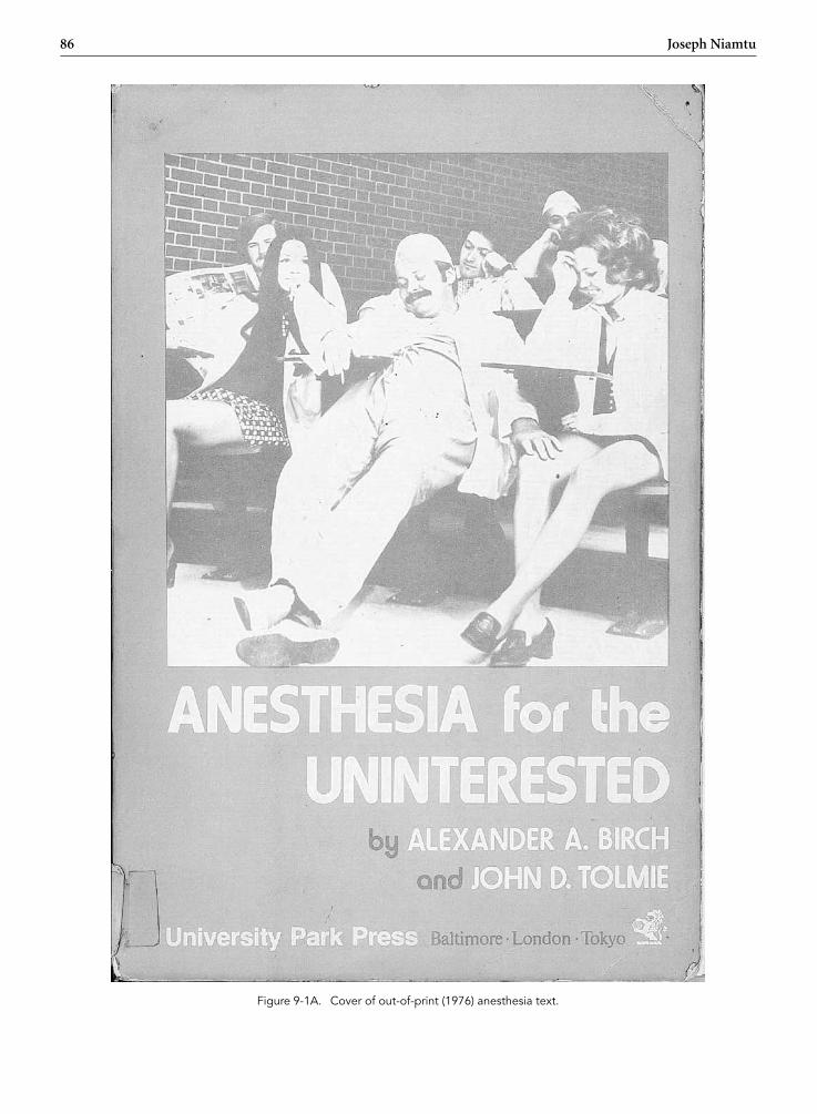
P1: PBU
cufx091-09 CUFX091/Friedberg 0 521 87090 9 Feb. 2, 2007 16:31
86 Joseph Niamtu
Figure 9-1A. Cover of out-of-print (1976) anesthesia text.

P1: PBU
cufx091-09 CUFX091/Friedberg 0 521 87090 9 Feb. 2, 2007 16:31
Local Anesthetic Blocks in Head and Neck Surgery 87
interference of nerve transmission by blocking the influx
of sodium through the excitable nerve membrane.8
SCIENCE IS SOMETIMES BORING!
When reading the following section, some of the infor-
mation is clearly boring see Fig. 9-1A. Finite anatomy was
boring for most medical and dental students, but it is ulti-
mately very important. In order to fully understand how
and when to perform a local anesthetic block, one must
appreciate the anatomy.
Sensory Anatomy of the Head and Neck
The main sensory innervation of the face is derived from
cranial nerve V (trigeminal nerve) and the upper cervical
nerves. see Fig. 9-2.
Sensory anatomy of the trigeminal nerve
The trigeminal nerve is the fifth of the twelve cranial nerves.
Its branches originate at the semilunar ganglion (Gasse-
rian ganglion) located in a cavity (Meckel’s cave) near the
apex of the petrous part of the temporal bone. Three large
nerves, the ophthalmic, maxillary, and mandibular, pro-
ceed from the ganglion to supply sensory innervation to
the face. Often referred to as “the great sensory nerve of
Figure 9-2. Sensory innervation of the head and neck is derivedfrom the trigeminal and upper cervical nerves.
the head and neck,” the trigeminal nerve is named for its
three major sensory branches. The ophthalmic nerve (V1),
maxillary nerve (V2), and mandibular nerve (V3) are liter-
ally “three twins” (trigeminal) carrying sensory informa-
tion of light touch, temperature, pain, and proprioception
from the face and scalp to the brainstem. The commonly
used terms V1, V2, and V3 are shorthand notation for
cranial nerve 5, branches 1, 2, and 3, respectively. In addi-
tion to nerves carrying incoming sensory information, cer-
tain branches of the trigeminal nerve also contain nerves
motor components. The ophthalmic and maxillary nerves
consist exclusively of sensory fibers; the mandibular nerve is
joined outside the cranium by the motor root. These out-
going motor components include branchial motor nerves
(i.e., nerves innervating muscles derived embryologically
from the branchial arches) as well as “hitchhiking” visceral
motor nerves (i.e., nerves innervating viscera, including
smooth muscle and glands). The trigeminal nerve exits
the trigeminal ganglion and courses “backward” to enter
the midlateral aspect of the pons at the brainstem.9
The ophthalmic nerve (V1) leaves the semilunar gan-
glion through the superior orbital fissure. The maxillary
nerve (V2) leaves the semilunar ganglion through the fora-
men rotundum at the skull base, and the mandibular nerve
(V3) leaves the semilunar ganglion through the foramen
ovale at the skull base (see Fig. 9-3 inset9). The remainder
of this chapter discusses only the sensory components of
this nerve system as they relate to local anesthetic blocking
techniques for cosmetic facial procedures.
Sensory anatomy of the ophthalmic nerve (V1)
The ophthalmic nerve, or first division of the trigeminal, is
a sensory nerve. It supplies branches to the cornea, ciliary
body, and iris; to the lacrimal gland and conjunctiva; to the
part of the mucous membrane of the nasal cavity; and to
the skin of the eyelids, eyebrow, forehead, and upper lateral
nose (see Fig. 9-3 V1). The smallest of the three divisions of
the trigeminal, it divides into three branches: the frontal,
the nasociliary, and the lacrimal.9 The frontal nerve divides
into the supraorbital and supratrochlear nerves providing
sensation to the forehead and anterior scalp.
The nasocillary nerve divides into four branches, two
of which supply sensory innervation to the face. These
two branches are the infratrochlear and the ethmoidal
nerves. The infratrochlear nerve supplies sensation to
the skin of the medial eyelids and side of the nose. The

P1: PBU
cufx091-09 CUFX091/Friedberg 0 521 87090 9 Feb. 2, 2007 16:31
88 Joseph Niamtu
Figure 9-3. Main branches of the trigeminal nerve supplying sensation to the respective facial areas. The inset shows the trigeminalganglion with the three main nerve branches.
terminal branch of the ethmoidal nerve is called the exter-
nal (or dorsal) nasal nerve. The ethmoidal nerve innervates
the skin of the nasal dorsum and tip. The lacrimal nerve
innervates the skin of the upper eyelid.
Local Anesthetic Techniques for theScalp and Forehead
The frontal nerve exits through a notch (in some cases
a foramen) on the superior orbital rim approximately
27 mm lateral to the glabellar midline. This supraorbital
notch is readily palpable in most patients. After exiting
the notch or foramen, the nerve traverses the corruga-
tor supercilli muscles and branches into a medial and lat-
eral portion. The lateral branches supply the lateral fore-
head and the medial branches supply the scalp. The supra-
trochlear nerve exits a foramen approximately 17 mm from
the glabellar midline (see Fig. 9-4) and supplies sensation
to the middle portion of the forehead. The infratrochlear
nerve exits a foramen below the trochlea and provides sen-
sation to the medial upper eyelid, canthus, medial nasal
skin, conjunctiva, and lacrimal apparatus.10
When injecting this area, it is prudent to always use
the free hand to palpate the orbital rim to prevent inad-
vertent injection into the globe! To anesthetize this area,
the supratrochlear nerve is measured 17 mm from the
glabellar midline and 1–2 cc of 2% lidocaine 1:100,000
epinephrine are injected (see Fig. 9-5 left). The supraor-
bital nerve is blocked by palpating the notch (and or mea-
suring 27 mm from the glabellar midline) and injecting
2 cc of local anesthetic solution (see Fig. 9-5 center). The
infratrochlear nerve is blocked by injecting 1–2 cc of local
anesthetic solution at the junction of the orbit and the
nasal bones (see Figure 9-5 right). In reality, one can block
all three of these nerves by simple injecting 2–4 cc of local
anesthetic solution from the central brow proceeding to
the medial brow Figure 9-6 shows the regions anesthetized
from these blocks.
Sensory anatomy of the maxillary nerve (V2)
The maxillary nerve or second division of the trigeminal
is a sensory nerve that crosses the pterygopalatine fossa
before traversing the orbit in the infraorbital groove and
canal in the floor of the orbit. It appears on the face at

P1: PBU
cufx091-09 CUFX091/Friedberg 0 521 87090 9 Feb. 2, 2007 16:31
Local Anesthetic Blocks in Head and Neck Surgery 89
Figure 9-4. The supraorbital nerve (SO) exits about 27 mmfrom the glabellar midline, and the supratrochlear nerve (ST) islocated approximately 17 mm from the glabellar midline. Theinfratrochlear nerve (IT) exits below the trochlea.
the infraorbital foramen as the infraorbital nerve.9 At its
termination, the nerve divides into branches that spread
out on the side of the nose, the lower eyelid, and the upper
lip, joining with filaments of the facial nerve.9 Terminal
branches include the following.
The zygomatic nerve arises in the pterygopalatine fossa,
enters the orbit by the inferior orbital fissure, and divides
Figure 9-6. The shaded areas indicate the anesthetized areasfrom supraorbital nerve (SO), supratrochlear nerve (ST), andinfratrochlear nerve (IT) blocks.
at the back of that cavity into two terminal branches, the
zygomaticotemporal and zygomaticofacial nerves.
The zygomaticotemporal branch runs along the lateral
wall of the orbit in a groove in the zygomatic bone before
passing through a foramen in the zygomatic bone and
entering the temporal fossa. It ascends between the bone
and substance of the temporalis muscle and pierces the
Figure 9-5. The forehead and scalp are blocked by a series of injections from the central to the medial brow.

P1: PBU
cufx091-09 CUFX091/Friedberg 0 521 87090 9 Feb. 2, 2007 16:31
90 Joseph Niamtu
temporal fascia about 2.5 cm above the zygomatic arch
where it is distributed to the skin of the side of the forehead
(see Fig. 9-3 V2).
The zygomaticofacial branch passes along the infero-
lateral angle of the orbit, emerges on the face through a
foramen in the zygomatic bone, and perforates the orbic-
ularis oculi and supplies the skin on the prominence of the
cheek (see Fig. 9-3 V2).
As the maxillary nerve traverses the orbital floor and
exits the infraorbital foramen, it branches into a plexus of
nerves that has the following terminal branches.
The inferior palpebral branches ascend behind the
orbicularis oculi muscle and supply the skin and conjunc-
tiva of the lower eyelid (see Fig. 9-3 V2).
The lateral nasal branches (rami nasales externi) supply
the skin of the side of the nose (see Fig. 9-3 V2).
The superior labial branches are distributed to the skin
of the upper lip, the mucous membrane of the mouth, and
labial glands (see Fig. 9-3 V2).
Local Anesthetic Techniques forthe Infraorbital Nerve Block
The infraorbital nerve exits the infraorbital foramen 4–7
mm below the orbital rim in an imaginary line dropped
from the medial limbus of the iris or the pupillary mid-
line. The anterior superior alveolar nerve branches from
the infraorbital nerve before it exits the foramen, and
thus some patients will manifest anesthesia of the ante-
rior teeth and gingiva if the branching is close to the fora-
men. Areas anesthetized include the lateral nose, anterior
cheek, lower eyelid, and upper lip on the injected side. This
nerve can be blocked either by the intraoral or extraoral
route.
To perform an infraorbital nerve block from an intraoral
approach, topical anesthesia is placed on the oral mucosa at
the vestibular sulcus just under the canine fossa (between
the canine and first premolar tooth) and left for several
minutes. The lip is then elevated and a 1.5-inch 27 gauge
needle is inserted in the sulcus and directed superiorly
toward the infraorbital foramen (see Fig. 9-7). The nee-
dle does not need to enter the foramen for a successful
block. The anesthetic solution needs only to contact the
vast branching around the foramen to be effective. It is
imperative to use the other hand to palpate the inferior
orbital rim to avoid injecting the orbit. Two to four cc of
2% lidocaine with 1:100,000 epinephrine is injected in this
area for the infraorbital block.
The infraorbital nerve can also be very easily blocked
by a facial approach. This is the preferred route of the
author. This may also be the preferred route in dental-
phobic patients. A 27 ga 0.5-inch needle is used and is
placed through the skin and aimed at the foramen in
a perpendicular direction. Two to four cc of local anes-
thetic solution is injected at or close to the foramen (see
Figure 9-7. The intraoral approach for local anesthetic block of the infraorbital nerve.

P1: PBU
cufx091-09 CUFX091/Friedberg 0 521 87090 9 Feb. 2, 2007 16:31
Local Anesthetic Blocks in Head and Neck Surgery 91
Figure 9-8. The facial approach for local anesthetic block of theinfraorbital nerve.
Fig. 9-8). Again, the other hand must constantly palpate
the inferior orbital rim to prevent inadvertent injection
into the orbit.
A successful infraorbital nerve block will anesthetize
the infraorbital cheek, the lower palpebral area, the lateral
nasal area, and superior labial regions, as shown in Fig-
ure 9-9.
Figure 9-9. Area of anesthesia from unilateral infraorbital nerveblock.
The aforementioned techniques provide anesthesia to
the lateral nasal skin but does not provide anesthesia to
the central portion of the nose. A dorsal (external) nasal
nerve block will supplement nasal anesthesia by providing
anesthesia over the area of the cartilaginous nasal dorsum
and tip. This supplementary nasal block is accomplished
by palpating the inferior rim of the nasal bones at the
osseous cartilaginous junction. The dorsal nerve (anterior
ethmoid branch of the nasocillary nerve) emerges 5–10
mm from the nasal midline at the osseous junction of the
inferior portion of the nasal bones (the distal edge of the
nasal bones) (see Fig. 9-10). The dotted line in Figure 9-14
shows the course of this nerve under the nasal bones before
emerging.
Two often-overlooked nerves in facial local anesthetic
blocks are the zygomaticotemporal and zygomaticofacial
nerves. These nerves represent terminal branches of the
zygomatic nerve. The zygomaticotemporal nerve emerges
through a foramen located on the anterior wall of the
temporal fossa. This foramen is actually behind the lateral
orbital rim posterior to the zygoma at the approximate
level of the lateral canthus (see Fig. 9-11).

P1: PBU
cufx091-09 CUFX091/Friedberg 0 521 87090 9 Feb. 2, 2007 16:31
Figure 9-10. The dorsal (external) nasal nerve is blocked subcutaneously at the osseous-cartilaginous junction of the distal nasal bones.
Figure 9-11. The zygomaticotemporal nerve is blocked by placing the needle on the concave surface of the posterior lateral orbitalrim.
92

P1: PBU
cufx091-09 CUFX091/Friedberg 0 521 87090 9 Feb. 2, 2007 16:31
Local Anesthetic Blocks in Head and Neck Surgery 93
Injection technique involves sliding a 1.5-inch nee-
dle behind the concave portion of the lateral orbital rim.
It is suggested that one closely examine this area on a
model skull prior to attempting this injection, as it will
make the technique simpler.
To orient for this injection, the doctor needs to palpate
the lateral orbital rim at the level of the frontozygomatic
suture (which is frequently palpable). With the index fin-
ger in the depression of the posterior lateral aspect of the
lateral orbital rim (inferior and posterior to the frontozy-
gomatic suture), the operator places the needle just behind
the palpating finger (which is about 1 centimeter posterior
to the frontozygomatic suture) (see Fig. 9-11). The needle
is then “walked” down the concave posterior wall of the
lateral orbital rim to the approximate level of the lateral
canthus. After aspirating, 1–2 cc of 2% lidocaine 1:100,000
epinephrine is injected in this area with a slight pumping
action to ensure deposition of the local anesthetic solution
at or about the foramen. Again, it is important to hug the
back concave wall of the lateral orbital rim with the needle
when injecting.
Blocking the zygomaticotemporal nerve causes anesthe-
sia in the area superior to the nerve including the lateral
orbital rim and the skin of the temple from above the zygo-
matic arch to the temporal fusion line (see Fig. 9-12 ZT).
The zygomaticofacial nerve exits through a foramen (or
foramina in some patients) in the inferior lateral portion
of the orbital rim at the zygoma. If the surgeon palpates the
junction of the inferior lateral (the most southwest portion
Figure 9-13. The zygomaticofacial nerve(s) is blocked by injecting the inferior lateral portion of the orbital rim.
Figure 9-12. The anesthetized areas from the zygomaticotempo-ral (ZT) and the zygomaticofacial nerve (ZF).
of the right orbit, if you will) portion of the lateral orbital
rim, the nerve emerges several millimeters lateral to this
point. By palpating this area and injecting just lateral to
the finger, this nerve is successfully blocked with 1–2 cc
of local anesthesia (see Fig. 9-13). Blocking this nerve will

P1: PBU
cufx091-09 CUFX091/Friedberg 0 521 87090 9 Feb. 2, 2007 16:31
94 Joseph Niamtu
result in anesthesia of a triangular area from the lateral
canthus and the malar region along the zygomatic arch
and some skin inferior to this area10 (see Fig. 9-12).
Total second division nerve block
An efficient and simple technique to obtain hemi midfa-
cial local anesthesia is to block the entire second division
or maxillary nerve. This will anesthetize the entire hemi-
maxilla and the unilateral maxillary sinus by blocking the
pterygopalatine, infraorbital, and zygomatic nerves and
their terminal branches. This is an easily learned tech-
nique involving an intraoral approach at the posterior lat-
eral palate (see Fig. 9-14). The maxillary nerve block via
the greater palatine canal was first described in 1917 by
Mendel.11 The greater palatine foramen is located ante-
rior to the junction of the hard and soft palate medial to
the second molar tooth (see Fig. 9-14 center). The fora-
men is usually found about 7 mm anterior to the hard and
soft palate junction. This junction is seen as a color change
such that the tissue overlying the soft palate is darker pink
than the tissue overlying the hard palate. The key to this
block is to place a 1.5-inch needle through the greater pala-
tine foramen. It sometimes takes multiple needle sticks to
localize the foramen. Due to the need for multiple sticks,
the palatal mucosa in this area is first infiltrated with
0.5 cc of lidocaine to facilitate painless location of the
greater palatine foramen. A 1.5-inch 25 or 27 ga needle is
bent to a 45 degree angle and will usually easily negotiate
the pterygopalatine canal, thereby placing the local anes-
thetic solution into the pterygopalatine fossa. The course
of the maxillary division of the trigeminal nerve (V2) is as
follows. The second division of the trigeminal nerve arises
from the gasserian ganglion in the medial cranial fossa
and exits the skull via the foramen rotundum (see Fig.
9-14 right). The nerve then traverses the superior aspect
of the pterygopalatine fossa, where it divides into three
major branches: the pterygopalatine nerve, the infraor-
bital nerve, and the zygomatic nerve.12 These nerves are
targeted in this block.
When the foramen is located, the needle should be
gently advanced. If significant resistance is encountered,
the needle should be withdrawn and redirected. Approx-
imately 5 percent of the population has been shown to
have tortuous canals that impede the needle tip and
in some patients this technique is not possible. It is
also important to aspirate before injecting to prevent
intravascular injection. When the needle is properly posi-
tioned (usually at a depth of 25–30 mm), the injection
(2–4 ml) should proceed over thirty to forty-five sec-
onds. Transient diplopia of the ipsilateral eye may occur.
This results from the local anesthetic diffusing superi-
orly and medially to anesthetize the orbital nerves. The
patient must be assured that if this phenomenon occurs,
it is transient. Again, this technique will anesthetize all
the terminal branches of the maxillary nerve with a single
injection.
Figure 9-14. The maxillary nerve block is performed by locating the greater palatine foramen (left), inserting a bent needle up thepterygopalatine canal (center) to inject local anesthetic into the ptergopalatine fossa (right). Notice the needle tip in the pterygopalatinefossa on the far right image. As the second division traverses this area, it is blocked at the main trunk.

P1: PBU
cufx091-09 CUFX091/Friedberg 0 521 87090 9 Feb. 2, 2007 16:31
Local Anesthetic Blocks in Head and Neck Surgery 95
Figure 9-15. The mental foramen is approached intraorally below the root tip of the lower second premolar (left) or from a facialapproach (right).
Sensory anatomy of the mandibular nerve (V3)
The mandibular nerve supplies the teeth and gums of the
mandible, the skin of the temporal region, part of the auri-
cle, the lower lip, and the lower part of the face (see Fig.
9-3 V3). The mandibular nerve also supplies the muscles
of mastication and the mucous membrane of the anterior
two thirds of the tongue. It is the largest of the three divi-
sions of the fifth cranial nerve and is made up of a motor
and sensory root.9
Sensory branches of the mandibular nerve include the
auriculotemporal nerve, which supplies sensation to the
skin covering the front of the helix and tragus (see Fig. 9-3).
The inferior alveolar nerve is the largest branch of the
mandibular nerve. It descends with the inferior alveolar
artery and exits the ramus of the mandible to the mandibu-
lar foramen. It then passes forward in the mandibular
canal, beneath the teeth, as far as the mental foramen,
where it divides into two terminal branches, incisive and
mental nerves. The mental nerve emerges at the men-
tal foramen and divides into three branches. One branch
descends to the skin of the chin, and two branches ascend
to the skin and mucous membrane of the lower lip. The
buccal nerve supplies sensation to the skin over the bucci-
nator muscle.9
Local Anesthetic Techniquesfor the Mental Nerve Block
The mental nerve exits the mental foramen on the hemi
mandible at the base of the root of the second premo-
lar (many patients may be missing a premolar due to
orthodontic extractions). The mental foramen is on the
average 11 mm inferior to the gum line (see Fig. 9-15).
There is variability with this foramen, like all foramina.
However, by injecting 2 to 4 cc of local anesthetic solution
about 10 mm inferior to the gum line or 15 mm inferior
to the top of the crown of the second premolar tooth, the
block is usually successful. In a patient without teeth, the
foramen is oftentimes located much higher on the jaw and
can sometimes be palpated. This block is performed more
superiorly in the denture-wearing patient. As stated earlier,
the foramen does not need to be entered because a suffi-
cient volume of local anesthetic solution in the general area
will be effective. By placing traction on the lip and pulling
it away from the jaw, the labial branches of the mental
nerve can sometimes be seen traversing through the thin
mucosa. The mental nerve gives off labial branches to the
lip and chin.
When anesthetized, the distribution of numbness will
be the unilateral lip down to the mentolabial fold, but
many times to the anterior chin and cheek, depending on
the individual furcating anatomy of that patient’s nerve
(see Fig. 9-16). The inferior alveolar nerve also supplies
sensory innervation to the chin pad. The mylohyoid nerve
may also innervate this area. To augment or extend the
area of local anesthesia on the chin, an inferior alveolar
nerve (mandibular dental block) block can be performed
instead of or with the mental nerve block. Additionally,
local skin infiltration in that area may assist.

P1: PBU
cufx091-09 CUFX091/Friedberg 0 521 87090 9 Feb. 2, 2007 16:31
96 Joseph Niamtu
Figure 9-16. This shows the anesthetized areas from a unilateralmental nerve block. Because of various anatomic factors, the areabelow the mentolabial fold or at the midline may share otherinnervation.
Local Anesthetic Techniques for the InferiorAlveolar Nerve Block (Intraoral)
Almost every person who has ever been to a dentist has
had this block and is aware of its effects, distribution, and
duration. This block is technically more difficult to master
but is easily learned. The basis of this technique involves
the deposition of local anesthetic solution at or about the
mandibular foramen on the medial mandibular ramus
where the inferior alveolar nerve enters the mandible (see
Fig. 9-17).
Detailed description of this technique is beyond the
scope of this review article but will be outlined as fol-
lows. The patient is seated upright and the surgeon places
the index finger on the posterior ramus and the thumb
in the coronoid notch on the anterior mandibular ramus
(see Fig. 9-17).
A 1.5-inch 27 ga needle is then directed to the medial
mandibular ramus at the level of the cusps of the upper sec-
ond molar and the needle is advanced halfway between the
thumb and index finger of the other hand that is grasping
the mandible.
Two cc of 2% lidocaine, 1:100,000 epinephrine is then
injected in a pumping motion to better the chances of
anesthetic solution contacting the nerve and foramen.
The needle can be slightly bent as shown in Figure 9-18
to negotiate the sometimes outward curvature of the
Figure 9-17. The target of the needle in the intraoral inferior alveolar nerve block is at the entrance of the nerve in the mandibularforamen on the medial ramus. The needle can be slightly bent with a medial angle to negotiate the flaring anatomy of the ramus. Themylohyoid nerve (inferior to needle) may or may not be blocked by this technique depending on its level of branching.

P1: PBU
cufx091-09 CUFX091/Friedberg 0 521 87090 9 Feb. 2, 2007 16:31
Local Anesthetic Blocks in Head and Neck Surgery 97
Figure 9-18. The mandibular nerve (V3) block places the localanesthetic just posterior to the lateral pterygoid plate, where thethird division of the trigeminal nerve exits the foramen ovale.The needle is walked off the pterygoid plate (1), and the localanesthetic solution is deposited in the region of the third divisionof the trigeminal nerve (2).
mandibular ramus. The surgeon should first aspirate to
avoid intravascular injection. Anesthesia from this block
sometimes takes five to ten minutes to take effect. Pro-
ficiency in this blocking technique requires practice but
is very useful in cosmetic facial procedures. In addition,
the ispilateral tongue is usually anesthetized with this
block. The area anesthetized includes the lower teeth and
gums, the chin, and skin on the lateral chin. The inferior
alveolar nerve block frequently includes the mylohyoid
nerve. In some patients, the mylohyoid nerve branches
above the area of inferior alveolar injection and in this
case needs a specific mylohyoid nerve block, as outlined
previously.
Local Anesthetic Techniques for the MandibularNerve (V3) Block (Facial Approach)
The mandibular nerve can also be blocked from a deep
injection as the nerve exits the foramen ovale, posterior
to the pterygoid plate13 (see number 2 in Fig. 9-18). This
technique requires more experience and has more poten-
tial complications than the intraoral approach.
The technique for performing this block begins with the
patient in supine position with the head and neck turned
away from the side to be blocked. The patient is asked to
open and close the mouth gently so that the operator can
identify and palpate the sigmoid notch. This is the area
between the mandibular condyle and the coronoid pro-
cess. This notch is located about 25 mm anterior to the
tragus. If one places their finger 25 mm anterior to the tra-
gus and opens and closes the jaw, the mandibular condyle
can be palpated with the jaw open. When the jaw is closed,
the finger will be over the sigmoid notch. A 22 ga, 8-cm
needle is inserted in the midpoint of the notch and directed
at a slightly cephalic and medial to angle through the notch
until the lateral pterygoid plate is contacted (number 1 in
Fig. 9-18). This is usually at a depth of approximately 4.5–
5.0 cm. Spinal needles frequently have measuring stops
that can be adjusted to the position of original contact
of the pterygoid plate. The needle is then withdrawn to
a subcutaneous position and carefully “walked off” the
posterior border of the pterygoid plate (arrows in Fig. 9-
18) in a horizontal plane until the needle no longer tou-
ches the plate and is posterior to it. The needle depth
should be the same as the distance on the needle stop
marker when the pterygoid plate was originally contacted.
The needle should not be advanced more than 0.5 cm
past the depth of the pterygoid plate because the supe-
rior constrictor muscle of the pharynx can be pierced eas-
ily.13 When the needle is in appropriate position, 5 cc of
local anesthetic solution can be administered. The area
anesthetized is shown as V3 in Figure 9-3. Complications
include hematoma formation and subarachnoid injection.
Again, this block should be learned in a proctored situation
and not be attempted by novice injectors.
Sensory anatomy of the scalp
The anterior scalp is anesthetized by injecting the branches
of V1 (supraorbital and supratrochlear nerves) and V2
(the zygomaticotemporal nerve). The greater and lesser
occipital nerves innervate the posterior scalp. The greater
auricular nerve supplies the lateral scalp (see Fig. 9-19).
The greater occipital nerve arises from the dorsal rami
of the second cervical nerve and travels deep to the cer-
vical musculature until it becomes subcutaneous slightly
inferior to the superior nuchal line.14 It emerges on this
line in association with the occipital artery. The artery is
the most useful landmark for locating the greater occipital
nerve.
Local Anesthetic Techniques to Block the Scalp
By performing the brow blocks (see Fig. 9-5), the cervical
plexus block (see Fig. 9-20), and the zygomaticotemporal

P1: PBU
cufx091-09 CUFX091/Friedberg 0 521 87090 9 Feb. 2, 2007 16:31
98 Joseph Niamtu
Figure 9-19. Innervation of the scalp where 1 = supratrochlearnerve, 2 = supraorbital nerve(s), 3 = zygomaticotemporal nerve,4 = greater auricular nerve, 5 = lesser occipital nerve, 6 = greateroccipital nerve.
Figure 9-20. The cervical plexus block is performed by making aline from the mastoid process (1) to the level of the transverseprocess of C6 (2), then finding the point halfway between thesetwo marks (X) just posterior to the sternocleidomastiod (dottedline). Local anesthetic is then injected perpendicularly, superiorly,and inferiorly in this region (the middle picture also shows thegreater occipital nerve, which is not part of the cervical plexus.)
block (see Fig. 9-11), most of the scalp is anesthetized,
except the posterior area. This is anesthetized by blocking
the greater occipital nerve. One can also perform a ring
block where wheals of local anesthetic are injected every
several centimeters around the entire scalp at about the
level of the eyebrows. About 30 cc of local is required to
perform a scalp-ring block.
Greater Occipital Nerve Block Techniquefor Posterior Scalp Anesthesia
The most efficient patient position is sitting upright with
the chin flexed to the sternum.15 The nerve is identi-
fied at its point of entry to the scalp, along the superior
nuchal line one third to one half the distance between
the mastoid process and the occipital protuberance in the
midline14 (see Fig. 9-21). Another measurement for locat-
ing the artery is 2.5–3.0 cm lateral to the occipital pro-
trubence.16 The patient will report pain upon compres-
sion of the nerve: the point at which maximal tenderness
is elicited can be used as the injection site. A 25 ga 5/8-
inch needle is used for the block. The occipital artery is
just lateral to the greater occipital nerve and can be used
as a pulsatile landmark. Two to four cc of local anesthetic
solution can be infiltrated on either side of the artery to
ensure proximity to the nerve. Figure 9-22 shows the der-
matomes anesthetized by blocking the greater occipital
nerve.
Sensory Anatomy of the Neck; Innervationof the Cervical Plexus
The cervical plexus is formed from the ventral rami of
the upper four cervical nerves (see Fig. 9-21 center). The
dorsal and ventral roots combine to form spinal nerves as
they exit through the intervertebral foramen. The ante-
rior rami of C2 through C4 form the cervical plexus. The
cervical plexus lies just behind the posterior border of
the sternocleido-mastoid muscle, giving off both superfi-
cial (superficial cervical plexus) and deep branches (deep
cervical plexus). The branches of the superficial cervical
plexus supply the skin and superficial structures of the
head, neck, and shoulder. The deep branches of the cervical
plexus innervate the deeper structures of the neck, includ-
ing the muscles of the anterior neck and the diaphragm
(phrenic nerve) and are not blocked for local anesthetic
procedures.

P1: PBU
cufx091-09 CUFX091/Friedberg 0 521 87090 9 Feb. 2, 2007 16:31
Local Anesthetic Blocks in Head and Neck Surgery 99
Figure 9-21. The greater occipital nerve is in close approximation to the artery of the same name (1). The nerve can be located bypalpating the artery and injecting just medial to it (2). Another landmark is injecting on the nuchal line, one third to one half the distancebetween the mastoid prominence and occipital protrubence (3 and 5). Number 4 in the diagram shows the lesser occipital nerve.
Figure 9-22. Blocking the entire ear (with the exception of thearea supplied by the vagus nerve) can be performed by insert-ing the needle at the black dots and infiltrating along the dottedlines. This will anesthetize the terminal branches of the auricu-lotemporal nerve, the lesser occipital nerve, and the anterior andposterior branches of the greater auricular nerve. The main trunksof these nerves could be blocked as detailed earlier in this article,but this terminal infiltration technique may be more convenient.
Superficial branches of the cervical plexus include
the following.
The lesser occipital nerve arises from the second (and
sometimes third) cervical nerve and emerges from the deep
fascia on the posterior lateral portion of the head behind
the auricle, supplying the skin and communicating with
the greater occipital, the great auricular, and the posterior
auricular branch of the facial.
The greater auricular nerve arises from the second and
third cervical nerves and divides into an anterior and a
posterior branch. The anterior branch is distributed to the
skin of the face over the parotid gland and communicates
in the substance of the gland with the facial nerve.
The posterior branch supplies the skin over the mastoid
process and on the back of the auricle, except at its upper
part; a filament pierces the auricle to reach its lateral sur-
face, where it is distributed to the lobule and lower part of
the concha. The posterior branch communicates with the
lesser occipital nerve, the auricular branch of the vagus,
and the posterior auricular branch of the facial nerve.
The cutaneous cervical nerve (cutaneus colli nerve, ante-
rior cervical nerve) arises from the second and third cervical
nerves and provides sensation to the antero-lateral parts
of the neck (see Fig. 9-21 center).
Local Anesthetic Techniques for the Cervical Plexus
This technique is used in cosmetic facial surgery to
block the superficial branches of the cervical plexus to

P1: PBU
cufx091-09 CUFX091/Friedberg 0 521 87090 9 Feb. 2, 2007 16:31
100 Joseph Niamtu
anesthetize skin of the lateral or anterior neck, the pos-
terior lateral scalp, and portions of the periauricular area
(see Fig. 9-2).
The technique involves lying the patient back with the
sternocleidomastiod flexed, exposing the mastoid process
and the transverse process of C6 (Chassaignac’s Tubercle)
(approximate level of the cricoid cartilage) (see Fig. 9-21
left). This line is divided in half at the posterior border of
the sternocleidomastoid to determine the injection point.
Another technique is to simply bisect the distance from the
origin and insertion of the sternocleidomastoid without
osseous landmarks. The success of this block involves a
larger volume of local anesthesia diffusing and spreading
out over a larger area rather than absolute accuracy of
the nerve position. 3 to 5 cc of local anesthetic solution
is injected subcutaneously with the needle perpendicular
to the skin. The needle is then redirected superiorly and
another 3 to 5 cc are injected. Finally, the needle is then
directed inferiorly and another 3 to 5 cc is injected. Figure
9-21 (center) shows the areas anesthetized by a cervical
plexus block.
Phrenic nerve involvement is rare with superficial cervi-
cal plexus block (more common with deep cervical blocks)
but technically possible as C3, 4, and 5 innervate the
diaphragm. Healthy patients can tolerate a hemi paral-
ysis of the diaphragm. However, caution must be used in
patients with cardiopulmonary problems because assisted
ventilation may be required. It must be kept in mind that
a bilateral block could potentially de-innervate the entire
diaphragm. To prevent unwanted spread of local anesthetic
solution, this injection is just subcutaneous in placement
and never done bilaterally.
Selected Area Blocks
Anesthesia for the ear
Four nerve branches supply sensory innervation to the ear.
The anterior half of the ear is supplied by the auriculotem-
poral nerve, which is a branch of the mandibular portion
of the trigeminal nerve. The posterior half of the ear is
innervated by two nerve branches derived from the cervi-
cal plexus: the great auricular nerve and the lesser occipital
nerve (see Fig. 9-22 center). The auditory branch of the
vagus nerve innervates the concha and external auditory
canal.
Although these nerves can be individually targeted with
blocks, a circumferential infiltration (ring block) will anes-
thetize the entire ear, except the concha and the external
auditory canal, which are innervated by the Vagus nerve
(CN X). The needle is inserted into the skin at the junc-
tion where the earlobe attaches to the head. The anesthetic
should be infiltrated while the needle is advanced to the
subcutaneous plane. Infiltration is made in a hexagonal
pattern around the entire periphery of the ear (see Fig. 9-
22). The chonal bowl and external auditory canal will need
separate infiltration. One should aspirate (as with all injec-
tions) prior to injection to prevent intravascular injection.
Anesthesia for the nose
The nose receives innervation from multiple nerves. The
supratrochlear and infratrochlear nerves innervate the
root, bridge, and upper portion of the side of the nose.
The infraorbital nerve supplies the skin on the side of
almost half of the lower nose. The external nasal branch
of the anterior ethmoidal nerve (dorsal nasal nerve) exits
between the nasal bone and the lateral nasal cartilage to
supply the skin over the dorsum of this part of the nose
to the tip.
Anesthetic techniques for blocking the nose vary with
the type of procedure being performed. To block the exter-
nal nasal structures (the bridge and tip), bilateral blocks
of the following nerves are performed: infraorbital, supra-
trochlear, infratrochlear, and the dorsal nasal nerves.
For internal nasal surgery such as rhinoplasty or nasal
trauma, the aforementioned blocks are performed in con-
junction with the following. The second division max-
illary block is valuable in providing supplemental anes-
thesia. This is shown in Figure 9-14. For septal anesthe-
sia, local anesthetic solution is deposited 1 cm in front
of the sphenoid rostrum to block the posterior and supe-
rior branches of the sphenoplatine nerve (see Fig. 9-23).
Bending a 1.5-inch needle will improve visualization while
injecting. The infiltrations are performed from posterior
to anterior to prevent needle puncture bleeding from
obscuring the field. Infiltration can also be made into the
inferior turbinates.
Blocking the lips
Minimally invasive cosmetic surgery techniques such as
filler injections may require local anesthesia of the lips.
Although the doctor could perform bilateral infraorbital
and mental nerve blocks, they present disadvantages.
Many practitioners are uncomfortable with these blocks,
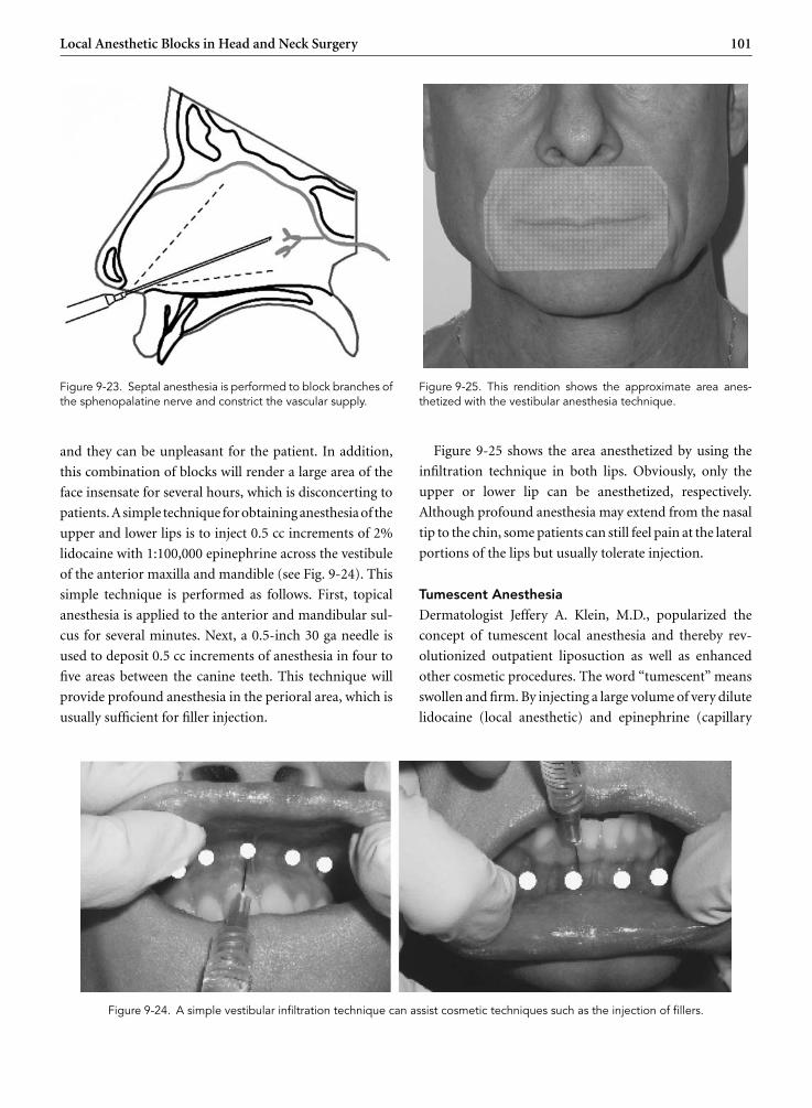
P1: PBU
cufx091-09 CUFX091/Friedberg 0 521 87090 9 Feb. 2, 2007 16:31
Local Anesthetic Blocks in Head and Neck Surgery 101
Figure 9-23. Septal anesthesia is performed to block branches ofthe sphenopalatine nerve and constrict the vascular supply.
and they can be unpleasant for the patient. In addition,
this combination of blocks will render a large area of the
face insensate for several hours, which is disconcerting to
patients. A simple technique for obtaining anesthesia of the
upper and lower lips is to inject 0.5 cc increments of 2%
lidocaine with 1:100,000 epinephrine across the vestibule
of the anterior maxilla and mandible (see Fig. 9-24). This
simple technique is performed as follows. First, topical
anesthesia is applied to the anterior and mandibular sul-
cus for several minutes. Next, a 0.5-inch 30 ga needle is
used to deposit 0.5 cc increments of anesthesia in four to
five areas between the canine teeth. This technique will
provide profound anesthesia in the perioral area, which is
usually sufficient for filler injection.
Figure 9-25. This rendition shows the approximate area anes-thetized with the vestibular anesthesia technique.
Figure 9-25 shows the area anesthetized by using the
infiltration technique in both lips. Obviously, only the
upper or lower lip can be anesthetized, respectively.
Although profound anesthesia may extend from the nasal
tip to the chin, some patients can still feel pain at the lateral
portions of the lips but usually tolerate injection.
Tumescent Anesthesia
Dermatologist Jeffery A. Klein, M.D., popularized the
concept of tumescent local anesthesia and thereby rev-
olutionized outpatient liposuction as well as enhanced
other cosmetic procedures. The word “tumescent” means
swollen and firm. By injecting a large volume of very dilute
lidocaine (local anesthetic) and epinephrine (capillary
Figure 9-24. A simple vestibular infiltration technique can assist cosmetic techniques such as the injection of fillers.

P1: PBU
cufx091-09 CUFX091/Friedberg 0 521 87090 9 Feb. 2, 2007 16:31
102 Joseph Niamtu
constrictor) into subcutaneous fat, the targeted tissue
becomes swollen and firm, or tumescent. The tumescent
technique is a method that provides local anesthesia to
large volumes of subcutaneous fat and thus permits lipo-
suction totally by local anesthesia. The tumescent tech-
nique may eliminate the need for both general anesthesia
and IV narcotics and sedatives.
The tumescent technique for liposuction (1) provides
local anesthesia, (2) constricts capillaries and prevents sur-
gical blood loss, and (3) provides fluid to the body by sub-
cutaneous injection so that no IV fluids are needed.18
Depending on the clinical requirements, a tumescent
anesthetic solution may contain a five- to fortyfold dilu-
tion of lidocaine found in commercially available for-
mulations of local anesthesia. Commercial solutions of
lidocaine used by dentists and anesthesiologists typically
contain 1 gm (1,000 mg) of lidocaine and 1 mg of
epinephrine per 50 ml (2% lidocaine with 1:200,000
epinephrine) of saline. In contrast, tumescent solutions of
local anesthesia contain approximately 0.5–1 gm of lido-
caine and 1 mg of epinephrine in 1,000 ml of NSS. This is a
twentyfold dilution of the commercial version of lidocaine
and epinephrine.18
Tumescent liposuction totally by local anesthesia has
proven to be extremely safe despite the use of unprece-
dented large doses of lidocaine and epinephrine. One
explanation for this remarkable safety is the extreme dilu-
tion of the tumescent local anesthetic solution. Large vol-
umes of dilute epinephrine produce intense constriction
of capillaries in the targeted fat, which in turn greatly
delays the rate of absorption of lidocaine and epinephrine.
Undiluted lidocaine and epinephrine are absorbed into
the bloodstream in less than an hour. Tumescent dilu-
tion causes widespread capillary constriction that causes
the absorption process to be spread over twenty-four to
thirty-six hours. This reduces peak concentration of lido-
caine in the blood, which in turn reduces the poten-
tial toxicity of a given dose of lidocaine (see Chap-
ter 8). Dentists typically use concentrated epinephrine,
which may cause a tachycardia, or rapid heart rate, if the
epinephrine is rapidly absorbed. When very dilute tumes-
cent epinephrine is used, the widespread vasoconstriction
slows the rate of epinephrine absorption, which in turn
prevents an increase in heart rate.18
Profound vasoconstriction (shrinkage of capillary
blood vessels) results from the tumescent infiltration of a
large volume of dilute epinephrine into subcutaneous fat.
Tumescent vasoconstriction is so complete that liposuc-
tion can be done with virtually no blood loss. In contrast,
the older forms of liposuction used before the invention of
the tumescent technique were associated with so much sur-
gical blood loss that autologous blood transfusions were
often routine.
Because the vasoconstriction delays lidocaine absorp-
tion, the local anesthetic remains in place in the fat
for many hours. This prolonged anesthesia may permit
surgery for up to ten hours after infiltration and can pro-
vide twenty-four to thirty-six hours of significant postop-
erative analgesia in some patients.
Maximum recommended lidocaine dosage is 40 mg ·kg−1 to 50 mg · kg−1 for tumescent liposuction when lido-
caine is greatly diluted. This is a relatively large dosage com-
pared to the 7 mg · kg−1 that is widely accepted as the “safe
maximum dose for lidocaine with epinephrine.” Anesthe-
siologists use nondiluted lidocaine for nerve blocks such
as epidural blocks.18
Because tumescent local anesthesia lasts so long, tumes-
cent liposuction is less painful and more pleasant than
liposuction under general anesthesia or IV sedation. With
tumescent local anesthesia, patients are able to avoid the
postoperative nausea and vomiting associated with gen-
eral anesthesia or IV opioids. Tumescent anesthesia is so
efficient at providing fluid to the body that it is unneces-
sary to administer IV fluids. There is a risk of dangerous
fluid overload if excessive IV fluids are given to a tumescent
liposuction patient.18
Tumescent Local Anesthesia for Facial Procedures
Although any part of the head or neck can, in theory, be
blocked, sometimes it is easier or advantageous to utilize
tumescent anesthesia instead of blocks. One big advantage
is the simultaneous hemostasis that accompanies the pain
control. Head and neck procedures that lend themselves to
tumescent local anesthesia include platysmaplasty (“neck-
lift”), rhytidectomy (facelift), brow and forehead lift, and
resurfacing procedures. This author utilizes tumescent
anesthesia for all of these except skin resurfacing. IV seda-
tion with reliance on the potency of ketamine will obtund
these patients. Niamtu finds that tumescent local anes-
thesia distorts the anatomy too much for his preferences.
Many practitioners favor this technique with laser resur-
facing or chemical peel. Generally, tumescent anesthesia

P1: PBU
cufx091-09 CUFX091/Friedberg 0 521 87090 9 Feb. 2, 2007 16:31
Local Anesthetic Blocks in Head and Neck Surgery 103
would be combined with selected local anesthetic blocks.
This adds extra time, material, and equipment to resur-
facing, so Niamtu has not embraced it.
For rhytidectomy, there exist numerous advantages
for the use of tumescent local anesthesia. The effects of
pain control and hemostasis are obvious. The ability of
the engorged tumescent anesthetic solution to hydrodis-
sect the subcutaneous plane is, in Niamtu’s opinion,
paramount. The efficacy of rhytidectomy hinges on the
correct tissue planes being dissected and manipulated.
Novice surgeons frequently become confused when spe-
cific tissue planes are required. By engorging the subcu-
taneous plane with 50–100 cc of tumescent anesthesia in
each pre- and postauricular areas as well as the 100–200
cc in the submental and cervical regions, the facelift can
be performed solely with local anesthesia (see Figs. 9-26
to 9-29).
Blocking the entire face
Although many cosmetic facial procedures are performed
with IV sedation or general anesthesia, many can be per-
formed with only local anesthetic techniques if one mas-
ters the blocks described in this chapter. Although this
author usually uses IV sedation for facelift, chemical peel,
and laser resurfacing, he has performed these procedures
with only local anesthesia. Even when using IV sedation
or general anesthesia, a prudent surgeon will utilize local
anesthetic techniques. This allows the anesthesia provider
to maintain the patient with less IV medication or gas and
Figure 9-26. Tumescent local anesthesia is infiltrated in the pre-and postauricular areas prior to facelift.
Figure 9-27. The preauricular and jowl tissues are engorged withtumescent in preparation for facelift.
provides superior postoperative pain control. Figure 9-30
shows the various dermatomes providing sensation to the
head and neck. By realizing the nerves that supply these
areas, a customized “anesthetic map” may be made by the
surgeon applicable to the operated areas.
Figure 9-28. The submental area is engorged with 100 cc oftumescent anesthesia in preparation for platysmaplasty.

P1: PBU
cufx091-09 CUFX091/Friedberg 0 521 87090 9 Feb. 2, 2007 16:31
104 Joseph Niamtu
Figure 9-29. The facelift flap is dissected fifteen minutes aftertumescent injection to the pre- and postauricular regions. Noticethe bloodless field, which speaks for the effectiveness of thetumescent technique.
Using the picture in Figure 9-30, the surgeon can cre-
ate a formula for which blocks are required for what
procedure. An example would be to select a procedure, say
endoscopic brow and forehead lift. Look at Figure 9-30 and
see what blocks correspond with which dermatomes. For
the endoscopic brow, one would need to block bilaterally
ST, SO, GO, LO, AT, and ZT. Because of anatomic vari-
ables, crossover may be needed with adjacent dermatomes,
but Figure 9-30 should be a good map and starting
point.
SUMMARY
A firm knowledge of the sensory neuroanatomy of the
head and neck can benefit the practice of cosmetic facial
surgery for both the surgeon and the patient. Although the
pathways of sensation for the head and neck are complex,
they can be easily and safely blocked by reviewing the basic
innervation patterns shown in Figure 9-30.
The entire sensory apparatus of the face is supplied
by the trigeminal nerve and several cervical branches.
There exist many patterns of nerve distribution anomaly,
cross innervation, and individual patient variation; how-
ever, by following the basic techniques outlined in this
chapter, the cosmetic surgeon should be able to achieve
pain control of the major dermatomes of the head and
neck.19−22
Figure 9-30. The major sensory dermatomes of the head andneck. AC = anterior cervical cutaneous colli; AT = auriculotempo-ral, B = buccal, EN = external (dorsal) nasal, GA = greater auric-ular, GO = greater occipital, IO = infraorbital, IT = infratroch-lear, LO = lesser occipital, M = mental, SO = supraorbital,ST = supratrochlear, ZF = zygomaticofacial, ZT = zygomatico-temporal.
REFERENCES
1. Hersh EV, Condouis GA: Local anesthetics: A review of theirpharmacology and clinical use. Compend Contin Educ Dent8:374,1987.
2. Jastak JT, Yagiela JA, Donaldson D: Local anesthesia of theoral cavity. Philadelphia, WB Saunders, 1995.
3. Covino BG, Vassalo HG: Chemical aspects of local anestheticagents. In Kitz RJ, Laver MB (eds.), Local Anesthetics: Mecha-nism of Action and Clinical Use. New York, Grune & Stratton,1976; p1.
4. Hersh EV: Local Anesthetics. In Fonseca RJ (ed.), Oraland Maxillofacial Surgery. Philadelphia, WB Saunders, 2000;p. 58.
5. Yagiela JA. Local Anesthetics. In Dionne RA, Phero JC (eds.),Management of Pain and Anxiety in Dental Practice. NewYork, Elservier, 1991; p. 109.
6. Malamed SF: Handbook of Local Anesthesia, 4th ed. St. Louis,Mosby,1997.
7. Aceves J, Machne X: The action of calcium and of local anes-thetics on nerve cells and their interaction during excitation.J Pharmacol Exp Ther 140:138,1963.

P1: PBU
cufx091-09 CUFX091/Friedberg 0 521 87090 9 Feb. 2, 2007 16:31
Local Anesthetic Blocks in Head and Neck Surgery 105
8. Stricharatz D: Molecular mechanisms of nerve block by localanesthetics. Anesthesiol 45:421,1976.
9. Gray H: Anatomy of the Human Body, 13th ed. Lea & Febiger,Philadelphia. 1918; p. 1158.
10. Zide BM, Swift R: How to block and tackle the face. PlastReconst Surg 101:840,1998.
11. Mendel N, Puterbaugh, PG: Conduction, Infiltration andGeneral Anesthesia in Dentistry, 4th ed. Dental Items of Inter-est Publishing Co. 1938; p. 140.
12. Mercuri LG: Intraoral second division nerve block. Oral Surg47:9,1979.
13. Brown DL: Atlas of Regional Anesthesia. Philadelphia, WBSaunders, 1999; p. 170.
14. Bonica TT, Buckley FO: Regional anesthesia with local anes-thetics. In The Management of Pain. 1990, p. 1883.
15. Wheeler AH: Therapeutic Injections for Pain Management.2001 www.emedicine.com/neuro/topic514.htm#target10.
16. Gmyrek R: Local Anesthesia and Regional Nerve Block Anes-thesia. 2002 www.emedicine.com/derm/topic824.htm.
17. Larrabee W, Msakielski KH: Surgical anatomy of the face.New York, Raven Press, Ltd., 1993; p. 83.
18. http//www.liposuction.com. Accessed 2–8–05.19. Niamtu J: Local anesthetic blocks of the head and neck for
cosmetic facial surgery, Part I: A review of basic sensory neu-roanatomy. Cosmet Dermatol 17:515,2004.
20. Niamtu J: Local Anesthetic blocks of the head and neck forcosmetic facial surgery, Part II: Techniques for the upper andmidface. Cosmet Dermatol 17:583,2004.
21. Niamtu J: Local anesthetic blocks of the head and neck forcosmetic facial surgery, Part III: Techniques for the maxillarynerve. Cosmet Dermatol 17:645,2004.
22. Niamtu J: Local anesthetic blocks of the head and neck forcosmetic facial surgery, Part IV: Techniques for the lowerface.Cosmet Dermatol 17:714,2004.

P1: PBU
cufx091-10 CUFX091/Friedberg 0 521 87090 9 Feb. 2, 2007 16:31
10 Local Anesthetics and Surgical Considerationsfor Body Contouring
Rodger Wade Pielet, M.D.
INTRODUCTION
LOCAL ANESTHESIA
BREAST AUGMENTATION AND PECTORAL AUGMENTATION
REDUCTION MAMMAPLASTY AND MASTOPEXY
SUCTION-ASSISTED LIPECTOMY
ABDOMINOPLASTY AND CIRCUMFERENTIAL BODY LIFT AND THIGH LIFT
MISCELLANEOUS ALLOPLASTIC BODY AUGMENTATION
INTRODUCTION
A significant number of techniques for proper infiltration
of local anesthetic for body contouring procedures can
be summarized on three levels. First, establish preemp-
tive analgesia and adequate vasoconstriction at all incision
sites. Second, provide both anesthesia and vasoconstric-
tion in all planes of dissection and manipulation. Third,
facilitate vasoconstriction to all vascular beds supplying
the surgical planes of dissection. The third objective is
best accomplished by having an understanding of the mus-
culocutaneous and fasciocutaneous vascular anatomy. In
many cases, these vascular pedicles are in close proximity
to the sensory nerves; but often, they need to be addressed
as distinct anatomic areas. A significant amount of local
infiltration occurs prior to the surgical scrub and prepa-
ration. This allows for an appropriate amount of time to
elapse for adequate analgesia and vasoconstriction.
Breast augmentation, for example, is the second most
requested surgical procedure in many aesthetic surgery
practices. Although there is no dominant vascular sup-
ply to the breast (Maliniak, 1943), the main contributors
are perforators from the internal mammary artery, the
lateral thoracic artery, and intercostal vessels (Fig. 10-1).
There are also perforators from the thoracoacromial and
thoracodorsal vessels to the pectoralis major muscle. This
understanding is important when performing either sub-
glandular or submuscular augmentation mammoplasty
with or without mastopexy.
The glandular tissue receives its sensory innervation
from the lateral mammary rami of the third through sixth
intercostal nerves and medial mammary rami of the sec-
ond through sixth intercostal nerves (Fig. 10-2). A very
separate anterior branch of the fourth intercostal nerve
supplies sensation to the nipple. The nipple and areolar
complex are always supplied by sensory nerves were seen
through the depths the glandular tissue and not by super-
ficial nerves (Fig. 10-3).
LOCAL ANESTHESIA
For proper analgesia as well as vasoconstriction at the
incision sites, Pielet prefers the use of 1% lidocaine with
epinephrine (1:100,000). This is preferable to the use
of bupivicaine with epinephrine because the potent and
prolonged vasodilitation of bupivicaine seems to out-
last the effects of the epinephrine and there is a slightly
higher increase of delayed postoperative bleeding Edi-
tor’s note: Bupivicaine does not have vasodilating prop-
erties. Bupivicaine for postoperative pain management
is discussed later in this chapter. Either 0.5% or 1%
lidoocaine with epinephrine is injected to infiltrate the
areas of vascular perforators or planes of dissection when
necessary.
106

P1: PBU
cufx091-10 CUFX091/Friedberg 0 521 87090 9 Feb. 2, 2007 16:31
Local Anesthetics and Surgical Considerations for Body Contouring 107
Figure 10-1. Dominant vascular supply to the breast.
Numerous authors have written about the use of tumes-
cent fluids to infiltrate the superficial and deep layers of
fat when performing liposuction. Tumescent infiltration
is also useful in other procedures, such as breast reduc-
tion and abdominoplasty. The typical tumescent solution
consists of 50 cc of 1% lidocaine plus 1 mg of epinephrine
per liter of normal saline or Lactated Ringers solution.
Other surgeons may choose to add up to 12.5 cc of 8.4%
sodium bicarbonate as well as various concentrations of
hyaluronic acid. Pielet has not found either bicarbonate
or hyaluronic acid to be necessary.
Figure 10-2. Nerve supply to the breast.
Figure 10-3. Local anesthetic injection to the incisional area forperiareolar breast augmentation.
When considering liposuction, for example, it is impor-
tant to have continuous communication with one’s anes-
thesiologist. Factors that must be monitored during these
procedures include the starting hematocrit of the patient,
the volume of tumescent fluid infiltrated, volume of fat
aspirated, volume of crystalloid administered through the
IV, and the amount of lidocaine used.
A useful rule of thumb predicts that if a lidocaine-
epinephrine solution is injected in the amount of 15–30 cc
per 100 cm2 of the area to be treated, the hematocrit will
fall approximately 1% for every 150 cc of fat aspirated. For
example, if 1,500 cc fat were removed by liposuction, the
hematocrit would be expected to fall by approximately
10% (Hetter, 1989). A one-to-one volume of fluid is
injected equal to the amount of lipo-aspirate from any
given area. Depending on the size of liposuction, this
often involves amounts of lidocaine that far exceed the
limits thought to be toxic (see Chapter 8). Because there
is delay in the peak levels for several hours after injection,
lidocaine doses as high as 35 mg · kg−1 have been found to
be completely safe in an outpatient setting (Klein, 1990)
(see Chapter 8).
BREAST AUGMENTATION AND
PECTORAL AUGMENTATION
The three most common incision sites for breast augmen-
tation include the inframammary fold, the peri-areola,
and the axilla. Pielet prefers to perform pectoral aug-
mentation entirely through an axillary incision. Pielet
does not perform the transumbilical approach (TUBA),

P1: PBU
cufx091-10 CUFX091/Friedberg 0 521 87090 9 Feb. 2, 2007 16:31
108 Rodger Wade Pielet
Figure 10-4. Parasternal injection of local anesthesia for breastaugmentation.
as this offers no real advantage over other incision sites.
Following infiltration of the incision areas, the medial
parenchymal breast tissue is infiltrated to block the neu-
rovascular perforators arising from the internal mammary
artery above the ribs (Fig. 10-5). This seems to be equally
effective as distinct, individual intercostal blocks and does
not carry the added minimal risk of pneumothorax, which
is slightly more common in very thin individuals. All injec-
tions can be accomplished with approximately 10–15 cc of
1% lidoocaine with epinephrine per side. Editor’s note: At
least 50–70 cc of 0.5% lidocaine with epinephrine is more
effective. Dr. Pielet is a plastic surgeon and client of Dr. Bar-
inholtz (Chapter 11). Inadequate local analgesia is a rea-
sonable explanation for Barinholtz inability to avoid opioids
with the MIA™ technique. Some surgeons may wish to use
a dilute tumescent solution in the subglandular or subpec-
toral spaces.
Figure 10-5. Marking for lateral intercostals injections in the ante-rior axillary line for breast augmentation.
Figure 10-6. Accufusor R© pain pump.
Following dissection of the pocket and assurance of
meticulous hemostasis, a small catheter is placed in the
pocket. The catheter is later connected to a postopera-
tive pain pump delivering a constant infusion of 0.25%
bupivicaine to each side. Some devices include an addi-
tional patient-controlled bolus (Fig. 10-6). It is also effec-
tive to place approximately 5–10 cc of 0.25% bupivicaine
in the pocket if the patient does not wish to have catheters
emerging from the skin for a few days after she goes home.
Editor’s note: Up to 50 cc of 0.25% bupivicaine (125 mg) is
considered a safe dose more effective analgesia will result from
using 20–25 cc per breast pocket. Bupivicaine used in these
applications has not demonstrated an increased risk of
postepinephrine, rebound vasodilatation, or hematoma.
REDUCTION MAMMAPLASTY AND MASTOPEXY
These two operations are performed in a very similar
manner. If one considers the breast simplistically, it is com-
prised of an “outer skin envelope” and “inner stuffing.”
The stuffing is basically made up of parenchyma, fat,
and sometimes an alloplastic implant above or below
the pectoralis major muscle. There are several different
procedures, techniques, and potential scar configurations
based on the patient’s needs and surgeon preference. In
general, a breast reduction involves the removal of both
components, skin and stuffing, whereas a breast lift is
usually the removal of skin only.
Most procedures require some degree of skin-flap
elevation above the parenchyma. The nipple/areolar
complex remains attached to the underlying parenchyma
in order to preserve neurovascular continuity. The excep-
tions to this principle are extremely severe cases of symp-
tomatic macromastia. In this instance, the resultant pedicle
would be insufficient to maintain the blood supply to the

P1: PBU
cufx091-10 CUFX091/Friedberg 0 521 87090 9 Feb. 2, 2007 16:31
Local Anesthetics and Surgical Considerations for Body Contouring 109
nipple/areola. The second exception would be for patients
whose medical or social (i.e., smoking) history precludes
adequate vascularity and/or safety. In these situations, an
en-bloc amputation of the skin, breast tissue, and fat with
replacement of the nipple/areolar complex is performed
as a free graft.
Regardless of the procedure, all incision sites are infil-
trated with 0.5% or 1% lidocaine with epinephrine. If
any area is to be de-epithelialized, intradermal placement
of dilute local with epinephrine is administered. This
facilitates the process of epidermal removal and aids in
hemostasis of this very vascular plane. The area of the
capsule of the breast that is the plane in which shin flaps
are elevated is infiltrated. These flaps are elevated only
as necessary to accomplish proper redraping of the skin
around the parenchymal pedicle. It is important to ele-
vate skin flaps to only within approximately 1 cm of the
pectoralis fascia. This dramatically decreases postopera-
tive pain. Any rent in the fascia, either sharply with scis-
sors or with electrocautery, or stimulation of the fascia
and underlying muscle produces significant increases in
postoperative pain and should be avoided.
SUCTION-ASSISTED LIPECTOMY
Liposuction remains one of the most commonly per-
formed surgical procedures in this country. For most
patients, the postoperative recovery and pain is directly
proportional to the magnitude of the procedure. In addi-
tion to the principles discussed above, it is important to
adequately infiltrate the treated areas and allow sufficient
time for vasoconstriction. It is possible to perform fat aspi-
rations in the range of 4–6 liters or more on an outpatient
basis using these techniques. Editor’s note: Complications
increase as volumes aspirated exceed 5,000 cc. The incision
port sites are infiltrated with either 0.5% or 1% lidocaine
with epinephrine prior to the surgical prep. There are
several published methods for preparation of the tumes-
cent solution. Very low concentrations of epinephrine
(1:400,000–800,000) will provide adequate vasoconstric-
tion. There are also many different recommendations for
the amount of fluid instilled into each area. In general, a
1:1 ratio of infiltrated fluid to the volume of anticipated
fat removal is utilized. Other surgeons choose to use a
‘super-wet’ technique.
In mid-2004, Pielet started using Vaser3 Assisted Lipos-
election (VAL) in addition to the standard tumescent
infiltration. The VAL has revolutionized the practice of
liposuction. VAL has significantly reduced the use of intra-
operative opioids as compared to other methods of tumes-
cent liposuction, including other applications of ultrasonic
technology. Decreased opioid requirement is likely related
to lesser tissue trauma and increased precision. More harsh
techniques require more vigorous action in both tunnel-
ing with the cannula as well as application of ultrasonic
energy. This decreased precision often results in placement
of the tip of the cannula beyond the area infiltrated with
the local anesthetic-containing tumescent solution. This,
obviously, negates the concept of preemptive analgesia and
also increases opioid requirements, intraoperative discom-
fort, and postoperative pain. Because there is more preci-
sion, less tissue trauma, and minimal opioid requirements,
patients actually complain of less postoperative pain.
ABDOMINOPLASTY AND CIRCUMFERENTIAL
BODY LIFT AND THIGH LIFT
There is enough similarity between these two procedures
to discuss proper infiltration of local anesthetic solutions.
Either of these procedures is combined with various ele-
ments of liposuction, and the principles outlined previ-
ously also apply in this situation. The areas of the inci-
sions are infiltrated in the skin as previously described. For
abdominoplasty, a skin flap is elevated up to the level of
the costal margin. To facilitate abdominoplasty, tumescent
solution is infiltrated for vasoconstriction and preemptive
analgesia, just above the fascia in addition to any areas
subjected to suction lipectomy (Fig. 10-7).
Figure 10-7. Electrocautery device use for reflection of lowerabdominal skin flap for abdominoplasty.

P1: PBU
cufx091-10 CUFX091/Friedberg 0 521 87090 9 Feb. 2, 2007 16:31
110 Rodger Wade Pielet
Figure 10-8. Skin infiltration prior to incision for abdominoplasty.
Modest amounts of fat may be aspirated along with sub-
dermal tunneling to facilitate the dissection (Fig. 10-8).
Electrocautery may be used to develop a skin flap which
is elevated up to the level of the costal margin (Fig. 10-9).
Editor’s note: Sometimes the level of analgesia from the
tumescent fluid is insufficient for the patient to tolerate the
electrical stimulus. Supplementing the tumescent with sub-
fascial injection of the rectus sheath may block the perforat-
ing fibers from the midline branch of the intercostals nerves.
Additionally, it may be helpful to proceed with blunt finger or
scissor dissection to develop the flap. Lastly, one can resort to
an additional bolus of 50 mg ketamine to avoid abandoning
an intravenous anesthetic; i.e. MIA™ technique (see Chapter
1) or intravenous general anesthesia (see Chapter 11).—BLF
Figure 10-9. Subdermal tunneling (with possible modest liposuc-tion) prior to reflection of skin flap for abdominoplasty.
Once again, great care is taken to dissect the flap slightly
above the fascia, attempting to avoid cutting or stimulat-
ing the fascia or underlying muscles. This is important
for three reasons. First, it allows visualization of vascular
perforators. They are cauterized or ligated before they can
retract into the fascia. Cut, retracted vessels complicate
the task of achieving hemostasis. Second, reduced stimu-
lation and trauma to the fascia and muscle decreases pain.
And third leaving a very thin but well-vascularized layer
of loose areolar tissue appears to decrease the incidence of
postoperative seroma formation. This is particularly help-
ful since the advent of more aggressive liposuction of the
abdominoplasty flap and adjacent areas. The placement
of closed-suction drains is a matter of preference and the
decision is usually made intraoperatively.
The majority of postoperative pain is initially related to
the fascial plication of the abdominal wall. There is a diver-
gence of opinion as to whether or not the imbrication of the
rectus fascia, the “internal corset” of the abdominoplasty,
requires “muscle” relaxation. Those who believe muscle
relaxants are required (see Chapters 11 and 13) also tend
to believe the rectus muscle itself is being brought to the
midline. Lengthy experience with PK MAC/MIA™ tech-
nique for abdominoplasty (see Chapters 1 and 4 ) has
reproducibly obtained adequate muscle relaxation for fas-
cial imbrication with adequate local analgesia. After a few
days, most patients complain less about this area and more
about the areas treated with liposuction.
Formerly, multiport Accufuser catheters were placed
along the fascia for postoperative pain management, but
an increase in the incidence of seromas was consistently
observed despite the use of drains. It is unlikely that this
is related to the small amount of fluid infused over time
but rather is due to the extreme effectiveness of these pain-
control devices. Essentially, the patient is extremely com-
fortable in the immediate postoperative period to such
an extent that they are excessively mobile and shearing
forces develop between the abdominal wall in the ele-
vated skin flap. This appears to be the etiology of the
higher incidence of seromas. A slight amount of discom-
fort does lead to splinting and better effective immo-
bilization of the abdominal wall in the first few days
after surgery. The aggressiveness of the liposuction of
the lateral flank/hip area through the abdominoplasty
incision may also facilitate seroma formation. The two
most important factors to decrease perioperative pain

P1: PBU
cufx091-10 CUFX091/Friedberg 0 521 87090 9 Feb. 2, 2007 16:31
Local Anesthetics and Surgical Considerations for Body Contouring 111
are elevation of the skin flap just above the fascia and
placement of a 25–50 cc bolus of 0.25% bupivicaine in the
fascia and the incision prior to closure.
The thigh lift, both medial and lateral, essentially in-
volves extensive tumescent liposuction, blunt dissection,
and mild elevation of skin flaps. All of these techniques
were outlined earlier in this chapter.
MISCELLANEOUS ALLOPLASTIC
BODY AUGMENTATION
In addition to the breast augmentation and pectoral aug-
mentation discussed previously, there has been a consider-
able increase in the number of requests for both calf aug-
mentation as well as buttock augmentation. In essence,
the dissection for calf augmentation is entirely above the
gastrocnemius muscle through an incision in the popliteal
fossa. Again, the incision line is injected prior to surgical
preparation and a small amount of dilute local and aes-
thetic is injected into the subcutaneous space where the
implants are to be placed. This is often performed as a
purely cosmetic procedure with the placement of one or
two implants on each side. It is also performed as a uni-
lateral procedure in individuals with either developmental
or traumatic asymmetry.
There are many different approaches for buttock
augmentation. Some surgeons prefer a supragluteal or
Figure 10-10. The entire pocket is elevated and correspondsprecisely to the preoperative markings. This technique aids inhydrodissection, elevation of the plane, hemostasis, and periop-erative pain management.
infragluteal incision as well as a single midline incision.
Pielet prefers bilateral paramedian incisions, as they seem
to yield the most acceptable cosmetic and functional result.
Dissection is carried down to the muscle and a subepime-
sial plane is dissected. Rather than injecting a bolus of local
anesthetic into the respective pocket, sequential aliquots
of dilute lidocaine with epinephrine are injected into the
undulating epimesium between the muscle fibers (see
Fig. 10-10).

P1: PBU
cufx091-11 CUFX091/Friedberg 0 521 87090 9 Feb. 2, 2007 16:50
11 Intravenous Anesthesia for Cosmetic Surgery
David Barinholtz, M.D.
PART II ALTERNATIVE ANESTHESIA APPROACHES IN COSMETIC SURGERY
INTRODUCTION
THE IDEAL ANESTHETIC
COMBINING AGENTS—COMPONENT THERAPY
ANESTHETIC AGENTS FOR TOTAL INTRAVENOUS ANESTHESIA (TIVA)
Benzodiazepines
Alpha2 Agonists
Clonidine
Dexmetetomidine
Propofol
Ketamine
Opioids
Nonopioid Analgesics
Acetaminophen
Nonsteroidal anti-inflammatory drugs (NSAIDs)
Adjunct Agents
Rocuronium
THE AIRWAY CONTINUUM AND IDEAL AIRWAY MANAGEMENT
THE ROLE OF NEUROPHYSIOLOGIC MONITORING IN TIVA
THE ROLE OF LOCAL ANESTHETICS
EMESIS—#1 NEMESIS
Who’s at Risk?
Types of PONV
Prevention and Treatment of Immediate PONV
Nonpharmacologic Therapy
Prevention and Treatment of Delayed PONV
PUTTING IT ALL TOGETHER
Preoperative Care—Prior to the Day of Surgery
Preoperative Care—Day of Surgery
Intraoperative Care
Postoperative Care
CONCLUSION
112

P1: PBU
cufx091-11 CUFX091/Friedberg 0 521 87090 9 Feb. 2, 2007 16:50
Intravenous Anesthesia for Cosmetic Surgery 113
INTRODUCTION
In the 1840s came the first case reports of successfully
anesthetizing patients for surgical procedures. The agents
used were inhalational agents, specifically diethyl ether and
nitrous oxide. Crawford Long, Horace Wells, and William
T. G. Morton will forever be credited with bringing the
benefits of anesthesia to patients undergoing surgery.1,3
No longer would surgical patients needlessly suffer. In this
era, the only ways to deliver an anesthetic systemically
was by inhalation or ingestion (hollow needles had not
yet been invented). Inhalation agents provided a rapid,
reliable, predictable way to anesthetize patients compared
with ingesting alcohol and/or opiates. Hollow needles were
introduced in the late 1800s.
Although various injectable adjuncts such as opiates,
sedative/hypnotics, dissociative agents, and muscle relax-
ants were developed in the early and mid 20th century,
inhalation agents (i.e., nitrous oxide and the halogenated
ether derivatives such as halothane, ethrane, isoflurane)
were the mainstays of anesthesia until the 1980s. How-
ever, they were far from ideal. Inhalation agents (volatile)
are fraught with a myriad of side effects, such as myocar-
dial depression, hypotension, arrhythmias, and postoper-
ative nausea and vomiting (PONV). Anesthesia was very
risky until the 1980s. The combination of cardiovascular
effects of inhalation agents, the routine use of muscle relax-
ants, and lack of sophisticated monitoring devices other
than ECG, NIABP, and spirometry (in addition to the fin-
ger on the pulse, stethoscope, and direct observation) was
the underlying reason. Anesthetic-related death rates were
generally quoted in the 1-in-10,000 range. With millions
of anesthetics being performed annually, the main focus
of the anesthesia community was to improve patient safety
as opposed to minimizing undesirable side effects such as
prolonged anesthetic effects and PONV.
Many things happened in the 1980s to revolutionize
anesthesia care. The first commercially marketed pulse
oximeter, the Nellcor N-100, was introduced in 1984. Pulse
oximetry dramatically reduced the incidence of adverse
hypoxic events. Not until 1990 did the ASA deem pulse
oximetry a “standard of care.” It was intuitively obvious to
early technology adapters that knowing the patient’s state
of oxygenation instantaneously was infinitely preferable
to relying on clinical signs and/or blood gases. Nonethe-
less, it was most disappointing that it took the ASA six
years to deem SpO2 a standard of care. Capnography
became a standard of care, dramatically reducing the inci-
dence of unrecognized esophageal intubations. The adop-
tion of these two innovations, along with a large influx of
very highly trained anesthesia providers into the profes-
sion, is widely believed to be responsible for the dramatic
reduction in anesthetic-related mortality.4 By the mid-
1990s, anesthetic-related mortality rates had decreased to
approximately 1 in 250,000.5
Another seminal event in the late 1980s that revolution-
ized anesthesia, especially ambulatory anesthesia, was the
introduction of propofol.6 Propofol was the first (and still
the only) rapid, ultra–short-acting sedative/hypnotic with
a lack of cumulative effects.7 Propofol could be admin-
istered continuously for prolonged anesthetics and still
allow patients to awaken rapidly without prolonged som-
nolence (or “hangover”).8,9 It is this quality, as well as an
inherent antiemetic effect, that has made propofol such a
popular agent for outpatient anesthesia.
By the mid-1990s, anesthesia care was extremely safe
and reliable, especially for healthy people having outpa-
tient surgery. The most popular anesthetic technique was
the “balanced” anesthetic. This technique takes advantage
of the utility of volatile agents combined with intravenous
agents for analgesia and muscle relaxation. Most of these
patients were intubated, paralyzed, and mechanically ven-
tilated. Although this technique is safe and reliable, it has
its limitations. Prolonged effects of volatile agents (even
desflurane and sevoflurane), muscle relaxants, and opi-
oids caused prolonged recovery times and a high incidence
of undesirable side effects such as PONV. Also, analgesia
was totally dependent on systemic opioids. Surgeons and
anesthesiologists were at odds about the use and safe doses
of local anesthetics (see Chapter 8). Systemic NSAIDs
were reported by some to increase hematoma formation.
Loss of NSAID analgesia meant increased reliance on opi-
oids and opioid-related PONV. There had to be a better
way.
In 1997, Barinholtz left the hospital-based anesthesia
world. After five years in academic anesthesia, the rapidly
expanding area of office-based anesthesia captured his
interest. Armed with the same knowledge as his colleagues,
Barinholtz set out to develop an anesthesia practice out-
side of the hospital setting that took advantage of all the
technologic and pharmacologic advances to provide the
safest state-of-the-art office-based anesthesia practice. For

P1: PBU
cufx091-11 CUFX091/Friedberg 0 521 87090 9 Feb. 2, 2007 16:50
114 David Barinholtz
the first time in his career, Barinholtz began to question
everything he did and why he did it. Does every patient
need to be intubated? Does every intubated patient need to
be paralyzed? Are intravenous agents (e.g., propofol) bet-
ter than volatile agents? Is a 20% PONV rate acceptable?
Are some of the techniques Barinholtz administered more
for his convenience than for the best interest of the
patients?
Some of Barinholtz’s first clients were plastic surgeons.
When he first started working with them in their office-
based surgical suites, he soon discovered that providing
a safe anesthetic wasn’t good enough. The “standard”
propofol induction, LMA placement, maintenance with
sevoflurane while judiciously titrating fentanyl, and a
dose of droperidol, was safe and reliable. Most patients
were discharged in less than two hours. Frequently, res-
cue antiemetics and opioids were being administered in
recovery. The biggest complaints were related to PONV
and pain management. Better outcomes were demanded!
The cosmetic surgery patient is not like a typical “elec-
tive” surgical patient. The cosmetic surgery patient isn’t
having surgery for a medical condition. These patients
desire to look better and expect to feel better. A patient
having surgery for a medical condition is most con-
cerned about getting better. In contradistinction, a cos-
metic surgery patient will tend to be highly critical of every
aspect of care preoperatively, intraoperatively, and post-
operatively, in addition to the achievement of the desired
cosmetic result. Once through this process, the cosmetic
surgical patient will ultimately answer the question, “Was
it worth it?” A rocky perioperative course due to anesthe-
sia can reflect poorly on the surgeon. Conversely, a won-
derful anesthetic experience will reflect very positively on
the surgeon and the anesthesia provider. Optimizing every
aspect of anesthetic care, from the preoperative evalua-
tion through the postoperative follow-up, became the new
goal.
What follows is the distillation of eight years of expe-
rience in office-based elective cosmetic surgery. What
evolved was a propofol-based, intravenous anesthetic,
guided by neurophysiologic monitoring, with minimal
airway intervention and judicious use of local anesthet-
ics. Based on vastly improved PONV rates and minimal
postoperative pain management, Barinholtz’s approach
provides an anesthetic for the cosmetic surgery patient
superior to the former “standard” (vide supra).
THE IDEAL ANESTHETIC
For more than 150 years, the professionals who have ded-
icated their careers to caring for people during one of the
scariest, most stressful and anxiety-provoking experiences
of their lives have strived to do so with caring, compassion,
and above all safety.
What would the ideal anesthetic look like? In a per-
fect world, the ideal anesthetic would have the following
characteristics:
1. SAFE—0% morbidity/mortality rate.
2. QUICK—minimum of time to achieve the desired
effect and wear off just as rapidly.
3. EASY/RELIABLE—simple to administer and have
very predictable effects.
4. EMERGENCE—wear off quickly without undesir-
able side effects.
5. ANALGESIC—pain-free experience.
6. AMNESTIC/SEDATIVE/HYPNOTIC (same cate-
gory because patients make no distinction among
the three)—from a patient’s perspective, as long as
they have no memory of anything they don’t desire
to remember perioperatively, it makes no difference
to them if they were actually completely asleep.
7. NO MOVEMENT—although this may not be the
largest preoccupation from an anesthesiologist’s
point of view, it is the single largest issue for a
surgeon. Patients talking during their surgery is
an additional issue. Whereas a patient may not be
aware or care if they are babbling incessantly during
surgery, it is very distracting and will upset even the
most tolerant surgeon.
No one agent or technology will achieve all seven of
these objectives. However, application of the current phar-
macologic agents and anesthetic monitoring technologies
comes very close.
COMBINING AGENTS—COMPONENT THERAPY
The best argument for an intravenous anesthetic reg-
imen is that one has independent control over every
anesthetic variable. Balanced anesthesia or TIVA relies
on hypnosis augmented by the addition of analgesics
and muscle relaxants. In contrast, an inhalational anes-
thetic provides hypnosis, analgesia, and a degree of muscle

P1: PBU
cufx091-11 CUFX091/Friedberg 0 521 87090 9 Feb. 2, 2007 16:50
Intravenous Anesthesia for Cosmetic Surgery 115
relaxation as an all-in-one package. A sedative/hypnotic
such as propofol will provide only unconsciousness and/or
amnesia. Dissociative agents (e.g., ketamine) and/or opi-
oids will provide systemic analgesia. If a small amount
of skeletal muscle relaxation is necessary for a rectus
muscle repair, for example, a small, subparalyzing dose
of a short-acting nondepolarizing agent (e.g., rocuro-
nium) provides the desired effect without paralyzing the
diaphragm, avoiding the need for intubation and positive
pressure ventilation. Adjuncts such as intravenous anti-
cholinergics, antiemetics, and vasoactive agents such as
labetolol, esmolol, and ephedrine provides unparalleled
control over every aspect of anesthetic effect and side
effect. More recently utilized agents, such as the centrally
acting alpha2 agonists clonidine and dexmetetomodine,
promise to improve the anesthetic even more. In order to
optimize the anesthetic, neurophysiologic monitoring for
depth of anesthesia and liberal use of local analgesia are
essential.
ANESTHETIC AGENTS FOR TOTAL INTRAVENOUS
ANESTHESIA (TIVA)
Benzodiazepines
For many years, preoperative administration of diazepam
for anxiolysis and perioperative amnesia was a mainstay
of most anesthetic regimens. In more recent years, the
more potent, shorter-acting, and more amnestic midazo-
lam replaced diazepam. For many, midazolam is still part
of virtually every anesthetic. Although a reliable amnes-
tic, it does have its drawbacks. In an elegant Level 1
study, Oxorn10 demonstrated a lack of propofol sparing
effect with 2 mg midazolam premedication in a nono-
pioid anesthetic. Oxorn reported that midazolam has an
antianalgesic effect.10,11 When administered PO for pedi-
atric patients, even for propofol/sevoflurane-based anes-
thetics it can be the rate-limiting factor in recovery.12 For
GI procedures, the use of even modest doses of mida-
zolam, with or without opioids, can result in prolonged
sedation and/or amnesia. Patients often cannot recall
results discussed with the gastroenterologist after the pro-
cedure. In contrast, patients receiving propofol, without
benzodiazepines, for these procedures not only remember
results discussed postoperatively but also recover much
more quickly. With the more widespread adminstration
of centrally acting alpha2 agonists such as clonidine13
and dexmetetomidine, which provide excellent anxiol-
ysis, the role of benzodiazepines is being called into
question.
Alpha2 Agonists
Clonidine
Although developed as an antihypertensive agent, cloni-
dine has attracted the interest of the anesthesia commu-
nity for many years. Over the past fifteen years, clonidine
has been administered by intravenous, epidural, spinal,
and intra-articular routes.14−18 Clonidine accentuates and
spares concomitantly administered anesthetic agents.19,20
Clonidine is particularly well suited for use in cosmetic
surgery patients.21,22 Its sedative and anxiolytic effects
make it an ideal preoperative agent, obviating the need
for midazolam. Its sedative and analgesic effects help min-
imize propofol usage,23−25 as well as opioids, thus mini-
mizing PONV. Its prolonged antiadrenergic effect helps
control blood pressure perioperatively, thus theoreti-
cally minimizing chances of hematomas postoperatively.
Clonidine has some limitations and concerns, however.
Administered orally (usually in the 0.1–0.3 mg range),26,27
it must be given thirty to sixty minutes prior to surgery.
Once given, the dose cannot be titrated. Although gener-
ally predictable, the desired de facto tranquilizing effect
is not always achieved. Clonidine 0.2 mg will achieve
a 2.5–5.0 ug · kg−1 blood level in patients weigh-
ing between 95–175 pounds. De facto tranquilization is
nearly always achieved with a therapeutic dose. Alter-
natively, sometimes the effect is more dramatic than
desired, requiring increased perioperative fluids and occa-
sional use of adrenergic agents to support blood pressure
(Table 11-1).
Dexmetetomidine
This centrally acting alpha2 agonist, FDA-approved for
sedating ICU patients, is eight times more potent than
clonidine.28 Recently, Mayer29 at the University of Illi-
nois and Shapiro30 at Harvard have been involved in
clinical trials in the perioperative use of dexmetetomi-
dine. This rapidly acting (approximately ten minutes),
short-acting, intravenous agent shows great promise in
outpatient anesthesia.28 Currently, however, its use is
cost-prohibitive in the price-sensitive world of cosmetic
surgery.

P1: PBU
cufx091-11 CUFX091/Friedberg 0 521 87090 9 Feb. 2, 2007 16:50
Tab
le1
1-1
.A
nest
heti
cag
ents
for
tota
lint
rave
nous
anes
thes
ia(T
IVA
)
Cla
ssD
rug
Site
-of-
acti
onPr
inci
pal
effe
ctR
ole
inco
mp
onen
tth
erap
y
Ben
zod
iaze
pin
esM
idaz
olam
Bd
z/g
aba
rece
pto
rsA
mne
sia/
anxi
olys
is/s
edat
ion
Preo
per
ativ
ean
xiol
ytic
/am
nest
icA
lpha
2ag
onis
tsC
loni
din
e(e
xist
sin
PO/I
Vfo
rmus
ually
adm
inis
tere
dPO
)
Dex
met
etom
odin
e(8
Xm
ore
pot
ent
than
clon
idin
eIV
infu
sion
)
Alp
ha2
rece
pto
rsin
CN
S
Alp
ha2
rece
pto
rsin
CN
S
Anx
ioly
sis/
sed
atio
n/an
alg
esia
dec
reas
esad
rene
rgic
outp
ut
Anx
ioly
sis/
sed
atio
n/an
alg
esia
dec
reas
esad
rene
rgic
outp
ut
Preo
per
ativ
ean
xiol
ytic
intr
aop
erat
ive
sed
atio
n/an
alg
esia
Preo
per
ativ
ean
xiol
ytic
intr
aop
erat
ive
sed
atio
n/an
alg
esia
Sed
ativ
e/hy
pno
ticPr
opof
olC
ereb
ralc
orte
xSe
dat
ion/
unco
nsci
ousn
ess
Mai
nse
dat
ive/
hyp
notic
/vol
atile
equi
vale
ntD
isso
ciat
ive
agen
tK
etam
ine
NM
DA
rece
pto
rsD
isso
ciat
ion/
anal
ges
iaD
isso
ciat
ive/
anal
ges
icO
pio
ids
Fent
anyl
Nar
cotic
Syst
emic
anal
ges
iaSy
stem
ican
alg
esia
Sufe
ntan
ilRe
cep
tors
Alfe
ntan
ilRe
inSe
ntan
ilN
SAID
sK
etor
olac
CO
X-2
sC
elec
oxib
,Rof
ecox
ib,V
ald
ecox
ib(a
llor
alex
cep
tva
ldec
oxib
-IV)
Cyc
loox
ygen
ase
inhi
bito
rsA
nti-i
nflam
mat
ory
anal
ges
iaLi
mite
db
ecau
seof
antip
late
let
effe
ctre
cent
conc
erns
with
CO
X-2
sPa
ra-a
min
ophe
nol
der
ivat
ives
Ace
tam
inop
hen
Inhi
bits
cent
ral
pro
stag
land
insy
nthe
sis
Ana
lges
iaLi
mite
d2◦
tolim
ited
effe
ctiv
enes
san
dhe
pat
otox
icco
ncer
ns
116

P1: PBU
cufx091-11 CUFX091/Friedberg 0 521 87090 9 Feb. 2, 2007 16:50
Intravenous Anesthesia for Cosmetic Surgery 117
Propofol
In 2007, no discussion of anesthesia for cosmetic surgery
can be complete without including propofol. A deriva-
tive of isopropylphenol, propofol has single-handedly rev-
olutionized outpatient anesthesia. With propofol as the
principal anesthetic agent, aided by judicious use of small
amounts of opioids and ketamine and guided by neuro-
physiologic monitoring, a reliable TIVA is possible. In
Barinholtz’s practice, every cosmetic procedure, from
facelifts to abdominoplasties to breast reductions to prone
liposuction, is performed with a propofol-based anesthetic
with no more airway intervention than an oropharyngeal
airway. The success of this technique depends on the clin-
ical vigilance of the anesthesiologist as well as neurophys-
iologic monitoring and local anesthetics.
Ketamine
Developed in the 1960s and popularized as a pediatric
and veterinary anesthetic agent, ketamine was largely over-
looked for use in cosmetic surgery until recently (see Chap-
ters 1 and 4).
Historically, large doses of ketamine administered to
pediatric and trauma patients (2–4 mg · kg−1),31 fre-
quently without amnestics such as midazolam, resulted
in profound dysphorias and/or hallucinations. Because of
unpredictable outcomes, ketamine fell out of favor with
anesthesiologists (vide supra).
In the 1990s, some began mixing smaller amounts of
ketamine with propofol with impressive results.32,33 Total
doses of ketamine of less than 1 mg · kg−1 administered
by bolus or continuous infusion in the presence of ade-
quate propofol thynosis attenuates the response to local
anesthetic injections and minimizes opioid requirements.
In fact, PK MAC in the presence of clonidine premed-
ication, BIS-monitored propofol to 60–75, and adequate
local anesthesia obviates the need for opioids altogether.34
Opioids
Unfortunately, in Barinholtz’ practice, despite the use of
clonidine and ketamine in conjunction with propofol, the
complete elimination of opioids has not been possible.
The surgeons, although adept at administering local anes-
thetic agents for the most part, aren’t perfect. Plastic sur-
geons take exception to interrupting surgery to administer
more local. Rather, they insist on increasing the depth of
the anesthetic. Perhaps, with more surgeon education, the
benefits of more local over increasing anesthetic depth will
be worth the very modest amount of time required to do so.
Barinholtz’ surgeons don’t view PONV as a large prob-
lem. If PONV were a large problem for plastic surgeons,
they would acquiesce to attempts to minimize if not elim-
inate opioids. However, Macario’s studies have shown
PONV is a large issue for patients.
With aggressive antiemetic strategies (vide infra),
patients experience approximately 1% incidence of PONV
in the immediate postoperative period. Nonetheless, elim-
ination of PONV is only one reason to eliminate opioids.
The addition of systemic analgesia transforms deep seda-
tion into intravenous general anesthesia. Opioids depress
the laryngeal or “life-preserving” reflexes thereby increas-
ing the probability of aspiration. Opioids depress respi-
ration mandating the routine administration of supple-
mental oxygen and monitoring of EtCO2. Supplemental
oxygen is a fire hazard in the presence of lasers and electro-
cautery devices particularly around the face. Lastly, opioids
fail to block noxious input to the brain from the surgical
field.
Interestingly, virtually all of the PONV that occurs in
these patients happens after taking postoperative opioids
at home (i.e. postdischarge or PDPONV). The surgeons
could certainly minimize patient calls related to PONV as
well as to pain if they adopted more local-analgesia-based
strategies, both intra- and postoperatively (vida infra).
Opioids will continue to be part of Barinholtz’ anesthetic
practice for the foreseeable future. However, the follow-
ing technique has evolved to specifically minimize opioid
requirements for all types of cosmetic surgery.
Nonopioid Analgesics
There are primarily two types of nonnarcotic analgesics
used for the purposes of preventing and treating periop-
erative pain.
Acetaminophen
A derivative of para-aminophenol, acetaminophen is
effective as an antipyretic and analgesic for mild to mod-
erate pain. Commonly used to treat headaches and mild
arthritic pain, its role in cosmetic surgery is limited. The
maximum recommended dosages are not very effective
in preventing or treating pain associated with cosmetic
procedures.35 Exceeding the maximum doses can be hep-
atotoxic and is not recommended.

P1: PBU
cufx091-11 CUFX091/Friedberg 0 521 87090 9 Feb. 2, 2007 16:50
118 David Barinholtz
Nonsteroidal anti-inflammatory drugs (NSAIDs)
The prototypical NSAID, ibuprofen, has been used for
many years as an effective analgesic for even moderate to
severe pain. In addition to its analgesic effect, ibuprofen
is a potent anti-inflammatory. Anti-inflammatory action
is an especially desirable feature that keeps swelling to a
minimum after dental/oral surgery as well as facial cos-
metic procedures. In the late 1980s, an injectable NSAID,
ketoralac, was introduced. The use of this drug soared.
It appeared that eliminating opioids was finally possible.
However, case reports began to appear in the surgical liter-
ature of postoperative bleeding complications linked to the
use of ketorolac (as well as other NSAIDs).36,39 Whether
a justified indictment or not, most cosmetic surgeons will
not allow their patients to take NSAIDs within two weeks
of surgery (before or after), let alone administer them peri-
operatively.
In the mid-1990s, a subclass of NSAIDs was devel-
oped.40 The cyclooxygenase subtype 2 inhibitors (COX-2)
take advantage of the fact that there are different types of
prostaglandins mediating inflammation and coagulation.
Two subtypes of cyclooxygenase act on arachadonic acid to
form prostaglandins. By inhibiting cyclooxygenase-2 (and
not cyclooxygenase-1), the prostaglandins that are respon-
sible for pain and inflammation are not formed. How-
ever, the prostaglandins responsible for proper platelet
function, formed through the action of cyclooxygenase-
1, are uninhibited. The result is an NSAID that treats pain
and inflammation without inhibiting platelet function.
Celecoxib was the first FDA-approved COX-2 inhibitor,
though it was originally developed to treat arthritis pain
without analgesia. Next, rofecoxib (Vioxx r©) proved even
more effective for postoperative pain.41,45 Many practi-
tioners, even cosmetic surgeons, began adopting the use
of rofecoxib perioperatively because it eliminated the fear
of bleeding complications. Results were promising. Then,
an even more potent COX-2 inhibitor, valdecoxib, was
introduced specifically for postoperative pain. The results
of clinical trials were impressive comparing valdecoxib.46
An injectable form of valdecoxib, parecoxib, was in phase
3 clinical trials and showed promise as a convenient peri-
operative analgesic.47
Unfortunately, in 2004, amid reports of cardiac deaths
associated with the chronic use of Vioxx r©, Merck removed
it from the market.48 As more data surfaced, valde-
coxib was also voluntarily withdrawn. Currently, celecoxib
(Celebrex r©) is the only commercially available COX-2
inhibitor. It is not as effective as either rofecoxib or valde-
coxib for postoperative pain, and there are now concerns
regarding this whole class of drugs.49 It is probably most
prudent at this time to avoid their routine use periopera-
tively until more research is done.
Adjunct Agents
Table 11-2 summarizes adjunct agents utilized in compo-
nent intravenous anesthesia. These agents are used mainly
to offset effects of the previously-mentioned agents.
Glycopyrrolate is administered to minimize secretions,
especially when using ketamine. The adrenergic agents
(ephedrine or epinephrine) can be used to offset the car-
diovascular effects of propofol, opioids, and, occasionally,
clonidine. The beta-blockers (and alpha/beta-blockers)
can be used to offset effects of epinephrine (from local
anesthetic injections) and cocaine. Titrated judiciously,
one can avoid significant tachycardia, bradycardia, hyper-
tension, or hypotension (Table 11-2).
Rocuronium
Virtually every cosmetic surgical procedure performed in
an office-based setting is conducted with a spontaneously
breathing, nonintubated patient. Muscle relaxants are not
routinely utilized, except in one specific instance. Specif-
ically, rocuronium is administered for the rectus muscle
repair portion of an abdominoplasty.
Nondepolarizing muscle relaxants inhibit the nicotinic
cholinergic receptors at the neuromuscular junction in
direct competition with acetylcholine relative to the con-
centration at the site. Different muscle groups have vary-
ing concentrations of cholinergic receptors. Therefore,
it is possible to partially paralyze some muscles with-
out affecting others in a clinically significant way (e.g.,
relaxing some muscles without completely paralyzing the
patient). Because the muscle with the highest concentra-
tion of cholinergic receptors is the diaphragm, it requires
the highest concentration of nondepolarizing agent to par-
alyze it. A dose of rocuronium exists that will effectively
relax the rectus muscle for surgical repair without affecting
diaphragmatic function. The ideal dose is approximately
25% of an intubating dose (or 10 mg of rocuronium for a
70-kg adult). Administered a few minutes prior to muscle
repair, it gives the surgeon ideal circumstances (virtually

P1: PBU
cufx091-11 CUFX091/Friedberg 0 521 87090 9 Feb. 2, 2007 16:50
Tab
le1
1-2
.A
dju
nct
agen
ts
Cla
ssD
rug
Site
-of-
acti
onPr
inci
pal
effe
ctR
ole
inco
mp
oten
tth
erap
y
Ant
icho
liner
gic
Gly
cop
yrro
late
Mus
carin
icre
cep
tors
Ant
isia
log
ogue
Dry
ing
agen
tSt
eroi
dD
exam
etha
sone
Cel
lsm
edat
ing
infla
mm
atio
nC
NS
Ant
i-infl
amm
ator
yan
tiem
etic
Ant
i-infl
amm
ator
yan
tiem
etic
But
yrop
heno
nes
(dop
amin
erg
ican
tag
onis
ts)
Dro
per
idol
Dop
amin
ere
cep
tors
Ant
iem
etic
Ant
iem
etic
5HT 3
anta
gon
ists
Ond
anse
tron
5HT 3
rece
pto
rsA
ntie
met
icA
ntie
met
icB
eta-
blo
cker
sEs
mol
olB
-ad
rene
rgic
rece
pto
rsD
ecre
ase
hear
tra
teD
ecre
ase
hear
tra
tere
spon
seto
epin
ephr
ine
inlo
cala
nd/o
rco
cain
eA
lpha
and
bet
ab
lock
ers
Lab
etol
olA
lpha
and
bet
aad
rene
rgic
rece
pto
rsD
ecre
ase
hear
tra
ted
ecre
ase
blo
odp
ress
ure
Offs
etef
fect
sof
epin
ephr
ine
and
/or
coca
ine
Ad
rene
rgic
agen
tEp
hed
rine
Dire
ctb
eta
agon
ist
ind
irect
alp
haan
db
eta
agon
ists
CN
SIn
crea
sehe
art
rate
incr
ease
blo
odp
ress
ure
antie
met
icO
ffset
hyp
oten
sive
effe
cts
ofan
esth
esia
antie
met
icA
dre
nerg
icag
ent
Epin
ephr
ine
Dire
ctal
pha
and
bet
aag
onis
tIn
crea
sehe
art
rate
incr
ease
blo
odp
ress
ure
Offs
ethy
pot
ensi
veef
fect
sof
anes
thes
iaN
ond
epol
ariz
ing
mus
cle
rela
xant
sRo
curo
nium
Nic
otin
ic-c
holin
erg
icre
cep
tors
Mus
cle
rela
xatio
nRe
lax
rect
usm
uscl
efo
rre
pai
r
119

P1: PBU
cufx091-11 CUFX091/Friedberg 0 521 87090 9 Feb. 2, 2007 16:50
120 David Barinholtz
identical to conditions encountered in the intubated, par-
alyzed, positive-pressure ventilated patient) to complete
the repair. Since rocuronium is so short acting and there is
usually at least one hour between the muscle repair and the
conclusion of surgery, it has not been necessary to adminis-
ter reversal agents. Also, this technique avoids a completely
paralyzed patient and the associated risks (airway, DVTs,
awareness). Muscle relaxation, not paralysis, is adminis-
tered. In hundreds of cases done with this technique in
the past eight years, no patient has required intubation or
become paralyzed at these doses. Succinylcholine (SCH)
is available in Barinholtz’s practice, but its use is relegated
to that of an emergency drug only.
The remaining adjunct agents are addressed in the sec-
tion on antiemetics.
THE AIRWAY CONTINUUM AND IDEAL
AIRWAY MANAGEMENT
The area of anesthesia that causes some of the most con-
troversy and is a significant factor in anesthetic-related
morbidity and mortality is airway management. Prior
to the invention of the cuffed endotracheal tube (ET)
by Guedel,50 patients were not routinely intubated for
surgery. Along with the ET and the discovery of mus-
cle relaxants,51 routine intubation with paralysis even for
minor surgeries became commonplace.52 Mask ventila-
tion, especially for short procedures such as myringotomy
and cystoscopy, was still common. However, by the early
1990s, the overwhelming majority of patients receiving
general anesthesia were intubated with an endotracheal
tube.
In the early 1990s, a revolutionary airway device was
introduced in the United States. Developed by Brain in
Great Britain, it was called the laryngeal mask airway
(LMA).53,54 This device gave practitioners an intermediate
choice between mask ventilation and endotracheal intu-
bation. Over the past fifteen years, the LMA has gained
widespread acceptance worldwide, especially in the ambu-
latory anesthesia arena. The LMA gives practitioners the
ability to maintain a patient’s airway, reliably deliver oxy-
gen (and other gases), and monitor end-tidal CO2, all with-
out the need for paralysis. Its success has spurred a host of
similar devices claiming similar advantages, for example,
the cuffed oropharygeal airway (COPA r©) and laryngeal
Combitube. Both the COPA r© and the Combitube r© claim
Figure 11-1. The airway continuum.
to offer most of the advantages of endotracheal intubation
without the invasiveness. However, all are deeply situated
in the pharynx (stimulating all the pharyngeal reflexes).
A 15 mm breathing connection facilitates coupling to an
anesthesia breathing circuit and, ultimately, an anesthe-
sia machine. Does the patient always need a supraglottic
device situated deep in the pharynx in order to maintain an
airway during deep sedation? Do all airway devices require
a 15 mm connector? The answer to these questions is no.
One method to explain and illustrate the concept is to
introduce the airway continuum (see Fig. 11-1). The con-
tinuum is simply a line that from left to right represents
increasing airway invasiveness. On the left is the untam-
pered natural airway and on the right is the most invasive
airway intervention, tracheostomy. In between are nasal
cannulae, oral and nasal airways, the supraglottic devices,
and endotracheal tubes. The continuum is divided into left
and right halves. The right half is the side on which all the
devices have a 15 mm connection for a breathing circuit.
These devices have the advantage of attaching to a fresh gas
source, the ability to monitor end-tidal CO2, and provid-
ing the ability for positive-pressure ventilation. However,
the presence of a 15 mm connector and the absence of
opioids means that patients can breathe, with or without
supplemental oxygen, spontaneously through a supraglot-
tic device without being connected to an anesthesia circuit.
Less is more.
The left half essentially comprises nasal cannulae, oral
airways, and nasal airways. Whereas nasal cannulae attach
to an oxygen source, traditionally oral airways and nasal
trumpets don’t. Also, none of these devices conveniently
provides the ability to monitor end-tidal CO2.

P1: PBU
cufx091-11 CUFX091/Friedberg 0 521 87090 9 Feb. 2, 2007 16:50
Intravenous Anesthesia for Cosmetic Surgery 121
Figure 11-2. Patient with modified oral airway in place for proneliposuction.
Recently, nasal cannulae that simultaneously deliver
oxygen and provide for end-tidal CO2 monitoring
have become commercially available. Several years ago,
Mallinkrodt patented a device fashioned as a nasal trum-
pet with the ability to administer oxygen and sample CO2.
One can fashion an oral airway device that can simultane-
ously deliver oxygen and sample end-tidal CO2. Figure 11-
2 shows a standard Guedel oral airway, flanges trimmed to
allow placement behind the teeth, with two lengths of oxy-
gen tubing placed into the opening in the airway: a short
length situated distally in the airway that can easily attach
directly to a male adapter for end-tidal CO2 monitoring,
and a longer length with the standard 5 in 1 connector,
or “Chistmas-tree” adapter, compatibility to attach to an
oxygen flowmeter. In the past six years, this device has been
employed in thousands of cosmetic procedures. These
Figure 11-4. Patient with modified oral airway in place for breastaugmentation, breast reduction, mastopexy or abdominoplasty.
Figure 11-3. Patient with modified oral airway in place for rhyr-tidectomy.
procedures include blepharoplasty, endoscopic browlift,
rhytidectomy, rhinoplasty, breast augmentation, breast
reduction, mastopexy, abdominoplasty, body lift, and lipo-
suction (including prone and lateral cases) (see Fig. 11-2,
11-3, and 11-4). This device facilitates the maintenance of
a patent airway, the reliable delivery of oxygen, and the
reliable monitoring of end-tidal CO2 (see Fig. 11-5).
There are two potential criticisms. First, this device does
not protect the airway. Second, one cannot immediately
ventilate the patient if necessary. These criticisms are valid
and underscore the importance of choosing appropri-
ate candidates without anticipated airway difficulty and
who are not at risk for aspiration (i.e., GERD). Also, one
must have the personnel, drugs, supplies, and equipment
(including Ambu r© bag, intubation equipment, LMAs,
cricothyroidotomy kit, and jet ventilator) to deal with
Figure 11-5. Modified oral airway with tubing to sample CO2 anddeliver oxygen.

P1: PBU
cufx091-11 CUFX091/Friedberg 0 521 87090 9 Feb. 2, 2007 16:50
122 David Barinholtz
every airway eventuality as described in the ASA difficult-
airway algorithm.55 Properly prepared, there is no reason
every skilled anesthesia provider cannot reproduce Barin-
holtz’s success in applying this technique. No patient has
suffered a serious complication with resulting harm from
its application in Barinholtz’s practice.
THE ROLE OF NEUROPHYSIOLOGIC
MONITORING IN TIVA
Since its first practical applications more than 150 years
ago, anesthesia care has had three main goals, namely,
(1) make patients unconscious/unaware of the gruesome
experience of having surgery, (2) immobilize the patient
so the surgeon can operate, and (3) don’t kill the patient.56
Historically, issues such as pain control and PONV pre-
vention were distant to these fundamental concerns.
While great strides have been made Because the 1840s
in all three of these areas, a review of the improvements
of the past fifty years is appropriate. Approximately fifty
years ago, curare, and subsequently, its derivatives, came
into clinical use. For procedures that require immobility,
there is a specific drug, receptor, and monitoring model.
As far as anesthetic mortality, the necessary technologies
have been developed to directly monitor and address the
concerns germane to this issue, specifically, ECG, NIABP,
SpO2, and EtCO2 (Fig. 11-6).
However, between 1842 and 1996, there was no way
to directly monitor the patient’s level of unconsciousness.
There has been an overwhelming amount of attention in
the anesthesia community, as well as the lay press, regard-
Figure 11-6. Monitor screen during sedation with modified oralairway. N.B. True end-tidal CO2 tracing.
ing the issue of “awareness under [general] anesthesia.”
The more germane issue for office-based as well as other
anesthesiologists is the much more common occurrence
of overanesthetizing patients. However, patients have also
heard the stories of awareness under anesthesia. Dozens of
accounts from patients of their own experiences (or those
of a close friend or relative) have been related to Barinholtz.
He is frequently asked how he can “make sure” this won’t
happen to them. The fact that patients are not paralyzed or
intubated is of great reassurance to them. Routinely mon-
itoring with BIS is an additional source of reassurance.
The techniques described in this chapter rely on intra-
venous anesthesia in spontaneously breathing, nonpara-
lyzed, nonintubated patients. Awareness is not a signifi-
cant concern as these patients can move (or even talk) if
they want or need to do so. Despite the fear of undermedi-
cating, the much more likely (and common practice) issue
is that of overanesthetizing.
Overmedicating produces more undesirable anesthetic
side effects (e.g., hypotension) intraoperatively, more
PONV, prolonged recoveries, and wasted drugs. In Barin-
holtz’s anesthesia practice, all drugs, supplies, and equip-
ment are the anesthesiologist’s responsibility. Further-
more, the anesthesia provider must be physically present
until all patients are recovered and discharged. It makes not
only good clinical sense but good business sense to employ
neurophysiologic monitoring. In the seven years since
employing BIS monitoring, propofol usage has decreased
by 20%. Patients rarely spend more than one hour recov-
ering, even after a prolonged (six-hour) anesthetic. The
foundation of the anesthetic care provided to Barinholtz’s
cosmetic surgery patients is the utilization of the TIVA
technique in conjunction with BIS monitoring.
Finally, directly monitoring the effects the anesthetics
have on the target organ—the brain—is more accurate
than indirect indices of anesthetic depth. The historically
used, indirect (and frequently inaccurate) cues to assess
depth of anesthesia such as heart rate, blood pressure,
tearing, and patient movement are no longer as relevant to
decision making as is the measure of the cerebral cortical
level of hypnosis (see Chapter 3).
Development of the BIS (and similar competing tech-
nologies, such as PSA 4000 and Entropy) and refin-
ing the technology (i.e., the BIS-XP platform) are
great advancements for anesthetic care. Anesthesiologists
should embrace BIS monitoring. Although BIS is not

P1: PBU
cufx091-11 CUFX091/Friedberg 0 521 87090 9 Feb. 2, 2007 16:50
Intravenous Anesthesia for Cosmetic Surgery 123
a perfect technology, neither is ECG, NIABP, or pulse
oximetry. BIS provides valuable, reliable, reproducible
data to help guide anesthetic care. Certainly, monitoring
the EEG effects of anesthesia makes more sense than recent
attempts to develop technologies to monitor and main-
tain fixed blood concentrations of anesthetic agents.57,58
A given concentration of agent in the blood may still have
a wide range of clinical effects on different patients. It is
important to monitor the effect on the brain directly (see
Chapter 3).
THE ROLE OF LOCAL ANESTHETICS
Administering propofol and ketamine, supplemented by
alpha2 agonists and opioids, can provide a TIVA with-
out relying on local analgesia. However, this is not an
advisable approach to the care of the cosmetic surgery
patient. Intraoperatively, one would have to anesthetize
the patient much more deeply. Although ketamine can
be used to provide some analgesia, opioids could not be
avoided. Postoperatively, pain relief would be completely
dependent on opioids. Prolonged recoveries, high PONV
rates, and inadequate (certainly suboptimal) pain relief
would be the outcome. In order for the patient to have
an optimal experience, adequate local analgesia must be
utilized.
Years ago, it was a struggle to get surgeons to employ
local analgesia as part of the surgical procedure. How many
anesthesiologists have heard their surgeon say, “Why do I
need local? That’s what you’re here for.” Luckily, today’s
enlightened cosmetic surgeon knows that employing local
analgesia is a valuable and necessary adjunct for the ulti-
mate success of the surgery. The concept of preemptive
analgesia is well established in both the anesthesia and
surgical literature.59–62
Postoperative local analgesia is achieved by using a long-
acting local anesthetic, such as bupivicaine, in the opera-
tive field before closing. Alternatively, placing a continuous
infusion of local anesthetic postoperatively in the form of
one of many commercially available pain-pumps will min-
imize, if not eliminate, the requirement for opioids.
Treating postoperative pain with local anesthetics, instead
of opioids, results in more effective pain relief.
The patient is made more mobile (minimizing risks of
thromboembolic phenomena), less prone to PONV and
postoperative hematomata, and less likely to experience
postoperative hypertension secondary to pain (again, min-
imizing the risk of a hematomata). This results in happier
patients and happier surgeons who aren’t getting called as
often. If a surgeon can effectively numb the area they are
operating on and operate without extending beyond the
anesthetized area (or with a willingness to administer more
local during the procedure if necessary), it is possible and
highly desirable to anesthetize the patient without using
any opioids (vide supra and see Chapter 4). But even in
the situation where a surgeon doesn’t maximize the use of
local (a predicament in which many anesthesiologists find
themselves), the liberal use of lidocaine greatly facilitates
minimizing intraoperative drug consumption, especially
opioids. The only caveat is to not exceed maximum safe
doses of local anesthetics (see Chapter 8 on lidocaine toxi-
city and Chapters 9 and 10 on local analgesia techniques).
EMESIS—#1 NEMESIS
The single biggest concern expressed by patients under-
going anesthesia for cosmetic surgery (or all surgery, for
that matter) is the fear of PONV. Studies have shown that
when questioned, patients indicate that the fear of PONV
is greater than the fear of postoperative pain and other
anesthetic side effects.63,64 Given the choice, most patients
would rather deal with pain than nausea and vomiting.
PONV doesn’t occur just when the patient is under the
anesthesiologist’s direct care. Patients are at risk for PONV
into the postdischarge, postoperative period, especially if
the surgeon has prescribed opioids for postoperative pain.
In order for the patient to have a PONV-free experience,
one must have a rational, organized approach for both
prevention and treatment of PONV, should it occur.
Who’s at Risk?
Theoretically, every patient receiving anesthesia is at risk
of PONV. Over the years, characteristics and risk factors
have emerged that identify patients who are at a higher
risk of experiencing PONV. These are (1) female gender,
(2) nonsmokers, (3) previous history of PONV, (4) hav-
ing a surgical procedure associated with a higher incidence
of PONV (e.g., gynecologic, laparoscopic, strabismus,
and various cosmetic procedures, especially of the face),
(5) history of motion sickness, (6) exposure to volatile
anesthetic agents, and (7) exposure to opioids (Table 11-3).
Several authors have attempted to assign point values to

P1: PBU
cufx091-11 CUFX091/Friedberg 0 521 87090 9 Feb. 2, 2007 16:50
124 David Barinholtz
Table 11-3. Risk factors for PONV
1. Female gender2. Nonsmoker3. Young age4. Previous history of PONV5. Emetogenic procedure6. History of motion sickness7. Volatile anesthetic agents8. Opioids
each of these risk factors in an attempt to assess an individ-
ual patient’s relative risk of experiencing PONV. Although
no model is perfect, clearly the more of these risk factors
that apply to an individual patient, the higher his or her
risk of PONV.65,68
Because the overwhelming majority of the cosmetic
surgery patients in Barinholtz’s practice are female non-
smokers who will most likely receive opioids intra- and
postoperatively (many of whom have a history of PONV
and motion sickness), all of these patients are treated as
at being high risk for PONV. Because of this proactive,
aggressive effort to prevent it, the PONV rate in Barin-
holtz’s practice is less than 1%.
Types of PONV
PONV can be separated into two types, immediate
and delayed. Immediate PONV occurs immediately after
surgery and during the recovery phase while the patient
is still under the direct care of the anesthesia provider.
Delayed PONV occurs after the patient has been dis-
charged. This could occur on the way home or up to several
days postoperatively. Learn to distinguish between these
two and develop strategies for prevention and treatment
of both. There are some guiding principles common to
both types of PONV.
The first is that prophylaxis is better than treatment. It
is far more desirable to prevent the problem altogether than
to have to deal with the consequences.
The second is that avoidance of opioids is the single
largest factor in preventing PONV.
Any or all of the strategies discussed herein can suc-
cessfully deal with PONV 99% of the time. If opioids are
somehow avoided, approximating 100% PONV-free out-
comes could be achieved with less prophylaxis.
Prevention and Treatment of Immediate PONV
In order to determine if the patient is at risk of immediate
PONV, delayed PONV, or both, focus on key elements of
the history. Patients with a previous history of PONV need
to be questioned carefully as to when the PONV occurred.
Did it occur in the recovery room, on the way home, or
after the patient returned home? Did it occur after taking
oral opioids for pain? Did they follow the instructions and
make sure to have a full stomach before taking oral opioids?
Did it linger for days after surgery? Does the patient have a
history of motion sickness (putting them at risk for PONV
on the ride home)? The answers to these very specific
questions can help the practitioner determine when and
where the patient is most at risk for developing PONV
and help arrive at a specific plan for both prevention and
treatment.
Table 11-4 lists specific factors that put patients at more
risk for immediate PONV. A previous history of imme-
diate PONV or an emetogenic procedure automatically
makes the patient at higher risk. Adding volatile agents
and/or opioids further increases the risk.
The most effective strategy for these patients is to make
certain they’re well hydrated (follow the ASA guidelines
and not only allow but encourage clear liquids up to three
hours before surgery), avoid volatile agents, and avoid opi-
oids. Whereas the first two admonitions are easy to accom-
plish, unfortunately opioids cannot always be avoided. If
a cosmetic surgery patient who is considered high risk
for immediate PONV receives opioids, he or she should
also receive multimodal therapy.69 Inspired by the works
of Scuderi, White, Gan, and Chung, it makes sense that
utilizing small doses of different classes of antiemetics to
disrupt the nausea/vomiting pathway at different points
will yield a higher success rate with a lower incidence of
side effects (e.g., dysphoria, somnolence, or tardive dysk-
inesia) than monotherapy.
Whereas Scuderi et al. describe various “cocktails,” the
one Barinholtz employs has been utilized clinically in the
treatment of more than 3,000 cosmetic surgery patients
over the past five years. Low-dose droperidol (0.625 mg
for the average adult) and dexamethasone (10 mg for the
average adult) shortly after induction, followed by low-
dose ondansetron (1 mg for the average adult) at the con-
clusion of the procedure, are administered.
The Barinholtz “cocktail” has been 99% effective in
the prevention and treatment of immediate PONV in his

P1: PBU
cufx091-11 CUFX091/Friedberg 0 521 87090 9 Feb. 2, 2007 16:50
Intravenous Anesthesia for Cosmetic Surgery 125
practice and is very cost effective for routine use. In the rare
instance of PONV in the PACU, additional ondansetron
(1 mg or 4 mg orally dissolving tablet [ODT r©]) is gener-
ally effective. Another effective approach can be to admin-
ister 50 mg IM ephedrine. This is effective even in the
normotensive patient. The effect appears to be centrally
mediated. Ephedrine “perks” up the patients and is usually
effective within ten to fifteen minutes after administration.
Pay close attention to these patients as they begin to
move around, stand up, get dressed, and walk. If the
therapy is effective and they remain nausea-free during
the getting-ready-to-leave phase, the patients can be dis-
charged without further therapy (as long as there are not
additional risk factors for delayed PONV). If, however, the
patient is still nauseated despite all of the above, further
therapy is required.
Nonpharmacologic Therapy
An effective adjunct to pharmacologic therapy is P6 accu-
point stimulation. Based on the principles of acupunc-
ture and backed by impressive clinical results in several
recent studies,70,71 stimulation of the P6 accupoint has
been shown to be very effective in the prevention and treat-
ment of PONV (as well as nausea/vomiting secondary to
motion sickness, pregnancy, and chemotherapy). Located
on the inside of the wrist, approximately 1 inch below the
palmar crease, in the distribution of the median nerve, the
P6 accupoint is easily identifiable. Whereas some authors
have chosen to needle the P6 accupoint or apply pressure in
order to stimulate it, the most effective practical approach
is electrostimulation. The Reliefband r© (Abbott Laborato-
ries) is the only device currently available and approved by
the FDA for electrostimulation of the P6 accupoint for the
prevention and treatment of PONV (as well as nausea and
vomiting from pregnancy, motion sickness, or chemother-
apy).70,71 Reliefband r© see Fig. 11.7 is easy to use, patient
controlled, and portable, and Barinholtz has employed it
as an adjunct to (or, in some cases, replacement of) phar-
macologic agents for PONV for more than five years. In
patients with resistant PONV, or in whom pharmocologic
agents are contraindicated, or in patients at risk of delayed
PONV, Reliefband r© has been a very clinically effective
as well as cost-effective approach in Barinholtz’s practice.
Combined with 5-HT3 antagonists (e.g., ondansetron),
it has been shown to be remarkably effective.72 Also, the
combination of Reliefband r© and ondansetron is easy for
Figure 11-7. Reliefband.
the patient to continue at home for up to several days
postoperatively if necessary.
Prevention and Treatment of Delayed PONV
Table 11-4 outlines specific factors that put a patient in
the high-risk category for delayed PONV. These are a pre-
vious history of delayed PONV, a history of motion sick-
ness, a history of ineffective (or questionable) treatment of
immediate PONV, and any prescription for oral opioids
for postoperative pain. For patients in this category, have
a strategy that is practical for home therapy. The options
are pills, orally dissolving tablets (ODTs), suppositories,
and P6 accupoint stimulation (vide supra).
Patients do not generally favor suppositories. Pills can
be hard to swallow in the face of active PONV. Also, some

P1: PBU
cufx091-11 CUFX091/Friedberg 0 521 87090 9 Feb. 2, 2007 16:50
Tab
le1
1-4
.A
sses
sing
risk
and
stra
teg
ies
for
pre
vent
ion
and
trea
tmen
tof
PON
V
Ris
kfa
ctor
sPr
even
tion
Trea
tmen
t
Imm
edia
tePO
NV
Prev
ious
hist
ory
imm
edia
tePO
NV
Emet
ogen
icp
roce
dur
eEx
pos
ure
tovo
latil
ean
esth
etic
Intr
aop
erat
ive
and
/or
pos
top
erat
ive
opio
ids
Mak
esu
rep
atie
ntis
wel
lhyd
rate
dp
reop
erat
ivel
y(e
ncou
rag
ecl
ear
liqui
ds
upto
3ho
urs
bef
ore
surg
ery)
Avo
idvo
latil
eag
ent
Avo
id(o
rat
leas
tm
inim
ize)
opio
idus
age
Mul
timod
alp
harm
acol
ogic
ther
apy
Ad
diti
onal
pha
rmac
olog
icag
ents
—co
nsid
erno
npha
rmac
olog
icth
erap
y(P
6ac
cup
oint
stim
ulat
ion)
Del
ayed
PON
VPr
evio
ushi
stor
yd
elay
edPO
NV
His
tory
ofm
otio
nsi
ckne
ssPr
escr
iptio
nfo
ror
alop
ioid
sfo
rp
osto
pp
ain
Inef
fect
ive
trea
tmen
tof
imm
edia
tePO
NV
Mak
esu
reim
med
iate
PON
Vre
solv
edb
efor
ed
isch
arg
ing
pat
ient
Inst
ruct
driv
eron
care
fuld
rivin
g(e
spec
ially
inp
atie
nts
with
hist
ory
ofm
otio
nsi
ckne
ss)
Avo
idp
osto
per
ativ
eop
ioid
s(e
mp
loy
loca
lan
esth
etic
sfo
rp
osto
pp
ain)
Reite
rate
need
for
full
stom
ach
bef
ore
taki
ng POop
ioid
s
Pres
crib
ePO
antie
met
icC
onsi
der
nonp
harm
acol
ogic
ther
apy
126
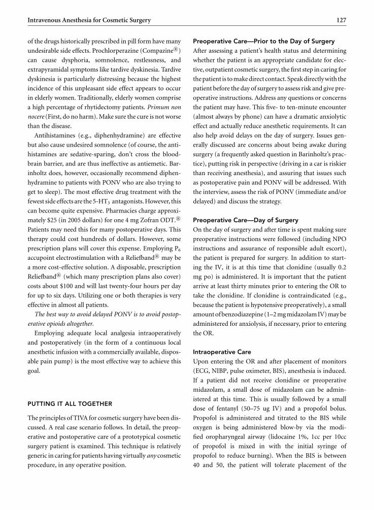
P1: PBU
cufx091-11 CUFX091/Friedberg 0 521 87090 9 Feb. 2, 2007 16:50
Intravenous Anesthesia for Cosmetic Surgery 127
of the drugs historically prescribed in pill form have many
undesirable side effects. Prochlorperazine (Compazine r©)
can cause dysphoria, somnolence, restlessness, and
extrapyramidal symptoms like tardive dyskinesia. Tardive
dyskinesia is particularly distressing because the highest
incidence of this unpleasant side effect appears to occur
in elderly women. Traditionally, elderly women comprise
a high percentage of rhytidectomy patients. Primum non
nocere (First, do no harm). Make sure the cure is not worse
than the disease.
Antihistamines (e.g., diphenhydramine) are effective
but also cause undesired somnolence (of course, the anti-
histamines are sedative-sparing, don’t cross the blood-
brain barrier, and are thus ineffective as antiemetic. Bar-
inholtz does, however, occasionally recommend diphen-
hydramine to patients with PONV who are also trying to
get to sleep). The most effective drug treatment with the
fewest side effects are the 5-HT3 antagonists. However, this
can become quite expensive. Pharmacies charge approxi-
mately $25 (in 2005 dollars) for one 4 mg Zofran ODT. r©
Patients may need this for many postoperative days. This
therapy could cost hundreds of dollars. However, some
prescription plans will cover this expense. Employing P6
accupoint electrostimulation with a Reliefband r© may be
a more cost-effective solution. A disposable, prescription
Reliefband r© (which many prescription plans also cover)
costs about $100 and will last twenty-four hours per day
for up to six days. Utilizing one or both therapies is very
effective in almost all patients.
The best way to avoid delayed PONV is to avoid postop-
erative opioids altogether.
Employing adequate local analgesia intraoperatively
and postoperatively (in the form of a continuous local
anesthetic infusion with a commercially available, dispos-
able pain pump) is the most effective way to achieve this
goal.
PUTTING IT ALL TOGETHER
The principles of TIVA for cosmetic surgery have been dis-
cussed. A real case scenario follows. In detail, the preop-
erative and postoperative care of a prototypical cosmetic
surgery patient is examined. This technique is relatively
generic in caring for patients having virtually any cosmetic
procedure, in any operative position.
Preoperative Care—Prior to the Day of Surgery
After assessing a patient’s health status and determining
whether the patient is an appropriate candidate for elec-
tive, outpatient cosmetic surgery, the first step in caring for
the patient is to make direct contact. Speak directly with the
patient before the day of surgery to assess risk and give pre-
operative instructions. Address any questions or concerns
the patient may have. This five- to ten-minute encounter
(almost always by phone) can have a dramatic anxiolytic
effect and actually reduce anesthetic requirements. It can
also help avoid delays on the day of surgery. Issues gen-
erally discussed are concerns about being awake during
surgery (a frequently asked question in Barinholtz’s prac-
tice), putting risk in perspective (driving in a car is riskier
than receiving anesthesia), and assuring that issues such
as postoperative pain and PONV will be addressed. With
the interview, assess the risk of PONV (immediate and/or
delayed) and discuss the strategy.
Preoperative Care—Day of Surgery
On the day of surgery and after time is spent making sure
preoperative instructions were followed (including NPO
instructions and assurance of responsible adult escort),
the patient is prepared for surgery. In addition to start-
ing the IV, it is at this time that clonidine (usually 0.2
mg po) is administered. It is important that the patient
arrive at least thirty minutes prior to entering the OR to
take the clonidine. If clonidine is contraindicated (e.g.,
because the patient is hypotensive preoperatively), a small
amount of benzodiazepine (1–2 mg midazolam IV) may be
administered for anxiolysis, if necessary, prior to entering
the OR.
Intraoperative Care
Upon entering the OR and after placement of monitors
(ECG, NIBP, pulse oximeter, BIS), anesthesia is induced.
If a patient did not receive clonidine or preoperative
midazolam, a small dose of midazolam can be admin-
istered at this time. This is usually followed by a small
dose of fentanyl (50–75 ug IV) and a propofol bolus.
Propofol is administered and titrated to the BIS while
oxygen is being administered blow-by via the modi-
fied oropharyngeal airway (lidocaine 1%, 1cc per 10cc
of propofol is mixed in with the initial syringe of
propofol to reduce burning). When the BIS is between
40 and 50, the patient will tolerate placement of the

P1: PBU
cufx091-11 CUFX091/Friedberg 0 521 87090 9 Feb. 2, 2007 16:50
128 David Barinholtz
oropharyngeal airway (usually somewhere between 1 and
2 mg · kg−1 of propofol has been administered by this
time).
After placement of the oropharyngeal airway and con-
firmation of a patent airway by end-tidal CO2 tracing,
ketamine (usually 50 mg IV) is administered. Two to
three minutes after ketamine administration, the surgeon
may inject the local analgesia. In addition to assuring the
ketamine has taken effect, BIS is usually maintained in
the range of 40–50 with the propofol infusion, and small
doses (25 ug increments) of fentanyl are administered and
titrated to a respiratory rate of approximately ten breaths
per minute (easily monitored by observing the end-tidal
CO2 tracing) before the surgeon is allowed to inject the local
analgesia. This (i.e., GA) will assure a virtually immobile
patient for local injections. Obviously, additional boluses
of propofol and/or fentanyl may occasionally need to be
administered (guided by BIS and respiratory rate, respec-
tively). If one decides to administer additional ketamine,
make sure the total dose doesn’t exceed 1 mg · kg−1. Higher
doses may be associated with undesirable side effects such
as dysphoria, prolonged recovery, and even PONV.
Once the surgery is underway (and appropriate antibi-
otics have been administered if necessary), the BIS is
allowed to come up and is generally maintained at 60
(+/−10) for the remainder of the procedure.
Prophylactic antiemetics are administered. At this point,
and for the remainder of the procedure, the propofol infu-
sion is titrated according to the BIS. Usual infusion rates are
75–120 ug · kg−1· min−1, but rates as low as 25 ug · kg−1
· min−1 or as high as 300 ug · kg−1· min−1 are some-
times required. The ability to respond to this patient
variation underscores the importance of BIS monitoring.
Additional opioids are titrated to achieve or maintain a
respiratory rate of ten to fifteen breaths per minute. Close
communication with the surgeon and close attention to
the surgery must be maintained in order to be able to
respond to the more and less stimulating parts of the
procedure by adjusting the propofol infusion and opi-
oid boluses accordingly. If muscle relaxation is required
for rectus muscle repair, utilize the technique described
earlier.
When the surgeon is placing the last stitches (or plac-
ing the garment on the patient), the propofol infusion is
discontinued. Low-dose ondansetron is administered. The
patient is allowed to awaken, which occurs typically within
three to five minutes of discontinuation of propofol. With
BIS >80, the patient will open his or her eyes and respond
to commands.
At this time the patient is transferred to a recliner and
taken to the PACU. Propofol is generally consumed at a
rate of approximately 50 cc · hr−1 for the average patient.
Opioid administration varies widely depending on patient
characteristics (e.g., history of smoking or drinking), pro-
cedure (nature and length), and surgeon’s skill with local.
If the procedure is one that historically is associated
with significant postoperative pain (i.e., abdominoplasty
or subpectoral breast augmentation), the surgeon will fre-
quently utilize a local anesthetic pain pump significantly
decreasing, if not eliminating, the need for opioids post-
operatively.
Postoperative Care
Once in PACU, patients are observed, postoperative issues
(i.e., pain, PONV) are addressed as necessary, and the
patients are discharged to go home in the company of
a responsible adult escort. Most patients (more than 90%)
arrive in PACU with an Aldrette score of 9 or greater, thus
putting them in phase recovery. It is the rare patient in
Barinholtz’s practice that spends more than one hour in
recovery.
As part of the postoperative instructions and follow-up,
Barinholtz takes on the burden of addressing postanes-
thetic complications directly. Patients and/or their fami-
lies are instructed to call the anesthesia provider if there
are anesthetic-related issues such as PONV. The surgeons
greatly appreciate this. Luckily, by employing these tech-
niques, calls are rare.
CONCLUSION
Office-based anesthesia has come a long way since
Dr. Crawford Long successfully administered the first ether
anesthetic in 1842. Advances in pharmacology and tech-
nology have made anesthesia better and safer than ever.
It is now a rare event (especially for healthy people hav-
ing elective cosmetic surgery) for patients to suffer their
demise under the anesthesiologist’s care. Improving the
quality of the perioperative experience has become a pri-
mary focus. In the future, with better drugs, better mon-
itoring technologies, and better delivery systems, further

P1: PBU
cufx091-11 CUFX091/Friedberg 0 521 87090 9 Feb. 2, 2007 16:50
Intravenous Anesthesia for Cosmetic Surgery 129
improvements in patient care should evolve toward these
ideal anesthetic.
REFERENCES
1. Long CW: An account of the first use of sulphuric ether byinhalation as an anesthetic in surgical operations. South MedSurg J 5:705,1849.
2. Heynick F: William T. G. Morton and “The Great Moment,”1944 Paramount Movie, 2003.
3. Wright AJ: Horace Wells, DDS and “Rebel with a Cause,”1815–1848, ASA Newsletter, American Society of Anesthesi-ologists, Park Ridge, IL, 63:6,1999.
4. Kitz RJ, Vandam LD: Scope of Modern Anesthetic Practice,in Miller RD (ed.): Anesthesia, 3rd ed., New York, ChurchillLivingstone, 1990, p 9.
5. Lagasse RS: Anesthesia Safety: Model or myth? A review ofpublished literature and analysis of current original data.Anesthesiol 97:1609,2002.
6. Fragen RJ: Diprivan (Propofol): A historical perspective.Semin Anesth 7:1,1988.
7. Rutter DV, Morgan M, Lumley J, Owen R: ICI 35 868(Diprivan): A new intravenous induction agent. Anaesthesia35:1188,1980.
8. Heath PJ, Kennedy DJ, Ogg TW, et al.: Which intravenousinduction agent for day surgery: A comparison of propo-fol thiopentone, methohexitone and etomidate. Anaesthesia43:365,1988.
9. Doze VA, Westphal LM, White PF: Comparison of propofolwith methohexital for outpatient anesthesia. Anesth Analg65:1189,1986.
10. Oxorn DC, Ferris LE, Harrington E, Orser BA: The effectsof midazolam on propofol-induced anesthesia: Propofoldose requirements, mood profiles, and perioperative dreams.Anesth Analg 85:553,1997.
11. Friedberg BL: Paradoxical increase in pain requirements withmidazolam premedication. Anesth Analg 99:1268,2004.
12. Cote CJ, Cohen IT, Santhanam S, et al.: A comparison ofthree doses of a commercially prepared oral midazolamsyrup in children. Anesth Analg 94:37,2002.
13. Pettinger WA: Drug therapy: Clonidine, a new antihyperten-sive drug. N Engl J Med 293:1179,1975.
14. Mannion S, Hayes I, Loughnane F, et al.: Intravenous but notperineural clonidine prolongs postoperative analgesia afterpso as compartment block with 0.5% levobupivacaine forhip fracture surgery. Anesth Analg 100:873,2005.
15. Strebel S, Gurzeler JA, Schneider MC, et al.: Small-doseintrathecal clonidine and isobaric bupivacaine for ortho-pedic surgery: A dose-response study. Anesth Analg 99:1231,2004.
16. Baker A, Klimscha W, Eisenach J, et al.: Intrathecal clonidinefor postoperative analgesia in elderly patients: The influenceof baricity on hemodynamic and analgesic effects. AnesthAnalg 99:128,2004.
17. Koch M, Famenne F, Deckers G, et al.: Epidural clonidineor sufentanil for intraoperative and postoperative analgesia.Anesth Analg 81:1154,1995.
18. Bernard JM, Kick O, Bonet F: Comparison of intravenousand epidural clonidine for postoperative patient-controlledanalgesia. Anesth Analg 81:706,1995.
19. Casati A, Magistris L, Fanelli G, et al.: Small-dose clonidineprolongs postoperative analgesia after sciatic-femoral nerveblock with 0.75% ropivacaine for foot surgery. Anesth Analg91:388,2000.
20. Joshi W, Reuben S, Kilaru P, et al.: Postoperative analgesiafor outpatient arthroscopic knee surgery with intra-articularclonidine and/or morphine. Anesth Analg 90:1102,2000.
21. Man D: Premedication with oral clonidine for facialrhytidectomy. Plast Reconstr Surg 94:214,1994.
22. Baker TM, Stuzin JM, Baker TJ, et al.: What’s new in aestheticsurgery? Clin Plast Surg 23:16,1996.
23. Friedberg BL, Sigl JC: Clonidine premedication decreasespropofol consumption during bispectral index (BIS)monitored propofol-ketamine technique for office-basedsurgery. Dermatol Surg 26:848,2000.
24. Higuchi H, Adachi Y, Dahan A, et al.: The interactionbetween propofol and clonidine for loss of consciousness.Anesth Analg 94:886,2002.
25. Higuchi H, Adachi Y, Arimura S, et al.: Oral clonidine pre-medication reduces the awakening concentration of propo-fol. Anesth Analg 94:609,2002.
26. Ishiyama T, Kashimoto S, Oguchi T, et al.: The effectsof clonidine premedication on the blood pressure andtachycardiac responses to ephedrine in elderly and youngpatients during propofol anesthesia. Anesth Analg 96:136,2003.
27. Taittonen MT, Kirvela OA, Aantaa R, et al.: The effect ofclonidine or midazolam premedication on perioperativeresponses during ketamine anesthesia. Anesth Analg 87:161,1998.
28. Arain SR, Ebert TJ: The efficacy, side effects, and recoverycharacteristics of dexmedotomidine versus propofol whenused for intraoperative sedation. Anesth Analg 95:461,2002.
29. Mayer D: Personal communication. October 2004.30. Shapiro F: Personal communication. November 2004.31. Corssen G, Domino EF: Dissociative anesthesia: Further
pharmacologic studies and first clinical experience with thephencyclidine derivative CI-581. Anesth Analg 45:29,1966.
32. Guit JBM, Koning HM, Costner ML: Ketamine as analgesiafor intravenous anesthesia with propofol (TIVA) anesthesia.Anaesthesia 46:24,1991.
33. Badrinath S, Avramov MN, Shadrick M, et al.: The use ofketamine-propofol combination during monitored anesthe-sia care. Anesth Analg 90:858,2000.
34. Friedberg BL: Propofol ketamine anesthesia for cosmeticsurgery in the office suite, chapter in Osborne I (ed.),Anesthesia for Outside the Operating Room. InternationalAnesthesiology Clinics. Baltimore, Lippincott, Williams &Wilkins, 41(2):39,2003.
35. White PF: The role of nonopoid analgesic techniques in themanagement of pain after ambulatory surgery. Anesth Analg94:577,2002.
36. Fauno P, Petersen KD, Husted SE: Increased blood loss afterpreoperative NSAID: Retrospective study of 186 hip arthro-plasties. Acta Orthop Scand 64:522,1993.

P1: PBU
cufx091-11 CUFX091/Friedberg 0 521 87090 9 Feb. 2, 2007 16:50
130 David Barinholtz
37. Robinson CM, Christie J, Malcolm-Smith N: Non-steroidalanti-inflammatory drugs, perioperative blood loss, andtransfusion requirements in elective hip arthroplasty.J Arthroplasty 8:607,1993.
38. Splinter WM, Rhine EJ, Roberts DW, et al.: Preoperativeketorolac increases bleeding after tonsillectomy in children.Can J Anaesth 43:560,1996.
39. Wierod FS, Frandsen NJ, Jacobsen JD, et al.: Risk of haem-orrhage from transurethral prostatectomy in acetylsalicylicacid and NSAID-treated patients. Scand J Urol Nephrol32:120,1998.
40. FitzGerald GA, Patrono C: Drug therapy: The coxibs,selective inhibitors of cyclooxygenase-2. N Engl J Med345:433,2001.
41. Reuben SS, Connelly NR: Postoperative analgesic effects ofcelecoxib or rofecoxib after spinal fusion surgery. AnesthAnalg 91:1221,2000.
42. Reuben SS, Bhopatkar S, Maciolek H, et al.: The preemptiveanalgesic effect of rofecoxib after ambulatory arthroscopicknee surgery. Anesth Analg 94:55,2002.
43. Turan A, Emet S, Karamanlioglu B, et al.: Analgesiceffects of rofecoxib in ear-nose-throat surgery. Anesth Analg95:1308,2002.
44. Buvanendran A, Kroin JS, Tuman KJ, et al.: Effects of peri-operative administration of a selective cyclooxygenase 2inhibitor on pain management and recovery of functionafter knee replacement: A randomized controlled trial. JAMA290:2411,2003.
45. Ma H, Tang J, White PF, et al.: Perioperative rofecoxibimproves early recovery after outpatient herniorrhaphy.Anesth Analg 98:970,2004.
46. Barton SF, Langeland FF, Snabes MD, et al.: Efficacyand safety of intravenous parecoxib sodium in relievingacute postoperative pain following gynecologic laparotomysurgery. Anesthesiol 97:306,2002.
47. Joshi GP, Viscusi ER, Gan TJ: Effective treatment of laparo-scopic cholecystectomy pain with intravenous followed byoral COX-e specific inhibitor. Anesth Analg 98:336,2004.
48. Kim PS, Reicin AS, Villalba L, et al.: Rofecoxib, Merck, andthe FDA. N Engl J Med 351:2875,2004.
49. Psaty BM, Furberg CD: COX-2 Inhibitors—Lessons in drugsafety. N Engl J Med 352:1133,2005.
50. Calverly RK: Anesthesia as a specialty: Past, present, andfuture, in Barash PG, Cullen BF, Stoelting RK (eds.), ClinicalAnesthesia. Philadelphia, Lippincott, 1989, p 21.
51. Griffith HR, Johnson GE: The use of curare in general anes-thesia. Anesthesiol 3:418,1942.
52. Calverley RK: Arthur E Guedel (1883–1956), in Repreht J,van Lieburg MJ, Lee JA, and Erdmann W (eds.), AnaesthesiaEssays on Its History. Berlin, Springer-Verlag, 1985, p 49.
53. Brain AIJ: The laryngeal mask airway: A new concept inairway management. Br J Anaesth 55:801,1983.
54. Brain AIJ, McGhee TD, McAteer EJ, et al.: The laryngeal maskairway: Development and preliminary trials of new type ofairway. Anaesthesia 40:356,1985.
55. ASA Publication: Practice Guidelines for Management of theDifficult Airway (last amended 10/16/02), p 21.
56. Calverley RK: Anesthesia as a specialty: Past, present andfuture. In Barash, Cullen, Stoelting (eds.), Clinical Anesthe-sia, Philadelphia, Lippincott, 1989, p 25.
57. Bruhn J, Bouillon TW, Ropcke H, et al.: A manual slide rulefor target-controlled infusion of propofol: Development andevaluation. Anesth Analg 96:142,2003.
58. Suttner S, Boldt J, Schmidt C, et al.: Cost analysisof target-controlled infusion-based anesthesia comparedwith standard anesthesia regimens. Anesth Analg 88:77,1999.
59. Woolf CJ, Chong MS: Preemptive analgesia: Treating post-operative pain by preventing the establishment of centralsensitization. Anesth Analg 77:362,1993.
60. Ong KS, Lirk P, Seymour RA: The efficacy of preemptiveanalgesia for acute postoperative pain management: A meta-analysis. Anesth Analg 100:757,2005.
61. McQuay HJ: Pre-emptive analgesia: A systematic review ofclinical studies. Ann Med 27:249,1995.
62. McQuay HJ: Pre-emptive analgesia. Br J Anaesth 69:1,1992.63. Macario A, Weinger M, Carney S, et al.: Which clinical anes-
thesia outcomes are important to avoid? The perspective ofpatients. Anesth Analg 89:652,1999.
64. Lee A, Gin T, Lau A, et al.: A comparison of patients’and health care professionals’ preferences for symptomsduring immediate postoperative recovery and the manage-ment of postoperative nausea and vomiting. Anesth Analg100:87,2005.
65. Pierre S, Corno G, Benais H, et al.: A risk score-dependentantiemetic approach effectively reduces postoperative nau-sea and vomiting—A continuous quality improvement ini-tiative. Can J Anesth 51:320,2004.
66. Apfel CC, Laara E, Koivuranta M, et al.: A simplified riskscore for predicting postoperative nausea and vomiting:Conclusions from cross-validations between two centers.Anesthesiol 91:693,1999.
67. Apfel CC, Kranke P, Eberhart LH, et al.: Comparison ofpredictive models for postoperative nausea and vomiting.Br J Anaesth 88:234,2002.
68. Apfel C, Korttila K, Abdalla M, et al.: A factorial trial of sixinterventions for the prevention of postoperative nausea andvomiting. N Engl J Med 350:2441,2004.
69. Scuderi P, James R, Harris L, et al.: Multimodalantiemetic management prevents early postoperative vom-iting after outpatient laparoscopy. Anesth Analg 91:1408,2000.
70. Gan TJ, Jiao K, Zenn M, et al.: A randomized controlled com-parison of electro-accupoint stimulation or ondansetronversus placebo for the prevention of postoperative nauseaand vomiting. Anesth Analg 99:1070,2004.
71. Zarate E, Mingus M, White PF, et al.: The use of transcuta-neous accupoint electrical stimulation for preventing nau-sea and vomiting after laparoscopic surgery. Anesth Analg92:629,2001.
72. White PF, Hamza M, Recart A, et al.: Optimal timing ofacustimulation for antiemetic prophylaxis as an adjunct toondansetron in patients undergoing plastic surgery. AnesthAnalg 100:367,2005.

P1: PBU
cufx091-12 CUFX091/Friedberg 0 521 87090 9 Feb. 2, 2007 19:35
12 Regional Anesthesia for Cosmetic Surgery
Holly Evans, M.D., F.R.C.P., and Susan M. Steele, M.D.
INTRODUCTION
PREOPERATIVE ASSESSMENT
General Assessment
Special Consideration: Coagulation Abnormalities
MONITORING AND SEDATION
Monitoring
Sedation for Block Placement
Intraoperative Sedation
SPINAL ANESTHESIA
Indications and advantages
Equipment
Local anesthetics and adjuvants
Adverse effects
EPIDURAL ANESTHESIA
Indications and advantages
Equipment
Local anesthetics and adjuvants
Adverse effects
PERIPHERAL NERVE BLOCKS
Paravertebral Nerve Blocks
Anatomy
Indications and advantages
Technique
Equipment
Local anesthetics and adjuvants
Adverse effectsIntercostal Nerve Blocks
Anatomy
Indications and advantages
Technique
Equipment
Local anesthetics and adjuvants
Adverse effects
RECOVERY AND DISCHARGE
Recovery
Discharge
SUMMARY
131
P1: PBU
cufx091-12 CUFX091/Friedberg 0 521 87090 9 Feb. 2, 2007 19:35
132 Holly Evans and Susan M. Steele
Table 12-1. Goals of anesthesia forcosmetic surgery
Intraoperative analgesiaIntraoperative anxiolysis ( ± amnesia)Postoperative analgesiaAbsence of postoperative side effects (nausea,
vomiting, sedation, urinary retention)Early return of baseline cognitive abilitiesTimely postoperative discharge
INTRODUCTION
Anesthesia for patients undergoing cosmetic surgery must
accomplish a number of important goals (Table 12-1).
Regional anesthesia, which includes both central neurax-
ial techniques as well as peripheral nerve blocks, has a
commendable safety profile1 and unique attributes that
allow these goals to be met. Nerve blocks provide dense
intraoperative anesthesia and analgesia. This minimizes
requirements for additional anesthetic, analgesic, or seda-
tive agents; consequently, side effects such as postopera-
tive nausea, vomiting, and sedation can be minimized.2,5
This enhances patient recovery and discharge and may also
improve patient satisfaction.6,8 Peripheral nerve blocks
with long-acting local anesthetic can extend the duration
of postoperative analgesia and further minimize opioid
use and associated side effects.2 This chapter describes the
use of central neuraxial blocks and peripheral nerve blocks
in cosmetic surgical patients. A discussion of appropriate
preoperative evaluation, monitoring, sedation, and nerve
block technique, as well as postoperative recovery and dis-
charge, is included.
PREOPERATIVE ASSESSMENT
General Assessment
Evaluation of patients contemplating regional anesthesia
should include a full history and physical examination
as well as investigations or consultations as indicated. A
complete history is obtained to document active medi-
cal issues, past medical history, prescription and nonpre-
scription medication use, allergies, and personal and fam-
ily anesthesia history. The physical examination involves
assessment of the patient’s height, weight, vital signs, and
airway. Cardiovascular, respiratory, and neurologic evalu-
ations are performed. In addition, the site where regional
anesthesia is to be administered is inspected. Laboratory
investigations are performed based on the patient’s under-
lying medical problems and the anticipated surgery. For
example, an otherwise healthy thirty-year-old woman hav-
ing breast augmentation may require no preoperative test-
ing. A sixty-year-old woman with hypertension treated
with a diuretic would be appropriately investigated with
electrolytes and an electrocardiogram prior to her
abdominoplasty. Similarly, consultations with other med-
ical specialists may be required for active medical issues.
The physiologic effects of regional anesthesia must be con-
sidered in the context of the patient’s underlying medical
condition(s) (Table 12-2). Subsequently, potential con-
traindications to regional anesthesia as well as ambulatory
surgery must be sought and addressed (see Tables 12-3,
12-4, and 12-5). Ideally, the preoperative anesthesia assess-
ment is performed several days prior to the scheduled pro-
cedure to enable such consultation, patient optimization,
and rescheduling if required.
The preoperative visit provides an ideal time for patient
education. Patients are instructed about fasting guidelines,
which medications to take the day of surgery, and the tim-
ing of medications and herbal remedies to be discontinued
preoperatively (see Ch. 14 & Appendix A). Outpatients
should be notified that they require a responsible care-
giver to accompany them to and from surgery and to stay
with them the night of surgery. In the process of obtain-
ing informed consent for anesthesia, patients should be
educated about the expected intra- and postoperative
Table 12-2. Physiologic effectsof neuraxial anesthesia
Cardiovascular SympathectomyVasodilationHypotension ( ± bradycardia)
Respiratory With high block:Subjective dyspneaImpaired active exhalationImpaired cough
Gastrointestinal Increased secretionsSphincter relaxationBowel constriction
Endocrine-Metabolic Inhibition of surgical stressresponse

P1: PBU
cufx091-12 CUFX091/Friedberg 0 521 87090 9 Feb. 2, 2007 19:35
Regional Anesthesia for Cosmetic Surgery 133
Table 12-3. Conditions associated with increasedrisk of neuraxial anesthesia
Condition Risk
Increased intracranialpressure
Brain stem herniation
Significant coagulopathy Spinal or epiduralhematoma
Uncorrected hypovolemia Shock, cardiovascularcollapse
Infection overlying needlepuncture site
Neuraxial infection
Systemic sepsis Neuraxial infectionSignificant aortic stenosis Cardiovascular collapseUnstable neurologic disease Neurologic deficitLocal anesthetic allergy Allergic reaction
course as well as the potential side effects and risks of the
anesthetic technique(s) contemplated.
Patients having cosmetic surgery are a unique subset.
They often have numerous questions, seek more infor-
mation than the general surgical population, and have
extremely high standards and expectations.
Table 12-4. Conditions associated with increasedrisk of peripheral nerve blocks
Condition Risk
Infection overlying needlepuncture site
Perineural infection
Significant coagulopathy Blood loss ± transfusionHematoma ± nervecompression
Uncorrected hypovolemia Shock, cardiovascularcollapse
Perineural tumor Tumor seeding andspread
Perineural vascularmalformation
Blood loss ± transfusionHematoma ± nervecompression
Unstable neurologicdisease
Neurologic deficit
Previous thoracic surgery Obliteration ofparavertebral space byscar tissuePneumothorax fromparavertebral block
Infection within chestcavity (i.e., empyema)
Paravertebral or neuraxialinfection fromparavertebral block
Local anesthetic allergy Allergic reaction
Table 12-5. Conditions associated with increasedrisk for ambulatory surgery
Condition Risk
Malignant hyperthermia Perioperative MHa
reactionObstructive sleep apnea,
obesityPostoperative airway
obstructionOxygen desaturation
Unstable systemic disease Exacerbation of symptomsAbsence of reliable
caregiverInsufficient assistance for
patient after dischargeSurgery involving
significant blood lossIntravascular volume
depletion and fluidshifts
Surgery associated withprolonged recovery
Potential for hospitaladmission
Surgery associated withsevere postoperativepain
Potential for hospitaladmission for analgesia
Local anesthetic allergy Allergic reaction
aMH = malignant hyperthermia
Special Consideration: Coagulation Abnormalities
As discussed, the patient’s underlying medical conditions
factor into the decision regarding the suitability of regional
anesthesia. Altered coagulation is one such condition that
deserves special mention as it increases the risk of neuraxial
hematoma following spinal or epidural anesthesia. Coagu-
lopathy may result from an intrinsic defect in the patient’s
coagulation system, from prescribed anticoagulant medi-
cation, or from nonpresciption drug use like garlic, ginger,
or ginko biloba (see Table 14-3 in Chapter 14).
Patients with an intrinsic bleeding diathesis such as
hemophilia or thrombocytopenia represent a subset of
patients with altered coagulation. Preoperative assessment
includes a focused bleeding history and physical examina-
tion. Appropriate laboratory evaluation depends on the
underlying disorder, and perioperative management usu-
ally occurs in conjunction with a hematologist. Individ-
uals with moderate and severe disease would rarely be
considered for ambulatory cosmetic surgery because of
the increased surgical and anesthetic risk. Patients with
mild disease having low-risk surgical procedures may
be suitable. However, these patients require a thorough
evaluation of their coagulation system and coordination
with their hematologist prior to being considered for

P1: PBU
cufx091-12 CUFX091/Friedberg 0 521 87090 9 Feb. 2, 2007 19:35
134 Holly Evans and Susan M. Steele
neuraxial anesthesia. The risk of neuraxial hematoma
increases as coagulation parameters fall out of the range
of normal. The risk-to-benefit ratio must be considered in
the context of this population of patients who are having
entirely elective procedures.
Patients with altered coagulation as a result of antico-
agulants used for thromboprophylaxis represent another
subset of anticoagulated patients. Preoperative evalua-
tion is as described previously. The American Society
of Regional Anesthesia guidelines for the perioperative
management of anticoagulation therapy for patients hav-
ing spinal or epidural anesthesia are summarized in
Table 12-6.9
Guidelines regarding the use of peripheral nerve blocks
for patients with altered coagulation due to an intrin-
sic bleeding diathesis or the use of thromboprophylactic
Table 12-6. American Society of Regional Anesthesia and Pain Medicine recommendationsregarding neuraxial anesthesia in the patient receiving thromboprophylaxis
Thromboprophylactic agent Recommendation
Antiplatelet Medication No contraindication with NSAIDsa
Discontinue ticlopidine 14 d in advanceDiscontinue clopidogrel 7 d in advanceDiscontinue GP IIb/IIIab inhibitors 8–48 h in advance
Unfractionated Heparin (Subcutaneous) No contraindicationConsider delaying heparin until after block if technical
difficulty anticipatedUnfractionated Heparin (Intravenous) Heparinize 1 h after neuraxial technique
Remove catheter 2–4 h after last heparin doseNo mandatory delay if traumatic
Low Molecular Weight Heparin Twice daily dosing:LMWHc 24 h after surgery, regardless of techniqueRemove neuraxial catheter 2 h before first LMWHc
doseSingle daily dosing (as per the following European
guidelines):Neuraxial technique 10–12 h after LMWHc
Next dose at least 4 h after needle or catheterplacement
Catheters removed 10–12 h after LMWHc and 4 hprior to next dose
Postpone LMWHc 24 h if traumaticWarfarin Document normal INRd after discontinuation (prior to
neuraxial technique)Remove catheter when INRd ≤ 1.5 (initiation of
therapy)Thrombolytics No data on safety interval for performance of
neuraxial technique or catheter removalFollow fibrinogen level
Herbal Therapy No evidence for mandatory discontinuation prior toneuraxial technique
Be aware of potential drug interactions
Adapted from Horlocker TT, Wedel DJ, Benzon HP, et al.: Regional anesthesia in the anticoagulated patient:Defining the risks (the second ASRA consensus conference on neuraxial anesthesia and anticoagulation). RegionalAnesthesia & Pain Medicine 28;172,2003.9aNSAIDs = nonsteroidal anti-inflammatory drugsbGP IIb/IIIa = glycoprotein IIb/IIIa inhibitorscLMWH = low molecular weight heparindINR = international normalized ratio

P1: PBU
cufx091-12 CUFX091/Friedberg 0 521 87090 9 Feb. 2, 2007 19:35
Regional Anesthesia for Cosmetic Surgery 135
agents are extrapolated from those described previously.9
Adherence to these guidelines should be more stringent
for peripheral nerve blocks performed near noncompress-
ible vessels (i.e., intercostal or sciatic nerve block) and for
those where hematoma formation could result in signifi-
cant injury to nerves or other surrounding structures (i.e.,
paravertebral or lumbar plexus block).10
MONITORING AND SEDATION
Monitoring
Intravenous access, supplemental oxygen, and appropri-
ate monitors are imperative to the safe practice of regional
anesthesia. Nerve blocks can produce significant physi-
ologic changes (see Table 12-2). Furthermore, multisys-
temic side effects may result from the intravascular absorp-
tion of large volumes of local anesthetic. Consequently,
monitoring with pulse oximetry, noninvasive blood pres-
sure, and electrocardiogram are required both during
block placement and intraoperatively.
Sedation for Block Placement
Sedation may be required during block placement. In this
setting, the goal is to relieve apprehension and pain as
well as to maintain a communicative patient able to con-
vey information about potential adverse effects of regional
anesthesia. For example, description of paresthesias allows
needle redirection and prevention of intraneural injection
of local anesthetic. And communication about tinnitus
and metallic taste allows discontinuation of injection of
local anesthetic and prevention of more serious symptoms
related to local anesthetic toxicity.
Intravenous agents are typically used for their titrata-
bility (see Table 12-7). Benzodiazepines provide anxiolysis
and anterograde amnesia. They raise the seizure threshold,
which is advantageous when large doses of local anesthetic
are used. The benzodiazepine midazolam is frequently
used owing to its rapid onset, relatively short half-life, and
lack of active metabolites. Analgesia and additional seda-
tion result from supplementation with opioid analgesics.
The potent, short-acting agent fentanyl is frequently emp-
loyed for this purpose. Benzodiazepines and opioids can
both be reversed with specific antagonists, and this further
enhances their safe application for perioperative sedation.
The half-life of reversal agents is sometimes shorter then
the drugs being reversal. Dependence on reversal agents
provides, the illusion of safety.
Ketamine affords an alternate option for analgesia with a
lower incidence of opioid-related side effects such as nau-
sea, vomiting, respiratory depression, and pruritus. The
dysphoria associated with ketamine may limit the use of
this agent; however, the concurrent use of midazolam can
minimize side effects.11 The alpha2 agonist, clonidine, also
offers analgesia with minimal opioid-related side effects,
though this agent may cause hypotension and bradycardia
as well as prolonged postoperative sedation. Nonpharma-
cologic anxiolysis is generated from verbal reassurance or
music.12 The importance of this cannot be overempha-
sized.
Intraoperative Sedation
Intraoperative sedation should enhance patient comfort
yet have minimal postoperative effects allowing for timely
patient recovery. Sedation can be continued intraoper-
atively using incremental doses of the agents described
herein. Alternatively, an infusion of a short-acting, eas-
ily titratable agent can be used (see Table 12-7). Target-
controlled infusion devices may be used to minimize drug
dose and associated side effects and have also been used to
provide patient-controlled sedation.13
The pharmacokinetics of propofol make it well suited
for sedation by continuous infusion. It has a rapid onset,
is easily titratable, and is associated with prompt awaken-
ing upon discontinuation of the infusion. The use of pro-
pofol has also been effective in reducing the incidence of
postoperative nausea and vomiting.14 The ultrashort-
acting potent opioid remifentanil is designed for contin-
uous infusion and provides sedation as well as additional
analgesia. Servin et al. compared sedation with an infus-
ion of remifentanil (0.1 ug · kg−1 · min−1 IV) or propofol
(50 ug · kg−1 · min−1 IV) in patients who received regional
anesthesia for their surgical procedure.15 Servin et al. fou-
nd that remifentanil provided greater analgesia, less amne-
sia, and a higher incidence of respiratory depression and
postoperative nausea and vomiting (PONV).15 Dexmede-
tomidine is a short-acting alpha2 agonist suitable for
continuous infusion. It provides improved postopera-
tive analgesia and has opioid-sparing effects compared
to propofol. However, it has a slower onset of action, a
longer duration of action after discontinuation of infusion,
and a greater incidence of postoperative hypotension and
sedation.16

P1: PBU
cufx091-12 CUFX091/Friedberg 0 521 87090 9 Feb. 2, 2007 19:35
136 Holly Evans and Susan M. Steele
Table 12-7. Selected intravenous agents used for sedation
Mechanism Specific agentsClass of agent of action Systemic effects Adverse effects and dose
Benzo-diazepines
Bind to GABAa
A receptorFacilitate central
nervous systeminhibition ofGABAa
SedationAnxiolysisAnterograde amnesiaAnticonvulsantSkeletal muscle relaxation
HypotensionRespiratory depressionProlonged sedation in
some patientsParadoxical excitement in
some patients
MidazolamIncremental doses of
20–30 ug/kg IV upto 0.1 mg · kg−1
Opioids Opioid receptor(i.e., mu, kappa,delta) agonists
Decreaseneurotransmitterrelease
SedationAnalgesiaNo amnesia
Nausea, vomitingPruritusRespiratory depressionUrinary retentionConstipationMuscle rigidityBradycardia
Fentanyl:Incremental doses of
0.5–1.5 ug · kg−1 IVAlfentanil:
5–10 ug · kg−1 IVRemifentanil:
0.025–0.2 ug · kg−1
Ketamine NMDAb receptorantagonist Mayalso interact withopioid receptors
Dissociative stateSedationAnalgesiaAmnesiaPharyngeal and laryngeal
reflexes maintained nearnormal
Sympathomimetic effectsBronchodilation
Emergence deliriumc
Psychosisc
Hallucinationsc
Increases salivarysecretions
NystagmusIncreases ICPd and IOPe
0.15–0.5 mg · kg−1
Alpha2
AgonistsSympathetic alpha2
receptor agonistsDecrease
neurotransmitterrelease
SedationAnalgesiaMinimal respiratory
depression
HypotensionBradycardiaProlonged sedation
(clonidine >
dexmetetomidine)
Clonidine1–2 ug · kg−1
IV or 0.2 mg poDexmedetomidineBolus: 0.4–1 ug · kg−1
IV over 10–20 minInfusion: 0.2–0.7 ug ·
kg−1 · hr−1 IVPropofol Presumed via
interaction withGABAa receptor
SedationAmnesiaAntipruriticAnticonvulsantAntiemetic
Pain on intravenousinjection
HypotensionRespiratory depression,
apneaSpontaneous excitatory
movementsSupports growth of
microorganisms
25–100 ug · kg−1 ·min−1 IV
aGABA = Gamma-amino-butyric acidbNMDA = N-methyl-D-aspartatecEditor’s note: This information is correct only when ketamine is given as a sole IV agent. Friedberg BL : Hypnotic doses of propofolblock ketamine induced hallucinations. Plast Reconstr Surg 91:196,1993. —BLFdICP = Intracranial pressureeIOP = Intraocular pressure

P1: PBU
cufx091-12 CUFX091/Friedberg 0 521 87090 9 Feb. 2, 2007 19:35
Regional Anesthesia for Cosmetic Surgery 137
Table 12-8. Potential advantages of spinalanesthesia for cosmetic surgery1,3,7
Ease of performanceDense anesthesiaMinimal additional sedation requiredPrompt return of awake stateLow incidence of PONVa
Reduced physiologic stress responseReduced thromboembolic complicationsLow incidence of local anesthetic toxicityCost savings
aPONV = postoperative nausea and vomiting
SPINAL ANESTHESIA
Indications and advantages
Spinal anesthesia provides rapid onset, bilateral surgical
anesthesia for cosmetic procedures of the abdomen and
lower extremities. Potential advantages are outlined in
Table 12-8.
Equipment
Two classes of spinal needles are available. Cutting nee-
dles, such as the Quincke or Greene, have a beveled distal
tip with an end orifice. Pencil-point needles, such as the
Whitacre or Sprotte, have a blunt distal tip and an ori-
fice on the side of the distal needle shaft. Pencil-point
needles provide an enhanced tactile feel as the needle is
advanced through the various tissues. Spinal needles in
current use have an inner stylet that prevents plugging of
the needle with skin or fat and subsequent deposition into
the epidural or subarachnoid space. Both classes of nee-
dles are available in a range of diameters and lengths. The
gauge describes the outer diameter of the needle. A larger
gauge represents a smaller diameter needle (i.e., a 29 ga
needle has a smaller diameter than a 22 ga needle). The
use of pencil-point and small-gauge needles is associated
with a lower incidence of postdural puncture headache
(PDPH).17 Small gauge needles can be more technically
difficult to use as they are more prone to deflection and
often have slower return of cerebrospinal fluid.
Local anesthetics and adjuvants
The selection of local anesthetic for spinal anesthesia is
based on the desired block onset, duration, and spread, as
well as the drug’s side-effect profile.
Table 12-9. Selected potential adverse effectsof spinal anesthesia1,9,27,64,77–83
Self-limiting Back painUrinary retentionTransient neurologic symptomsPostdural puncture headache
Rare but serious Epidural abscessSpinal or epidural hematomaMeningitisCauda equina syndromeIsolated nerve injuryTotal spinal anesthesiaBradycardia and cardiac arrest
Lidocaine and mepivacaine both have a rapid onset and
short to intermediate duration of action.18,19 Lidocaine
has been associated with transient neurologic symptoms,
which has significantly curtailed its use, particularly in the
outpatient population.20,26 In addition, there is some evi-
dence that this complication may also occur with mepiva-
caine.27 Procaine is another short-acting agent. However,
its use has been limited by frequent block failure.28
A preparation of chloroprocaine without the preserva-
tive methylparaben or the antioxidant bisulfite has recently
been used for spinal anesthesia.29,34 In a crossover study of
volunteers, intrathecal chloroprocaine (40 mg) was com-
pared to lidocaine (40 mg).29 Investigators found that
both provided adequate anesthesia. However, the chloro-
procaine group had faster resolution of sensory block by
twenty-three minutes as well as earlier time to ambula-
tion and voiding by thirty minutes. Transient neurologic
symptoms occurred in 87.5% in the lidocaine group and
none in the chloroprocaine group. In a study of simi-
lar design, spinal anesthesia with chloroprocaine (40 mg)
was compared to low-dose bupivacaine (7.5 mg).35 Both
agents had a similar peak block height and time to peak
block; however, complete regression of sensory and motor
block, ambulation and voiding occurred over one hour
later in the bupivacaine group. Despite the beneficial
profile described, the addition of epinephrine to hyper-
baric chloroprocaine has been associated with transient
neurologic symptoms and nonspecific, transient, flu-like
symptoms.30 Transient neurologic symptoms are rare with
bupivacaine and levobupivacaine.36 These agents have a
long duration of action, which can potentially delay recov-
ery and discharge of ambulatory patients.37 A reduction

P1: PBU
cufx091-12 CUFX091/Friedberg 0 521 87090 9 Feb. 2, 2007 19:35
138 Holly Evans and Susan M. Steele
in dose has been used in an attempt to shorten the dura-
tion of action; however, this can potentially lead to block
failure.37 Intrathecal ropivacaine also has a long duration
of action.38–42 However, some studies have shown earlier
recovery of sensory and motor function when compared to
bupivacaine, which may be advantageous to outpatients.42
The naturally occurring lumbar lordosis and thoracic
kyphosis affect the spread of both hypo- and hyperbaric
solutions. A hyperbaric solution injected into a lumbar
interspace (near the peak of the lumbar lordosis) gravi-
tates cephalad to the thoracic kyphosis and caudally to the
sacral nerve roots in a patient in the supine position (Fig.
12-1). A mid-thoracic block may also result from a hypo-
baric solution if the patient remains sitting for several
minutes after injection. Unilateral spinal anesthesia can be
achieved with either hypo- or hyperbaric solutions admin-
istered to patients in the lateral decubitus position.
The effect of spinal local anesthetics can be impacted
by the addition of adjuvant medications. Intrathecally
administered opioids are commonly used adjuvants that
act by binding to opiate receptors in the spinal cord.43
They enhance the quality of spinal anesthesia and increase
the duration of sensory anesthesia. As a result, the dose of
spinal local anesthetic can be reduced, facilitating recov-
ery and outpatient discharge. Ben-David et al. illustrated
this in a population of patients who received 5 mg hyper-
baric spinal bupivacaine.44 The incidence of inadequate
block was 24% in the group who received bupivacaine
alone, whereas there were no failed blocks in the group
who received 10 ug intrathecal fentanyl with bupivacaine.
Pruritus is a known side effect of intrathecal opioids and
is usually mild and self-limiting.45 The highly lipophilic
agent fentanyl (10–25 ug) is well suited for ambulatory
Figure 12-1. Distribution of hyperbaric local anesthetic solutionin the intrathecal space after injection at L3–4 (at the apex of thelumbar lordosis) in a patient turned immediately supine. Notehigher concentrations in midthoracic and sacral regions as thelocal anesthetic solution runs “down hill.” Reproduced with per-mission from Neuraxial Blocks by Stevens RA, in Regional Anes-thesia and Analgesia, edited by Brown DL; WB Saunders; 1996.
anesthesia for cosmetic surgery as it has a rapid onset and
short duration of action.44,46,48 Hydrophilic opioids, such
as morphine (0.1–0.3 mg), have a longer duration of effect
and more extensive rostral spread within the neuraxis. As a
result, intrathecal morphine can provide prolonged post-
operative analgesia to inpatients.
The risk of delayed respiratory depression contraindicates
the use of intrathecal morphine for outpatients.
The alpha2 agonist clonidine increases the quality and
duration of sensory and motor block when administered
intrathecally.34 Side effects such as hypotension, brady-
cardia, and sedation are common with intrathecal doses
between 75 and 225 ug, and this may limit the use of
this agent in outpatients.49 A number of investigators have
studied a lower dose of intrathecal clonidine (15 ug) and
have shown enhanced block quality with limited systemic
side effects.34,50
Vasoconstrictors such as epinephrine (0.2–0.3 mg) and
phenylephrine (2–5 mg) can increase the density and dura-
tion of lidocaine spinal block.51,56 Prolonged motor block
and urinary retention may limit more widespread use of
these agents. In addition, phenylephrine has been associ-
ated with the development of transient neurologic symp-
toms.54,56
Adverse effects
If spinal anesthesia is planned for outpatient cosmetic
surgery, appropriate patient selection based on a thor-
ough preoperative assessment and meticulous technique
can significantly minimize the risk. Nevertheless, potential
risks do exist (see Table 12-9). Patients must be informed
about these risks and should be provided with appropriate
contact information in the event that problems arise.
Back pain is estimated to occur in 11% of surgical
patients twenty-four to thirty-six hours after spinal anes-
thesia.57 (Any additional discomfort, beyond the surgical,
in the cosmetic surgical patient may be cause for com-
plaint. Therefore, the possibility of back pain must be dis-
closed to adjust the patient’s expectations as well as to
obtain an informed consent.) Back pain may result from
needle trauma, from intrinsic effects of the local anesthetic,
or from ligamentous strain. Urinary retention is related to
inhibition of detrusor muscle activity from S3 nerve root
block. Risk factors include male gender, age greater than
sixty years, surgery duration greater than 120 minutes, and
systemic analgesia.58,59

P1: PBU
cufx091-12 CUFX091/Friedberg 0 521 87090 9 Feb. 2, 2007 19:35
Regional Anesthesia for Cosmetic Surgery 139
Transient neurologic symptoms involve pain or dyses-
thesia in the legs or buttocks that occurs within twenty-
four hours of spinal anesthesia.60,61Risk factors include
the use of lidocaine, possibly mepivacaine, ambulatory
surgery, and the lithotomy position. Treatment is symp-
tomatic and spontaneous recovery typically occurs within
days.
A postdural puncture headache (PDPH) is a fronto-
occipital headache that occurs in a sitting or standing
position and is immediately improved upon assuming a
supine position.62 The incidence is greater with cutting
and large bore spinal needles, a transversely directed nee-
dle bevel, young age, and female sex.63,64 Treatment is ini-
tially conservative with fluid hydration, analgesics, and
possibly caffeine.65 An epidural blood patch can be con-
sidered, although spontaneous resolution generally occurs
in one to two weeks.
Serious neurologic injury related to spinal anesthesia
is extremely rare.66,69 Nonetheless, any new neurologic
deficit should be promptly assessed in the event that a
potentially reversible etiology exists. An epidural abscess
presents with progressive sensory or motor deficits, bowel
or bladder dysfunction, fever, or back pain.70,72 A neu-
raxial hematoma may present similarly.70 Magnetic reso-
nance imaging can establish the diagnosis of either condi-
tion. Following diagnosis, immediate surgical evacuation
of the abscess or hematoma may be required. Subsequently,
abscesses are also managed with antiobiotics.73 Menin-
gitis is suspected if the patient presents with headache,
fever, nuchal rigidity, or a new neurological deficit.73,75
Cerebrospinal fluid analysis can yield the diagnosis, and
systemic antibiotic therapy is begun early. Cauda equina
syndrome presents with pain in the lower back or legs,
lower extremity sensorimotor deficit, and bowel or blad-
der dysfunction.70,75 Risk factors include the use of 5%
hyperbaric lidocaine and continuous spinal anesthesia
with microcatheters. Emergent investigation and treat-
ment are necessary. The diagnosis and management of
other neurologic complications (i.e., isolated nerve injury)
can be greatly facilitated with the assistance of a consultant
neurologist.
Total spinal anesthesia results from excessive cepha-
lad spread of the spinal block. Apnea, bradycardia,
profound hypotension, and cardiac arrest can result.76
Prompt recognition and initiation of supportive therapy
is crucial.
Table 12-10. Selected potential adverse effectsof epidural anesthesia1,64,73,77,82,105,106
Self-limiting Back painUrinary retentionPostdural puncture headache
Rare but serious Local anesthetic toxicityTotal spinal anesthesiaCardiac arrestIsolated nerve injuryCauda equina syndromeEpidural abscessMeningitisEpidural hematomaRetained catheter
EPIDURAL ANESTHESIA
Indications and advantages
Epidural anesthesia achieves bilateral surgical anesthesia
with similar advantages to spinal anesthesia (Table 12-10).
In addition, epidurals provide the option for extended
postoperative analgesia for patients admitted to hospital.
Thoracic epidurals have applications for cosmetic pro-
cedures involving the breast and thorax, whereas thora-
columbar epidurals are more suited to abdominal, lower
extremity, and buttock surgery.
Equipment
In order to facilitate catheter placement, epidural needles
have a curved distal tip (i.e., Tuohy or Hustead) and a larger
diameter (i.e., 16–18 ga) than spinal needles. Multiorificed,
polyamide epidural catheters have a closed end and three
orifices at the distal tip. This design enhances even distri-
bution of local anesthetic. The loss-of-resistance syringe
is a 3 or 5 ml size and is made of glass or plastic.
Local anesthetics and adjuvants
The selection of local anesthetic for epidural anesthesia is
based on the desired block onset and duration as well as
the drug’s side-effect profile. The mass of the local anes-
thetic administered also affects onset time and duration
as well as depth of anesthesia and the extent of cranio-
caudal spread.77,79 In addition, the volume of injectate
affects the extent of the block.79 Other factors such as preg-
nancy and age may also be important.80,82 When dosed
with concentrated local anesthetic, the epidural catheter

P1: PBU
cufx091-12 CUFX091/Friedberg 0 521 87090 9 Feb. 2, 2007 19:35
140 Holly Evans and Susan M. Steele
provides surgical anesthesia. Chloroprocaine has a fast
onset and short duration of action.83,85 A number of stud-
ies have investigated the use of epidural chloroprocaine for
ambulatory surgical procedures and have found onset of
maximal block in 16 minutes, a time to ambulation of 78
minutes, and outpatient discharge as soon as 130 minutes
after completion of surgery.83,85 This agent has historically
been associated with back pain following epidural admin-
istration of large and repeated doses.86,87 In addition, there
are reports of persistent neurologic deficits and adhesive
arachnoiditis following accidental intrathecal administra-
tion.88 Preservatives and antioxidants in older prepara-
tions were likely causal. Recent data supporting the safe
intrathecal use of preservative-free chloroprocaine may
lead to more frequent epidural use of this agent.
Lidocaine and mepivacaine both have a rapid onset and
short to intermediate duration of action.89 Bupivacaine,
levobupivacaine, and ropivacaine have a long onset and
duration. These agents would not be suitable for ambula-
tory procedures; however, they are effective in providing
extended postoperative analgesia for patients admitted to
hospital. When a bolus dose of concentrated long-acting
local anesthetic is administered epidurally, ropivacaine is
selected preferentially in order to minimize the risk of
cardiovascular toxicity associated with bupivacaine.90 In
addition, ropivacaine provides a greater separation of sen-
sory and motor block.
Adjuvant medications can be used to enhance epidural
anesthesia. Epidural opioids can improve block quality,
though they may be associated with postoperative nau-
sea and vomiting, pruritus, or urinary retention. Short-
acting agents such as fentanyl are preferentially chosen for
ambulatory patients. Long-acting epidural morphine is
reserved for inpatients because of the risk of delayed res-
piratory depression. Clonidine (100–200 ug epidurally)
enhances the duration of epidural anesthesia.91 However,
side effects such as hypotension, bradycardia, and seda-
tion may impede safe outpatient recovery and discharge.92
Vasoconstrictors such as epinephrine (2.5–5 ug · ml−1)
can enhance the quality of the block, prolong the duration
of short-acting local anesthetics, and decrease the risk of
local anesthetic toxicity.93 Bicarbonate (0.1 mEq · ml−1)
increases speed of onset of chloroprocaine, lidocaine, and
mepivacaine.94,96
A continuous epidural can be used to provide postop-
erative analgesia.97 The combination of low-dose opioid
(i.e., fentanyl 5 ug · ml−1) and dilute ropivacaine, lev-
oubupivacaine, or bupivacaine (i.e., 0.1–0.2%) is used to
provide sensory anesthesia with minimal opioid- or local-
anesthetic–related side effects. For example, this combi-
nation minimizes the motor block and hypotension that
result from higher concentrations of local anesthetic and
reduces the pruritus typically seen with larger doses of
neuraxial opioids.
Adverse effects
Epidural anesthesia shares many of the potential side
effects and complications described for spinal anesthesia
(see Table 12-9).
Back pain occurs in up to 31% at twenty-four to thirty-
six hours after surgery and is more common than with
spinal anesthesia (i.e., > 11%). Urinary retention can occur
as discussed previously. PDPH can result from uninten-
tional dural puncture with an epidural needle. Presenta-
tion and treatment have been previously outlined.
Epidural anesthesia is associated with the risk of sys-
temic local anesthetic toxicity. This may result from sys-
temic absorption of a large volume of local anesthetic
administered epidurally or from inadvertent intravascu-
lar injection. Prompt recognition of the symptoms of
local anesthetic toxicity is required in order to initiate
timely treatment (see Table 12-11). Total spinal block and
Table 12-11. Signs and symptoms of localanesthetic toxicity
1. Disorientation, restlessness2. Tremor3. Metallic taste4. Perioral paresthesias5. Tinnitus6. Auditory hallucinations7. Muscle spasms8. Tonic/clonic seizures9. Coma
10. Respiratory arrest11. Cardiac arrest, ventricular fibrillation12. Death
Editor’s note: Signs 1–6 may be seen only in patientsreceiving regional anesthesia with minimal or no addi-tional sedation. In particular, seizure threshold will beelevated in patients receiving either benzodiazepine orpropofol. A-V dissociation and hypotension may precede#11, cardiac arrest. —BLF

P1: PBU
cufx091-12 CUFX091/Friedberg 0 521 87090 9 Feb. 2, 2007 19:35
Regional Anesthesia for Cosmetic Surgery 141
subsequent cardiac arrest can occur following acciden-
tal intrathecal injection of a large volume of local anes-
thetic intended for the epidural space. This was described
in the context of spinal anesthesia along with isolated nerve
injury, cauda equina syndrome, epidural abscess, menin-
gitis, and epidural hematoma. Finally, catheter shearing
with retention of catheter fragments in vivo has been
reported.98,99 To minimize this risk, avoid withdrawing
the catheter through the epidural needle.
PERIPHERAL NERVE BLOCKS
Paravertebral Nerve Blocks
Anatomy
The thoracic paravertebral space is a wedge-shaped space
present on either side of the vertebral column100,101 (see
Fig. 12-2). It is bound anterolaterally by the parietal
pleura and medially by the posterolateral aspect of the
vertebral body, the intervertebral disks, the interverte-
bral foramina, and its contents. The superior costotrans-
verse ligaments represent the posterior boundary of the
space. These ligaments extend from the superior aspect
of each rib to the inferior aspect of the transverse process
above.
The endothoracic fascia exists within the paraverte-
bral space between the parietal pleura and the superior
costotransverse ligament.102 This deep thoracic fascia is
attached anteriorly to the periosteum of the sternum and
the perichondrium of the costal cartilages (see Fig. 12-3).
Posteriorly, it fuses with the periosteum of the verte-
bral bodies and is continuous with the prevertebral fascia
(see Fig. 12-4). The endothoracic fascia divides the par-
avertebral space into an anterior extrapleural paravertebral
compartment and a posterior subendothoracic paraver-
tebral compartment.103 The anterior section contains
the sympathetic trunk and loose connective tissue called
the subserous fascia. The posterior segment contains the
sympathetic rami communicantes, the spinal/intercostal
nerves with their associated dorsal ramus, and the seg-
mental spinal blood vessels. The spinal nerves are in fact
groups of small nerve rootlets surrounded by fatty tissue.
There is no encompassing fascial sheath, which enhances
the nerve’s susceptibility to local anesthetic blockade in
this location.
Figure 12-2. Transverse section at level of intervertebral foramen. Reproduced with permission from Eason MJ and Wyatt R, in Anaes-thesia 34:638,1979.108

P1: PBU
cufx091-12 CUFX091/Friedberg 0 521 87090 9 Feb. 2, 2007 19:35
142 Holly Evans and Susan M. Steele
Figure 12-3. Paravertebral saggital section of the thorax portraying the extent of the endothoracic fascia. Reproduced with permissionfrom Karmakar MK, in Regional Anesthesia & Pain Medicine 25(3):325–7;2000.110
Each thoracic paravertebral space communicates with
adjacent spaces superiorly and inferiorly across the heads
and necks of the ribs. This communication occurs pre-
dominantly in the anterior extrapleural compartment,
whereas the posterior subendothoracic compartment is
more segmental. The thoracic paravertebral space is con-
tinuous with the intercostal space laterally and the epidural
space medially via the intervertebral foramen101 (see Fig.
12-5). Communication with the contralateral paraverte-
bral space can occur either through the epidural space or
the prevertebral space.102
The thoracic paravertebral space is limited caudally by
the origin of the psoas muscle. Nevertheless, the endotho-
racic fascia continues below the diaphragm as the fas-
cia transversalis, and continuity between the thoracic and
lumbar paravertebral spaces occurs via the medial and lat-
eral arcuate ligaments.103
Indications and advantages
Paravertebral nerve block involves injection of local anes-
thetic close to the spinal nerves as they emerge from the
intervertebral foramen and pass through the paraverte-
bral space. Unilateral or bilateral segmental block can be
performed. Long-acting local anesthetics provide intraop-
erative anesthesia and up to twenty-three hours of postop-
erative analgesia.104 Thoracic paravertebral blocks are use-
ful for cosmetic surgical procedures of the breast and/or
chest wall (see Chapter 10). Thoracolumbar paravertebral
nerve blocks are suitable for lower abdominal surgery such
as abdominoplasty (Table 12-12).
Klein et al. studied the efficacy of thoracic paraverte-
bral nerve blocks for breast augmentation or reconstruc-
tion. They randomized sixty patients to T1-7 paraverte-
bral blocks with bupivacaine or general anesthesia. The
group that received the nerve blocks had decreased opi-
oid requirements in the postanesthesia care unit (0.8 ±2.0 mg vs 3.6 ± 4.0 mg of morphine equivalent; p =0.001), lower verbal analog pain scores for the first seventy-
two hours postoperatively (p < 0.05), and reduced nausea
scores at twenty-four hours (p = 0.04) compared to the
general anesthesia group. Similar beneficial effects were
noted in comparable studies involving breast surgery and

P1: PBU
cufx091-12 CUFX091/Friedberg 0 521 87090 9 Feb. 2, 2007 19:35
Regional Anesthesia for Cosmetic Surgery 143
Figure 12-4. Anatomy of the thoracic paravertebral space. Reproduced with permission from Karmakar MK, in Anesthesiology 95(3):771–80,2001.109
lower abdominal procedures.105 Table 12-13 summarizes
these advantages.
Technique
An intravenous is placed, monitors are attached, and resus-
citation equipment is made available. Light sedation may
be provided. The patient can be placed lateral decubitus or
prone, though identification of landmarks is greatly facil-
itated in the sitting position.
The neck is flexed forward, the back is rounded, and
the shoulders are maximally relaxed. Levels to be blocked
are chosen based on the nature of the surgical procedure
(Table 12-14). The superior aspect of the spinous pro-
cess is identified and a mark is placed 2.5 cm lateral to
it (Fig. 12-6). Because of the extreme angulation of the
thoracic spinous processes, this mark overlies the trans-
verse process of the vertebra below. For example, the T5
spinous process is at the same horizontal level as the T6
transverse process. The spinous processes project more
horizontally in the lumbar spine. As a result, the palpable
lumbar spinous process is usually in line with the trans-
verse processes of the same vertebra. The most prominent
spinous process in the neck corresponds to C7, the lower
border of the scapula is T7, and the intercristal line marks
L4. The skin is cleaned with disinfectant and subcutaneous
infiltration of local anesthetic is given at all needle entry
sites.
A number of ways to identify the paravertebral space
have been described. The paravertebral space can be
located when a change in resistance (or “pop”) is felt as
a Tuohy needle passes through the costotransverse lig-
ament106 (Fig. 12-7). A 10 cm, 22 ga Tuohy needle is
attached to extension tubing and a 20 ml syringe contain-
ing local anesthetic. The needle is inserted perpendicular
to the skin 2.5 cm lateral to the midpoint of the supe-
rior border of the spinous process. The needle is advanced
to contact the transverse process and the depth is noted.
The transverse processes in the high thoracic spine (T1,2)
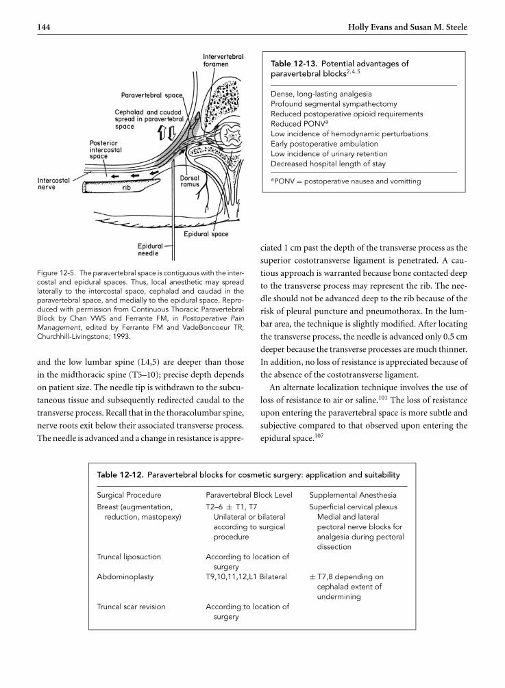
P1: PBU
cufx091-12 CUFX091/Friedberg 0 521 87090 9 Feb. 2, 2007 19:35
144 Holly Evans and Susan M. Steele
Figure 12-5. The paravertebral space is contiguous with the inter-costal and epidural spaces. Thus, local anesthetic may spreadlaterally to the intercostal space, cephalad and caudad in theparavertebral space, and medially to the epidural space. Repro-duced with permission from Continuous Thoracic ParavertebralBlock by Chan VWS and Ferrante FM, in Postoperative PainManagement, edited by Ferrante FM and VadeBoncoeur TR;Churchhill-Livingstone; 1993.
and the low lumbar spine (L4,5) are deeper than those
in the midthoracic spine (T5–10); precise depth depends
on patient size. The needle tip is withdrawn to the subcu-
taneous tissue and subsequently redirected caudal to the
transverse process. Recall that in the thoracolumbar spine,
nerve roots exit below their associated transverse process.
The needle is advanced and a change in resistance is appre-
Table 12-13. Potential advantages ofparavertebral blocks2,4,5
Dense, long-lasting analgesiaProfound segmental sympathectomyReduced postoperative opioid requirementsReduced PONVa
Low incidence of hemodynamic perturbationsEarly postoperative ambulationLow incidence of urinary retentionDecreased hospital length of stay
aPONV = postoperative nausea and vomitting
ciated 1 cm past the depth of the transverse process as the
superior costotransverse ligament is penetrated. A cau-
tious approach is warranted because bone contacted deep
to the transverse process may represent the rib. The nee-
dle should not be advanced deep to the rib because of the
risk of pleural puncture and pneumothorax. In the lum-
bar area, the technique is slightly modified. After locating
the transverse process, the needle is advanced only 0.5 cm
deeper because the transverse processes are much thinner.
In addition, no loss of resistance is appreciated because of
the absence of the costotransverse ligament.
An alternate localization technique involves the use of
loss of resistance to air or saline.101 The loss of resistance
upon entering the paravertebral space is more subtle and
subjective compared to that observed upon entering the
epidural space.107
Table 12-12. Paravertebral blocks for cosmetic surgery: application and suitability
Surgical Procedure Paravertebral Block Level Supplemental Anesthesia
Breast (augmentation,reduction, mastopexy)
T2–6 ± T1, T7Unilateral or bilateralaccording to surgicalprocedure
Superficial cervical plexusMedial and lateralpectoral nerve blocks foranalgesia during pectoraldissection
Truncal liposuction According to location ofsurgery
Abdominoplasty T9,10,11,12,L1 Bilateral ± T7,8 depending oncephalad extent ofundermining
Truncal scar revision According to location ofsurgery

P1: PBU
cufx091-12 CUFX091/Friedberg 0 521 87090 9 Feb. 2, 2007 19:35
Regional Anesthesia for Cosmetic Surgery 145
Table 12-14. Selected potential adverse effects ofparavertebral blocks and their incidence4,114
Adverse Effect
Self-limiting Failed blockVascular punctureInjection-site hematomaPain at injection siteEpidural spreadPostdural puncture headacheBrachial plexus blockHorner’s syndrome
Rare but serious PneumothoraxPulmonary hemorrhageLocal anesthetic toxicityNerve injuryIntrathecal injection, total spinalanesthesiaSeptic complications
Others have used nerve stimulation to identify the spinal
nerve roots in the paravertebral space. A 21 or 22 ga short-
bevel insulated stimulating needle is advanced to contact
transverse process. The needle is subsequently redirected
while connected to a nerve stimulator providing a current
output greater than 1.5 mA. Paraspinal muscle contraction
occurs initially as the needle passes through these muscles.
On further needle advancement, intercostal or abdomi-
nal muscle contractions are observed depending on the
dermatomal level of the block. The stimulating current is
decreased to 0.5–0.6 mA while appropriate muscle con-
traction is maintained. Isolated posterior spinal muscle
contraction is not accepted as this may represent either
direct muscle stimulation or stimulation of the posterior
ramus of the spinal nerve root after it diverges from the
spinal nerve. An additional option involves pressure mea-
surement to confirm paravertebral needle placement.108
When the needle tip is in the erector spinae muscle, mea-
sured pressure is higher during inspiration than expira-
tion. Within the paravertebral space, there is a sudden
lowering of pressures and a pressure inversion occurs.
Expiratory becomes higher than inspiratory pressure.
A further method to identify the paravertebral space
involves the injection of contrast material. Contrast may
distribute linearly as it spreads superiorly and inferiorly;
this is presumed to occur within the anterior compart-
ment. Alternatively, there may be cloud-like dispersal of
Figure 12-6. Superficial skin markings for left breast surgery.Reproduced with permission from Greengrass R and Steele S,in Techniques in Regional Anesthesia & Pain Management 2(1):8–12, January 1998.
contrast dye within one or two spinal segments; this is
thought to occur when the needle is in the posterior com-
partment.109
Some advocate a technique whereby after contact with
the transverse process, the needle is redirected superior to
it108,109 (Fig. 12-7). Using this approach, it is possible to
inadvertently contact rib first and walk off superiorly into
the pleura and lung. In addition, this technique blocks the
nerve root and dermatomal segment above the transverse
process contacted. The authors prefer redirecting the nee-
dle caudad to the transverse process.110 If rib is contacted
first, caudal redirection will bring the needle into contact
with the transverse process at a shallower depth. Conse-
quently, more accurate estimation of the depth of the par-
avertebral space is made and the risk of pneumothorax is
minimized.
When the needle is properly sited, an assistant aspirates
the syringe for blood, cerebrospinal fluid, and air. If air
is detected, it is likely that penetration of the pleura and
lung has occurred. The needle should be removed and
patient stability ensured. Similar steps are taken if blood
or cerebrospinal fluid is detected. When the paraverte-
bral space is correctly identified, there is no resistance to
injection of local anesthetic. Smaller volumes of local anes-
thetic (3–5 ml per level) are used for thoracic paravertebral

P1: PBU
cufx091-12 CUFX091/Friedberg 0 521 87090 9 Feb. 2, 2007 19:35
146 Holly Evans and Susan M. Steele
Figure 12-7. Needle “walked off” transverse process into paravertebral space. Reproduced with permission from Greengrass R andSteele S, in Techniques in Regional Anesthesia & Pain Management 2(1),8–12, January 1998.
blocks, whereas larger volumes (5–7 ml per level) are used
in the lumbar area. Doses may need to be reduced when
multiple and/or bilateral levels are blocked. Patients will
often experience a pressure paresthesia upon injection of
local anesthetic. Block adequacy is evaluated in a number
of ways. Sensory anesthesia and motor block are sought
in the appropriate dermatomes. For example, successful
upper thoracic paravertebral block is associated with loss
of sensation over the breast and upper chest wall. In addi-
tion, motor block of the intercostal muscles occurs and
can be confirmed by inspection and palpation of limited
hemithorax expansion. Vasodilation and warmth in the
corresponding distribution provides evidence of a suc-
cessful sympathetic block.
Using the majority of the techniques described, it is dif-
ficult to determine whether the needle tip exists in the
anterior extrapleural paravertebral compartment or the
posterior subendothoracic paravertebral compartment of
the thoracic paravertebral space. As discussed previously,
the pattern of spread of contrast dye may help with
this differentiation. Adequate block will result follow-
ing multilevel injection into either compartment; how-
ever, more extensive craniocaudad spread occurs when
the needle is in the anterior compartment (mean 4.5
dermatomal levels) compared to when it exists in the
posterior compartment (mean 2.3 dermatomal levels).111
These results reflect more complete cephalocaudad conti-
nuity between anterior segments. Consequently, it is here
that a single-level injection of a larger volume of local
anesthetic or placement of a continuous catheter should
occur.
Continuous paravertebral block can provide extended
postoperative analgesia. This technique can be used both
in patients admitted to hospital as well as outpatients.110

P1: PBU
cufx091-12 CUFX091/Friedberg 0 521 87090 9 Feb. 2, 2007 19:35
Regional Anesthesia for Cosmetic Surgery 147
A large-bore Tuohy needle is first used to identify the par-
avertebral space. Subsequently, a flexible catheter with a
single distal orifice is threaded 1–2 cm into the space.
Equipment
The equipment required depends on the technique used
to identify the paravertebral space. As described previ-
ously, commonly used equipment consists of a 10 cm
22 ga Tuohy needle with wings and centimeter gradations.
This is attached to extension tubing and a 20 ml syringe
containing local anesthetic. If loss of resistance is utilized,
a loss of resistance syringe is also needed. When a nerve-
stimulation technique is used, a 10 cm 21–22 ga insulated
short-bevel needle and a nerve stimulator are required.
And when pressure measurement is undertaken, a three-
way stopcock, pressure tubing, and a pressure transducer
are employed. A sterile skin marker and ruler should also
be included.
Local anesthetics and adjuvants
The local anesthetic agent selected is based on the desired
onset and duration of the nerve block. Block onset occurs
within ten minutes and surgical anesthesia within forty
minutes following injection of short-acting local anes-
thetic.112 Analgesia lasts up to twenty-three hours when
long-acting local anesthetic is used.106 Epinephrine is fre-
quently added to enhance block quality and limit sys-
temic toxicity. The proximity of the paravertebral space
to the intercostal space raises concern about significant
systemic absorption of local anesthetic following paraver-
tebral block. However, plasma local anesthetic levels have
been below toxic range when standard doses are used.113
In addition, systemic absorption is similar when a single-
level injection of 20 ml is given at T3-4 compared to when
injections of 4 ml each are given over five levels between
T2 and T6 (total 20 ml).113
Adverse effects
The adverse effects of paravertebral blocks are summa-
rized in Table 12-15. Vascular puncture, injection site
hematoma, and pain at the site of needle puncture are
usually mild, self-limiting, and respond to symptomatic
treatment. Epidural spread may be due to excessively
medial needle placement. A more extensive block than
expected may result and hypotension may occur.114
Brachial plexus block or Horner’s syndrome may result
Table 12-15. Comparison between paravertebraland intercostal nerve blocks
Paravertebral nerveblocks
Intercostal nerve blocks
Segmental sensory andmotor block
Segmental sensory andmotor block
Posterior ramus blocked Posterior ramus notreliably blocked
Sympathetic block Sympathetic chain notreliably blocked
Some LA spread toadjacent levels
Minimal LA spread toadjacent levels
Continuous catheter canprovide multilevelanalgesia
Continuous catheterusually provides onlysingle-level analgesia
Risk of pneumothorax Risk of pneumothoraxRisk of vascular puncture Risk of vascular punctureRisk of epidural spread,
dural punctureLower risk of epidural
spread, dural punctureLess systemic LA
absorptionSignificant LA systemic
absorption
from cephalad extension of injected local anesthetic. The
features of Horner’s syndrome include ptosis, miosis, and
anhydrosis and result from block of the sympathetic stel-
late ganglion.
Pleural puncture following paravertebral block is rare.
Pneumothorax results infrequently and is suspected if a
pleural “pop” is felt during needle advancement, if air
is aspirated into the syringe attached to the block nee-
dle, or if the patient develops an irritating cough or a
sharp pain in chest or shoulder. Resulting pneumothoraces
are often small enough to warrant conservative manage-
ment.100 However, when a chest tube is indicated and hos-
pital admission is required, this significantly impacts the
patient’s postoperative recovery. A case report describes
pulmonary hemorrhage as a complication following
paravertebral block in a patient who had previous thoracic
surgery.115 In addition, this patient population may have
pleural scarring affecting paravertebral anatomy, and this
can increase the risk of pneumothorax. Local anesthetic
toxicity (Table 12-11) can result from the use of large vol-
umes of local anesthetic or from accidental intravascular
injection. Precalculation of the maximum allowable dose
of local anesthetic and meticulous aspiration technique to
detect intravascular needle location should minimize this
risk. Nerve injury is rare, though a case report describes

P1: PBU
cufx091-12 CUFX091/Friedberg 0 521 87090 9 Feb. 2, 2007 19:35
148 Holly Evans and Susan M. Steele
a patient who developed chronic segmental thoracic pain
following paravertebral nerve block. Intrathecal injection,
dural puncture, and postdural puncture headache may
result from overly medial needle placement or from pen-
etration of a dural cuff that extends into the paraverte-
bral space.116 Significant resuscitation may be required
depending on the dose of local anesthetic injected intrathe-
cally and the resulting symptoms.
Intercostal Nerve Blocks
Anatomy
The intercostal space is continuous with the lateral aspect
of the paravertebral space. Upon exiting the paraverte-
bral space, the spinal nerve becomes the intercostal nerve.
Medial to the angle of the rib, the intercostal nerve is found
between the pleura and the fascia of the internal intercostal
muscle. The angle of the rib is located 6 to 8 cm from mid-
line. At this point, the intercostal nerve continues its course
between the internal and the innermost intercostal mus-
cles. The nerve lies in close proximity to the segmental
intercostal vessels, but its relation to the subcostal groove
is variable.117 Each intercostal nerve has four branches
(Fig. 12-8). The gray ramus communicans passes anteri-
orly to the sympathetic ganglion, and the posterior ramus
passes posteriorly to supply the skin and muscle in the
paravertebral area. These branches are more reliably anes-
thetized with paravertebral than intercostal nerve blocks.
The intercostal nerve also gives rise to lateral and anterior
cutaneous branches.
Indications and advantages
An intercostal nerve block is indicated for analgesia follow-
ing breast or chest-wall procedures and is predominantly
used when paravertebral or epidural blocks are not indi-
cated. The differences between paravertebral and inter-
costal blocks are outlined in Table 12-15.
Technique
An intravenous is placed, monitors are attached, and
resuscitation equipment is made available. Light sedation
may be provided. The patient is typically placed prone,
although the lateral decubitus, sitting, or supine positions
can also be used. In the prone position, a pillow is placed
under the upper abdomen to promote thoracic spine flex-
ion and to widen the intercostal spaces. The patient’s arms
hang over the edge of the bed to displace the scapulae
laterally (see Fig. 12-8).
Figure 12-8. Longitudinal section to show direction of needle: (a) above, (b) below: transverse process or rib. Reproduced with permis-sion from Eason MJ and Wyatt R, in Anaesthesia 34(7):638–42,1979.108

P1: PBU
cufx091-12 CUFX091/Friedberg 0 521 87090 9 Feb. 2, 2007 19:35
Regional Anesthesia for Cosmetic Surgery 149
Figure 12-9. (A) Intercostal nerve block: patient positioning. (B) The index finger displaces the skin up over the rib. The needle isinserted at the tip of the finger and rests on the rib. The needle is walked off the lower rib edge and inserted 3 to 5 mm. (C) Anintercostal nerve and its branches. Reproduced with permission from Nerve Blocks by Wedel DJ, in Anesthesia, 5th edition, edited byMiller RD; Churchhill-Livingstone; 2000.
A paraspinal line is drawn 5 to 9 cm lateral to the midline
where the posterior angulation of the ribs is most easily
palpable. In order to avoid the scapula, the line diverges
medially in the upper thoracic segments. This location is
chosen for the nerve block as it is proximal to the diver-
gence of the lateral cutaneous branch. The inferior border
of each rib is marked along the length of the paraspinal
line. The appropriate levels to be blocked are identified
according to the surgical procedure. The skin is cleaned
with disinfectant and sterile drapes are placed. The anes-
thesiologist stands to the patient’s side. A subcutaneous
skin wheal is placed at the mark representing the inferior
border of the angle of the rib at each level to be blocked.
The left hand is used to move the skin wheal so that it over-
lies the rib. The right hand holds a 3–4 cm 22 ga short bevel
needle attached to a 10 ml syringe and advances it onto the
rib (Fig. 12-9). The left hand grasps the needle while the
right hand holds the syringe. The needle is subsequently
walked in a caudal direction past the inferior border of the
rib and advanced 3–5 mm past the depth at which the rib
was contacted. A subtle change in resistance if often felt as
the needle passes through the internal intercostal muscle.
After negative aspiration for blood, 3–5 ml of local anes-
thetic is injected and the nerve block is repeated at each of
the desired levels. The most caudal rib to be anesthetized is
blocked first. Subsequent nerve blocks gradually proceed
in a cephalad direction.
Equipment
A 3–4 cm 22 ga short bevel needle and a 10 ml syringe are
required.
Local anesthetics and adjuvants
There may be significant systemic absorption of local anes-
thetic following intercostal nerve block. Therefore, the
dose of local anesthetic used should be carefully calculated.
Ropivacaine offers long-acting anesthesia with an accept-
able safely profile. Epinephrine can be added to further
reduce the risk of toxicity.
Adverse effects
Pneumothorax is rare (0.42%), despite the close proxim-
ity of the lung and pleura.118 The risk of local anesthetic
toxicity is related to the large volumes of local anesthetic

P1: PBU
cufx091-12 CUFX091/Friedberg 0 521 87090 9 Feb. 2, 2007 19:35
150 Holly Evans and Susan M. Steele
used to obtain an adequate block and to the significant
systemic absorption that occurs in this highly vascular
area.
RECOVERY AND DISCHARGE
Recovery
Patients require a period of monitoring postoperatively
because both surgery and anesthesia can be associated
with significant physiologic changes. Standard monitors
such as pulse oximeter, noninvasive blood pressure, and
electrocardiogram are used. In addition, the vigilance of a
nurse with resuscitation skills is of critical importance. A
physician with advanced cardiac life-support skills should
be immediately available.
A plan is required for analgesia as block resolution
occurs. This transition is managed in the postanesthesia
care unit following spinal or epidural anesthesia. For both
groups, multimodal analgesia is ideal. This involves the
used of regular, scheduled doses of acetaminophen and
nonsteroidal anti-inflammatory medication in addition
to oral opioid tablets.
Discharge
Many cosmetic surgical procedures are performed on an
ambulatory basis. Traditional outpatient discharge criteria
include adequate analgesia; absence of side effects such as
nausea, vomiting, and sedation; as well as the ability to
ambulate, void, and tolerate fluid intake.
Recent literature has challenged the requirements to
drink and void prior to discharge.119,120 In a randomized
trial, Jin et al. documented the feasibility of discharging
patients prior to fluid intake.119 They found no differ-
ence in the incidence of postoperative nausea and vomit-
ing when they compared a group of patients who drank
prior to discharge versus a group who did not drink.
Patients with a history of urinary retention, men, those
who have had spinal anesthesia, and those who have had
anal, urogenital, and hernia surgery are known to be at
high risk of postoperative urinary retention. Mulroy and
colleagues have shown the viability of discharging ambu-
latory patients who have received short-acting spinal or
epidural anesthesia without requiring them to void.120
In their study, they used bladder ultrasound to estimate
the volume of urine prior to determining suitability of
discharge. Forty-six patients had less than 400 ml urine
and were discharged home without voiding. They were
instructed to return to hospital if they had not voided
within eight hours; however, all were able to void sponta-
neously. Twenty-three patients had urine volume greater
than 400 ml. Most (97.7%) voided spontaneously during
the subsequent sixty minutes in which they were detained
in hospital; however, three patients required catheteriza-
tion. The quantification of urine volume with bladder
ultrasound and a defined protocol certainly led to their
success and excellent results. The use of this protocol in
cosmetic surgical patients who have received regional anes-
thesia remains unexplored.
Finally, all patients should receive appropriate contact
numbers should complications arise following discharge,
and contingency plans should be in place for those patients
who do not meet discharge criteria and who require admis-
sion to an inpatient facility.
SUMMARY
Regional anesthesia has broad application for cosmetic
surgical procedures. Neuraxial techniques provide rapid-
onset, dense anesthesia with minimal postoperative seda-
tion, nausea, and vomiting. Spinal anesthesia, in partic-
ular, has been tailored for day-case procedures by the
use of low doses of local anesthetics, short-acting agents,
dose-sparing adjuvants such as opioids, and small-gauge,
pencil-point needles. As a result, side effects have been
minimized and there is timely recovery of motor strength
as well as ability to ambulate and to void.
Paravertebral nerve blocks uniquely offer dense
intraoperative anesthesia and prolonged postoperative
analgesia with rare side effects. This technique is surpris-
ingly underutilized given its benefits. With appropriately
selected patients, surgical procedures, and nerve block
techniques, regional anesthesia can facilitate successful
cosmetic surgery and result in positive patient outcome
with high patient satisfaction.
REFERENCES
1. Auroy Y, Benhamou D, Bargues L, et al.: Major complica-tions of regional anesthesia in France: The SOS RegionalAnesthesia Hotline Service. Anesthesiol 97:1274,2002.
2. Naja MZ, Ziade MF, Lonnqvist PA: Nerve-stimulatorguided paravertebral blockade vs. general anaesthesia for

P1: PBU
cufx091-12 CUFX091/Friedberg 0 521 87090 9 Feb. 2, 2007 19:35
Regional Anesthesia for Cosmetic Surgery 151
breast surgery: A prospective randomized trial. Eur J Anaes-thesiol 20:897,2003.
3. Borgeat A, Ekatodramis G, Schenker CA: Postoperative nau-sea and vomiting in regional anesthesia: A review. Anesthe-siol 98:530,2003.
4. Pusch F, Freitag H, Weinstabl C, et al.: Single injection par-avertebral block compared to general anaesthesia in breastsurgery. Acta Anaesthesiol Scand 43:770,1999.
5. Klein SM, Bergh A, Steele SM, et al.: Thoracic paravertebralblock for breast surgery. Anesth Analg 90:1402,2000.
6. Gebhard RE, Al-Samsam T, Greger J, et al.: Distal nerveblocks at the wrist for outpatient carpal tunnel surgery offerintraoperative cardiovascular stability and reduce dischargetime. Anesth Analg 95:351,2002.
7. Song D, Greilich NB, White PF, et al.: Recovery profilesand costs of anesthesia for outpatient unilateral inguinalherniorrhaphy. Anesth Analg 91:876,2000.
8. Williams BA, Kentor ML, Vogt MT, et al.: Femoral-sciaticnerve blocks for complex outpatient knee surgery are associ-ated with less postoperative pain before same-day discharge:A review of 1,200 consecutive cases from the period 1996–1999. Anesthesiol 98:1206,2003.
9. Horlocker TT, Wedel DJ, Benzon HP, et al.: Regional anes-thesia in the anticoagulated patient: Defining the risks (thesecond ASRA consensus conference on neuraxial anes-thesia and anticoagulation). Reg Anesth Pain Med 28:172,2003.
10. Klein SM, D’Ercole F, Greengrass RA, et al.: Enoxaparinassociated with psoas hematoma and lumbar plexopathyafter lumbar plexus block. Anesthesiol 87:1576,1997.
11. Deng XM, Xiao WJ, Luo MP, et al.: The use of midazolamand small-dose ketamine for sedation and analgesia duringlocal anesthesia. Anesth Analg 93:1174,2001.
12. Koch ME, Kain ZN, Ayoub C, et al.: The sedative and anal-gesia sparing effect of music. Anesthesiol 89;300,1998.
13. Milne SE, Kenny GN: Future applications for TCI systems.Anaesthesia 53(S1):56,1998.
14. Apfel CC, Korttila K, Abdalla M, et al.: A factorial trial ofsix interventions for the prevention of postoperative nauseaand vomiting. N Engl J Med 350:2441,2004.
15. Servin FS, Raeder JC, Merle JC, et al.: Remifentanil sedationcompared with propofol during regional anaesthesia. ActaAnaesthesiol Scand 46:309,2002.
16. Arain SR, Ebert TJ: The efficacy, side effects, and recov-ery characteristics of dexmedotomidine versus propofolwhen used for intraoperative sedation. Anesth Analg 95:461,2002.
17. Halpern S, Preston R: Postdural puncture headache andspinal needle design. Meta-analyses. Anesthesiol 81:1376,1994.
18. Pawlowski J, Sukhani R, Pappas AL, et al.: The anestheticand recovery profile of two doses (60 and 80 mg) of plainmepivacaine for ambulatory spinal anesthesia. Anesth Analg91:580,2000.
19. Zayas VM, Liguori GA, Chisholm MF, et al.: Dose responserelationships for isobaric spinal mepivacaine using the com-bined spinal epidural technique. Anesth Analg 89:1167,1999.
20. Zaric D, Christiansen C, Pace NL, et al.: Transient neu-rologic symptoms (TNS) following spinal anaesthesia withlidocaine versus other local anaesthetics. Cochrane DatabaseSyst Rev 2:CD003006,2003.
21. Pollock JE: Transient neurologic symptoms: Etiology,risk factors, and management. Reg Anesth Pain Med27:581,2002.
22. Salazar F, Bogdanovich A, Adalia R, et al.: Transient neuro-logic symptoms after spinal anaesthesia using isobaric 2%mepivacaine and isobaric 2% lidocaine. Acta AnaesthesiolScand 45:240,2001.
23. Johnson ME: Potential neurotoxicity of spinal anesthesiawith lidocaine. Mayo Clin Proc 75:921,2000.
24. Freedman JM, Li DK, Drasner K, et al.: Transient neurologicsymptoms after spinal anesthesia: An epidemiologic studyof 1,863 patients. Anesthesiol 89:633,1998.
25. Liguori GA, Zayas VM, Chisholm MF: Transient neuro-logic symptoms after spinal anesthesia with mepivacaineand lidocaine. Anesthesiol 88:619,1998.
26. Hampl KF, Schneider MC, Ummenhofer W, et al.: Transientneurologic symptoms after spinal anesthesia. Anesth Analg81:1148,1995.
26. Liguori GA, Zayas VM: Repeated episodes of transient radi-ating back and leg pain following spinal anesthesia with1.5% mepivacaine and 2% lidocaine. Reg Anesth Pain Med23:511,1998.
27. Le Truong HH, Girard M, Drolet P, et al.: Spinal anesthesia:a comparison of procaine and lidocaine. Can J Anaesth48:470,2001.
28. Kouri ME, Kopacz DJ: Spinal 2-chloroprocaine: A compar-ison with lidocaine in volunteers. Anesth Analg 98:75,2004.
29. Smith KN, Kopacz DJ, McDonald SB: Spinal 2-chloroprocaine: A dose-ranging study and the effect ofadded epinephrine. Anesth Anal 98:81,2004.
30. Vath JS, Kopacz DJ: Spinal 2-chloroprocaine: The effect ofadded fentanyl. Anesth Analg 98:89,2004.
31. Warren DT, Kopacz DJ: Spinal 2-chloroprocaine: The effectof added dextrose. Anesth Analg 98:95,2004.
32. Yoos JR, Kopacz DJ: Spinal 2-chloroprocaine for surgery: Aninitial 10-month experience. Anesth Analg 100:553,2005.
33. Davis BR, Kopacz DJ: Spinal 2-chloroprocaine: The effectof added clonidine. Anesth Analg 100:559,2005.
34. Yoos JR, Kopacz DJ: Spinal 2-chloroprocaine: a comparisonwith small-dose bupivacaine in volunteers. Anesth Analg100:566,2005.
35. Gonter AF, Kopacz DJ: Spinal 2-chloroprocaine: A com-parison with procaine in volunteers. Anesth Analg 100:573,2005.
36. Glaser C, Marhofer P, Zimpfer G, et al.: Levobupivacaineversus racemic bupivacaine for spinal anesthesia. AnesthAnalg 94(1):194,2002.
37. Liu SS, Ware PD, Allen HW, et al.: Dose-response charac-teristics of spinal bupivacaine in volunteers. Clinical impli-cations for ambulatory anesthesia. Anesthesiol 85:729,1996.
38. Wille M: Intrathecal use of ropivacaine: A review. ActaAnaesthesiol Belg 55:251,2004.
39. Wahedi W, Nolte H, Klein P: Ropivacaine for spinal anes-thesia. A dose-finding study. Anaesthesist 45:737,1996.

P1: PBU
cufx091-12 CUFX091/Friedberg 0 521 87090 9 Feb. 2, 2007 19:35
152 Holly Evans and Susan M. Steele
40. Kallio H, Snall EV, Kero MP, et al.: A comparison ofintrathecal plain solutions containing ropivacaine 20 or 15mg versus bupivacaine 10 mg. Anesth Analg 99:713,2004.
41. McNamee DA, McClelland AM, Scott S, et al.: Spinalanaesthesia: Comparison of plain ropivacaine 5 mg · ml−1
with bupivacaine 5 mg ·ml−1 for major orthopaedic surgery.Br J Anaesth 89:702,2002.
42. Gautier PE, De Kock M, Van Steenberge A, et al.: Intrathecalropivacaine for ambulatory surgery. A comparison betweenintrathecal bupivacaine and intrathecal ropivacaine forknee arthroscopy. Anesthesiol 91:1239,1999.
43. Cousins MJ, Mather LE: Intrathecal and epidural adminis-tration of opioids. Anesthesiol 61:276,1984.
44. Ben-David B, Solomon E, Levin H, et al.: Intrathecal fen-tanyl with small-dose dilute bupivacaine: Better anesthesiawithout prolonging recovery. Anesth Analg 85:560, 1997.
45. Ballantyne JC, Loach AB, Carr DB: Itching after epiduraland spinal opiates. Pain 33:149,1988.
46. Goel S, Bhardwaj N, Grover VK: Intrathecal fentanyl addedto intrathecal bupivacaine for day case surgery: A random-ized study. Eur J Anaesthesiol 20:294,2003.
47. Gupta A, Axelsson K, Thorn SE, et al.: Low-dose bupiva-caine plus fentanyl for spinal anesthesia during ambulatoryinguinal herniorrhaphy: A comparison between 6 mg and50.5 mg of bupivacaine. Acta Anaesthesiol Scand 47:13, 2003.
48. Liu S, Chiu AA, Carpenter RL, et al.: Fentanyl prolongs lido-caine spinal anesthesia without prolonging recovery. AnesthAnalg 80:730,1995.
49. Eisenach JC, De Kock M, Klimscha W: Alpha2 adrenergicagonists for regional anesthesia. A clinical review of cloni-dine (1984–1995). Anesthesiol 85:655,1996.
50. De Kock M, Gautier P, Fanard L, et al.: Intrathecal ropi-vacaine and clonidine for ambulatory knee arthroscopy: Adose-response study. Anesthesiol 94:574,2001.
51. Kito K, Kato H, Shibata M, et al.: The effect of varied dosesof epinephrine on duration of lidocaine spinal anesthesiain the thoracic and lumbosacral dermatomes. Anesth Analg86:1018,1998.
52. Moore JM, Liu SS, Pollock JE, et al.: The effect ofepinephrine on small-dose hyperbaric bupivacaine spinalanesthesia: Clinical implications for ambulatory surgery.Anesth Analg 86:973,1998.
53. Chiu AA, Liu S, Carpenter RL, et al.: The effects ofepinephrine on lidocaine spinal anesthesia: A cross-overstudy. Anesth Analg 80:735,1995.
54. Sakura S, Sumi M, Sakaguchi Y, et al.: The addition ofphenylephrine contributes to the development of transientneurologic symptoms after spinal anesthesia with 0.5%tetracaine. Anesthesiol 87:771,1997.
55. Vaida GT, Moss P, Capan LM, et al.: Prolongation of lido-caine spinal anesthesia with phenylephrine. Anesth Analg65:781,1986.
56. Leicht CH, Carlson SA: Prolongation of lidocaine spinalanesthesia with epinephrine and phenylephrine. AnesthAnalg 65:365,1986.
57. Seeberger MD, Lang ML, Drewe J, et al.: Comparison ofspinal and epidural anesthesia for patients younger than50 years of age. Anesth Analg 78:667,1994.
58. Petros JG, Rimm EB, Robillard RJ: Factors influencing uri-nary tract retention after elective open cholecystectomy.Surg Gyn Obstet 174:497,1992.
59. Lamonerie L, Marret E, Deleuze A, Lembert N, Dupont M,Bonnet F. Prevalence of postoperative bladder distensionand urinary retention detected by ultrasound measurement.Br J Anaesth 92:544,2004.
60. Hodgson PS, Neal JM, Pollock JE, et al.: The neurotoxicityof drugs given intrathecally (spinal). Anesth Analg 88:797,1999.
61. Pollock JE, Neal JM, Stephenson CA, et al.: Prospectivestudy of the incidence of transient radicular irritation inpatients undergoing spinal anesthesia. Anesthesiol 84:1361,1996.
62. Lybecker H, Moller JT, May O, et al.: Incidence and pre-diction of postdural puncture headache. A prospectivestudy of 1021 spinal anesthesias. Anesth Analg 70:389,1990.
63. Norris MC, Leighton BL, DeSimone CA: Needle bevel direc-tion and headache after inadvertent dural puncture. Anes-thesiol 70:729,1989.
64. Aguilera Celorrio L, Martinez-Garbizu I, Saez de EquilazIzaola JL, et al.: Influence of needle bevel, age and sex on theappearance of post-puncture headaches. Rev Esp AnestesiolReanim 36:16,1989.
65. Camann WR, Murray RS, Mushlin PS, et al.: Effects oforal caffeine on postdural puncture headache. A double-blind, placebo-controlled trial. Anesth Analg 70:181,1990.
66. Auroy Y, Narchi P, Messiah A, et al.: Serious complicationsrelated to regional anesthesia: Results of a prospective surveyin France. Anesthesiol 87:479,1997.
67. Moore DC, Bridenbaugh LD: Spinal (subarachnoid) block.A review of 11,574 cases. JAMA 195:907,1966.
68. Dripps RD, Vandam LD: Long-term follow-up of patientswho received 10,098 spinal anesthetics: Failure to discovermajor neurological sequelae. JAMA 156:1486,1954.
69. Vandam LD, Dripps RD: A long-term follow-up of 10,098spinal anesthetics. II. Incidence and analysis of minor sen-sory neurological defects. Surg 38:463,1955.
70. Moen V, Dahlgren N, Irestedt L: Severe neurological com-plications after central neuraxial blockades in Sweden 1990–1999. Anesthesiol 101:950,2004.
71. Wedel DJ, Horlocker TT: Risks of regional anesthesia –Infectious, septic. Reg Anesth 21(6S):57,1996.
72. Loarie DJ, Fairley HB: Epidural abscess following spinalanesthesia. Anesth Analg 57:351,1978.
73. Kilpatrick ME, Girgis NI: Meningitis—A complication ofspinal anesthesia. Anesth Analg 62:513,1983.
74. Burke D, Wildsmith JA: Meningitis after spinal anaesthesia.Br J Anaesth 78:635,1997.
75. Aromaa U, Lahdensuu M, Cozanitis DA: Severe complica-tions associated with epidural and spinal anaesthesias inFinland 1987–1993. A study based on patient insuranceclaims. Acta Anaesthesiol Scand 41:445, 1997.
76. Caplan RA, Ward RJ, Posner K, et al.: Unexpected cardiacarrest during spinal anesthesia: A closed claims analysis ofpredisposing factors. Anesthesiol 68:5,1988.

P1: PBU
cufx091-12 CUFX091/Friedberg 0 521 87090 9 Feb. 2, 2007 19:35
Regional Anesthesia for Cosmetic Surgery 153
77. Scott DB, McClure JH, Giasi RM, et al.: Effects of concen-tration of local anaesthetic drugs in extradural block. Br JAnaesth 52:1033,1980.
78. Park WY, Hagins FM, Rivat EL, et al.: Age and epidural doseresponse in adult men. Anesthesiol 56(4):318,1982.
79. Liu SS, Ware PD, Rajendran S: Effects of concentration andvolume of 2-chloroprocaine on epidural anesthesia in vol-unteers. Anesthesiol 86:1288,1997.
80. Arakawa M: Does pregnancy increase the efficacy of lumbarepidural anesthesia? Internat J Obstr Anesth 13:86, 2004.
81. Fagraeus L, Urban BJ, Bromage PR: Spread of epidural anal-gesia in early pregnancy. Anesthesiol 58:184,1983.
82. Nydahl PA, Philipson L, Axelsson K, et al.: Epidural anes-thesia with 0.5% bupivacaine: Influence of age on sensoryand motor blockade. Anesth Analg 73:780,1991.
83. Neal JM, Deck JJ, Kopacz DJ, et al.: Hospital discharge afterambulatory knee arthroscopy: A comparison of epidural2-chloroprocaine versus lidocaine. Reg Anesth Pain Med26:35,2001.
84. Allen RW, Fee JP, Moore J: A preliminary assessment ofepidural chloroprocaine for day procedures. Anaesthesia48:773,1993.
85. Kopacz DJ, Mulroy MF: Chloroprocaine and lidocainedecrease hospital stay and admission rate after outpatientepidural anesthesia. Reg Anesth 15:19,1990.
86. Stevens RA, Urmey WF, Urquhart BL, et al.: Back painafter epidural anesthesia with chloroprocaine. Anesthesiol78:492,1993.
87. Stevens RA, Chester WL, Artuso JD, et al.: Back pain afterepidural anesthesia with chloroprocaine in volunteers: Pre-liminary report. Reg Anesth 16:199,1991.
88. Reisner LS, Hochman BN, Plumer MH: Persistent neuro-logic deficit and adhesive arachnoiditis following intrathe-cal 2-chloroprocaine injection. Anesth Analg 59:452,1980.
89. Terai T, Yukioka H, Fujimori M: A double-blind compar-ison of lidocaine and mepivacaine during epidural anaes-thesia. Acta Anaesthesiol Scand 37:607,1993.
90. Groban L, Deal DD, Vernon JC, et al.: Cardiac resusci-tation after incremental overdosage with lidocaine, bupi-vacaine, levobupivacaine, and ropivacaine in anesthetizeddogs. Anesth Analg 92:37,2001.
91. Klimscha W, Chiari A, Krafft P, et al.: Hemodynamic andanalgesic effects of clonidine added repetitively to con-tinuous epidural and spinal blocks. Anesth Analg 80:322,1995.
92. Nishikawa T, Dohi S: Clinical evaluation of clonidine addedto lidocaine solution for epidural anesthesia. Anesthesiol73:853,1990.
93. Bromage PR, Burfoot MF, Crowell DE, et al.: Quality ofepidural blockade. I. Influence of physical factors. Br JAnaesth 36:342,1964.
94. Stevens RA, Chester WL, Grueter JA, et al.: The effect of pHadjustment of 0.5% bupivacaine on the latency of epiduralanesthesia. Reg Anesth 14:236,1989.
95. DiFazio CA, Carron H, Grosslight KR, et al.: Comparisonof pH-adjusted lidocaine solutions for epidural anesthesia.Anesth Analg 65:760,1986.
96. Capogna G, Celleno D, Tagariello V: The effect of pH adjust-ment of 2% mepivacaine on epidural anesthesia. Reg Anesth14:121,1989.
97. Block BM, Liu SS, Rowlingson AJ, et al.: Efficacy ofpostoperative epidural analgesia: A meta-analysis. JAMA290:2455,2003.
98. Staats PS, Stinson MS, Lee RR: Lumbar stenosis compli-cating retained epidural catheter tip. Anesthesiol 83:1115,1995.
99. Sakuma N, Hori M, Suzuki H, et al.: A sheared off andsequestered epidural catheter: A case report. Masui-Jap JAnesthesiol 53:198,2004.
100. Eason MJ, Wyatt R: Paravertebral thoracic block—A reap-praisal. Anaesthesia 34:638,1979.
101. Karmakar MK: Thoracic paravertebral block. Anesthesiol95:771,2001.
102. Karmakar MK, Chung DC: Variability of a thoracic paraver-tebral block. Are we ignoring the endothoracic fascia? RegAnesth Pain Med 25(3):325,2000.
103. Saito T, Den S, Tanuma K, et al.: Anatomical bases for par-avertebral anesthetic block: Fluid communication betweenthe thoracic and lumbar paravertebral regions. Surg &Radiol Anat 21:359,1999.
104. Weltz CR, Greengrass RA, Lyerly HK: Ambulatory surgicalmanagement of breast carcinoma using paravertebral block.Ann Surg 222:19,1995.
105. Naja MZ, el Hassan MJ, Oweidat M, et al.: Paraverte-bral blockade vs general anesthesia or spinal anesthe-sia for inguinal hernia repair. Middle East J Anesthesiol16:201,2001.
106. Greengrass R, Buckenmaier CC: Paravertebral anaesthe-sia/analgesia for ambulatory surgery. Best Pract Res ClinAnaesthesiol 16:271,2002.
107. Naja Z, Lonnqvist PA: Somatic paravertebral nerve block-ade. Incidence of failed block and complications. Anaesthe-sia 56:1184,2001.
108. Richardson J, Cheema SP, Hawkins J, et al.: Thoracic par-avertebral space location. A new method using pressuremeasurement. Anaesthesia 51:137,1996.
109. Naja MZ, Ziade MF, El Rajab M, et al.: Varying anatomi-cal injection points within the thoracic paravertebral space:Effect on spread of solution and nerve blockade. Anaesthesia59:459,2004.
110. Buckenmaier CC, 3rd, Klein SM, Nielsen KC, et al.: Con-tinuous paravertebral catheter and outpatient infusion forbreast surgery. Anesth Analg 97:715,2003.
111. Saito T, Den S, Cheema SP, et al.: A single-injection, multi-segmental paravertebral block-extension of somatosensoryand sympathetic block in volunteers. Acta AnaesthesiolScand 45:30,2001.
112. Lemay E, Guay J, Cote C, et al.: The number of injectionsdoes not influence local anesthetic absorption after paraver-tebral blockade. Can J Anaesth 50:562,2003.
113. Richardson J, Sabanathan S: Thoracic paravertebral analge-sia. Acta Anaesthesiol Scand 39:1005,1995.
114. Thomas PW, Sanders DJ, Berrisford RG: Pulmonary haem-orrhage after percutaneous paravertebral block. Br J Anaesth83:668,1999.

P1: PBU
cufx091-12 CUFX091/Friedberg 0 521 87090 9 Feb. 2, 2007 19:35
154 Holly Evans and Susan M. Steele
115. Bigler D, Dirkes W, Hansen R, et al.: Effects of thoracic par-avertebral block with bupivacaine versus combined thoracicepidural block with bupivacaine and morphine on pain andpulmonary function after cholecystectomy. Acta Anaesthe-siol Scand 33:561,1989.
116. Lekhak B, Bartley C, Conacher ID, et al.: Total spinalanaesthesia in association with insertion of a paravertebralcatheter. Br J Anaesth 86:280,2001.
117. Hardy PA: Anatomical variation in the position of the prox-imal intercostal nerve. Br J Anaesth 61(3):338,1988.
118. Moore DC, Bridenbaugh LD: Pneumothorax: Its inci-dence following intercostal nerve block. JAMA 182:1005,1962.
119. Jin FL, Norris AM, Chung F: Should adult patients drinkbefore discharge from ambulatory surgery? Anesth Analg .87:306,1998.
120. Mulroy MF, Salinas FV, Larkin KL, et al.: Ambulatorysurgery patients may be discharged before voiding aftershort-acting spinal and epidural anesthesia. Anesthesiol97:315,2002.

P1: PBU
cufx091-13 CUFX091/Friedberg 0 521 87090 9 Feb. 2, 2007 19:36
13 General Inhalation Anesthesia for Cosmetic Surgery
Meena Desai, M.D.
INTRODUCTION
PATIENT SELECTION FOR THE COSMETIC SURGICAL SUITE
GENERAL ANESTHETIC CONCERNS FOR STANDARD AESTHETICPROCEDURES
Rhytidectomy or Facelift
Preoperative considerations
Intraoperative considerations
Emergence considerations
Postprocedure considerations
Browlift
Endoscopic browlift
Breast Augmentation
Preoperative considerations
Intraoperative considerations
Emergence considerations
Postprocedure considerations
Endoscopic breast implants
Mastopexy
Intraoperative considerations
Liposuction
Preoperative concerns
Intraoperative concerns
Postprocedure considerations
Rhinoplasty
Intraoperative concerns
Emergence concerns
Postprocedure concerns
Abdominoplasty
Preoperative considerations
Intraoperative concerns
Emergence concerns
Postprocedure concerns
Laser Facial Resurfacing
Intraoperative concerns
Postprocedure concerns
155

P1: PBU
cufx091-13 CUFX091/Friedberg 0 521 87090 9 Feb. 2, 2007 19:36
156 Meena Desai
Blepharoplasty
Emergence concerns
PRACTICAL CONSIDERATION OF CLINICAL ISSUES
Fast Tracking and Discharge Criteria
Pain medication in recovery
Pain management
PONV
MALIGNANT HYPERTHERMIA (MH) PROTOCOL
THROMBOEMBOLISM
Recommendations according to risk stratification
Low-risk patients
Moderate-risk patients
High-risk patients
CONCLUSION
INTRODUCTION
The ultimate in consumer-driven medical care is the
business of cosmetic surgery. These are purely elective
procedures performed for the convenience and wishes of
the buyer patient and at the convenience of the patient.
Traditional ideology of surgical care is maximized as
service is optimized. The maximization of service should
never, however, compromise the prevailing medical,
surgical, or anesthetic standard of care. Meeting the
standard of care is the minimum requirement of the
cosmetic surgical practice.
Much has been written regarding the safety of general
anesthesia in the office.1−4 One should have a structure and
plan for the administration of care that adheres to known
standards. The care offered to entice the consumer to an
individual practice and practitioner is multifaceted. Sev-
eral studies have found that the friendliness and courtesy
of the staff were top predictors of patient satisfaction.5 The
rendering of anesthetic care must be cognizant of the con-
sumerism of plastic surgery and be sensitive of delivering
their care with a practice philosophy suited to the surgical
practice. Care is often beyond expectations as patients and
their idiosyncratic requests are willingly accommodated.
In this chapter, some of the specific particulars of general
anesthetic care are provided as they relate to individual
procedures. There are also some methods of care that cater
to the consumerism milieu.
Many patients respond to the branding of names in rela-
tionship to the care that is rendered. Hospital-based care
may be analogized as the “Cadillac,” whereas the plastic
surgical care that is provided in an ASC or office-based
cosmetic surgical suite may be analogized as the “Rolls
Royce.” Attempts are made to preempt postoperative nau-
sea and vomiting (PONV) instead of addressing it after it
occurs. Comforters are provided for blankets, linens have
lace, neck pillows have aromatherapy, heat, and so forth.
This is the “spa” surgical experience.
Keeping abreast of the cosmetic surgery literature pro-
vides some assurance that one is collaborating with a sur-
geon that also has the highest regard for the standard of
care. This may vary from state to state. Florida, for exam-
ple, currently has modeled its office regulations around
those of the AAAASF, namely, level: local only, level II,
intravenous (sedation), and level III (general anesthesia).
Additionally, surgical societies may have consensus
statements and practice standards that are changing and
are prudent to regard. Some practices may have outside
or independent peer review as a gauge of practice qual-
ity. Alternatively, the surgeon who is part of a hospital
staff is subject to peer review from that avenue. Postpro-
cedure care and patient satisfaction are vital to a surgical
practice. In association with the surgical practice, check
anesthetic parameters of patient care and satisfaction as
well.

P1: PBU
cufx091-13 CUFX091/Friedberg 0 521 87090 9 Feb. 2, 2007 19:36
General Inhalation Anesthesia for Cosmetic Surgery 157
Screen one’s surgeons as carefully as one would one’s
anesthesia colleagues. Verify their licenses and credentials.
Check their references and run a query via the AMA data
bank or the National Practitioner data bank. What is the
surgeon’s rate of infection? How many “redos” or “touch-
ups” does the practice do?
What is the admission rate to the acute care center or
hospital postprocedure? In how many litigious encounters
is your surgeon involved? Do not hesitate to “credential”
them within your practice much as one do one’s own staff
and reciprocally provide the same information to your
surgeon. Encourage one’s staff to keep all of your practice
credentialing files up to date. If one is in a state that has
no local or state guidelines, then it is incumbent on one
to develop them. The office-based guidelines approved by
the American Society of Anesthesiologists (ASA)6 are an
excellent template for the development of one’s own inter-
nal guidelines.
PATIENT SELECTION FOR THE COSMETIC
SURGICAL SUITE
An invaluable service that can be monitored by the anes-
thesia group is appropriate patient selection for the facility.
Desai’s group screens every patient well in advance of the
date of the procedure. The screening process begins as
soon as the patient is scheduled. Testing is procedure and
history specific. Algorithms may be developed and imple-
mented to make the flow of the preanesthesia interviews
and laboratory screening succinct.
Desai’s screening criteria are constantly updated and
revised as the literature and experience warrants. Pre-
existing medical conditions can be predictors of adverse
events in the ambulatory setting.7−10 The American Col-
lege of Cardiology and the American Heart Association
have updated guidelines for Perioperative Cardiovascular
Evaluation for Noncardiac Surgery11 that are useful for
appropriate patient evaluation and selection. The British
Journal of Anaesthesia also has an excellent review that
helps one judge perioperative cardiac risk.12
Many surgeons’ practices track patients with postoper-
ative follow-up calls.
Satisfaction with the anesthesia services may not always
be a part of this interview. Therefore, the anesthesia prac-
tice should also follow up to ensure a high level of patient
satisfaction, specifically with their anesthesia care. These
Table 13-1. Selection list of patients undergoinggeneral anesthesia criteria that suggest a patientmay be unsuitable for a procedure in the office suite
1. Unstable angina2. History of myocardial injury within three to six
months3. Severe cardiomyopathy4. Uncontrolled hypertension5. End stage renal disease on dialysis6. Sickle-cell anemia7. Patient on major organ transplant list8. Active multiple sclerosis9. Severe chronic obstructive pulmonary disease
(COPD)10. Abnormal airway (difficult intubation and mask
ventilation9
11. Malignant hyperthermia12. Acute illegal substance abuse13. Morbid obesity: body mass index10
14. Dementia15. Psychological instability: rage/anger problems16. Myasthenia gravis17. Recent CVA within three months18. Obstructive sleep apnea11
19. Patients with implanted electrical devices (i.e.,AICD)
calls and the tracking of postoperative information can be
utilized to maintain a very high standard of practice and a
very high standard of patient satisfaction.
GENERAL ANESTHETIC CONCERNS FOR
STANDARD AESTHETIC PROCEDURES
In the fast-paced, efficiency driven environment, it
behooves one to employ the best use of time to ensure
patient satisfaction and safety in a mode that is also cost
conscious. An ideal general anesthetic technique should
provide smooth and rapid induction, optimal operating
conditions, and a rapid recovery with minimal or no side
effects. One would like to tailor the anesthetic technique
to promote fast-tracking.
Several comparisons have been done to facilitate the
selection of the inhalation agents desflurane, isoflurane,
or sevoflurane.13−17 Although one may have varied results
with each agent and each anesthesiologist, the differences
in time can be minor. The use of a Bispectral Index
(BIS) monitor correlates increasing sedation and loss of

P1: PBU
cufx091-13 CUFX091/Friedberg 0 521 87090 9 Feb. 2, 2007 19:36
158 Meena Desai
consciousness. BIS can facilitate the titration of the anes-
thetic to ensure a rapid emergence.19−24 Studies in the past
however, have not been clear on the impact of the monitor
on real cost saving,25 nor is it proven to reduce the risk
of awareness.26 Recent studies show cost savings range
between 21–39% with BIS monitoring.27,28 N.B. The B-
Aware trial29 also recently demonstrated a statistically sig-
nificant 82% reduction in intraoperative awareness under
anesthesia, whereas the SAFE2 Trial30 recently demon-
strated a similarly significant 77% reduction in intraoper-
ative awareness under anesthesia.
The ongoing discussion of the use of nitrous oxide in
the balanced inhalational anesthetic technique has propo-
nents and antagonists.17,31 In Desai’s experience deliver-
ing the “Rolls Royce” of care, the use of nitrous oxide has
been eliminated as it may increase the incidence of PONV
in some patients. The choice of opioids has also been
studied.32,33 The short-acting remifentanil (Ultiva r©) ver-
sus fentanyl should be reviewed within any given practice.
Remifentanil is clearly more costly. Its very short-acting
profile may not leave the homebound postsurgical patient
with adequate analgesia. Additionally, it has been shown
to have a higher frequency of hypotension in equivalent
doses. Inasmuch as many cosmetic surgical procedures
involve postoperative pain, Desai preferentially adminis-
ters fentanyl. Fentanyl allows for a comfortable patient and
facilitates timely discharge. Changes in clinical practice
that are geared toward well-titrated, short-acting agents
can substantially affect how a patient feels and can shorten
the time to discharge.34
Rhytidectomy or Facelift
The purpose of the facelift is to decrease skin wrinkling and
rejuvenate the appearance of the face with the removal of
excess skin and the suspension of facial fascia and tissues.
Most patients seeking rhytidectomy are over the age
of forty-five. This is a procedure for which the anesthetic
technique has a great deal of variation. Much of it is depen-
dent on the choice of anesthesia by the surgeon as well as
the skills of the surgeon with that choice of anesthesia. The
concepts to consider in general anesthesia and facelifts fol-
low.
Preoperative considerations
These patients are often elderly and require proper screen-
ing. A thorough examination of comorbid conditions is
important to ensure they are maximally optimized for
elective surgery. These procedures are often four or more
hours and may have a significant impact on the patient. A
thorough review of all herbal and other medications also
should be noted, as many may impact on clotting, and
care should be taken to stop all herbals for at least two
weeks (see Chapter 14 and Appendix A). Premedication
with clonidine is useful for sedation as well as for helping
with hemodynamic control.
Intraoperative considerations
The endotracheal tube is inserted and secured in a few
different ways. The surgeon must work around the tube
and the anesthesiologist must be able to see that it is stable
and connected during the case. Each surgeon will have a
preferred method of securing the tube and one must adapt
to it. Endotracheal tubes, either oral or oral RAE r©, may be
secured with sterile bio-occlusive dressing onto the chin or
tied to the canine or front teeth with wire or dental floss.
Some anesthesiologists prefer to use a laryngeal mask air-
way (LMA) to deliver inhalational anesthesia. In any case,
it is important not to distort the face when securing any
airway device. Doing so is a powerful subliminal message
of the anesthesiologist’s indifference to the surgeon’s task!
Indifference is anathema to cooperation.
The eyes will be protected with gel or eye shields during
the case. The control of blood pressure during the pro-
cedure is key. Maintain the pressure 20–30% lower than
baseline during the resection and then bring it back up
close to baseline prior to closing. Restoring normotension
allows the surgeon to observe additional oozing prior to
closing when the patient reaches their normotensive range.
Keep fluid load to a minimum as bleeding is min-
imal. There are no appreciable third-space losses with
rhytidectomy. Administer maintenance fluid only, as
excess fluid administration can contribute significantly to
facial edema. Some surgeons will request dexamethasone
to help minimize edema. Doses from 4–10 mg have been
safely used.
Emergence considerations
Extubate the patient without coughing and bucking as
both acts increase venous return and bleeding. Most sur-
geons want complete dressings applied on or before awak-
ening. Many will hold pressure to minimize postextuba-
tion bleeding. The head dressing is circumferential of the

P1: PBU
cufx091-13 CUFX091/Friedberg 0 521 87090 9 Feb. 2, 2007 19:36
General Inhalation Anesthesia for Cosmetic Surgery 159
face and neck. One needs to make sure the dressing is not
too constricting around the neck. Elevate the head as soon
as it is feasible as it helps with postprocedure swelling.
Postprocedure considerations
As local anesthetic is used for facelifts, there is not much
postoperative pain in the immediate recovery area. PONV
is a great concern, and a multimodal regimen plan may
have better outcomes. Some surgeons prefer to observe
the patient in recovery for a period of time in excess of
that required for discharge according to Aldrete scores.
Browlift
This procedure serves to lift “excess” skin over the eye as
aging and gravity pull the eye and eyebrow down from the
supraorbital rim.
This procedure also is used to remove excess skin and
wrinkling on the brow. There are a few common types
of browlifts that are incisional. There is the full coronal
incisional browlift and the side oblique incisional browlift.
The incisions are within the hairline for the full coronal
lift, and the undermining of tissues is done under direct
visualization. The oblique incisional lift places the incision
at the start of the hairline oblique to the brow. There is
some blood loss with the scalp incision and dissection
as control is gained after incision. There is not the same
concern over the use of muscle relaxants for browlift as
applies to rhytidectomy. One may elect to intubate and
paralyze the patient. Considerations about coughing on
extubation and increase in blood pressure are similar to
those for rhytidectomy. Often pressure is applied to the
dressings upon extubation.
Endoscopic browlift
The browlift procedure lends itself well to the endoscopic
approach. The approach involves three to five minimal
incisions and is dissected endoscopically. The repair and
suspension may be held together with installed screw hard-
ware in the frontal mid-scalp well within the hairline. This
approach may involve a longer operative time as com-
pared to the incisional lift but does leave less scarring for
the patient.
Breast Augmentation
Preoperative considerations
Breast augmentation is an extremely popular procedure.
Although many of the patients are in the forty-and-
younger age groups, all ages of patients may present them-
selves (see Chapter 15).
The preoperative interview is the ideal time to address
postprocedure pain-management strategy and expecta-
tions. The nature of pain post–breast implantation should
be discussed. The quality of the pain is that of pressure
and heaviness. As a weight has been added to their chest
and many will have a firm circumferential pressure dress-
ing, this sensation, of pressure and heaviness, is acutely
experienced with each breath. The expectation should be
that pain is to be expected, however, it should be tolerable.
The stretching of skin in a very short time period is often
felt as a deep ache in the shoulders and back. Submus-
cular dissection and implant placement will also involve
postprocedure deep muscular pain. Submuscular implant
placement produces spasm as well as pain. Postoperative
oral diazepam can be very helpful in relieving pain sec-
ondary to spasm.
Intraoperative considerations
One of the key factors for a good surgical result is to
have total control of the bleeding and oozing in the breast
pocket. Blood in the pocket is known to increase the inci-
dence of capsule contracture and breast immobility. The
placement of the implants also requires position changes
intraprocedurally from supine to sitting.
Airway circuits and IV lines of sufficient length should
enable a smooth and timely transition from supine to sit-
ting during the procedure.
The position of the arms is at the discretion of the sur-
geon. Whether the arm placement is at the patient’s side or
out on arm boards, check pressure points and abduction
angles. Also, ensure the arms will transition safely from
supine to sitting as placement of implants is confirmed.
Consider the effects of position as pertains to vascular tone
and blood pressure and be prepared to treat accordingly
as one may change supine to sitting positions frequently.
Awareness of serious surgical complications would encom-
pass pneumothorax and uncontrolled bleeding from the
thoracic vessels. A plan for transfer and admission to an
acute-care facility should be in place in the event that com-
plications cannot be definitively addressed in the office
surgical suite setting.
Emergence considerations
As in much of plastic and cosmetic surgery, the prefer-
ence of the surgeon is key. Many will want all dressings

P1: PBU
cufx091-13 CUFX091/Friedberg 0 521 87090 9 Feb. 2, 2007 19:36
160 Meena Desai
on prior to extubation. Dressings will involve wound cov-
ering as well as bras and elastic bands or bandages that
are positioned to mold the placement of the implant in
the pocket. If the surgeon would prefer all dressings in
place, one must maintain an adequate level of anesthesia
that keeps the patient still as they are sat up. The ideal
awakening would involve extubation without coughing as
increases in venous pressures may increase the chance of
bleeding and oozing in the pocket.
Postprocedure considerations
Pain management solutions may include continuous infu-
sion pumps (i.e., On-Q, r© or others) and intercostal and
incisional blocks.
Endoscopic breast implants
Most breast implants are still placed using the traditional
inframammary approach. Alternative approaches are used
by some surgeons as a marketing tool in their practices.
For example, the larger incision of the inframammary
approach is excellent for silicone implants. As often as
not, the peri-areaolar approach is used for inflatable saline
implants.
Endoscopic breast implants allow for a smaller inci-
sion and dissection under direct endoscopic view. The
approaches may be either transaxillary or transumbilical
for the placement of breast implants. The anesthetic con-
cerns remain similar to the other various approaches of
breast implant placement.
Mastopexy
Mastopexy may be performed alone or in conjunction with
augmentation. Reduction mammoplasty will have similar
position change concerns as breast size, shape, and contour
are manipulated.
Intraoperative considerations
The duration of the case is directly related to the extent
of the mastopexy and its repair. As cosmetic surgery is
assessed with appearance, each incision is painstakingly
closed by hand. Blood loss and fluid-balance concerns in
reduction mammoplasty are dependent on the extent of
the resection, the amount of tissue, and the extent of the
flaps in question. Some surgeons may apply nitropaste
0.5–1.0 inch to the nipple areola complex to increase blood
flow to the flap. The effects of the nitropaste are usually
subclinical. However, attention must be paid to the poten-
tial effects on the patient’s vital signs.
Liposuction
Contouring lipoplasty is a very common plastic surgery
procedure. This procedure is not a weight-loss procedure
but is a body-contouring procedure. Almost any area of
the body is amenable to liposculpture. The most common
method to perform liposuction is the tumescent tech-
nique. This technique involves the instillation of saline
mixed with a dilute solution of lidocaine (0.05–0.1%) and
epinephrine (1:1,000,000) into the areas of lipoplasty (see
Chapter 8).
The lidocaine in the mixture is a method of analge-
sia, and the epinephrine component is to aid hemostasis.
The tumescent technique takes advantage of the tissue-
binding capacity of lidocaine with this dilute concentra-
tion, where there is the slow uptake of lidocaine into the
bloodstream with serum level peaking as late as 10–14
hours after the infusion. The surgeon keeps a precise tally
of the infusion amount of each tumesced site, and spe-
cific attention is given to tumescent input and aspirate
output.
The most common method of suction is the use of a
power suction canister. The suction cannulae vary in the
number and placement of the suction ports and have a
varied diameter depending on the effect desired. Fibrous
areas of the body are difficult to suction and also are less
conducive to the even spread of the tumescent fluid. These
difficult fibrous areas may be suctioned using an ultra-
sonic suction that will “liquefy” the fat before the area
is resuctioned with the standard liposuction machine to
remove the liquefied fat. The ultrasonic liposuction adds
another element of complexity (viz., timing its use) as
there is an association of seroma formation with increased
time usage. Additionally, heat injury is a concern with this
technology, and particular care must be taken to watch
and protect the skin.
Much has been discussed in the plastic surgical litera-
ture over the amount of liposuction volume. The consen-
sus would lead the prudent plastic and cosmetic surgeon to
suction volumes less than 5,000 cc of aspirate. The removal
of larger volumes may require overnight stays and addi-
tional postprocedure monitoring of fluid balance, blood
loss, and pain management.

P1: PBU
cufx091-13 CUFX091/Friedberg 0 521 87090 9 Feb. 2, 2007 19:36
General Inhalation Anesthesia for Cosmetic Surgery 161
Preoperative concerns
Liposuction is a potentially serious surgery and is of a
moderately invasive nature.
The mortality rate is 19 in 1,000, where the most com-
mon cause of death is from thromboembolism.35
Thromboembolic concerns need to be addressed to
minimize risk. Particular attention should be paid to
herbal and other medications that may elevate this risk
(see Chapter 14 and Appendix A). Oral contraceptives
(and smoking) will increase this risk, and these issues must
be discussed by the practice with the patient. Preoperative
education should include discussions on early ambulation
and exercises to ward against venous pooling. The char-
acter of liposuction pain is often that of incisions, and
patients may experience some interior orthostatic symp-
toms (described as “burning”) in the first twelve to twenty-
four hours. The discomfort after twenty-four hours will be
that of a deep ache that is accentuated when the muscles
under the suctioned fat are being used. The patient may
continue to ooze fluid from the first twenty-four hours.
The patient should have immediate home help for that
time.
Intraoperative concerns
The most immediate anesthetic concern is the fluid man-
agement of this patient, along with adequate analgesia.
Much of the tumescent fluid is absorbed by the patient,36
even though it appears that much is aspirated out with
the fat. With the use of epinephrine, the blood loss is
limited.37,38 One can observe the amount of hemoglobin
staining or “redness” of the aspirate. Using the “wet” tech-
nique of liposuction, one can keep the fluid requirements
to a minimum, “maintenance only”37 level. Amounts of
liposuction greater than 4,000 cc should be replaced with
0.25 ml intravenous crystalloid per ml of aspirate removed
over 4,000.38 Other intraoperative complications must
be considered in the diagnostic differential if difficulties
arise. These complications include viscus perforation, pul-
monary edema, vascular perforation, fat embolism, local
anesthetic toxicity, and hypothermia.37 Positioning and
repositioning the patient from side-to-side and supine-
to-prone is frequently done as one infuses the tumescent
solution to all the sites. The surgeon allows it to work and
then repositions for suctioning. This is an intraoperative
challenge as one must be careful to position safely and
continuously be aware of body alignment and all pres-
sure points. The anesthesiologist must also ensure that all
monitoring cables and intravenous tubing will allow for
ease of movement during repositioning. The anesthesi-
ologist must prepare ahead with padding and position-
ing devices because they are often managing much of the
“moves” on their own. It is most helpful to have all the
devices handy as one also has to be careful with the instru-
mented airway. Attention to the patient’s temperature is in
order because hypothermia is a real risk.36 The patient has
many exposed areas to the loss of convective heat. The
infiltration of tumescent fluid produces losses of body
heat. Often the skin is wet because the surgeon checks
for evenness of liposculpture. There is additional heat loss
through evaporation. Methods to increase patient temper-
ature need to involve Bair r© huggers and keeping OR tem-
peratures higher than normal. The procedure concludes
with feathering the edges of the fat deposit that involves
the suction cannula, providing a transition between the
suctioned and residual fat to provide a natural contour.
This feathering will trespass into less anesthetized areas
because the tumescent solution will not have been infused
there. There is often transient pain at the end of proce-
dure. This is best addressed with short-acting hypnotics
to manage hyperdynamic changes, which will resolve sud-
denly at the end of the procedure. The liposuction gar-
ment is a snug elastic garment applied immediately at the
end of the procedure to control fluid sequestration. The
compression of the potential “third space” created by the
suction of the fat is largely eliminated by the use of com-
pression garments. Failure to appreciate this issue has led
some anesthesia providers to administer overzealous fluid
replacement. Inappropriate fluid management can lead to
dilutional anemia, dilution of platelets, and other clotting
factors, sometimes with fatal outcomes. The garment is
worn for several weeks to smooth the skin and promote
retraction to the newly sculpted areas.
Postprocedure considerations
Despite the use of lidocaine tumescent, there can be pain
in the “feathered” areas. One must ensure that the patient
is comfortable as they are assessed in PACU. Our practice
utilizes nalbuphine (Nubain r©) in doses of 5–10 mg IVP in
PACU. In doses exceeding 10 mg, there is often an increased
element of sedation that will delay discharge. Warming the
patient with a Bair r© hugger in the PACU is also very useful
after liposuction.

P1: PBU
cufx091-13 CUFX091/Friedberg 0 521 87090 9 Feb. 2, 2007 19:36
162 Meena Desai
Rhinoplasty
Rhinoplasty is a common procedure that involves con-
touring the nose. Repairs can be simple or quite involved,
as many aspects may need to be manipulated to achieve
the desired result.
Intraoperative concerns
Rhinoplasty is a procedure well suited to general anesthe-
sia with an instrumented airway. The endotracheal tube is
positioned over the mandible and the oral RAE r© tube with
its curve may be quite helpful in its profile. An armored
or flexible style LMA will also do well for administering
inhalation anesthesia. The tube may be easily taped to
the patient’s chin to keep it out of the surgeon’s field of
vision. The flexible tube’s advantage is the additional bal-
lotment of the esophagus, reducing the amount of blood
that can drip down into the stomach. Blood in the stomach
is a well-recognized cause of PONV following rhinoplasty.
Insertion of the flexible LMA is facilitated by lubricating
an uncuffed #5 endotracheal tube and using it as a stylet
to stiffen the outer tube.
Extensive repair with osteotomies may involve bleeding,
which may compromise the airway and put the patient
at a higher risk for aspiration. The use of an endotra-
cheal tube with throat packing may help in controlling
how much blood goes down the esophagus and into the
stomach. Attention must be paid to see that the throat
packs are removed. Unfortunately, even when dampened
with saline, the throat packs are still very abrasive to the
esophageal mucosa. Since the patient will likely have a sore
throat complaint from the endotracheal tube, they may
have difficulty understanding why swallowing is painful
as well. Limiting the cuff inflation of the LMA to the least
effective amount will lower the incidence of sore throat
complaints compared with the endotracheal tube throat
pack combination. Cosmetic surgery patients will com-
plain if the IV insertion hurt. They will not fail to complain
about a sore throat. “Rolls Royce” anesthesia care means
eliminating any and all avoidable patient complaints.
Emergence concerns
The application of the surgical splint and dressing at the
completion of the rhinoplasty is an art and varies with
every surgeon. Awaken the patient after the dressing is
in place and the splint has stiffened and contoured. The
awakening is a challenge because the patient should not
cough, which will increase bleeding, and yet must be
awake to guard against aspiration. In addition, because
one cannot exert proper mask pressure with a “new nose”
without potentially injuring the repair, the patient must
be awake at extubation to eliminate the propensity for
laryngospasm.
Postprocedure concerns
The patient is generally comfortable because local has been
used during the procedure. Nausea and vomiting may be
a problem and will need to be addressed.
Abdominoplasty
This is a procedure that is designed to remove excess
skin and skin laxity and to remove fat with the abdom-
inal skin flap. Liposuction does not address the issue of
excess skin, and abdominoplasty is the definitive proce-
dure to solve this problem. Most abdominoplasty patients
are past child-bearing age and many are moderately over-
weight. The number of men undergoing this procedure
is also steadily increasing.39 The procedure is moderately
invasive, and patients will need a thorough workup pre-
operatively. The procedure is extraperitoneal and often
accompanied by liposuction.
Liposuction may be done to the hips and flanks. Aggres-
sive liposuction of the flap is to be avoided because it com-
promises the blood supply to the flap and increases the
chance of flap necrosis.
Preoperative considerations
Abdominoplasty patients are particularly at increased risk
for thromboembolic events. Teaching must be done to
advise patients of this particular risk, to inform them of
their postoperative role in the recognition of the symp-
toms of this complication, and to instruct them in their
role in decreasing the incidence of this complication. Many
patients also describe muscular back pain, back muscle
spasm, and tightness in the first twenty-four hours. These
discomforts can be best addressed with local heat, ice ther-
apy, and comfortable positioning.
Intraoperative concerns
Abdominoplasties can be performed with general anesthe-
sia, oral endotracheal intubation, and muscle paralysis.
After the imbrication of the rectus muscle sheath Mus-
cle paralysis is not required for sufficient conditions to

P1: PBU
cufx091-13 CUFX091/Friedberg 0 521 87090 9 Feb. 2, 2007 19:36
General Inhalation Anesthesia for Cosmetic Surgery 163
imbricate the rectus muscle sheath. Less is more is com-
pleted, the patient will be positioned in a flex position with
the back up and the knees bent. The back is raised and the
patient is flexed at approximately 30–40 degrees. This flex-
ion allows the abdominal flap to be closed under tension.
The patient is kept in this position throughout the remain-
der of the procedure and in the PACU. Drains are usually
placed and removed in seven to ten days. If pain-infusion
systems are to be used, they are placed and primed at this
time.40
Emergence concerns
Because the sutures are closed under tension, one should
try and extubate without coughing. Coughing can increase
intra-abdominal pressure, which can impact on the repair.
Postprocedure concerns
The greatest immediate concern is to ensure the patient is
comfortable with tolerable pain. Adequate opioid analge-
sia intraoperatively is key.
Many practices are now utilizing local pain-infusion
pumps that will deliver local anesthetic to the site over
a number of days. Home instructions need to stress the
necessity of moving about after surgery and the impor-
tance of leg and feet exercises that can be done to deter
venous pooling.
Laser Facial Resurfacing
Laser facial resurfacing serves to reduce fine-line wrin-
kles and to even out skin coloration. It can also be useful
to lessen facial scarring owing to previous injury. This is
particularly applicable to acne scarring. The laser seeks
to damage a skin layer that with healing will come in
with stronger connective tissue and “tighten” the face.
The insult generated by the laser is similar in quality to a
second-degree burn. The procedure itself is quite painful
and does require adequate analgesia. The duration of the
procedure may change from fifteen to forty-five minutes
depending on the area to be covered and the number of
passes to be made.
Intraoperative concerns
Protect the eyes with shield protectors during the pro-
cedure. A major concern is that of fire in and around
the patient. Precaution should be taken to use a laser-
resistant endotracheal tube. The patients should be insu-
lated with water-soaked drapes and gauze. Water-soaked
gauze should be applied to the endothracheal tube and
placed over the eyes. Critical attention must be paid to
ensure the laser beam does not come into contact with
the endotracheal tube. The endotracheal tube provides the
least leakage of oxygen to the areas and is therefore the best
option versus another airway device choice. One must also
keep the inspired oxygen concentration to a minimum in
case of airway fire.
Postprocedure concerns
Adequate analgesia is key after this procedure. When ade-
quate intraoperative analgesia was supplied for laser facial
resurfacing, only 12 percent of patients required postop-
erative analgesia and the PONV rate was zero.41 In com-
parison, when intraoperative analgesia was inadequate,
70 percent of patients required postoperative analgesia
and the PONV rate was 35 percent!42 The patient often
remarks that the face feels like it is on fire. Occlusive oint-
ment applied topically shields the face and does allevi-
ate some pain. Cool compresses and cold-water-soaked
gauze also aid in decreasing the pain. Narcotic analgesia
given intraoperatively may need to be supplemented in the
PACU.
Blepharoplasty
Blepharoplasty and cosmetic eye surgery is performed to
improve the appearance of the eye. The patient usually
wishes to alter excess skin wrinkling and puffiness due to
excess fat deposition. General anesthesia is provided at the
request of the patient or surgeon. Most often these cases
are done with IV sedation; however, some patients may
not be able to tolerate four quadrant blephroplasty or may
wish to have general anesthesia.
Emergence concerns
The most critical time is that of emergence and extubation.
One must attempt to ensure that the patient is extubated
without coughing and bucking. Patients are less likely to
cough or buck on an LMA compared to an endotracheal
tube because any increase in venous and arterial pressure
is deleterious to the surgical result and may lead to uncon-
trolled bleeding. Keeping the head elevated at least 30
degrees will aid in reducing the venous pressure impacting
on the surgical site.

P1: PBU
cufx091-13 CUFX091/Friedberg 0 521 87090 9 Feb. 2, 2007 19:36
164 Meena Desai
PRACTICAL CONSIDERATION
OF CLINICAL ISSUES
Fast Tracking and Discharge Criteria
The length of postoperative stay among ambulatory surgi-
cal patients is mainly determined by the type of surgery and
adverse events such as excessive pain, PONV, dizziness,
drowsiness, and unexpected cardiovascular events.43 An
approach that incorporates concepts to avoid pitfalls and
increase the feasibility of fast tracking is well reviewed.44,45
One should utilize a modified Aldrete score for bypass-
ing the intensive PACU and also develop and use Post-
anesthesia Discharge Scoring System (PADS) criteria for
determining home readiness.46 New ideas in ambulatory
postprocedure care also allow for oral fluids in selected
patients but not as routine for all discharge protocols. The
issue of voiding before discharge can also be individual-
ized, as it may not be necessary in those patients at low
risk for urinary retention.47
Pain medication in recovery
Oftentimes the general surgical cosmetic suite has one OR
and one acute recovery bay. The nursing staff, though qual-
ified, is limited, and the efficient flow of patients is depen-
dent on minimal to no delays in the discharge area. The
greatest concerns of patients in the PACU relate to pain
management48,49 and PONV issues.
Pain management
Excepting rhytidectomies, most cosmetic surgical proce-
dures involve some postprocedure pain. The adequate dos-
ing of an analgesic intraoperatively may be the key to
prompt discharge in the PACU. The patient that awak-
ens with a tolerable comfort level will fare better fulfilling
the discharge and home-readiness protocols of the cen-
ter. Desai’s practice largely uses intraoperative fentanyl
because it provides cost-effective analgesia and allows for
timely discharge with comfort. The patient may resume
oral medications at home as soon as they begin to feel
pain. Rescue analgesia is primarily done after the patient
is assessed and a pain score value is determined. For
pain score values 7 or less, treatment with nalbuphine is
instituted in rapid incremental doses of 5, 7.5, or 10 mg.
Nalbuphine, a mixed agonist/antagonist opioid, has a good
analgesic profile and a respiratory ceiling. The patient is
still monitored after the intravenous dose before being
Figure 13-1. Abdominoplasty patient shown at the conclusion ofthe procedure with pain pump.
safely discharged home. Dosing greater than 10 mg does
often lead to unwanted side effects, such as drowsiness,
that may delay discharge. For pain score values 8 or greater,
dose with up to two incremental doses of 25 ug of fentanyl
intravenously in the PACU.
Many surgeons are also employing continuous infusion
pain-management techniques that involve local anesthetic
pumps. These pumps may be patient-controlled as well
as continuous infusion via indwelling soaker catheters.
They are most commonly used for abdominal surgeries
and breast surgeries (see Figures 13-1, 13-2, and 13-3).
The catheters are placed prior to closing, and the initial
dosing of local anesthetic is delivered prior to awakening.
They are typically kept in place for three to five days and
then removed at a postprocedure office visit. Many sur-
geons may also place local anesthesia on incision sites to
Figure 13-2. Additional view of pain pump for abdominoplastypatient.

P1: PBU
cufx091-13 CUFX091/Friedberg 0 521 87090 9 Feb. 2, 2007 19:36
General Inhalation Anesthesia for Cosmetic Surgery 165
Figure 13-3. Close-up view of pain pump for abdominoplastypatient.
decrease postoperative discomfort. Others may also use
intercostals or other local field blocks. Many plastic and
cosmetic surgeons are averse to using nonsteroidal anti-
inflammatory medications (NSAIDs) because they do not
wish to do anything that may increase the risk of bleed-
ing. Intravenous ketorolac (Toradol, r©) has an increased
bleeding and oozing profile that has made cosmetic sur-
geons less amenable to its use. The recent controversies of
the COX-2 inhibitors have led many patients and plastic
surgeons away from their use.
PONV
The other main concern for patients is nausea and vomit-
ing after the procedure. For many in this satin-gloved ser-
vice, even the feeling of nausea is distasteful. Many recent
analyses have developed a model for predicting high- and
low-risk anesthesia groups for PONV.50,51 At this level of
consumerism, one must attend to all aspects of the surgi-
cal experience by considering all patients as a PONV risk.
Many patients will consider paying out of pocket for the
avoidance of this complication.53
A simplified PONV risk score includes the female gen-
der, history of motion sickness or PONV, nonsmoker,
and the use of postoperative opioids as being predictive
of risk.54,55 Although high-dose opioids are implicated in
increasing nausea, it has been shown that usual opioid
doses used in the course of outpatient surgery do not pro-
mote an increased incidence of PONV.56
The use of reversal agents are also implicated in increas-
ing the incidence of PONV; however, their use is guided by
clinical necessity.57,58 Consider the guidelines or consensus
statements regarding PONV.51 A multimodal approach
may be a successful regime.59,60 The timing of antiemetic
dosing has been shown to affect its efficacy. It appears
that 5-HT3 blockers, such as ondansteron, may be best
administered immediately before the end of surgery for
the greatest efficacy.61,62 The use of a second dose of a
5-HT3 blocker has been shown to have a diminished effi-
cacy as a rescue drug.52,62
Prophylactic intravenous administration of dexametha-
sone immediately after induction rather than at the end
of anesthesia was most effective in preventing PONV.63 A
single prophylactic dose of dexamethasone has not been
shown to have any clinically relevant toxicity in otherwise
healthy patients. The combination of dexamethasone and
a 5-HT3 receptor antagonist may be a more efficacious
combination that either one administered alone.64,65
The use of a multidrug regime can reduce the nausea
and vomiting propensity of an inhalational general anes-
thetic. Mandatory po fluid intake is unnecessary as a dis-
charge criterion.66 Patients also need to be counseled as
to how to deal with the car ride home, especially if they
have a propensity toward motion sickness. They should
be advised to recline the chair, close their eyes, and sit
quietly. Additionally, they should be told to avoid sudden
movements, which can increase the incidence of motion
sickness.
MALIGNANT HYPERTHERMIA (MH) PROTOCOL
In Desai’s practice, extreme diligence is used obtaining in-
depth patient history as soon as the case is booked. The
collection of patient anesthetic history often precedes the

P1: PBU
cufx091-13 CUFX091/Friedberg 0 521 87090 9 Feb. 2, 2007 19:36
166 Meena Desai
procedure by two weeks. Under no circumstances should a
patient with a personal or a family history of MH undergo
a triggering anesthetic in an office-based setting.
The level of staffing, materials, and testing required in
case of an attack of malignant hyperthermia would not be
adequate in most office locations. One keeps an office loca-
tion that performs triggering anesthetics equipped with
dantrolene, iced fluids, bicarbonate, mannitol, insulin,
and so forth as advised by the many societies (i.e., JACHO,
MHAUS, AAAASF, and AAAHC) to ensure the initiation
of timely treatment. Guidelines and policies are available
on the MHAUS web site.67 One should routinely have prac-
tice drills for how to deal with this as well as other emer-
gencies in the office cosmetic suite setting. Preparedness
may be lifesaving.
THROMBOEMBOLISM
Thromboembolism is a dreaded complication of surgery.
Deep venous thrombosis and pulmonary embolus can
cause significant morbidity and even death. Certain guide-
lines have been published that can significantly reduce the
incidence of this complication.68 The plastic and cosmetic
surgeon walks a fine line between postoperative bleeding
and thromboembolism. Abdominoplasty has one of the
highest rates of deep venous thrombosis and pulmonary
embolus in plastic surgery.69
Recent investigations into deaths in Florida have shown
a significant association of pulmonary embolus as cause of
death. Thromboembolic risk increases when abdomino-
plasty is combined with other aesthetic procedures.70 The
association of pulmonary embolus and abdominoplasty
may be related to the reduction of ease of superficial
venous drainage from the pelvis and legs. Adding suction-
assisted lipectomy to abdominoplasty does not increase the
risk of deep venous thrombosis or pulmonary embolus.70
The American Society of Plastic Surgery has formulated
a task force on Deep Venous Thrombosis Prophylaxis
and has established some guidelines.68 It is recommended
that patients be stratified according to their risk of deep
venous thrombosis and pulmonary embolus. The low-risk
group represents patients who have known risk factors,
require surgical procedures of thirty minutes or less, and
are under the age of forty (see Table 13-2). A moderate risk
exists for patients who are greater than forty years of age,
require procedures longer than thirty minutes, or are tak-
Table 13-2. Risk factors for deep venousthrombosis and pulmonary embolism68
1. Virchow’s triad (stasis, hypercoaguability, vascularinjury)
2. Immobilization (e.g., from surgery or a fracture)3. Malignancy4. Thrombophlebitis5. Pregnancy, and for six to twelve weeks postpartum6. Extremity trauma7. Hormone replacement therapy or oral
contraceptives8. Smoking9. Obesity (body mass index >30)
10. Recent myocardial infarction or cerebrovascularaccident
11. Previous history of deep vein thrombosis(pulmonary embolism)
12. History of radiation therapy (especially pelvic)13. Antiphospholipid antibody syndrome14. Homocystinemia15. Polycythemia16. Other hypercoaguable states
a. Abnormal protein C or Sb. Factor V Leidenc. Abnormal factors XIII, IX, X
ing oral contraception or undergoing hormone replace-
ment therapy. Although general anesthesia for less than
thirty minutes does not cause significant venous pool-
ing, a linear increase in the risk of deep venous throm-
bosis occurs with general anesthesia times of greater than
one hour.71 High-risk patients are those who have addi-
tional risk factors from the moderate-risk group such as
malignancy, immobilization, obesity, and hypercoagulable
states.
Recommendations according to risk stratification
LOW-RISK PATIENTS. Position the patient comfortably on
the operating table with slight knee flexion and a pillow to
enhance popliteal venous return. Avoid external pressure
on the legs or constricting garments.
MODERATE-RISK PATIENTS. Observe the same comfortable
positioning and the use of intermittent pneumatic com-
pression garments worn before, during, and after general
anesthesia until the patient is fully awake. If possible, these
patients should stop taking risky medications at least one
week before surgery, although it is unclear in the literature
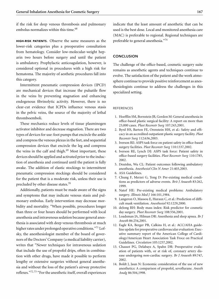
P1: PBU
cufx091-13 CUFX091/Friedberg 0 521 87090 9 Feb. 2, 2007 19:36
General Inhalation Anesthesia for Cosmetic Surgery 167
if the risk for deep venous thrombosis and pulmonary
embolus normalizes within this time.68
HIGH-RISK PATIENTS. Observe the same measures as the
lower-risk categories plus a preoperative consultation
from hematology. Consider low-molecular-weight hep-
arin two hours before surgery and until the patient
is ambulatory. Prophylactic anticoagulation, however, is
considered optional in procedures with a high risk for
hematoma. The majority of aesthetic procedures fall into
this category.
Intermittent pneumatic compression devices (IPCD)
are mechanical devices that increase the pulsatile flow
in the veins by preventing stagnation and enhancing
endogenous fibrinolytic activity. However, there is no
clear-cut evidence that ICPDs influence venous stasis
in the pelvic veins, the source of the majority of lethal
thromboemboli.
These mechanics reduce levels of tissue plasminogen
activator inhibitor and decrease stagnation. There are two
types of devices for use: foot pumps that encircle the ankle
and compress the venous plexus in the feet, and sequential
compression devices that encircle the leg and compress
the veins in the calf and thigh.69 Most important, these
devices should be applied and activated prior to the induc-
tion of anesthesia and continued until the patient is fully
awake. The addition of elastic stockings to intermittent
pneumatic compression stockings should be considered
for the patient that is a moderate risk, unless their use is
precluded by other disease states.70
Additionally, patients must be made aware of the signs
and symptoms that may indicate venous stasis and pul-
monary embolus. Early intervention may decrease mor-
bidity and mortality. “When possible, procedures longer
than three or four hours should be performed with local
anesthesia and intravenous sedation because general anes-
thesia is associated with deep venous thrombosis at much
higher rates under prolonged operative conditions.”71 Lof-
sky, the anesthesiologist member of the board of gover-
nors of the Doctors’ Company (a medical liability carrier),
writes that “Newer techniques for intravenous sedation
that include the use of propofol drips, often in combina-
tion with other drugs, have made it possible to perform
lengthy or extensive surgeries without general anesthe-
sia and without the loss of the patient’s airway protective
reflexes.”72,73 “For the anesthetic itself, overall experiences
indicate that the least amount of anesthetic that can be
used is the best dose. Local and monitored anesthesia care
(MAC) is preferable to regional. Regional techniques are
preferable to general anesthesia.”74
CONCLUSION
The challenge of the office-based, cosmetic surgery suite
remains as anesthetic agents and techniques continue to
evolve. The satisfaction of the patient and the work atmo-
sphere continue to provide positive reinforcement as anes-
thesiologists continue to address the challenges in this
specialized setting.
REFERENCES
1. Hoefflin SM, Bornstein JB, Gordon M: General anesthesia inoffice-based plastic surgical facility: A report on more than23,000 cases. Plast Reconstr Surg 107:243,2001.
2. Byrd HS, Barton FE, Orenstein HH, et al.: Safety and effi-cacy in an accredited outpatient plastic surgery facility. PlastReconstr Surg 112:636,2003.
3. Iverson RE: ASPS task force on patient safety in office-basedsurgery facilities. Plast Reconstr Surg 110:1337,2002.
4. Iverson RE, Lynch DJ: ASPS task force: Patient safety inoffice-based surgery facilities. Plast Reconstr Surg 110:1785,2002.
5. Deutshn, Wu CL: Patient outcomes following ambulatoryanesthesia. Anesthesiol Clin N Amer 21:403,2003.
6. ASA Guidelines.7. Chung F, Merzei G, Tong D: Pre-existing medical condi-
tions as predictors of adverse events. Brit J Anaesth 83:262,1999.
8. Natof HE: Pre-existing medical problems: Ambulatorysurgery. Illinois Med J 166:101,1984.
9. Langeron O, Massou E, Huraux C, et al.: Prediction of diffi-cult mask ventilation. Anesthesiol 92:1229,2000.
10. deJong RH: Body mass index: Risk predictor for cosmeticday surgery. Plast Reconstr Surg 108:556,2001.
11. Loadsman JA, Hilman DR: Anesthesia and sleep apnea. Br JAnesth 86:254,2001.
12. Eagle KA, Berger PB, Calkins H, et al.: ACC/AHA guide-line update for preoperative cardiovascular evaluation: Exec-utive summary report of the American College of Cardi-ology/American Heart Association Task Froce on PracticalGuidelines. Circulation 105:1257,2002.
13. Chassot PG, Delabays A, Spahn DR: Preoperative evalu-ation of patients with, or at risk of, coronary artery dis-ease undergoing non-cardiac surgery. Br J Anaesth 89:747,2002.
14. Boldt J, Jaun N: Economic consideration of the use of newanesthetics: A comparison of propofol, sevoflurane. AnesthAnalg 86:504,1998.
P1: PBU
cufx091-13 CUFX091/Friedberg 0 521 87090 9 Feb. 2, 2007 19:36
168 Meena Desai
15. Montes FR, Trillos JE, Rincon IE, et al.: Comparison of totalintravenous anesthesia and sevoflurane-fentanyl anesthesiafor outpatient otorhinolaryngeal surgery. J Clin Anesth 14:324,2002.
16. Larsen B, Seitz A, Larsen R: Recovery of cognitive func-tion after remifentanil-propofol anesthesia. Anesth Analg90:168,2000.
17. Lebenborm-Mansour MH, Pandit SK, Kothary SP, et al.:Desflurane vs. propofol anesthesia: A comparative analysis.Anesth Analg 76:936,1993.
18. Joshi GP: Inhalational techniques in ambulatory anesthesia.Anesthesiol Clin N Am 21:263,2003.
19. Gan TJ, Glass PS, Windsor A, et al.: Bispectral index mon-itoring allows faster emergence and improved recovery. BISutility study group. Anesthesiol 87:808,1997.
20. Song D, Joshi G, White PF: Titration of volatile anestheticsusing Bispectral index. Anesthesiol 87:842,1997.
21. Kalkman CH, Drummond JC: Monitors of depth of anes-thesia (editorial). Anesthesiol 96:784,2002.
22. Drover DR, Lemmens HJ, Pierce ET, et al.: Patient state index:Titration of delivery and recovery from propofol. Anesthesiol97:82,2002.
23. Song D: Titration of volatile anesthetics using bispectralindex facilitates recovery. Anesthesiol 87:842,1997.
24. Pavlin DJ, Hong JY, Friund PR, et al.: The effect of bispec-tral index monitoring on endtidal gas concentration. AnesthAnalg 93:613,2001.
25. Yli-Hankala A, Vakkuri A, Annila P, et al.: EEG bispectralindex monitoring in sevoflurane or propofol. Acta Anesthe-siol Scand 43:545,1999.
26. O’Connor MF, Daves SM, Tung A, et al.: BIS monitoringto prevent awareness during general anesthesia. Anesthesiol94:520,2001.
27. Mak S, Crowley J: The utility of the Bispectral Index vs.standard practice anesthetic care: A meta analysis of ran-domized trials comparing drug reduction and recoverytime. Poster presentation at the 2002 annual meeting ofSAMBA.
28. Rosow C, Manberg PJ: Bispectral Index monitoring: Mon-itoring during critical events. Anesthesiol Clin N Am19:947,2001.
29. Myles PS, Leslie K: Bispectral index monitoring to pre-vent awareness during anaesthesia: The B-Aware randomisedcontrolled Trial. The Lancet 363:1757,2004.
30. Ekman A, Lindholm ML, Lennmarken C, et al.: Reductionin the incidence of awareness using BIS monitoring. ActaAnaesthesiol Scand 48:20,2004.
31. Tang J, Chen L, White PF, et al.: Use of propofol for office-based anesthesia. J Clin Anesth 11:226,1999.
32. Song D, Whitten C, White P: Use of remifentanil duringanesthetic induction. Anesth Analg 88:734,1999.
33. Joshi GP, Warner DS, Twersky RS, et al.: A comparison ofremifentanil and fentanyl adverse effect profile. J Clin Anesth14:494,2002.
34. Apfelbaum JL, Walawander CA, Grascla TH, et al.: Elim-inating intensive postoperative care in same day surgerypatients using short acting anesthetics. Anesthesiol 97:66,2002.
35. Grazer FM, deJong RH: Fatal outcomes from liposuction:Census survey of cosmetic surgeons. Plast Reconstr Surg105:436,2000.
36. Kenkel JM, Lipschitz AH, Luby M, et al.: Hemodynamicphysiology and thermoregulation in liposuction. PlastReconstr Surg 114:503,2004.
37. deJong RH, Grazer FM: Perioperative management of cos-metic liposuction. Plast Reconstr Surg 107:1039,2001.
38. Trott SA, Beran SJ, Rohrich RJ, et al.: Safety considera-tion and fluid resuscitation in liposuction: An analysis of53 patients. Plast Reconstr Surg 102:2220,1998.
39. Steele SM, Nielsen KC, Klein SM (eds.): Ambulatory Anes-thesia Perioperative Analgesia. New York, McGraw Hill,2005.
40. Casac L, Jewell M: Non-narcotic acute pain relief after aes-thetic surgery. Aesth Surg J 22:493,2002.
41. Friedberg BL: Facial laser resurfacing with propofol-ketamine technique: Room air, spontaneous ventilation(RASV) anesthesia. Dermatol Surg 25:569,1999.
42. Blakely KR, Klein KW, White PF, et al.: A total intravenoustechnique for outpatient facial laser resurfacing. Anesth Analg87:827,1998.
43. Chung F, Mezei G: Factors contributing to a prolonged stayafter ambulatory surgery. Anesth Analg 89:1352,1999.
44. Joshi GP: Fast tracking in outpatient surgery. Curr OpinAnesthesiol 14:635,2001.
45. Marshall SI, Chung F: Discharge criteria and complicationafter ambulatory surgery. Anesth Analg 88:508,1999.
46. Joshi GP, Twersky R: Fast tracking in ambulatory surgery.Ambul Surg 8:185,2000.
47. Joshi GP: New concepts in recovery after ambulatory surgery.Ambul Surg 10:167,2003.
48. McHugh GA, Thoms GM: The management of pain follow-ing day case surgery. Anaesthesia 57:270,2002.
49. Pavlin DJ, Chen C, Penaloza DA: Pain as a complicatingrecovery and discharge after ambulatory surgery. AnesthAnalg 95:627,2002.
50. Marcus JR, Few JW, Chao JD, et al.: Prevention of emesis.Plast Reconstr Surg 109:2487,2002.
51. Gan TJ, Meyer T, Apfel CC, et al.: Consensus guidelines formanaging postoperative nausea and vomiting. Anesth Analg97:62,2003.
52. Gupta AM, Wu CL, Elkassabany N, et al.: Does the rout-ing prophylactic use of antiemetics affect the incidence ofpostdischarge nausea? Anesthesiol 99:488,2003.
53. Gan TJ, Sloan F, de L Dear G, et al.: How much are patientswilling to pay to avoid postoperative vomiting? Anesth Analg92:393,2001.
54. Apfel CC: A simplified risk score for predicting postoperativenausea. Anesthesiol 91:693,1999.
55. Cohen MM, Duncan PG, Debor DP, et al.: The postoperativeinterview: Assessing risk factors for nausea and vomiting.Analg 78:7,1994.
56. Cepeda MS, Gonzalez F, Granados V, et al.: Incidence of nau-sea and vomiting in outpatients undergoing general anesthe-sia. J Clin Anesth 8:324,1996.
57. Geldner G, Wulf H: Muscle relaxants suitable for day casesurgery. Eur J Anesthesiol 18(S23):43,2001.

P1: PBU
cufx091-13 CUFX091/Friedberg 0 521 87090 9 Feb. 2, 2007 19:36
General Inhalation Anesthesia for Cosmetic Surgery 169
58. Tramer MR, Fuchs-Buder T: Omitting antagonism of neu-romuscular block: Effect on postop nausea. Br J of Anaesth82:379,1999.
59. Habib AS, Gan TJ: Combination antiemetics: What is theevidence? Int Anesthesiol Clin 41:119,2003.
60. White PF, Hamza MA, Recart A, et al.: Optimal timingof acustimulation for emetic prohylaxis as an adjunct toondansetron in patients undergoing plastic surgery. AnesthAnalg 100:367,2005.
61. Tang J, Wang B, White P, et al.: The effect of timing ofondasetron administration on its efficacy, cost-effectiveness.Anesth Analg 86:274,1998.
62. Barbosa MV, Nahas FX, Ferreira LM: Ondansetron for theprevention of postoperative nausea and vomiting: Which isthe best dosage for aesthetic surgery patients? Aesth PlastSurg 28:33,2004.
63. Wang JJ, Ho ST, Tzen JI, et al.: The effect of timing ofdexamethasone administration on it efficacy. Anesth Analg91:136,2000.
64. Henzi I, Walder B, Tramer M: Dexamethasone for the pre-vention of postoperative nausea and vomiting. Anesth Analg90:186,2000.
65. Fuji Y, Tanaka H, Toyooka H: The effects of dexametha-sone on posantiemetics in female patients. Anesth Analg85:913,1997.
66. Jin FL, Norris A: Should adult patients drink fluids beforedischarge from ambulatory surgery? Can J Anesth 87:306,1998.
67. MHAUS: www.mhaus.org.68. Most D, Koslow J, Heller J: Thromboembolisn in plastic
surgery. Plast Reconst Surg 115:20,2005.69. Jewell M: Prevention of DVT in aesthetic surgery. Aesth Surg
J 2:161,2001.70. Reinisch JF, Bresnick SD, Walker JW, et al.: Deep venous
thrombosis and pulmonary embolus after facelift. PlastReconstr Surg 107:157,2001.
71. McDevitt NB: American Society of Plastic and Recon-structive Surgeons – DVT prophylaxis. Plast Reconstr Surg104:1923,1999.
72. Lofsky AS: Deep venous thrombosis and pulmonaryembolism in plastic surgery office procedures. The Doctors’Company Newsletter. Napa, CA, 2005 ww.thedoctors.com/risk/specialty/anesthesiology/J4254.asp
73. Friedberg BL: Propofol-ketamine technique, dissociativeanesthesia for office surgery: A five year review of 1,264 cases.Aesth Plast Surg 23:70,1999.
74. Laurito CE: Anesthesia provided at alternative sites, inBarasch PG, Cullen BF, Stoelting RK (eds.), Clinical Anes-thesia, 4th ed., Philadelphia, Lippincott, Williams & Wilkins,2001; p1343.

P1: PBU
cufx091-13 CUFX091/Friedberg 0 521 87090 9 Feb. 2, 2007 19:36
170

P1: PBU
cufx091-14 CUFX091/Friedberg 0 521 87090 9 Feb. 2, 2007 19:36
14 Preanesthetic Assessment of the CosmeticSurgery Patient
Norman Levin, M.D.
PART III OTHER CONSIDERATIONS FOR ANESTHESIA IN COSMETIC SURGERY
INTRODUCTION
PAST MEDICAL HISTORY
Hypertension
Cardiac Disease
Diabetes Mellitus
Herbal Supplements
PAST ANESTHETIC HISTORY
Malignant Hyperthermia (MH)
PREGNANCY
PHYSICAL EXAMINATION
PREOPERATIVE TESTS
Preoperative Tests Should Not Be Routinely Ordered
Electrocardiogram (EKG)
Laboratory Tests
Chest Radiographs
PREOPERATIVE PREPARATION
Anxiety
Prevention of Postoperative Nausea and Vomiting (PONV)
Prevention of Aspiration Pneumonitis
SUMMARY
INTRODUCTION
Over the past two decades, outpatient cosmetic surgical
procedures have grown at an exponential rate, progress-
ing from simple procedures in a physician’s office surgi-
cal suite to a broad spectrum of procedures in offices and
freestanding ambulatory surgery centers in addition to the
hospital setting. This number continues to grow as more
office surgery suites and ambulatory surgical centers con-
tinue to open. This rapid growth in ambulatory surgery
would not have been possible without the changing role
of the anesthesiologist and the development of better and
shorter acting anesthetic medications.
The preanesthetic evaluation of the surgical patient is
the first duty of an anesthesiologist to a patient. Klafta
and Rozien identified six interrelated goals of the pre-
anesthetic evaluation.1 The first is to assess health and
ensure physical readiness for anesthesia-requiring proce-
dures. The second is to devise a mutually agreeable anes-
thetic plan and to educate the patient about it. The third
is to reduce the psychological and physiological conse-
quences of anxiety. The fourth is to plan postoperative care
and pain therapy. The fifth is to coordinate patient care in
a way that decreases total cost and improves outcomes.
The sixth is to obtain informed consent for anesthesia
(Table 14-1).
171

P1: PBU
cufx091-14 CUFX091/Friedberg 0 521 87090 9 Feb. 2, 2007 19:36
172 Norman Levin
Table 14-1. Six preanesthetic goals
1. Assess health and physical readiness for surgery2. Devise an anesthetic plan and inform patient thereof3. Reduce anxiety4. Plan postoperative care and pain management5. Coordinate patient care6. Obtain informed consent for anesthesia
The preanesthetic evaluation provides one of the most
formidable challenges for anesthesiologists and represents
a focused assessment to address issues relevant to the safe
administration of anesthesia. The preanesthetic evalua-
tion gives the anesthesiologist the opportunity to meet,
comfort, and allay any anxiety that the patient might have
concerning the surgical procedure. The preanesthetic eval-
uation is also an opportunity to assess the health of the
patient, to educate the patient concerning anesthesia, and
to obtain pertinent information concerning the patient’s
medical history and an informed consent.
In the past, only “healthy” patients were acceptable can-
didates for ambulatory (or office-based) surgery. However,
in cosmetic surgery over the years, the number of patients
with more severe medical problems has increased through
the utilization of different anesthetic techniques. The pre-
anesthetic assessment of the cosmetic patient is of utmost
importance and should not be different than that of any
patient undergoing other types of surgical procedures in
any setting, whether it be in ambulatory surgical centers,
office surgical suites, or hospital surgical suites. However,
with the continued growth of ambulatory surgery centers
and office-based surgical procedures, this has placed the
anesthesiologist in a position of being the most involved
in the direct medical care of the patient.
Previously unacceptable, high-risk patients, many of
whom comprise cosmetic patients, are now being done in
outpatient surgical centers. As a result, a new role has been
created for the anesthesiologist. That is why the preanes-
thetic assessment is very important and most beneficial in
identifying medical problems, previous anesthetic prob-
lems, family history of anesthetic problems, medications
and/or herbal supplements patients might be taking, and
the hour at which food or liquids were taken.
During the preanesthesia evaluation, information
should be reviewed from multiple sources, including the
patient’s medical records, other physician evaluations and
physical examinations, and medical and laboratory tests,
in addition to the patient’s interview.
Because of the importance of the preanesthetic assess-
ment, this chapter reviews many of the aspects involved in
the anesthetic management of the patient undergoing cos-
metic surgery. Many cosmetic surgeons utilize the services
of other medical specialties, such as internal medicine, for
the medical workup and the appropriate laboratory tests.
In addition to the internist giving clearance for surgery, it
must be emphasized that the anesthesiologist is ultimately
responsible for the pre- and postoperative management of
that patient.
PAST MEDICAL HISTORY
Obtain information concerning the patient’s past medical
history. Although the ASA patient-physical-status classi-
fication is widely accepted, it is only a gross predictor of
the overall outcome and not a predictor of anesthetic risk
(Table 14-2). In the past, the majority of patients undergo-
ing cosmetic surgery were healthy individuals (ASA phys-
ical status 1 or 2). However, over the years, the number
of patients with various severe medical problems (ASA
physical status 3) has increased, and these patients have
undergone outpatient cosmetic surgery with safety. This
has been possible because of newer and better medications
for medical conditions, in addition to faster-acting anes-
thetic agents and the improvement of anesthesia monitor-
ing equipment. Natof concluded that ASA physical status
3 patients, whose systemic diseases were well controlled
preoperatively, were at no higher risk for postoperative
complications than ASA 1 or 2 patients.2 Many cosmetic
patients come to surgery with numerous medical condi-
tions, but only the most common ones are discussed in
this chapter.
Table 14-2. ASA patient physical status
ASA 1 A normal healthy patientASA 2 A patient with a mild systemic diseaseASA 3 A patient with a severe systemic disease that
limits activityASA 4 A patient with severe systemic disease that is
a constant threat to lifeASA 5 A moribund patient who is not expected to
survive without the operationASA 6 A declared brain-dead patient whose organs
are being removed for donor purposes

P1: PBU
cufx091-14 CUFX091/Friedberg 0 521 87090 9 Feb. 2, 2007 19:36
Preanesthetic Assessment of the Cosmetic Surgery Patient 173
HypertensionA large number of patients undergoing cosmetic surgery
have a history of hypertension and are on blood pressure
medications. These patients should continue their medi-
cations until and including the day of surgery. Diuretics
should not be taken before surgery to limit the possibility
of intraoperative enuresis.
At what level of blood pressure in hypertensive patients
would it be acceptable to proceed with surgery? According
to Dix, there is little evidence to support canceling surgery
in patients who present for surgery with systolic blood
pressures between 140 and 179 mm Hg and diastolic blood
pressures between 90 and 109 mm Hg.3
Patients with systolic blood pressures between 180 and 210
mm Hg are three times more likely to suffer postoperative
myocardial ischemia than those with a preoperative blood
pressure of 120 mm Hg.4
There are some patients who come to the operating
room without a history of hypertension, yet the anesthe-
siologist finds hypertension to be present. Anxiety con-
cerning the procedure can cause the blood pressure to
be elevated. Some of these patients could have a cardiac
abnormality. Approximately 10% of the general popula-
tion has an increased incidence of altered left-ventricular
structure and function when mildly stressed.5
An increase in blood pressure can also occur when
epinephrine is injected by the surgeon for surgical hemostasis.
An elevated blood pressure can occur in patients under-
going general anesthesia, especially during the induction
of anesthesia and intubation with an endotracheal tube.
Compared with direct laryngoscopy, a lower incidence of
hypertension in hypertensive patients can be achieved dur-
ing laryngeal intubation when utilizing a lightwand device
or a laryngeal mask airway (LMA) Fastrack™.6 There is an
increasing recognition that systolic blood pressure is as
important or more important than diastolic blood pres-
sure in elderly patients. Older patients with systolic hyper-
tension are at increased risk for stroke and cardiac events.7
Control the patient’s blood pressure during the preanes-
thesia period or immediately prior to the induction of
anesthesia or before the injection of epinephrine by the
surgeon.
Beta-blockers are used as the first line of antihy-
pertensive drugs for the treatment of hypertension.
Beta-blockade in the perioperative period decreases peri-
operative cardiac morbidity and mortality. There are many
cases in which beta-blocker use can be justified, but cer-
tainly on any patient with hypertension in the periop-
erative period. If there are no strong contraindications,
atenolol, metoprolol, or labetolol could be considered in
such a patient.8,9 Patients at risk of cardiovascular morbid-
ity are increasingly receiving perioperative beta-blocker
therapy.10 For those patients in whom beta-blockers might
be contraindicated (i.e., asthmatics), hydralazine or a
nitrate should be considered. Patients will tolerate pres-
sure elevator but not heart rate elevation. It is mandatory
to avoid tachycardia.
Cardiac Disease
Many older patients coming for cosmetic surgery have an
abnormal cardiac history and are under good control with
pharmacological management. There are many commonly
seen dysrhythmias such as sinus tachycardia, sinus brady-
cardia, and ventricular and atrial premature beats.
Bradycardia is frequently seen in patients who might be
athletically active or on medications accounting for their
slow heart rate. In other patients, tachycardia might be a
new problem needing further study and treatment prior
to surgery.
Alternatively, tachycardia might be due only to anxi-
ety with the release of endogenous catecholamines. Seda-
tives or cardiac medications might be necessary to control
the dysrhythmia prior to commencing with the surgical
procedure. As previously mentioned, most cosmetic sur-
geons use epinephrine for hemostasis, and this can
cause the dysrhythmia to become more exaggerated and
severe.
Patients with a history of myocardial infarction (MI)
should not have a cosmetic surgery for at least six months
following the occurrence. Multiple studies have demon-
strated an increased incidence of reinfarction if the MI
was within six months of surgery.11−13 Patients with unsta-
ble acute coronary syndromes, such as unstable angina or
decompensated congestive heart failure of ischemic origin,
are at high risk of developing further decompensation,
myocardial necrosis, and death during the perioperative
period. Patients with unstable acute coronary syndromes
should not be considered for cosmetic (or any) surgery,
unless absolutely necessary.14
Based on observations of the surgery on thousands of
patients, the author notes that neither increasing age nor
the presence of stable preexisting disease has any effect
on the incidence of postoperative complications in the
surgical outpatient setting.

P1: PBU
cufx091-14 CUFX091/Friedberg 0 521 87090 9 Feb. 2, 2007 19:36
174 Norman Levin
Diabetes Mellitus
There are many patients coming for cosmetic surgery with
a history of diabetes mellitus. Diabetes mellitus (DM) is
the most commonly encountered endocrinopathy and is
a progressive disease of glucose dysregulation. This carbo-
hydrate intolerance frequently results in significant acute
and long-term systemic sequelae. The goal of the anesthe-
siologist is to maintain the patient in a physiological state
to mimic normal metabolism.
Avoid hypoglycemia. Administer exogenous glucose, if
necessary. Prevent excessive hyperglycemia, ketoacido-
sis, and electrolyte disturbances. Administer exogenous
insulin when needed. The well-controlled, diet-treated
patient with NIDDM (non–insulin-dependent diabetes
mellitus) does not require any type of special treatment.
Patients on oral hypoglycemic drugs should continue
their medication until the evening before surgery. Patients
with well-controlled insulin-dependent diabetes mellitus
(IDDM) may not need any adjustment in their usual
subcutaneous insulin dosage. In all diabetic patients, an
Accu-chek r© or some other method of blood sugar deter-
mination should be performed during the immediate
preoperative evaluation, and insulin medication, where
appropriate, should be administered.
Most IDDM patients check their blood sugars on a reg-
ular basis. Many internists like to control their patient’s
insulin dosage for surgery and suggest an amount of
insulin to be taken on the day of surgery. If the patient’s
physician does not recommend an insulin regimen, the
anesthesiologist should suggest one. A commonly used
approach is to reduce the intermediate preparation of
insulin by one fourth (25%) to one half (50%) the usual
daily dosage the morning of surgery. If regular insulin
is part of the morning schedule, the intermediate-acting
insulin dose may be increased by 0.5 unit for each unit of
regular insulin.
When a patient receives insulin and is not eating, an intra-
venous of glucose solution should be started in the preoper-
ative period to minimize the chance of hypoglycemia during
or after surgery.
Herbal Supplements
Obtain information during the preanesthesia evaluation
about the use of herbal medications that are often found
in herbal dietary supplements, diet pills, muscle builders,
and so-called power drinks (see Appendix A). The use
of over-the-counter herbal medications during the peri-
operative period is an area of both enormous consumer
enthusiasm and physician concern.15 Several studies have
suggested that patients undergoing surgery appear to use
herbal medications significantly more frequently than the
general population.16,17 Many cosmetic surgical patients
have not only a concern for their appearance but also
a great concern for their health. As a result, they often
take herbal medications to improve their physical well-
being. Kaye and colleagues reported that nearly a third of
the patients in an ambulatory surgical setting admitted to
using herbal medications, and over 70% of those patients
failed to disclose their herbal medicine use during their
routine preoperative assessment.17
The danger to patients is that morbidity and mortality
with herbal medications may be more likely in the peri-
operative period because of the polypharmacy and phys-
iologic alterations that occur during that time.18 Adverse
reactions that may be caused by supplements include pro-
longed bleeding, interference with anesthesia, cardiovas-
cular disturbances, and interactions with pharmaceuticals
(Table 14-3). Also, it may be extremely difficult to dif-
ferentiate cause and effect related to surgery versus the
use of herbal medications when dealing with postopera-
tive complications such as myocardial infarction, stroke,
coagulation disorders, prolonged effects of anesthetics,
and interference with medications necessary for patient
care.
The five most popular herbal products in the United
States are (1) Gingko Biloba, (2) St. John’s Wort, (3)
Ginseng, (4) Garlic, and (5) Echinacea. Gingko Biloba can
inhibit platelet function, causing intraoperative bleeding
(see Chapter 12 for the particular risk with neuraxial
blockade). St. John’s Wort has multiple drug interactions
and is contraindicated with MAOIs and SSRIs. Ginseng
can interact with cardiac and hypoglycemic agents.
Garlic can inhibit platelet function, causing increased
bleeding. Echinacea can cause immunosuppression and
a potential for hepatotoxicity.19,20 Ephedra (ma huang)
is contained in many supplements. Ephedra is danger-
ous because it indirectly causes release of endogenous
catecholamines. Increased catecholamines contribute to
perioperative instability with hypertension, tachycardia,
dysrhythmia, and potentially myocardial infarction.
Anesthesiologists must inquire specifically as to the use
of herbal medications from their patients.

P1: PBU
cufx091-14 CUFX091/Friedberg 0 521 87090 9 Feb. 2, 2007 19:36
Preanesthetic Assessment of the Cosmetic Surgery Patient 175
Table 14-3. Supplements contraindicated in the perioperative period
Supplement Common Use(s) Adverse Effects
Echinacea Simulates immune system, used incommon cold and bronchitis
Can cause hepatotoxicity, maydecrease effectiveness ofcorticosteroids
Ephedra(ma huang)
CNS stimulant, diet aid, bronchodilator,nasal decongestant
Death, tachycardia, hypertension,myocardial infarction
Garlic Blood pressure and lipid lowering,antithrombotic, antiviral
Affects platelet aggregation, avoidwith anticoagulants
Ginger Antispasmodic, antiemetic Can prolong bleeding time,contraindicated with anticoagulants
Ginkgo Biloba Enhances blood flow, alleviates vertigoand tinnitus
Can increase bleeding time,contraindicated in patients onanticoagulants
Ginseng Improves physical and cognitiveenhancer, antioxidant
May interact with cardiac andhypoglycemic medication,contraindicated with anticoagulants
Goldenseal Anti-inflammatory, diuretic May worsen edema and hypertensionKava kava Anxiolytic, sedative, analgesic May potentiate barbiturates,
antidepressants, and generalanesthesia
Licorice Gastric and duodenal ulcers andbronchitis
May cause hypertension,hypokalemia, and edema
Melatonin Insomnia, jet lag May potentiate barbiturates andgeneral anesthetics
St. John’s Wort Antidepressant Multiple drug interactions,contraindicated with MAOIs andSSRIs
When asked about medications for medical reasons, many
patients feel that herbals are completely safe and do not con-
sider them to be drugs.
The ASA recommends that supplements producing
adverse effects be avoided for at least two weeks prior to
surgery and one week after surgery.
PAST ANESTHETIC HISTORY
A review of the patient’s past anesthetic and surgical his-
tory is important. The review can make the anesthesiol-
ogist aware of prior anesthetic problems and/or compli-
cations that the patient or a family member might have
experienced. This will enable the physician to develop a
better anesthetic plan for the care, comfort, and safety of
the patient.
Malignant Hyperthermia (MH)
Although extremely rare, malignant hyperthermia is
a potentially fatal complication of anesthesia. During
the preoperative evaluation, the anesthesiologist should
obtain from the patient or a family member information
concerning previous anesthetic problems. This is espe-
cially important for those patients who have experienced
or are susceptible to MH. In those individuals, prepara-
tions for safe administration of anesthesia are necessary
to avoid a catastrophic outcome. A treatment plan for
MH should be available in every anesthetizing location.
MH-triggering agents (i.e., halothane, enflurane, isoflu-
rane, desflurane, sevoflurane, and succinylcholine) should
not be used on patients susceptible to MH or their undi-
agnosed relatives.
All facilities, including ambulatory surgery centers and
offices, where MH-triggering anesthetics are administered
should stock dantrolene sodium for injection.
With the avoidance of the MH-triggering agents, pre-
anesthesia treatment with dantrolene is not recommended
for most MH-susceptible patients. Neither propofol nor
ketanine are triggering agents.

P1: PBU
cufx091-14 CUFX091/Friedberg 0 521 87090 9 Feb. 2, 2007 19:36
176 Norman Levin
PREGNANCY
Occasionally patients who are not aware of being preg-
nant are scheduled for cosmetic surgery. Mandatory
pregnancy testing on all females during their reproductive
years (between ages twelve and fifty years) is a controversial
issue. Spontaneous abortion and teratogenic effects may
occur during the first trimester of pregnancy.21
Teratogenic effects of anesthetics are probably minimal to
nonexistent and have never been conclusively demonstrated
in humans.
The drugs that were of most concern included nitrous
oxide and the benzodiazepines.22,23 In animal studies,
nitrous oxide, if not combined with a halogenated (sympa-
tholytic) agent, may cause vasoconstriction of the uterine
vessels with a decrease in uterine blood flow. No adverse
effect of nitrous oxide has been demonstrated in human
pregnancy. The maintenance of uterine perfusion and
maternal oxygenation to preserve fetal oxygenation are
the keys to any anesthetic during pregnancy, with the
avoidance of maternal hypoxia and hypotension being
essential.
If, during the presurgical workup, a positive pregnancy
test occurs, delay the surgery until after the delivery. Delay
may avoid any suspicion of anesthetic involvement causing
either the spontaneous abortion or fetal abnormalities that
might occur.
PHYSICAL EXAMINATION
One of the major responsibilities of the anesthesiologist is
to physically evaluate the cosmetic surgical patient imme-
diately before surgery concerning the risk of anesthesia and
of the procedure to be performed. At a minimum, a focused
preanesthetic physical examination should include an
assessment of the airway, lungs, and heart with documen-
tation of the vital signs.
The importance of the preanesthesia airway examina-
tion of the cosmetic surgical patient cannot be under-
stated. It is not important whether the proposed anesthetic
administered to the patient is local with intravenous seda-
tion, total intravenous anesthesia (TIVA), or general anes-
thesia. The unexpected need for airway support, whether
by endotracheal intubation, LMA, or other means, should
be evaluated in advance for the possibility of being a diffi-
cult airway. If an emergency arises and the need for endo-
tracheal intubation should become necessary, it is impera-
tive to determine which patient might present as a difficult
intubation.
The failure of the anesthesiologist to monitor and main-
tain a patent airway, when involved with the patient
undergoing cosmetic surgery, is one of the most common
causes of anesthesia-related morbidity and mortality (see
Chapter 18).
Keeping the airway patent and protected is an important
function of the anesthesiologist. Mallampati suggested
that the size of the base of the tongue is an important fac-
tor in determining the degree of difficulty of direct laryn-
goscopy.24 A relatively simple grading system that involves
the preoperative ability to visualize the faucial pillars, soft
palate, and base of uvula was designed as a means of pre-
dicting the degree of difficulty in laryngeal exposure. Khan
modified Mallampati’s classification utilizing the upper-
lip bite test as an acceptable option for predicting difficult
intubation as a simple, single test.25
PREOPERATIVE TESTS
Preoperative Tests Should Not Be RoutinelyOrdered
The ASA Task Force on Preanesthesia Evaluation con-
cluded that routine preoperative tests do not make an
important contribution to the process of preoperative
assessment and management of the patient by the anes-
thesiologist.27 It is the patient’s underlying condition and
the likelihood that the results will affect the anesthetic
plan that should guide the choice of preprocedure labo-
ratory tests, chest x-ray, and EKG. Preoperative tests may
be ordered on a selective basis for optimizing periopera-
tive management. Most cosmetic surgical patients come
to surgery having had a workup by an internist or other
physician giving medical clearance for the surgery. Often
a request is made to the anesthesiologist for specific tests
or studies. The ASA Task Force on Preanesthesia Evalua-
tion concluded that there should not be rules for ordering
preoperative tests but only for selected clinical situations.26
Depending on the patient’s clinical situation, the following
studies might act as a guide in preoperative testing.
Electrocardiogram (EKG)
Patients with a known history of cardiovascular disease
should have an EKG. The ASA Task Force feels that age alone
is not an indication for an EKG. However, many others feel

P1: PBU
cufx091-14 CUFX091/Friedberg 0 521 87090 9 Feb. 2, 2007 19:36
Preanesthetic Assessment of the Cosmetic Surgery Patient 177
that having a baseline EKG for patients over forty years of
age might be beneficial.
Laboratory Tests
For active healthy patients, laboratory tests might not be
necessary for males below fifty years of age. For females in
this age range, only hemoglobin (or hematocrit) should be
necessary. Patients with chronic diseases (e.g., hyperten-
sion, diabetes) should have the appropriate additional lab-
oratory studies (e.g., electrolytes, glucose) as indicated for
a medical condition. Also, patients with an unexplained
hemoglobin less than 10 gm · dl−1 should undergo fur-
ther evaluation prior to elective cosmetic surgery. Testing
should rule out other diseases (e.g., liver disease, anemia,
bleeding, and other hematological disorders) that could
influence perioperative mortality and morbidity.
Chest Radiographs
Unless the patient has clinically acute pulmonary symp-
toms, routine chest x-rays are not necessary.
PREOPERATIVE PREPARATION
Anxiety
The preoperative assessment and communication with
the patient is essential in obtaining information. Many
presurgical patients are very anxious in spite of a tele-
phone conversation with the anesthesiologist the night
before surgery.27 However, Levin has found that a call
to the patient the night before surgery has the effect
of allaying anxiety and decreasing the need for preop-
erative sedative medications. In the past, it has been
shown that the preoperative visit by the anesthesiologist is
more effective than preoperative barbiturate medication in
reducing perioperative anxiety.28 A significant amount of
anxiety is present at least six days prior to surgery in unpre-
pared patients.29 High levels of anxiety are often associ-
ated with other adverse outcomes, such as an increased
incidence of emesis. Frequently, cosmetic surgical patients
who need anxiolytics are not given preoperative medi-
cations because of concern for prolonged recovery and
discharge from the facility. However, midazolam, with a
relatively short half-life and the lack of significant side
effects produces excellent amnesic and anxiolytic proper-
ties without any delay in discharge from the facility (see
Chapter 7). Oxorn found a threefold incidence of patients
requesting postoperative analgesia in a group that received
preoperative midazolam compared to a group that did
not.29a
Prevention of Postoperative Nausea andVomiting (PONV)
As part of the preoperative evaluation, it is important to
identify patients in whom PONV occurred following a
previous surgical procedure or are at a high risk for devel-
oping such a complication. PONV is often a limiting factor
in the early discharge of ambulatory surgery patients, in
addition to being a leading cause of unanticipated hospital
admission.30,31
Typically, the administration of opioids, as part of a
preanesthetic regimen or during the course of an anes-
thetic, is known to increase the incidence of PONV. Gen-
eral anesthesia carries a higher risk of PONV than regional
anesthesia, major conduction anesthesia (subarachnoid
or epidural block), intravenous anesthesia, or monitored
anesthesia care. Perhaps the most controversial aspect of
general anesthesia is the independent risk associated with
the administration of nitrous oxide.
Many patients prefer the avoidance of PONV than the
avoidance of postoperative pain.32
Not all patients should receive PONV prophylaxis.
However, patients at small risk for PONV are unlikely to
benefit from prophylaxis and would be put at unneces-
sary risk from the potential side effects of antiemetics.
Thus, prophylaxis should be reserved for those patients at
moderate to high risk for PONV. Individuals with four
primary risk factors for PONV were reported by Apfel et
al.33 as patients receiving balanced inhalation anesthesia:
female sex, nonsmoking status, history of PONV, and opi-
oid use. The incidence of PONV with the presence of
none, one, two, three, or all four of these risk factors was
approximately 10%, 20%, 40%, 60%, and 80%, respec-
tively. Reduce the risk of PONV whenever clinically practi-
cal. Patients receiving general anesthesia had an elevenfold
increase in risk for PONV compared with those receiving
regional anesthesia.34
Propofol, administered for the induction and main-
tenance of anesthesia, effectively reduced early PONV
incidence.35 Oxygen supplementation restricted to the
intraoperative period also halved the risk of PONV.36
Hydration can also reduce the incidence of PONV.37
Avoiding nitrous oxide and volatile inhaled anesthetics

P1: PBU
cufx091-14 CUFX091/Friedberg 0 521 87090 9 Feb. 2, 2007 19:36
178 Norman Levin
and minimizing intraoperative and postoperative opioid
use reduced the incidence of PONV.33,38−43 Scuderi et al.44
tested the efficacy of a multimodal approach to reducing
PONV. Their multimodal approach consisted of preop-
erative anxiolysis, aggressive hydration, oxygen, prophy-
lactic antiemetics (droperidol and dexamethasone at the
induction with ondansetron at the end of surgery), total
IV anesthesia with propofol and remifentanil, and ketoro-
lac. No nitrous oxide or neuromuscular blockade was used.
Patients who received multimodal therapy had a 98% com-
plete response rate, compared with a 76% response rate
among patients receiving antiemetic monotherapy and a
59% response rate among those receiving routine anes-
thetic plus saline placebo.
The introduction of serotonin antagonists (specifi-
cally the 5-HT3 subgroup) in the early 1990s offered
considerable promise for the management of PONV.
Ondansetron, dolasetron, and granisetron are the drugs
approved for PONV prophylaxis. Prophylactic adminis-
tration of this class of medication has been shown to
decrease the incidence of PONV in various patient pop-
ulations. Comparisons of ondansetron and dolasetron
for PONV prophylaxis suggest that there are no clini-
cally or statistically significant differences between these
medications.45
Recently, combinations of antiemetics administered
prophylactically appear to be more effective than either
antiemetic alone. For instance, the combination of
ondansetron and droperidol is more effective than either of
the two medications alone.46 The same is true for the com-
bination of ondansetron and dexamethasone.47 Dimen-
hyrinate (Dramamine r©) 50 mg IM or IV may also be a
useful antiemetic, especially for patients prone to motion
sickness. The timing of administration of antiemetics is
very important. Some medications work best when given
preoperatively, and others work best when given intra- or
postoperatively. Dexamethasone administered at prophy-
lactic doses of 2.5–5 mg has been found to effectively pre-
vent nausea and vomiting48,49 and is most effective when
administered before the induction of anesthesia as it peaks
one to two hours after administration.50
Serotonin Receptor Antagonists are most effective when
given at the end of surgery.51,52 For ondansetron, the opti-
mal dose for prophylaxis seems to be 4 mg administered
intravenously at the end of surgery, prior to emergence
from anesthesia. The optimal dose of dolasetron appears
to be 12.5 mg and the timing of administration for prophy-
laxis appears to be less important than for ondansetron.
For droperidol, the optimal dose is 0.625–1.25 mg and
mostly effective when given at the end of surgery. Droperi-
dol in doses of either 0.625 mg or 1.25 mg compares favor-
ably with ondansetron 4 mg in outpatients. In fact, the
higher dose of droperidol (i.e., 1.25 mg) is more effective
than ondansetron in preventing nausea.53
In December 2001, the FDA issued a “black box” warning
for droperidol in response to patient deaths associated with
cardiac rhythm abnormalities.
Metoclopramide stimulates gastric emptying and is
administered at the end of surgery. Metoclopramide’s use-
fulness is uncertain except in patients where gastric stasis
is an issue. Prochlorperazine (Compazine r©) is as effective
as ondansetron but can cause extrapyramidal symptoms.
Transdermal scopolamine (Transderm-Scop r©) has an
onset of action of four hours. Thus, it should be applied
far enough in advance to ensure it has time to start
working, but it can cause visual disturbances and dry
mouth. The various side effects caused by some of these
medications – sedation, dysphoria, cardiac effects, and
extrapyramidal reactions – have been a concern for
clinicians, particularly when treating outpatients.
Prevention of Aspiration Pneumonitis
Many patients with gastrointestinal disorders, such as
decreased lower esophageal sphincter tone, hiatal her-
nia, and gastroesophageal reflux, are at risk for aspira-
tion of gastric contents into the lungs. When patients
are anesthetized, aspiration can occur with active vom-
iting or passive regurgitation, most commonly during
the induction of anesthesia, when the airway is unpro-
tected. Patients must be informed of the presurgery fast-
ing requirements either by the anesthesiologist or the sur-
geon, who must also explain the possible complications if
not followed. During the immediate preanesthesia assess-
ment, obtain information concerning the hour at which
food or drink was ingested. For patients not at risk for
aspiration, the “ASA Practice Guidelines for Preoperative
Fasting” recommends that patients may ingest clear liq-
uids until two hours prior to surgery and should avoid
solids or nonhuman milk for six or more hours prior to
surgery.54

P1: PBU
cufx091-14 CUFX091/Friedberg 0 521 87090 9 Feb. 2, 2007 19:36
Preanesthetic Assessment of the Cosmetic Surgery Patient 179
There is controversy concerning the administration of
prophylactic medication for prevention of pulmonary
injury from aspiration of gastric contents. Decreas-
ing the volume and pH of the gastric contents can
reduce the risk of pulmonary aspiration. The prophy-
lactic use of medications in all patients to prevent pul-
monary aspiration can be associated with unwanted side
effects. However, patients with predisposing factors for
this complication may be candidates for prophylactic
treatment.
Medications used to prevent aspiration pneumonitis
include H2-receptor antagonists, a dopamine antagonist,
and nonparticulate oral antacids. The H2-receptor antag-
onists cimetidine and ranitidine are effective in decreasing
gastric-acid secretion and raising the pH. However, nei-
ther cimetidine nor ranitidine influence the acid already
present in the stomach. Ranitidine should be given two
hours prior to surgery because it peaks in two hours.55
Metoclopramide, a dopamine antagonist, reduces gastric
volume by stimulating gastric emptying without any effect
on the pH and increases lower esophageal sphincter tone.56
Sodium citrate (0.3M, 30 ml) and Bicitra, r© both nonpar-
ticulate oral antacids, are effective in raising pH, but can
increase gastric volume. Used in conjunction with meto-
clopropamide and when prophylaxis is desired, the onset
of action is immediate.57
Particulate antacids should not be used, as they can
worsen the pulmonary damage if aspirated.
However, according to the “ASA Practice Guidelines
for Preoperative Fasting,” the use of these medications
to decrease the risks of pulmonary aspiration in those
patients who are not at an increased risk for this com-
plication is not recommended.54
SUMMARY
The preanesthetic evaluation of the cosmetic patient is one
of the most important functions for the anesthesiologist.
The number of patients with a variety of severe medi-
cal conditions undergoing cosmetic surgery in an outpa-
tient surgical setting continues to increase and, because
of this, the anesthesiologist is the physician most involved
in the direct medical care of the patient. As much infor-
mation as possible concerning the patient’s medical and
anesthetic history, with appropriate medical records and
laboratory tests, should be obtained and reviewed in the
preoperative period for a better and safer patient anesthetic
experience.
REFERENCES
1. Klafka JM, Rozien MF: Current understanding of patient’sattitudes toward and preparation for anesthesia: A review.Anesth Analg 83:1314,1996.
2. Natof HE: Pre-existing medical problems. Ambulatorysurgery. IL Med J 166:101,1984.
3. Dix P, Howell S: Survey of cancellation rate of hypertensivepatients undergoing anesthesia and elective surgery. Br JAnaesth 86:7889,2001.
4. Howell SJ, Hemming AE, Allman KG, et al.: Predictors ofpostoperative myocardial ischaemia. The role of intercur-rent arterial hypertension and other cardiovascular risk fac-tors. Anaesthesia 52:107,1997.
5. Muscholl MW, Hense HW, Brockel U, et al.: Changes inleft ventricular structure and function in patients with whitecoat hypertension: Cross sectional survey. Br Med J 317:565,1998.
6. Kihara S, Brimacombe J, Yaguchi Y, et al.: Hemodynamicresponses among three tracheal intubation devices in nor-motensive and hypertensive patients. Anesth Analg 96:890,2003.
7. Cannel WB: Risk stratification in hypertension: Newinsights from the Framingham study. Am J Hypertension13:3S,2000.
8. Auerbach AD, Goldman L: Beta-blockers and reductionof cardiac events in noncardiac surgery. Scientific review.JAMA 287:1435,2002.
9. Auerbach AD, Goldman L: Beta-blockers and reduction ofcardiac events in non-cardiac surgery. Clinical applications.JAMA 287:1445,2002.
10. Howell SJ, Sear JW, Foex P: Peri-operative beta-blockade:A useful treatment that should be greeted with cautiousenthusiasm. Br J Anaesth 86:161,2001.
11. Tarhan S, Moffitt EA, Taylor WF, et al.: Myocardial infarc-tion after general anesthesia. JAMA 220:1451,1972.
12. Rao TLK, Jacobs KH, El-Etr AA: Reinfarction followinganesthesia in patients with myocardial infarction. Anesthe-siol 59:499,1983.
13. Shah KB, Kleinman BS, Sami H, et al.: Reevaluation ofperi-operative myocardial infarction in patients with priormyocardial infarction undergoing non-cardiac operations.Anesth Analg 71:231,1990.
14. Shah KB, Kleinman BS, Rao T, et al.: Angina and other riskfactors in patients with cardiac diseases undergoing non-cardiac operations. Anesth Analg 70:240,1990.
15. Ang-Lee MK, Moss J, Yuan C: Herbal medicine and peri-operative care. JAMA 286:208,2001.
16. Tsen LC, Segal S, Pothier M, et al.: Complementary andalternative medicine use should be included in preoperativeevaluations. Anesthesiol 931:148,2000.
P1: PBU
cufx091-14 CUFX091/Friedberg 0 521 87090 9 Feb. 2, 2007 19:36
180 Norman Levin
17. Kaye AD, Clarke RC, Sabar R, et al.: Herbal medicines: Cur-rent trends in anesthesiology practice – A hospital survey. JClin Anesth 12:468,2000.
18. Bovil JG: Adverse drug interactions in anesthesia. J ClinAnesth 9 (Suppl 6):3S,1997.
19. Ernest E: The risk-benefit profile of commonly used herbaltherapies: Ginko, St. John’s Wort, Ginseng, Echinacea, SawPalmetto, and Kava. Ann Int Med 136:42,2002.
20. Petry JJ: Surgically significant nutritional supplements.Plast Reconstr Surg 97:233,1996.
21. Boiven JF: Risk of spontaneous abortion in women occupa-tionally exposed to anesthetic gases: A meta-analysis. OccupEnviron Med 54:541,1997.
22. Mazze RI: Halothane prevents nitrous oxide teratogenic-ity in Sprague-Dawley rats; folic acid does not. Teratology38:121,1988.
23. Shiono PH, Mills JL: Oral clefts and diazepam use duringpregnancy. N Engl J Med 311:919,1984.
24. Mallampati SR, Gatt SP, Gugino LD, et al.: A clinical signto predict difficult tracheal intubation: A prospective study.Can Anaesth Soc J 32:429,1985.
25. Khan ZH, Kashfi A, Ebrahimkhani E: A comparison of theupper lip bite test (a simple new technique) with modifiedMallampati classification in predicting difficulty in endotra-cheal intubation: A prospective blinded study. Anesth Analg96:595,2003.
27. Practice advisory for preanesthesia evaluation: A report bythe American Society of Anesthesiologists task force on pre-anesthesia evaluation. Anesthesiol 96:490,2002.
28. McLeanne GJ, Cooper R: The nature of peri-operative anx-iety. Anesthesia 45:153,1990.
29. Egbert LD, Battit GE, Turndorf H, et al.: The value of pre-operative visit by the anesthetist. JAMA 185:553,1963.
29a. Oxorn DC, Ferris LE, Harrington E, Orser BA: The effects ofmidazolam on propofol-induced anesthesia: Propofol doserequirements, mood profiles, and perioperative dreams.Anesth Analg 85:553,1997.
30. Johnson M: Anxiety in surgical patients. Psychol Med10:145,1980.
31. Gold BS, Kitz DS, Lecky JH, et al.: Unanticipated admis-sion to the hospital following ambulatory surgery. JAMA262:3008,1989.
32. Fortier J, Chung F, Su J: Unanticipated admission afterambulatory surgery: A prospective study. Can J Anaesth45:612,1998.
33. Macario A, Weinger M, Carney S, et al.: Which clinicalanesthesia outcomes are important to avoid? Anesth Analg89:652,1999.
34. Apfel CC, Laara E, Koivuranta M, et al.: A simplified riskscore for predicting postoperative nausea and vomiting.Anesthesiol 91:693,1999.
35. Sinclair DR, Chung F, Mezei G: Can post-operative nauseaand vomiting be predicted? Anesthesiol 91:109,1999.
36. Visser K, Hassink EA, Bonsel GJ, et al.: Randomized con-trolled trial of total intravenous anesthesia with propofolversus inhalation anesthesia with isoflurane-nitrous oxide:Post-operative nausea with vomiting and economic analy-sis. Anesthesiol 95:616,2001.
37. Goll V, Ozan A, Greif R, et al.: Ondansetron is no moreeffective than supplemental intraoperative oxygen for pre-vention of postoperative nausea and vomiting. Anesth Analg92:112,2001.
38. Yogendran S, Asokumar B, Cheng DC, et al.: A prospectiverandomized double-blinded study of the effect of intra-venous fluid therapy on adverse outcomes on outpatientsurgery. Anesth Analg 80:682–6,1995.
39. Apfel CC, Kranke P, Eberhart LH, et al.: Comparison ofpredictive models for postoperative nausea and vomiting.Br J Anaesth 88:234,2002.
40. Tram M, Moore A, McQuay H: Meta-analytic comparisonof prophylactic anti-emetic efficacy for postoperative nau-sea and vomiting: Propofol anaesthesia vs. omitting nitrousoxide vs. total i.v. anaesthesia with propofol. Br J Anaesth78:256,1997.
41. Apfel CC, Katz MH, Kranke P, et al.: Volatile anaestheticsmay be the main cause of early but not delayed postoperativevomiting: A randomized controlled trial of factorial design.Br J Anaesth 88:659,2002.
42. Sukhani R, Vazquez J, Pappas AL, et al.: Recovery afterpropofol with and without intra-operative fentanyl inpatients undergoing ambulatory gynecologic laparoscopy.Anesth Analg 83:975,1996.
43. Mniche S, Rsing J, Dahl JB, et al.: Non-steroidal anti-inflammatory drugs and the risk of operative site bleedingafter tonsillectomy: A quantitative systematic review. AnesthAnalg 96:68,2003.
44. Polati E, Verlato G, Finco G, et al.: Ondansetron versusmetoclopramide in the treatment of postoperative nauseaand vomiting. Anesth Analg 85:395,1997.
45. Scuderi PE, James RL, Harris L, et al.: Multi-modal anti-emetic management prevents early postoperative vomit-ing after outpatient laparoscopy. Anesth Analg 91:1408,2000.
46. Zarate E, White P, Klein KW, et al.: A comparison of the costsand efficacy of ondansetron versus dolasetron for antiemeticprophylaxis. Anesth Analg 90:1352,2000.
47. Riley TJ, McKenzie R, Tantisira BR, et al.: Droperidol-ondansetron combination versus droperidol alone for post-operative control of emesis after total abdominal hysterec-tomy. J Clin Anesth 10:6,1998.
48. Sanchez-Ledesma MJ, Lopez-Olaondo L, Pueyo FJ, et al.:A comparison of three anti-emetic combinations for theprevention of post-operative nausea and vomiting. AnesthAnalg 95:1590,2002.
49. Wang JJ, Ho ST, Lee SC, et al.: The use of dexamethasone forpreventing postoperative nausea and vomiting in femalesundergoing thyroidectomy: A dose-ranging study. AnesthAnalg 91:1404,2000.
50. Wang JJ, Ho ST, Tzeng JI, et al.: The effect of timing of dex-amethasone administration on its efficacy as a prophylacticanti-emetic for post-operative nausea and vomiting. AnesthAnalg 91:136–9,2000.
51. Domino KB, Anderson EA, Polissar NL, et al.: Compar-ative efficacy and safety of ondansetron, droperidol, andmetoclopramide for preventing post-operative nausea andvomiting: A meta-analysis. Anesth Analg 88:1370,1999.

P1: PBU
cufx091-14 CUFX091/Friedberg 0 521 87090 9 Feb. 2, 2007 19:36
Preanesthetic Assessment of the Cosmetic Surgery Patient 181
52. Sun R, Klein KW, White PF: The effect of timing ofondansetron administration in outpatients undergoing oto-laryngologic surgery. Anesth Analg 84:331,1997.
53. Graczyk SG, McKenzie R, Kallar S, et al.: Intravenousdolasetron for the prevention of post-operative nausea andvomiting after outpatient laparoscopic gynecologic surgery.Anesth Analg 84:325,1997.
54. Fortney JT, Gan TJ, Graczy KS, et al.: A comparison of theefficacy, safety, and patient satisfaction of ondansetron ver-sus droperidol as anti-emetics for elective outpatient surgi-cal procedures. S3A-409 and S3A-410 Study Groups. AnesthAnalg 86:731,1998.
55. ASA practice guidelines for preoperative fasting and the use
of pharmacologic agents to reduce the risk of pulmonaryaspiration: Application to healthy patients undergoing elec-tive procedures. Anesthesiol 90:896,1999.
56. Manchikanti L, Colliver JA, Roush JR, et al.: Evaluation ofranitidine as an oral antacid in outpatient anesthesia. SouthMed J 78:818,1985.
57. Hey VMF, Ostrick DG, Mazumder JK, et al.: Pethidine,metoclopramide and gastro-oesophageal sphincter. Anaes-thesia 36:173,1981.
58. Manchikanti L, Grow JB, Colliver JA, et al.: Bicitra r©
(sodium citrate) and metoclopramide in outpatient anes-thesia for prophylaxis against aspiration pneumonitis. Anes-thesiol 63:378,1985.

P1: PBU
cufx091-15 CUFX091/Friedberg 0 521 87090 9 Feb. 2, 2007 19:38
15 Psychological Aspects of Cosmetic Surgery
David B. Sarwer, Ph.D., Canice E. Crerand, Ph.D., and Lauren M. Gibbons, B.A.
INTRODUCTION
COSMETIC PROCEDURES OF THE FACE
Rhinoplasty
Anti-Aging Procedures
Minimally Invasive Anti-Aging Procedures
Other Facial Procedures
Facial skeletal procedures
Acne treatment
Vitiligo
Hair Loss and Hair Transplantation
COSMETIC PROCEDURES OF THE BODY
Cosmetic Breast Augmentation
Descriptive characteristics and motivational factors
Studies of pre- and postoperative psychological status
Body-image dissatisfaction
Psychosocial outcomes and postoperative complications
Breast implants and suicide
Lipoplasty and Abdominoplasty
Lipoplasty
Abdominoplasty
Other Body Enhancement Procedures
Body-contouring implants
Genital enhancement
Tattoos and body piercing
PSYCHIATRIC DISORDERS AMONG COSMETIC SURGERY PATIENTS
Body Dysmorphic Disorder
Clinical features
Nonpsychiatric and psychiatric treatments
Variants of BDD
Eating Disorders
SUMMARY
182

P1: PBU
cufx091-15 CUFX091/Friedberg 0 521 87090 9 Feb. 2, 2007 19:38
Psychological Aspects of Cosmetic Surgery 183
INTRODUCTION
According to the American Society of Plastic Surgeons
(ASPS), over fifteen million Americans underwent a plas-
tic surgical procedure in 2003.1 The majority of plastic
surgical procedures consisted of relatively new, minimally
invasive, nonsurgical procedures. These numbers, while
familiar to many plastic surgeons, are often staggering to
other medical professionals and lay persons who have little
idea of the number of Americans who turn to medicine
to enhance their physical appearance. Nevertheless, these
numbers likely underestimate the number of procedures
performed annually, as they do not account for nonplastic
surgeon physicians who offer these treatments. In partic-
ular, the preceding numbers do not reflect the surgical
activity of either the American Academy of Cosmetic Sur-
geons (AACS) or the American Society of Dermatologic
Surgeons (ASDS).
The growth in popularity of cosmetic surgery and
related treatments can be attributed to several factors.2−4
Changes in the medical and surgical communities, includ-
ing improvements in safety and direct-to-consumer mar-
keting, have likely contributed to the growth. The mass
media and entertainment industries have long champi-
oned cosmetic surgery, perhaps no more so than dur-
ing the current era of “reality-based” television programs
such as “Extreme Makeover” and “The Swan.” The vir-
tually inescapable bombardment of mass-media ideals of
beauty, coupled with the discontent that many people, par-
ticularly women, experience with regard to their physi-
cal appearance, have likely contributed as well.5,6 Finally,
society’s acceptance of the use of medicine to enhance
appearance, perhaps paired with a greater awareness of the
importance of physical appearance in daily life, has poten-
tially fueled cosmetic surgery’s increase in popularity. Plas-
tic surgeons and mental health professionals have long
been interested in the psychological aspects of cosmetic
surgery. Understanding the psychological characteristics
of patients who desire and undergo cosmetic proce-
dures is important for practical reasons. Cosmetic pro-
cedures are often considered analogous to psychological
interventions; many patients report increased satisfaction
with their appearance, as well as psychological improve-
ments, postoperatively.7 Nevertheless, these procedures
are likely not appropriate for all individuals, particularly
those with certain characteristics or psychiatric disorders.
Thus, an understanding of the psychological functioning
of cosmetic surgery patients is an important part of max-
imizing treatment outcomes. This chapter reviews studies
of the pre- and postoperative psychosocial functioning of
persons who seek and undergo cosmetic surgery. A review
of the psychological studies of individuals who have under-
gone the most common facial procedures begins the chap-
ter. The second major section discusses studies of persons
who have undergone cosmetic procedures of the body. The
chapter concludes with a discussion of the psychiatric dis-
orders that may be most relevant to this population—body
dysmorphic disorder and eating disorders.
COSMETIC PROCEDURES OF THE FACE
Rhinoplasty
Rhinoplasty is traditionally one of the most popular cos-
metic surgical procedures, with over 350,000 performed
in 2003.1 The psychological characteristics of rhinoplasty
patients have received as much research attention as those
of persons who have undergone any cosmetic procedure.
The first reports date back to the 1940s and 1950s. These
investigations, as well as studies conducted into the 1960s,
relied heavily on clinical interviews and suggested that
patients were highly psychopathological.8,9 Early inves-
tigators typically conceptualized the desire for rhinoplasty
from a psychodynamic perspective, the prevailing theo-
retical orientation in psychiatry at the time. The nose was
often thought to symbolize the penis, and the desire for
rhinoplasty was believed to represent the patient’s uncon-
scious displacement of sexual conflicts onto the nose.10
For the adolescent female patient, the desire for rhino-
plasty was interpreted as an attempt to remove elements
of her father’s personality from her own.11
During the 1970s and 1980s, a “second generation”
of research in cosmetic surgery began to include valid
and reliable measures to assess patients’ psychological
characteristics.2,3 Studies of this era found less pre-
operative psychopathology and several noted postop-
erative benefits.12−17 For example, investigations that
used the Minnesota Multiphasic Personality Inventory
(MMPI), likely the most widely used measure of per-
sonality, reported that the personality profiles of rhino-
plasty patients were essentially normal preoperatively, and
no changes were observed postoperatively.14,15 Unfortu-
nately, many of these studies suffered from methodological

P1: PBU
cufx091-15 CUFX091/Friedberg 0 521 87090 9 Feb. 2, 2007 19:38
184 David B. Sarwer, Canice E. Crerand, and Lauren M. Gibbons
problems, such as small sample sizes and the lack of appro-
priate control groups, which call into question the valid-
ity of the findings. Studies through the present have used
improved methodologies, including reliable and valid self-
report measures and clinical interviews with established
diagnostic criteria.18−25 Most studies also have included
pre- and postoperative assessments with appropriate con-
trol groups. These studies have suggested that most rhino-
plasty patients are psychologically healthy individuals. As
noted by at least one study,25 the desire for rhinoplasty
may be understood as an increased dissatisfaction with
the size and/or shape of the nose, rather than a symptom
of psychopathology. These findings are consistent with the
experiences of most present-day cosmetic surgeons.
Anti-Aging Procedures
Rhytidectomy and blepharoplasty are two of the most pop-
ular surgical procedures for those interested in restoring
a more youthful appearance. In 2003, 128,667 facelifts
and 246,633 blepharoplasty procedures were performed
in the United States.1 (As these procedures are often per-
formed concurrently, studies of these patient populations
are reviewed together.)
Early reports suggested that facelift patients were quite
psychopathological. Patients were often characterized as
dependent and depressed; approximately 70% of patients
received a preoperative psychiatric diagnosis.26 However,
the majority reported postoperative improvements in
well-being and did not experience postoperative “emo-
tional disturbances.”26 Studies that used standardized self-
report measures noted similar improvements in psycho-
logical symptoms postoperatively.24,27
Other studies have examined the body-image concerns
of these patients. In one of the first empirical studies
investigating the body-image concerns of cosmetic surgery
patients, nearly half of the patients studied sought facelift
or blepharoplasty procedures.28 They reported higher lev-
els of dissatisfaction with the feature for which they sought
surgery, but they did not report increased dissatisfaction
with their overall body image.28 Rhytidectomy and/or ble-
pharoplasty patients have reported greater investment in
their appearance as well as greater satisfaction with their
overall body image as compared to women who sought
rhinoplasty.25 Postoperatively, patients reported decreases
in body-image dissatisfaction for the feature that was
treated but no changes in overall body image.29
Minimally Invasive Anti-Aging Procedures
Minimally invasive anti-aging procedures have surpassed
the popularity of the more traditional anti-aging surgical
procedures. For example, nearly 2.9 million botulinum
toxin (Botox r©) injections were performed in 2003, mak-
ing it the most popular of all cosmetic treatments.1 The
toxin is typically injected into areas of the face (i.e., fore-
head creases, “crow’s feet”) in order to reduce the appear-
ance of wrinkling. It is also effective at treating excessive
sweating. Other popular minimally invasive procedures
include fat injections, collagen injections, chemical peels,
dermabrasion, and laser resurfacing. Like Botox r© injec-
tions, these procedures can improve the appearance of
wrinkled, scarred, or sun-damaged skin. In 2004, the
Food and Drug Administration (FDA) approved a product
specifically designed to improve lipo-atrophy in persons
with HIV disease. New generations of customized facial
implants also are being used to treat this condition.30 It
is quite possible that these products will also be used for
cosmetic purposes in persons without HIV disease in the
near future.
Despite their popularity, little is known about the psy-
chological characteristics or body-image concerns of the
patients who seek these procedures. A German study of
thirty patients who received Botox r© injections for facial
lines examined post-treatment social outcomes and atti-
tudes toward appearance.31 Over half of those studied
reported improvements in their appearance and nearly
50% reported greater confidence in their appearance.31
A recent study of 178 patients seeking laser skin resur-
facing reported that 18% received prior treatment for
depression.32 A third study evaluated the psychosocial
benefits associated with alpha-hydroxy acid, a topical treat-
ment that is used to reduce roughness and fine wrinkling.
Patients noted significant improvements in appearance
and relationship satisfaction following treatment.33
Other Facial Procedures
Facial skeletal procedures
Some patients request more “atypical” procedures that
involve bone contouring and/or grafting as well as the
insertion of cheek, chin, or other facial implants. One study
described fifteen patients who sought extensive symmet-
rical facial skeletal recontouring procedures in order to
address discontent with facial width.34 These “facial width

P1: PBU
cufx091-15 CUFX091/Friedberg 0 521 87090 9 Feb. 2, 2007 19:38
Psychological Aspects of Cosmetic Surgery 185
deformity” patients reported concerns with minor, largely
unnoticeable anatomic deviations.
Preoperative psychiatric interviews revealed that the
majority experienced significant impairment in psychoso-
cial functioning, though only three (of fifteen) received
a formal psychiatric diagnosis. The clinical descriptions
of these patients, however, suggest that some may have
been suffering from body dysmorphic disorder, dis-
cussed in detail herein. Postoperatively, patients reported
improvements in body image and psychosocial function-
ing, although psychometric measures were not used to
assess these changes.34
Although requests for some of these facial-widening
procedures are rare, more patients are requesting pro-
cedures such as cheek and chin implants in order to
change the structural appearance of their faces. In 2003,
over 28,000 chin or cheek implantation procedures were
performed.1 The popularity of these procedures under-
scores the need for more research regarding the psycho-
logical characteristics and body-image concerns of these
patients.
Acne treatment
Many patients present to plastic surgeons, dermatologists,
or other professionals complaining of acne or acne-related
scarring. This is not surprising, considering that acne
affects at least 80% of adolescents.35 The occurrence of
acne typically decreases with age; however, it may persist
through adulthood for a minority of persons.36
Historically, the psychosocial effects of acne have
been dismissed, largely as it was considered a non–
life-threatening, age-related cosmetic condition.37 More
health professionals now recognize the impact acne may
have on psychological and social well-being. Up to 50%
of adolescents experience psychological difficulties asso-
ciated with acne, including body-image concerns, poor
self-esteem, social isolation, and depression.38 Studies of
patients who seek acne treatment have found similar
results.37,39−41
For some, the distress may become so severe that it con-
tributes to suicidal ideation or suicide attempts.40,42 Facial
acne patients, compared to those with truncal acne, appear
to be particularly vulnerable to the psychological effects
of the disease, experiencing lower self-esteem and greater
body-image dissatisfaction.41 The distress appears to be
related to the self-perceived rather than objective severity
of the acne.43 Acne patients report impairments in quality
of life on par with those of other chronic medical condi-
tions, such as epilepsy and diabetes.44
Acne treatment appears to result in improvements
in psychosocial functioning.45,46 Kellett and Gawkrodger
reported significant reductions in anxiety and depression,
but not general emotional distress, following treatment
with isotretinoin.39 The authors concluded that some of
the psychological effects of acne may remain despite suc-
cessful treatment. This finding makes intuitive sense, given
that acne can result in permanent scarring. Extended dura-
tion of acne and acne excoriee (skin picking) are associated
with greater likelihood of scarring.47 Even in the absence
of residual cutaneous scars, emotional scars may remain.
Cash and Santos found that former adolescent acne suf-
ferers, especially women and those recalling more subjec-
tively severe acne, reported less current facial satisfaction
and more body-image dysphoria than peers who did not
have facial acne as teenagers.48 The psychosocial distress
associated with acne has implications for medical profes-
sionals who treat acne-related scars. These providers may
be able to assess the psychological effects of this disorder
and provide appropriate mental health treatment referrals
when warranted.49
Vitiligo
Vitiligo is a progressive condition characterized by loss of
skin pigmentation, resulting in irregular hypopigmented
patches.50 Generalized vitiligo, the most common form, is
characterized by bilateral, symmetric depigmentation of
the face (particularly periorificial area), neck, torso, wrists,
and legs.51 The prevalence of vitiligo is estimated to be
about 1–2% of the world population.52 Age of onset is typ-
ically childhood or young adulthood.51 Although there is
no cure, medical treatments include topical cosmetics, use
of psoralens and UVA light to stimulate repigmentation
(PUVA), corticosteroids, and surgical skin grafting.51,52
Similar to acne, the psychological distress associated
with vitiligo is often underestimated.50 The condition
appears to have a negative impact on the social and emo-
tional well-being of its sufferers.53 Patients often report dif-
ficulties with body image, self-esteem, and quality of life.50
Similar psychological sequelae have been reported among
patients with other chronic skin conditions, including
eczema and psoriasis.40,54,55

P1: PBU
cufx091-15 CUFX091/Friedberg 0 521 87090 9 Feb. 2, 2007 19:38
186 David B. Sarwer, Canice E. Crerand, and Lauren M. Gibbons
A more extensive review of the psychological characteristics
of patients who seek dermatological treatment is beyond the
scope of this chapter; interested readers are directed to these
reviews and books.56−58
Encouragingly, cognitive-behavioral psychotherapy
appears to be a successful treatment for the psychologi-
cal distress associated with these conditions.50
Hair Loss and Hair Transplantation
Head hair possesses considerable cultural, social, and per-
sonal significance.59,60 As a result, hair loss can be a psycho-
logically difficult experience for some. There are a number
of hair-loss conditions; the most prevalent is androgenetic
alopecia (AGA), or common genetically predisposed hair
loss. AGA is a progressive condition mediated by andro-
genic metabolism (especially dihydrotestosterone). The
receding frontal hairline and vertex balding is visibly evi-
dent in the majority of men. AGA also occurs in a signifi-
cant minority of women, although the pattern of alopecia
is one of diffuse thinning.
AGA can be a very distressing condition for both gen-
ders, albeit more troubling for women.61,62 Among men,
increased distress is associated with earlier onset of hair
loss. It is also experienced by men who are more psycho-
logically invested in their appearance and by younger men
not involved in an intimate relationship.63,64 The effects of
AGA on women may be more extensive, including lower
self-esteem.64 As with many other conditions, the subjec-
tive severity of AGA is more related to its psychosocial
impact than are objective or clinical indices of severity.61
Most outcome data for AGA treatment comes
from large, controlled clinical trials of minoxidil and
finasteride.61,65,66 The psychological outcome measures in
these studies have focused largely on patients’ perceptions
of, and satisfaction with, resultant hair growth and gen-
erally support the efficacy of the treatments. There is a
dearth of outcomes research using more psychologically
sophisticated measures. One uncontrolled study of 144
men treated with topical minoxidil confirmed moderate
hair growth and improvements on hair-specific quality-of-
life measures (e.g., hair-loss distress and perceived social
noticeability of the hair loss) but not on more global mea-
sures of anxiety, depression, or self-esteem.67
In recent years, there have been major advances in sur-
gical methods of hair replacement, including micrograft-
ing and flap techniques, especially for men with AGA.
Hair transplantation is a more certain and permanent
method of hair replacement in less time. The popular-
ity of hair transplantation has decreased in the past sev-
eral years, likely due to the availability of prescription and
over-the-counter medical treatments for AGA. Surpris-
ingly, no formal studies have investigated the psycholog-
ical characteristics of persons who seek hair transplan-
tation or the psychological changes that may occur after
treatment. Given the prevalence of AGA, studies are clearly
warranted.
COSMETIC PROCEDURES OF THE BODY
Cosmetic Breast Augmentation
Despite the controversy surrounding silicone-gel–filled
breast implants, the number of American women who
have undergone cosmetic breast augmentation (primarily
with saline-filled implants) has increased by no less than
600% in the past decade.1 The dramatic increase is remark-
able considering that in 1992 the FDA issued a moratorium
on the use of silicone-gel–filled implants because of con-
cerns related to the physical safety of the implants. Several
studies and literature reviews since have suggested that sil-
icone breast implants are not associated with specific dis-
eases, including cancer and connective tissue disease.68−72
These findings, along with others, motivated two breast-
implant manufacturers to reapply for FDA approval for
silicone-gel–filled implants in early 2005.
Many studies have examined the psychological char-
acteristics of women who undergo cosmetic breast aug-
mentation. Some have provided important information
on the characteristics of women interested in the proce-
dure. Others have investigated the psychological changes
typically experienced postoperatively.
Descriptive characteristics and motivational factors
The average breast augmentation patient appears to be
quite different than the stereotypical one. The typical
woman is European-American, in her late twenties or early
thirties, and is married with children.73−83 Many of these
women pursue augmentation with the goal of return-
ing their breasts of their former, pre-childbirth size and
shape. In contrast, the stereotypical patient is thought to
be younger, single, and interested in breast augmentation
as a way to facilitate the development of a romantic rela-
tionship. Nevertheless, women from their late teens to mid

P1: PBU
cufx091-15 CUFX091/Friedberg 0 521 87090 9 Feb. 2, 2007 19:38
Psychological Aspects of Cosmetic Surgery 187
forties of varying ethnic backgrounds and relationship sta-
tus present for breast augmentation. Given the increasing
popularity of the procedure, there likely is no “typical”
patient.
Several factors likely motivate women to undergo breast
augmentation.84 Intrapsychic factors describe the internal
motivations for surgery and the resulting effects on psy-
chological status. Interpersonal factors concern the impor-
tance of the appearance of the breasts in marital, sexual,
and social relationships. Women anticipate an improved
quality of life, body image, and self-esteem, as well as
increased marital and sexual satisfaction following breast
enlargement.77,82,83,85−88 Informational and medical fac-
tors also are thought to play a role in the decision to seek
augmentation. Women who undergo breast augmentation
obtain a great deal of information about breast implants
from the mass media85,89,90 and appear to be aware of many
of the risks associated with the procedure.85,87,91
Nevertheless, women who receive breast implants dif-
fer from their peers in several ways. Women with breast
implants are more likely to have had more sexual part-
ners, report a greater use of oral contraceptives, be younger
at their first pregnancy, and have a history of terminated
pregnancies as compared to other women.92−95 They have
been found to be more frequent users of alcohol and
tobacco.93−95 They also have a higher divorce rate.82,83
Finally, they have been reported to have a below-average
body weight,85,92−96 leading to concern that some may be
experiencing eating disorders (vide infra).
Studies of pre- and postoperative psychological status
Numerous studies have investigated the preoperative psy-
chological status of women interested in breast augmen-
tation. As with the studies of persons interested in facial
procedures, the early generations of research in this area
relied primarily on clinical interviews to assess psycho-
logical functioning.84 More often than not, these stud-
ies described breast-augmentation patients as experienc-
ing increased symptoms of depression, anxiety, guilt, and
low self-esteem.81−83,97,98 Fewer studies have examined
the effects of breast augmentation on psychological func-
tioning. Most of these studies have reported improve-
ments, or at least no change, in self-esteem and depres-
sive symptoms postoperatively.74,81,97 Subsequent studies
were more likely to use valid and reliable psychometric
measures to assess relevant characteristics. They typically
have found significantly less psychopathology than the
interview-based investigations. For example, two stud-
ies of breast-augmentation patients who used psychome-
tric measures, including the MMPI, found little evidence
of psychopathology.76,79 Few investigators have used psy-
chometric measures to assess changes following surgery.
One study found a decrease in depressive symptoms after
surgery; another reported increased symptoms in 30%
of patients in the immediate postoperative period.99,100
Although generally considered more valid and reliable
than the clinical interview studies, these studies have also
suffered from methodological problems.
Nevertheless, two tentative conclusions can be drawn
from this research.2,101,102 First, breast-augmentation can-
didates likely present for surgery with a variety of psycho-
logical symptoms and conditions. Whether some of these
conditions serve as contraindications for surgery has yet
to be established. Second, given the limited number of
studies that have specifically investigated the psychosocial
benefits of breast augmentation, it is premature to defini-
tively conclude that the procedure confers more general
psychological benefits.
Body-image dissatisfaction
The most profound psychological effects of breast aug-
mentation may occur in the realm of body image. An
increasing amount of attention has been paid to the rela-
tionship between body image and cosmetic surgery over
the past decade.2,7,102−104 Empirical studies have suggested
that cosmetic-surgery patients report increased body-
image dissatisfaction prior to surgery.25,85,96,103,105,106
Others have found postoperative improvements in body
image.29,87,107−110
The body-image concerns of breast-augmentation
patients have been described in several reports.76,77,79 For
example, more than 50% of breast-augmentation patients
reported significant behavioral avoidance (e.g., avoidance
of being seen undressed) in response to negative feelings
about their breasts.111 Compared to women similar in
breast size not pursuing breast augmentation, surgery can-
didates reported greater dissatisfaction with their breasts,
greater investment in their overall appearance, and greater
concern with their appearance in social situations.85,96
Augmentation candidates also rated their ideal breast size,
as well as the breast size preferred by women, as sig-
nificantly larger than did controls.96 Finally, prospective

P1: PBU
cufx091-15 CUFX091/Friedberg 0 521 87090 9 Feb. 2, 2007 19:38
188 David B. Sarwer, Canice E. Crerand, and Lauren M. Gibbons
patients reported more frequent teasing about their phys-
ical appearance and more frequent use of psychotherapy
than controls, suggesting that some breast-augmentation
candidates may be experiencing negative emotional con-
sequences as a result of their breast dissatisfaction.96
Women who undergo breast augmentation experi-
ence improvements in their body image postopera-
tively, as suggested by clinical reports and empirical
studies.29,76,78,81,87,108 In one of the largest studies of
psychosocial outcomes following breast augmentation,
greater than 90% of patients reported an improved body
image two years postoperatively.87
Psychosocial outcomes and
postoperative complications
Clinical reports and empirical studies suggest that the
vast majority of women are satisfied with the outcome
of breast augmentation.78,87,99,112,113 Patient satisfaction
and body-image improvements, however, may be tem-
pered by the occurrence of a postoperative complication.
Up to 25% of women experience a surgical or implant-
related complication.114−116 The most common complica-
tions are implant rupture/deflation, capsular contracture,
pain, breast asymmetry, scarring, loss of nipple sensation,
and breast-feeding difficulties.68,−72,114−118
Approximately 10% of women who receive breast implants
for cosmetic purposes experience a complication within five
years of implantation.114,115
In a study of 749 women who received breast implants
in the United States before 1991 (prior to the FDA ban on
silicone-gel–filled implants), 23.8% experienced compli-
cations severe enough to require additional surgery.115
The most common complication was capsular contrac-
ture (73.6% of complications, 17.5% of women), fol-
lowed by implant rupture (24.2% of complications, 5.7%
of women), hematoma (24.2% of complications, 5.7% of
women), and wound infection (10.7% of complications,
2.5% of women). Large-scale studies in Europe have cor-
roborated these findings.69,70,116−118
At least three studies have suggested that the experience
of a complication is negatively related to postoperative
satisfaction.87,91,119 In a large prospective study, Cash and
colleagues87 found that although women typically report
improvements in self-image and body image after breast
augmentation, those who experienced postoperative com-
plications reported less favorable improvements. At six
months after surgery, women who experienced a “socially
detectable” complication, such as significant capsular con-
tracture, expressed less surgical and body-image satisfac-
tion compared to women with nonsocially detectable or
no complications. By twenty-four months after surgery,
the groups did not differ in satisfaction. However, women
with “socially detectable” complications viewed the risk-
benefit ratio of surgery less favorably compared to those
with less visible or no complications.
Breast implants and suicide
In the past few years, four large epidemiological studies
in the United States and Europe designed to investigate
the relationship between breast implants and mortality
found an unexpected relationship between breast implants
and suicide.120−123 The suicide rate (as obtained from
patients’ death records) was two to three times greater
among patients with breast implants as compared to either
patients who underwent other cosmetic surgical proce-
dures or population estimates.
The exact nature of the relationship between breast
implants and suicide is unclear. Some psychological vari-
able(s), yet to be specified, may explain this relation-
ship. Some women may enter into surgery with unrealistic
expectations about the effect that breast augmentation will
have on their lives. When these expectations are not met,
they may become despondent, depressed, and potentially
suicidal. Alternatively, women who experience postopera-
tive complications, particularly those that they believe are
a consequence of their implants but that have not been
found to be statistically associated with breast implants
(e.g., autoimmune and connective tissue diseases), may
become depressed as a result of a lack of perceived or real
attention from the medical community. Although specu-
lative, both of these hypotheses have some intuitive appeal.
As described earlier, women seeking breast augmen-
tation may present for surgery with certain unique pre-
operative personality characteristics that may predispose
them to commit suicide. Several are, in and of them-
selves, associated with an increased risk of suicide. Joiner
has argued that these personality characteristics could
actually account for an even higher suicide rate than
found in the epidemiological investigations.124 He fur-
ther suggests that postoperative improvements in body
image may produce a protective effect from the other-
wise increased risk. Jacobsen and colleagues found an

P1: PBU
cufx091-15 CUFX091/Friedberg 0 521 87090 9 Feb. 2, 2007 19:38
Psychological Aspects of Cosmetic Surgery 189
increased prevalence of preoperative psychiatric hospi-
talizations in women who received breast implants as
compared to women who underwent other forms of cos-
metic surgery or breast reduction.121 These results sug-
gest that the increased suicide rate among women who
have breast implants likely reflects some underlying psy-
chopathology rather than a direct relationship with the
implants.125−126 Obviously, additional prospective epi-
demiological and empirical studies of the relationship
between breast implants and suicide are needed.
Lipoplasty and Abdominoplasty
The United States is in the midst of an obesity epidemic.
Approximately two thirds of American adults are now
considered to be overweight or obese, as defined by a body
mass index (BMI) >25 kg · m−2.127 Obesity is associated
with increased body-image dissatisfaction as well as sev-
eral significant comorbidities, including coronary heart
disease, hypertension, type II diabetes, osteoarthritis, and
sleep apnea.128 Although Americans spend billions of dol-
lars annually in efforts to lose weight, successful long-
term weight control proves elusive for most. Although
designed for body-contouring purposes, many individ-
uals erroneously believe that lipoplasty (liposuction)
and adominoplasty are permanent solutions to weight
problems.
Lipoplasty
Over 320,000 men and women underwent lipoplasty in
2003, making it the most popular surgical procedure.1
Unlike the sizable literature on the psychological char-
acteristics of breast-augmentation patients, few, if any,
studies have specifically investigated the pre- and post-
operative psychological status of liposuction patients. As
with all cosmetic procedures, patients’ expectations of the
postoperative result are critical to a successful outcome.
Many patients mistakenly believe that liposuction leads
to significant weight loss. The typical weight loss associ-
ated with liposuction has not been well documented. One
pilot study of fourteen overweight women reported a mean
weight loss of 5.1 kg by six weeks postoperatively, with an
additional 1.3 kg weight loss by four months.129 Studies
investigating changes in lipids and insulin sensitivity fol-
lowing liposuction have been equivocal.130−132
Many patients erroneously believe that fat deposits will
never return to the treated areas. Although liposuction
reduces the number of fat cells in a local area of the body,
the remaining fat cells may still expand if weight increases.
The “average” person has about two million fat cells.
Similarly, some patients may believe that liposuction
will result in “washboard abs” and smooth thighs. Unfor-
tunately, if fat cells are not removed in a consistent fashion,
residual pockets of fat may remain. Most patients, how-
ever, report satisfaction with their results and maintain a
more proportional shape, even if they do gain some weight
postoperatively.133,134 Between 40% and 50% reported
weight gain after surgery and up to 29% claimed that their
fat returned to the site of the surgery.133,134
Persons with excessive weight or shape concerns require
particular attention prior to lipoplasty. Women and men
with formal eating disorders, as discussed in detail herein,
may seek lipoplasty as an inappropriate compensatory
behavior to control their weight. In a case report of two
women with bulimia nervosa who underwent lipoplasty,
the request for surgery was accompanied with an unreal-
istic expectation that surgery would result in an improve-
ment of eating-disorder symptoms.135 Postoperatively,
both women reported a worsening of their bulimic and
depressive symptoms, and one woman reported a weight
gain of twenty-five pounds in three months.135 Unfortu-
nately, little else is known about the relationship between
eating disorders and lipoplasty.
Abdominoplasty
The number of men and women who seek abdomino-
plasty has increased steadily over the past decade. The
popularity may be a result of the increasing numbers of
individuals with extreme obesity who are now undergo-
ing bariatric surgery (“stomach stapling”) for weight loss.
Bariatric procedures typically result in a weight loss of
approximately one third of operative body weight. In addi-
tion, the procedure often results in significant improve-
ments in obesity-related comorbidities and psychosocial
status.136 Unfortunately, many patients are left with excess
folds of skin and fat on the abdomen, arms, and thighs
following the massive weight loss. This redundant skin
may contribute to increased body-image dissatisfaction128
and, as a result, may lead patients to seek abdomino-
plasty and related procedures. In 2003, approximately
52,000 individuals underwent abdominoplasty and other
body-contouring procedures following weight loss associ-
ated with bariatric surgery.1 Although no formal studies

P1: PBU
cufx091-15 CUFX091/Friedberg 0 521 87090 9 Feb. 2, 2007 19:38
190 David B. Sarwer, Canice E. Crerand, and Lauren M. Gibbons
exist, case reports suggest that these individuals experience
psychosocial improvements and a decrease in the physical
discomfort associated with the excess skin.137
Only one study has documented the psychosocial
changes associated with abdominoplasty. Eight weeks after
the surgery, women reported significant improvements
in overall body-image dissatisfaction, abdominal dissat-
isfaction, and self-conscious avoidance of body exposure
during sexual activity.107 Patients did not report signifi-
cant improvements in self-concept or general life satisfac-
tion. These results are consistent with other postoperative
studies, suggesting that the impact of cosmetic surgery
procedures may be limited to specific improvements of
body-image discontent but not necessarily more general
psychosocial functioning.29
Other Body Enhancement Procedures
There are an almost limitless number of procedures that
can be performed to enhance the body. The following dis-
cussion will focus on body-contouring implants, genital
enhancement, and tattoos and body piercing.
Body-contouring implants
An increasing number of individuals are using pec-
toral, calf, gluteal, and other body-contouring implants
to improve their appearance. Very little is known about
the psychological motivations and characteristics of per-
sons who seek these implants or the psychological changes
that may occur postoperatively.138 Many of the features
shaped by these implants are typically covered by cloth-
ing. As a result, the changes in appearance are not read-
ily visible to others. Thus, it is quite possible that some
individuals who undergo these procedures may be suf-
fering from body dysmorphic disorder or its associated
condition, muscle dysmorphia, which are discussed later.
Some of these patients, may also be HIV positive and
have had loss of muscle mass secondary to their disease
process.
Genital enhancement
An unknown number of men and women who are dissat-
isfied with the appearance of their genitalia pursue what
has been called “genital enhancement” or “genital beau-
tification” procedures. Men may undergo procedures to
lengthen or widen their genitals. Women may seek surgery
to reduce the size of the labia minora. Although these
“defects” are sometimes thought to be functional (imped-
ing urination or adversely affecting sexual functioning),
there is a significant aesthetic component. Patients typ-
ically report that they are motivated for surgery out of
embarrassment, either when undressed or wearing tight
clothing.139−140 Little else, however, is known about the
psychological characteristics of these patients.138 Consid-
ering the nature of these procedures, it is possible that
a significant percentage of these patients are suffering
from body dysmorphic disorder or other psychiatric dis-
orders with a delusional or psychotic component. The
plastic-surgery literature includes several case reports of
individuals who have performed “do-it-yourself” surg-
eries, such as injecting their genitals with various oils and
substances.141−143
Tattoos and body piercing
Up to twenty million Americans are estimated to have
tattoos.144 Tattoos are found on 3–8% of the general
population and 10–13% of adolescents (ages twelve to
eighteen).145 More than half are found on women.146 Inter-
estingly, requests for tattoo removal appear to be on the
rise as well, perhaps because of the development of more
effective laser removal tools.146 In a study of 105 individu-
als seeking tattoo removal, 61% reported embarrassment
as a consequence of their tattoo(s), and 26% reported a
less positive body image.146
Accurate estimates of the number of Americans who
have undergone body piercing are lacking. Body pierc-
ing, particularly when ear piercing is considered, may be
even more prevalent than tattoos, as piercings are less
expensive, less difficult to obtain, and may be consid-
ered less permanent. However, they can result in lifelong
complications such as scarring and blood-borne infec-
tious diseases, as well as more temporary complications
such as abscesses.147,148 In a study of 454 college students,
51% had at least one body piercing (including ears), with
17% experiencing a medical complication (e.g., bleed-
ing, local trauma, and bacterial infections).149 Tongue
piercings can be prone to infection and can result in
swelling, chipped teeth, speech impediment, and nerve
damage.150
The presence of tattoos or body piercings in adoles-
cents may be a marker for other risk-taking behavior.145

P1: PBU
cufx091-15 CUFX091/Friedberg 0 521 87090 9 Feb. 2, 2007 19:38
Psychological Aspects of Cosmetic Surgery 191
Adolescents with tattoos and/or body piercings were more
likely to have engaged in risky behaviors such as drug use
and sexual activity and were at increased risk for disor-
dered eating and suicide.145 Crawford and Cash found that
pierced and/or tattooed college students scored higher on
a measure of excitement seeking and were more likely to
smoke cigarettes and engage in binge drinking relative to
their “unmarked” peers.151 Pierced/tattooed students also
reported more body-image dissatisfaction, despite being
very pleased with their “body art.” Perhaps body dissat-
isfaction is an impetus to obtain body art to “improve”
one’s appearance.
PSYCHIATRIC DISORDERS AMONG COSMETIC
SURGERY PATIENTS
All of the major psychiatric diagnoses can likely be found
within the large population of cosmetic-surgery patients.
There is some evidence that body dysmorphic disor-
der may occur with greater frequency among cosmetic-
surgery populations as compared to the general popula-
tion. Given the relationship of body-image dissatisfaction
and cosmetic surgery, eating disorders such as anorexia
and bulimia also warrant consideration.
Body Dysmorphic Disorder
Body dysmorphic disorder (BDD) is defined as a preoc-
cupation with an imagined defect in appearance (or if a
slight physical defect is present, the person’s concern is
exaggerated) that results in significant emotional distress
or impairment in functioning.152 Although not recog-
nized as a formal psychiatric disorder in the United States
until 1987,152 descriptions of persons with the distinctive
symptoms first appeared in the American dermatology
and plastic-surgery literatures much earlier. Reports in
the dermatology literature described patients presenting
with “dermatological nondisease,”154 whereas those in the
plastic-surgery literature detailed “minimal deformity”
and “insatiable” patients.155,156 These patients typically
reported dissatisfaction with their postoperative results.
BDD is estimated to occur in 0.5% to 2% of the gen-
eral population.157,158 Rates of 2.5% to 5% have been
reported in university samples.159,162 The condition, how-
ever, appears to be far more common among patients
presenting for cosmetic surgery. The first study in the
United States suggested that 7% of female cosmetic-
surgery patients met criteria for BDD.28 A recent study
of patients seeking only facial cosmetic procedures found
that 8% met diagnostic criteria.163 Among international
samples, rates of BDD among cosmetic-surgery patients
range from 9% to 53%.164−167 Rates of 9–15% have been
reported in patients seeking dermatological treatment,
most commonly for acne.168−170 Methodological differ-
ences in the assessment of symptoms are likely responsi-
ble for the wide range of rates reported. Though preva-
lence rates are unknown, patients with BDD also request
treatment from orthodontists, maxillofacial surgeons, and
paraprofessionals.171−174
Clinical features
Age of onset for BDD is typically late adolescence. The
disorder occurs with equal frequency among men and
women. Most clinical and demographic features appear
to be similar between genders.175,176 Although any body
part can be a source of preoccupation, patients typically
report concerns with the skin, face, nose, and hair.171,175,176
Preoccupation with more than one feature is common.171
Although the course of BDD tends to be chronic, symp-
tom severity, areas of concern, and insight may vary over
time.175,177
Patients often experience intrusive thoughts about their
“defects.” Some may recognize the exaggerated nature of
these concerns, whereas others may hold more delusional
beliefs about their appearance.178,179 Patients with BDD
often engage in compulsive behaviors, such as skin picking,
mirror checking, camouflaging, and reassurance seeking
often as a means of decreasing their distress.177,180−184 The
condition frequently results in significant emotional dis-
tress, impairment in social and occupational functioning,
and decreased quality of life.184−187 Self-harm and suici-
dality are relatively common.175,177,181,186,187
Nonpsychiatric and psychiatric treatments
Individuals with BDD often seek cosmetic and dermato-
logical treatments as a means of decreasing their appear-
ance concerns.171,173,175,186,187 In the largest study to date of
the use of aesthetic treatments by BDD patients (n = 250),
76% sought and 66% received treatment, with dermato-
logical procedures and cosmetic surgery being the most
popular.171 Similarly, in a sample of 200 persons with BDD,

P1: PBU
cufx091-15 CUFX091/Friedberg 0 521 87090 9 Feb. 2, 2007 19:38
192 David B. Sarwer, Canice E. Crerand, and Lauren M. Gibbons
nonpsychiatric treatment was sought by 71% and received
by 64%.173 The most commonly received treatments were
topical acne agents, rhinoplasty, collagen injections, elec-
trolysis, and tooth whitening.173
Studies suggest that nonpsychiatric treatments often are
ineffective at reducing the preoccupation with appear-
ance. Greater than 80% have been found to be dissatis-
fied with the results of cosmetic treatments.187 Two studies
have indicated that the majority of nonpsychiatric treat-
ments received by patients with BDD result in either no
change or a worsening in symptoms.171,173 Following treat-
ment, some patients develop new appearance preoccupa-
tions. Others may threaten or enact legal action and/or
violence against their surgeons.188−190 Because of these
issues, the presence of BDD is often considered a con-
traindication for cosmetic procedures.104,191,192 Selective-
serotonin-reuptake-inhibitor antidepressant medications
and cognitive-behavioral therapy appear to be more effec-
tive strategies for treating BDD.193−205
Variants of BDD
Muscle dysmorphia, referring to a preoccupation with
being insufficiently large and muscular, is considered a
form of BDD.206 Patients with muscle dysmorphia tend
to weight lift and diet in a compulsive manner; they also
engage in other checking and camouflaging behaviors (i.e.,
layering clothing to appear larger). Some use anabolic
steroids in order to compensate for their perceived
lack of muscularity. Individuals with muscle dysmorphia
typically experience significant social and occupational
impairment, often because their exercise and eating reg-
imens are so time-consuming. The prevalence of mus-
cle dysmorphia is unknown. Estimates suggest that 5% of
nonprofessional weightlifters and 9% of individuals with
BDD have the condition.207
Another possible variant of BDD, “botulinophilia,” was
recently described. The condition is characterized by per-
sistent demands for Botox r© injections to treat excessive
sweating (hyperhidrosis), despite any clinical evidence of
a physical problem.208
In summary, a significant minority of cosmetic-surgery
patients appear to have BDD. Cosmetic treatments, how-
ever, appear to be an ineffective treatment for the condi-
tion. Treatment providers need to be aware of the potential
for BDD in their patients and to provide appropriate men-
tal health referrals when necessary.191,192,209
Eating Disorders
Given the disproportionate amount of concern that indi-
viduals with anorexia and bulimia nervosa place on their
appearance, these disorders may occur with increased fre-
quency among those who seek cosmetic surgery.138 The
distinguishing feature of anorexia nervosa is a fanatical
pursuit of thinness related to an overwhelming fear of
becoming fat.152 Patients with bulimia nervosa are gen-
erally distinguished from those with anorexia on the basis
of relatively normal weight and the presence of binge
eating and purging.152 The normal weight of bulimic
patients frequently makes them more difficult to identify
than anorexic patients. Persons with both conditions may
erroneously believe that cosmetic surgery will improve
their immense dissatisfaction with their bodies and
self-esteem.
Presently, there is no information on the rate of anorexia
or bulimia among cosmetic-surgery patients; investiga-
tion has been limited to case reports. Women with
both disorders have experienced an exacerbation of their
eating-disorder symptoms following breast augmentation,
lipoplasty, rhinoplasty, and chin augmentation.135,210,211
Interestingly, a case report of five breast-reduction patients
with bulimia suggested that four of the five women
experienced an improvement in their eating-disorder
symptoms and psychological distress postoperatively.212
Impressively, the improvement in eating-disorder symp-
toms was maintained ten years postoperatively.213
SUMMARY
Studies suggest that persons who seek cosmetic procedures
experience a wide variety of psychological symptoms.
Although early studies conceptualized the desire for cos-
metic treatments as being indicative of psychopathology,
recent investigations utilizing improved methodologies sug-
gest that most cosmetic-surgery patients are psychologically
“normal.” This finding is consistent with the experiences
of most cosmetic-treatment providers today. Body-image
dissatisfaction, rather than psychopathology, appears to
provide a more reasonable explanation as to why individ-
uals seek to change their appearance.
Future studies are needed to address the motiva-
tions of patients who seek cosmetic procedures (par-
ticularly body-contouring procedures) and the relation-
ship of body image and preoperative psychopathology

P1: PBU
cufx091-15 CUFX091/Friedberg 0 521 87090 9 Feb. 2, 2007 19:38
Psychological Aspects of Cosmetic Surgery 193
to treatment outcome. Although more men now seek
cosmetic enhancements, particularly for hair loss, empir-
ical evaluations of gender differences in body image and
other psychological characteristics among those who seek
cosmetic procedures are lacking. Future studies should
incorporate appropriate control groups and standard-
ized pre- and postoperative assessments (including struc-
tured clinical interviews and psychometrically sound
self-report measures). Although several studies have sug-
gested that patients experience psychosocial improve-
ments after surgery, studies investigating the endurance
of these changes beyond the first or second postoperative
year are needed.
The studies reviewed in this chapter have implications
for clinical practice. Clearly, it is not uncommon for
patients requesting cosmetic procedures to experience psy-
chosocial distress, particularly those seeking treatments
for chronic skin conditions. Furthermore, a minority of
patients likely present with serious psychiatric disorders,
such as BDD and eating disorders. Patients complaining of
minimal appearance flaws or excessive emotional distress
should be evaluated for BDD. Additionally, patients pre-
senting for body procedures, such as lipoplasty, abdomino-
plasty, and breast augmentation, should be assessed for
symptoms of eating disorders. Because persons with
these disorders may be more likely to present for surgi-
cal rather than psychiatric treatment, cosmetic-treatment
providers are in a unique position to identify such patients
and to provide appropriate referrals to mental health
professionals.
REFERENCES
1. American Society of Plastic Surgeons. National clearing-house of plastic surgery statistics. Arlington Heights, IL:ASPS;2004.
2. Sarwer DB, Crerand CE: Body image and cosmetic medicaltreatments. Body Image: Internat J Res 1:99,2004.
3. Sarwer DB, Magee L, Crerand CE: Cosmetic surgery andcosmetic medical treatments, in JK Thompson (ed.), Hand-book of Eating Disorders and Obesity. Hoboken, John Wiley& Sons, Inc., 2004;p718.
4. Cash TF: Body image and cosmetic surgery, in SarwerDB, Pruzinsky T, Cash TF, Goldwyn RM, Persing LA,Whitaker LA (eds.), Psychological Aspects of Reconstructiveand Cosmetic Surgery: Empirical, Clinical, and Ethical Issues.Philadelphia, Lippincott, Williams & Wilkens, in press.
5. Sarwer DB, Magee L, Crerand CE: Cosmetic surgery andcosmetic medical treatments, in Thompson JK (ed.), Hand-
book of eating disorders and obesity. Hoboken, John Wiley &Sons, Inc. 2004;p718.
6. Sarwer DB, Magee L: Physical appearance and society, inSarwer DB, Pruzinsky T, Cash TF, Goldwyn RM, Persing LA,Whitaker LA (eds.), Psychological Aspects of Reconstructiveand Cosmetic Surgery: Empirical, Clinical, and Ethical Issues.Philadelphia, Lippincott, Williams & Wilkens, in press.
7. Sarwer DB, Wadden TA, Pertschuk MJ, et al.: The psychol-ogy of cosmetic surgery: A review and re-conceptualization.Clin Psychol Rev 18:1,1998.
8. Linn L, Goldman IB: Psychiatric observations concerningrhinoplasty. Psychosom Med 11:307,1949.
9. Hill G, Silver AG: Psychodynamic and esthetic motivationsfor plastic surgery. Psychosom Med 12:345,1950.
10. Book HE: Sexual implications of the nose. Compr Psychiatry12:450,1971.
11. Gifford S: Cosmetic surgery and personality change: Areview and some clinical observations, in Goldwyn RM(ed.), The Unfavorable Result in Plastic Surgery: Avoidanceand Treatment, 1st ed., New York, Little, Brown, 1973; p 1.
12. Hay GG: Psychiatric aspects of cosmetic nasal operations.Br J Psychiatry 116:85,1970.
13. Robin AA, Copas JB, Jack AB, et al.: Reshaping the psyche:The concurrent improvement in appearance and mentalstate after rhinoplasty. Br J Psychiatry 152:539,1988.
14. Wright MR, Wright WK: A psychological study of patientsundergoing cosmetic surgery. Arch Otolaryngol 101:145,1975.
15. Micheli-Pellegrini V, Manfrida GM: Rhinoplasty and itspsychological implications: Applied psychology observa-tions in aesthetic surgery. Aesth Plast Surg 3:299,1979.
16. Hay GG, Heather BB: Changes in psychometric test resultsfollowing cosmetic nasal operations. Br J Psychiatry 122:89,1973.
17. Marcus P: Psychological aspects of cosmetic rhinoplasty.Br J Plast Surg 37:313,1984.
18. Ercolani M, Baldaro B, Rossi N, et al.: Short-term outcomeof rhinoplasty for medical or cosmetic indication. J Psycho-som Res 47:277,1999.
19. Ercolani M, Baldaro B, Rossi N, et al.: Five-year follow-upof cosmetic rhinoplasty. J Psychosom Res 47:283,1999.
20. Goin MK, Rees TD: A prospective study of patients’ psycho-logical reactions to rhinoplasty. Ann Plast Surg 27:210,1991.
21. Hern J, Hamann J, Tostevin, P, et al.: Assessing psychologicalmorbidity in patients with nasal deformity using the COREquestionnaire. Clin Otolaryngol 27:359,2002.
22. Hern J, Rowe-Jones J, Hinton A: Nasal deformity and inter-personal problems. Clin Otolaryngol 28:121,2003.
23. Borges-Dinis P, Dinis M, Gomes A: Psychosocial conse-quences of nasal aesthetic and functional surgery: A con-trolled prospective study in an ENT setting. Rhinology 36:32,1998.
24. Rankin M, Borah GL, Perry AW, et al.: Quality-of-life out-comes after cosmetic surgery. Plast Reconstr Surg 102:2139,1998.
25. Sarwer DB, Whitaker LA, Wadden TA, et al.: Body imagedissatisfaction in women seeking rhytidectomy or blepharo-plasty. Aesth Surg J 17:230,1997.

P1: PBU
cufx091-15 CUFX091/Friedberg 0 521 87090 9 Feb. 2, 2007 19:38
194 David B. Sarwer, Canice E. Crerand, and Lauren M. Gibbons
26. Webb WL, Slaughter R, Meyer E, et al.: Mechanisms of psy-chosocial adjustment in patients seeking ‘face-lift’ opera-tion. Psychosom Med 27:183,1965.
27. Goin MK, Burgoyne RW, Goin JM, et al.: A prospective psy-chological study of 50 female facelift patients. Plast ReconstrSurg 65:436,1980.
28. Sarwer DB, Wadden TA, Pertschuk MJ, et al.: Bodyimage dissatisfaction and body dysmorphic disorder in 100cosmetic surgery patients. Plast Reconstr Surg 101:1644,1998.
29. Sarwer DB, Wadden TA, Whitaker LA: An investigation ofchanges in body image following cosmetic surgery. PlastReconstr Surg 109:363,2002.
30. Binder WJ, Bloom DC: The use of custom-designed midfa-cial and sub-malar implants in the treatment of facial wast-ing syndrome. Arch Facial Plast Surg 6:394,2004.
31. Sommer B, Zschocke I, Bergfeld D, et al.: Satisfaction ofpatients after treatment with botulinum toxin for dynamicfacial lines. Dermatol Surg 29:456,2003.
32. Koch RJ, Newman JP, Safer DL: Psychological predictors ofpatient satisfaction with laser skin resurfacing. Arch FacialPlast Surg 5:445,2003.
33. Fried RG, Cash TF: Cutaneous and psychosocial benefits ofalpha hydroxyl acid use. Percept Mot Skills 86:137,1998.
34. Edgerton MT, Langmann MW, Pruzinsky, T: Patients seek-ing symmetrical recontouring for ‘perceived’ deformitiesin the width of the face and skull. Aesth Plast Surg 14:59,1990.
35. Krowchuk DP: Managing acne in adolescents. Ped Clin NAm 47:841,2000.
36. White GM: Recent findings in the epidemiologic evidence,classification, and subtypes of acne vulgaris. J Am Acad Der-matol 39:S34,1998.
37. Koo J: The psychosocial impact of acne: Patients’ percep-tions. J Am Acad Dermatol 32:S26,1995.
38. Baldwin HE: The interaction between acne vulgaris and thepsyche. Cutis 70:133,2002.
39. Kellett SC, Gawkrodger DJ: The psychological and emo-tional impact of acne and the effect of treatment withisotretinoin. Br J Dermatol 140:273,1999.
40. Gupta MA, Gupta AK: Depression and suicidal ideationin dermatology patients with acne, alopecia areata, atopicdermatitis, and psoriasis. Br J Dermatol 139:846,1998.
41. Papadopoulos L, Walker C, Aitken D, et al.: The relation-ship between body location and psychological morbidityin individuals with acne vulgaris. Psychology, Health, andMedicine 5:431,2000.
42. Cotterill JA, Cunliffe WJ: Suicide in dermatological patients.Br J Dermatol 137:246,1997.
43. Krowchuk DP, Stancin T, Keskinen R, et al.: The psy-chosocial effects of acne in adolescents. Pediatric Dermatol8:332,1991.
44. Mallon E, Newton JN, Klassen A, et al.: The quality of life inacne: A comparison with general medical conditions usinggeneric questionnaires. Br J Dermatol 140:672,1999.
45. Rubinow DR, Peck GL, Squillace KM, et al.: Anxiety anddepression in cystic acne patients after successful treatmentwith isotretinoin. J Am Acad Dermatol 17:25,1987.
46. MacDonald Hull S, Cunliffe WJ, et al.: Treatment of thedepressed and dysmorphic acne patient. Clin Exp Dermatol16:210,1991.
47. Layton AM: Optimal management of acne to preventscarring and psychological sequelae. Am J Clin Dermatol2:135,2001.
48. Cash TF, Santos M: Remembrance of things past: The vesti-gial psychological effects of adolescent acne in early adulthood.Unpublished manuscript, Old Dominion University, Nor-folk, VA;2004.
49. Crerand CE, Cash TF, Whitaker LA: Cosmetic surgery of theface, in Thompson JK (ed.), Handbook of eating disordersand obesity. Hoboken, John Wiley & Sons, Inc., 2004;p718.
50. Papadopoulos L, Bor R, Legg C: A preliminary investigationinto the effects of cognitive-behavioural therapy. Br J MedPsychol 72:385,1999.
51. Kovacs SO: Vitiligo. J Am Acad Dermatol 38:647,1998.52. Hartmann A, Brocker EB, Becker JC: Hypopigmentary skin
disorders: Current treatment options and future directions.Drugs 64:89,2004.
53. Kent G, Al’Abadie M: Psychologic effects of vitiligo: A crit-ical incident analysis. J Am Acad Dermatol 35:895,1996.
54. Rapp SR, Feldman SR, Exum ML, et al.: Psoriasis causes asmuch disability as other major medical diseases. J Am AcadDermatol 41:401,1999.
55. Novartis. Eczema patients face a lifetime of isolation, bul-lying, discrimination, and under-performance at schooland work (2004). www.novartis.com. International Studyof Life with Atopic Eczema. Results presented at the Euro-pean Academy of Dermatology and Venereology, Florence,Italy.
56. Koo J, Lebwohl A: Psychodermatology: The mind and skinconnection. Am Fam Physician 64:1873,2001.
57. Gupta MA, Gupta AK: Psychiatric and psychologicalco-morbidity in patients with dermatological disorders:Epidemiology and management. Am J Clin Dermatol4:833,2003.
59. Kligman AM, Freeman B: History of baldness: From magicto medicine. Clin Dermatol 6:83,1988.
60. Morris D: Bodywatching: A field guide to the human species.New York, Crown, 1985.
61. Cash TF: The psychosocial consequences of androgeneticalopecia: A review of the research literature. Br J Dermatol141:398,1999.
62. Cash TF: The psychology of hair loss and its implicationsfor patient care. Clin Dermatol 19:161,2001.
63. Cash TF: Psychological effects of androgenetic alopeciaamong men. J Am Acad Dermatol 26:926,1992.
64. Cash TF, Price V, Savin R: The psychosocial effects ofandrogenetic alopecia on women: Comparisons with bald-ing men and female control subjects. J Am Acad Dermatol29:568,1993.
65. Kaufman KD, Olsen EA, Whiting D, et al.: Finasteride in thetreatment of men with androgenetic alopecia. J Am AcadDermatol 39:578,1998.
66. Pharmacia & Upjohn Co. Rogaine r© Extra Strength for Men(5% Minoxidil Topical Solution) for non-prescription use.Data presented at FDA Non-prescription Drug Advisory

P1: PBU
cufx091-15 CUFX091/Friedberg 0 521 87090 9 Feb. 2, 2007 19:38
Psychological Aspects of Cosmetic Surgery 195
Committee Meeting. July 16, 1997. Pharmacia & Upjohn,Kalamazoo, MI.
67. McNulty P, Buesching DP, Patrick DL, et al.: Change inquality of life in a study of male minoxidil users. Drug InfoJ 27:871,1993.
68. Bondurant S, Ernester VR, Herdman R: Committee onthe Safety of Silicone Breast Implants, Division of HealthPromotion and Disease Prevention. Safety of SiliconeBreast Implants. Washington, DC: National Academy Press;2000.
69. Holmich LR, Kjoller K, Fryzek JP, et al.: Self-reported dis-eases and symptoms by rupture status among unselectedDanish women with cosmetic silicone breast implants. PlastReconstr Surg 111:723,2003.
70. Jensen B, Wittrup IH, Friss S, et al.: Self-reported symptomsamong Danish women following cosmetic breast implantsurgery. Clin Rheumatol 21:35,2002.
71. Sanchez-Guerrero J, Colditz GA, Karlson EW, et al.: Siliconebreast implants and the risk of connective-tissue diseasesand symptoms. N Engl J Med 332:1666,1995.
72. Silverman BG, Brown SL, Bright RA, et al.: Reported com-plications of silicone gel breast implants: An epidemiologicreview. Ann Intern Med 124:744,1996.
73. Edgerton MT, McClary AR: Augmentation mammaplasty:Psychiatric implications and surgical indications. PlastReconstr Surg 21:279,1958.
74. Edgerton MT, Meyer E, Jacobson WE: Augmentationmammaplasty II: Further surgical and psychiatric evalu-ation. Plast Reconstr Surg 27:279,1961.
75. Druss RG: Changes in body image following augmentationbreast surgery. Int J Psychoanal Psychother 2:248,1973.
76. Baker JL, Kolin IS, Bartlett ES: Psychosexual dynamics ofpatients undergoing mammary augmentation. Plast Recon-str Surg 53:652,1974.
77. Schlebusch L: Negative bodily experience and preva-lence of depression in patients who request augmentationmammaplasty. S Afr Med J 75:323,1989.
78. Young VL, Nemecek JR, Nemecek DA: The efficacy of breastaugmentation: Breast size increase, patient satisfaction, andpsychological effects. Plast Reconstr Surg 94:958,1994.
79. Shipley RH, O’Donnell JM, Bader KF: Personality char-acteristics of women seeking breast augmentation. PlastReconstr Surg 60:369,1977.
80. Goin JM, Goin MK: Changing the body: Psychologicaleffects of plastic surgery. Baltimore, Lippincott, Williams &Wilkens, 1981.
81. Sihm F, Jagd M, Pers M: Psychological assessment beforeand after augmentation mammaplasty. Scan J Plast Surg12:295,1978.
82. Beale S, Lisper H, Palm B: A psychological study ofpatients seeking augmentation mammaplasty. Br J Psychi-atry 136:133,1980.
83. Schlebusch L, Levin A: A psychological profile of womenselected for augmentation mammaplasty. S Afr Med J 64:481,1983.
84. Sarwer DB, Nordmann JE, Herbert JD: Cosmetic breast aug-mentation surgery: A critical overview. J Womens Health9:843,2000.
85. Didie ER, Sarwer DB: Factors that influence the decision toundergo cosmetic breast augmentation surgery. J WomensHealth 12:241,2003.
86. Birtchnell S, Whitfield P, Lacey JH: Motivational factorsin women requesting augmentation and reduction mam-maplasty. J Psychosom Res 34:509,1990.
87. Cash TF, Duel LA, Perkins LL: Women’s psychosocialoutcomes of breast augmentation with silicone gel-filledimplants: A 2-year prospective study. Plast Reconstr Surg109:2112,2002.
88. Meyer L, Ringberg A: Augmentation mammaplasty-psychiatric and psychosocial characteristics and outcomein a group of Swedish women. Scand J Plast Reconstr Surg21:199,1987.
89. Larson DL, Anderson RC, Maksud D, et al.: What influencespublic perceptions of silicone breast implants? Plast ReconstrSurg 94:318,1994.
90. Palcheff-Wiemer M, Concannon MJ, Cohn VS, et al.: Theimpact of the media on women with breast implants. PlastReconstr Surg , 92:779,1993.
91. Handel N, Wellisch D, Silverstein MJ, et al.: Knowledge, con-cern and satisfaction among augmentation mammaplastypatients. Ann Plast Surg 30:13,1993.
92. Brinton LA, Brown SL, Colton T, et al.: Characteristics of apopulation of women with breast implants compared withwomen seeking other types of plastic surgery. Plast ReconstrSurg 105:919,2000.
93. Cook LS, Daling JR, Voigt LF, et al.: Characteristics ofwomen with and without breast augmentation. JAMA277:1612,1997.
94. Fryzek JP, Weiderpass E, Signorello LB, et al.: Charac-teristics of women with cosmetic breast augmentationsurgery compared with breast reduction surgery patientsand women in the general population of Sweden. Ann PlastSurg 45:349,2000.
95. Kjoller K, Holmich LR, Fryzek JP, et al.: Characteris-tics of women with cosmetic breast implants comparedwith women with other types of cosmetic surgery andpopulation-based controls in Denmark. Ann Plast Surg50:6,2003.
96. Sarwer DB, LaRossa D, Bartlett S, et al.: Body image con-cerns of breast augmentation patients. Plast Reconstr Surg112:83,2003.
97. Ohlsen L, Ponten B, Hambert G: Augmentation mam-maplasty: A surgical and psychiatric evaluation of theresults. Ann Plast Surg 2:42,1978.
98. Napoleleon A: The presentation of personalities in plasticsurgery. Ann Plast Surg 31:193,1993.
99. Schlebusch L, Marht I: Long-term psychological seque-lae of augmentation mammaplasty. S Afr Med J 83:267,1993.
100. Meyer L, Ringberg A: Augmentation mammaplasty—Psychiatric and psychosocial characteristics and outcomein a group of Swedish women. Scand J Plast Reconstr Surg21:199,1987.
101. Honigman R, Phillips KA, Castle DJ: A review of psychoso-cial outcomes for patients seeking cosmetic surgery. PlastReconstr Surg 113,1229,2004.

P1: PBU
cufx091-15 CUFX091/Friedberg 0 521 87090 9 Feb. 2, 2007 19:38
196 David B. Sarwer, Canice E. Crerand, and Lauren M. Gibbons
102. Sarwer DB, Gibbons LM, Crerand CE: Body dysmorphicdisorder and aesthetic surgery, in Nahai F (ed.), The art ofaesthetic surgery: Principles and techniques. Quality MedicalPublishing (in press).
103. Sarwer DB, Pertschuk, MJ, Wadden TA, et al.: Psychologicalinvestigations in cosmetic surgery: A look back and a lookahead. Plast Reconstr Surg 101:1136,1998.
104. Sarwer DB, Didie ER: Body image in cosmetic surgicaland dermatological practice, in Castle DJ, Phillips KA(eds.), Disorders of Body Image. Petersfield, UK, WrightsonBiomedical Publishing Ltd.,2002; p 37.
105. Pertschuk MJ, Sarwer DB, Wadden TA, et al.: Body imagedissatisfaction in male cosmetic surgery patients. Aesth PlastSurg 22:20,1998.
106. Simis KJ, Verhulst FC, Koot HM: Body image, psychosocialfunctioning, and personality: How different are adolescentsand young adults applying for plastic surgery? J Child Psy-chol Psychiatry 42:669,2001.
107. Bolton MA, Pruzinsky T, Cash TF, et al.: Measuring out-comes in plastic surgery: Body image and quality of lifein abdominoplasty patients. Plast Reconstr Surg . 112:619,2003.
108. Banbury J, Yetman R, Lucas A, et al.: Prospective analysis ofthe outcome of subpectoral breast augmentation: Sensorychanges, muscle function, and body image. Plast ReconstrSurg 113:701,2004.
109. Dunofsky M: Psychological characteristics of women whoundergo single and multiple cosmetic surgeries. Ann PlastSurg 39:223,1997.
110. Kilman PR, Sattler JI, Taylor J: The impact of augmenta-tion mammaplasty: A follow-up study. Plast Reconstr Surg80:374,1987.
111. Sarwer DB, Bartlett SP, Bucky LP, et al.: Bigger is notalways better: Body image dissatisfaction in breast reduc-tion and breast augmentation patients. Plast Reconstr Surg101:1956,1998.
112. Park AJ, Chetty U, Watson ACH: Patient satisfaction fol-lowing insertion of silicone breast implants. Brit J Plast Surg49:515,1996.
113. Wells KE, Cruse CW, Baker JL, et al.: The health statusof women following cosmetic surgery. Plast Reconstr Surg93:907,1994.
114. Cunningham BL, Lokeh A, Gutowski KA: Saline-filledbreast implant safety and efficacy: A multicenter retrospec-tive review. Plast Reconstr Surg 105:2143,2000.
115. Gabriel SE, Woods JE, O’Fallon WM, et al.: Complicationsleading to surgery after breast implantation. N Engl J Med336:677,1997.
116. Kjoller K, Holmich LR, Jacobsen PH, et al.: Epidemiologicalinvestigation of local complications after cosmetic breastimplant surgery in Denmark. Ann Plast Surg 48:229,2002.
117. Fryzek JP, Signorello LB, Hakelius L, et al.: Self-reportedsymptoms among cosmetic breast implant and breastreduction surgery. Plast Reconstr Surg 107:206,2001.
118. Fryzek JP, Signorello LB, Hakelius L, et al.: Local com-plications and subsequent symptom reporting amongwomen with cosmetic breast implants. Plast Reconstr Surg107:214,2001.
119. Fiala TG, Lee WPA, May JW: Augmentation mammoplasty:Results of a patient survey. Ann Plast Surg 30:503,1993.
120. Brinton LA, Lubin JH, Burich MC, et al.: Mortalityamong augmentation mammoplasty patients. Epidemiol-ogy 12:321,2001.
121. Jacobsen PH, Holmich LR, McLaughlin JK, et al.: Mortalityand suicide among Danish women with cosmetic breastimplants. Arch Intern Med 164:2450,2004.
122. Koot VC, Peeters PH, Granath F, et al.: Total and causespecific mortality among Swedish women with cosmeticbreast implants: A prospective study. Br Med J 326:527,2003.
123. Pukkala E, Kulmala I, Hovi SL, et al.: Causes of death amongFinnish women with cosmetic breast implants, 1971–2001.Ann Plast Surg 51:339,2003.
124. Joiner TE: Does breast augmentation confer risk of or pro-tection from suicide? Aesth Surg J 23:370,2003.
125. McLaughlin JK, Lipworth L, Tarone RE: Suicide amongwomen with cosmetic breast implants: A review of theepidemiologic evidence. J Long Term Eff Med Implants13:445,2003.
126. Sarwer DB: Discussion of causes of death among Finnishwomen with cosmetic breast implants, 1971–2001. ByPukkala E, et al. Ann Plast Surg 51:343,2003.
127. Hedley AA, Ogden CL, Johnson CL, et al.: Prevalence ofoverweight and obesity among US children, adolescents andadults, 1999–2002. JAMA 291:2847,2004.
128. Sarwer DB, Thompson JK, Cash TF: Obesity and bodyimage in adulthood. Psych Clin NA 28:69,2005.
129. Giese SY, Bulan EJ, Commons GW, et al.: Improvements incardiovascular risk profile with large-volume liposuction:A pilot study. Plast Reconstr Surg 108:510,2001.
130. Baxter RA: Serum lipid changes following large-volume suc-tion lipectomy. Aesth Surg J 17:213,1997.
131. Klein S, Fontana L, Young VL, et al.: Absence of an effect ofliposuction on insulin action and risk factors for coronaryheart disease. N Engl J Med 350:2549,2004.
132. Samdal F, Birkeland KI, Ose L, et al.: Effect of large-volume liposuction on sex hormones and glucose and lipidmetabolism in females. Aesth Plast Surg 19:131,1995.
133. Dillerud E, Haheim LL: Long term results of blunt suctionlipectomy assessed by a patient questionnaire survey. PlastReconstr Surg 92:35,1993.
134. Rohrich RJ, Broughton G, Horton B, et al.: The key to long-term success in liposuction: A guide for plastic surgeons andpatients. Plast Reconst Surg 114:1945,2004.
135. Willard SG, McDermott BE, Woodhouse L, et al.: Lipoplastyin the bulimic patient. Plast Reconstr Surg 98:276,1996.
136. Sarwer DB, Wadden TA, Fabricatore AN: Psychosocial andbehavioral aspects of bariatric surgery. Obesity Res, in press.
137. Rhomberg M, Pulzi P, Piza-Katzer H: Single-stage abdom-inoplasty and mastopexy after weight loss following gastricbanding. Obesity Surg13:418,2003.
138. Sarwer DB, Didie ER, Gibbons LM: Cosmetic surgery of thebody, in Thompson JK (ed.), Handbook of eating disordersand obesity. Hoboken, John Wiley & Sons, Inc., 2004; p 718.
139. Choi HY, Kim KT: A new method for aesthetic reduction oflabia minora (the de-epithelialized reduction labioplasty).Plast Reconstr Surg 105:419,2000.

P1: PBU
cufx091-15 CUFX091/Friedberg 0 521 87090 9 Feb. 2, 2007 19:38
Psychological Aspects of Cosmetic Surgery 197
140. Perovic SV, Radojicic ZI, Djordjevic ML, et al.: Enlarge-ment and sculpturing of a small and deformed glans. J Urol170:1686,2003.
141. Behar TA, Anderson EE, Barwick WJ, et al.: Sclerosinglipogranulomatosis: A case report of scrotal injection ofautomobile transmission fluid and literature review of sub-cutatneous injection of oils. Plast Reconstr Surg 91:352,1991.
142. Bhagat R, Holmes IH, Andrzjej K, et al.: Self-injection witholive oil: A cause of lipoid pneumonia. Chest 107:875,1995.
143. Cohen JL, Kreoleian CM, Krull EA: Penile paraffinoma:Self-injection with mineral oil. J Am Acad Dermatol45:S222,2001.
144. Greif J, Hewitt W, Armstrong ML: Tattooing and bodypiercing: Body art practices among college students. ClinNurs Res 8:368,1999.
145. Carroll ST, Riffenburgh RH, Roberts TA, et al.: Tattoos andbody piercings as indicators of adolescent risk-taking behav-iors. Pediatrics 109:1021,2002.
146. Armstrong ML, Stuppy DJ, Gabriel DC, et al.: Motivationfor tattoo removal. Arch Dermatol 132:412,1996.
147. Wright J: Modifying the body: Piercing and tattoos. NursStand10:27,1995.
148. Tweeten SS, Rickman LS: Infectious complications of bodypiercing. Clin Infect Dis 26:735,1998.
149. Mayers LB, Judelson DA, Moriarty BW, et al.: Prevalence ofbody art (body piercing and tattooing) in university under-graduates and incidence of medical complications. MayoClin Proc 77:29,2002.
150. Farah CS, Harmon DM: Tongue piercing: Case report andreview of current practice. Aust Dent J 43:387,1998.
153. Crawford Y, Cash TF: Tattooing and body piercing amongcollege students: Relationships with personality and bodyimage. Unpublished manuscript, Old Dominion University,Norfolk, VA.
154. American Psychiatric Association. Diagnostic and statisticalmanual of mental disorders. 4th ed., text rev. Washington, D.C., APA Press, 2000.
155. American Psychiatric Association. Diagnostic and statisticalmanual of mental Disorders. 3rd ed., rev. Washington, D. C.,APA Press,1987.
156. Cotterill JA: Dermatological non-disease: A common andpotentially fatal disturbance of cutaneous body image. Br JDermatol 104:611,1981.
157. Edgerton MT, Jacobson WE, Meyer E: Surgical-psychiatricstudy of patients seeking plastic (cosmetic) surgery: Ninety-eight consecutive patients with minimal deformity. Br J PlastSurg13:136,1960.
158. Knorr NJ, Edgerton MT, Hoopes JE: The ‘insatiable’ cos-metic surgery patient. Plast Reconstr Surg 40:285,1967.
159. Otto MW, Wilhelm S, Cohen LS, et al.: Prevalence of bodydysmorphic disorder in a community sample of women.Am J Psychiatry 158:2061,2001.
160. Faravelli C, Salvatori S, Galassi F, et al.: Epidemiology ofsomatoform disorders: A community survey in Florence.Soc Psychiatry Psychiatr Epidemiol 32:24,1997.
161. Bohne A, Keuthen NJ, Wilhelm S, et al.: Prevalence of symp-toms of body dysmorphic disorder and its correlates: Across-cultural comparison. Psychosomatics 43:486,2002.
162. Bohne A, Wilhelm S, Keuthen NJ, et al.: Prevalence of bodydysmorphic disorder in a German college student sample.Psychiatry Res 109:101,2002.
163. Cansever A, Uzun O, Donmez E, et al.: The prevalence andclinical features of body dysmorphic disorder in collegestudents: A study in a Turkish sample. Compr Psychiatry44:60,2003.
164. Sarwer, DB, Cash, TF, Magee, L et al.: Female college stu-dents and cosmetic surgery: An investigation of experiences,attitudes, and body image. Plast Reconstr Surg 115:931,2005.
165. Crerand CE, Sarwer DB, Magee L, et al.: Rate of bodydysmorphic disorder among patients seeking facial plasticsurgery. Psychiatric Annals 34:958,2004.
166. Aouizerate B, Pujol H, Grabot D, et al.: Body dysmorphicdisorder in a sample of cosmetic surgery applicants. EurPsychiatry. 18:365,2003.
167. Vindigni V, Pavan C, Semenzin M, et al.: The importance ofrecognizing body dysmorphic disorder in cosmetic surgerypatients: Do our patients need a preoperative psychiatricevaluation? Eur J Plast Surg 25:305,2002.
168. Ishigooka J, Iwao M, Suzuki M, et al.: Demographic fea-tures of patients seeking cosmetic surgery. Psychiatry ClinNeurosci 52:283,1998.
169. Vargel S, Ulusahin A: Psychopathology and body image incosmetic surgery patients. Aesth Plast Surg 25:474,2001.
170. Phillips KA, Dufresne RG, Wilkel C, et al.: Rate of bodydysmorphic disorder in dermatology patients. J Am AcadDermatol 42:436,2000.
171. Dufresne RG, Phillips KA, Vittorio CC, et al.: A screen-ing questionnaire for body dysmorphic disorder in a cos-metic dermatologic surgery practice. Dermatol Surg 27:457,2001.
172. Uzun O, Basoglu C, Akar A, et al.: Body dysmorphic disorderin patients with acne. Compr Psychiatry 44;415,2003.
173. Phillips KA, Grant JE, Siniscalchi J, et al.: Surgical andnonpsychiatric medical treatment of patients with body dys-morphic disorder. Psychosomatics 42:504,2001.
174. Cunningham SJ, Feinmann C: Psychological assessment ofpatients requesting orthognathic surgery and the relevanceof body dysmorphic disorder. Br J Orthod 25:293,1998.
175. Crerand CE, Phillips KA, Menard W, et al.: Non-psychiatricmedical treatment of Body Dysmorphic Disorder. Psycho-somatics (in press).
176. Cunningham SJ, Bryant CJ, Manisali M., et al.: Dysmor-phophobia: Recent developments of interest to the maxillo-facial surgeon. Br J Oral Maxillofac Surg . 34:368,1996.
177. Phillips KA, Diaz S: Gender differences in body dysmorphicdisorder. J Nerv Ment Dis 185:570,1997.
178. Perugi G, Akiskal HS, Giannotti D, et al.: Gender-relateddifferences in body dysmorphic disorder (dysmorphopho-bia). J Nerv Ment Dis 185:578,1997.
179. Phillips KA, McElroy SL, Keck PE, et al.: Body dysmorphicdisorder: 30 cases of imagined ugliness. Am J Psychiatry150:302,1993.
180. Phillips KA, McElroy SL: Insight, overvalued ideation,and delusional thinking in body dysmorphic disorder:Theoretical and treatment implications. J Nerv Ment Dis181:699,1993.

P1: PBU
cufx091-15 CUFX091/Friedberg 0 521 87090 9 Feb. 2, 2007 19:38
198 David B. Sarwer, Canice E. Crerand, and Lauren M. Gibbons
181. Phillips KA, McElroy SL, Keck PE, et al.: A comparison ofdelusional and non-delusional body dysmorphic disorderin 100 cases. Psychopharmacol Bull 30:179,1994.
182. Phillips KA, Castle DJ: Body dysmorphic disorder, in CastleDJ, Phillips KA (eds.), Disorders of body image. Hampshire,England, Wrighton Biomedical Publishing, 2002; p 101.
183. Phillips KA, Taub SL: Skin picking as a symptom of bodydysmorphic disorder. Psychopharmacol Bull 31:279,1995.
184. Koblenzer CS: Psychodermatology of women. Clin Derma-tol 15:127,1997.
185. Rosen JC, Reiter J, Orosan P: Cognitive-behavioral bodyimage therapy for body dysmorphic disorder. J Consult ClinPsychol 63:263,1995.
186. Phillips KA: The broken mirror. New York, Oxford Univer-sity Press, 1996.
187. Phillips KA: Quality of life for patients with body dysmor-phic disorder. J Nerv Ment Dis 188:170,2000.
188. Veale D: Outcome of cosmetic surgery and ‘DIY’ surgery inpatients with body dysmorphic disorder. Psychiatr Bull RColl Psychiatr 24:218,2000.
189. Veale D, Boocock A, Gournay K, et al.: Body dysmor-phic disorder: A survey of fifty cases. Br J Psychiatry169:196,1996.
190. Sarwer DB: Awareness and identification of body dysmor-phic disorder by aesthetic surgeons: Results of a survey ofAmerican Society for Aesthetic Plastic Surgery members.Aesth Surg J 22:531,2002.
191. Leonardo J: New York’s highest court dismisses BDD case.Plastic Surgery News 2001 July:1–9.
192. Yazel L: The serial-surgery murder. Glamour 1999 May:108–114.
193. Sarwer DB, Pertschuk MJ: Cosmetic Surgery, in KornsteinSG, Clayton AH (eds.): Textbook of Women’s Mental Health.New York, NY, Guilford, 2002; p 481.
194. Sarwer DB: Psychological considerations in cosmeticsurgery, in Goldwyn, Cohen (eds.), The Unfavorable Resultin Plastic Surgery. Philadelphia, Lippincott, Williams, &Wilkens, 2001; p 14.
195. Hollander E, Allen A, Kwon J, et al.: Clomipramine vs.Desipramine crossover trial in body dysmorphic disorder.Arch Gen Psychiatry 56:1033,1999.
196. Hollander E, Liebowitz M, Winchel R, et al.: Treatment ofbody-dysmorphic disorder with serotonin reuptake block-ers. Am J Psychiatry146:768,1989.
197. Perugi G, Giannotti D, Di Vaio S, et al.: Fluvoxamine in thetreatment of the body dysmorphic disorder (dysmorpho-phobia). Int Clin Psychopharmacol 11:247,1996.
198. Phillips KA. Body dysmorphic disorder: Clinical aspects andtreatment strategies. Bull Menninger Clin 62:A33,1998.
199. Phillips KA, Dwight MM, McElroy SL: Efficacy and safety offluvoxamine in body dysmorphic disorder. J Clin Psychiatry59:165,1998.
200. Phillips KA, Albertini RS, Siniscalchi JM, et al.: Effectivenessof pharmacotherapy for body dysmorphic disorder: A chartreview study. J Clin Psychiatry 62:721,2001.
201. Phillips KA, Albertini RS, Rasmussen SA: A randomizedplacebo-controlled trial of fluoxetine in body dysmorphicdisorder. Arch Gen Psychiatry 59:381,2002.
202. Phillips KA, Najjar F: An open-label study of citalopram inbody dysmorphic disorder. J Clin Psychiatry 64:715,2003.
203. Looper KJ, Kirmayer LJ: Behavioral medicine approachesto somatoform disorders. J Consult Clin Psychol 70:810,2002.
204. Neziroglu FA, Yaryura-Tobias JA: Exposure, response pre-vention, and cognitive therapy in the treatment of bodydysmorphic disorder. Behav Ther 24:431,1993.
205. Veale D, Gournay K, Dryden W, et al.: Body dysmorphicdisorder: A cognitive behavioural model and pilot ran-domised controlled trial. Behav Res Ther. 34:717,1996.
206. Wilhelm S, Otto MW, Lohr B, et al.: Cognitive behaviorgroup therapy for body dysmorphic disorder: A case series.Behav Res Ther 37:71,1999.
207. Sarwer DB, Gibbons LM, Crerand CE: Treating body dys-morphic disorder with cognitive-behavior therapy. Psychi-atric Annals 34:934,2004.
208. Pope HG, Gruber AJ, Choi P, et al.: Muscle dysmorphia:An under-recognized form of body dysmorphic disorder.Psychosomatics 38:548,1997.
209. McIntosh VV, Britt E, Bulik CM: Cosmetic breast augmen-tation and eating disorders. N Z Med J107:151,1994.
210. Olivardia, R: Mirror, mirror on the wall, who’s the largestof them all? The features and phenomenology of muscledysmorphia. Harvard Rev Psychiatry 9:254,2001.
211. Harth W, Linse R: Botulinophilia: Contraindication fortherapy with botulinum toxin. Int J Clin Pharmacol Ther39:460,2001.
212. Sarwer DB, Crerand CE, Didie ER: Body dysmorphic dis-order in cosmetic surgery patients. Facial Plast Surg 19:113,2003.
213. Yates A, Shisslak CM, Allender JR, et al.: Plastic surgery andthe bulimic patient. Int J Eat Disord 7:557,1988.
214. Losee JE, Serletti JM, Kreipe RE, et al.: Reductionmammaplasty in patients with bulimia nervosa. Ann PlastSurg 39:443,1997.
215. Losee JE, Jiang S, Long DE, et al.: Macromastia as an etiologicfactor in bulimia nervosa: 10-year follow-up after treat-ment with reduction mammoplasty. Ann Plast Surg 52:452,2004.

P1: PBU
cufx091-16 CUFX091/Friedberg 0 521 87090 9 Feb. 2, 2007 19:38
16 The Business of Office-Based Anesthesiafor Cosmetic Surgery
Marc E. Koch, M.D., M.B.A.
INTRODUCTION
CHOOSING AN APPROPRIATE CLIENT
Due Diligence and Surgeon’s Credentials
BE PREPARED
The Stark Act, Malpractice Liability, and Compliance Issues
SELF-PROMOTION
Sales, Marketing, and Business Development
WHO’S THE COMPETITION?
MAINTAINING ONE’S CLIENTELE
BILLING AND COLLECTIONS FOR COSMETIC SURGERY CASES—BILLINGAND PAYMENT
CONCLUSION
INTRODUCTION
The practice of office-based anesthesiology (OBA) is
nearly a century old.1 However, published articles on the
subject did not appear in the medical literature until 1981.2
As with traditional applications, the goal of anesthesia in
the office setting is to provide patients with a lack of aware-
ness of surrounding events, to keep the patient still to
allow the surgery to take place, to enable access for the sur-
geon through muscles to bones and body cavities. All cos-
metic surgery avoids body cavities and is therefore, min-
imally curative to prevent dangerous surges in hemody-
namics.
Compared to hospitals and licensed ambulatory surgery
centers, office-based medical practices currently have to
abide by significantly fewer regulations. Therefore, it is
imperative that physicians adequately investigate areas
taken for granted in the hospital or ambulatory surgi-
cal facility, such as organizational structure, governance,
facility construction, and logistical equipment, as well as
policies and procedures, including fire, safety, drugs, emer-
gencies, staffing, training, and unanticipated patient trans-
fers.3
In addition to the core functions of any business, OBA
possesses many unique elements compared to traditional
hospital-based practice. At its core, OBA more closely
resembles any other community-based referral practice
with a long list of business considerations. The benefits
of OBA have made it one of the fastest growing sectors
in anesthesiology. Patients enjoy the heightened privacy,
efficiency, and familiarity of an office setting (lower costs,
too). Surgeons appreciate the increased convenience and
control of operating in their own offices. Many save time
in travel and eliminate many of the hassles associated with
hospitals and surgery centers. For an anesthesiologist, an
office-based practice can usually provide a better lifestyle
with unique challenges. Catching this wave of the future
requires careful consideration of these unique circum-
stances, pressures, and challenges.
There are many business issues an anesthesiologist expe-
riences when entering the cosmetic surgery market. This
chapter covers some of the more important business issues
199

P1: PBU
cufx091-16 CUFX091/Friedberg 0 521 87090 9 Feb. 2, 2007 19:38
200 Marc E. Koch
that any physician should consider before embarking in a
career that, either in whole or in part, includes cosmetic
and plastic surgery.
CHOOSING AN APPROPRIATE CLIENT
Due Diligence and Surgeon’s Credentials
The first thing one needs to take care of is due diligence.
This means carefully researching and making sure that
things are what they seem to be and that the circumstances
of the opportunity have been adequately and appropriately
represented. The main concern here is not to enter a sit-
uation where the economic and clinical well-being of the
anesthesiologist can be held in the balance.
Cosmetic surgery is one of those specialties that are
inhabited by various competitors from multiple special-
ties. For instance, the “gold” standard in cosmetic surgery
is certainly a board-certified plastic surgeon. Plastic sur-
geons have generally completed a residency in general
surgery, a fellowship in plastic surgery, and some addi-
tional training in special techniques. These physicians
are board-certified by the ABMS specialty of plastic and
reconstructive surgery. A 2006 court case in California
held that board-certified cosmetic surgeons were equiv-
alent to ABMS-certified plastic surgeons. The California
Medical Board is considering an appeal. Having said that,
one must note that cosmetic surgery is also peformed by
dentists, oral surgeons, dermatologists, general surgeons,
ENTs, obstetrician-gynecologists, and even gastroenterol-
ogists. In this regard, the specialty is unlike anesthesi-
ology. Anesthesia may be administered by anesthesiol-
ogists, nurse anesthetists, and anesthesia assistants. The
medical specialty of anesthesiology is practiced only by
anesthesiologists.
Oral surgeons and dentists have become involved
through extension of their related area of expertise. In
other words, oral and maxillofacial surgery, which was
once a profession that was limited to the teeth and the
structures that support the mouth, has now expanded
to the point where some oral surgeons are performing
rhinoplasty, facelift, liposuction on the neck, and facial
laser resurfacing. Anesthesiologists need to understand
that these alternate providers are the people who are actu-
ally providing care. Some of them may or may not have
completed medical school, internship, and formal resi-
dency training.
Although not a hard and fast rule, it seems as though
most oral and maxillofacial surgeons tend to limit their
cosmetic surgery to the head, neck, and related structures.
If, in fact, the oral surgeon is providing service in an area
that is already somewhat saturated by cosmetic surgeons,
it may not be unusual that competition will be based on
price. Anesthesiologists should be especially cautious of
alternate providers, such as oral and maxillofacial sur-
geons, if during negotiations for anesthesia fees the con-
tractee appears to be aggressive.
Dermatology is another example of a specialty that has
been competing on the cosmetic surgery front. No deaths
were reported from dermatologic cosmetic surgical offices
in Florida between 2000 and 2003 in the Coldiron paper.4
Liposuction was formerly a procedure associated with gen-
eral anesthesia and a substantial amount of blood loss. One
of the reasons this technique was safe and successful was
that it utilized an anesthesiologist. In 1987, it was shown
that by using a high volume of dilute local anesthesia with
epinephrine, and by encouraging homeostasis, less tissue
trauma and a safer overall technique for patients would
result.5,6 Just as there are good and bad board-certified
plastic surgeons, there are dermatologists (and other cos-
metic surgeons) who are aware and those who are unaware
of the pharmaceutical limitations of high-volume local
anesthetics (see Chapter 8).
Ask questions (vide infra) and make sure that the anes-
thesiologist who is embarking on a career that includes
the coverage of cosmetic surgery clients obtains all the
information necessary. Only then can the anesthesiologist
gauge his comfort level and determine if it meshes well
with the opportunity at hand.
Beyond establishing the surgeon’s credentials, do not
forget about real life experience. With rapidly emerging
changes in technology, it is not unusual for new tech-
niques, new drugs, and new procedures to be offered to
patients. The skill of the surgeon, however, needs to be
evaluated. Is this the first time a given surgeon is providing
a procedure? Is this the tenth time? Is this the twentieth
time? Nevertheless, it is important that each individual
anesthesiologist set their own guidelines as to what they
consider a necessary and indicated amount of experience
prior to providing anesthesia to a given client. Another
hint about the surgeon’s experience is to inquire about the
“redo” or reoperation rate. In most competent practices,
it is 1–2%. A 5–10% redo rate should raise a red flag! A

P1: PBU
cufx091-16 CUFX091/Friedberg 0 521 87090 9 Feb. 2, 2007 19:38
The Business of Office-Based Anesthesia for Cosmetic Surgery 201
marginally competent practice may be completely unpre-
pared to provide those statistics because a “redo” would
not be considered particularly unusual.
BE PREPARED
The Stark Act, Malpractice Liability, andCompliance Issues
The Stark Act is generally known as the “self-referral”
law because it basically prohibits physicians from refer-
ring Medicare patients for certain health services to enti-
ties in which they (or immediate family members) have a
financial relationship. Office-based cosmetic surgery prac-
tices are not affected by the Stark Act. These health ser-
vices include laboratories, physical/occupational/speech
therapy, radiology and imaging, radiation therapy, DME,
home health, prosthetics, outpatient prescriptions, and in-
patient and out-patient hospital services, among others.
Originally, there was an exception for physicians referring
Medicare patients to an entity where they had an owner-
ship interest. However, under the 2003 Medicare Modern-
ization Act, that exception was limited to exclude specialty
hospitals.
It is rare that a cosmetic procedure will be covered by
insurance, especially by Medicare.
The basic issues are no different from malpractice lia-
bility for anesthesiologists providing anesthesia services
during any other kind of procedure or venue (see Chap-
ter 18). In order for malpractice liability to have occurred,
two conditions must be met. First, the patient must be
harmed. Second, the anesthesiologist must depart from
the standard of care. Thus, theoretically at least, a patient
with an undesirable outcome must still prove that the anes-
thesiologist’s care was not within the standards of other
anesthesiologists under the same circumstances. With that
said, however, one cannot rule out the sympathy that a
jury might feel for a patient who suffers an injury while
undergoing cosmetic surgery. It is sometimes difficult to
overcome the juror’s prejudice regarding cosmetic surgery.
The average juror often feels that the surgery is probably
unnecessary in the first place and that the physicians are
undertaking a purely money-making pursuit rather than
helping a truly sick patient.
Another consideration is that, unlike a hospital, a com-
pany employing the anesthesiologist is vicariously liable
for the anesthesiologist’s negligence. It therefore behooves
any such company to obtain separate and additional insur-
ance.
Compliance issues are somewhat less significant for the
anesthesiologist providing care in the office-based cos-
metic surgery setting. On the assumption that the vast
majority of cosmetic surgeries are not paid for by any insur-
ance carrier, the usual Health Insurance Portability and
Accountability Act (HIPAA) requirements do not apply.
The federal Anti-kickback Statute is not applicable, nor is
the Federal False Claims Act and its requirements regard-
ing proper coding and billing. However, rules regarding
physician conduct are enforceable.
However, in those cases in which the cosmetic surgery
is, in fact, paid for by an insurance company, including the
Center for Medicaid and Medicare Services (CMS), all of
these statutes must be complied with.
SELF-PROMOTION
Sales, Marketing, and Business Development
One of the most important components of OBA is spread-
ing the word about one’s services and capabilities. There-
fore, marketing, sales, and business development is a cru-
cial investment for OBA providers. Developing new clients
within the specialty of cosmetic surgery can be a challenge.
When presenting an anesthesia solution to cosmetic sur-
geons, there are two questions that immediately arise:
1. Are your anesthesiologists board-certified?
2. What are your fees?
Board certification is important in part because of a
large number of cosmetic surgery practices being accred-
ited by national accrediting agencies such as AAAASF,
AAAHC, and JCAHO. Although these organizations do
not require that the anesthesiologist be board-certified,
they do require that the anesthesia provider have the
appropriate credentials to manage patients at whatever
level of sedation and anesthesia is achieved. In addition,
owing to the length of the cases, along with the level of inva-
siveness of many of them, working with a board-certified
anesthesiologist may also help the surgeon with malprac-
tice insurance and liability.
Because members of the American Society of Plastic
Surgeons (ASPS) are required to obtain accreditation in
order to operate in their office, accreditation is a major

P1: PBU
cufx091-16 CUFX091/Friedberg 0 521 87090 9 Feb. 2, 2007 19:38
202 Marc E. Koch
component in marketing to plastic and cosmetic surgery
clients.
In order to market oneself as a premier anesthesia group,
consider aligning oneself with the three major office-based
surgery accreditation organizations (JCAHO, AAAASF,
and AAAHC). This signifies to a cosmetic surgeon that the
anesthesiologist is dedicated to patient safety. The neces-
sary policies, procedures, and processes will be in place in
order to administer a safe anesthetic. Sometimes, cosmetic
surgeons may even advertise the fact that they utilize an
accredited anesthesia group.
Anesthesia cash fees vary from town to town depend-
ing on the availability of anesthesiologists, as well as the
competitive marketplace of cosmetic surgeons. In the New
York metropolitan area, for example, there is a high volume
of cosmetic surgeons, making it an extremely competitive
marketplace. The cosmetic surgeons who have lowered
their fees in an effort to attract their share of the market
will frequently expect the anesthesiologist to do the same.
If an anesthesiologist is working primarily at a hospital and
is covering a cosmetic surgeon to supplement his income,
he may choose to negotiate his fees downward. However,
if the cosmetic surgeon is using an anesthesia group that
is specializing in outpatient anesthesia, then there may be
less room to negotiate because of the anesthesia group’s
higher overhead and overall costs.
In general, the anesthesia rates will be charged hourly,
with the first hour ranging between $400 and $600 and
subsequent hours ranging from $225 to $400. These hourly
rates are charged on a per-case basis. Some practices will
charge flat fees per case; however, this is normally done
after performing the surgery a minimum of three times
with the surgeon to gauge how long the procedure takes
them and to price it accordingly. The offering of a flat
day rate is cost effective for surgeons who can schedule
two or more cases, or five or more hours of anesthesia
time. Flat day rates range from $1800 to $3000 and may
or may not include medications and supplies or ancillary
staff.
Logistical arrangements vary and the anesthesiologist
may bring their own medications and supplies at an added
fee of $175–$250 per case. It behooves the surgeon to
review closely what materials and personnel the anesthe-
siologist is supplying and to make sure that he is equipped
to handle any untoward event. One possible scenario is
for the anesthesia group, or solo provider, to adhere to
state guidelines and/or accreditation guidelines. The sur-
geon then knows that a high level of patient safety issues
has been addressed. Once this is realized, the surgeon may
conclude that he is better off having the anesthesia medica-
tions and supplies provided by the anesthesiologist, since
he may bring an added layer of protection.
Although the lowest hourly rate may win the surgeons’
business initially, they may soon realize they are compro-
mising their schedules to work with the lower fee anes-
thesia providers who may provide coverage only during
non-hospital hours. Weighed in with the knowledge that
they may be receiving the anesthesia provider post-call,
after eight to twelve hours of work, the cost-savings ratio-
nale often dissipates and inquiries into additional coverage
options resume.
The cosmetic surgeons who appear to be most satisfied
with their anesthesia coverage arrangement and have been
successful in maintaining a lengthy relationship with their
group are more often than not the ones who view the
anesthesia service as an extension of their own surgical
practice. Therefore, the surgeons may place a high “worth”
on what is brought to the table.
Marketing to cosmetic surgeons is very different than
marketing to other office-based specialties. One significant
difference is that, in most cases, the patient pays out-of-
pocket for the anesthesia instead of billing an insurance
company. In order to be successful, OBA providers must
offer the most competitive daily and hourly rates. Since
patients are primarily responsible for the costs, accepting
all types of payments, including credit cards, makes good
business sense.
Cosmetic surgeons often cater to an educated, afflu-
ent population. These are generally people who look for
seals of approval, such as board certification and accredi-
tation. For this particular clientele, it is important to build
one’s practice with exemplary physicians and highlight
their impressive credentials in promotional materials.
It is vitally import that an anesthesiologist demonstrate
respect for the patient, surgeon, and office staff. Marketing
materials should stress the fact that the anesthesiologist is
a guest in the plastic surgeon’s office. Promise to deliver the
quality of care that their patients expect or even demand.
The cosmetic surgery specialty necessitates flexibility
and reliability from anesthesia providers. Promote the
fact that a large group will guarantee coverage for reg-
ular clients and can often provide last-minute or back-up

P1: PBU
cufx091-16 CUFX091/Friedberg 0 521 87090 9 Feb. 2, 2007 19:38
The Business of Office-Based Anesthesia for Cosmetic Surgery 203
coverage for cosmetic surgeons utilizing other, often
smaller, anesthesia groups.
WHO’S THE COMPETITION?
Competition is present in any kind of business. Medicine
and cosmetic surgery is no exception. The cosmetic sur-
geons compete as do anesthesiologists who primarily work
in the ambulatory environment. One strives to provide
excellent clinical care and to keep on the forefront of the
latest techniques that improve outcomes and patient sat-
isfaction. Cosmetic surgeons and their patients want great
anesthesiologists too, but unlike other specialties, the sur-
geons are very cost conscious. One might even say cost
savvy.
There is no insurance claim. Instead, the patient writes
a check or uses a credit card. This patient wants it all—a
great surgery team and a cost perceived as affordable. Cos-
metic surgeries are growing at an astounding rate. Patients
are price shopping and are not shy about comparing
prices.
Competition for the anesthesia component of cosmetic
surgery comes in a variety of forms. First, there is the sur-
geon himself, who may opt to do a local anesthetic. Alter-
natively, a “conscious sedation” may be administered with
the nurse (hopefully) monitoring the patient. Surgeon-
administered anesthesia is becoming progressively less fre-
quent as cases emerge that have had negative outcomes.
The ASPS and other cosmetic surgery organizations have
encouraged their members to conform to standards, such
as becoming accredited or state licensed. This is also the
case for the surgeon supervising a nurse anesthetist, with-
out the presence of an anesthesiologist; another (poten-
tially) risky situation for all involved.
There are also varying types of anesthesiologist-to-
anesthesiologist competition for cosmetic cases. These
cases can be very desirable to many doctors, as it is cash
in hand and no paperwork. Additionally, the setting is
often very “posh.” The patients are normally younger
and healthy, so complications tend to be minimal. Many
hospital-based anesthesiologists vie for these cases and
moonlight on their post-call day, vacation, or holiday time
off. It is a great way to supplement one’s income, especially
for doctors recently out of residency. There is even a grow-
ing trend of full-time freelancers that transit from office
to office. A doctor can work a 9 to 5 schedule and have
a great deal of freedom. These two types of arrangements
offer pretty much just the doctor’s services. Moonlighters
and freelancers often require the cosmetic surgeon to sup-
ply all the anesthesia equipment, medications, supplies,
and so forth. This arrangement may not be a suitable one
for all cosmetic surgery practices.
On the other hand, there are anesthesia groups that have
found their way into the cosmetic surgery niche. Again, the
lure of upfront payment and no insurance claims to deal
with is an attraction to groups with idle full-time equiv-
alent (FTE) time. Because cosmetic surgery is a booming
market, groups are also adding to their staff to accommo-
date the cosmetic surgery office-based surgical facilities
(OBSFs). These groups can be the type that has a large
hospital contract and does cosmetic surgery at an ambu-
latory surgery center (ASC). Or they can be the type that
provides service at a smaller community hospital and does
multiple ASCs and OBSFs. And last there is the unique
type of group that focuses solely on ambulatory anesthe-
sia, devoting full time to OBSFs and ASCs.
The arrangement with these anesthesia groups does vary
from providing only the anesthesiologist’s service (as in
the moonlighter or freelancers) to providing everything
related to anesthesia. Still, other permutations may be
everything in between these two extremes. Because the
full-time groups are larger and have a behind-the-scenes
staff, the cosmetic surgeon can negotiate the type of service
arrangement that fits best with his practice. When in this
competitive environment, it is important to know one’s
competition and make sure that proposals are “apples-
to-apples” comparisons. Otherwise, another group’s rates
may look more attractive than one’s own rates.
Competition in cosmetic surgery will probably get more
intense as the type of procedures increase and the technol-
ogy enhancements enable more cases to be done in the
ambulatory setting. When partnering with cosmetic sur-
geons to provide anesthesia, concentrate on longevity. Try
to become a trusted member of the team, not just another
charge on the bill. Work with the surgeon. Be flexible
and keep abreast of market conditions that affect rates.
Volunteer to do comarketing events. Contribute to the
cost of advertising. Do one’s best to promote the cosmetic
surgery industry and that surgeon’s individual practice.
Create a win-win, mutually beneficial partnership to help
ensure a long-term relationship that will also be financially
rewarding.
P1: PBU
cufx091-16 CUFX091/Friedberg 0 521 87090 9 Feb. 2, 2007 19:38
204 Marc E. Koch
MAINTAINING ONE’S CLIENTELE
As more and more anesthesia providers dabble in the office
and ambulatory arena, it’s important to develop policies
that distinguish one’s group from the rest. Once clients
are brought on board, one needs to maintain and nur-
ture those business relationships. Having a dedicated staff
person or “client advocate” to address concerns on a con-
sistent and one-on-one basis will yield high dividends for
the future.
Cosmetic surgeons oftentimes have sporadic schedules
and untimely surgical procedures; therefore, scheduling
is an evolving and dynamic process. One of the major
aspects of scheduling is to ensure every physician is sched-
uled to cover the correct facility according to licensure,
certifications, type of procedures, and travel time. Each
facility should have a core group of three to four physi-
cians who consistently provide that client’s anesthesia
according to the credentialing idiosyncrasies mentioned
previously.
BILLING AND COLLECTIONS FOR COSMETIC
SURGERY CASES—BILLING AND PAYMENT
Be flexible. That’s the bottom line for anesthesia billing.
Plastic-surgery cases can fall under two categories:
Elective cosmetic surgery—These procedures are not
medically necessary. There are flat-fee agreements accord-
ing to procedures and special agreements per surgeon:
in 2005 dollars, for example, $600 for the first hour of
anesthesia services, $300 for each additional hour, and
$200 for medications and supplies used for anesthesia.
($600/$300/$200). To be accommodating, it’s advisable to
accommodate surgeons’ individual policies of collecting
payment from patients. There are surgeons who collect
both the procedure and anesthesia fee from patients prior
to the procedure. In this case, the payment is forwarded to
the anesthesia provider.
Other surgeons collect a combined fee for the procedure
and the anesthesia. These surgeons will then cut a check for
the anesthesia portion. In a third scenario, usually for spo-
radic cosmetic surgeons, patients are given an estimated
anesthesia fee, and a check or credit card is given to the
anesthesiologist/billing staff. If at the end of the proce-
dure the estimated time is higher or lower than originally
quoted, the patient is credited or charged the difference.
Charge entry is performed as usual (patient demograph-
ics, procedure, and diagnosis are entered referencing the
anesthesia grid and/or surgeon’s superbill). Upon charge
entry, it’s a good idea to reconcile the number of cases billed
versus the number of cases scheduled and completed. This
also ensures that payments are received according to spe-
cific fee schedules.
Medical necessity plastic surgery—These cases are usu-
ally billed through insurance carriers and are subject
to individual payor contracts and negotiated reimburse-
ment rates. In some cases, patients will be responsible for
deductibles, coinsurances, and/or copays. Some insurance
carriers request and require medical-necessity notes from
the surgeon in order to proceed with the anesthesia pay-
ment. If payment claims are denied, patients should be
billed according to the surgeon’s agreement.
The following are some example cases that have consti-
tuted as medical necessity depending on the diagnosis:
1. A severed limb/digit hand or foot
2. Breast reduction due to back problems
3. Cleft lip
4. Insertion of prosthesis (mastectomy due to breast
cancer)
5. Bell’s palsy paralysis (corrective eye surgery)
Whether elective or medically necessary, all cosmetic/
plastic cases must provide and complete the following:
1. A detailed anesthesia record
2. A signed consent form
3. Q/A form
4. Demographics
5. Insurance, when applicable
CONCLUSION
The business of OBA has a multitude of unique attributes
compared to the hospital and surgery center environ-
ments. Comfortable working hours and a more intimate
relationship with patients and physicians can provide the
forum for a considerable amount of professional res-
onance. In addition, the limited resources, the itiner-
ant nature of the practice, and the need to innovate on
the spur of the moment can make for both variety and

P1: PBU
cufx091-16 CUFX091/Friedberg 0 521 87090 9 Feb. 2, 2007 19:38
The Business of Office-Based Anesthesia for Cosmetic Surgery 205
excitement. The gamut of anesthesia techniques, patient
comorbidities, and surgeon expectations is generally no
more homogeneous than that found in traditional loca-
tions. But, then again, no specific area of anesthesia prac-
tice or venue is devoid of challenges. Although OBSFs are
very different from other settings, this distinction does
not necessarily make it superior or inferior. There is lit-
tle doubt that some of the growing pains experienced by
trailblazing ambulatory surgery centers have been and will
continue to be felt by nascent OBSF practices as the indus-
try evolves and develops. For the anesthesiologist, a meld
between business person and clinician is becoming more
a rule than an exception, and efforts to maintain and pro-
mote professional sovereignty will help forge continued
growth of this unique practice setting.
REFERENCES
1. Waters RM: The downtown anesthesia clinic. Am J Surg 33:71,1919.
2. Vinnik CA: An intravenous dissociation technique for out-patient plastic surgery: tranquility in the office surgical facil-ity. Plast Reconstr Surg 67:199,1981.
3. Mihalcik JA: The anesthesiologist and office-based anesthesiapractice. ASA Newsletter. Park Ridge, IL, American Societyof Anesthesiologists. 60:20,1996.
4. Koch ME, Giannuzzi R, Goldstein RC: Office anesthesiology.North Am Clin 17:395,1999.
5. Coldiron B, Shreve BA, Balkrishnan R, et al.: Patient injuriesfrom surgical procedures performed in medical offices: Threeyears of Florida data. Dermatol Surg , 30:1435,2004.
6. Klein JA: The tumescent technique for liposuction surgery.J Am Acad Cosmetic Surg 4:263,1987.
7. Klein JA: Tumescent Liposuction. Saint Louis, MO, Mosby,2000.

P1: PBU
cufx091-17 CUFX091/Friedberg 0 521 87090 9 Feb. 2, 2007 19:39
17 The Politics of Office-Based Anesthesia
David Barinholtz, M.D.
INTRODUCTION
WHAT IS OBS/OBA?
OBA Trends
Problems/Issues That Have Emerged
Morbidity and mortality
Defining the problem
Interpreting the data
PROPOSED SOLUTIONS TO PERCEIVED PROBLEMS
Banning General Anesthesia
Requiring Accreditation in the Office-Based Setting
What is accreditation?
Requiring Hospital Privileges/Board Certification
What about board certification? Is that necessary?
Placing Limits on Procedures
Limits on patients
Limits on procedures
Limiting time
Limiting combination of procedures
Limits on liposuction
Mandatory Reporting of Adverse Events
What the states are doing/have done?
CONs
State Licenses
SCOPE-OF-PRACTICE ISSUES
Anesthesiologists vs. nurse anesthetists
Operator/anesthetist
The specific standards may differ between accrediting bodies
Plastic vs. cosmetic surgeons
Dentists
General and pediatric dentists
Oral surgeons
Dentist anesthesiologists
Podiatrists
Anesthesiologist “extenders”
Propofol and RNs
206

P1: PBU
cufx091-17 CUFX091/Friedberg 0 521 87090 9 Feb. 2, 2007 19:39
The Politics of Office-Based Anesthesia 207
Current Status of Office-Based Activities at the State Level
Legislation
Regulations
Guidelines
Professional Society Activities
Federal Government Issues
Stark Law amendments
Effect of government issues on reimbursement
Reasons for denial of payment in POBS
State Licensure
Accreditation
Other reimbursement issues
Site-of-Service Differentials
Taxes
THE FUTURE
INTRODUCTION
Whereas Parts I and II of this book is dedicated to dis-
cussing clinical issues regarding anesthesia for cosmetic
surgery, this chapter delves into the broad-based issues
surrounding the locale where much of cosmetic surgery is
being done, the physician’s office.
Over the past twenty years, the number of cosmetic as
well as other invasive procedures performed in the office-
based setting has skyrocketed. Unfortunately, along with
this increase have come a few, highly publicized, bad out-
comes resulting in patient’s deaths. Along with these deaths
comes the question, why? Why are these things occurring
in office-based surgical (OBS) facilities at a higher rate than
hospitals or ambulatory surgery centers (ASCs) (or are
they)? The answer to this highly charged, complex issue is
itself highly charged and complex, and playing out all over
the country at the state legislative level, state regulatory
level, between professional societies, among the accred-
iting bodies, and, of course, the payers. At the moment,
however, the patient’s lives are at stake.
In this chapter, the salient issues are elucidated regard-
ing office-based surgery (OBS) and office-based anesthe-
sia (OBA). However, the reader is cautioned. This chapter
can discuss and present only what specifically is going on
as of the time of its writing. There is constantly evolving
activity at many levels. Anyone who is pondering becom-
ing involved in this practice should exercise due diligence
regarding the regulatory/legislative climate in the state
in which one is contemplating an office-based anesthesia
practice.
WHAT IS OBS/OBA?
OBS/OBA refers to procedures being performed within
the confines of a physician’s office suite, not licensed by
the state as a hospital or ASC. (However, some states do
require OBS settings to be licensed.) Other defining fea-
tures include (1) the idea that the majority of activity in the
office suite is office visits, not ambulatory surgery, and (2)
the procedure room/OR is open only to physicians who
are members of that medical practice.
OBA Trends
According to SMG Marketing Group, in 1984 fewer than
500,000 surgical procedures nationwide were being per-
formed in a physician’s office-based setting (POBS).
SMG was a marketing research company based in Chicago
that had been following ambulatory surgery trends since the
late 1970s. SMG was acquired by Verispan and, in 2004,
the surgical-trends project was terminated. By 2005, the

P1: PBU
cufx091-17 CUFX091/Friedberg 0 521 87090 9 Feb. 2, 2007 19:39
208 David Barinholtz
Table 17-1. 2004 top 5 femalea cosmetic surgicalprocedures
1. Liposuction 292,4022. Breast implants 264,0413. Eyelid surgery 200,6674. Nose reshaping 195,5045. Facelift 103,994
aAccording to the ASAPS, women accounted for 87% of thenine million cosmetic surgery patients.∗∗Editor’s note: These figures do not include the same pro-cedures performed by members of the AACS or ASDS andlikely significantly underestimate the true numbers. —BLF
number of OBS was estimated to be approximately ten mil-
lion (Table 17-1). In 2004, according to ASAPS, nine mil-
lion cosmetic procedures were performed. This figure does
not include cosmetic procedures performed by members
of the American Academy of Cosmetic Surgeons (AACS)
or members of the American Society of Sermatologic Sur-
geons (ASDS). Both AACS and ASDS members perform
cosmetic surgery. Therefore, the figure ten million is likely
a substantial underestimation.
To put this in perspective, the total number of outpatient
surgeries in the United States in 2005 was projected to be
approximately forty million. Of these, approximately half,
or twenty million, would be performed in hospital out-
patient departments (HOPD). The other twenty million
are virtually equally divided between ASCs and POBS1
(Fig. 17-1). Why has this trend developed?
The main force behind this trend is clearly economic.
During the 1980s and 1990s, third-party payers paid fees
for procedures done in freestanding ASCs that were sub-
stantially lower than the identical procedures done in the
HOPD. In the 1990s and beyond, these same payers paid
fees that were even lower in OBS. Over the past fifteen years,
payers have been nudging and, more recently, forcing pro-
cedures into the office. This is evidenced most recently by
CMS’s elimination of ASC codes.2 Elimination of these
codes mean ASCs can no longer be reimbursed for these
procedures. CMS (formerly Medicare) has simultaneously
created significant site-of-services differentials, reimburs-
ing physicians more to perform these procedures in the
office. One of the main reasons surgeons started taking
their cosmetic procedures to the office is to have control
over the costs that were spiraling out of control in the hos-
pitals and ASCs. Added to the cost considerations are the
facts that many physicians have been seeing their profes-
sional reimbursements cut and malpractice costs rising.
Facility fees generated by procedures done in-office can
augment one’s net income or “bottom line.”
Other benefits of OBS include control over the OR, from
the scheduling of cases to the drugs, supplies, equipment,
and personnel. Convenience for the surgeon, efficiency of
an office-based surgical practice, as well as patient pref-
erence, comfort, and privacy round out the reasons why
OBS has grown in popularity. Recent technologic advances
have also moved procedures from the traditional OR to
the POBS.3 Procedures for liposuction, benign prostatic
hypertrophy (BPH), sterilization, and endometrial bleed-
ing are but a few examples.
Problems/Issues That Have Emerged
On the surface, these trends all appear positive. The
payers are paying out less. Physicians are experiencing
increased remuneration and more control over their prac-
tices. Patients are delighted to be cared for in the doc-
tor’s office and to not have go to a hospital or ASC. How-
ever, under the surface, dark clouds are gathering. In most
states, physicians are licensed to “practice medicine in all
of its branches.” Additionally, most states do not or have
not (until very recently) regulated surgery in POBS. There
have emerged physicians who are performing procedures
in their offices beyond the scope of their education, train-
ing, and experience.
What follows generally in these office-based situations
are inadequate environments that were improperly staffed
and equipped. This is a recipe for disaster, and disaster is
just what has happened.
A few highly publicized tragedies occurred. A woman
dies having liposuction in her plastic surgeon’s office in
California.4 A woman dies having laser surgery in her oph-
thalmologist’s office in Atlanta.5 Some people die in offices
in Florida having liposuction.6 A plastic surgeon performs
a breast augmentation on a healthy woman in his office in
Florida, is unable to resuscitate her, and she dies.7
If one looks into the details surrounding these deaths
and many others that have occurred in POBS over the
past several years, the same theme emerges. Virtually every
one of the cases involved an anesthesia mishap: overdose
of local anesthetic; overdose of sedatives and analgesics;
prolonged surgery without DVT prophylaxis; inade-
quate monitoring; inadequate or nonexistent emergency

P1: PBU
cufx091-17 CUFX091/Friedberg 0 521 87090 9 Feb. 2, 2007 19:39
The Politics of Office-Based Anesthesia 209
Table 17-2. Procedure-related office-based deaths (13) in Florida, March 2000 and March200310
Procedure Surgeon Facility Boards Hospital priv
1. Abdominoplasty Liposuction Plastic AAAASF yes yes2. Breast reduction Plastic None yes yes3. Abdominoplasty Plastic None yes yes4. Rhinoplasty Facial None yes yes
plastic5. Liposuction laser resurf Plastic AAAASF yes yes6. Abdominoplasty, hernia Plastic AAAHC yes yes7. Facelift Plastic AAAASF yes yes8. TAB OB/GYN None yes yes9. Hemodialysis cath insert Radiol AAAHC yes yes
10. Dialysis cath repl Radiol None yes yes11. Colonoscopy GI None yes yes12. Colonoscopy GI None yes yes13. Liver biopsy Radiol None yes yes
Editor’s note: Seven of thirteen deaths were in cosmetic surgery offices! —BLF
resuscitation, drugs, and equipment; inadequate person-
nel properly trained in anesthesia, resuscitation and airway
management. Combinations of one or more of these were
involved in these cases. What is going on? (See Table 17-2.)
Morbidity and mortality
Obviously, patient safety must remain at the heart of
OBS concerns. Are people having more complications
requiring hospitalization and/or dying more frequently in
POBS than in HOPD and ASCs? The answer is presently
unknown!
DEFINING THE PROBLEM. In 2005, there wasn’t compre-
hensive, definitive data quantifying the exact numbers of
procedures done in each of these settings, nor was there
data on the relative morbidity and mortality rates. All of the
accrediting bodies had the data from each individual orga-
nization they accredit, including numbers of procedures,
hospitalizations, deaths, types of anesthesia, and so forth.
Unfortunately, the data had not been pooled. With pooled
data, at least the morbidity and mortality rates in HOPD,
ASCs, and the approximately 2,000 accredited POBS prac-
tices would be known. Differences, if they exist, could be
dissected out. Until then, there are smaller databases from
sources such as Medicare and The National Ambulatory
Medical Care Survey, from which data was extrapolated.
More recently, there is the data from the state of Florida.
Unfortunately, small differences of opinion in which pieces
of data are to be considered, and differences in data col-
lection methods among settings, can result in distinctly
different conclusions (vide infra).
INTERPRETING THE DATA. One of the first studies to alert
the medical community that a serious problem may exist
with POBS was published in 2000 by Grazer and deJong.8
Based on results of voluntary surveys sent out to board-
certified plastic surgeons, a study was published that
claimed that the mortality rate for in-office lipoplasty is
approximately 1 in 5,000. The major cause of death in this
study was pulmonary embolism, followed by anesthesia-
related mishaps. This study has been widely criticized for
its methods and data interpretation. The statistical short-
comings of this article precluded its publication in the
anesthesia literature. It is highly improbable that these fig-
ures would stand if extrapolated to the number of liposuc-
tion cases performed nationally. Grazer and deJong’s8 fig-
ure is widely at variance with the mortality rate of approx-
imately 1 in 250,000 usually cited by the anesthesia com-
munity.
In 1999, the state of Florida passed a moratorium
on OBS requiring general anesthesia after approximately
eighteen people died over a period of two years between
1997 and 1999 in POBS. Following those deaths and that
moratorium, Florida has since enacted some of the strictest

P1: PBU
cufx091-17 CUFX091/Friedberg 0 521 87090 9 Feb. 2, 2007 19:39
210 David Barinholtz
regulations governing OBS. Within those regulations is a
requirement for mandatory reporting of all deaths and/or
hospital transfers from POBS.
The Florida data has been analyzed, but opinions differ
on the interpretation of the data. In 2003, Vila et al. pub-
lished a study in Archives of Surgery utilizing the Florida
data that had been collected from April 1, 2000, to April 1,
2002.9 Based on the analysis and interpretation of the data,
Vila et al. concluded that the relative risk of adverse inci-
dents and death was twelve times greater in POBS than
ASCs.9
In December 2004, Coldiron and Venkat published two
studies in Dermatologic Surgery.10,11 These studies used
three years of Florida data collected from March 2000
to March 2003. The conclusions these studies made were
in stark contrast to Vila’s conclusions. In Venkat’s study,
it was concluded that surgery in a POBS was approxi-
mately 50% to 90% safer than surgery performed in an
ASC.11 Coldiron’s evaluation of the data was even more
interesting. Coldiron10 criticized the Vila et al.9 interpre-
tation of and inclusion of some data. Based on two IV
sedation deaths compared with five general anesthesia
deaths, Coldiron concluded that this increased risk was
mostly due to cosmetic surgeries performed under general
anesthesia.10
Coldiron suggests a ban on general anesthesia in POBS
as the way to solve the problem.10 Requirements such as
board certification, accreditation, and hospital privileges
would do little to alter the situation.10
So, is there a problem or not? Are there more deaths
and/or adverse occurrences in POBS than in ASCs or
HOPD? Or do different mandatory reporting require-
ments in POBS versus ASCs or HOPD make it appear so?
Are anesthesiologists and board-certified plastic surgeons
responsible for most of the problem? Is accreditation a
good idea or not?
These are discussed here in, along with other issues that
have been considered and/or implemented in the name of
patient safety in POBS (vide infra).
PROPOSED SOLUTIONS TO
PERCEIVED PROBLEMS
Banning General Anesthesia
As Coldiron pointed out in his evaluation of the Florida
data, 52% of the cases of death or hospital transfer involved
cosmetic surgery performed under general anesthesia.10
In a discussion of his findings in the March 2005 issue of
Cosmetic Surgery Times, Guttman suggests a ban on general
anesthesia in the office setting.12
Further supporting Coldiron’s argument is the fact that
during the ninety-day moratorium on general anesthesia
in POBS, there were no deaths related to general anesthesia.
Most anesthetic-related mishaps in hospitals and ASCs are
also related to general anesthesia. Most surgical patients
in hospitals and ASC are there for medically indicated
surgery.
Banning general anesthesia in these hospitals and ASCs
would also save many lives. Two very highly publicized
cosmetic surgery deaths in 2004 occurred as complications
of general anesthesia at Manhattan Eye and Ear Hospital,
not an office.
General anesthesia is inherently risky, and the anesthe-
sia community is to be credited with making great strides
in patient safety. In the regulated, accredited world of
hospitals and ASCs, one thing is certain: the only prac-
titioners credentialed and privileged to administer and/or
supervise deep sedation or general anesthesia are persons
who demonstrate adequate training, education, and expe-
rience as determined by very specific criteria outlined in
the process. Hence, only fully qualified anesthesiologists
and nurse anesthetists will obtain these privileges. Fur-
thermore, people with a history of significant problems—
serious malpractice issues, medical board issues, and so
forth—will not be granted these privileges.
In contrast, in the office-based environment this process
frequently is in the hands of the surgeon/owner.
Without the benefit of a peer-review process, no detailed
and agreed-on specifics exist among the accrediting bodies
as to what defines “adequate training, education, and expe-
rience.” With surgeons motivated to have cost-effective
anesthesia in their office, one can see how an unsafe
provider might fall through the cracks. However, if one
adopts mechanisms for credentialing and privileging in
POBS similar to those in hospitals and ASCs for anesthe-
sia providers, one may achieve similar success. However,
general anesthesia is never risk free.
One point Coldiron also brought up in his discussion
is that “restrictions on office procedures could poten-
tially limit patients’ access to necessary medical care.”10
In that spirit, let’s not throw out the baby with the bath-
water. Banning of general anesthesia would seriously limit

P1: PBU
cufx091-17 CUFX091/Friedberg 0 521 87090 9 Feb. 2, 2007 19:39
The Politics of Office-Based Anesthesia 211
access to necessary care for hundreds of thousands of
patients per year. Hospitals and ASCs could not absorb
the ten million procedures per year, most of which are
performed under deep sedation/general anesthesia, and
most of which are medically necessary. Oral surgery, pedi-
atric dentistry, gastroenterology, orthopedic surgery, podi-
atric surgery, urology, gynecology, and otolaryngology
are but a handful of specialties where patients would be
significantly impacted by this restriction, and it is not
necessary.
In practices where these processes of credentialing and
privileging are followed, the safety record is indeed impres-
sive. Data compiled by two AAAHC-accredited office-
based anesthesia practices over the past ten years demon-
strate almost 200,000 anesthetics (virtually every one deep
sedation or general anesthesia) in POBS settings without a
single death or other negative outcome.14 A series of over
23,000 patients in eighteen years was recently published
in Plastic and Reconstructive Surgery without a death or
negative outcome.15
In 2003, the Journal of Oral and Maxillofacial Surgery
published the results of a prospective study involv-
ing seventy-nine oral surgeons at fifty-eight study sites
and data from 34,578 patients cared for between Jan-
uary and December of 2001.16 In this study, 71.9% of
patients received deep sedation/general anesthesia (24,737
patients) without a death.
Two patients required hospitalization—one for an aller-
gic reaction to cefazolin and one who aspirated. Both
patients recovered fully. Based on data collected by the
Oral Maxillofacial Surgery National Insurance Corpora-
tion (OMSNIC), between 1988 and 2001 the incidence of
death or serious brain injury was 1.28 per 1 million anes-
thetics administered. Clearly, when practiced correctly by
qualified practitioners in appropriate environments, gen-
eral anesthesia in the office-based setting can be as safe as
or safer than in hospitals and ASCs.
Requiring Accreditation in the Office-Based Setting
At the core of every state’s hospital treatment act, ambu-
latory surgery center treatment act, and office-based leg-
islative and regulatory initiatives is accreditation by one
of the major accrediting bodies. The Joint Commission
on Accreditation of Healthcare Organizations (JCAHO)
accredits virtually every hospital. The Accreditation Asso-
ciation for Ambulatory Health Care (AAAHC) accredits
most ASCs, with the balance being JCAHO accredited.
In the office arena, the majority of facilities that have
achieved accreditation have done so through the Amer-
ican Accreditation Association for Ambulatory Surgical
Facilities (AAAASF), formerly the American Accredita-
tion Association for Plastic Surgical Facilities (AAAPSF),
an organization founded by plastic surgeons in 1980 to
address the accreditation needs of the office-based plastic
surgical facilities. Most of the remaining offices are accred-
ited by AAAHC through their office-based accreditation
program.
Even JCAHO and, more recently, the American Osteo-
pathic Association (AOA) have developed office-based
accreditation programs.
What is accreditation?
With subtle yet sometimes significant differences, the
essential elements of accreditation are the same. When an
accrediting body with the help of its surveyor(s) exam-
ines an organization, they all address the same core
issues (Table 17-3). The differences between the accred-
iting bodies as they relate to office-based anesthesia and
surgery are outlined in Table 17-4. Coldiron and many
others argue that accreditation in POBS would have no
impact on patient safety and hence is an unnecessary
intrusion.10 Coldiron states, in the paper’s abstract,
“requiring office accreditation, board certification and
hospital privileges, would have little effect on overall safety
of surgical procedures.”10
Certainly, compelling arguments are made especially in
the situation where local anesthetics are used alone or in
combination with minimal sedation. In these situations of
Level or Class A facilities, little is to be gained by requir-
ing the accreditation process. In fact, states that do regu-
late OBS recognize this and don’t require accreditation for
these facilities (Table 17-5).
Table 17-3. Core issues of accrediting bodies
1. Facilities and environment2. Governance and administration3. Quality of care4. Medical records5. Peer review and quality improvement6. Credentialing and privileging7. Emergency preparedness

P1: PBU
cufx091-17 CUFX091/Friedberg 0 521 87090 9 Feb. 2, 2007 19:39
212 David Barinholtz
Table 17-4. AAAASF level of surgery and facility definition
Level of FacilitySurgery Class Definition
I A Provides for minor surgical procedures performed under topical and local Infiltrationblocks with or without oral or intramuscular preoperative sedation. Excluded arespinal, epidural, axillary, stellate ganglion blocks, regional blocks (e.g.,interscalene), supraclavicular, infraclavicular, and intravenous regional anesthesia.These methods are appropriate for Class B and C facilities.
II B Provides for minor and major surgical procedures performed in conjunction withoral, parenteral, or intravenous sedation or under analgesic or dissociative drugs.
III C Provides for major surgical procedures that require generala or regional blockanesthesia and support of vital bodily functions.
aEditor’s note: The MIA™ technique is not “general anesthesia.” See Chapter 1, Appendix 1-1. —BLF
However, Surgery Level II or III (B or C Class Facili-
ties) are another matter. In these facilities, more invasive
surgeries with deeper levels of anesthesia are being per-
formed, and it is in those facilities where the majority
of problems arise. It is also these facilities that account
for the bulk of the increased caseload in POBS as pro-
cedures shifted from HOPD and ASCs. Is it possible the
perceived increase in complications is an uncovering of
a “hidden epidemic” of similar occurrences in hospi-
tals and ASCs as they shift to POBS? The only way to
know for sure is to adopt the same reporting mechanisms
in all settings. However mandatory reporting does not
necessitate mandatory accreditation. In the meantime, be
conservative.
For over fifty years in hospitals and twenty-five years
in ASCs, the medical community and the public have
accepted accreditation as the minimum acceptable stan-
dards of care. The same minimum standards could be
applied to offices that offer identical services. Coldiron
points out in the Florida data that four (57%) of the seven
cosmetic surgery deaths were in accredited offices.10 Par-
ticularly in light of the small numbers, accreditation to
guarantee patient safety is not an especially compelling
argument.
Barinholtz will be the first to admit accreditation is no
panacea. Bad things do happen in accredited facilities (as
illustrated by the two aforementioned deaths at the pres-
tigious Manhattan Eye and Ear Hospital).
Table 17-5. Similarities and differences between various accreditation organizations
Accreditation body AAAASF AAAHC JCAHO
Medicare Deemed Status Yes Yes YesBoard Certification of Surgeon Yes No NoPhysician Supervision of Anesthesiaa Yes Yes YesAdditional Education Requirements
for NonanesthesiologistsSupervising
Yes No No
Accreditation Cycle 3 yrs 6 mos, 1 yr, or 3 yrs 3 yrsApproximate Base Costb $675–$1,000 $2,990 $3,975Corporate Website aaaasf.org aaahc.org jcaho.org
aThis requirement may not apply in the event a state’s governor has opted out of the physiciansupervision of nonanesthesiologist anesthesia-providers requirement.bCost for an accreditation survey may be influenced by the number of offices to be accredited, thenumber of surgeons and surgical specialties, and whether or not a facility is asking for Medicare“deemed” status.

P1: PBU
cufx091-17 CUFX091/Friedberg 0 521 87090 9 Feb. 2, 2007 19:39
The Politics of Office-Based Anesthesia 213
Accrediting bodies are constantly evaluating the stan-
dards and how to improve on them. The Office of the
Inspector General of the Department of Health and
Human Services issued a report in 2002 stating that the
accrediting bodies and state agencies have to do a better
job with quality oversight.17
No one, however, is suggesting abandoning the accredi-
tation process. Identify the problems and fix them. Patients
and practitioners in Surgery Level II or III (B or C Class
Facilities) may benefit. The best way to benefit is to live the
standards every day.
In hospitals and ASCs, many practitioners from many
disciplines as well as nurses and administrators provide
a check-and-balance system to assure standards are being
complied with on a daily basis, not just every two or three
years. Offices, on the other hand, are isolated, and it is usu-
ally up to one surgeon, one anesthesia provider, and one
nurse (the latter two often being employed by the surgeon)
to make sure the standards are being followed. The advan-
tages of accreditation are lost without a commitment by
all involved.
Requiring Hospital Privileges/Board Certification
As significant numbers of procedures have moved from
hospitals and ASCs to POBS, another concern has arisen.
Are practitioners performing procedures in their offices
because they could not obtain privileges in a peer-reviewed
institution owing to inadequate training, education, and
experience?
Are physicians with “inadequate training, education,
and experience” responsible for a disproportionate share
of adverse outcomes? Alternatively, are some very qual-
ified, very capable practitioners being kept off staff of
hospitals and ASCs for political and/or economic reasons?
The answers to both questions are yes. This is the crux
of the controversy surrounding the issue of whether only
board-certified physicians with hospital privileges should
be allowed to perform the same procedures in the office-
based setting.
Hospital privileges and board-certification require-
ments are very touchy subjects. However, the solutions
are not as complicated and cumbersome as one might
think. The overwhelming majority of physicians would
agree that although licensed to practice medicine in
all of its branches, not all physicians are qualified to
practice every specialty. Hospitals and ASCs have cre-
dentialing committees to evaluate whether practition-
ers have the qualifications to perform the procedures
they request. By and large, the process works well, but
sometimes the reasons for denial of privileges has noth-
ing to do with qualifications and everything to do with
politics.
One example may be when plastic surgeons influence
committees to deny cosmetic privileges to ENT surgeons,
dermatologists, and maxillofacial surgeons despite ade-
quate training. Another example may be when orthopedic
surgeons keep podiatrists off staff. These are just a couple
of examples of political/economic credentialing. So how
does one discern between these situations and those where
privileges are denied because of lack of qualifications? It is
not currently possible. The answer may lie in an alternative
credentialing mechanism.
As states regulate OBS and attempt to assure the pub-
lic that practitioners are safe, several have adopted multi-
ple ways to accomplish this. One is to require accredita-
tion. Accrediting bodies address standards qualifications
for practitioners. The problem is that accrediting bod-
ies don’t specifically describe what constitutes adequate
education, training, and experience. The only one that
goes into detail is the AAAASF, which requires proof of
board certification and hospital privileges. AAAHC and
JCAHO are much more vague. In order for accreditation
to adequately address this, those organizations must define
specifically what constitutes proper education, training,
and experience. Another way is for the state to require
hospital privileges, but this has significant limitations (vide
supra). Yet one other solution is for the state medical board
to develop alternative credentialing mechanisms for peo-
ple that don’t have hospital privileges. There are ways to
assure the public practitioners performing procedures in
every setting have proper qualifications without unfairly
restricting one’s ability to practice. However, specific ways
to assure the public would have been more constructive
than the preceding generalization.
What about board certification? Is that necessary?
According to the American Board of Medical Specialties
(ABMS) website, approximately 90% of physicians prac-
ticing in the United States are board certified.
So this question is germane to only 10% of physicians.
Should these people be allowed to perform surgery in-
office (or anywhere)?

P1: PBU
cufx091-17 CUFX091/Friedberg 0 521 87090 9 Feb. 2, 2007 19:39
214 David Barinholtz
Most hospitals and ASCs won’t grant privileges to
non–board-certified/eligible physicians. Many third-party
payers won’t enter into contracts with non–board-
certified/eligible physicians.
Most medical malpractice carriers won’t write poli-
cies for non–board-certified/eligible physicians. (Cover-
age may not be available for activities outside these physi-
cians’ primary field.) So the reality is that few of these
people are out there. Unfortunately, in the case of office-
based cosmetic surgery, there are some people who don’t
disclose to their carrier what procedures they perform, are
subject to no regulations in the office, and are licensed to
practice medicine in all of its branches. Thus, they can and
do perform all manner of procedures for which they may
not be qualified.
Are these people responsible for more complications?
According to the American Society of Aesthetic Plastic
Surgeons (ASAPS), an analysis of the Florida data shows
an approximately threefold increase in the risk of an in-
office death in the hands of a non–board-certified cosmetic
surgeon compared to a board-certified plastic surgeon. A
fact not supported by the Coldiron study.10
The plastic surgeons [ASAPS] didn’t dissect out the data
on board-certified dermatologists performing tumescent
liposuction under local anesthesia. Significantly, there were
no deaths in this group. It would appear that board certi-
fication per se has little merit. In its recent publication of
“Core Principles for Office-Based Surgery,” The Ameri-
can College of Surgeons advocates board certification for
physicians.18 This document was unanimously approved
by all three major accrediting bodies—American Society
of Aesthetic Plastic Surgeons (ASAPS), American Society
of Cosmetic Surgery (ASCS), and the Dermatologic Sur-
gical Society (DSS)—as well as a host of other surgical and
anesthesia societies.
Placing Limits on Procedures
Another tactic that has been employed in the name of
patient safety has been to limit what procedures can be
performed in the office and on whom. States have enacted
limits on the length of surgery, liposuction volumes, and
combinations of certain procedures. The concern here is
that this has been done by legislation. That is not to say
these measures don’t have merit; it is, however, very dis-
concerting when lay legislators decide to legislate medical
judgment. These issues could be dealt with at the accred-
itation and medical-board level. The limits are discussed
herein.
Limits on patients
Widely supported is that not all patients are appropriate
candidates for outpatient surgery, let alone in an office-
based setting. Furthermore, not all settings are equal.
Compared to an office-based setting, the hospital has an
ICU, a code team, respiratory therapy, and other services
that make this setting most appropriate for any patient at
perceived increased risk to require these services.
No matter how prepared a freestanding outpatient facil-
ity is (ASC or POBS), it does not possess the personnel and
resources for emergencies that a hospital does. This is why
patients have to be chosen with care in POBS. Only ASA
physical status I and II patients should be routinely cared
for in this setting. ASA physical status III and IV patients
are generally not appropriate candidates. In the final anal-
ysis, it is more prudent to avoid emergencies than to be
prepared to handle them when they arise.
Limits on procedures
Most states that regulate OBS, as well as the ASA, surgi-
cal societies, and accrediting bodies, have statements to
the effect that procedures should be of such duration and
complexity as to expect them to be completed in a rea-
sonable period of time, and the patient should be able to
be discharged in a reasonable amount of time. However,
specifics are notoriously absent. Some states have legis-
lated limits. Unless this slippery slope is to continue and
every individual procedure is to be legislated, the medical
boards and accrediting bodies must take a stand.
Limiting time
Some states have adopted regulations limiting the time a
surgical procedure can be done safely in the office-based
setting. Typically, these limits range from four to six hours.
Although common sense may dictate this, there are no data
to support this. It is, however, reasonable to assume that
the longer the procedure, the more potential for anesthetic
morbidity, hypothermia, hypovolemia, and thromboem-
bolic phenomena.
It would behoove the medical community to extract and
look at the data of adverse incidents as related to length of
procedure. In the meantime, some commonsense limits
would be appropriate. Most hospitals, ASCs, and offices

P1: PBU
cufx091-17 CUFX091/Friedberg 0 521 87090 9 Feb. 2, 2007 19:39
The Politics of Office-Based Anesthesia 215
that have limits do not allow elective outpatient procedures
anticipated to take more than six hours. This would appear
to be appropriate.
Limiting combination of procedures
Because Florida had a cluster of deaths after liposuction
combined with abdominoplasty, a ninety-day moratorium
was placed on liposuction performed within fourteen days
of an abdominoplasty in 2004. Upon further review, the
office surgery rules in January of 2005 were amended, lim-
iting the amount of liposuction performed in conjunction
with any procedure. The incidences were more likely due
to combinations of procedures resulting in a prolonged
(greater-than-six-hour) anesthetic than to a specific com-
bination of procedures. Establishing reasonable time limits
should adequately address this situation.
Limits on liposuction
Because a seemingly disproportionate number of patients
having liposuction in the office-based setting experience
complications, some states have put specific limits on lipo-
suction volumes. Others are considering this. Again, this
is more an issue in the duration of surgery than a specific
problem with liposuction. Liposuctions beyond 5,000–
6,000 cc generally take more than six hours. It may be
difficult to justify going beyond that in one session for the
stated reason.
On average, 1 liter of tumescent solution contains 50 cc
of 1% lidocaine or 500 mg. Five liters amounts to 2,500
mg or 35 mg · kg−1 for a 70-kg person. Current “industry
standards” recommend no more than 35–50 mg · kg−1 of
lidocaine when administered in the dilute concentration
for liposuction (see Chapter 8). These limits seem reason-
able and certainly should not impact patients’ access to
care.
Mandatory Reporting of Adverse Events
As stated earlier, one of the problems in discussing patient
safety in OBS is that there are no comprehensive and accu-
rate data. Florida made the reporting of adverse events
mandatory in all settings, a requirement other states
should follow.
This way each entity would be able to track adverse
incidences and spot trends, if they exist (i.e., in differences
between settings, providers, procedures). Rational deter-
minations could then be made based on undisputed facts
as to which measures make sense and which don’t.
What the states are doing/have done?
CONs. Historically, in order to assure proper allocation of
healthcare resources and avoid duplicative, wasteful ser-
vices to exist in any given area, many states adopted Certifi-
cate of Need (CON) laws. Under these programs, the gov-
ernor of the state appoints a board whose job is to review
applications for licensure of new healthcare facilities. The
board is supposed to evaluate the proposed new healthcare
facility and—based on issues such as location, existence
of similar facilities that offer similar services, population
trends, sources of funding, and overall soundness of the
plan—make a determination as to whether the facility is
needed in the community. If the determination is positive,
then a CON is issued, clearing the way for state licensure.
If the determination is negative, no CON is issued, and the
project does not go forward. Currently, thirty-seven states
have laws requiring CONs for state licensure of hospitals,
twenty-seven states require CONs for ASCs, and thirteen
states have no CON law. A few states—Connecticut, Penn-
sylvania, and Rhode Island—have recently established laws
requiring a CON and licensure of single-specialty, office-
based surgical facilities (Table 17-6). Other states are con-
sidering expanding or resurrecting CON laws to include
OBS facilities as well. Is this a good idea?
If one asks the federal government, the answer to this
question appears to be “no.” In July of 2004, the Federal
Trade Commission and Department of Justice in Wash-
ington, D.C., issued the results of a five-year study eval-
uating CON programs in states that still have them. The
main conclusion of this report is that CON boards and
the CON process is a corrupt system fraught with undue
influence of special-interest groups such as local hospitals
and the hospital lobby. The CON boards create an anti-
competitive environment that restricts consumer access to
care and keeps health-care costs artificially high. Based on
the results of this study, those departments recommended
that states that still have CON laws abolish them.19 Why
then, in the light of this study’s results, are some states
expanding and resurrecting CON laws?
CON laws are, undoubtedly, influenced by the hospi-
tal lobbies. They argue a doomsday scenario in which
there is a hospital and/or ASC on every corner, diluting
and duplicating health-care resources to such an extent

P1: PBU
cufx091-17 CUFX091/Friedberg 0 521 87090 9 Feb. 2, 2007 19:39
216 David Barinholtz
Table 17-6. States with CON laws, summary prepared 02/05/2002
Certificate of need (CON)CON laws exempting
ASCs No CON
1. Alabama 1. Arkansas 1. Arizona2. Alaska 2. Florida 2. California3. Connecticut 3. Louisiana 3. Colorado4. Delaware 4. Missourib 4. Idaho5. District of Columbia 5. Nebraska 5. Indiana6. Georgia 6. New Jersey 6. Kansas7. Hawaii 7. Ohio 7. Minnesota8. Illinois 8. Oklahoma 8. New Mexico9. Iowa 9. Oregon 9. North Dakota
10. Kentucky 10. Wisconsin 10. Pennsylvania11. Maine 11. South Dakota12. Maryland 12. Texas13. Massachusetts 13. Utah14. Michigan 14. Wyoming15. Mississippi16. Montana17. Nevadaa
19. New Jersey20. New York21. North Carolina22. Rhode Island23. Tennessee24. Vermont25. Virginia26. Washington27. West Virginia
aLas Vegas, Reno, and all ASC includes major medical are exempt from CONequipment over $1 million.bCON may be required in other counties over 100K.
as to make it impossible for any health-care institution to
survive. One need only look at states without CON laws
to see how ludicrous this contention is. In fact, in these
states, market forces have not only worked to assure ade-
quate distribution of healthcare resources but have also
kept costs under control by healthy free-market competi-
tion. Clearly, CONs are not the answer. What about state
licensure?
STATE LICENSES. CONs aside, state licensure is simply the
process by which one applies for and receives a license
to operate a health-care facility. Typically, the sequence is
as follows: Once a completed application is received, the
state will require the organization to undergo an accred-
itation survey by one of the major accrediting organiza-
tions, a state inspection, and a Medicare inspection. Upon
successful completion, a time-limited license is granted.
At the time of renewal, the process is repeated. Virtually
every hospital in the United States is licensed by the state
using this process. Forty-three states require licensure of
ASCs. To date, three states, Connecticut, Pennsylvania, and
Rhode Island, require state licensure of OBS facilities. Ari-
zona requires licensure of such organizations that provide
general anesthesia. But is licensure necessary in OBS/OBA?
Is it realistic to expect states to inspect and license all 50,000
OBS facilities? Both answers are “no.”
As stated earlier, at the core of state licensure is the
requirement for accreditation. The accrediting bodies
JCAHO, AAAHC, and AAAASF have done an admirable
(though, admittedly, not perfect) job of assuring patient

P1: PBU
cufx091-17 CUFX091/Friedberg 0 521 87090 9 Feb. 2, 2007 19:39
The Politics of Office-Based Anesthesia 217
safety and quality of care in hospitals, ASCs, and OBS
facilities.
If Medicare reimbursement is expected by the facility, it
must also undergo a Medicare survey. All of the accredit-
ing bodies have been given “deemed status” by Medicare
to perform these surveys. The state inspection is probably
the most redundant and superfluous part of the process.
Rarely do state inspections uncover a problem missed by
the accrediting bodies. Also, it is unrealistic to expect state
medical boards and/or regulatory bodies to have the man-
power with the appropriate expertise to inspect every OBS
facility.
The accrediting bodies, on the other hand, already
inspect and accredit more than 5,000 hospitals, almost
4,000 ASCs, and approximately 2,000 OBS facilities, and
counting.
All the major accrediting bodies have developed accred-
itation programs for OBS facilities and are currently
increasing their surveyor ranks to accommodate the
rapidly increasing demand for surveys. Other accrediting
organizations, such as the American Osteopathic Associ-
ation (AOA) program, have been developed. In assuring
safety and quality of care in hospitals and ASCs, who better
to turn to to assure a similar standard of care in OBS than
the experts?
SCOPE-OF-PRACTICE ISSUES
As states grapple with all of these concerns, there are other
issues heating up at the state level. These battles generally
involve scope-of-practice issues (vide infra).
Anesthesiologists vs. nurse anesthetists
There are few issues in medicine that garner more debate
than the scope-of-practice battle between anesthesiolo-
gists and nurse anesthetists. This battle played out nation-
ally in the late 1990s as the American Association of Nurse
Anesthetists (AANA) lobbied the U.S. Congress to have
the Medicare rule requiring physician supervision of nurse
anesthetists abolished. In the end, the rule was maintained
but with the ability of individual state governors to opt out
(see Table 17-7).
At the heart of this matter is a discussion of patient
safety. The anesthesiologists’ argument was that when an
anesthesiologist is involved in a patient’s care, the risk
of adverse outcomes is lower and quality of care is bet-
ter. A University of Pennsylvania (U of P) study demon-
Table 17-7. States that have opted out of the nurseanesthetist physician supervision rule
1. Alaska2. Idaho3. Iowa4. Kansas5. Minnesota6. Montana7. Nebraska8. New Hampshire9. New Mexico
10. North Dakota11. Oregon12. Washington13. South Carolina14. Tennessee (considering an opt-out)
strated a substantially higher failure-to-rescue rate when a
patient under the care of a nurse anesthetist not supervised
by an anesthesiologist suffers an adverse event compared
to the rate when an anesthesiologist is involved.20 The
nurse anesthetists responded that the U of P study was
flawed, that unsupervised nurse anesthesia care is every
bit as safe as supervised nurse anesthesia care, and that the
motivations of the anesthesiologists were economic and
political.
Aside from the thirteen opt-out states, in the remain-
ing thirty-seven states an anesthesiologist or the operating
practitioner (i.e., the surgeon) by law must supervise nurse
anesthetists. It is the rare hospital or ASC that doesn’t have
staff anesthesiologists directly administering or supervis-
ing the administration of every anesthetic. Over 95% of
anesthetics in the United States in hospitals and ASCs are
either directly administered or supervised by anesthesiol-
ogists. Even in the small number of institutions that don’t
have an anesthesiologist on staff, there are other clinical
and ancillary resources to respond in case of an adverse
event (e.g., code team, ER staff, ICU staff).
In the office-based setting, approximately ten million
patients per year are being treated. The rate of adverse
outcomes in this setting is unknown. There is no break-
down on the percentage of these ten million patients
having cosmetic surgery as opposed to medically indi-
cated procedures. The relative percentages of anesthe-
siologist and nurse anesthetist administered anesthetics
are also unknown. Last, it is also unknown how many

P1: PBU
cufx091-17 CUFX091/Friedberg 0 521 87090 9 Feb. 2, 2007 19:39
218 David Barinholtz
anesthetics are administered (or directed) by the operating
practitioner without the involvement of an anesthesia
professional. What is known is that 2,000 of the 50,000
OBS facilities are accredited. It is unknown how many of
the remaining 48,000 OBS facilities may be inadequately
staffed and equipped. It may be that the staff in these 48,000
facilities is inadequately trained in many or all aspects of
care. This may even include the surgeon. Some surgeons
are not only performing procedures in-office for which
they do not have ABMS training (vide supra), but they are
also choosing to perform or supervise anesthetics that go
beyond their scope of training and expertise. However the
same argument could be made for an ABMS plastic sur-
geon, trained before the advent of liposuction, who took
a weekend post-graduate course or learned from the lipo-
suction supply salesman.
POBS are facilities with one operating room, one
surgeon, one anesthesia provider, and no code team. It is
not unreasonable to expect the same minimum standard
of care that exists in hospitals and ASCs. However, without
a dedicated code team this standard may be difficult to
achieve.
The accrediting bodies all address administration of
and supervision of anesthesia. Although the specifics of
the standards may vary, all accrediting bodies attempt
to assure that persons administering and/or supervising
anesthesia have the proper education, training, and expe-
rience. So, why then do the AANA and its state compo-
nent societies oppose making accreditation mandatory in
the office-based setting? The main argument made by the
nurse anesthetists against mandatory accreditation, and
the basis of several lawsuits brought by state nurse anes-
thesia societies, is that it will restrict their ability to practice.
However, nurse anesthetists currently practice in hospitals
and ASCs, all of which are accredited. The accrediting bod-
ies would have no reason to deny accreditation in a facility
where nurse anesthetists are being properly supervised as
required by law in thirty-seven states. The argument makes
no sense.
Operator/anesthetist
If there is one thing that anesthesiologists and nurse
anesthetists can agree on, it’s that few other healthcare
professionals possess the skills and abilities to perform
anesthetics safely. With that being said, by state law,
there are several other categories of practitioners that
can provide anesthetics. In every state physicians (MDs
and DOs) are licensed to practice medicine in all of
its branches. Oral surgeons are trained and licensed in
deep sedation/general anesthesia. Podiatrists are licensed
in most states to provide and/or supervise all levels of
sedation but not general anesthesia.
There is now a movement afoot, fueled by the lead-
ership of the gastroenterology societies, to allow RNs
(not nurse anesthetists) to administer propofol for endo-
scopies. Although these practitioners can and do safely
provide sedation and analgesia in hospitals and ASCs,
these settings are regulated and accredited. These prac-
titioners must demonstrate proficiency and adequate
training prior to receiving privileges. JCAHO, AAAHC,
and AAAASF all have standards addressing this.
The specific standards may differ between
accrediting bodies
Many professional organizations, including the ASA,
American College of Surgeons, AANA, and AMA, have
comprehensive guidelines regarding surgery and anes-
thesia in the office-based setting. ASAPS mandates to its
membership that their facilities be accredited or they risk
losing membership.21
In February of 2005, the Board of Regents of the Amer-
ican College of Surgeons voted unanimously to adopt a
similar policy. Virtually every one of these organizations
frowns on the operating practitioner providing (either
directly or by having staff, usually an RN, provide) anes-
thetics beyond local anesthesia and “conscious sedation.”
If accreditation was adopted as the standard of care in
the office-based setting, then the same minimum stan-
dards that exist in hospitals and ASCs could be applied to
the office-based setting.
Practitioners would have to demonstrate adequate edu-
cation, training, and experience as well as assure envi-
ronments are adequately staffed and equipped. The states
would merely have to police whether the facility is
accredited.
Plastic vs. cosmetic surgeons
If the largest scope-of-practice issue in OBS is between
anesthesiologists and nurse anesthetists, then the second
largest one is between plastic and cosmetic surgeons.
For the purpose of this discussion, “plastic” surgeons
refers to ABMS board-certified/eligible plastic surgeons

P1: PBU
cufx091-17 CUFX091/Friedberg 0 521 87090 9 Feb. 2, 2007 19:39
The Politics of Office-Based Anesthesia 219
who perform cosmetic procedures. “Cosmetic” surgeons are
physicians in other specialties who also perform cosmetic
procedures.
In the 1980s, as cosmetic surgeons began operating
in their offices, Grazer and other plastic surgeons raised
the “hospital privileges” issue to “warn” the public that
they should not have office surgery with a physician who
did not have hospital or ASC privileges. Anesthesiologists
practicing in the office-based setting need to be aware of
differences in training as well as proficiency in performing
surgery. A crude guideline to proficiency can be proce-
dural times. Two hours in surgery for a “virgin” breast
augmentation is reasonable; six hours is not. Four hours in
surgery for a standard open rhytidectomy (facelift) with no
added procedures is about average; eight hours in surgery
is bordering on the unreasonable. Another index of com-
petency may be the reoperation or “redo” rate. In most
practices, 1–2% would be reasonable to expect, 10% would
be unreasonable.
A (potentially) good rule of thumb to assure one is work-
ing with an ABMS certified/eligible surgeon is to require
proof of hospital privileges for surgeons requesting to per-
form in-office surgery. The gray area occurs when practi-
tioners claim to have adequate training but are being kept
off hospital staffs for political and/or economic reasons, as
discussed previously.
Some states have developed alternative credentialing
programs for these practitioners. Also the accrediting bod-
ies are generally blind to board certification as long as the
surgeon can demonstrate appropriate education, train-
ing, and experience. The issue here is not to enter into
the turf battle between plastic surgeons, facial plastic ENT
surgeons, or dermatologic surgeons on who should be per-
forming rhinoplasties, facelifts, and liposuction. The issue
is to know when one is working with the family practi-
tioner (FP) performing liposuction or the oral surgeon
doing breast augmentation.
The accreditation process may help assure that only
properly qualified practitioners are performing procedures
in the office-based setting.
Dentists
As previously stated, dentists are also licensed by the states
to administer anesthetics. Basically, dentists fall into three
categories: general dentists, oral surgeons, and dentist
anesthesiologists.
GENERAL AND PEDIATRIC DENTISTS. In virtually every
state, if general or pediatric dentists wish to perform
anesthesia in their office (aside from local anesthesia and
nitrous oxide), they must apply for a special permit. There
are basically two types of special permits, one that covers
conscious to moderate sedation, and one that provides for
deep sedation to general anesthesia. Both permits require
the dentist to go through extra training. Generally, the
lesser permit (frequently referred to as Special Permit A)
requires a training course of 50 to 100 hours and has clin-
ical requirements. The permit for deep sedation/general
anesthesia requires training in the 1,000- to 2,000-hour
range (frequently referred to as Special Permit B). Most
general or pediatric dentists who want to provide sedation
obtain the lesser permit. In most states (except for the opt-
out states), dentists can supervise nurse anesthetists only
to the extent they are licensed. So a dentist with a Special
Permit A cannot supervise a nurse anesthetist providing
general anesthesia (requiring a Special Permit B).
When a pediatric dentist has an uncooperative child
requiring dental work, the dentist needs a way to be able
to safely control the child. Many of these dentists try to take
these children to a hospital or ASC for general anesthesia.
Unfortunately, the reimbursement climate is such that the
dental insurance carriers say general anesthesia is a “med-
ical service” and refuse payment. On the other side, the
medical insurance carriers claim this is a dental and not
a medical problem and refuse payment. Even if the den-
tal carrier will cover general anesthesia, dental plans have
annual maximums in the $1,000 to $3,000 range. After the
dental work is paid for, there is little left to cover anesthe-
sia and facility expenses. This has forced many pediatric
dentists into treating children in their office with various
combinations of physical and pharmacological restraints
without a qualified anesthesia provider present, with pre-
dictably disastrous outcomes (see Chapter 5).
After some highly publicized cases and segments on tele-
vision shows like “60 Minutes,” “Dateline,” and “20/20,”
states are starting to respond. Since 1995, twenty-eight
states have passed laws requiring third-party medical
insurance carriers to pay for general anesthesia for these
patients.22 Although a good start, these laws still have many
loopholes that render them ineffective.
ORAL SURGEONS. Aside from anesthesiologists and nurse
anesthetists, the largest group of providers with significant

P1: PBU
cufx091-17 CUFX091/Friedberg 0 521 87090 9 Feb. 2, 2007 19:39
220 David Barinholtz
anesthesia training is oral/maxillofacial surgeons. Oral
surgery residency training programs provide six months
to one year of training exclusively in anesthesia. In these
programs the oral surgery residents are being taught side-
by-side with anesthesia residents. States recognize this
and grant oral surgeons licensure to provide deep seda-
tion/general anesthesia. By and large, the oral surgery com-
munity has done an admirable job with patient safety (see
Chapter 5). The American Association of Oral and Max-
illofacial Surgeons (AAOMS) has an accreditation pro-
gram whose anesthesia standards mirror the other accred-
iting bodies. By the rules imposed on them by their own
professional society, all oral surgeons performing surgery
and anesthesia in their offices must go through accredita-
tion by this program in all fifty states.
Oral surgeons perform cosmetic surgery. At first
thought one might say, “These people are dentists,
of course they can’t perform cosmetic surgery!” How-
ever, oral surgeons perform all manner of reconstructive
surgery on the face, maxilla, mandible, and skull. Why
can’t an oral surgeon do a chin implant or blepharoplasty?
Frequently, this comes down to hospital and local politics.
It has more to do with “turf” battles and medical elitism
than common sense. Oral surgeons who have the training
and experience and frequently do orthognathic procedures
are qualified to perform all manner of facial cosmetic pro-
cedures. On the other hand, there is no amount of oral sur-
gical training that will qualify one to perform breast aug-
mentation or abdominoplasties. Many states are currently
addressing this issue. For example, the California Medical
Board quashed a move by the oral surgeons to include facial
cosmetic surgery under the “mouth and related structures”
portion of the dental regulations. However, recent efforts
by these surgeons have succeeded. Botox r©, Restylane r©,
and rhytidectomy can now be had at the dental office.
Caveat emptor.
DENTIST ANESTHESIOLOGISTS. There is a small group of
dental professionals—called dentist anesthesiologists—
who complete an anesthesia residency training program
after dental school. There are only a few anesthesia res-
idencies that will accept dental-school graduates. There
is no separate, recognized subspecialty board certification
for these individuals. Hence, in the United States there
are only approximately 200 of these individuals. Although
most of these professionals practice exclusively in the den-
tal community, some do work with physicians. In Penn-
sylvania, there are dental anesthesiologists who work with
cosmetic surgeons. At the University of Illinois, dentist
anesthesiologists ran the anesthesia division at the Eye and
Ear Infirmary for many years, training medical anesthesia
residents and oral surgery residents performing anesthet-
ics for ophthalmic and ENT surgery. Other than granting
permits or licenses for general anesthesia, there is no sepa-
rate licensure or certification for dentist anesthesiologists.
In Japan, where there is separate licensure and certification,
dentist anesthesiologists make up a significant portion of
the anesthesia professionals in that community. It might
behoove the American Dental Association to consider cre-
ating a separate certification program for this subspecialty
in order to encourage more professionals to take this path.
This could help alleviate the anesthesia-provider shortage
in the United States (see Chapter 5).
Podiatrists
No discussion on state regulations and legislative issues
regarding anesthesia would be complete without dis-
cussing podiatrists. Podiatrists are individuals who attend
a four-year podiatric medical school. Upon completion
(and sometimes doing a residency, although this is not
mandatory), podiatrists can perform surgery on the foot
and ankle.
Podiatrists are allowed by state laws to provide and/or
supervise local anesthesia, regional anesthesia, and/or
intravenous sedation for podiatric procedures, with the
specific exclusion of general anesthesia. Currently, many
podiatrists have their own freestanding facilities and per-
form all manner of surgeries such as procedures on
bunions, hammertoes, and ankle arthroscopies requiring
anesthesia.
In many of these centers, podiatrists are supervising
nurse anesthetists. When states amend laws addressing
OBA/OBS, it is imperative that they include the podiatrists.
Mandatory accreditation for OBA/OBS would assure these
settings are proper and safe.
Anesthesiologist “extenders”
The increasing demand for anesthesia services in North
America has led to two different types of anesthesiolo-
gist “extenders.” In Canada, respiratory technicians have
been pressed into service under anesthesiologist supervi-
sion in the hospital-based socialized system. Once patients

P1: PBU
cufx091-17 CUFX091/Friedberg 0 521 87090 9 Feb. 2, 2007 19:39
The Politics of Office-Based Anesthesia 221
are induced, intubated, placed on a ventilator, and have
their vital signs stabilized, maintenance of the anesthetic
is turned over to a respiratory technician. By contrast, in
the United States, the services of a new class of helper,
called the anesthesia assistant (AA), has evolved to serve
a similar function. Currently, only a few states recognize
this type of provider. The nurse anesthetist community is
not enamored with this development.
Propofol and RNs
As any anesthesia provider who practices ambulatory anes-
thesia knows, propofol is a godsend. This short-acting,
quick-recovery drug has revolutionized outpatient anes-
thesia.
The anesthesia community has a commendable safety
record with regard to propofol. However, the anesthesia
community appears to be the victim of its own success.
There is such a good safety record with propofol that a
false sense of security has emerged. Other non–anesthesia-
trained practitioners are now attempting to administer
propofol. In March of 2004, all three major gastroenterol-
ogy societies came out with a joint statement advocating
nurse-administered propofol sedation (NAPS).23 In this
statement, they erroneously classify propofol administra-
tion as “conscious sedation.” The reality of NAPS is that
when propofol is administered for endoscopy, it is a level
of hypnosis compatible with general anesthesia (i.e., BIS
45–60). (See Chapter 1, Appendix 1.1.)
Licensed and/or accredited hospitals and ASCs, through
their own credentialing and privileging process, by and
large do not allow non–anesthesia providers to administer
propofol. However, as freestanding endoscopy centers are
popping up all over the country, this is quickly becoming
a large patient-safety issue. Most of these centers are not
subject to state CON and licensing laws, and few juris-
dictions require accreditation. As the population is aging
and more of these procedures are done outside hospitals
and ASCs and in centers without institutional support
such as code teams, something needs to be done. Already
thirteen states, by nursing statute, specifically prohibit RNs
(except nurse anesthetists) from administering propofol
in any setting.24 Six states, however, do allow this, and the
issue is not specifically addressed by standards requiring
an anesthesia professional to administer propofol in any
of its accredited facilities. AAAHC and JCAHO are also
addressing this issue.
Current Status of Office-Based Activitiesat the State Level
Now that myriad office-based issues involved have been
elucidated, what has been done thus far to assure patient
safety and quality of care in the office-based setting?
Table 17-7 summarizes the current status of legislative
regulatory and medical board activities in the states
that address them. Currently, approximately twenty-
four states have addressed OBS/OBA. There are seven
additional states with activities in development. This
still leaves nineteen states with no regulatory legislative
activities. The ways this has been approached varies from
state to state, with some states requiring accreditation,
some having recreated the accreditation process at the
state regulatory level, others having addressed only
specific elements of patient safety (e.g., requiring ACLS
certification of providers of OBS/OBA) while ignoring
other critical elements, and still others having issued
only guidelines and recommendations. To help decipher
and understand the table, one may find the following
definitions of terms helpful.
LEGISLATION. Legislation means that a law was passed by
the state mandating these changes. Persons not in com-
pliance are in direct violation of the law. If apprehended,
violators can face significant penalties.
REGULATIONS. Regulations are rules adopted by the state
medical board or its equivalent. These rules have as much
enforceability as laws, the only difference being that the
regulatory agency already has statutory authority, obvi-
ating the need for additional legislation. There is some
disagreement over what statutory authority the regulatory
body has, and many of these regulations have been chal-
lenged in court.
GUIDELINES. Guidelines are more like suggestions.
Although many organizations have adopted guidelines for
the safe practice of OBS/OBA, at the end of the day, they
are unenforceable. These guidelines can supply a wonder-
ful template for states looking for guidance in developing
standards, but without it becoming a law or a regulation,
it has no enforceability or “teeth.”
Professional Society Activities
In 1995, in response to the lack of attention the orga-
nized anesthesia community was giving to anesthesia and

P1: PBU
cufx091-17 CUFX091/Friedberg 0 521 87090 9 Feb. 2, 2007 19:39
222 David Barinholtz
surgery in the office-based setting, Barry L. Friedberg,
M.D., formed the Society for OFfice Anesthesiologists
(SOFA). The purpose of this society was to bring together
anesthesiologists who practice in this setting to share ideas
in the name of elevating patient safety and quality of care.
In 1996, a small group of anesthesiologists, including this
author, at the University of Illinois in Chicago started the
Society for Office-Based Anesthesia (SOBA), an organiza-
tion with similar goals. Soon, SOFA and SOBA merged.
Over the next few years, membership expanded to over
500, and several educational meetings were held. Word of
these activities reached the ASA and SAMBA, and these
organizations began addressing the unique issues in the
office-based environment. In 1999, the ASA convened a
task force, and in October of that year issued “Guidelines
for Office-Based Anesthesia.”25
Also in 1994, the American College of Surgeons issued
“Guidelines for Optimal Office Based Surgery,” a manual
that addresses all the salient issues to assure safe surgi-
cal practices in the office-based setting. Interestingly, this
manual virtually recreates the accreditation process (a sec-
ond edition was published in 2000).26
In 2000, ASAPS issued a statement to its membership.
This statement mandated to the membership that if Level
I or II surgery was being performed in their office facility,
the facility be accredited. Failure to comply could result in
loss of membership in the society.21
In 2002, the Federation of State Medical Boards (FSMB)
in its “Report of the Special Committee on Outpatient
Surgery” published guidelines for the safe practice of
anesthesia and surgery in POBS.27 Within these guide-
lines is a recommendation that all states require accredi-
tation or create their own standards (using FSMB model
guidelines).
Then, in 2003, the American Medical Association
(AMA) and the American College of Surgeons (ACS)
both issued public policy statements on improving patient
safety in office-based surgery.18 These statements both
contain the same ten core principles. The principles are
well thought out and address all the issues that have been
discussed in this chapter. Furthermore, these core princi-
ples have been endorsed by all of the accrediting bodies, all
of the major surgical societies and organizations (includ-
ing the ASAPS, AACS, and ASDS), the ASA, Federation
of State Medical Boards, and many state medical societies.
Clearly, virtually all of organized medicine is now speak-
ing with one voice when it comes to office-based surgery.
There probably isn’t a reader of this chapter who doesn’t
belong to or isn’t affiliated with one of these organiza-
tions. From a patient-safety and/or regulatory perspec-
tive, and certainly from a medico-legal perspective, one
should strive to become familiar with these core princi-
ples and insure compliance. Ignore them at one’s (and
one’s patient’s) peril.
Federal Government Issues
Traditionally, when it comes to regulating the practice
of medicine, the federal government has deferred to the
states. However, there are exceptions. Federal law requires
physicians and nurse anesthetists who receive Medicare
reimbursement to follow the physician supervision rule.
The federal law did provide a mechanism for states to opt
out. Recently, there have been other federal actions that
may impact OBS/OBA.
Stark Law amendments
Last year, on the heels of the Stark Law amendments that
ban physician ownership of surgical hospitals, initiatives
were introduced to extend this to ASCs. If passed it would
make it illegal for physicians to own any part of an ASC at
which they operate. Some versions of this amendment are
attempting to include single-specialty OBS facilities. This
would mean that if physicians want to do surgery in their
own offices, they are not allowed to own their office. This
is clearly a Catch-22.
Effect of government issues on reimbursement
Whereas reimbursement for elective cosmetic surgery and
anesthesia is primarily done on a cash basis, many anesthe-
sia providers also work in settings where third-party payers
largely control reimbursement. The third-party payers do
recognize the cost savings in the OBS setting. However,
various political and bureaucratic issues present several
obstacles to reimbursement.
REASONS FOR DENIAL OF PAYMENT IN POBS
State Licensure. The reasons for denial of reimbursement
abound, but at the top of the list is state licensure. It is
the policy of Blue Cross (BC) /Blue Shield (BS) and Medi-
care that a facility not licensed by the state as a hospital or
ASC will not be reimbursed for facility-related expenses.

P1: PBU
cufx091-17 CUFX091/Friedberg 0 521 87090 9 Feb. 2, 2007 19:39
The Politics of Office-Based Anesthesia 223
However, this is not entirely true (vide infra). Because BC/BS
and Medicare are the two largest payers in the country,
their policies have a significant impact on the ability of
physicians to run a successful office-based surgical prac-
tice. Many states have CON requirements for licensure,
which makes it very difficult if not impossible for a physi-
cian to obtain a license for an OBS center.
Accreditation. Many other third-party payers, such as
Aetna, Cigna, and United Healthcare, will reimburse OBS
facilities for their facility-related expenses as long as they
are accredited by one of the major accrediting bodies. It
is the attitude of these carriers that accredited facilities
provide care that is just as safe as licensed hospitals and
ASCs. And because they can simultaneously assure their
subscribers safe care and save money, to them it’s a “no-
brainer.”
OTHER REIMBURSEMENT ISSUES
Site-of-Service Differentials. Medicare and BC/BS will not
directly reimburse nonlicensed facilities for facility-related
expenses (vide supra). Instead, they have created the “site-
of-service differential.” Recognizing the cost savings on a
whole host of procedures (urologic, gynecologic, orthope-
dic, podiatric, and gastroenterologic, to name a few), third-
party payers are reimbursing physicians performing these
procedures in their office a higher professional fee than
they would if the same procedures were done in the hospi-
tal or ASC. The differential can range from a few hundred
to over a thousand dollars a case. Physicians don’t have to
have their facility licensed or accredited to get the increased
fee. These carriers appear to be talking out of both sides of
their mouths. Indeed, Medicare, in 2005, published over
100 CPT codes for which they will no longer reimburse
hospitals or ASCs. The reason given for the elimination of
many of these codes is that they are being performed in
OBSs more than 50% of the time. Clearly, the intent is to
force procedures into the more cost-effective office-based
environment. But they are picking and choosing with the
potential for compromising patient safety by not requiring
accreditation or licensure.
Taxes. As market forces have shifted 50% of outpatient
surgery outside the hospital, the better payers tend to make
up a higher proportion of these cases, as discussed previ-
ously. The hospitals are left with higher proportions of the
poorer paying Medicare, Medicaid, and BC/BS. Hospitals
are feeling this financial loss and turning to state legis-
latures for relief. Some of the initiatives have focused on
expanding CON and licensure requirements, others have
focused on banning physician ownership of ASCs, and yet
others are imposing new taxes.
In 2004, the state of New Jersey enacted two laws
impacting outpatient surgery. One law imposes a 6% tax
on cosmetic surgeons on gross revenues received for all
cosmetic procedures (including Botox r© injections). The
other law imposes a 3.5% tax on gross revenues of for-
profit ASCs.
Illinois is considering a similar cosmetic surgery tax.
The monies from these taxes are supposed to go to support
charity care at hospitals (in Illinois the money is earmarked
for stem-cell research). Hopefully, these laws will be suc-
cessfully challenged on constitutional and commonsense
grounds. These laws make no sense.
What does make sense is creating a level playing field for
outpatient surgery by requiring accreditation of all such
facilities, HOPD, ASC, and POBS, and making BC/BS and
Medicare reimburse all accredited facilities (thereby saving
taxpayers and subscribers potentially billions of dollars).
Allow the free-market economy to do what it does best. Bal-
ance quality with cost. Hospitals need to step up to the
plate, cut their bureaucratic bloat and other waste, and
compete instead of lobbying state legislatures to continue
to buoy up their inefficient, obsolete model.
THE FUTURE
The future of office-based surgery and anesthesia appears
to be on the right track. The states are addressing it, and
the professional societies appear to have a clear consensus
and have created a reasonable set of standards to guide
the states. The accrediting bodies are specifically address-
ing POBS with reasonable, unobtrusive, inexpensive, one-
day surveys to help to assure patients receive safe care
in this setting. Although some fine-tuning of scope-of-
practice issues, accreditation standards, alternative cre-
dentialing mechanisms, and a few other issues needs to
occur, patient-care issues appear to be on the right track.
Mandatory reporting and the ability to track trends will be
beneficial—not only in POBS but also in all arenas where
procedures are being performed—in making reasonable,

P1: PBU
cufx091-17 CUFX091/Friedberg 0 521 87090 9 Feb. 2, 2007 19:39
224 David Barinholtz
rational decisions on what needs to be done in the efforts
to continuously improve patient safety and quality of
care.
REFERENCES
1. SMG Marketing Group: Forecast of surgical volume in hos-pital/ambulatory setting: 1981–2006. 1999; p27.
2. Federal Register, Part III, Department of Health and HumanServices, Centers for Medicare and Medicaid Services, 42CFR Part 416, Medicare Program: Update of AmbulatorySurgical Center List of Covered Procedures; Proposed Rule,November 26, 2004, p69182.
3. Klein JA: The tumescent technique for liposuction surgery.J Am Acad Cosmetic Surg 4:263,1987.
4. Allen JE: Boom in liposuction treatment carries risk. Asso-ciated Press, August 24, 1997.
5. Hayden T, Sieder JJ: Death by Nip and Tuck. NewsweekAugust 9, 1999, p58.
6. Associated Press: Report: 18 died after basic cosmeticsurgery. The Palm Beach Post, March 7, 1999, p28A.
7. Associated Press: Expert: Sarasota doctor used too muchanesthesia in fatal surgery. HeraldToday.com January 6, 2005.
8. Grazer FM, deJong, RH: Fatal outcome from liposuction:census survey of cosmetic surgeons. Plast Reconstr Surg105:436,2000.
9. Vila H, Soto R, Cantor A, et al.: Comparative outcomes anal-ysis of procedures performed in physician offices and ambu-latory surgery centers. Arch Surg 138:991,2002.
10. Coldiron B, Shreve E, Balkrishnan R: Patient injuries fromsurgical procedures performed in medical offices: Threeyears of Florida data. Dermatol Surg 30:1435, 2004.
11. Venkat AP, Coldiron B, Balkrishnan R, et al.: Lower adverseevent and mortality rates in physician offices compared withambulatory surgery centers: A reappraisal of adverse eventdata. Dermatol Surg 30:1444,2004.
12. Guttman C: Office-Based Surgery Deaths: Who is Most atFault? Cosmetic Surgery Times March 2005; p4.
13. Laurito CE: Anesthesia provided at alternative sites, inBarasch PG, Cullen BF, Stoelting RK (eds.), Clincal Anesthe-sia, 4th ed., Philadelphia, Lippincott, Williams & Wilkins,2001; p1343.
14. Koch M, Barinholtz D: Combined data from two AAAHC
accredited office-based anesthesia practices over a ten yearperiod. Personal Communication, March 2005.
15. Hoeffin SM, Bornstein JB, Martin G, et al.: General anes-thesia in an office-based plastic surgical facility: A report onmore than 23,000 consecutive office-based procedures undergeneral anesthesia with no significant anesthetic complica-tions. Plast Reconstr Surg 107:243,2001.
16. Perrot DH, Yuen J, Andreson RV, et al.: Office-basedambulatory anesthesia: Outcomes of clinical practice oforal and maxillofacial surgeons. J Oral Maxillofacial Surg61:983,2003.
17. Rehnquist J: Quality Oversight of Ambulatory SurgicalCenters: A System in Neglect. Department of Health andHuman Services, Office of Inspector General, February 2002,OEI-01–00–00450.
18. American College of Surgeons: Statement on Patient SafetyPrinciples for Office-Based Surgery Utilizing ModerateSedaton/Analgesia, Deep Sedation/Analgesia, or GeneralAnesthesia. Bulletin of the American College of Surgeons2004; p89.
19. Department of Justice/Federal Trade Commission: Improv-ing Health Care: A Dose of Competition. July 2004,Chapter 8.
20. Silber, Williams SV, Krakauer H, et al.: Hospital and patientcharacteristics associated with death after surgery. A studyof adverse occurrence and failing to rescue. Medical Care30:65A,1992.
21. ASAPS Communications Department, American Societyof Plastic Surgeons, American Society for Aesthetic PlasticSurgery, Inc., Policy Statement on Accreditation of OfficeFacilities, Society Statement issued February 2000.
22. American Dental Association, Department of State Govern-ment Affairs, #47 Associated Medical Costs, July 25, 2003.
23. AGA News Release: Three Gastroenterology SpecialtyGroups Issue Joint Statement on Sedation in Endoscopy.American Gastroenterological Association, March 8, 2004.
24. Meltzer, B: RNs Pushing Propofol. Outpatient Surgery Mag-azine, Paoli, PA, Herrin Publishing Partners LP, 7:28,2003.
25. ASA Statement: Guidelines for Office-Based Anesthesia.Approved by ASA House of Delegates, October 13, 1999.
26. American College of Surgeons: Guidelines for OptimalOffice-Based Surgery, 2nd Ed., 2000.
27. Federation of State Medical Boards, “Report of the specialcommittee on outpatient surgery (BD Rpt 02–3),” 2002.

P1: PBU
cufx091-18 CUFX091/Friedberg 0 521 87090 9 Feb. 2, 2007 19:40
18 Staying Out of Trouble: The Medicolegal Perspective
Ann Lofsky, M.D.
INTRODUCTION
PATIENT SELECTION
FACILITY SELECTION
DOCUMENTATION
MONITORING
INTRAOPERATIVE MANAGEMENT
Emergency Planning
Fluid Management
LIPOSUCTION
OPERATING ROOM FIRES
THE RECOVERY PERIOD
Location
Recovery Room Staffing
Monitoring
PATIENT DISCHARGE
WHAT TO DO IF YOU ARE SUED
Don’t Panic
Don’t Discuss It
Get the Facts Down
Spend Time on Activities You Enjoy
INTRODUCTION
From a pricing standpoint, malpractice carriers do not
routinely rate anesthesiologists who work in plastic
surgery offices any differently from those who work in
hospital operating rooms, but the claims they generate
often do have issues that are unique to the plastic surgery
population or to an office environment. An anesthesi-
ologist working in an office is often the only one there
who is skilled in airway and fluid management, and any
additional help required, in terms of personnel or equip-
ment, may be located some distance away. Office operating
rooms are regulated by state requirements that vary widely,
and anesthesia equipment typically runs the gamut from
state of the art to frankly antique.
Despite all these considerations, from a legal standpoint,
the standard of care—which is defined as what a simi-
larly trained, competent physician might have chosen to
do given the same circumstances—does not vary between
office and hospital operating rooms. An anesthesiologist
working in a small plastic surgery suite OR is held to the
same standard of care as if the case were done in the oper-
ating room of a large metropolitan hospital a few miles
225

P1: PBU
cufx091-18 CUFX091/Friedberg 0 521 87090 9 Feb. 2, 2007 19:40
226 Ann Lofsky
away. This practice also includes the handling of any and
all unforeseen complications that might occur.
Complications resulting in malpractice litigation
against anesthesiologists can stem from problems in any
stage of the process, from the patient preoperative evalu-
ation through discharge. The following is a review of the
most common categories of these claims with an emphasis
on the factors that make claims resulting from cosmetic
surgeries unique.
PATIENT SELECTION
Cosmetic surgery cases are, by definition, elective. When
complications occur related to the preoperative condition
of the patient, the argument that “This patient needed
to have the surgery regardless” never applies. This places
an extra burden on anesthesiologists to ensure that each
patient is optimized for surgery preoperatively and that
pertinent medical conditions have been sufficiently evalu-
ated. (N.B. All claims described in this chapter in italics are
composites, incorporating details from numerous closed
malpractice cases.)
A twenty-year-old woman, 95 lbs. and 5′ 2′′, presented
to an office surgery center for breast augmentation under
general anesthesia. She gave no pertinent medical history
and had no prior surgeries. She tolerated the surgery and
anesthesia without incident, but in the recovery room, she
became obtunded and began seizing, which did not stop
with intravenous benzodiazepines. She developed wide-
complex bradycardia, progressing to a full cardiac arrest. The
paramedics were called and she was resuscitated and trans-
ferred to a hospital, where she was ultimately declared brain
dead.
One of the issues in this case was that well known to
the patient’s family and primary care practitioner, she
had struggled with anorexia and had lost more than
30 pounds in the three months prior to her surgery. Lab-
oratory work after the arrest demonstrated severe elec-
trolyte abnormalities thought to have contributed to the
intractable seizures and arrhythmia. The anesthesiologist
and surgeon were both criticized for failing to question the
patient about recent weight changes (see BDD in Ch. 15)
and for requiring neither a history and physical nor clear-
ance from the patient’s primary care physician. The office
preoperative questionnaire contained no inquiries about
illicit drug or diuretic use.
This case is somewhat unusual in that a patient may have
intentionally failed to disclose pertinent medical informa-
tion, possibly owing to fears that the surgery might not
have gone forward. It is not a secret, however, that some-
times patients do seek cosmetic surgery for largely psycho-
logical reasons, and the anesthesiologist should at least be
alert for “red flags” that might indicate patients are not
entirely forthcoming regarding their medical condition or
habits. If still unsure, a physician can ask that a patient be
sent for a complete preoperative evaluation and clearance.
A sixty-five-year-old woman was scheduled for a facelift.
She gave a history of smoking, high blood pressure, and ele-
vated cholesterol. Her preoperative evaluation consisted of a
CBC and EKG , both felt by the anesthesiologist to be within
normal limits. The surgery was performed under local seda-
tion. Two hours into the procedure, she developed ST seg-
ment changes and nitroglycerin paste was applied with some
improvement. Her blood pressure then fell but responded to
ephedrine and fluids. The procedure was completed in four
hours, but in recovery, the patient remained hypotensive and
the EKG monitor showed multifocal PVCs. She was trans-
ferred to a university hospital, where she ruled-in for acute
myocardial infarction. The cardiologist there read her pre-
operative EKG as showing left ventricular strain and possible
lateral ischemia. In deposition, he stated that, had he seen
that EKG preoperatively, he would have ordered a treadmill
exam or stress echocardiogram before clearing this patient
for surgery.
An allegation in this case was the fact that the anes-
thesiologist had failed to seek cardiology evaluation or
clearance for this patient prior to surgery. Consider
the possibility of undiagnosed underlying disease. From
a medical-legal standpoint, if physicians fail to obtain
indicated consultations, they can be held to the standard of
care of physicians in the specialty they could have referred
to, which in this case would be cardiology. If a reasonable
and prudent cardiologist would have cancelled the case
based on the preoperative EKG, an anesthesiologist might
also be found negligent for failing to do so.
In the real world of anesthesia practice, comfortable
working relationships develop between surgeons and anes-
thesiologists. An anesthesiologist who works solely in one
physician’s office may feel that he or she is in essence
an employee and therefore required to do whatever the
surgeon needs. The standard of care for any anesthesia
provider, however, requires independent judgment. No

P1: PBU
cufx091-18 CUFX091/Friedberg 0 521 87090 9 Feb. 2, 2007 19:40
Staying Out of Trouble: The Medicolegal Perspective 227
one is better able to assess a patient’s ability to withstand
a given anesthetic than someone trained in that specialty.
An anesthesiologist should always be prepared to defend
the choice of the anesthetic for any patient and for the
decision to proceed with the surgery.
From the medical-legal perspective, the anesthesiolo-
gist is the final gatekeeper. Regardless of what the sur-
geon has planned or what the specialists have cleared the
patient for, the ultimate decision of whether or not to pro-
ceed with the case is always in the hands of the person
who pushes the induction dose. If something doesn’t feel
right—perhaps a patient with an active upper respiratory
infection, or someone who appears pale or lethargic—it
is always within the anesthesiologist’s rights and respon-
sibilities to either reschedule the case or obtain additional
information.
Again, because there is never an urgent threat to life or
limb in cosmetic surgery cases, they can always be safely
delayed for medical reasons or to obtain additional stud-
ies. Although perhaps a genuine fact of life and some-
thing anesthesiologists do take into consideration, con-
cerns such as “The surgeon never would have used me
again if I had cancelled another case” or “additional test-
ing would have been too expensive for this patient” will not
likely be viewed sympathetically by jury members in court.
FACILITY SELECTION
Cosmetic surgery cases may be performed in hospital oper-
ating rooms, surgery centers, or office-based ORs. The
explanation for why any given case was done at a certain
facility should, hopefully, be something more substantial
than “that was just where it was scheduled.” Many fac-
tors are obviously taken into account, including the sur-
geons’ and anesthesiologists’ schedules, patients’ prefer-
ences, their insurance statuses, and patients’ medical con-
ditions. Medicolegally, the patient’s medical concerns take
precedence over all others. One never wants to be sheep-
ishly forced to admit that you made a decision primarily
based on financial issues.
One surgeon, doing a facelift on a high-profile patient,
opened his office OR on a Saturday, when it was nor-
mally closed, and had only one nurse present in addi-
tion to the anesthesiologist. As everyone was anxious to
leave the office, the patient was discharged home thirty
minutes after the procedure finished. The patient subse-
quently developed intractable vomiting with rupture of a
suture line. Her caretaker was forced to call the paramedics,
who transported her to a large metropolitan emergency
room for treatment, as the office had then closed. Although
this patient suffered no complications (other than severe
embarrassment), it was argued this was simply a surgical
case done in the wrong place at the wrong time with the
wrong staffing.
Another issue is whether, for any given patient, the facil-
ity is appropriate for the surgery anticipated. Some surgery
centers and offices have rules as to whether they will accept
ASA 3 and 4 patients. Higher risk patients such as the mor-
bidly obese, insulin-dependent diabetics, and sleep apnea
patients might not be appropriate for every setting. Should
a patient with moderately severe asthma have surgery at a
facility without access to respiratory therapy and breathing
treatments?
Should a procedure with a possible large blood loss be
done at a site without access to a blood bank? Should a
patient with an extensive cardiac history be done only in
a facility with the ability to urgently admit and monitor
overnight, if needed?
These are questions that need to be addressed on a case-
by-case basis, but it is always better if the surgeon, anes-
thesiologist, and medical director (if one exists) have pre-
viously agreed on policies in place as to which patients are
and are not appropriate for the outpatient office setting.
A thirty-two-year-old female, 5′ 3′′ and 335 lbs., had bilat-
eral breast reduction performed in an office surgery facility
under general anesthesia. Postoperatively, she developed res-
piratory difficulty and had rales consistent with pulmonary
edema. She was reintubated, and the paramedics were called
for transfer to the local hospital. This was technically diffi-
cult because the gurney did not fit into the building’s elevator
and they had to carry her down six flights of stairs, delay-
ing her arrival at the emergency room. She alleged cognitive
difficulties secondary to prolonged hypoxia.
For procedures that may be excessively long or com-
plicated, facilities with the ability to admit and monitor
patients overnight might be more appropriate. Accord-
ing to Dr. Mark Gorney, a past president of the American
Society of Plastic Surgeons and former medical director of
The Doctors Company, a medical malpractice insurance
carrier, reviews of malpractice claims indicate that plastic
surgery procedures longer than six hours do seem to have a
higher complication rate overall. “That doesn’t mean you

P1: PBU
cufx091-18 CUFX091/Friedberg 0 521 87090 9 Feb. 2, 2007 19:40
228 Ann Lofsky
shouldn’t do them, but you should take that into account
in your decision making process.”1 Surgeries expected to
last exceedingly long might be better scheduled in more
acute care environments, or consideration could be given
to staging them into two or more smaller and shorter
procedures.
When problems develop, a common question posed by
plaintiffs’ attorneys is “Why did you decide to operate
on the patient there?” Even though it is often the sur-
geon’s decision where to book a case, the final decision
over whether or not a patient can be safely anesthetized in
any given situation is still considered to be the anesthesi-
ologist’s.
No one can force you to start a case wherein you don’t feel
comfortable.
If an anesthesiologist has reservations, the time to voice
them is obviously before the case begins.
DOCUMENTATION
Good documentation, including legible and complete
anesthesia records, can significantly improve the chances
of defending a malpractice claim. A panel of experts
reviewing cosmetic surgery malpractice cases, where an
anesthesiologist was a named defendant, found only one
out of eight had adequate documentation of an informed
consent for anesthesia.2 Whenever possible, the informa-
tion regarding the planned anesthetic should be provided
by the anesthesiologist, not the surgeon. A single sen-
tence related to anesthesia buried in a surgical consent may
not offer sufficient protection to an anesthesiologist if an
adverse event occurs.2 One or two sentences regarding the
informed consent, written by the anesthesiologist, can go
a long way toward making a malpractice claim defensible.
The informed consent need not be extensive, but it
should at least mention the type of anesthesia planned
(sedation, general, or regional) and the most common and
severe injuries possible. A sample informed consent for
a general anesthetic might simply read: “Risks explained
including possible sore throat, dental injury, pneumo-
nia, and death. Questions answered. Patient concurs.”
No patient entering surgery wants to hear about possible
death. This can, however, be phrased in a reassuring light:
“Anesthesia is becoming safer all the time. Death related
to surgery is extremely uncommon these days, but I need
to mention this as a rare complication of anesthesia.”
That advisory is important from a malpractice stand-
point because patients who have consented to the remote
possibility of death will have a hard time arguing that they
never would have had anesthesia had they known a dental
crown could be loosened.3
It is also a good idea, when consenting patients for seda-
tion or regional blocks, to mention that general anesthesia
is a remote possibility should the chosen alternative prove
unsatisfactory. The patient should clearly understand what
type of anesthesia is anticipated and whether there are any
decisions to be made. If there were reasonable alternatives
and you failed to mention that fact in advance (or docu-
ment that such a discussion took place), it could become
an issue in the event of litigation. Although informed con-
sent is rarely the main reason why patients file lawsuits
against anesthesiologists, it may become a secondary issue
when complications related to the anesthesia or surgery
occur.
Sometimes patients may not completely understand
what monitored anesthesia care (MAC) or intravenous
sedation is.
They may have a mindset that they will be completely
unconscious during the procedure and then become
frightened or angry if they are aware during the surgery
being performed. A substantial number of malpractice
claims for awareness do occur in patients having planned
intravenous sedation or regional blocks where general
anesthesia was never anticipated. In these cases, patient
expectations and understanding are key. Listen carefully to
patients’ concerns and wishes preoperatively. If a patient
is adamant about not wanting to see or hear anything at all
during surgery, this needs to be addressed early on. Either
the patient can be led to understand and agree to the rea-
sons for sedation, or consideration needs to be given to
changing the plan to a general anesthetic.
There is sometimes a tendency, when procedures are
performed in small offices, to do things less formally.
Charting standards for anesthesia, however, are univer-
sal. Always adhere to all specialty standards and guide-
lines regarding the documentation of vital signs, oxime-
ter, and end-tidal CO2 readings, where appropriate, no
matter how simple or short a procedure might be. In hos-
pitals, charts are often reviewed for completeness by med-
ical records or medical staff committees. This may never
occur in some offices. No chart will be more thoroughly
reviewed, however, than one involved in a malpractice

P1: PBU
cufx091-18 CUFX091/Friedberg 0 521 87090 9 Feb. 2, 2007 19:40
Staying Out of Trouble: The Medicolegal Perspective 229
action, no matter where the surgical care it describes took
place.
A fifty-eight-year-old overweight man had a facelift per-
formed in a plastic surgery office operating room under gen-
eral anesthesia. In the recovery room, he required several
doses of intravenous morphine. Subsequently, he was noted
to have shallow, labored respirations and was given nalox-
one and transferred to a hospital for overnight admission.
The remainder of his course was uneventful, but he filed
suit, claiming injuries and emotional distress. Review of the
records found no mention of informed consent for anesthesia
and no recorded vital signs for the entire two-hour recov-
ery room stay. Although the surgeon, anesthesiologist, and
nurses all testified that the patient was continuously on a
pulse oximeter in the recovery area, and that blood pressure
and pulse were checked automatically at intervals, it was felt
this case would be very hard to defend as to standard of care
owing to the lack of appropriate documentation.
Anesthesiologists need to be proactive in charting every
case as if it could be the one involved in a malpractice
action because, of course, this cannot be known with cer-
tainty in advance. In medical malpractice handling, “If you
didn’t write it down, it didn’t happen.” Although that may
seem harsh, it can be necessary if a physician’s routine is
not documented anywhere in the medical record. Because
many other things are documented, the implication may
be that if you forgot to chart it, maybe you also forgot to
do it or check it, no matter what “it” turns out to be.
MONITORING
Anesthesiologists performing plastic procedures should
use all standard monitors including blood pressure,
EKG, pulse oximeter, and end-tidal CO2 (for general
anesthetics).
Vital signs should be recorded at regular intervals on
the anesthesia record. When anesthesia records are not
meticulous as to monitoring, it can make claims difficult
to defend, even when the problem is seemingly unrelated.
Sloppy anesthesia records may imply sloppy anesthesia
technique to a jury who will have little other tangible evi-
dence to view at trial.
It is also, of course, important who is doing the monitor-
ing. It is not uncommon for surgeons to provide their own
sedation for cases or to medically direct nurses or ancil-
lary personnel to administer drugs for them. Although it is
certainly preferable to have dedicated anesthesia providers
present, if such is not the case, it is highly recommended
that someone other than the surgeon be designated to
watch the patient and the monitors while the operation
is taking place. There have been a number of disastrous
outcomes that occurred when everyone’s attention was
focused on the operation and not on the patient.
Whenever anesthesia is provided in an office, some-
one present should be skilled in emergency airway man-
agement and Advanced Cardiac Life Support (ACLS)
protocols.
Anesthesiologists should keep their Code skills up to
date and be aware of current ACLS guidelines. The author
once attended a weekend ACLS course where another anes-
thesiologist excused his failure to correctly manage a sim-
ulated Code situation by stating, “I only work in plas-
tic surgery offices. I’ll never need this.” As malpractice
cases will attest, plastic surgery offices are certainly not
exempt from cardiac arrests, and anesthesiologists work-
ing in them will be expected to handle emergencies as any
skilled physician would.
A thirty-two-year-old man presented for a cosmetic eye
procedure. A nurse administered intravenous midazolam
and fentanyl for pain and agitation at the surgeon’s direction.
A pulse oximeter was the only monitor used, but as the patient
was moving frequently, it was either silenced or removed. At
the conclusion of the one-hour procedure, the drapes were
removed and the patient was noted to be profoundly cyan-
otic. All attempts at resuscitation were unsuccessful.
Although hypoxemia is not unique to plastic surgery set-
tings, a special warning is warranted regarding the silenc-
ing of monitor alarms.
Many anesthesia “disaster” claims occur because the pulse
oximeter alarm is silenced and the anesthesiologist’s atten-
tion is temporarily diverted.
Often these happen in the seemingly most innocuous
of circumstances, such as sedation cases with supposedly
awake patients or in long, otherwise uneventful surgeries
where anesthesiologists might be tempted to let their guard
down, leaving the head of the bed or engaging in activi-
ties such as talking on the phone or reading. In these cir-
cumstances, the audible alarms on the monitors are the
patients’ safety nets, and disabling them for other than
extremely brief episodes (i.e., Bovie interference) is ill
advised. There is simply no defense for failing to use the
monitors or failing to use them correctly.4

P1: PBU
cufx091-18 CUFX091/Friedberg 0 521 87090 9 Feb. 2, 2007 19:40
230 Ann Lofsky
A similar scenario was the likely cause of the demise of
Olivia Goldsmith, author of The First Wives’ Club, who
in January 2004 was scheduled for a rhytidectomy at the
Manhattan Eye and Ear Hospital. Information obtained
from the New York Department of Health, through the
Freedom of Information Act, is strongly suggestive. How-
ever, because of the medicolegal ramifications of the case,
the complete story will not likely emerge (vide supra).
It is also strongly encouraged that appropriate moni-
tors be available both during the procedure and during
the recovery period—especially the capability for pulse
oximetry. Patients are variably awake after anesthesia
and can have unexpected reactions to postoperative pain
medication.
If another case has begun in the operating room using
the sole set of available monitors, what will be left for a
patient in recovery who needs them? Every office surgery
site should have protocols for monitoring patients in
recovery, and the anesthesiologist should be aware of them
and able to have input regarding their appropriateness. The
anesthesiologist is responsible for a patient until they have
safely recovered from the effects of anesthesia, and there-
fore should be notified if any vital signs are considered
abnormal.
It is, of course, not the monitors that are watching the
patient. It is the person watching (and listening to) the
monitors. There have been malpractice claims filed where
patients were left to recover alone in rooms far away from
all medical and office personnel, who failed to hear the
monitors alarming. Obviously, if no one can hear the
alarms on monitors, they are essentially of no use at all.
Anesthesiologists should know who will be present with
their patients for the entirety of their recovery periods and
feel comfortable that they have the ability both to detect
and react appropriately to any and all alarms.
INTRAOPERATIVE MANAGEMENT
Emergency Planning
As has been mentioned, the handling of emergencies in
office operating rooms can be more difficult than in a hos-
pital OR. Help may be far away, so the need for it must be
anticipated. Paramedics might need to be summoned or
the patient transferred by other means to an emergency
room or intensive care area before it is too late. Part of
this involves the anesthesiologist’s recognizing and acting
on the fact that things may be getting out of hand. Situ-
ations that might appropriately be handled with a “wait-
and-see” attitude in a hospital might require different han-
dling in a remote office location. If breathing treatments or
inhalers are not available in an office, then even moderate
wheezing that could exacerbate might be a cause for alarm
and reason to consider transferring the patient or aborting
the case.
A thirty-five-year-old female presented to an office OR
for abdominal and thigh liposuction with monitored anes-
thesia care. The surgeon injected a mixture of bupivi-
caine, lidocaine, and epinephrine. Her pulse and heart
rate increased substantially, and the surgeon complained
because of increased bleeding, so the anesthesiologist injected
hydralazine and a beta-blocker. The blood pressure and
pulse rate started to fall precipitously and did not respond to
atropine and fluids. The blood pressure was no longer obtain-
able by monitor. The anesthesiologist searched the drawers of
the medication cart and could find no injectable ephedrine,
neosynephrine, or epinephrine. The locked emergency cart
for the facility contained airway equipment, but no drugs.
The paramedics were called and responded approximately
ten minutes later. Administering epinephrine intravenously,
they stabilized the patient and transferred her to an emer-
gency room, but she was eventually declared brain dead and
removed from the ventilator.
In offices, there may be no person designated to stock
anesthesia equipment and drugs. The anesthesiologist
should personally ensure that all emergency drugs and
equipment are available and up-to-date. Emergency air-
way devices, such as laryngeal mask airways (LMAs),
should be present as well as appropriately sized endotra-
cheal tubes and laryngoscopes. Drugs should be checked
at intervals to remove outdated vials and replenish used
items. It is advisable to develop a checklist for emergency
medications such as those stocked in hospital operating
rooms. Although rarely used in offices, they can make the
difference in avoiding catastrophic consequences owing
to delays in the arrival of urgently needed emergency
supplies.
An anesthesiologist may have worked with the same
surgeon in the same office for years. Together, they will
likely have developed a routine and a rapport that allows
them to anticipate problems and be prepared with solu-
tions. Sometimes, however, anesthesiologists are called at
the last minute and asked to work at sites with which they

P1: PBU
cufx091-18 CUFX091/Friedberg 0 521 87090 9 Feb. 2, 2007 19:40
Staying Out of Trouble: The Medicolegal Perspective 231
are totally unfamiliar. Time should always be allotted to
become familiar with the anesthesia equipment, OR proce-
dures, and supply system of any new facility. An emergency
situation will not be the best time to realize you don’t know
where needed items are kept.
Anesthesiologists should be aware of the availability
and location of emergency supplies, including ACLS drugs
and equipment, and where the nearest defibrillator is. If
dantrolene were required for an unanticipated malignant
hyperthermic reaction, would you know where to get it?
Although it’s not likely these things will be needed on any
given case, playing the odds works only until it doesn’t.
Anesthesiologists are always expected to be prepared for
the worst. Even though rare occurrences, complications
such as pneumothorax and pulmonary embolism can and
do occur with plastic surgery procedures. The anesthesi-
ologist needs to be both alert to the symptoms and signs
of such unusual problems and be immediately prepared to
treat them according to accepted guidelines.
A twenty-three-year-old woman presented for bilateral
reduction mammoplasty. The surgery proceeded unevent-
fully. At the end of the case, the surgeon performed bilateral
intercostals nerve blocks with 0.25% bupivicaine for postop-
erative pain control. The vital signs became unstable, with
falling blood pressure and oxygen saturation. Suspecting a
reaction to the local anesthetic, the anesthesiologist admin-
istered ephedrine and epinephrine. The patient remained
intubated and on the ventilator. When she failed to stabi-
lize, the paramedics were summoned. On arrival they noted
poor breath sounds bilaterally. A needle was placed in a
left intrathoracic space with an immediate outflow of air.
The patient was ultimately diagnosed with bilateral tension
pneumorthoraces.
As is the case with any unusual complication, if the pos-
sibility is never considered, it is unlikely it will be treated
appropriately. When a patient becomes unstable and fails
to respond to standard treatments, it is always a good idea
to mentally run through a differential diagnosis of possi-
ble causes and rule out the worst-case scenarios clinically
rather than simply treating the most likely cause. Uncom-
mon complications happen uncommonly, but that doesn’t
mean they won’t happen to you! The index of suspicion
for pneumothorax should also be raised any time needles
are used around the chest cavity, especially for intercostal
blocks. A spontaneous pneumothorax, unrelated to injec-
tions around the chest, is far harder to suspect. It is more
likely for the second scenario to play out in a patient with
preexisting COPD.
Although statistical studies may not currently be avail-
able, it does seem from reviews of medical malpractice
claims that the outcomes for patients who arrest in remote
sites such as offices and surgery centers are not as good as
those for patients in the operating rooms of fully staffed
hospitals. Even the fastest paramedics, it seems, cannot
always get there in time to resuscitate patients and avoid
serious anoxic brain injuries. Anesthesiologists, therefore,
need to make sure they have all the supplies available that
they might require to stabilize their own patients in the
event of serious complications.
Fluid Management
Accurate assessments of fluid intake and output can be a
problem in longer surgical cases. Large-volume liposuc-
tion (i.e., >5,000 cc) may involve considerable fluid shifts
that may make intraoperative management difficult. The
California Medical Board discourages >5,000 cc liposuc-
tion in the office-based setting. Florida has also limited
office-based liposuction to 4,000 cc. The use of compres-
sion garments tends to obliterate the “third space” created
by the removal of fat deposits.
Efforts should be made to make sure the patient’s urine
output remains in a reasonable range (at least 60 cc·hour−1) as measured by a Foley catheter for longer surg-
eries. Some thought should be given, when administering
many liters of crystalloid, as to whether the patient actu-
ally may need blood or blood products. Just because it
isn’t available in the office doesn’t mean a patient doesn’t
need it. Aborting a surgical case or transferring a patient
is never an easy or pleasant process for the anesthe-
siologist, but when malpractice claims are reviewed by
experts using 20/20 hindsight, it may be determined that
that was the only appropriate decision considering the
circumstances.
The necessity of giving many liters of intravenous crys-
talloid in order to stabilize a patient’s vital signs or keep
up urine output may be a sign to the anesthesiologist that
things are getting out of hand. Malpractice cases reviewed
where intravenous intake is in the 10-liter-and-up range
in an office setting often have end results that might have
been avoided had consideration been given to obtaining
laboratory work or transfusing blood. One wonders, in
reviewing such cases, if there was a discussion between the

P1: PBU
cufx091-18 CUFX091/Friedberg 0 521 87090 9 Feb. 2, 2007 19:40
232 Ann Lofsky
surgeon and anesthesiologist as to how the case was going
and how much more surgery was anticipated.
Blood loss can be quite difficult to determine in pro-
cedures such as liposuction, where blood is mixed pri-
marily with other fluids. Clearly, errors can be made on
both sides. Too much intravenous crystalloid causes a
dilutional anemia and fluid overloaded state, Circulating
clotting factors will be similarly diluted whereas too lit-
tle leads to hypovolemia with hypotension and low urine
output.
Anesthesiologists may be accustomed to using labora-
tory work, such as blood counts and electrolyte studies
or monitored central venous pressures to guide them, but
these may not always be available or feasible in every office
setting. If extensive fluid shifts are expected or possible,
the availability of chemistry and hematology labs and the
ability to do invasive monitoring might be considerations
in deciding where best to do a specific case.
A fifty-two-year-old previously healthy woman had large-
volume liposuction, a facelift, and breast implants performed
in a plastic surgery office under general anesthesia. Blood
pressure initially was 130/85, but several hours into the case,
it began to run 80–100 mm Hg systolic. This responded to
intravenous fluid boluses of normal saline. After nine hours
of surgery, the anesthesiologist had given 12 liters of fluid
and the patient’s urine output totaled 500 cc. She was extu-
bated at the end of the case, but in recovery, her respirations
became progressively more labored. Auscultation revealed
bilateral rales and wheezes. She was given intravenous
furosemide, but was eventually transferred by paramedics
to a medical center, where she was treated for pulmonary
edema.
When procedures exceed the time and blood loss orig-
inally estimated, it is crucial for the anesthesiologist to
discuss with the surgeon whether the case should proceed.
Multiple procedure cases can be stopped before new pro-
cedures are begun, and the patient can return on another
day. It can be difficult to defend claims where the surgery
was allowed to proceed under circumstances that should
have caused the anesthesiologist concern.
LIPOSUCTION
Since its introduction into the United States, liposuction
has advanced from a procedure for minor body contour-
ing to one with the ability to recontour multiple body
areas, with large volumes of fat aspirated.5 Whenever
liposuction is performed, an anesthesiologist should be
aware of the extent of the procedure, including how much
local anesthesia is being used and how much volume is
estimated to be aspirated, and should work with the sur-
geon to determine a safe limit for the patient (see Chap-
ter 8). Some facilities have their own policies as to what
the upper limits for acceptable liposuction volumes are
considered to be. Even if no such guidelines exist, anes-
thesiologists should be aware of what the standards in
the community are and what specialty societies currently
recommend.
The American Society of Plastic Surgeons (ASPS) issued
practice advisories on liposuction in 2003 and 2004.
Although it is important to remember that specialty soci-
eties do not establish the medical-legal standard of care,
many physicians have chosen to adhere to their guide-
lines. One recommendations states, “Regardless of the
anesthetic route, large volume liposuction (>5,000 cc total
aspirate) should be performed in an acute care hospital or
in a facility that is either accredited or licensed. This gen-
erous loophole leaves open the possibility that >5,000 ccs
of aspiration could be performed in an AAAASF accred-
ited office facility. Liposuction volumes exceeding 5,000 cc
have been associated with higher morbidity and mortal-
ity. Postoperative vital signs and urinary output should be
monitored overnight in an appropriate facility by qualified
and competent staff who are familiar with perioperative
care of the liposuction patient.”6,7 Although there is noth-
ing magical about the 5,000-cc number, what appears clear
is that the complication rate rises as the volume of fat aspi-
rated increases and possibly as the number of anatomical
sites aspirated increases as well.6,7
A fifty-eight-year-old man had surgery in an office operat-
ing room that included liposuction of 7,500 cc, a facelift, and
abdominoplasty. The patient’s blood pressure was running
between 120 and 130 systolic, but after four hours, systolic
pressures were in the 80s to 90s with a pulse rate of 110. The
anesthesiologist gave volume, which raised the blood pressure
and lowered the pulse rate. By the end of the seven-hour case,
the patient had received 11 liters of normal saline. Blood loss
was estimated at 1,500 cc. In the recovery area, the patient
remained hypotensive and appeared pale and dusky. He was
transferred to an emergency room, where his hemoglobin and
hematocrit were measured at 5 g/dL and 15%. He went on
to have a very stormy hospital course.

P1: PBU
cufx091-18 CUFX091/Friedberg 0 521 87090 9 Feb. 2, 2007 19:40
Staying Out of Trouble: The Medicolegal Perspective 233
Whenever the tumescent liposuction technique is uti-
lized, intake and output measurements should be made of
the fluid injected by the surgeon. Because a large propor-
tion of the residual fluid will become intravascular, this
should be taken into account in estimating intravenous
fluid requirements. Patients with large volumes of resid-
ual fluid from the wetting solution are at risk for fluid
overload and should be observed for an extended period
of time with consideration given to prophylactic diuretic
treatment.6,7
The anesthesiologist should additionally be aware of
the total dose of local anesthetic given by the surgeon
and be alert to potential signs of toxicity. Whereas lido-
caine used in wetting solutions for liposuction is variably
absorbed, it can still result in toxic blood levels. The ASPS
suggests limiting lidocaine dose to levels of 35 mg · kg−1,
with the admonition that this level may not be safe in
patients with low protein and other medical conditions.6,7
It has also been recommended that epinephrine doses not
exceed 0.07 mg · kg−1, although apparently doses as high as
10 mg · kg−1 have been safely used.6,7 (See Chapter 8).
There is a safety concern when multiple surgical proce-
dures are combined, such as in the case described previ-
ously. The ASPS practice advisory recommends that large-
volume liposuction not be combined with certain other
procedures, such as abdominoplasty, because of the inci-
dence of serious complications noted.6,7
Communication between the surgeon and anesthesiol-
ogist is, therefore, crucial. It is important that the anesthe-
siologist be included in the planning process and be fully
aware of the length and extent of the surgical procedure
contemplated. It is always a judgment call to decide when
a surgery is simply becoming “too much” for any given
patient, but the correct decision will likely be obvious to a
malpractice-claims reviewer using 20/20 hindsight.
Multiple or lengthy procedures may better be divided
and accomplished in separate operations.
Patients usually prefer to “get it all over with in one ses-
sion.” When the “one session” approach is explained as
a potential safety issue, it is much more easily accepted.
Obviously, the time for an anesthesiologist to voice con-
cerns is ideally before the procedure gets underway. Once
the surgery has begun, the anesthesiologist can keep the
surgeon apprised of how much has been aspirated—
especially when the volumes become large. It is never too
late to stop a procedure if the anesthesiologist has real con-
cerns about the patient’s well-being. A defense attorney or
jury member may someday ask, “Why didn’t you say some-
thing to the surgeon if you were concerned?” There is rarely
a good answer to that question. Either you weren’t con-
cerned when you should have been or you failed to speak
up about it.
OPERATING ROOM FIRES
Intraoperative fires are not unique to plastic surgery. How-
ever, it is particularly devastating to a patient who has
come in for a cosmetic procedure to end up with a dis-
figuring burn. When procedures are performed on the
face under sedation, there is a necessary proximity of the
operative site to supplemental oxygen provided by nasal
cannula or facemask that makes this a time of particular
risk.
The three ingredients necessary to combustion are
(1) an increased oxygen environment, (2) a flammable
substance, and (3) a heat source. Judging by numerous
malpractice cases, an oxygen pool around the face, a paper
drape, and a surgical cautery device are more than suffi-
cient to satisfy these requirements.
A sixty-three-year-old man developed second- and third-
degree burns on his face when a cautery device ignited
the nasal cannula and drapes during a blepharoplasty. The
patient was given propofol during the injection of local anes-
thesia, but at the time of the fire was completely awake. He
was receiving oxygen 4L flow continuously through the nasal
cannula. The charted oxygen-4 saturations were all 100%.
The patient sued because of the physical complications of
the burn and also alleged psychological trauma from having
witnessed the flames.
Always an issue in malpractice claims involving burns
is whether the surgeon and anesthesiologist discussed dis-
continuing the oxygen while a cautery or laser device was
in use. Avoiding fires on facial cases might involve little
more than an acknowledgment that combustion is a risk
and that the surgeon agrees to inform the anesthesiolo-
gist before a heat source is used so that the oxygen can be
temporarily turned off. When burns occur, the anesthesi-
ologist is often asked why the oxygen was in use at the time.
“Because I always use it on awake sedation cases” is not a
very compelling response to that question. If the recorded
oxygen saturation was near 100% at the time, it could be
argued that the patient did not really require supplemental
P1: PBU
cufx091-18 CUFX091/Friedberg 0 521 87090 9 Feb. 2, 2007 19:40
234 Ann Lofsky
oxygen at that moment and could easily have tolerated a
short period without it.
If the reason for providing supplemental oxygen is
patient comfort because of stuffiness under the drapes,
consideration should be given to switching to compressed
air—which comprises only 21% oxygen, lessening the
risk of fire. If a patient does require an enriched oxy-
gen environment because of partial airway obstruction
or desaturation without it, an argument may be made by
plaintiffs’ attorneys or experts that the anesthetic could
have been more safely managed utilizing intubation or
a laryngeal mask airway (LMA) to provide higher oxy-
gen concentrations in an enclosed system, rather than
insufflating increased flows of oxygen near the operative
site.
Avoid using the drapes as a tent to enrich the entire area
with oxygen. This essentially creates an oxygen balloon
that may be ignited by sparks, causing the drapes to engulf
in flames as if a bomb had been detonated. Any oxygen
delivered through a cannula or mask tends to naturally
pool under the drapes and may remain there for some
time after the flow meter has been turned off.
Additional risk management suggestions for preventing
burns include using moist sponges and towels to drape
off the surgical field, keeping the electrocautery device in
a holster when not in use, and avoiding foot pedal con-
trols that might accidentally be deployed by stepping on
them. Flammable agents such as alcohol or tincture-based
products should be avoided as skin-preparation agents.
Petroleum-based eye ointments should be used with cau-
tion in eye surgeries as they are potentially flammable.8 If
fires do occur, all drapes and flammables should be imme-
diately removed.
Oxygen and nitrous oxide should be immediately dis-
continued until the fire is extinguished. Use sterile water,
if possible, to douse the fire. It is strongly suggested that all
operating room areas have a fire extinguisher and that the
anesthesiologist be aware of its location and how to use
it. Ongoing care involves management of the burn and a
frank discussion with the patient and family. An accurate
record of all events surrounding the incident should be
kept—preferably in the patient’s medical record.8
Fires around the face may be largely preventable. Mal-
practice cases involving OR fires are often indefensible
and can result in substantial losses. The most critical ele-
ment in preventing these claims does seem to be good
communication among the OR team and an awareness
that such an event is always a potential risk.
THE RECOVERY PERIOD
Judging by malpractice claims, the recovery period may
be one of the most dangerous for plastic surgery patients.
Part of this likely has to do with the variable monitoring
standards used in offices and surgery centers compared
with hospitals’ postanesthesia recovery areas. As has been
stated, claims have been seen for patients left to recover
alone in remote areas, far away from any medical person-
nel. Because the anesthesiologist remains responsible for
the patient until safely recovered from the anesthetic, any
mishaps during this period may incur substantial liability
for the anesthesiologist as well. An anesthesiologist plan-
ning to perform anesthesia for a plastic surgery procedure
should be aware of all recovery-room policies and should
feel comfortable that the following have been satisfactorily
addressed.
Location
The location of the recovery room should be central
enough to be easily accessible to all personnel who might
be required. It should be close enough to the operating
room to safely transport patients who are still under the
effects of anesthesia. Recovery areas should be located so
as to assure that someone would hear audible alarms or a
patient calling for assistance. Otherwise, an assigned nurse
should be continuously present. The danger is that seem-
ingly awake patients arriving in the recovery room might
be considered “finished” in the minds of the nurses who
have other duties to attend to. As anesthesiologists are well
aware, the level of consciousness of any patient can vary
widely depending on the amount of stimulation and the
addition of any postoperative pain medication. Patients
must be frequently reassessed in this critical period.
A fifty-six-year-old woman had a three-hour facelift per-
formed under general anesthesia. She was transported to the
office operating room’s recovery area, which was a converted
room on the opposite side of the office from the OR. The
plastic surgeon later explained that “Patients liked it there
because it was quiet and private.” She received 50 micrograms
of fentanyl intravenously for pain, which was repeated thirty
minutes later. She stated she was comfortable and was awake
and conversant.

P1: PBU
cufx091-18 CUFX091/Friedberg 0 521 87090 9 Feb. 2, 2007 19:40
Staying Out of Trouble: The Medicolegal Perspective 235
The nurse left the area to help get the operating room
ready for the next patient. When she returned to check on
the recovering patient about fifteen minutes later, she found
her cyanotic and in full respiratory arrest. The woman was
still attached to a pulse oximeter that was alarming, but
it could not be heard by anyone in the operating room area.
Although a secretarial station was located directly across from
the room, the staff who normally worked there were out on
lunch break during this episode.
Cases like this one are often indefensible because they
represent a simple failure to monitor at-risk patients effec-
tively. Anesthesiologists may also be named in claims such
as these because they have allowed their patients to recover
in areas they should have known were understaffed or
unsafe. If patients are “fast-tracked” and recovered in the
operating room until ready to stay in the waiting room
or discharge area, the anesthesiologist needs to be present
until the patient is sufficiently awake, and then the patient
can be supervised by qualified personnel.
Recovery Room Staffing
At least as important a factor as where the recovery room
is located is who will be responsible for monitoring the
patient there. Typically, the anesthesiologist is available
until the patient is stable, and then a nurse or someone
else capable of medically evaluating the patient watches the
vital signs and monitors the patient’s needs for additional
medication. In short-staffed facilities, it is important that
whoever is assigned to the recovery room does not have
competing responsibilities likely to draw attention away
from a recovering patient.
It can be an invitation to error if the person watching
the patient has little or no medical training. Not every
patient will be able to communicate that they are in trouble
and someone must be alert for subtle signs before things
progress to an emergency situation. More than one claim
has involved nonmedical personnel mistaking a recovering
patient’s being quiet for stability, while failing to recognize
oversedation and respiratory insufficiency until it was too
late.
Recovery-room personnel ought to have clear guide-
lines from the surgeon and anesthesiologist as to exactly
when the physicians should be notified (i.e., when vital
signs fall outside of specifically set parameters). Anesthesi-
ologists should avoid writing pain-medication orders with
wide limits that leave dosing decisions largely up to some-
one else. Anesthesiologists might feel comfortable doing
this in hospital recovery rooms, where they are familiar
with highly skilled recovery-room nurses. However, unfa-
miliar staff at another facility might handle those orders
very differently. For example, ordering morphine sulfate
2–4 mg intravenously every five minutes as needed for pain
could result in large doses being given to a patient over a
relatively short interval. If the anesthesiologist writes the
orders, he could find himself held at least partially respon-
sible for the results—even if he were no longer in atten-
dance.
It is safest to be as specific as possible with postoper-
ative orders, giving the recovery-room nurses some idea
of when patients should be medicated and specifying pain
scores and respiratory rates for which medication should
be held. It is best to anticipate where problems could
develop and to take steps to prevent them before they
occur.
A continuously present office staff member, who was not an
RN, recovered a twenty-three-year-old breast-augmentation
patient. The only monitor used was an EKG. When
the patient became bradycardic, the monitoring personnel
assumed it was due to the fact that the patient was sleeping
soundly. It was only when frequent PVCs and bizarre com-
plexes appeared that the surgeon was summoned and a Code
was called. The patient developed anoxic brain damage.
Monitoring
Monitoring in the recovery area is at least as important as
monitoring in the operating room—if not more so. The
anesthesiologist may not be in continuous attendance and
may need to rely on the monitors and alarms to notify
other personnel of a potential problem. Recovery areas
should have a full complement of monitors, including at
least EKG, pulse oximeter, and blood-pressure monitoring
capabilities. There should be a separate set of monitors for
the recovery area if there is a possibility that a procedure
might simultaneously be done elsewhere in the facility that
would require the same monitors.
A forty-eight-year-old woman presented for breast aug-
mentation and liposuction, which was performed unevent-
fully under general anesthesia. She spent between one and
one-and-a-half hours in recovery and was medicated twice
during that period with intravenous meperidine for pain. As
she was standing up to get dressed in preparation for going
home, she suddenly stated that she felt faint and collapsed on

P1: PBU
cufx091-18 CUFX091/Friedberg 0 521 87090 9 Feb. 2, 2007 19:40
236 Ann Lofsky
the floor, unarousable. Although there were single readings
recorded for oxygen saturation, blood pressure, and pulse on
her arrival in recovery, and a short EKG strip, those monitors
were on a wheeled cart that had been returned to the oper-
ating room by the anesthesiologist when he began another
general anesthetic. There were no physicians available and no
other functional monitors in the facility with which to even
assess the patient. The paramedics were summoned and on
arrival found the patient to be in ventricular fibrillation. It
was impossible to determine what the etiology of the syncope
had been since the patient was completely unmonitored at
the time.
From a malpractice-defense standpoint, probably the
single most important monitor is the pulse oximeter.
Should a patient develop problems, documentation that
the oxygen saturation was always monitored and in a sat-
isfactory range or that any desaturations were promptly
noticed and corrected can go a long way toward proving
that the medical care provided was standard of care. It is
crucial to pick up hypoxia as soon as it occurs and imper-
ative that it be treated appropriately and promptly.
Airway equipment should be readily available in the
recovery room, including supplemental oxygen. Someone
with airway-management skills needs to be available while
a patient is still recovering from an anesthetic. From a
medicolegal standpoint, charting is as important in recov-
ery as it is in the OR. Blood pressure, pulse, and oxygen-
saturation levels should be charted on the recovery-room
record at least every fifteen minutes. Physicians should be
notified of any instability and written parameters should
exist for what is considered outside of normal limits.
Any medications should be charted with accurate times
and dosages administered. As risk managers admonish,
“It isn’t what you do; it’s what you chart that counts!”
Some complications may be unavoidable risks of anesthe-
sia, but the failure to pick them up promptly and treat
them aggressively is often a major problem leading to
litigation.
Reviewers of medical malpractice claims that contain
issues regarding plastic surgery and recovery have rec-
ommended that a physician remain in the facility and be
readily available during the full recovery period until the
last patient has been discharged.2 This may be either the
anesthesiologist or the surgeon. However, an anesthesi-
ologist should verify that the surgeon will remain before
personally leaving the area. It is further advised that when-
ever a patient is placed under anesthesia, someone with
advanced life support (ACLS) training will be available
on-site. Complications still can and do occur in recovery,
and someone who is capable of handling them must be
present.
PATIENT DISCHARGE
Unlike other surgical specialties, plastic-surgery malprac-
tice claims frequently involve issues regarding patients’
discharge plans after the procedure. This may concern the
timing of discharge or the decision of where to send the
patient. Cosmetic-surgery patients may be admitted to a
hospital, discharged home, sent to outside facilities with
skilled nursing available, or to “hotels” with little medical
capabilities. Clearly, each of these facilities is appropriate
for some patients, but the determination of which is best
for any given patient is a decision that should be made after
considering the patient’s preference, the surgeon’s postop-
erative concerns, and the anesthesiologist’s evaluation of
the patient’s medical status.
Some anesthesiologists simply defer this decision to oth-
ers, assuming it is outside of their customary responsibili-
ties. From a liability standpoint, though, if it is deemed
that a patient ultimately suffered from residual anes-
thetic effects or from inadequately monitored electrolyte
or blood-count abnormalities, a resulting lawsuit might
well include the anesthesiologist in addition to the sur-
geon. When a patient has had extensive blood loss or
fluid shifts intraoperatively and laboratory work has not
yet been obtained, it would be prudent to discharge that
patient to a facility capable of obtaining laboratory studies
and monitoring vital signs to ensure that values remain in
an acceptable range. If the patient is continuing to bleed
postoperatively, this may also require postdischarge mon-
itoring, even if the patient was quite stable throughout the
recovery period.
A sixty-two-year-old woman with hypertension and a
smoking history had a bilateral blepharoplasty and browlift
with abdominal and thigh liposuction performed in an office
operating room late in the afternoon. The blood loss was
estimated at 500 cc. This was replaced with 3,500 cc. of
intravenous crystalloid over the six-hour case. The patient’s
vital signs were stable in the recovery room. After two doses
of intramuscular opioids for pain, she was discharged after
one-and-a-half hours in recovery in the care of a nurse, who

P1: PBU
cufx091-18 CUFX091/Friedberg 0 521 87090 9 Feb. 2, 2007 19:40
Staying Out of Trouble: The Medicolegal Perspective 237
routinely watched patients overnight in a spare room of her
own home.
On arrival at her home, the nurse noted the patient was
quite drowsy and dosing on and off. Several hours later, she
complained of a feeling that she could not catch her breath.
The surgeon was contacted by phone and he suggested loos-
ening the bandages around her face. This was done, and the
patient again fell asleep. When the nurse returned to check on
her several hours later, she had no spontaneous respirations.
Paramedics were called, but she could not be resuscitated.
One concern in the malpractice claim that resulted from
this case was the decision to discharge this patient to a pri-
vate home in the light of the fact that she had had exten-
sive surgery involving fluid shifts, she was elderly, and she
had underlying risk factors. Although a nurse was present,
there was no ability to check blood pressure or oxygen
saturation. Another issue was the nurse’s and surgeon’s
responsibility for acting conservatively when the patient
complained of shortness of breath and possible difficulty
breathing.
As is the case with the recovery room, it matters not
only what capabilities a postdischarge facility has but also
who will be monitoring the patient while there. Although
plastic-surgery hotels may have skilled nursing available,
they may not necessarily be assigned to every patient. If an
anesthesiologist feels that someone with medical training
should be checking on a patient after discharge, this should
actively be communicated to the surgeon. Too many mal-
practice cases involve lay caregivers admitting after an
adverse outcome that a patient complained of shortness of
breath or dizziness or that they appeared pale or confused,
but that they were unsure of how significant that was or
what they should do about it.
Clearly, many patients can be safely recovered at home
or at hotels if they are given good discharge instructions
and if there is a family member or caregiver available who
understands the potentially troublesome symptoms and
signs for which they should be on the alert. It would cer-
tainly be worthwhile for anesthesiologists to be aware of the
discharge instructions that are given to their own patients.
Patients should be instructed whom to call in the event
of questions and to dial 911 or proceed to an emergency
room for potentially life-threatening concerns.
If there are conditions specific to an individual patient
(e.g., an asthmatic history, insulin-controlled diabetes),
the anesthesiologist should make sure that the patient has
been instructed on how best to handle this after discharge
and knows whom to call with questions or concerns—
whether the surgeon, anesthesiologist, or primary care
practitioner. If patients have been instructed to resume
insulin or use inhalers, this should ideally be commu-
nicated in writing—and explained both to the patients
and to the individuals accompanying them home. Patients
may not completely understand or remember what is told
to them in the immediate postop period because of the
residual effects of sedative anesthetic agents. All discharge
instructions should be documented in the medical record
as well.
Factors that may be important in deciding where a par-
ticular patient will go after discharge include the surgical
procedure performed, the patient’s condition during and
after the surgery, and the preoperative medical conditions.
Physicians should not be reluctant to change discharge
plans if one of these variables changes. It is not uncom-
mon for surgical procedures to run longer than planned
or for patients to remain more somnolent in recovery than
was expected.
One condition that has received increased attention in
the anesthesiology literature in recent years is obstructive
sleep apnea. Patients with sleep apnea are at risk not only
during the operative period but postoperatively as well,
especially if they are being given parenteral opioids.9 Many
patients with sleep apnea do not obtain formal sleep stud-
ies and have not been diagnosed at the time they present
for surgery. The majority of them do not present with
the classic Pickwickian appearance of obese males with
short thick necks. Many of them have no obvious physi-
cal presentations—but they do have physiologically more
compliant or narrowed upper airways.
Even if patients are completely unaware they have this
condition, they may still be at substantial risk for serious
postoperative apneic periods and even death. In order to
identify individuals at risk, patients must be asked specif-
ically whether their sleeping partners have ever advised
them of loud snoring or whether they suffer from excessive
daytime somnolence that interferes with daily life func-
tions. Patients who answer affirmatively to these questions
should be advised about the possibility of sleep apnea and
treated similarly to those who carry that diagnosis.9
When patients with sleep apnea are given postopera-
tive pain medication, consideration should be given to
whether pulse oximeter monitoring is appropriate during

P1: PBU
cufx091-18 CUFX091/Friedberg 0 521 87090 9 Feb. 2, 2007 19:40
238 Ann Lofsky
this period to detect hypoxic apneic episodes. Pulse oxime-
ter monitoring can be accomplished safely in a variety of
settings—the critical factors being the presence of a mon-
itor to identify hypoxic episodes and a person capable of
responding to the alarms and taking appropriate action.
Patients who normally wear continuous positive pressure
airway devices (CPAP) at night should be advised to wear
them in the postoperative period if at all possible.
Reviews of malpractice claims indicate that when
patients have tight abdominoplasty incision closures, they
may also be at risk from adverse respiratory events, even
if they have no apparent underlying predisposing factors.
Although this does not seem to be well described in the
anesthesia literature, some plastic surgeons indicate they
are aware that increased abdominal pressure can make the
work of breathing more difficult. Having to overcome this
pressure during inspiration may make respirations shal-
lower than normal, and patients might be at risk, similar
to sleep apnea patients, from opioid pain medications and
during sleep. If patients have tightly closed abdominoplas-
ties, if they complain at all of feeling short of breath, or
if they require substantial doses of parenteral pain med-
ication, consideration should be given to postoperative
monitoring with pulse oximetry or placement in a facility
where they will be closely observed overnight.
When patients are sent to unmonitored facilities after
discharge, special attention should be given to the timing
of discharge. The emphasis in recent years has been on
“fast-tracking,” as newer shorter-acting anesthetic agents
make recovery times quicker, and as more complex cases
are being done under sedation or monitored anesthesia
care (MAC). Still, every facility should have standard-
ized discharge criteria in place that includes evaluations
of consciousness, oxygen saturation, circulation, respi-
ration, and activity level. Problems in any one of these
areas should be promptly addressed and rectified prior
to sending a patient home. A patient with a preoperative
oxygen saturation of 99% on room air and a postoperative
reading of 92% should not simply be sent home because
they meet minimal criteria for saturation. There should
be some evaluation of what the problem is (e.g., underly-
ing bronchospastic disease, fluid overload, negative pres-
sure pulmonary edema, splinting, aspiration pneumonia,
apneic episodes). Consideration should strongly be given
to whether such a patient needs further recovery time,
transfer to an acute care facility, or supplemental oxygen
overnight. That is a medical determination best made by
a physician.
The timing of the last medications given should also
be taken into consideration when determining a patient’s
readiness for discharge. A patient may be wide awake, con-
versant, breathing well, and well saturated, but if they have
received (opioid) pain medication within the last half hour,
all could change within a short period of time. Patients
may also have allergic reactions to medication, and suffi-
cient time should be allotted for those effects to become
apparent before release from the facility.
A fifty-seven-year-old man underwent an abdomino-
plasty with liposuction in a plastic surgeon’s office. He arrived
in the recovery room in the early afternoon. One hour later,
he was medicated with 75 mg of meperidine intramuscularly.
Twenty minutes after that, he was described in the nursing
notes as ready for discharge, and he left the office in the pres-
ence of the surgeon and anesthesiologist.
The patient’s wife phoned the surgeon shortly thereafter.
She stated that whereas he was initially awake and alert, he
soon fell asleep and was snoring loudly in the car. On arrival
home, he was too sleepy to walk. The surgeon told her to leave
the man lying down until he was more awake. A short time
later, the wife noted he was not breathing. The patient was
taken to an emergency room, but he was declared dead on
arrival. An autopsy listed the cause of death as “respiratory
arrest in the recovery phase of general anesthesia.”
Although there are no concrete guidelines for how long
a patient should be observed in recovery after general
anesthesia, one hour is certainly within reason, and the
patient described did exceed that time in postoperative
recovery. The problem arose in the interval of only fifteen
minutes between the dose of intramuscular meperidine
and the discharge home. Fifteen minutes was barely time
for the drug to act and certainly not the point of maxi-
mal effect. Reviewers felt an interval of one hour after the
last intramuscular opioid would have been more reason-
able. Reviewers also suggested that giving the drug intra-
venously instead of intramuscularly would have had the
advantages of acting sooner and clearing faster from the
patient’s system.
Not infrequently, it is the anesthesiologist who is order-
ing medication in recovery. Although anesthesiologists
may not even be present in the office when patients are
discharged, they may find themselves liable for the deci-
sions of others to discharge patients home while still under

P1: PBU
cufx091-18 CUFX091/Friedberg 0 521 87090 9 Feb. 2, 2007 19:40
Staying Out of Trouble: The Medicolegal Perspective 239
the effects of anesthetics or postoperative medications. It
would, therefore, be wise for anesthesiologists to be aware
of and have input into the discharge policies of all facilities
where they will be anesthetizing patients.
WHAT TO DO IF YOU ARE SUED
Being sued is a fact of life for most anesthesiologists with
busy practices. Although anesthesiology currently has one
of the lowest frequencies for lawsuits among all medical
specialties, the average anesthesiologist is still sued approx-
imately once every eight years. The good news is that the
vast majority of those lawsuits are successfully defended,
with between 80–90% of them closing without any pay-
ments being made to patients.
Don’t Panic
The stress of a lawsuit may leave an anesthesiologist feeling
isolated and alone. Unlike physicians in many other spe-
cialties, anesthesiologists do not usually have consistent
and loyal patient bases. They may have only transient rela-
tionships with the other physicians with whom they work.
Often it may seem like one is only as good as your last case.
Anesthesiologists sued for the first time frequently report
feelings of depression or dread or feel like their career is
in jeopardy. Familiarization with the legal process and the
knowledge that many other anesthesiologists have suc-
cessfully trod this same pathway can help alleviate that
anxiety. The malpractice process can be long and drawn
out, with many months elapsing between interviews with
claims representatives, meetings with attorneys, and depo-
sitions. However, anesthesiologists need to remember that
life goes on.
Although sufficient attention should be devoted to the
legal process to ensure its proper functioning, this should
not substantially impact the performance of job func-
tions or one’s personal life. It is simply one more thing
an anesthesiologist must deal with in an often overbur-
dened schedule. It may well take years for a legal case to
slog toward completion, with mounds of paperwork gen-
erated in the process, but this should not be the central
focus of life. The vast majority of one’s anesthesia peers
have likely been through similar processes, although it is
not a subject often publicly discussed. It may well be, as
one physician put it, “the cost of doing business for the
business we are in.”
Don’t Discuss It
Although it is tempting to review the facts and details of
a case with friends and colleagues, technically all discus-
sions about a claim are legally discoverable by the plain-
tiff ’s attorney, with the exception of the formal peer-review
process and discussions with one’s own attorney and mal-
practice company claims representative. It is not uncom-
mon for the patient’s attorney to ask in a deposition, “With
whom, if anyone, did you discuss this case?” An affirmative
answer provides an opportunity to subpoena any individ-
uals who might be able to furnish information regarding
your mindset or conclusions about the anesthesia care. An
unexpected patient death can be the stimulus for post-
traumatic stages disorder (PTSD) for the involved anes-
thesiologist. Do not hesitate to seek psychiatric help. Your
therapist’s conversation with you is nondisconversable.
Clearly, there will be discussions with the patient and
family. Refusing to discuss the case with them at all only
gives the impression one is hiding something or is afraid.
Although physicians are encouraged to be honest and open
with patients and their significant others regarding com-
plications or untoward outcomes, it is important to avoid
placing blame or admitting one feels at fault. Patients have
a right to know the basic facts regarding what has hap-
pened, and they additionally want to feel that their physi-
cians care. “I am very sorry this happened” is an empathetic
show of support. However, “I really wish now that we had
handled this differently” may be an invitation to litigation.
Patients’ families often have amazing recall for what was
told to them immediately after complications occur. Care
should be paid to what is said in the heat of the moment.
Get the Facts Down
Write a detailed narrative of the facts as they occurred from
your perspective and keep it at home or separate from the
patient’s medical record. This is for the anesthesiologist
and his defense team to have access to all pertinent infor-
mation, and it should be documented while it is still fresh in
one’s mind. This can serve as a starting point for explaining
the case to an attorney and claims representative. Obtain
copies of the pertinent medical records if possible. Once a
case enters litigation, medical records may be sequestered
and it can be months or even years before they are
subpoenaed.
One important caution here: DO NOT ALTER THE
MEDICAL RECORD after the fact. Although it might

P1: PBU
cufx091-18 CUFX091/Friedberg 0 521 87090 9 Feb. 2, 2007 19:40
240 Ann Lofsky
be tempting to make the records more perfect or clearer,
records alterations are frequently discovered as such and
will only impugn one’s credibility and honesty. If some-
thing is not correct or important information has been
omitted from the patient’s chart, it is permissible to add
an addendum, clearly labeled as such, and dated at the
actual time it was written. Remember that medical records
may have already been copied by the time one goes back
and reviews them and that it is important that all copies
be the same, or alternately that there be a very credible
explanation for why that is not so.
The medical record is not the place to plead the case.
What is charted should be pertinent information regarding
the care of the patient. A patient’s chart is not the place
to explain in detail why something occurred or why there
was no one at fault. Stick to the facts. There will be plenty
of time for explanations during the legal process. Placing
self-serving notes in the record in an attempt to convince
potential plaintiffs attorneys of one’s innocence may give
the impression that one is more concerned about one’s
own welfare than the patient’s.
Plaintiff’s attorneys may simply review charts to see
if there is anything that seems negligent. One physician
attempting to blame someone else is simply a red flag.
Basically, this ensures that each physician will make the
case against the other, and the only one who will likely
win will be the patient. Juries often conclude in such cir-
cumstances that someone must be at fault if they are both
blaming each other, so why not give the patient the money?
It is always important to avoid public “finger pointing.” If
one honestly believes that a nurse or surgeon is at fault,
there will be opportunities to explain this to one’s attorney,
who will know best how to handle that information.
It is also helpful if time can be spent researching the per-
tinent medical literature. If there is information available
that might be relevant to one’s case, the anesthesiologist
can furnish and explain this to his defense team. This may
include online searches of medical journals for conditions
similar to those experienced by the patient or reviews of
relevant medical society guidelines and standards. If there
is information available, it is always preferable to know
this in advance and to be prepared to address it rather
than having it be a surprise to the anesthesiologist by the
plaintiff ’s attorney experts. The anesthesiologist’s attor-
ney will designate an expert on his behalf who will likely
independently search the literature and help establish the
standard of care.
Spend Time on Activities You Enjoy
Stress, overwork, and sleep deprivation can have only neg-
ative effects on an anesthesiologist’s mental state and job
function. After being sued, it is more important than ever
to have healthy outlets for recreation and stress release.
Run, ski, do yoga, meditate, or find something that helps
one get one’s mind onto something more positive. As
many anesthesiologists who have successfully navigated
the malpractice litigation process will attest, “This too shall
pass.”10
REFERENCES
1. Personal communication, Mark Gorney, M.D. The DoctorsCompany, www.thedoctors.com.
2. Bristow J, Charles D, Gorney M, et al.: Plastic Surgery andAnesthesia: A Claims and Risk Reduction Workshop, TheDoctors Company Risk Management Advisory, 2000. www.thedoctors.com/risk/specialty/plasticsurgery/J4221.asp.
3. Lofsky, AS: Guidelines for Risk Management in Anesthe-siology, The Doctors Company Risk Management Advisory,1998. www.thedoctors.com/risk/specialty/anesthesiology/J3221.asp.
4. Lofsky, AS: Alarms Save Lives, The Doctors CompanyRisk Management Bulletin, 2001. www.thedoctors.com/risk/bulletins/alarms.asp.
5. American Society of Plastic Surgeons: Practice Advi-sory on Liposuction: Executive Summary, 2003. www.plasticsurgery.org/loader.cfm?url//commonspot/security/getfile.cfm&PageID/765.
6. Iverson, RE, Lynch DJ, and ASPS Committee on PatientSafety: Practice Advisory on Liposuction. Plast Reconstr Surg113:1478,2004.
7. Friedberg BL: Inaccuracies and omissions with the report ofThe ASPS Committee on Patient Safety Practice Advisory onLiposuction. Plast Reconstr Surg 117:2142,2005.
8. Gorney M, Lofsky AS, Charles, DM: Playing with Fire, TheDoctors Company Risk Management Advisory, 2002. www.thedoctors.com/risk/bulletins/fireinor.asp.
9. Lofsky AS: Sleep Apnea and Narcotic PostoperativePain Medication: A Morbidity and Mortality Risk, TheDoctors Company Risk Management Bulletin, 2001.www.thedoctors.com/risk/bulletins/sleepapnea.asp.
10. Lofsky AS: You Are Not Alone: On Being Sued, The DoctorsAdvocate, 4th Quarter, 2000. www.thedoctors.com.

P1: PBU
cufx091-19 CUFX091/Friedberg 0 521 87090 9 Feb. 2, 2007 19:41
appendix A
A Guide to Perioperative Nutrition
David Rahm, M.D.
According to the author, nutritional supplementation in
the period before and after surgery can have a signifi-
cant impact on surgical outcome by reducing bruising,
swelling, and inflammation; promoting wound healing;
enhancing immunity; and reducing oxidation generated
by surgery and anesthetic agents. However, supplements
must be administered judiciously; some popular herbal
products are contraindicated before and after surgery.
Insufficient nutrition impairs wound healing and leaves
surgical patients more susceptible to perioperative com-
plications. By addressing nutritional status and provid-
ing focused guidance on nutritional supplementation,
the aesthetic surgeon can positively influence surgical
outcome.
OBESITY, AGING, AND NUTRITION
The risk of death from comorbid conditions increases
exponentially as weight increases.1−3 Patients who are
poorly nourished, obese, and, especially, diabetic are par-
ticularly prone to surgery-related complications, includ-
ing wound infection and poor healing. Most Ameri-
cans consume diets too high in calories and deficient in
essential nutrients. More than 70% of American adults
do not even get two thirds of the recommended daily
allowance (RDA) for one or more nutrients; consump-
tion of fruits and vegetables is notably poor.4−8 American
meals, loaded with packaged, processed, nutrient-poor
foods, contribute to marginal deficiencies that result in a
shortage of micronutrients and antioxidants that are par-
ticularly important to surgical patients undergoing anes-
thesia, trauma, and wound healing. Older patients are
more susceptible to wound-healing problems because of
the interactions of body systems, environmental stresses,
and disease. Although they have the capacity to heal well,
older patients have a slower recovery rate. Aging also pro-
duces dysregulation of the immune system resulting from
changes in cell-mediated immunity. Deficits in micronu-
trients such as zinc, selenium, and vitamin B6 (common
in older adults) have a negative influence on immune
response. Because aging and malnutrition exert cumu-
lative influences on immune response, many older peo-
ple have poor cell-mediated immune response and are
therefore at increased risk of infection. The appropri-
ate use of nutritional supplements can be particularly
helpful in improving immune response in aging sur-
gical patients with protein, energy, and micronutrient
deficits.
HERBAL MEDICINES, PHARMACEUTICALS,
AND ANESTHESIA
With the greater availability of nutritional supplements,
many Americans use herbal preparations for the manage-
ment of specific symptoms and to combat changes asso-
ciated with aging. For example, men use saw palmetto to
treat benign prostatic hyperplasia, and women use dong
quai to relieve menopausal symptoms. Many herbal users
do not understand the interaction of herbal medicines
with pharmaceuticals or anesthesia, and so surgeons must
pay careful attention to the use of these products by their
patients. A smaller proportion of Americans use supple-
ments to maintain good health. The authors of a study
published recently in the Journal of the American Medi-
cal Association9,10 concluded that all adults should take
a daily multivitamin. This recommendation is based on
research demonstrating that a multivitamin may help pre-
vent chronic disorders such as heart disease, some cancers,
and osteoporosis. For aesthetic surgeons, knowing which
nutrients to include and which to exclude is the basis of
perioperative supplementation.
241

P1: PBU
cufx091-19 CUFX091/Friedberg 0 521 87090 9 Feb. 2, 2007 19:41
242 David Rahm
SUPPLEMENTS TO AVOID DURING
THE PERIOPERATIVE PERIOD
For several years, discussion about which nutritional sup-
plements are contraindicated during the perioperative
period has been widespread.11−14Although many familiar
supplement and botanical therapies are valuable, their use
around the time of surgery can be problematic. The five
most popular herbal products in the United States, Ginkgo
biloba, St. John’s wort, ginseng, garlic, and echinacea,15,16
can all have negative side effects during this time. Adverse
reactions that may be caused by supplements include pro-
longed bleeding, interference with anesthesia, cardiovas-
cular disturbances, and interactions with pharmaceuti-
cals. It is recommended by the American Society of Anes-
thesiologists that supplements producing these effects be
avoided for at least two weeks before surgery and for at least
one week after.17 Table A-1 lists popular supplements that
should be discontinued during the perioperative period.
SUPPLEMENTS RECOMMENDED FOR USE
DURING THE PERIOPERATIVE PERIOD
Many herbs and nutraceuticals are potentially useful dur-
ing the perioperative period. Aesthetic surgeons can incor-
porate nutritional guidance and supplementation into
patient-care regimens to mitigate complications and opti-
mize outcomes. Simple, short-term guidance on nutri-
tional therapies can also be effective in enhancing patient
satisfaction. Recent statistics indicate that the likelihood
that a surgical patient will present with poor dietary habits
is quite high.4−8 Although a surgeon cannot change a
patient’s eating habits and lifestyle choices in the lim-
ited time between consultation and surgery, a focused
approach to nutrition is practical. It is important for
patients to know that caloric restriction is not recom-
mended during the perioperative period. Patients fre-
quently believe that the inactivity of recovery will cause
weight gain. However, the trauma of surgery and the subse-
quent wound-healing process increase metabolic require-
ments by 10% to 100%.18 Cutting calories during the peri-
operative period can therefore impair wound healing; the
reduction or elimination of beneficial nutritional supple-
ments during the perioperative period can deprive the
body of vital nutrients when they are most needed. Patients
who normally take supplements may experience dimin-
ished outcomes if they are instructed to discontinue all
supplements rather than to eliminate only those that are
contraindicated. I recommend that surgical patients aug-
ment the diet during the perioperative period with nutri-
tional supplements. Although there is no universal agree-
ment regarding supplements and dosages in the periop-
erative period, Table A-2 shows nutrients that are useful
for individuals undergoing aesthetic surgery; this listing
is based on the best available data and recent expert rec-
ommendations. Perioperative supplementation can have
a significant and measurable effect on surgical outcome
by favorably affecting four primary mechanisms: reduc-
tion of oxidation generated by surgery and anesthetic
agents; enhancement of immunity; reduction of bruis-
ing, swelling, and inflammation; and promotion of wound
healing.
OXIDATION AND ANTIOXIDANTS
Many anesthetic agents are a considerable source of cellular
oxidation, causing formation of reactive oxygen species or
free radicals, which in turn cause tissue damage and affect
wound healing. Excessive free radicals have many harm-
ful effects, including suppression of immune function,
disruption of normal cell activity, increased lipid peroxi-
dation, and abnormal cross-linking of protein molecules
resulting in tissue stiffness. Beneficial antioxidants can
deactivate unstable free-radical molecules resulting from
surgery, thereby playing an important role in the preven-
tion of further damage. The administration of specific
nutrients and compounds before surgery can help protect
patients against the more common forms of injury and
oxidation induced by anesthesia and surgery.19 The pres-
ence of these antioxidants in the cell can either prevent
free-radical formation or minimize damage by interrupt-
ing an oxidizing chain reaction. The body also produces
its own antioxidant defenses, including several enzymes
such as catalase, superoxide dismutase, and glutathione
peroxidase; all three can also be taken in supplement form.
However, it is usually simpler for patients to take supple-
ments that enhance the activity of these naturally produced
enzymes than to take the enzyme supplements directly.
Vitamins and minerals, such as carotenoids, vitamins A
and C, selenium, and bioflavonoids, act as antioxidants.
Because antioxidant systems and requirements in vari-
ous body organs differ, a combination of these substances

P1: PBU
cufx091-19 CUFX091/Friedberg 0 521 87090 9 Feb. 2, 2007 19:41
Appendix A 243
Table A-1. Supplements contraindicated during the perioperative period
Supplement Use Adverse effects
Bilberry (Vaccinium myrtillus) Visual acuity; antioxidant Antiplatelet activity, inhibition of clotformation
Dong quai (Angelica sinensis) Relief of menopausal disorders,menstrual cramps
May potentiate anticoagulantmedications
Echinacea (Echinacea angustifolia) Immune-system stimulant Can cause hepatoxicity;contraindicated with hepatoxicdrugs (e.g., anabolic steroids,methotrexate)
∗Ephedra (Ma huang) CNS stimulant, appetite suppressant,antiasthmatic, nasal decongestant,bronchodilator
Hypertension, tachycardia,cardiomyopathy dysrhythmia,myocardial infarction
Feverfew (Tanacetum parthenium) Migraine preventive; used to relieveallergy symptoms
May affect clotting components;contraindicated with warfarin andother anticoagulants
Fish oil Contains omega-3 derivatives DHAand EPA; used to treathypercholesterolemia and increasedtriglyceride levels
EPA and DHA inhibit platelet adhesionand aggregation; excessive dosescan inhibit wound healing
Garlic (Allium sativum) Antispasmodic, antiseptic, antiviral,antihypertensive; used to treathypercholesterolemia
Contraindicated with warfarin andother anticoagulants, NSAIDs,aspirin
Ginger (Zingiber officinale) Antiemetic, antispasmodic Risk of prolonged clotting time;contraindicated with warfarin andother anticoagulants, NSAIDs,aspirin
Ginkgo (Ginkgo biloba) Antioxidant; enhances cerebral bloodflow, alleviates vertigo and tinnitus
Inhibition of platelet activity factor;contraindicated with warfarin andother anticoagulants, NSAIDs,aspirin
Ginseng (Panax gingseng,P. quinquefolium)
Improves physical and cognitiveperformance; antioxidant
May interact with cardiac andhypoglycemic agents;contraindicated with warfarin orother anticoagulants, NSAIDs,aspirin
Goldenseal (Hydrastis canadensis) Mild laxative; reduces inflammation May worsen swelling and high bloodpressure
Hawthorne (Crataegus laevigata) Used for ischemic heart disease,hypertension, angina, and chroniccongestive heart disease
Potentiates actions of digitalis andother cardiac glycosides
Kava kava (Piper methysticum) Sedative, analgesic, muscle relaxant,anxiolytic
May potentiate CNS effects ofbarbiturates, antidepressants,antipsychotics and generalanesthesia
Licorice (Glycyrrhiza g/abra) Used to treat gastric and duodenalulcers, gastritis, and bronchitis
May cause high blood pressure,hypokalemia, and edema
Melatonin Used for jet lag, insomnia, andseasonal affective disorder
May potentiate CNS effects ofbarbiturates and general anesthetics
Red clover (Trifolium pretense] Used to relieve symptoms ofmenopause
May potentiate existing anticoagulantmedications
St. John’s wort (Hypericumperforation)
Antidepressant for mild to moderatedepression
Contraindicated with other MAOIs orSSRIs; photosensitivity; multipledrug interactions

P1: PBU
cufx091-19 CUFX091/Friedberg 0 521 87090 9 Feb. 2, 2007 19:41
244 David Rahm
Table A-1. (Continued)
Supplement Use Adverse effects
Valerian (Valeriana officinalis) Sleep aid, mild sedative Contraindicated with sedatives andanxiolytics
Vitamin EAntioxidant; used in treatment of
cardiovascular diseaseAnticlotting activity may prolong
bleeding timeYohimbe (Corynanthe yohimbe) Aphrodisiac, sexual stimulant Hypertension; tachycardia; increases
potency of anesthetic agents
CNS = Central nervous system; DHA = docosahexaenoic acid; EPA = eicosapentaenoic acid; MAOI = monoamine oxidaseinhibitor; NSAID = nonsteroidal anti-inflammatory drug; SSRI = selective serotonin-reuptake inhibitor.
may provide the best protection against free-radical dam-
age. The current evidence does not favor the use of large
(megadoses) of individual nutrients. Administration of
smaller, more measured doses of a broad spectrum of sev-
eral supplements is recommended for antioxidant pro-
tection. With surgery, decreases in blood and tissue lev-
Table A-2. Supplements recommended for use in the perioperative period
Supplement/nutrient Mechanism of action Dosage range
Vitamin A (carotenoid or retinolpalmitate)
Antioxidant; required for new cell growthand maintenance and repair ofepithelial tissue
15,000–25,000 lU/d(carotenoid/palmitate blend) limituse to 4 wk
Vitamin C (ascorbic acid) Antioxidant; necessary for tissue growthand repair; primary role in formation ofcollagen
500–750 mg daily (divided doses)
B vitamins “Anti-stress” group of water-solublevitamins; necessary for multiplemetabolic pathways
Best taken as B-100 complex: thiamine(B1) riboflavin (B2), niacin (B3),pyridoxine (B6), biotin, pantothenicacid, folic acid, cobalamin (B12),choline, inositol
Zinc Antioxidant; essential for protein synthesisand collage formation
15–21 mg/d
Selenium Antioxidant; inhibits oxidation of fats,protects vitamin E
150–210 mcg/d
Copper Required for cross-linking of collagen andelastin; required for formation ofhemoglobin, red blood cells, and bone
1.5–2 mg/d
Arnica montana Administered in homeopathic remedy forbruising and swelling
Bromelain Proteolytic enzyme; used to minimizeinflammation and soft-tissue injury
1,500 mg/d, 2,000–3,000 MCU/d
Flavonoids (quercetin and citrusbioflavonoids)
Antioxidant, anti-inflammatory; functionwith vitamin C to prevent bruising andsupport immune function
600–1,500 mg/d
MCU = Milk clotting units
els of several nutrients have been documented. At the
same time, surgery and anesthesia can increase antioxidant
requirements. For example, a decrease in the plasma con-
centration of vitamin C during the postoperative period,
frequently affecting patients and associated with organ
failure, has been postulated to be caused by increased

P1: PBU
cufx091-19 CUFX091/Friedberg 0 521 87090 9 Feb. 2, 2007 19:41
Appendix A 245
radical-scavenging activity in response to surgical
trauma.20 Nutrient deficiency may be exacerbated because
patients typically fast before and after surgery. For the
many patients who take antioxidant supplements regularly
and may build up an increased requirement, a shortage of
these nutrients is particularly detrimental.
IMMUNITY
Marginal nutritional status and aging are associated with
alterations in cellular physiology and immune function,
both important factors for the surgical patient. Anesthe-
sia, stress, and pain may also substantially alter the immune
system, with potential affects on postoperative function.
Nearly all nutrients play a crucial role in maintaining opti-
mal immune response; deficient or excessive intake can
negatively affect immune status and pathogen suscepti-
bility. Because nutrient status contributes to immuno-
competence, the lack of certain nutrients can suppress
immune functions that are fundamental to host protec-
tion. Excessive caloric intake and obesity can also influ-
ence immune mechanisms. Obesity can promote diabetes,
which can significantly alter the immune state. In addition,
immunity becomes weaker with aging, and this trend is
enhanced by poor nutrition. Zinc is particularly impor-
tant to the immune system, playing a vital role in more
than 300 enzymes that facilitate chemical reactions needed
for immune function. Zinc is required for development
and activation of T-lymphocytes, and even a moderate
deficiency can adversely affect the immune system.21 Zinc
deficiency can be manifested in increased susceptibility
to a variety of pathogens through many pathways, rang-
ing from the barrier of the skin to gene regulation within
lymphocytes.22 Zinc affects these key immunologic medi-
ators because of its role in basic cell functions such as
DNA replication, RNA transcription, cell division, and cell
activation. Small doses of zinc supplements can increase
T-lymphocyte levels and have the potential to decrease
the incidence of postsurgical infection and its associated
complications. Vitamin A also helps regulate the immune
system.23,24 Studies in animal models and cell lines show
that vitamin A and related retinoids play a major role in
immunity, including lymphopoiesis, cytokine expression,
antibody production, and the function of nearly all white
blood cells. In particular, natural killer cells, macrophages,
and lymphocytes are activated by vitamin A. Vitamin
A has been documented to boost immune responses in
the elderly, people with marginal nutrition, and patients
undergoing surgery.
BRUISING, SWELLING, AND INFLAMMATION
An innovative approach used in aesthetic surgery to reduce
inflammation, swelling, and bruising is to provide tis-
sue levels of selective anti-inflammatory agents before the
induction of anesthesia and surgery. With injury caused
by surgery comes the release of vasoactive substances and
pain-inducing chemicals. This proinflammatory process
can be attenuated by botanical compounds such as brome-
lain, an enzyme derived from pineapple stem. Brome-
lain supplementation before and after surgery has been
shown to reduce swelling, bruising, healing time, and pain.
Bromelain’s effectiveness as a selective anti-inflammatory
agent has been demonstrated in several published double-
blind studies. It is most commonly used to treat inflam-
mation and soft-tissue injuries, and it has been shown to
speed healing from bruises and hematomas.25 Bromelain
treatment after blunt injury to the musculoskeletal system
results in reductions in swelling, pain (at rest and during
movement), and tenderness.26 Presurgical administration
of bromelain can accelerate visible signs of healing.27,28
Bromelain has low toxicity in the recommended dosage
ranges, and in human clinical trials it has been gener-
ally well tolerated and free of side effects. Recently, aes-
thetic surgeons have become familiar with the benefits of
bromelain in the treatment of inflammation and resorp-
tion of hematomas. A second herbal remedy that has been
touted widely for use in plastic surgery is Arnica mon-
tana, which is administered in a homeopathic dilution
and has the potential to reduce pain and swelling and
to improve healing of soft-tissue injuries. Many aesthetic
surgeons have recommended this compound, but, like all
homeopathic remedies, arnica is the subject of consider-
able debate in conventional medical circles. The available
evidence from clinical trials indicates that homeopathic
arnica’s toxicity is negligible and that arnica is safe for use
in the perioperative period.29 However, cosmetic surgeons
should be cautious. In a small percentage of patients who
take excessive doses of arnica before surgery, bleeding and
bruising during surgery may be increased. It is probably
wise to advise patients to refrain from taking arnica dur-
ing the preoperative period. Until homeopathy is better

P1: PBU
cufx091-19 CUFX091/Friedberg 0 521 87090 9 Feb. 2, 2007 19:41
246 David Rahm
understood, it would be wise to keep an open mind with
regard to arnica and to maintain communication with
patients who use it.
WOUND HEALING
Wound healing is an orderly progression including inflam-
mation, epithelialization, angiogenesis, and the accumu-
lation of cells necessary to heal the tissue. Like many other
bodily functions, wound healing is often straightforward
and successful. However, a patient in poor health may not
heal so easily. From a nutritional standpoint, raw mate-
rials are required for the formation of new tissues and
blood vessels. This complex activity can be severely ham-
pered by a diet lacking in essential nutrients. Several of
the nutrients listed in Table A-2 can affect wound heal-
ing. Vitamin C is a key requirement for proper wound
healing. Adequate levels of vitamin C are necessary for
function of the enzyme protocollagen hydroxylase, which
produces collagen, the primary constituent of granula-
tion tissue. The importance of vitamin C in the wound-
healing process has long been recognized. It is evident
from clinical experience and reported studies that wound
healing requires more vitamin C than diet alone can easily
provide.30 The need for daily replenishment through sup-
plements is increased because vitamin C is water-soluble
— any excess is excreted rather than stored. Ophthalmol-
ogists routinely administer vitamin C to patients under-
going corneal transplantation, in which optimal wound
healing is critical. The relative safety and effectiveness
of vitamin A in surgical patients is well documented.31
Vitamin A’s significant wound-healing activity is related
to the use of corticosteroids before surgery. Anti-
inflammatory corticosteroids significantly impair wound
healing by interfering with inflammation, fibroblast pro-
liferation, collagen metabolism, and reepithelialization.
These actions are mediated by the antagonism of var-
ious growth factors and cytokines. Vitamin A restores
the inflammatory response and promotes epithelialization
and the synthesis of collagen and ground substances.16
As noted in Table A-2, a typical recommended daily dose
is 25,000 IU for no longer than four weeks total periop-
eratively. Reported incidences of vitamin A toxicity are
relatively rare, averaging fewer than ten cases per year
from 1976 to 1987. The overconsumption of vitamin A
supplements, occurring after ingestion of 500,000 IU or
>100X the RDA), typically produce symptoms that are
reversible.32 The use of vitamin A does require some
caution: It should not be used by pregnant women and
should be used only for short periods in women who may
become pregnant. The importance of perioperative nutri-
tion is growing with the increased likelihood that surgical
patients will have age- and obesity-related problems. It is
important for surgeons to make patients aware of supple-
ments that are known to cause perioperative problems and
to recommend supplements that boost patient nutrition
in the critical period surrounding surgery.
REFERENCES
1. Eckel RH, Krauss RM: American Heart Association call toaction: Obesity as a major risk factor for coronary heartdisease. Circulation 97:2099,1998.
2. Allison DB, Fontaine KR, Manson JE, et al.: Annualdeaths attributable to obesity in the United States. JAMA282:1530,1999.
3. Calle EE, Thun MJ, Petrelli JM, et al.: Body mass index andmortality in a prospective cohort of US adults. N Engl J Med341:1097,1999.
4. Block G: Dietary guidelines and the results of food consump-tion surveys. Am J Clin Nutr 53:356S,1999.
5. Kant AK, Schatzkin A: Consumption of energy-dense,nutrient-poor foods by the US population: Effect on nutrientprofiles. J Am Coll Nutr 13:285,1994.
6. Breslow RA, Subar AF, Patterson BH, et al.: Trends in foodintake. The 1987 and 1992 National Health Interview Sur-veys. Nutr Cancer 28:86,1997.
7. Kant AK, Schatzkin A, Block G, et al.: Food group intake pat-terns and associated nutrient profiles of the US population.J Am Diet Assoc 91:1532,1991.
8. van der Wielen RP, deWild GM, de Groot LC, et al.: Dietaryintakes of energy and water-soluble vitamins in different cat-egories of aging. J Gerontol A Biol Sci Med Sci 51:B100,1996.
9. Fletcher RH, Fairfield KM: Vitamins for chronic dis-ease prevention in adults: Clinical applications. JAMA287:3127,2002.
10. Fairfield KM, Fletcher RH: Vitamins for chronic disease pre-vention in adults: Scientific review. JAMA 287:3116,2002.
11. O’Hara M, Kiefer D, Farrell K, et al.: A review of 12 com-monly used medicinal herbs. Arch Fam Med 7:523,1998.
12. Miller LG: Herbal medicinals: Selected clinical considera-tions focusing on known or potential drug-herb interactions.Arch Intern Med 158:2200,1998.
13. Larkin M: Surgery patients at risk for herb-anesthesia inter-actions. Lancet 354:1362,1999.
14. Cupp MJ: Herbal remedies: Adverse effects and drug inter-actions. Am Fam Physician 59:1239,1999.
15. Ernst E: The risk-benefit profile of commonly used herbaltherapies: Ginko, St. John’s Wort, Gingseng, Echinacea, SawPalmetto, and Kava. Ann Intern Med 136:42,2002.
16. Petry JJ: Surgically significant nutritional supplements. PlastReconstr Surg 97:233,1996.

P1: PBU
cufx091-19 CUFX091/Friedberg 0 521 87090 9 Feb. 2, 2007 19:41
Appendix A 247
17. Ang-Lee MK, Moss J, Yuan CS: Herbal medicine and peri-operative care. JAMA 286:208,2001.
18. White DA, Baxter M: Hormones and metabolic control, 2nded. London, UK: Edward Arnold 1994.
19. Kelly FJ: Use of antioxidants in the prevention and treatmentof disease. J Int Fed Clin Chem 10:21,1998.
20. Irvin TT: Vitamin C requirements in postoperative patients.Int Vitamin Nutr Res Suppl 23:277,1982.
21. Beck FW, Prasad AS, Kaplan J, et al. Changes in cytokineproduction and T cell subpopulations in experimentallyinduced zinc-deficient humans. Am J Physiol 272:E1002,1997.
22. Shankar AH, Prasad AS: Zinc and immune function: Thebiological basis of altered resistance to infection. Am J ClinNutr 68:447S,1998.
23. Ross AC: Vitamin A and retinoids, in Shils ME, Olson J, ShikeM, et al.(eds.), Modern nutrition in health and disease, 9thed. Baltimore, Williams & Wilkins, 1999.
24. Gerster H: Vitamin A functions, dietary requirements andsafety in humans. Int J Vitam Nutr Res 67:71,1997.
25. Blonstein JL: Control of swelling in boxing injuries. Practi-tioner 203:206,1969.
26. Masson M: Bromelain in blunt injuries of the locomotorsystem. A study of observed applications in general practice.Fortschr Med 113:303,1995.
27. Tassman GC, Zafram JN, Zayan GM: Evaluation of a plantproteolytic enzyme for the control of inflammation and pain.J Dent Med 19:73,1964.
28. Tassman GC, Zafram JN, Zayan GM: A double-blindcrossover study of a plant proteolytic enzyme in oral surgery.J Dent Med 20:51,1965.
29. Lawrence WT: Arnica, Safety and Efficacy Report. PlastReconstr Surg 15:1164,2003.
30. Bartlett MK, Jones FM, Ryan AE: Vitamin C and woundhealing: Ascorbic acid content and tensile strength of healingwounds in human beings. N Engl J Med 226:474, 1942.
31. Wicke C, Halliday B, Allen D, et al.: Effects of steroids andretinoids on wound healing. Arch Surg 135:1265,2000.
32. Bendich A, Langseth L: Safety of vitamin A. Am J Clin Nutr2:358,1989.

P1: PBU
cufx091-20 CUFX091/Friedberg 0 521 87090 9 Feb. 2, 2007 19:41
appendix B
Reflections on Thirty Years as an Expert Witness
Norig Ellison, M.D.
INTRODUCTION
Threat of malpractice litigation is a fact of life in American
Medicine and well recognized. That the threat and its costs
vary greatly by both specialty and geography is equally well
recognized (e.g., an academic anesthesiologist in Philadel-
phia pays more than three times what comparable insur-
ance costs in San Francisco). Physicians in California credit
the Medical Injury Compensation Reform Act (MICRA)
of 1975 for their low premiums and physicians in Pennsyl-
vania attribute high premiums to an inability to convince
the state legislature to pass a “MICRA” equivalent. This
failure currently has the potential to effect healthcare in
Pennsylvania adversely. For example:
1. Premiums for category 5 (highest risk) specialists
are more than $200,000 annually.
2. Young physicians trained in high-risk specialties are
electing to go elsewhere.
3. Hospitals are closing labor and delivery suites to
avoid carrying insurance coverage for same.
On the positive side, in Pennsylvania, anesthesia has moved
progressively from category 5 to category 3 over the past
twenty years. This move reflects the national improvement
in the safety of the anesthetized patient (Fig. B-1).
WHO CAN BE AN EXPERT WITNESS?
Most witnesses in a trial do not express an opinion – that is,
they only testify to the events which they have “witnessed”
or to the facts as they know them. In contrast, expert wit-
nesses are specifically recruited by lawyers on both sides
of a case to express their opinions on the issue, especially
on medical malpractice cases. Why is this so? The “jury
of peers,” who will judge the facts, lack the expertise to
take the facts as presented to them by the “fact witnesses”
and reach a conclusion. Lawyers on both sides will bring
in expert witnesses to educate the jurors on what con-
stitutes “Standard of Care” and if a particular treatment
was appropriate. In fact, in a medical malpractice case, the
jury’s decision often will depend on which side’s expert
witnesses are the most credible.1
As a defense expert witness, I have had frequent
opportunity to review the statements of plaintiff ’s expert
witnesses. Often as I compare my analysis to that of the
plaintiff ’s expert witness, I marvel how two individuals
educated in the same profession and practicing the same
specialty can profess such divergent opinions as to stan-
dard of care in general or a specific physician’s practice in
particular after reviewing the same set of documents.
“Peer” is defined as “one of equal standing with
another.”2 If a pediatric cardiac anesthesiologist is being
sued, does that mean the jurors will be pediatric cardiac
anesthesiologists? Absolutely not! In addition to the fact
that it would be unlikely to find a sufficient number of
them within a given jurisdiction to serve as jurors, physi-
cians would almost certainly be eliminated from consid-
eration by the plaintiff ’s lawyer because of potential bias
toward the defending physician. That helps explain why,
as previously mentioned, the jury may lack the expertise
to reach a conclusion based on just the facts.
Surely, however, the expert witnesses on both sides
would certainly be a pediatric cardiac anesthesiologist.
Unfortunately, this is not required. In thirty-seven states,
expert witnesses are required only to possess a medical
license in their state of residency (Table B-1).
While it may be true that, at one time, there was a
“conspiracy of silence” that kept physicians from tes-
tifying against other physicians, that day is long gone.
Why do physicians testify on behalf of the plaintiff?
Some might testify to discourage “bad physicians” from
practicing medicine. Another motivating factor is the
248

P1: PBU
cufx091-20 CUFX091/Friedberg 0 521 87090 9 Feb. 2, 2007 19:41
Appendix B 249
0.001
0.00075
0.00005
0.00025
01940 1950 1960 1970
Year
1980 1990 2000
0
10,000
20,000
30,000
40,000
Mortality
Rate
ASA
Membership
Figure B-1. Anesthesia mortality versus ASA membership. Theinverse relationship between mortality and available anesthesiol-ogists is clearly shown by the exponential decrease in mortalitybeing mirrored by an exponential increase in ASA membership.ASA, American Society of Anesthesiologists. From the AmericanSociety of Anesthesiologists with permission of the publisher.
financial rewards that can be obtained. Regardless, the
AMA Board of Trustees has affirmed that it encourages
physicians to recognize their ethical duty as learned profes-
sionals to assist in the administration of justice by serving
as experts.3
Table B-1. States with no expertwitness provisions
Alabama NebraskaAlaska NevadaArizona New HampshireCalifornia New JerseyDistrict of Columbia New MexicoGeorgia New YorkHawaii North CarolinaIllinois North DakotaIndiana OklahomaIowa OregonKansas PennsylvaniaKentucky South CarolinaLouisiana South DakotaMaine UtahMaryland VermontMassachusetts VirginiaMichigan WashingtonMinnesota WisconsinMissouri WyomingMontana
Reprinted from Ellison (1) with permission of the publisher.
Who should be recruited as defense expert witnesses?
Partners or close associates would be acquainted with the
local standard of care, but the plaintiff ’s lawyer will quickly
bring out the close association and thereby plant the pos-
sibility of biased testimony in the juror’s mind.
In selecting any expert witness, be it for the plaintiff
or the defendant, consideration should be given to such
obvious issues as a similar area of practice, certification
in the specialty, and experience in the subspecialty (e.g.,
pain, critical care) if appropriate, and national reputation
as evidenced by publications or positions held in national
specialty organizations. Less obvious, but equally impor-
tant, is the impression the witness will make on the jury. A
distinctive accent, be it from abroad or just another region
of the country, may offend jurors who are parochial. Expert
witnesses must also be able to respond quickly to opposing
lawyers’ attempts to impugn their testimony.
CLOSED CLAIMS PROJECT OF THE AMERICAN
SOCIETY OF ANESTHESIOLOGISTS (ASA)
In 1984, the ASA Closed Claims Project began to collect
data from closed claim files of (currently thirty-five) co-
operating malpractice insurance companies. This data
identified the major causes of anesthesia-related patient
injury. In this way, ASA can determine where to place
emphasis when trying to improve both the care and safety
of the anesthetized patient.4
While the cooperating insurance companies cover more
than 60% of the practicing anesthesiologist in America, the
total number of anesthetics administered by these anesthe-
siologists is unknown—thus, there is no denominator to
go with the numerator and an incidence cannot be calcu-
lated. However, after twenty years of data collection, it is
possible to look at trends over time and the response to
interventions. Each year the June issue of the ASA Newslet-
ter features reports from the Professional Liability Com-
mittee and these reports reflect both these trends and the
responses to interventions. For example:
1. Claims for death and brain damage have decreased,
confirming that the severity of anesthesia-related
damage has decreased (Fig. B-2).
2. Conversely, the claims for nerve damage have
remained constant. In certain susceptible patients,
nerve injury may occur in spite of conventionally

P1: PBU
cufx091-20 CUFX091/Friedberg 0 521 87090 9 Feb. 2, 2007 19:41
250 Norig Ellison
% OF TOTAL CLAIMS IN TIME PERIOD
n=674 n=2904 n=783
N = 4,459 Claims
1970-79 1990-94
Death
Brain damage
Nerve injury
1980-89
50%
40% 41%*
15%*
15%12%
9%*
16%
32%
22%* 18%
30%
20%
10%
0%
Figure B-2. The incidence of death, brain damage, and nerve injury as a percentage of total claims in a given time period. A significantreduction in the proportion of claims for death and brain damage occurred between 1970–79 and 1990–1994 (p 0.01, test). Reproducedwith permission from Anesthesiol 91:552,1999.
accepted methods of positioning and padding.5
Therefore, the occurrence, especially of an ulnar
neuropathy postoperatively, does not necessarily
mean malpractice.
3. Claims for respiratory damaging events have clearly
decreased (Fig. B-3). ASA’s first practice parameter,
Management of the Difficult Airway, was approved
in 1992 and revised in 2002 in response to the recog-
nition of this major risk.6 The decrease reflects favor-
ably on the effect of the parameter. The advent of
1975-79
*
*Perc
enta
ge
0
10
20
30
40
50
1980-89 1990+
Figure B-3. Claims for respiratory damaging events as a propor-tion of all claims is the database for each time period. ∗p < 0.5for the 1975–90 and 1990+ time periods. Reproduced with per-mission from ASA Newsletter 60:11,1996.
pulse oximetry in the mid 1980s and adoption of
capnographic confirmation of tracheal intubation
as an ASA standard at that time undoubtedly con-
tributed to this improvement also.
MALPRACTICE INSURANCE CLAIMS
A larger, but more general, source of data regarding
malpractice litigation is available from the Physician’s
Insurance Association of America that summarizes data
reported by eighteen physician-owned insurance compa-
nies covering all specialties in every region of the country.7
This data source permits a comparison of anesthesia-
related claims to other specialties. For example, in terms of
average payout, over $250,000 of all cases between 1985–
1997 anesthesia ranked third (Table B-2) and, in terms of
average payout of all claims paid, anesthesia ranks eighth
(Table B-3).
An analysis of “the most expensive locations” helps
explain why anesthesia premiums are what they are. Nearly
half the claims (46.6%) originate in the two locations
where anesthesiologists primarily work: the operating
room and the labor and delivery suite (Fig. B-4).
The most common reason for malpractice claims in
general as well as for anesthesia and surgery are listed
in Table B-4. In all twenty-four specialties, “no medical

P1: PBU
cufx091-20 CUFX091/Friedberg 0 521 87090 9 Feb. 2, 2007 19:41
Appendix B 251
Table B-2. Expert witness requirements by state
State Rules
Arkansas Prohibits testimony from expert witnesses whose compensation depends on outcome of suit.Health care provider shall not be required to give expert opinion testimony against himself orherself except with respect to discovery
Colorado Expert witness must be licensed physician and substantially familiar with standard of care on dateof injury
Connecticut Expert witness must be licensed physician practicing for five years before date of injuryDelaware Expert testimony on deviation from applicable standard unless panel found negligence to have
occurred and caused injury complained of Expert witness must have knowledge of locality orsimilar locality in order to testify
(Locality rule: any Delaware physician in active practice may testify as to standard of care)Florida Expert testimony by licensed physician in same practice or practicing for five years before
claim filedIdaho Expert witness must have knowledge of community standardsMississippi Expert witness must be licensed physicianOhio Expert testimony limited to licensed physician or surgeon who devotes three-quarters of his or her
time to active clinical practice or teachingRhode Island Only those persons whose knowledge, skill, experience or training qualifies them as experts will
be permitted to testifyTennessee Expert witness must be licensed in Tennessee or contiguous state and practice for one year
preceding date of injuryTexas Expert witness must be practicing physician or training medical residentsWest Virginia Expert witness must be licensed physician and engaged in the same or substantially similar
medical field as defendant
Reprinted from Ellison (1) with permission of the publisher.
misadventure” (NMM) was listed among the top three rea-
sons, but only in anesthesia and psychiatry was NMM the
top reason. NMM means the physician did nothing wrong
but was involved in the patient’s care in some way—and
on occasion contributed to the payment, obviously some-
times significantly.
A more complete analysis of anesthesia-related claims is
provided in Figure B-5. The troika of death, brain damage,
and peripheral neuropathy comprises 62% of all claims.
In the remaining 38%, another four categories comprising
14% presumably are related to anesthesia procedures: air-
way trauma, pneumothorax (central line cannulation or
high peak conspiratory pressure), headache (post-lumbar
puncture), and aspiration.
MALPRACTICE INSURANCE COSTS
A recent survey of forty-six medical liability insurance car-
riers found the average premium for an anesthesiologist
was $20,611, but the range was from $3,458 to $62,400!8
Part of this range can be explained by history of lawsuits
and performance of high-risk procedures such as invasive
pain management. The remainder is essentially geograph-
ically determined with the highest premiums occurring in
Florida, Illinois, Michigan, and Ohio.
Interestingly, when 1985 premiums are adjusted for
inflation and compared to the 2004 premiums, the former
is 35% higher (Fig. B-6). The aforementioned improve-
ments in patient safety due to the adoption of monitors
and practice standards/guidelines have been credited for
these savings.
It is paradoxical, and at the same time the administra-
tion of anesthesia is becoming safer, malpractice insur-
ance premiums are increasing. Why? Mills has addressed
the issue of increasing premiums.9 While adverse patient
outcomes are the underlying factor in establishing rates,
the costs associated in both resolving and defending claims
have increased between 1994 and 2000, 84% for the former
and 39% for the latter. Volatile jury awards have also con-
tributed (Table B-5). Superimposed on these factors have

P1: PBU
cufx091-20 CUFX091/Friedberg 0 521 87090 9 Feb. 2, 2007 19:41
252 Norig Ellison
Table B-3. Which specialties have the biggest payouts?
% of paid claims$250,000 and over Average payout
1. Neurology 24 $662,7152. Pediatrics 26 662,2753. Anesthesiology 21 639,1534. Surgery, Ob/Gyn 26 631,8905. Radiation therapy 26 626,5906. Dermatology 12 607,9977. Pathology 25 603,2088. Neurosurgery 30 598,8509. Surgery, cardiovascular
and thoracic19 584,722
10. Emergency medicine 16 575,62211. Cardiology,
non-surgical22 575,123
12. Gynecology 12 574,33313. Gastroenterology 13 559,33614. Psychiatry 16 548,21715. Surgery, general 17 532,38916. Otorhinolaryngology 19 523,41317. Internal medicine 19 523,16718. Radiology 15 503,77819. FP/GP 14 500,22920. Surgery, orthopedic 16 490,90921. Surgery, urologic 14 485,34522. Ophthalmology 16 466,62523. Surgery, plastic 8 461,257
For claims that lead to indemnity payments of at least $250,000, some spe-cialists take a harder hit than others. As a group, Ob/Gyns wind up with thebiggest total payout. Data are from 1985–1997.
been insurance-industry–related factors. Between 1994
and 2000, there was significant competition within the
malpractice insurance industry, resulting in a reluctance to
increase premiums despite increased losses. This resulted
in several large companies becoming insolvent and oth-
ers, including St. Paul, which was the largest malprac-
tice insurer, leaving the malpractice market completely.
Both the loss of competition with the decrease in insur-
ers and a decrease in investment income, the latter
a national factor totally unrelated to the malpractice
issue, have further contributed to the premium increase
(Table B-6).
Malpractice “hot spots”
Patient’s room
Labor and delivery room
Operating room
Critical-care unit
11.6
% of claims
6.9
33.9
1.7
Radiology department
Emergency department
Other inpatient
Total outpatient
3.8
6.7
6.1
30.2
Figure B-4. Malpractice “hot spots.” Reproduced with permission from Medical Economics, August 24, 1998, p 118.

P1: PBU
cufx091-20 CUFX091/Friedberg 0 521 87090 9 Feb. 2, 2007 19:41
Appendix B 253
Table B-4. How 23 specialties compare in number of claims
% closed withClaims payout Average payout
Surgery, Ob/Gyn 22,217 36.43 $216,392Internal medicine 20,319 27.36 153,028FP/GP 17,372 37.42 122,172Surgery, general 16,812 36.06 143,415Surgery, orthopedic 15,729 30.07 130,563Radiology 8,162 29.96 127,466Surgery, plastic 6,105 29.47 83,379Anesthesiology 5,940 36.65 176,544Pediatrics 4,783 29.61 226,818Ophthalmology 4,516 30.29 133,252Surgery, cardiovascular
and thoracic4,159 24.01 164,727
Surgery, urologic 3,849 30.32 122,087Neurosurgery 3,798 28.74 235,738Otorhinolaryngology 2,530 32.20 151,282Emergency medicine 2,217 28.37 140,038Neurology 2,194 20.48 215,358Cardiology 1,978 18.85 182,453Dermatology
(non-surgical)1,874 32.17 103,285
Gynecology 1,812 32.76 109,333Psychiatry 1,557 22.86 136,021Radiation therapy 1,297 22.66 208,879Gastroenterology 1,105 21.88 127,315Pathology
(non-surgical)1,006 30.72 204,955
Overall, 89.5 percent of the claims recorded by the PIAA from 1985 through 1997have been closed. In the nearly 32 percent of cases that resulted in an indemnitypayout, the average paid was $154,910.The figures in this table give only a general idea of how specialties compare. Thenumber of claims is not weighted according to the number of physicians in eachspecialty. Reprinted from Preston (7).
Figure B-5. Most common complications in the ASA Closed Claims Project database. Some claims involve multiple complications.Figures have been rounded. Reproduced with permission from ASA Newsletter 60:15, 1996.

P1: PBU
cufx091-20 CUFX091/Friedberg 0 521 87090 9 Feb. 2, 2007 19:41
254 Norig Ellison
Inflation–Adjusted Anesthesia Malpractice Premiums
$35,000
$30,000
$25,000
$20,000
$15,000
$10,000
$5,000
$0
Evaluation Year
Pre
miu
m in
200
4 D
olla
rs
1985 2002 2003 2004
$31,514
$16,095
$21,778
$20,611
Figure B-6. Inflation-adjusted anesthesia malpractice premiums.Mean premiums for liability insurance. For anesthesiologists inthe United States adjusted for inflation in 2004 dollars using theU.S. Consumer Price Index. Adjusted premiums during 2002–04were still more than 30% below these in 1985. Reproduced withpermission ASA Newsletter 68:6,2004.
Table B-5. What specialists are sued for
Averagepayout
All FieldsImproper performance $134,360No medical misadventure 139,411Errors in diagnosis 169,037Failure to supervise or monitor case 189,461Medication errors 114,192
Surgery, generalImproper performance $144,419No medical misadventure 135,377Errors in diagnosis 180,318Failure to supervise or monitor case 160,570Performed when not indicated or
when contraindicated136,931
Anesthesiology (top five causes)No medical misadventure $168,107Improper performance 118,074Intubation problems 228,514Problems monitoring patient during
surgery270,224
Tooth injuries 8,333Internal medicine (top five causes)
Errors in diagnosis $178,189No medical misadventure 123,117Improper performance 123,910Failure to supervise or monitor case 160,944Medication errors 108,418
Table B-6. Factors that Influence malpracticepremium rates
Medical Quality of careNature and severity of injuryDefensibilityDocumentation
Legal Volatility of jury awardLevel of tort reformLimits of coverageIncreasing defense costs
Economic Validity of rate levelCompetition entering and exiting
malpractice marketIncreases and decreases in investment
income
Reprinted from Mills (9) with permission of the AmericanSociety of Anesthesiologists
PREVENTIVE MEDICINE
The term “defensive medicine” has been used to describe
one’s practice of ordering unnecessary tests to protect
against lawsuits. Here the term “preventive medicine” is
used to describe steps that are taken to avoid preventable
errors. To prevent patient mix-up or wrong side/site
surgery, three steps are recommended:
1. Both the anesthesiologist and the circulating nurse
independently or jointly confirm the patient’s name
and the planned procedure with the patient on
arrival in the operating room.
2. Prior to induction, the surgeon or his designee mark
the operative site.
3. After the induction and prior to the incision, a
“TIMEOUT” is called where the surgeon, anesthe-
siologist, and nurse jointly identify the patient and
agree on the procedure. Recording the timeout on
the anesthesia record is encouraged.
Documentation legibly and contemporaneously of the
administration of anesthesia is essential. If a second
sheet is needed before the time graph is filled up, going
on to a second sheet is preferred to a cramped inade-
quately documented record. Equally important is a care-
ful documentation of what may be done in the PACU or
ICU. The Anesthesia Patient Safety Foundation advocates

P1: PBU
cufx091-20 CUFX091/Friedberg 0 521 87090 9 Feb. 2, 2007 19:41
Appendix B 255
electronic records to address both the legibility and timing
issue.
The increasing use of electronic instruments in the OR
hastened the demise of ether and cyclopropane as anes-
thetic agents, thus eliminating the risk of explosion from
these agents. Today, intra-operative fires most commonly
involve head and neck procedures with the surgical instru-
ment, either electrocautery or laser, as the ignition source.
Use of supplemental oxygen will increase this risk. Com-
munication and coordination between surgeon and anes-
thesiologist are essential to prevent this risk.
Burns due to inappropriate attempts to warm
patients—either with heated bags or unauthorized use
of thermal blankets/hot air sources—are other clearly pre-
ventable injuries.
EXPERT WITNESS CASE REVIEW
Long before a malpractice case comes to trial, expert wit-
nesses will be recruited by both the plaintiffs’ and defen-
dants’ lawyers. Indeed, if the former’s expert concludes
that there is no evidence of malpractice, that may be the
end of the case. Unfortunately, plaintiff ’s lawyers always
seem able to find an anesthesiologist who is willing to say
anything to anybody for a price.
Indeed, a major problem regarding expert witness tes-
timony has been dealing with these “professional wit-
nesses” who appear willing to testify anywhere, anytime,
to anything. Defense counsel for many years cautioned
about taking any action against these individuals for fear
of being accused of witness tampering. The American
Academy of Neurosurgeons (AANS) deserves credit for
standing up and challenging irresponsible witnesses who
develop new theories of causation unsupported by scien-
tific evidence to explain how/why a physician commit-
ted malpractice. The AANS expelled a member who was
considered to have offered irresponsible testimony. This
case went all the way to the U.S. Supreme Court, which
affirmed the right of a professional society to police its
members.
In 2004, the ASA House of Delegates approved a mecha-
nism for reviewing testimony of expert witnesses in closed
cases and, if appropriate, recommending sanctions. These
may include either suspension or expulsion of members
who are found to have provided irresponsible testimony.
One problem is that the anesthesia professional witness
61%32%
6%
61%32%
6%
1%
Dropped/DismissedDropped/Dismissed
Outcome of Malpractice Case Closed in 2001
Sources: Physician Insurers Association of America
SettlementsSettlements
Defense VerdictsDefense Verdicts
Plaintiff Verdict
Figure B-7. Outcome of malpractice cases closed in 2001. Repro-duced with permission from Physician Practice 13:32,2003.
may not belong to the ASA. In these cases, at least the tes-
timony offered can be labeled irresponsible and in future
cases, this fact can be introduced to attack the credibility
of the professional expert witness.
FINAL REFLECTIONS
The good news is that only 7% of malpractice cases go
to trial and 85% of those return a verdict for the defense
(Fig. B-7). The bad news is that 32% of cases are settled,
invariably with a payment that may be substantial. How-
ever, in many cases, a small payment is made “to make the
case go away,” the thought being that the expense of a trial
would be greater than a small payment.
Nevertheless, the current system is broken. Many
patients are compensated who have not been injured. The
“no medical misadventure” discussed previously attests to
this.
Patients who are injured may not be compensated due
to failure to file claims or inadequate legal representation.
Do I know of any such cases? No, but the statement has
been repeated so many times that I suspect somewhere,
sometime there may have been a patient who was not com-
pensated.
A new system of medical injury compensation is needed.
Alternatives include binding arbitration by impartial pan-
els, no-fault insurance, specialized health courts such as
currently exist in the areas of taxes, worker’s compensa-
tion, and labor issues.

P1: PBU
cufx091-20 CUFX091/Friedberg 0 521 87090 9 Feb. 2, 2007 19:41
256 Norig Ellison
REFERENCES
1. Ellison N: Role of the Expert Witness in Malpractice Litiga-tion. Problems in Anesthesia 13:515, 2004.
2. Merriam-Webster’s Dictionary, 10th ed. Springfield, MA,Merriam-Webster Inc., 1993.
3. Reardon TR, et al.: Expert witness testimony. AMA Boardof Trustees Report 5-A-98, June 1998 Handbook of AMAHouse of Delegates.
4. Cheney FW: The ASA closed claims project. Anesthesiol91:552,1999.
5. Cheney FW: Perioperative ulnar nerve injury—A contin-uing medical and liability problem. ASA Newsletter. ParkRidge, IL, American Society of Anesthesiologists 62:10,1998.
6. Caplan RA, Benumof JL, Berry FA, et al.: Practice guidelinesfor management of the difficult airway. Anesthesiol 2003;98:1269–1277.
7. Preston JH: Malpractice danger zones. Medical EconomicsAugust 24, 1998, p 106.
8. Domino KB: Availability and cost of professional liabilityinsurance. ASA Newsletter. Park Ridge, IL, American Societyof Anesthesiologists 60:5,2004.
9. Mills EC: Why are my malpractice insurance rates increasing?ASA Newsletter. Park Ridge, IL, American Society of Anes-thesiologists 66:13,2002.
10. Administrative procedure for expert witness testimony. ASA2004 House of Delegates Handbook. Park Ridge, IL, AmericanSociety of Anesthesiologists, p. 410.

P1: PBU
0521870909ind CUFX091/Friedberg 0 521 87090 9 Feb. 27, 2007 15:52
Index
abdominoplastygeneral anesthesia for, 162local anesthesia for, 109, 111minimally invasive anesthesia r© for,psychological aspects of, 189respiration risks of, 238
accreditationfor anesthesiologists, 201for dentist anesthesiologists, 48differing standards for, 212, 218issues in, 211for office-based anesthesia, 14, 211, 212reimbursement and, 223
Accreditation Association for AmbulatoryHealth Care (AAAHC), 211
acetaminophendental procedures and, 55in MIA™, 5, 9in TIVA, 116, 117
acne treatment, 185adrenergic alpha-agonists
dental anesthesia and, 55regional anesthesia and, 136for TIVA, 115, 116
adrenergic beta-agonists, 17, 118, 119Advanced Cardiac Life Support (ACLS),
17, 229adverse events, 79, 215. See also
complications, perioperativeaging, 184, 241airway continuum, 120airway patency management
algorithm for, 8, 18in cosmetic procedures, 121in dental procedures, 49, 50–53devices for, 18, 120, 121, 122interventions for, 12legal issues in, 236
Aldrete score, 164allergy history, patient’s, 18alloplastic body augmentation, 111alopecia, 186ambulatory surgery (AS)
growth of centers for, 171vs. office-based surgery, 207
risks associated with, 78, 133American Academy of Cosmetic Surgery
(AACS), 183American Association for Accreditation of
Ambulatory Surgery Facilities(AAAASF), Inc., 11, 211, 212
American Board of Anesthesiology(ABA), Inc., 48
American Dental Association (ADA), 47American Dental Board of Anesthesiology
(ADBA), 48American Osteopathic Association
(AOA), 211American Society for Aesthetic Plastic
Surgery (ASAPS), 77American Society for Dermatologic
Surgery (ASDS), 77, 183American Society of Anesthesiologists
(ASA)clinical levels of sedation by, 13Closed Claims Project of, 249, 253membership/specialties of, 48mortality vs. membership in, 249office-based guidelines by, 157physical status classifications by, 172
American Society of Plastic andReconstructive Surgeons (ASPRS), 79
American Society of Plastic Surgeons(ASPS)
accreditation and, 201DVT task force by, 166liposuction statistics by, 77plastic surgery statistics by, 183
American Society of Regional Anesthesia(ASRA) and Pain Medicine, 134
analgesia, adequateBIS monitoring and, 3inferences regarding, 8, 45levels of, 11patient movement and, 3, 8, 42
analgesia, preemptiveclonidine and, 9dissociative effect for, 44, 46essential concepts of, 43ketamine and, 5
in MIA™, 12, 42–45non-opioid (NOPA), 42postoperative nausea/pain and, 4
analgesics. See non-opioid analgesics;opioid analgesics
anatomy. See sensory anatomyandrogenetic alopecia (AGA), 186anesthesia
goals of, 132history of, 86primary components of, 37
anesthesia practitioner, 218anesthesiologist
attitude of, 20, 43due diligence by, 200education by, 19, 21–22, 44as final gatekeeper, 226, 228medical care by, 172MIA™ and, 45vs. nurse anesthetist, 217questions to ask, 56
anesthetic agents, selecting, 114, 157anesthetic toxicity, 140, 149anorexia, 192antacids, 179anti-aging procedures, 184antibiotics, 74anticoagulants, 134antidepressants, 74antiemetic agents
dental anesthesia and, 56prophylactic use of, 19, 59, 178
antifungals, 75antihypertensives, 74anti-inflammatory agents. See
non-steroidal anti-inflammatoryagents (NSAIDs)
Anti-kickback Statute, 201antioxidants, 242antiseizure agents, 75anxiety, 177anxiolysis,aspiration, 49, 162aspiration pneumonitis, 178assessment. See pre-anesthetic assessment
257

P1: PBU
0521870909ind CUFX091/Friedberg 0 521 87090 9 Feb. 27, 2007 15:52
258 Index
assistance for sedation, 11, 12, 13asthma history, patient’s, 18
back pain, 138, 140bariatric surgery, 189benzodiazepines
ketamine and, 39in regional anesthesia, 136for sedation, 13for TIVA, 115, 116
beta-blockers. See adrenergicbeta-agonists
bicarbonate, 140billing/payment for anesthesia care, 204,
208, 222bispectral analysis, 26Bispectral Index™
components of, 25range of, 26, 27sedation levels and, 3validating/assessing, 26–28
Bispectral Index™ monitoringadvantages of, for patient, 21as case management tool, 8, 43in dental anesthesia, 54differentiating MIA™, 11during general anesthesia, 32, 157ketamine and, 4, 6overview of, 3patient movement and, 43, 45pharmacologic responses and, 35–36with supraglottic device, 12titrating propofol with, 13, 17in TIVA, 122
BIS™ algorithm, 26BIS™ monitor, 25BIS™ trend, 30“blanched” surgical field, 8, 43, 45blepharoplasty, 163, 184blood loss, 78, 232blood sugar, 8, 16board certification. See certificationbody augmentation/contouring, 111,
186–191body dysmorphic disorder (BDD), 191body-image dissatisfaction, 187, 189,
191body lift, circumferential, 109body piercing, 190bone contouring/grafting, 184“botulinophilia,” 192botulinum toxin injection, 184brachial plexus block, 147brain abnormalities, 35–36, 50breast augmentation, 159, 186–189breast surgery/procedures
local anesthesia for, 107, 108using PK technique, 61, 63
bromelain, 245browlift, 102, 159bruising, 245bulimia nervosa, 192bupivacaine, 4, 9, 140burns, 233, 255buttock augmentation, 111, 190butyrophenones, 119
caffeine, 8, 16calf augmentation, 111, 190cardiac arrest, 139, 141cardiac destabilization, 17, 74cardiac patients, 173catheterization, urinary, 8, 15central nervous system function,
monitoring, 33–35Certificate of Need (CON) laws, 215, 216certification, 48–49, 201, 213cervical plexus, 98, 99cheek implant, 184chemical peel, 184chest radiograph, 177children
ketamine in, 39lidocaine in, 75PK technique used for, 65, 66
chin implant, 184“chinner,” 12chloroprocaine, 137, 140clonidine
dental anesthesia and, 55for epidural anesthesia, 140as premedication for MIA r©, 7, 8, 11, 41for spinal anesthesia, 138for TIVA, 115, 116
Closed Claims Project (ASA), 249, 253coagulation abnormalities, 133, 134cocaine, 85collagen injection, 184compliance issues, 201complications, perioperative
due to combustion, 233, 255legal issues in, 231reducing, 245of tumescent liposuction, 78–80
conflict of interest, 201conscious sedation, 11consciousness, recovery of, 27consciousness monitoring. See also
Bispectral Index™ monitoringin clinical situations, 35–37CNS function and, 33–35defined, 24EEG in, 24–25during general anesthesia, 30–33monitors for, 25, 28–30as neurophysiologic monitoring, 122
consent, informed patient, 21, 22, 228consultations, 226consumer of anesthesia care. See patient
as consumercontinuing education (CE), 48corticosteroids, 185cosmetic surgery
business of, 156competition in, 203growth of, 183risk expectations for, 1top procedures for, 208
cosmetics, topical, 185cost considerations
general anesthesia and, 157of malpractice insurance, 251office-based anesthesiology and, 202,
204office-based surgery and, 208
coughing, 18, 52COX-2 inhibitors, 55, 116, 118
dairy products, 16Deep Venous Thrombosis Prophylaxis
task force (ASPS), 166dementia, 50dental anesthesia
airway management in, 50–53education/certification for, 47–49pain control in, 55patient monitoring in, 52, 53, 54technique for, 48, 54types of dentists for, 219–220
dental procedures, cosmetic, 47, 49–50dentist anesthesiologist (DA), 48, 220dermabrasion, 184desflurane, 157devices, medical/surgical, 36. See also
supraglottic devicesdexamethasone, 55, 119, 178dexmedetomidine
vs. clonidine in MIA™, 9dental anesthesia and, 55for TIVA, 115, 116
diabetic patients, 16, 174diazepam, 6, 9, 40dietary supplements. See herbal
supplements; mineral supplements;vitamin supplements
diphenhydramine, 9discharge criteria, 164, 236–239dissociative effect
defined, 5, 9essential concepts of, 41and ketamine in MIA™, 41–42preemptive analgesia and, 44
diuretics, 15documentation, 54, 228–229, 239, 254

P1: PBU
0521870909ind CUFX091/Friedberg 0 521 87090 9 Feb. 27, 2007 15:52
Index 259
dopamine antagonist, 179droperidol, 178
ear, 99, 100eating disorders, 192education/training
by anesthesiologist, 21–22, 44of dentist anesthesiologist, 48–49of patient, 20–22tumescent liposuction and, 78
elective cosmetic surgery, 1, 204electric stimulator device, 56, 125electrocardiogram (EKG) monitoring, 17,
176, 229electrocautery devices, 35electroencephalogram (EEG) monitoring,
24–26, 33–35electromyogram (EMG) monitoring,
33–34emergence, monitoring during, 32emergency contingencies
for endotracheal intubation, 176legal issues in, 229, 230limiting surgeries to avoid, 214in office-based setting, 81, 166, 199for operating room fires, 233
emesis. See nausea and vomiting (PONV),postoperative
emetogenic agents, 4, 9, 14emetogenic anesthesia, 4, 14endotracheal intubation, 31, 50–52endotracheal tube
for dental anesthesia, 51development of, 120during laser resurfacing, 163during rhinoplasty, 162
end-tidal carbon dioxide (EtCO2)monitoring, 30
in anesthetic regimens, 29for dental anesthesia, 53legal issues in, 229in MIA™ technique, 3, 14in TIVA, 120, 121
enuresis, 8, 15ephedrine,epidural anesthesia, 139–141epidural spread, 147epinephrine
lidocaine and, 73for nerve blocks, 147, 149for spinal anesthesia, 137tachycardia due to, 17for TIVA, 118toxicity of, 80
error, iatrogenic, 79, 215EtCO2 monitoring. See end-tidal carbon
dioxide (EtCO2) monitoringetomidate, 36
evaluation, patient. See pre-anestheticassessment
expert witness. See witness, experteye, surgery around. See blepharoplasty;
infraorbital nerve block; supraorbitalnerve
eye protection, 51, 163
facelocal anesthetic techniques for, 102,
103, 104psychological aspects and, 183–186sensory innervation of, 87–88
facelift, 158, 184facial implants, 184facial resurfacing, 163facial skeletal procedures, 184facility for anesthesia care. See also
office-based anesthesiology (OBA)dental care and, 56evaluating proposed, 215legal issues for selecting, 227–228PK technique and, 60screening for, 157staffing, 235
fast tracking, 157, 164, 238fat injection, 184Federal False Claims Act of 1985, 201federal regulations, 222fees, 202fentanyl
dental anesthesia and, 55for epidural anesthesia, 140for general anesthesia, 158for TIVA, 116
finasteride, 186fires, operating room, 233, 2555-HT3 inhibitors
dental anesthesia and, 56to prevent PONV, 178for TIVA, 119
fluid managementcosmetic surgery and, 7, 15legal issues in, 231liposuction and, 161in MIA™ technique, 14to prevent PONV, 177
fluid shift, third space, 79, 161food intake
cosmetic surgery and, 8MIA™ technique and, 14, 15–16prior to surgery, 178
foreheadanesthesia techniques for, 88–90, 102sensory innervation of, 89
gag reflex, hyperactive, 49, 52general inhalation anesthesia
banning, in office-based settings, 210BIS definition of, 3comparing techniques for, 157consciousness monitoring during,
30–33defined, 11informing patient of, 228vs. MIA™ technique, 13, 15preemptive analgesia and, 5, 9, 42, 44screening for office-based, 157
genital enhancement, 190gluteal augmentation, 111, 190glycopyrrolate
as premedication for MIA, 7side effect of, 20for TIVA, 118, 119
gynecological procedures, 64, 65
H2-receptor antagonists, 179hair replacement procedures, 186hallucinations, 44head
local anesthetic techniques for, 88–90sensory innervation of, 87–88, 104
headache, postoperative, 8, 16after epidural anesthesia, 140after spinal anesthesia, 139
hematomas, 245hemodynamic monitoring, 29hemorrhage, 78, 232hepatic history, patient’s, 19herbal supplements, 174, 175, 242,
243–244HIPAA (Health Insurance Portability
and Accountability Act of 1996), 18,201
histamine H2 antagonists, 179history, medical. See pre-anesthetic
assessmentHorner’s syndrome, 147hospital privileges, 213hydration. See fluid managementhyperhidrosis, 192hypertension, 15, 17, 173hyperthermia, 165, 175hypnosis, 3, 8, 11hypoglycemia, 8, 16hypo-pigmentation, 185hypothermia, 161
iatrogenic error, 9, 79, 215ibuprofen, 118immunity, 245immunosuppressants, 75induction of anesthesia, 31inferior alveolar nerve block (intraoral),
96inflammation, 245

P1: PBU
0521870909ind CUFX091/Friedberg 0 521 87090 9 Feb. 27, 2007 15:52
260 Index
information, patient. See pre-anestheticassessment
infraorbital nerve block, 90–94infratrochlear nerve, 89infusion pumps, 61innervation. See sensory anatomyInstitute of Medicine (IOM), 49insurance for cosmetic surgery, 18intercostal nerve block, 148
vs. paravertebral nerve block, 147technique for, 148, 149
intervention for sedation, 11, 12, 13dental procedures and, 48
intraoperative considerations. See alsocomplications, perioperative;consciousness monitoring; specificsurgery
legal issues in, 230for MIA™ technique, 3, 5, 14, 17for TIVA, 127for tumescent liposuction, 81
intravenous anesthesia. See also sedation,intravenous; total intravenousanesthetic (TIVA)
combining agents in, 114history/development of, 69, 113–114informing patient about, 228
intravenous anesthetics, 136Iraq, 70–71isoflurane, 157
jaundice patients, 19Joint Commission on Accreditation of
Healthcare Organizations (JCAHO),211
ketamineBIS monitoring and, 36CNS action of, 41dental anesthesia and, 55dosing, and side effects, 39–41, 44in MIA™ technique, 5, 8, 9in PK MAC, 11, 60in regional anesthesia, 136tachycardia due to, 17tips for administering, 6for TIVA, 116, 117
ketorolac, 9, 178
labetalol, 17laboratory tests, 177laryngeal mask airway. See supraglottic
deviceslaryngeal reflexes, 11, 14–17laryngospasm, 5, 14laser resurfacing, 42, 163, 184lawsuits. See malpractice litigationlegal issues. See liability; standard of care
length of procedures, 227–233level-of-consciousness monitoring.
See consciousness monitoringlevobupivacaine, 140liability
discharge and, 236documentation and, 228“hot spots” for, 252informed consent and, 22, 228liposuction and, 231occurrence of, defined, 201in office-based setting, 218, 225during recovery, 132, 234, 236
licensure, 215lidocaine
for epidural anesthesia, 140history of, 85for laryngospasm, 5mega-dosing, 77–82in MIA™ technique, 4for patient movement, 45pharmacology of, 73as premedication for MIA, 7for spinal anesthesia, 137topical, 51toxicity of, 74, 75–77, 80toxicology of, 73–75in tumescent liposuction, 77–82, 102
lipectomy. See suction-assisted lipectomy(SAL)
lipo-atrophy, 184lipoplasty, 75, 189liposuction
ASPRS guidelines for, 79fluid management and, 7, 15, 231legal issues and, 232limiting volumes for, 215terminology for, 75
liposuction, tumescentbenefits, 77–78complications/pitfalls, 78–80general anesthesia for, 160implications/suggestions for, 80–82legal issues and, 233overview, 101, 109
lips, 100, 101litigation. See malpractice litigationlocal anesthesia
for body contouring, 106–107for ear, 99, 100for face, 102, 103history of, 84for lips, 100for nose, 92, 100role of, 123tips for, 8tumescent, 101, 102, 103, 104in United Kingdom, 61, 62
local anesthesia blocksfor cervical plexus, 99of inferior alveolar nerve, 96of infraorbital nerve, 90–94of mandibular nerve (V3), 90, 97of maxillary nerve, 94of mental nerve, 95, 96for scalp/forehead, 88–90, 97, 98in United Kingdom, 61, 62of zygomaticotemporal/facial nerves,
91, 92, 93local anesthetics
for dental anesthesia, 52for epidural anesthesia, 139mechanism of action of, 85for peripheral nerve blocks, 147for spinal anesthesia, 137toxicity of, 140, 149
location for anesthesia care. See facility foranesthesia care
MAC. See monitored anesthesia care(MAC)
malignant hyperthermia (MH), 165, 175malpractice insurance
claims for, 250, 253costs for, 251payouts by, 252rates for, 225, 254
malpractice liability. See liabilitymalpractice litigation, 239, 248, 254, 255mammaplasty, 108mandibular nerve/block, 95, 97marketing, 201–203mass casualty anesthesia, 68–71mastopexy, 108, 160maxillary nerve/block, 88, 94media, mass, 183medical record. See documentationMedicare patients, 201
reimbursement for, 208, 217, 222medications, patient’s prescription, 15, 16mental nerve block, 95, 96mepivacaine, 137, 140methylenedioxymethamphetamine
(MDMA), 39metoclopramide, 178, 179MIA™ technique. See minimally invasive
anesthesia (MIA) r©
midazolam, 7, 116, 177military medicine, 68–71mineral supplements, 242, 244, 245minimally invasive anesthesia (MIA) r©
algorithms for, 6, 8, 18classification of, 11, 13, 15defined, 5, 11, 41, 63drugs compared to those used with, 42educating surgeon regarding, 19, 44

P1: PBU
0521870909ind CUFX091/Friedberg 0 521 87090 9 Feb. 27, 2007 15:52
Index 261
errors to avoid in, 9, 44, 45major principles in, 8, 20premedication in, 6–7rationale for, 1–3, 12types of surgeries using, 2, 10
minimally invasive surgery/proceduresgrowth of, 183psychological aspects of, 184using MIA™ technique, 2
minoxidil, 186monitored anesthesia care (MAC), 11monitoring, patient, 24, 229, 235. See also
Bispectral Index™ monitoring;consciousness monitoring; types ofphysiologic monitoring
morbidity/mortalityand ASA membership, 249and breast implants, 188of dental anesthesia, 52and herbal supplements, 174of liposuction, 75–77, 78, 161in office-based setting, 208–209
morphine, 140muscle dysmorphia, 192muscle relaxants, 51, 119
nasopharyngeal airway. See supraglotticdevices
nausea and vomiting (PONV),postoperative
anesthesia and, 59dental anesthesia and, 55general anesthesia and, 165MIA™ and, 8, 9, 19, 40–41PK technique and, 67preventing, 19, 126, 177–178risk for/types of, 123–125therapy/treatment for, 125–127
neckanesthesia techniques for, 99, 102, 103sensory innervation of, 87–88, 98, 104
nerve block. See local anesthesia blocks;neuraxial blocks; peripheral nerveblocks
neuraxial blocksASRA on, 134physiologic effects of, 132risks associated with, 133safety of, 132types of, 137–141
neurologic symptoms/injury, 140. See alsobrain abnormalities 138
neurophysiologic monitoring. See alsoBispectral Index™ monitoring;consciousness monitoring, 122
nitrous oxide (N2O)BIS monitoring and, 35in dental anesthesia, 53–54
in general anesthesia, 158and PONV, 60, 177
N-methyl-D-aspartate (NMDA)receptors, 5, 39–43
“no medical misadventure” (NMM), 250non-opioid analgesics, 117–118non-opioid preemptive analgesia
(NOPA), 42non-steroidal anti-inflammatory agents
(NSAIDs), 55, 116, 118nose
general anesthesia for, 162local anesthetic techniques for, 92, 100,
101psychological aspects and, 183
nurse anesthetist, 48, 217nurses, office-based, 22, 218, 221nutrition, 243. See also food intake; herbal
supplements; mineral supplements;vitamin supplements 241
nystagmus, horizontal, 40–41
obesity, 189, 241occipital nerve block, greater, 98office-based anesthesiology (OBA). See
also facility for anesthesia careaccreditation for, 14ASA guidelines for, 157business of, 199, 201–203competition to, 203defined, 207development of, 113differing standards for, 212, 218expectations of, 20–22financial aspects of, 202, 204growth of, 171importance of BIS in, 53–54, 122importance of ketamine in, 36morbidity/mortality of, 76outcomes of, 19professional organizations for, 221regulation of, 15, 156, 199safety of, 78, 166solutions to problems with, 210
office-based surgery (OBS)defined, 207expectations of, 156growth/benefits of, 207nursing care in, 22outcomes in, 56placing limits on, 214problems/issues with, 208–209rationale for, 1–3, 9safety of, 13, 15, 81, 82types of, 2
On-Q r© pump, 9Operation Iraqi Freedom (OIF), 70–71ophthalmic nerve, 87
opioid analgesicsdental anesthesia and, 55, 56epidural anesthesia and, 140general anesthesia and, 158MIA™ and, 17, 42MIA™ and, 9PK technique and, 60regional anesthesia and, 136TIVA and, 116, 117
oral device. See supraglottic devicesoral pharyngeal airway. See supraglottic
devicesotoplasty, 65, 66oxidation, cellular, 242oxygen administration
avoiding combustion in, 233laser resurfacing and, 42to prevent PONV, 177in TIVA, 120, 121
oxygen saturation monitoringin dental anesthesia, 53laryngospasm and, 5legal issues in, 229, 236in PK MAC, 6during sedation, 12
pacemakers, 35pain management, intraoperative, 84, 85pain management, postoperative
after general anesthesia, 164after MIA™, 8–9anesthesia affecting, 5dental anesthesia and, 55preemptive analgesia and, 4, 8, 43, 44
paravertebral nerve block, 141–148advantages of, 144anatomy of areas for, 141, 142, 143, 144vs. intercostal nerve block, 147procedures using, 144technique for, 145, 146
patient as consumerbusiness of enticing, 156dental anesthesia and, 56maintaining, 204media’s influence on, 183questions to ask by, 56target markets of,
patient information/history. Seepre-anesthetic assessment
patient movementsduring anesthesia, 3, 9BIS monitoring and, 42, 43ketamine and, 8
patient selection/screening. Seepre-anesthetic assessment
payment. See reimbursementpayment for anesthesia care. See
reimbursement

P1: PBU
0521870909ind CUFX091/Friedberg 0 521 87090 9 Feb. 27, 2007 15:52
262 Index
pectoral augmentation, 107, 190peripheral nerve blocks, 132, 133,
141–150pharyngeal airway. See supraglottic
devicespharyngeal reflexes, 12, 49phobia, dental, 50physical examination, 176physical status classifications (ASA), 172physician’s office-based setting (POBS),
209. See also office-based surgery(OBS) 207
PK MAC. See propofol-ketamine (PK)MAC
plastic surgery/surgeonsvs. cosmetic, 204, 218liposuction and, 75, 78, 160numbers of, 183PONV and, 117reimbursement for, 204speciality of, 200
platysmaplasty. See neck procedurespneumonitis, aspiration, 178pneumothorax, 147, 149podiatrists, 220post anesthetic care unit (PACU), 164Postanesthesia Discharge Scoring System
(PADSS), 164post-dural puncture headache (PDPH),
139, 140postoperative symptoms/management.
See also recovery period; specificcosmetic procedure
after general anesthesia, 164after peripheral nerve blocks, 147, 149after regional anesthesia, 138, 140in anesthetic regimens, 5, 8in dental anesthesia, 55ketamine and, 40–41, 43, 44legal issues during, 234in MIA™ regimen, 8–9, 19, 20, 21in PK regimen, 16, 59, 67in TIVA regimen, 123–127, 128
pre-anesthetic assessment. See alsopreoperative considerations
anesthetic history in, 175goals for, 172information obtained for, 16, 17–19legal issues in, 226–227medical history in, 172for office-based GA, 157overview, 171physical examination in, 176preexisting conditions in, 19, 166pre-operative tests in, 176verifying information in, 254
pregnancy status, patient’s, 18, 176premedication, 6–7, 177
preoperative considerations. See alsospecific surgery
limiting patients for, 214for MIA™, 14, 15–16for regional anesthesia, 132–135for TIVA, 127
prescription medications, patient’s,15, 16
preventive medicine, 254privacy statute (HIPAA), federal, 18procaine, 85professional organizations, 221propofol
body weight and, 17incremental induction of, 5, 17for MIA™, 8, 12, 13, 41, 44patient movement and, 44PONV and, 177in regional anesthesia, 136registered nurse and, 221for TIVA, 116, 117
propofol-ketamine (PK) MACadministering, 4with BIS monitoring. See minimally
invasive anesthesia (MIA) r©
breast surgery using, 61, 63definition of, 11development of, 6gynecological procedures using, 64, 65intercostal nerve block and, 62for military/mass casualties, 68–71otoplasty using, 65, 66in United Kingdom, 60–61, 62, 66
psoralens, 185psychiatric disorders, 183, 191–192psychological considerations, 183, 192pulmonary edema, 80pulse oximeter. See oxygen saturation
monitoring
radiograph, 177recovery period. See also postoperative
symptoms/managementlegal issues during, 234regaining consciousness in, 27for regional anesthesia, 150
reflexes, life-protecting, 14regional anesthesia
IV agents for sedation in, 136monitoring/sedation in, 135overview of, 132preoperative assessment for, 132–135recovery and discharge for, 150
regulation. See also accreditation; licensureof dental anesthesia, 48–49, 53at federal level, 222of office-based anesthesia, 15, 156, 199at state level, 221
reimbursement, 204, 208, 222remifentanil, 56, 158, 178re-operation rate, 200rhinoplasty, 12, 162, 183rhytidectomy, 102, 158, 184risk management, 235, 256. See also safety
233, 254risk-benefit, 1, 3rocuronium, 118, 119ropivacaine, 140, 149
safetydental anesthesia and, 48, 56general anesthesia and, 157during laser resurfacing, 42, 163of liposuction, 75–77of MIA™ technique, 13nurse anesthetist and, 217of office-based anesthesia, 199in office-based setting, 82of office-based surgery, 13, 15, 81, 166pre-anesthetic assessment and, 172of regional anesthesia, 132
scalplocal anesthetic techniques for, 88–90,
97, 98sensory innervation of, 89, 97, 98
scar removal, 185, 190scope of practice issues, 217scopolamine, transdermal, 178screening, patient. See pre-anesthetic
assessmentsecond division nerve block, total, 94sedation, intravenous. See also total
intravenous anesthetic (TIVA)MIA™ technique as, 11, 13, 15unpleasant memories of, 20
sedation levelsBIS levels and, 3dental procedures and, 48described, 11interventions for, 11in regional anesthesia, 135, 136
selective serotonin reuptake inhibitors(SSRIs), 74
self-promotion. See marketingsensory anatomy
of face, 88of head/neck, 87–88, 104of mandibular nerve, 95of maxillary nerve, 88of neck (cervical plexus), 98of scalp/forehead, 89, 97, 98
sensory perception, 44, 49serotonin agents. See 5-HT3 inhibitors;
selective serotonin reuptakeinhibitors (SSRIs)
sevoflurane, 157

P1: PBU
0521870909ind CUFX091/Friedberg 0 521 87090 9 Feb. 27, 2007 15:52
Index 263
site-of-service differentials, 223skin, sensory perception of, 44skin grafting, 185skin pigmentation loss, 185sleep apnea, 237smoking status, patient’s, 17Society for Office Anesthesiologists
(SOFA), 222sodium citrate, 179sphenopalatine nerve block, 101spinal anesthesia, 137–139spinal block, total, 139, 140SpO2. See oxygen saturation monitoringstaffing, 235standard of care
documenting, 228legally defined, 225malpractice liability and, 201by professional societies, 221scope of practice and, 217state-regulated, 221
Stark Law/amendments, 201, 222state licenses/regulation, 216, 221, 222suction-assisted lipectomy (SAL), 75, 109suicide, 188supplements, dietary. See herbal
supplements; mineral supplements;vitamin supplements
supraglottic devicescoughing due to, 18for dental anesthesia, 52development of, 120for emergencies, 230sedation assistance by, 12
supraorbital nerve, 89
supratrochlear nerve, 89surgeon
as anesthesia practitioner, 218anesthesiologist and, 226credentials of, 200MIA™ technique and, 19–20, 45plastic vs. cosmetic, 218
swelling, 245
tachycardia, 17target controlled infusion (TCI) pumps,
61tattoo removal, 190tax, cosmetic surgery, 223thigh lift, 109thromboembolism
after general anesthesia, 166liposuction and, 80, 161
thromboprophylaxis, 134time limits for procedures, 214timeout, 254topical anesthetics, 52total intravenous anesthetic (TIVA)
anesthetic/adjunct agents for,115–120
case scenario for, 127–128development of, 6four “S”s of, 69local anesthesia and, 123vs. MIA™, 13military medicine and, 68, 70–71neurophysiologic monitoring in, 122positive experiences with, 20to prevent PONV, 178TARGIT on, 69–71
trigeminal nerve, 87, 88Triservice Anesthesia Research Group
Initiative on TIVA (TARGIT), 69–71tumescent anesthesia
for facelift, 102, 103, 104for liposuction. See liposuction,
tumescentfor necklift, 102, 103
ultraviolet light treatment, 185United Kingdom, 60
vasoconstriction. See “blanched” surgicalfield
ventricular tachycardia/fibrillation, 17vestibular infiltration technique, 101vital sign monitoring
considerations in, 29legal issues in, 229in MIA™ technique, 3, 14
vitamin supplements, 242, 244, 245, 246vitiligo, 185
weight, body, 17, 45, 189, 192“wind-up” phenomenon, 5, 41, 44witness, expert
requirements for, by state, 251reviewing testimony of, 255states with no provision for, 249who can be, 248
wound healing, 246
zinc, 245zygomaticotemporal/facial nerves/block,
91, 92, 93