Embed Size (px)
Citation preview

Dr.k.vanya

Bony orbit Angle of the medial and lateral walls of the orbit is 45°
so the optical axis forms approimately 23 °with both medial &lateral walls.
The medial walls of the 2 orbits are parallel to each other.


Extraocularmusles(EOM):-6
Four recti:- Superior rectus
Inferior rectus
Medial rectus
Lateral rectus

Two oblique muscles:-
Superior oblique
Inferior oblique
In addition levator palpebrae superioris also present &it inserts into upper eye lid for elevating palpebralfissure.
The 4 rectii arises from fibrous ring (annulus of zinn)around optic foramen.

Vertical recti(sup.&inf. Rectus ) run in line with orbital axis & are inserted infront of equator.
They form an angle of 23° with visual axis.

Superior rectus Arises from upper part of annulus o zinn.
Below the attachment of levator M.
Continuous with attachment of med.,&lat. Recti
Pierces tenon’s capsule &it is inserted into sclera 7.7 mm from superior limbus.
Length 48 mm;width 9mm.
N.supply:-sup.divison of oculomotor N.
B.supply:-lat. Muscular branch of ophthalmic A.

Inferior rectus Shortest of all recti
Arises from lower part of optic foramen.
Attached to sclera at 6.5 mm from inferior limbus
Lies b/w globe and inf.oblique.
Also attached to fascial sheath of lower lid.
Length 40mm;width 9mm
N.supply:-branch of inf divison of oculomotor N.
B.supply:-medial muscular branch of ophthalmic A.

Medial rectus Largest ocular M& stronger than lateral rectus.
Arise from medial & inferior sides of optic foramen
Passing along medial wall of orbit ;inserts 5.5mm from nasal limbus.
Length 40mm;thicker than other EOM.
N.supply:-inf.divison of oculomotor N.
B.supply:-medial muscular branch of ophthalmic A.
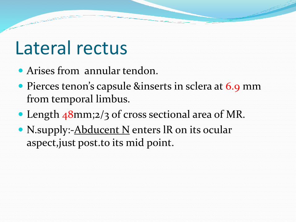
Lateral rectus Arises from annular tendon.
Pierces tenon’s capsule &inserts in sclera at 6.9 mm from temporal limbus.
Length 48mm;2/3 of cross sectional area of MR.
N.supply:-Abducent N enters lR on its ocular aspect,just post.to its mid point.

Spiral of tillaux Imaginary line joining the insertions of the 4 recti and
is an important anatomical landmark when performing surgery.
The insertions are located progressively further away from the limbus in a spiral pattern.
the medial rectus insertion is closest .
Superior rectus is farthest.


Obliques are inserted behind equator & form an angle of 51° with visual axis.

Superior oblique Longest& thinnest EOM.
Arises from common origin at the apex of orbit; superomedial to optic foramen.
Runs forward to trochlea(cartilaginous ring at upper&inner angles of orbit)
After threading through this it becomes tendinous
It changes its direction completely and runs over the globe under SR to attach above & lat, to posterior pole.

Ant.fibres of S.O tendon-intorsion
Post.fibres of S.O tendon-extorsion
N.supply:-Trochlear N(4) after dividing into 2-3 branches enters muscle superiorly.
B.supply:-superior muscular branch of ophthalmic A.

Inferior oblique Only EOM not arising from apex of orbit
It arises anteriorly from lower & inner orbital walls near lacrimal fossa.
Running below inf.rectus& attaches below&lat. to post.pole of globe.
N.supply:-Inf.divison of oculomotor N.
B.supply:-Infraorbital &medial muscular branches of ophtalmic A.

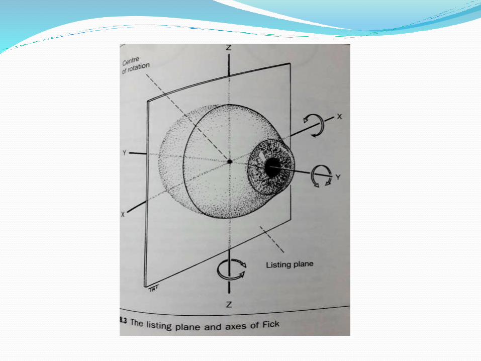
Action of extraocular muscles Rotation around centre of rotation
Centre of rotation lies 12/13 mm behind cornea.
3 types of rotation:
1. Rotation around fick vertical axis Z—side to side
2. “ “ fick horizontal axis X– up&down
3. “ “ fick antero posterior axis– torsion

Uniocular movements Ductions – only one eye is open,the other covered/closed
tested by asking the pt. to follow a target in each direction of gaze.
Types of ductions:-
1. Adduction
2. Abduction
3. Supraduction
4. Infraduction
5. Incycloduction
6. excycloduction

Binocular movements Versions:-both eyes open,attempting to fixate a target
&moving in same direction.
Binocular ,simultaneous,conjugate movements in same direction.
Abduction of one eye accompanied by adduction of other eye is called conjugate movements.

Types of versions:-
Dextroversion&laevo version
Elevation&depression
Dextro elevation&dextro depression
Laevo elevation& laevo depression

Torsional movements/righting reflexes:-
When you tilt head to maintain upright image.
Vergences:-binocular,simultaneous,disjugate/disjunctive movements (opp.direction)
Convergence– simultaneous adduction
Divergence– outward movement from convergent position

Types of convergence Reflex ----tonic
----proximal
----fusional
----accommodative
voluntary

Actions of EOM
ACTION PRIMARY SECONDARY TERTIARY
MR ADDUCTION ------ ---------
LR ABDUCTION ------ ---------
SR ELEVATION INTORSION ADDUCTION
IR DEPRESSION EXTORSION ADDUCTION
SO INTORSION DEPRESSION ABDUCTION
IO EXTORSION ELEVATION ABDUCTION

Both obliques have same tertiary action because inserted behind the center of rotation,
pull post. pole of globe medially
when they contract ant.portion of eye so it causes abduction

Both recti have same tertiary action bcz they inserted anterior to centre of rotation
pull ant.portion of globe medially so it causes adduction

Synergists:-ref.to muscles having same primary action in same eye.
Ex:-sup.rectus & inf.oblique----elevators
inf.rectus&sup.oblique-----depressors
Antagonists:-having opp.action in same eye
Ex:-sup.&inf. Recti
sup.&inf.oblique

Yoke muscle(contralateral synergists):-
Ref. to pair of muscles (one from each eye) which contract simultaneously during version movements.
Ex :-in dextroversion RLR &LMR
Contralateral antagonist:-pair of muscle (one from each eye)having an opposite action.
Ex:-in dextroversion RLR & LLR

Diagnostic positions of gaze:-9
1 Primary position of gaze:-assumed by eyes when
fixating a distant object with head erect.
6 cardinal positions :- to test 12 EOM in their main
field of action

1. Dextroversion
2. Laevo version
3. Dextro elevation
4. Leavo elevation
5. Dextro depression
6. Laevo depression
+2 Elevation
Depression


Laws of ocular motility1. Hering’s law of equal innervation:- during any
conjugate movement equal & simultaneous innervation flows to yoke muscles

2. Sherrington law of reciprocal innervation :-
inc.innervation to an EOM is accompanied by reciprocal dec. in innervation to its antagonist.
Ex:-RMR & RLR

Supranuclear control of ocular movements:-
1. Saccadic system
2. Smooth pursuit system
3. Vergence system
4. Vestibular system
5. Optokinetic system
6. Position maintenance system

Saccadic system:-saccades are sudden,jerky,conjugate,movementsas the gaze shifts from one object to other.
voluntary(normal)
invoiuntary(peripheral,auditory,visualstimuli)

Smooth pursuit eye movements:-
Tracking movements of eye as they follow moving object
Voluntary movements
When the velocity of moving object inc. replaced by small saccades(“catchup saccades”)

Vergence movement:
Allow focussing an object which moves away from/towards observer.
Very slow disjugate movements

Vestibular eye movement:-
Effective in compensating for effects of head movements in disturbing visual fixation
Through vestibular system

Optokinetic system:-
a movement following the moving scene , succeeded by rapid saccade in opp.direction
Position maintenance system:-
Helps to maintain specific gaze by rapid micro movements called “flicks” & slow micro movements called “drifts”.

Bibilography Adler’s physiology of eye -11th edition
Wolff’s anatomy of eye -8th edition
Parson’s disease of eye-21st edition
Jack.j.kanski brad bowlingclinical ophthalmology -7th edition
A.k.Khurana comprehensive ophthalmology -5th edition