Embed Size (px)
Citation preview
Imaging of Cardiac Tumors
dr amol gulhane DNB Resident CARE hospital , hyd
•Prevalence of 0.002-0.3%
• About 75 percent of all primary cardiac tumors -benign neoplasms.
•Remaining 25 percent of primary cardiac tumors -malignant neoplasms-metastatic MC
Metastatic involvement of the heart is 30 times more prevalent than primary cardiac tumors.
Presurgical assessment of malignant cardiac tumors with the use of MDCT and MRI allow determination of the resectability of tumors and planning for the reconstruction of cardiac chambers.
Echocardiography is the best imaging modality to depict small masses that arise from the cardiac valves.
Visualization of extracardiac extension is suboptimal with the use of transthoracic echocardiography for the evaluation of malignant cardiac masses.
TEE overcomes the limited acoustic window of the transthoracic mode, the airways and lungs can be obstacles for imaging of the aortic arch, pulmonary arteries and some systemic and pulmonary veins
MRI is presently the modality of choice to evaluate cardiac tumors. High contrast resolution and multiplanar capability allow a specific diagnosis and optimal evaluation of myocardial infiltration, pericardial involvement and extracardiac extension.
The use of ECG-gated MDCT has better soft tissue contrast than echocardiography and can definitively characterize fatty content and calcifications.
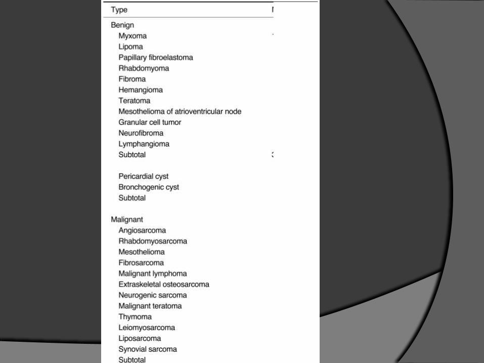

Relative incidence of benign heart tumors
% of Group
TUMOR Adults Children Infants
MyxomaLipomaPapillary fibroelastomaRhabdomyomaFibromaHemangiomaTeratomaMesothelioma of AV nodeGranular cell tumorNeurofibromaLymphangiomaHamartoma
462116235131110
1500
46155
1340101
000
65124
1820000
BENIGN TUMORS
Myxomas
Myxomas are the most common benign tumor found in adults(4th -7th decades).
classic triad of symptoms - cardiac obstructive symptoms related to the obstruction of blood flow , embolic events, and constitutional symptoms such as fever, malaise and weight loss.
It ocurs in the middle age, 40-70 years, and has anassociation with pituitary adenoma, testicular tumours andCushing's disease.
• Majority sporadic; some are familial (autosomal dominant transmission) or part of a syndrome1. Carney complex – spotty skin pigmentation,
myxomas, endocrine overactivity, schwannomas
2. NAME syndrome – nevi, atrial myxoma, myxoid neurofibroma, ephelides
3. LAMB syndrome – lentigines, atrial myxoma, blue nevi
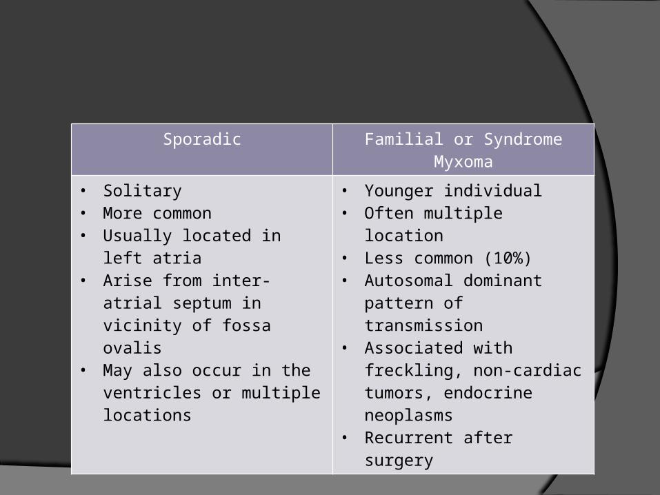
Sporadic Familial or Syndrome Myxoma
• Solitary• More common• Usually located in left atria• Arise from inter-atrial septum
in vicinity of fossa ovalis• May also occur in the
ventricles or multiple locations
• Younger individual• Often multiple location• Less common (10%)• Autosomal dominant pattern
of transmission• Associated with freckling, non-
cardiac tumors, endocrine neoplasms
• Recurrent after surgery
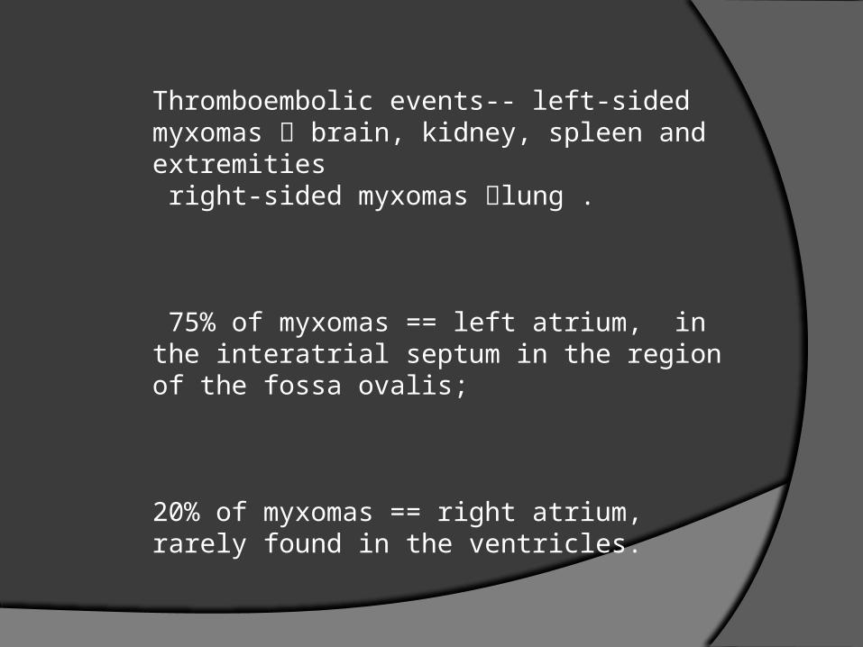
Thromboembolic events-- left-sided myxomas brain, kidney, spleen and extremities right-sided myxomas lung .
75% of myxomas == left atrium, in the interatrial septum in the region of the fossa ovalis;
20% of myxomas == right atrium, rarely found in the ventricles.
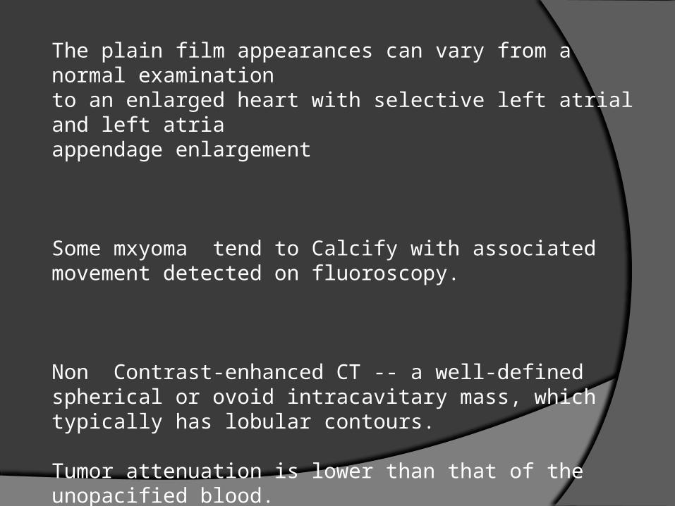
The plain film appearances can vary from a normal examinationto an enlarged heart with selective left atrial and left atriaappendage enlargement
Some mxyoma tend to Calcify with associated movement detected on fluoroscopy.
Non Contrast-enhanced CT -- a well-defined spherical or ovoid intracavitary mass, which typically has lobular contours.
Tumor attenuation is lower than that of the unopacified blood.
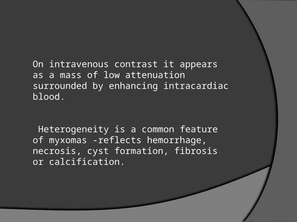
On intravenous contrast it appears as a mass of low attenuation surrounded by enhancing intracardiac blood.
Heterogeneity is a common feature of myxomas -reflects hemorrhage, necrosis, cyst formation, fibrosis or calcification.
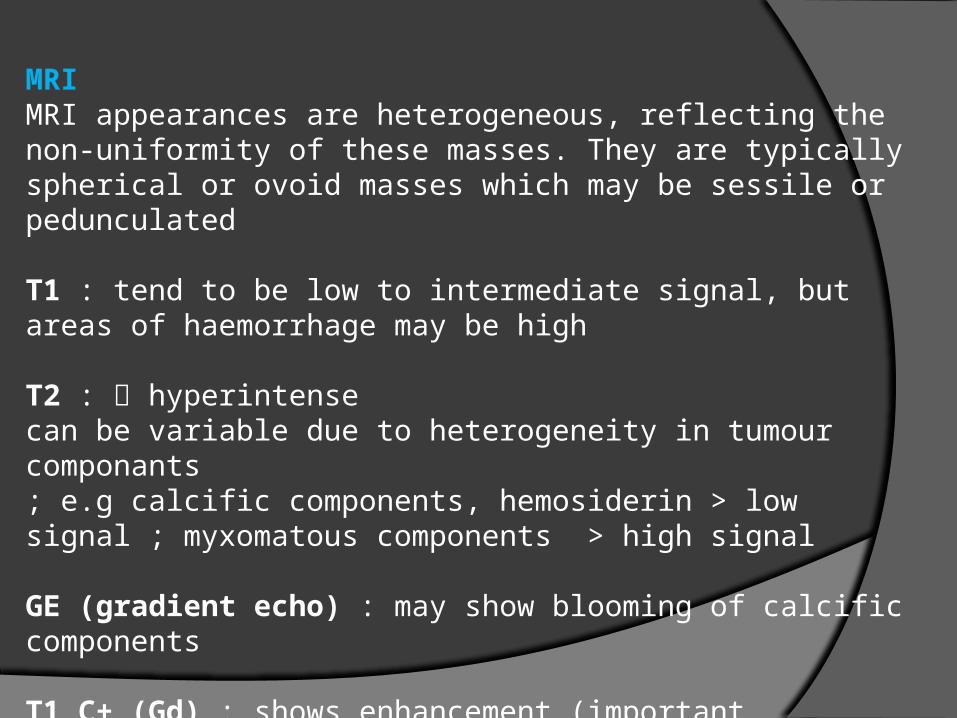
MRIMRI appearances are heterogeneous, reflecting the non-uniformity of these masses. They are typically spherical or ovoid masses which may be sessile or pedunculated T1 : tend to be low to intermediate signal, but areas of haemorrhage may be high
T2 : hyperintense can be variable due to heterogeneity in tumour componants; e.g calcific components, hemosiderin > low signal ; myxomatous components > high signal
GE (gradient echo) : may show blooming of calcific components
T1 C+ (Gd) : shows enhancement (important discriminator from a thrombus)
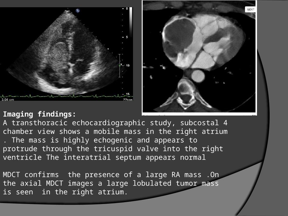
Imaging findings:A transthoracic echocardiographic study, subcostal 4 chamber view shows a mobile mass in the right atrium . The mass is highly echogenic and appears to protrude through the tricuspid valve into the right ventricle The interatrial septum appears normal
MDCT confirms the presence of a large RA mass .On the axial MDCT images a large lobulated tumor mass is seen in the right atrium.
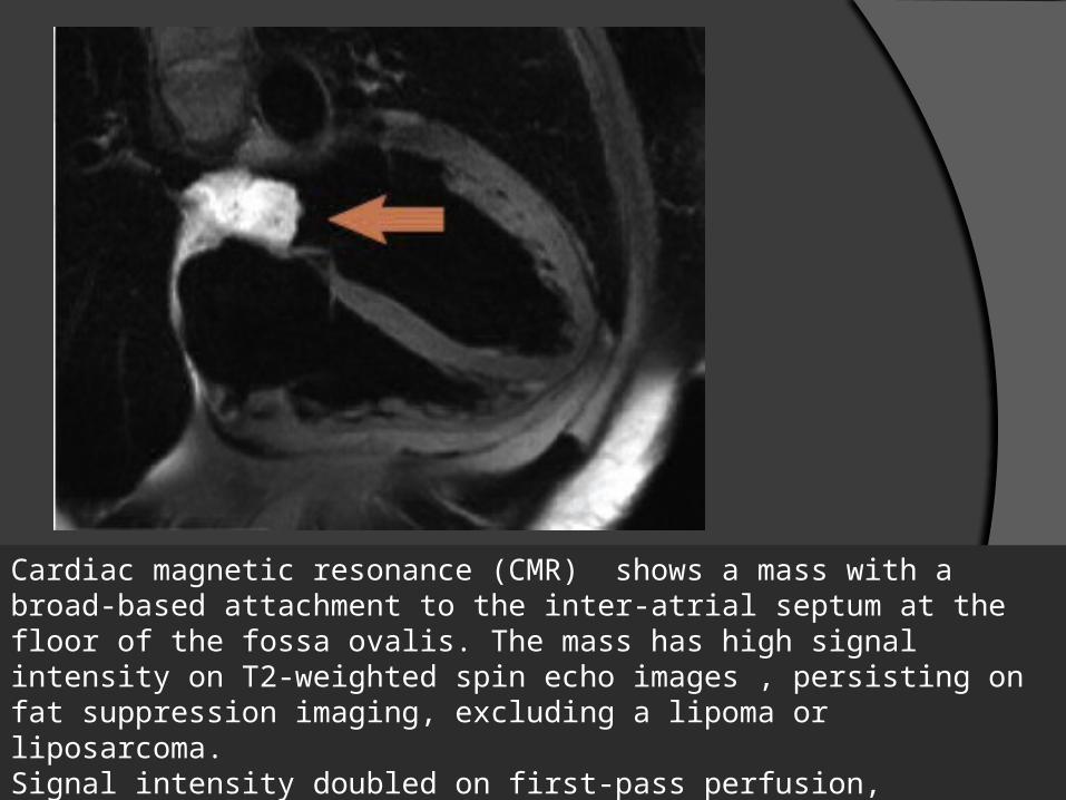
Cardiac magnetic resonance (CMR) shows a mass with a broad-based attachment to the inter-atrial septum at the floor of the fossa ovalis. The mass has high signal intensity on T2-weighted spin echo images , persisting on fat suppression imaging, excluding a lipoma or liposarcoma. Signal intensity doubled on first-pass perfusion, excluding thrombus. Delayed enhancement imaging revealed patchy hyperenhancement suggestive of cystic cavitation, features all strongly suggestive of a myxoma.
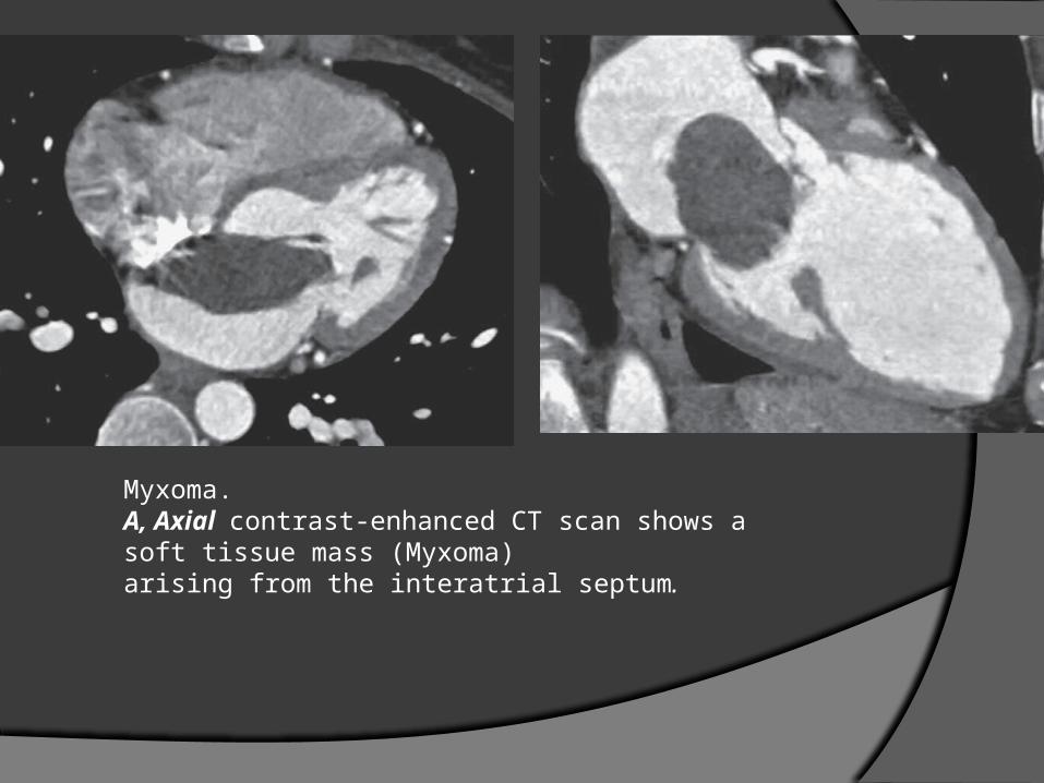
Myxoma. A, Axial contrast-enhanced CT scan shows a soft tissue mass (Myxoma)arising from the interatrial septum.
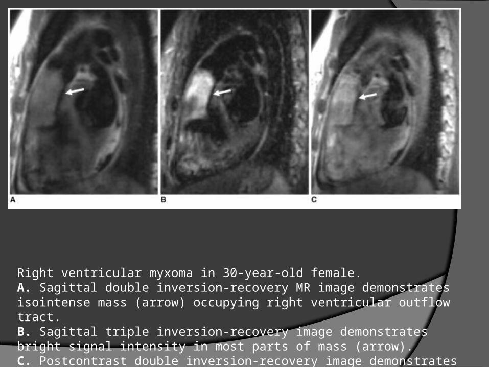
Right ventricular myxoma in 30-year-old female.A. Sagittal double inversion-recovery MR image demonstrates isointense mass (arrow) occupying right ventricular outflow tract.B. Sagittal triple inversion-recovery image demonstrates bright signal intensity in most parts of mass (arrow).C. Postcontrast double inversion-recovery image demonstrates hyperenhancement of mass (arrow).
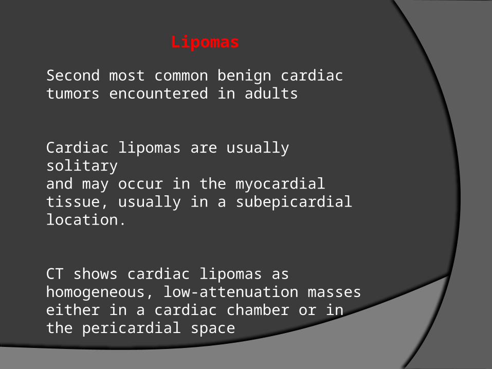
Lipomas
Second most common benign cardiac tumors encountered in adults
Cardiac lipomas are usually solitaryand may occur in the myocardial tissue, usually in a subepicardial location.
CT shows cardiac lipomas as homogeneous, low-attenuation masses either in a cardiac chamber or in the pericardial space
• If subepicardial Compression of the heart Pericardial effusion
• If subendocardial With intracavitary extension, may
produce symptoms characteristic of their location
• Most common chambers affected: LV, RA, IAS
MR homogeneous increased signal intensity seen on the T1- and T2-weighted ; suppressed on fat saturated sequences.
As with soft-tissue lipomas, cardiac lipomas do not show enhancement with the administration of a contrast material.
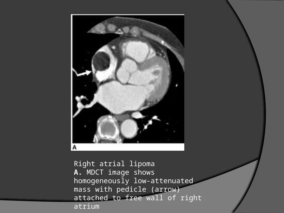
Right atrial lipoma A. MDCT image shows homogeneously low-attenuated mass with pedicle (arrow) attached to free wall of right atrium
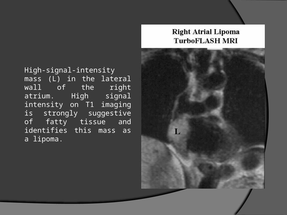
High-signal-intensity mass (L) in the lateral wall of the right atrium. High signal intensity on T1 imaging is strongly suggestive of fatty tissue and identifies this mass as a lipoma.
Papillary Fibroelastomas
rare lesion, usually affecting older adults
Benign endocardial papillomas that mainly affect the cardiac valves and account for approximately 75% of all cardiac valvular tumors
It is composed of multiple papillary fronds, which predisposeit to form thrombi.
Because of the risk of lethalembolization to the coronary or cerebral circulation, surgicalresection is the treatment of choice.
Papillary fibroelastomas are usually not observed on CT or MR images as they are small (< 1.5 cm in diameter) and are attached to the moving valves .
MR imaging typically demonstrates the presence of a mass on a valve leaflet or on the endocardial surface .
These tumors can create turbulence in the blood flow, which might be demonstrated with the use of cine MR imaging
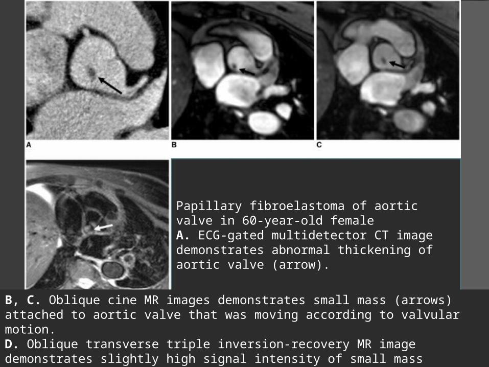
Papillary fibroelastoma of aortic valve in 60-year-old femaleA. ECG-gated multidetector CT image demonstrates abnormal thickening of aortic valve (arrow).
B, C. Oblique cine MR images demonstrates small mass (arrows) attached to aortic valve that was moving according to valvular motion.D. Oblique transverse triple inversion-recovery MR image demonstrates slightly high signal intensity of small mass (arrow).
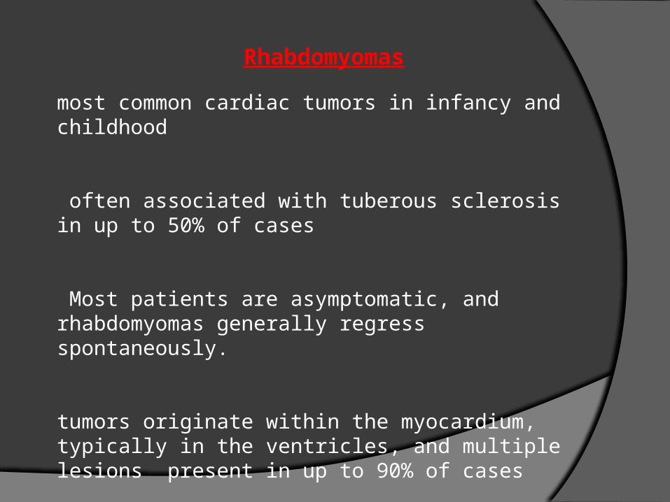
Rhabdomyomas
most common cardiac tumors in infancy and childhood
often associated with tuberous sclerosis in up to 50% of cases
Most patients are asymptomatic, and rhabdomyomas generally regress spontaneously.
tumors originate within the myocardium, typically in the ventricles, and multiple lesions present in up to 90% of cases
Echocardiography is usually used for evaluationof these tumors.
On noncontrast-enhanced CT scans, rhabdomyomas are typically denser than the adjacent myocardiumand may have areas of fat density.
Enhances on contrast CT.
MR isointense to marginally hyperintense as compared with the myocardium on T1-weighted images and hyperintense on T2-weighted images
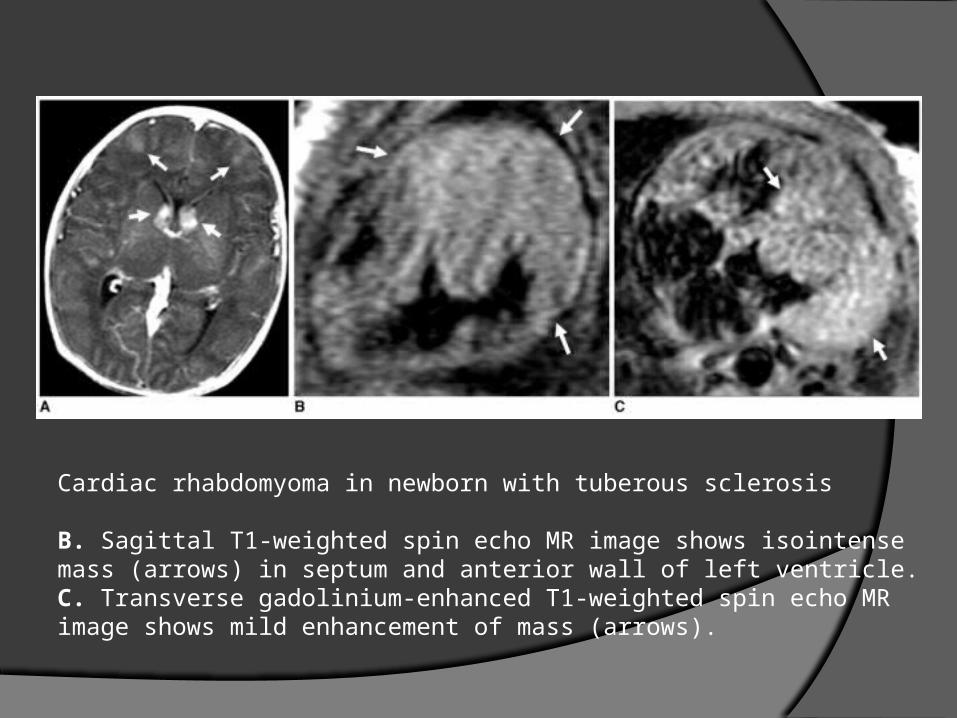
Cardiac rhabdomyoma in newborn with tuberous sclerosis
B. Sagittal T1-weighted spin echo MR image shows isointense mass (arrows) in septum and anterior wall of left ventricle.C. Transverse gadolinium-enhanced T1-weighted spin echo MR image shows mild enhancement of mass (arrows).
Fibromas
mainly affect infants and children, second most common tumors found in this age group
Grossly, the lesions are solid tumors that arise within the myocardium and can grow to a size that obliterates the cavity.
CT homogeneous masses with soft-tissue attenuation, may be either sharply marginated or infiltrative. Calcification is often observed.
MRhomogeneously isointense to hypointense relative to the myocardium on T1- and T2-weighted images due to a dense, fibrous nature. For the same reason, these tumors often show delayed enhancement on gadolinium-enhanced study
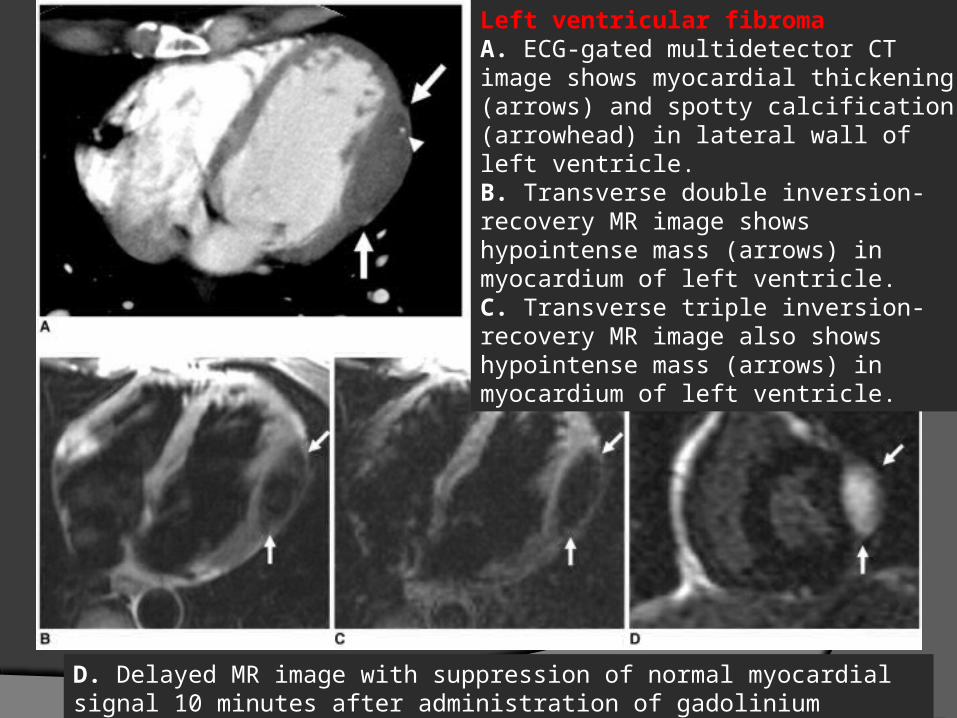
Left ventricular fibroma A. ECG-gated multidetector CT image shows myocardial thickening (arrows) and spotty calcification (arrowhead) in lateral wall of left ventricle.B. Transverse double inversion-recovery MR image shows hypointense mass (arrows) in myocardium of left ventricle.C. Transverse triple inversion-recovery MR image also shows hypointense mass (arrows) in myocardium of left ventricle.
D. Delayed MR image with suppression of normal myocardial signal 10 minutes after administration of gadolinium demonstrates hyperenhancement of mass (arrows).
Hemangiomas
benign vascular tumors
5-10% of benign tumors
Cardiac hemangiomas are heterogeneous on precontrast CT images and show intense contrast enhancement
As with hepatic hemangiomas, these tumors typically show intermediate signal intensity on T1-weighted images and hyperintense on the T2-weighted images

Cavernous hemangioma of left atrial appendage
A. Transverse double inversionrecovery MR image shows intermediate signal intensity mass (arrows) in left atrial appendage.B. Transverse triple inversion-recovery MR image shows hyperintense mass (arrows) with smooth margin.
C. Coronal gadolinium-enhanced double inversion-recovery MR image shows strong enhancement of lesion (arrows).
PRIMARY MALIGNANCIES
Angiosarcomasmost common cardiac sarcomas
Adults. M>F
the tumors tend to occur in the right atrium and involve the pericardium.
usually cause right-sided heart failure or tamponade
Presentation is late, and there is often the presence of metastases at the time of diagnosis, particularly to the lung.
Invasive behavior is a feature of malignant lesions with pericardial or pleural effusion.
CT low-attenuation mass in the right atrium, might be irregular or nodular
Heterogenous enhancement
Cardiac sarcomas heterogeneous signal intensity on MR images , with blood-filled spaces within the neoplasm seen on T2 as high signal intensity
A papillary appearance can be observed as a specific MR feature of an angiosarcoma, with a nodular area of high signal intensity interspersed within areas of intermediate signal intensity seen on T1- and T2-weighted images
In cases with diffuse pericardial infiltration, linear enhancement along the vascular spaces seen ( "sunray" appearance )
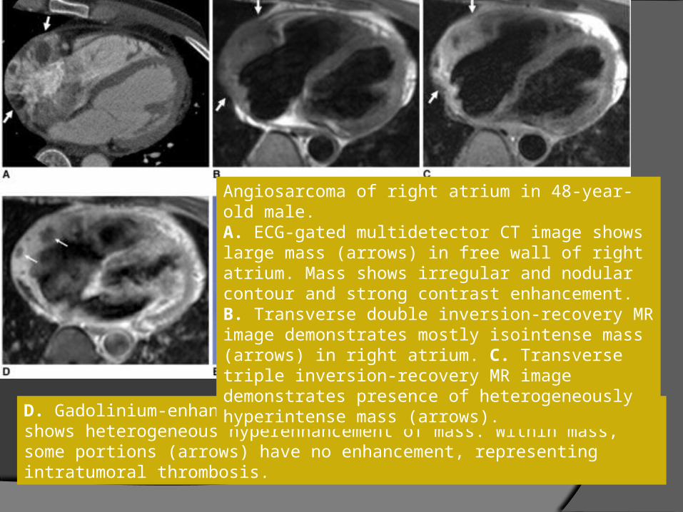
D. Gadolinium-enhanced double inversion-recovery MR image shows heterogeneous hyperenhancement of mass. Within mass, some portions (arrows) have no enhancement, representing intratumoral thrombosis.
Angiosarcoma of right atrium in 48-year-old male.A. ECG-gated multidetector CT image shows large mass (arrows) in free wall of right atrium. Mass shows irregular and nodular contour and strong contrast enhancement.B. Transverse double inversion-recovery MR image demonstrates mostly isointense mass (arrows) in right atrium. C. Transverse triple inversion-recovery MR image demonstrates presence of heterogeneously hyperintense mass (arrows).
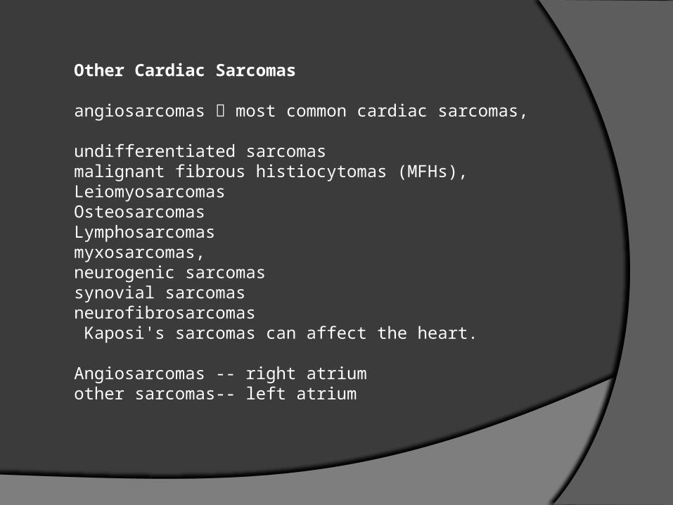
Other Cardiac Sarcomas
angiosarcomas most common cardiac sarcomas,
undifferentiated sarcomasmalignant fibrous histiocytomas (MFHs), LeiomyosarcomasOsteosarcomasLymphosarcomasmyxosarcomas, neurogenic sarcomassynovial sarcomasneurofibrosarcomas Kaposi's sarcomas can affect the heart.
Angiosarcomas -- right atriumother sarcomas-- left atrium

MFHs left atrium , attached to the posterior wall
MFH -- nonspecific signal intensity MRI
A MFH arises from the posterior wall of the left atrium and can extend into the pulmonary veins
Myxoma does not extent to PV
MFH
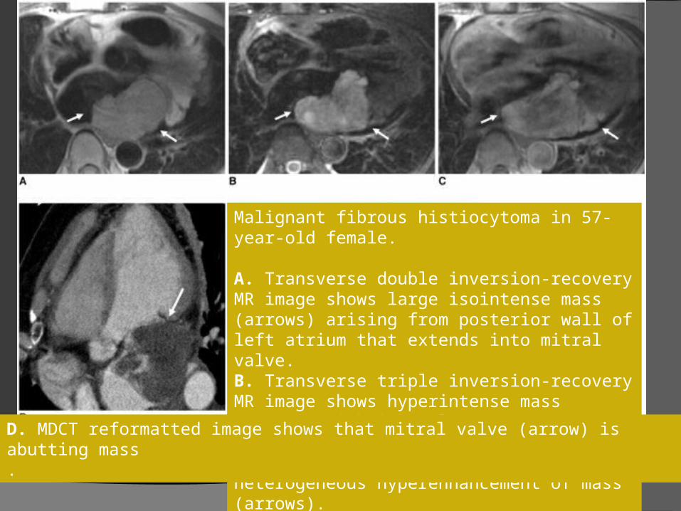
Malignant fibrous histiocytoma in 57-year-old female.
A. Transverse double inversion-recovery MR image shows large isointense mass (arrows) arising from posterior wall of left atrium that extends into mitral valve.B. Transverse triple inversion-recovery MR image shows hyperintense mass (arrows) with irregular contour.C. Gadolinium-enhanced double inversion-recovery MR image shows heterogeneous hyperenhancement of mass (arrows).
D. MDCT reformatted image shows that mitral valve (arrow) is abutting mass.
Primary cardiac osteosarcomas
usually arise from the posterior wall of theleft atrium near the pulmonary veins.
metastatic osteosarcomas,usually involve the right atrium.
May calcify and may be confused with calcifi ed myxoma.
OSlocation in the posterior wall of the left atrium
myxomas septal location
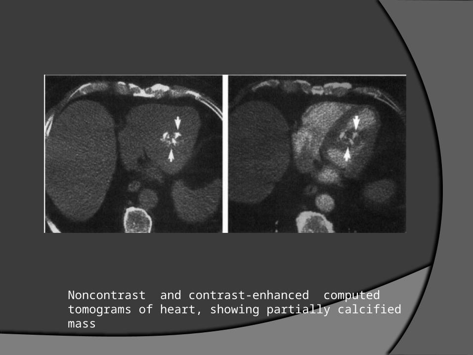
Noncontrast and contrast-enhanced computed tomograms of heart, showing partially calcified mass
Lymphoma
Up to 25% of patients with lymphoma have cardiac involvement at autopsy
Primary cardiac lymphoma (lymphoma limited to the heart and/orpericardium) is very rare.
Primary cardiac lymphoma is usually a B-cell lymphoma.
most common locationright heart, usually the right atrium
Associated pericardial effusion is common
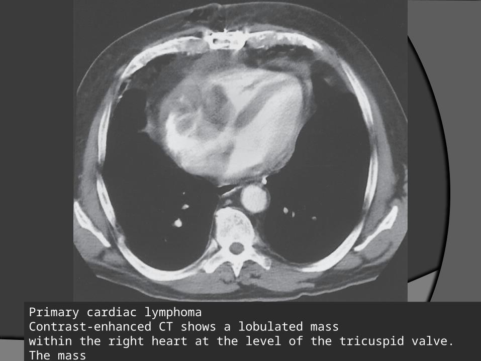
Primary cardiac lymphoma Contrast-enhanced CT shows a lobulated masswithin the right heart at the level of the tricuspid valve. The massextends into the right atrial and right ventricular cavities
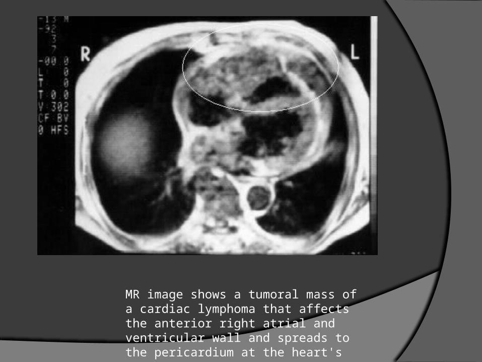
MR image shows a tumoral mass of a cardiac lymphoma that affects the anterior right atrial and ventricular wall and spreads to the pericardium at the heart's apex.
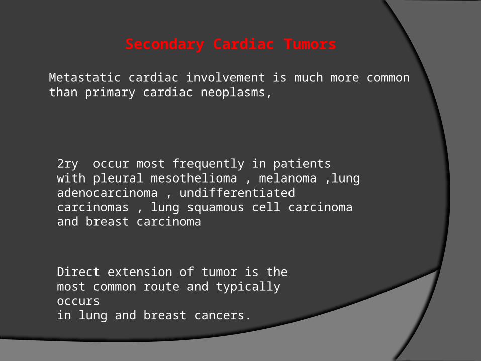
Secondary Cardiac Tumors
Metastatic cardiac involvement is much more commonthan primary cardiac neoplasms,
2ry occur most frequently in patients with pleural mesothelioma , melanoma ,lung adenocarcinoma , undifferentiated carcinomas , lung squamous cell carcinoma and breast carcinoma
Direct extension of tumor is the most common route and typically occursin lung and breast cancers.
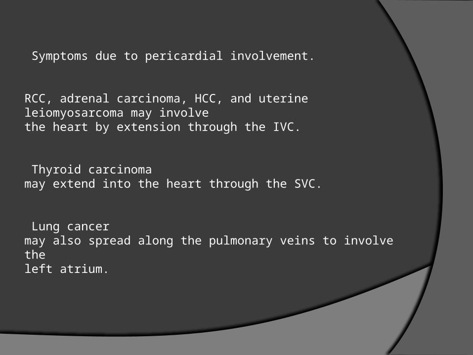
Symptoms due to pericardial involvement.
RCC, adrenal carcinoma, HCC, and uterine leiomyosarcoma may involvethe heart by extension through the IVC.
Thyroid carcinomamay extend into the heart through the SVC.
Lung cancermay also spread along the pulmonary veins to involve theleft atrium.
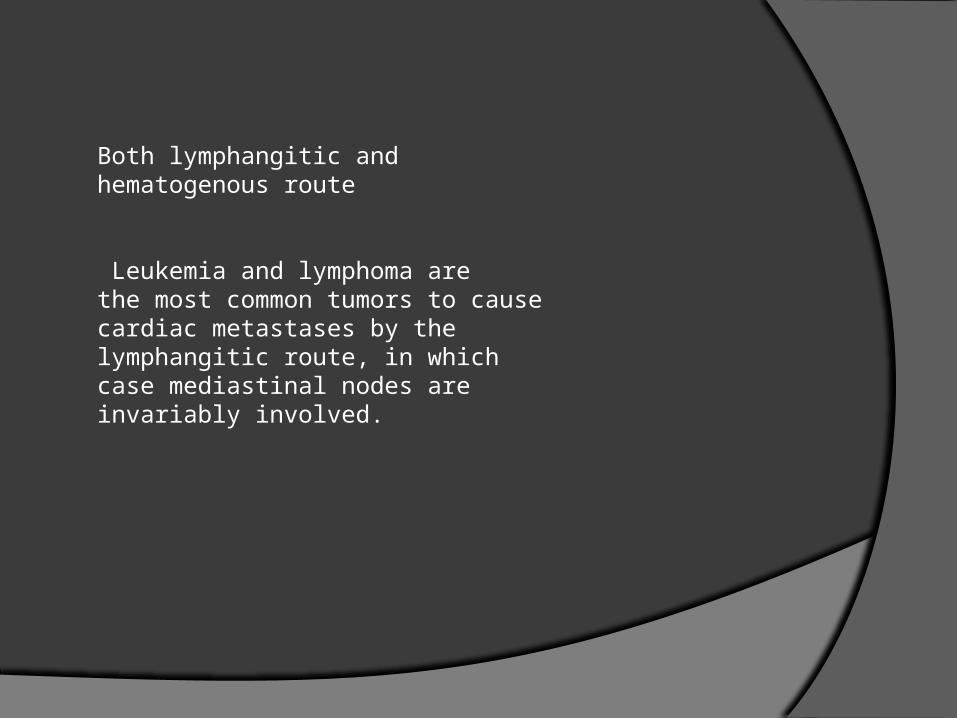
Both lymphangitic and hematogenous route
Leukemia and lymphoma arethe most common tumors to cause cardiac metastases by the lymphangitic route, in which case mediastinal nodes are invariably involved.
Lesions That Mimic Cardiac Tumors
Thrombus
Thrombus within the heart may mimic a cardiac mass.
In the atria, thrombus usually involves the appendages
In the ventricle, thrombus usually occurs over an area of hypokinesis such as a myocardial infarction or within a ventricular aneurysm.
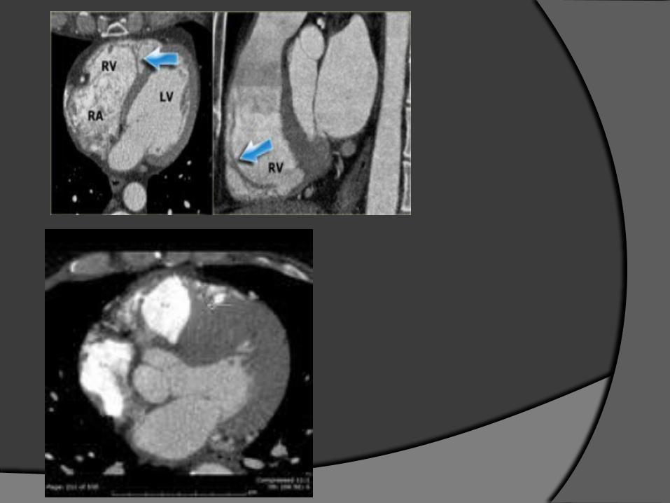
Lipomatous Hypertrophy of the Interatrial Septum
Fat may be detected in the AV grooves and the interatrialseptum.
Normally, fat in the interatrial septum measuresless than 1 cm anterior and posterior to the fossa ovalis.
LHIS results in increased fat deposition in the interatrial septum.
This may mimic an atrial tumor on an echocardiogram
Typically, the lesion is dumbbell-shapedbecause of the sparing of the region of the fossa ovalis
CT and MRI are able to accuratelydepict the fatty nature of the lesion
Pathologically, the lesion is not a true encapsulatedLipoma . fetal or brown fat intermixed with myocardial cells.
Treatment is directed at controlling thearrhythmia, if present.

Normal Anatomic Structures
Normal anatomic structures that may be mistaken for amass include a prominent moderator band in the right ventricle,
The crista terminalis is a vertically orientated smooth muscle ridge extending from the SVC to the IVC.
its size and shape can be variable and it may protrude into the RA, it may be mistaken as an intracardiac mass or thrombus.
Prominent crista terminalis
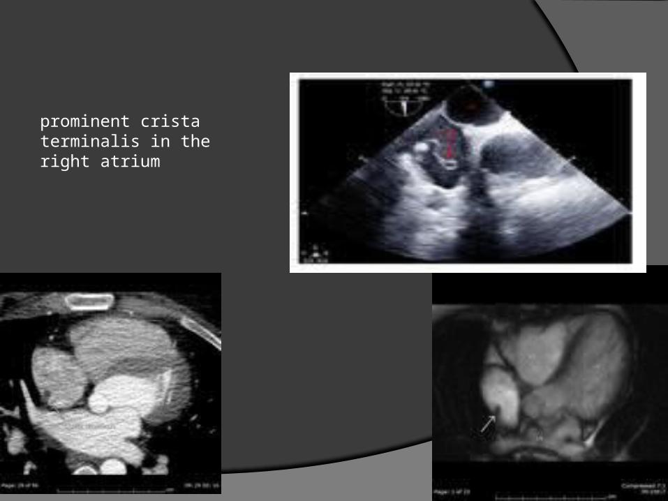
prominent crista terminalis in theright atrium
The RAA has a triangular or pyramidal shape with a wide base opening and rough trabeculation of pectinate muscles.
The pectinate muscle in the RAA may be misinterpreted as a mass or thrombus.
the pectinate muscle have parallel course on imaging
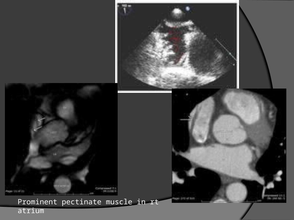
Prominent pectinate muscle in rt atrium
Most malignant tumors are likely to be treated with radiation or chemotherapy
Most beingn cardiac tumours are usually treated surgically

Thank u