Embed Size (px)
DESCRIPTION
Citation preview

AIDS
Megha J Nair6th Batch

INTRODUCTION
Acquired Immunodeficiency Syndrome
First indication in 1981 with reports from New York and Los Angeles-sudden outbreak of two rare diseases-kaposi’s sarcoma and Pnuemocystis carinii

HUMAN IMMUNODEFICIENCY VIRUS
Causative agent of AIDS Spherical enveloped virus about 90-120 nm in
size Genome is diploid composed of two identical
single stranded positive RNA copies Along with viral RNA – reverse transcriptase
enzyme-characteristic feature of retrovirus When virus infects cell-reverse trancriptase
transcribe the single stranded RNA to double stranded RNA nand then to double stranded DNA(PROVIRUS)-integrates into human genome!
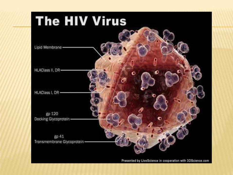

During viral replication , when the virus buds out of the host cell surface membrane-acquires a lipoprotein envelop-consists of lipid derived from the host cell membrane and glycoproteins which are virus coded
Major virus coded envelop protein –projecting knob like spikes and anchoring transmembrane pedicles
Spikes-major surface component of virus which binds to CD4 receptors on host cells…transmembrane proteins help in cell fusion

PATHOGENESIS
Transmitted mainly through sexual contact or through blood transfusion
Transmitted when the virus enters the blood or the tissues and come in contact with suitable host cells
Receptor for virus –CD4 antigen-thus infect any cells bearing CD4 antigen on surface-primarily the CD4+ helper T cell
Others include B lymphocytes,monocytes and macrophages such as specialised macrophages of lungs langerhan cells in dermis.

After fusion of virus with the cell-HIV genome uncoated and internalised into the cell
Action of reverse transcriptase enzyme-double stranded DNA integrated into the human genome with the help of integrase-causes latent infection
Long and variable incubation period is due to latency
Primary pathogenic mechanism-due to damage caused to the CD4 T lymphocytes
T4 cells decrease in number,CMI reduces T4:T8 ratio reverses Helper T cell function-essential for B cell
function-polyclonal activation of Bcells -hypergammaglobulinemia

CLINICAL MANIFESTATIONS
Not primarily to viral cytopathology but due to failure of immune responses
AIDS – only the last stage in the wide spectrum of clinical features

STAGES OF EVOLUTION
Acute HIV infection
Asymptomatic or latent infection
Persistent generalised lymphadenopathy
AIDS related complex
AIDS

ACUTE HIV INFECTION
Within 3-6 weeks of infection
Low grade fever,malaise,head ache,lymphadenopathy,sometimes with rashes arthropathy resembling glandular fever.Spontaneous resolution within weeks
Tests for HIV antibodies negative in early stage and get positive during its course-seroconversion illness.
ASYMPTOMATIC AND LATENT INFECTION
Symptomless infection Positive HIV antibodies test Passes through various stages-CD4
lymphocytopaenia,minor oppurtunistic infection,persistent generalised lymphadenopathy,ARC and full blown AIDS
Viral multiplication goes on-immune response mounnted by host-only helps in limiting viral load
Tcell-500 from 1000/microlitre-acute infection and when 200 or less , clinical AIDS
PGL
Enlarged lymph nodes
Atleast 1 cm or more
In two or more non contiguous extrainguinal sites-that persists for more than 3 months in the absence of any current illness or medication

PGL

ARC
Patients with considerable immunodeficiency suffering from various constitutional symptoms or minor oppurtunistic infections
Fatigue , unexplained fever , persistent diarrhoe , marked weight lose-symptoms-also gen. lymphadenopathy and splenomegaly
Oppurtunistic inf.-oral candidiasis,herpes zoster,hairy cell luekoplakia,salmanellosis or tb

ORAL THRUSH
AIDS
• End stage disease-irreversible breakdown of immune system-progressive oppurtunistic infection and malignancies.
• Dry cough , dyspnea , fever , recurrent pneumonia
• GIT-thrush , herpetic steatites , gingivitis , hairy luekoplakia or kaposi’s sarcoma.A characteristic intestinal pathogen is cryptosporidium
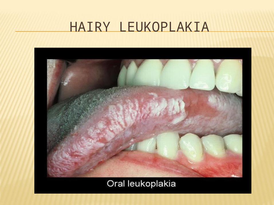
HAIRY LEUKOPLAKIA

CNS-toxoplasmosis and cryptococcosis,lymphomas of CNS also seen.also can cross blood brain barrier and cause encephalopathy leading to loss of higher function-then dementia
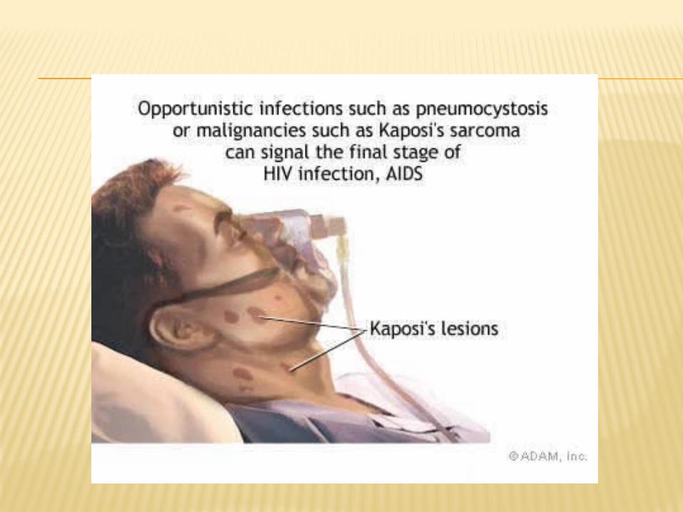
Malignancies-kaposi’s sarcoma(nonmetastasing mucosal or cutaneous tuour of endothelial orgin)Hodgkin and Non Hodgkin lymphomas
Babies born to infected mothers-also positive-

KAPOSI’S LESIONS


DIAGNOSIS
IMMUNOLOGICAL TESTS: Total luecocyte and lymphocyte count Tcell subset assay-ratio inversion.Absolute
CD4 cell count less than 200/cubic mm Platelet cout will show thrombocytopaenia Raised IgG and IgA levels Diminished CMI by skin tests Lymph node biopsy
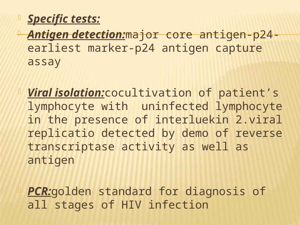
Specific tests: Antigen detection:major core antigen-
p24-earliest marker-p24 antigen capture assay
Viral isolation:cocultivation of patient’s lymphocyte with uninfected lymphocyte in the presence of interluekin 2.viral replicatio detected by demo of reverse transcriptase activity as well as antigen
PCR:golden standard for diagnosis of all stages of HIV infection

Antibody detection: ELISA Test
Western blot test-HIV proteins seperated by their electrophoretic mobility by poly acrylamide gel electrophoresis are blotted on to the strips of nitrocellulose paper.strips are reacted with test sera and then with enzyme conjugated anti human globulin
TREATMENT
Treatment and prophylaxis of infections and tumours
General managementImmunorestortive
measures(administration of interluekin 2,thymic factors etc)
Specific anti-HIV drugs(anti retroviral drugs)

THANK YOU