Embed Size (px)
Citation preview

Dr.Arun Naragund
Asst.Prof. Dept. of Shareera Rachana
Shri J.G.C.H.S. Ayurvedic Medical College, Ghataprabha -591321
SPLEEN

• The spleen consists of a large encapsulated mass
of vascular and lymphoid tissue
• It is situated in the upper left quadrant of the
abdominal cavity between the fundus of the
stomach and the diaphragm.
• Its shape varies from a slightly curved wedge to a
‘domed' tetrahedron.
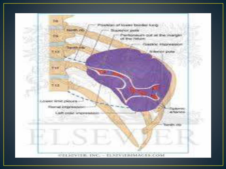
• Its long axis lies approximately in the plane of the
tenth rib. (45˚ with horizontal plane)
• Its posterior border is approximately 4 cm from the
mid-dorsal line at the level of T 10
• Its anterior border usually reaches the mid-
axillary line.


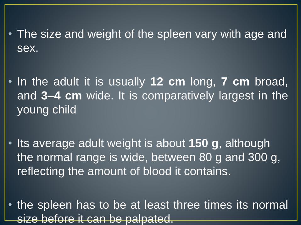
• The size and weight of the spleen vary with age and
sex.
• In the adult it is usually 12 cm long, 7 cm broad,
and 3–4 cm wide. It is comparatively largest in the
young child
• Its average adult weight is about 150 g, although
the normal range is wide, between 80 g and 300 g,
reflecting the amount of blood it contains.
• the spleen has to be at least three times its normal
size before it can be palpated.
• TWO ENDS : Anterior
(EXTRE MITIES/POLES) Posterior
• THREE BORDERS : Superior
Inferior
Intermediate
• TWO SURFACES : Diaphragmatic
Visceral
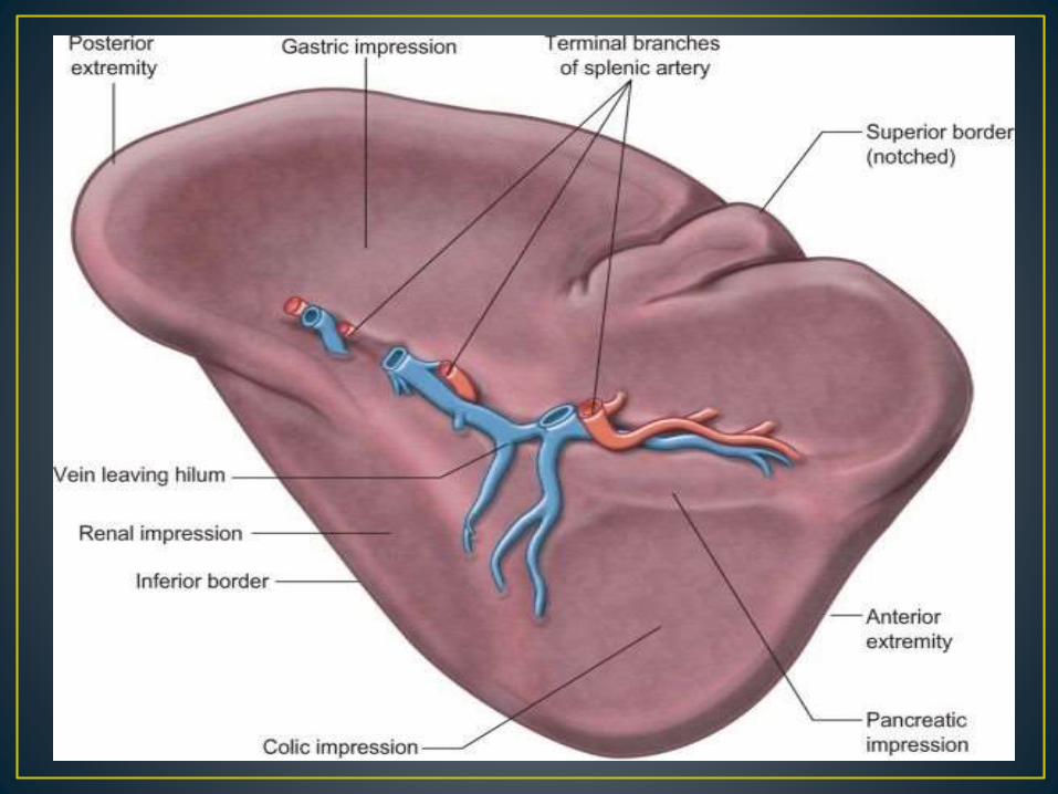
• The posterior extremity, or superior pole, usuallyfaces the rounded vertebral column. Rests on upperpole of left kidney.
• The anterior extremity, or inferior pole, is larger,expanded & more like a border .
• The superior border have one or two notches thathave persisted from the lobulated form of the spleenin early fetal life
• The inferior border is more blunt and rounded thanthe superior border and corresponds in position tothe lower margin of the eleventh rib
• The intermediate border is rounded & directed toright.
• The diaphragmatic surface is convex and smooth and faces
mostly superiorly and laterally .
• The diaphragmatic surface is related to left dome of the
diaphragm which separates it from the basal pleura, the
lower lobe of the left lung and the 9-11 left ribs
• The visceral surface faces inferomedially with impressions.
• The gastric impression is broad ,concave It is separated from
the stomach by a peritoneal recess.
• The renal impression is slightly concave by the left kidney &
left suprarenal gland.
• The colic impression lies at the inferior pole ,and is usually
flat.
• The pancreatic impression is often small
• The hilum of the spleen is a long fissure pierced by splenic
artery and vein ,nerves and lymphatics enter and leave the

• The spleen is almost entirely covered by peritoneum that
adheres firmly to its capsule, and is separated from the
stomach and left kidney by recesses of the greater sac
• The splenorenal (lienorenal) ligament is formed from two
layers of peritoneum. The anterior layer & The posterio layer.
• The splenic vessels lie between the layers of the
splenorenal ligament:
• The tail of the pancreas is usually present in its lower
portion.
• The gastrosplenic ligament also has two layers. The
posterior layer is at the splenic hilum and over the posterior
aspect of the stomach.
• The anterior layer reaches the greater curvature of the
stomach anteriorly.
• The short gastric and left gastroepiploic branches of the
splenic artery pass between its layers.
• The phrenicocolic ligament extends from the splenic flexure
to the diaphragm at the level of the eleventh rib.
• It is continuous with the peritoneum of the lateral end of the
transverse mesocolon


• The spleen is supplied exclusively from the splenic artery.
• This is the largest branch of the coeliac axis and its course
is among the most tortuous in the body, above the superior
border of the pancreas and descend to lie behind the gland.
• The splenic artery runs in the splenorenal ligament
posterior to the tail of the pancreas.
• It divides into two or three main branches before entering
the hilum of the spleen. As these branches enter the hilum
they divide further into four or five segmental arteries that
each supply a segment of the splenic tissue.
• The splenic artery gives off various branches to the
pancreas in its course and gives off short gastric arteries to
the stomach just prior to dividing or from its terminal
branches.
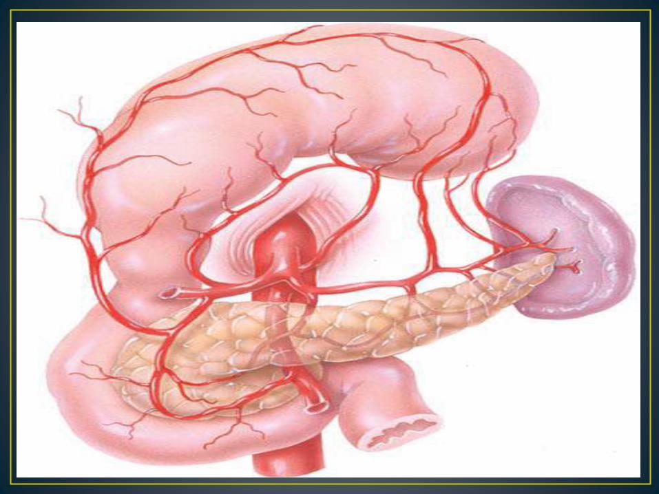
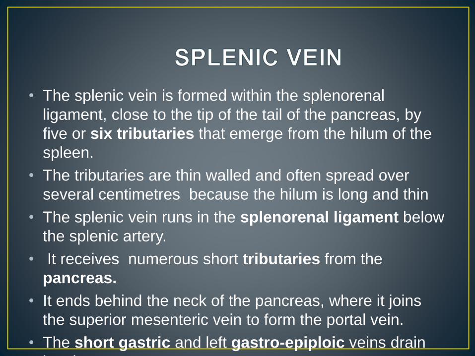
• The splenic vein is formed within the splenorenal
ligament, close to the tip of the tail of the pancreas, by
five or six tributaries that emerge from the hilum of the
spleen.
• The tributaries are thin walled and often spread over
several centimetres because the hilum is long and thin
• The splenic vein runs in the splenorenal ligament below
the splenic artery.
• It receives numerous short tributaries from the
pancreas.
• It ends behind the neck of the pancreas, where it joins
the superior mesenteric vein to form the portal vein.
• The short gastric and left gastro-epiploic veins drain
into it.

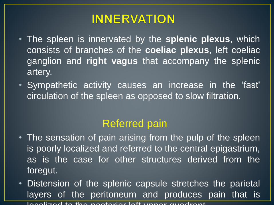
• The spleen is innervated by the splenic plexus, which
consists of branches of the coeliac plexus, left coeliac
ganglion and right vagus that accompany the splenic
artery.
• Sympathetic activity causes an increase in the ‘fast'
circulation of the spleen as opposed to slow filtration.
Referred pain
• The sensation of pain arising from the pulp of the spleen
is poorly localized and referred to the central epigastrium,
as is the case for other structures derived from the
foregut.
• Distension of the splenic capsule stretches the parietal
layers of the peritoneum and produces pain that is
localized to the posterior left upper quadrant.

• The spleen is essentially concerned with phagocytosis and
immunity .
• In the fetus it is also an important site of haemopoiesis.
• In spleenectomy functions are assumed by the liver and by
other lymphoid tissues.

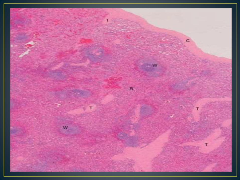
• Microscopically, the parenchymal tissue of the spleenconsists of two major components, white pulp and redpulp.
• The white pulp is composed of lymphoid tissue in whichB and T lymphocytes mature and proliferate underantigenic stimulation.
• The red pulp is a unique filtration device that enablesthe spleen to clear particulate material from the blood asit perfuses the spleen.
• It is composed of a complex system of interconnectedspaces populated by large numbers of phagocyticmacrophages that remove effete red blood cells,microorganisms, cellular debris and other particulatematter from the circulation.

Spleenic Artery
Spleenic vein
White
Pulp RED
PULP
Venous sinus
Splenic cord
Central
Artery
TRABECULACAPSUL
E

• The segmental splenic arteries enter the hilum and ramify
in the trabeculae throughout the organ.
• The splenic veins are similar in number to the arterial
branches.
• Small arteries tapering to arterioles pass through the white
pulp then turn abruptly to form penicillar branches which,
after a course of approximately 0.5 mm, pass out of the
white pulp into the marginal zone and red pulp.
• The passage of blood through the vascular compartments
between the arterioles and splenic veins is referred to
collectively as the intermediate circulation of the spleen.
• Ultimately, blood is passed to the venous sinusoids from
which it enters venules leading to small veins that run
within trabeculae, and thence into larger veins that drain
the spleen at its hilum


THANK YOU