Embed Size (px)
Citation preview


The Art of Radiology


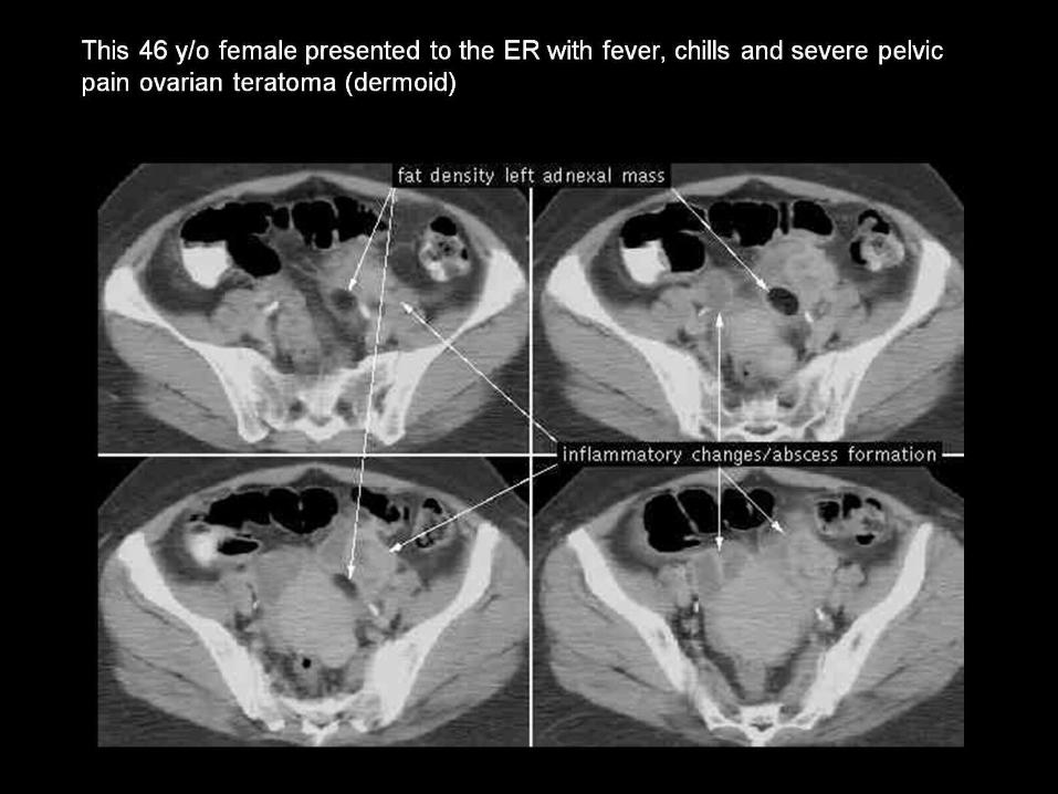
- Tubo Ovarian Abscess & Ova ( )rian Teratoma Dermoid


CT scan shows the ovarian blood vessels (OBV) coursing in the upper pelvis lateral to the ureters.

Normal ovary

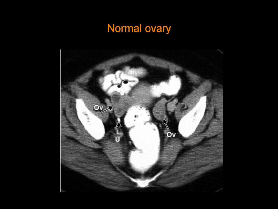
Normal ovary

Normal ovary

The ovarian blood vessels (OBV) are lateral to the ureters (U) in the lower abdomen

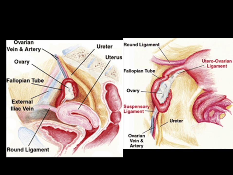
The ovarian blood vessels are continuous with the suspensory ligaments (SL), which appear thicker than the vessels .
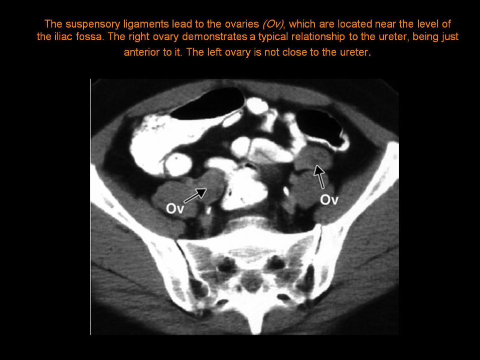
The suspensory ligaments lead to the ovaries (Ov), which are located near the level of the iliac fossa. The right ovary demonstrates a typical relationship to the ureter, being just anterior to it.
The left ovary is not close to the ureter .


The suspensory ligament

broad ligament (BL) ,

BL = broad ligament, M = complex mass in the left ovary

utero-ovarian ligament (UOL) .

Surgically transposed ovary in a young patient with cervical cancer .
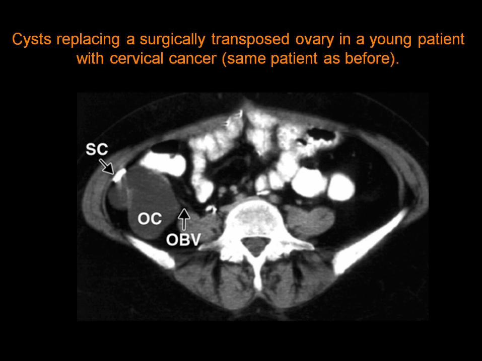
Cysts replacing a surgically transposed ovary in a young patient with cervical cancer (same patient as before) .

Differentiation of the ovaries from enlarged lymph nodes in a patient with metastatic cervical cancer .

Normal ovary and an enlarged lymph node at the pelvic sidewall in a patient with metastatic cervical cancer .

Cystic lymph node mass simulating an adnexal mass .
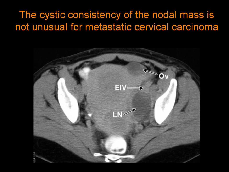
The cystic consistency of the nodal mass is not unusual for metastatic cervical carcinoma


Pelvic cysts of ovarian origin.
SLA = ovarian attachment of the right suspensory ligament

Determining the origin of pelvic masses in a 32-year-old woman. suspensory ligament (SL) leads to it. The bilateral cystic lesions proved to be endometriomas.
ULM = uterine leiomyoma pedunculated from the fundus.

Posterolateral displacement of the ureter by a large myomatous uterus.left ureter (U)
.

Tracking the ovarian vein to a recognizable suspensory ligament joining a pelvic mass in a 48-year-old woman .

Ovarian vein leading to a pelvic-abdominal mass in a 32-year-old woman .
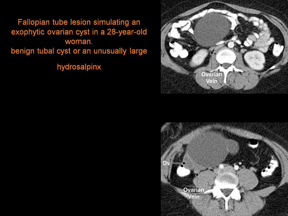
Fallopian tube lesion simulating an exophytic ovarian cyst in a 28-year-old woman.
benign tubal cyst or an unusually large hydrosalpinx


67-year-old woman with benign serous ovarian tumor. ovarian vascular pedicle" sign revealing organ of origin of a pelvic
mass lesion on helical ct
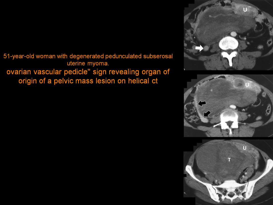
51-year-old woman with degenerated pedunculated subserosal uterine myoma. ovarian vascular pedicle" sign revealing organ of origin of a pelvic mass lesion
on helical ct

36-year-old woman with Krukenberg's tumor.
OVARIAN VASCULAR PEDICLE" SIGN REVEALING ORGAN OF ORIGIN OF A PELVIC MASS LESION ON HELICAL CT
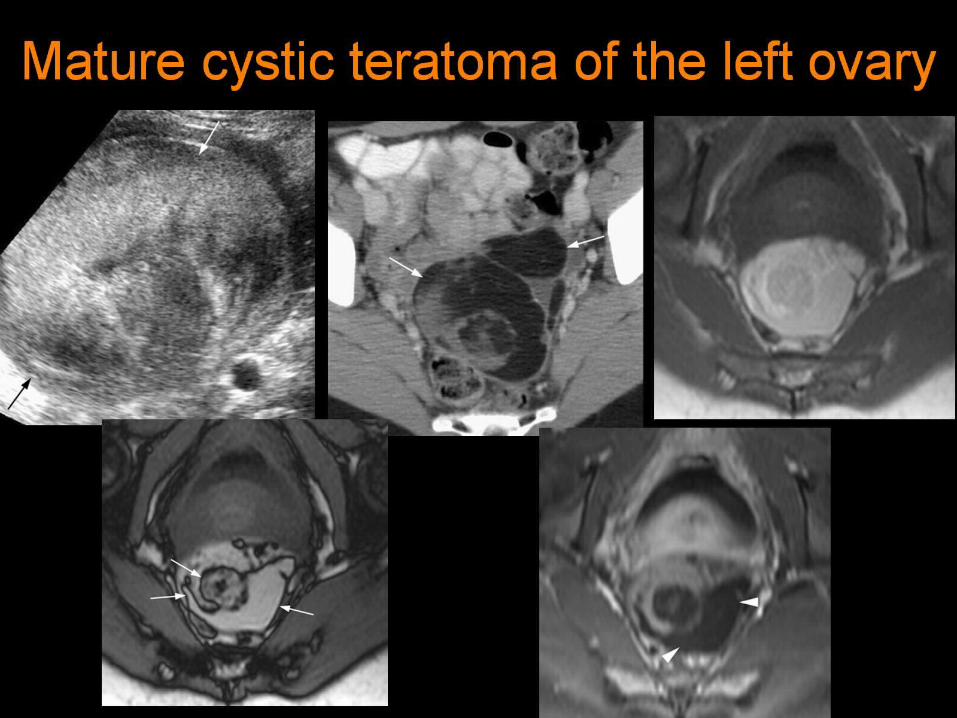
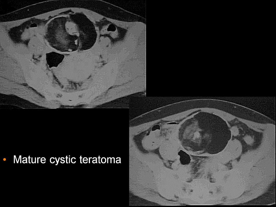
Mature cystic teratoma of the left ovary

Retroperitoneal liposarcoma

Right ovarian cyst. CT of the pelvis with contrast shows an approximate 5cm round well-defined low-density mass (arrow) in the right adnexal consistent with an ovarian
cyst.

dermoid cyst

Tubo-ovarain abscessTOA

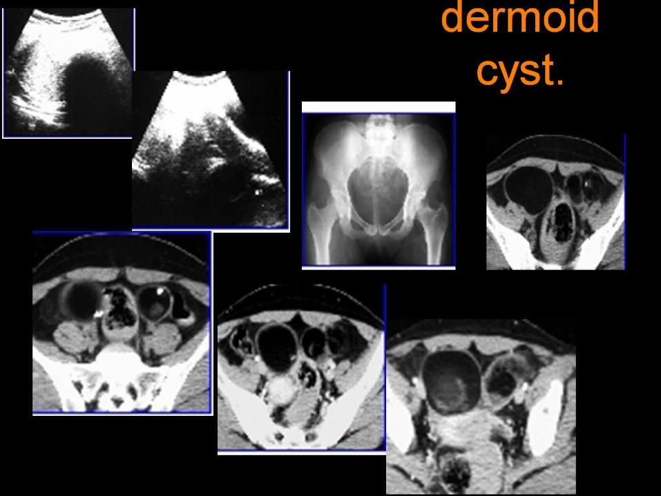
dermoid cyst.

Small cell carcinoma of the ovary, hypercalcemic type

Benign serous cystadenoma in a 49-year-old woman

Benign mucinous cystadenoma in a 26-year-old woman

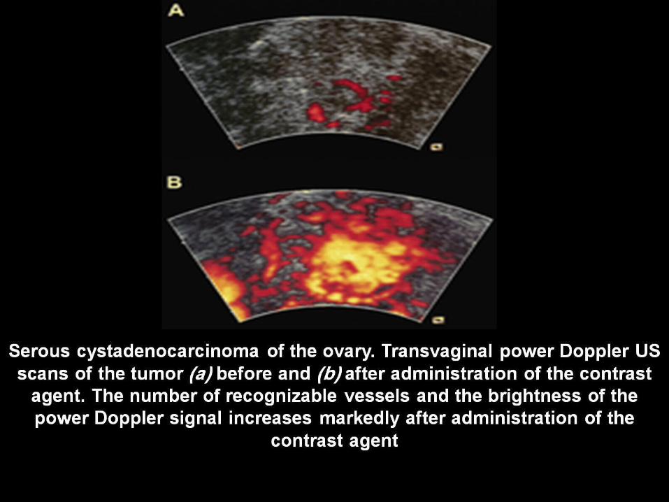
Serous cystadenocarcinoma of the ovary with peritoneal carcinomatosis in a 60-year-old woman .

Bilateral serous cystadenocarcinomas in a 50-year-old woman..
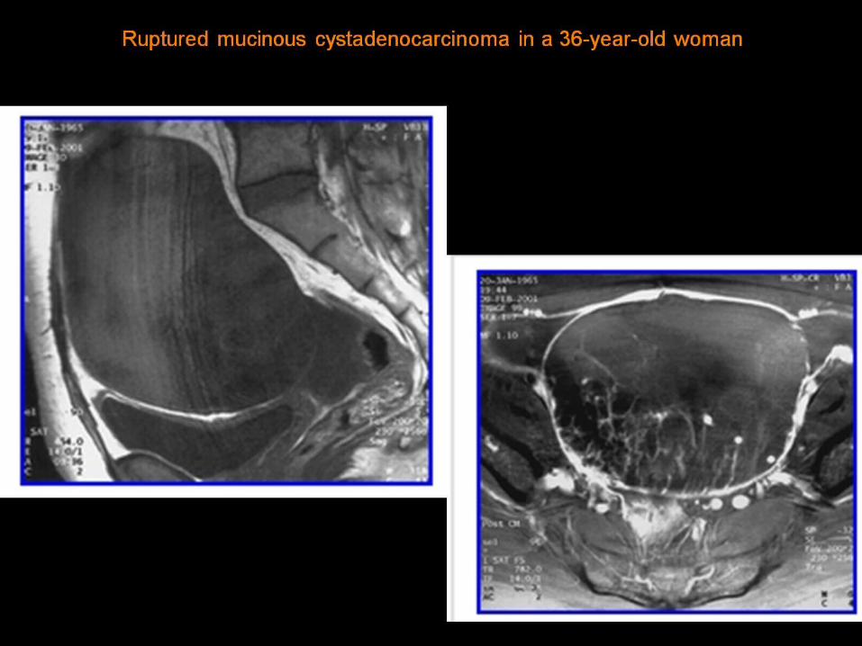
Ruptured mucinous cystadenocarcinoma in a 36-year-old woman

Borderline mucinous tumor in a 20-year-old woman .
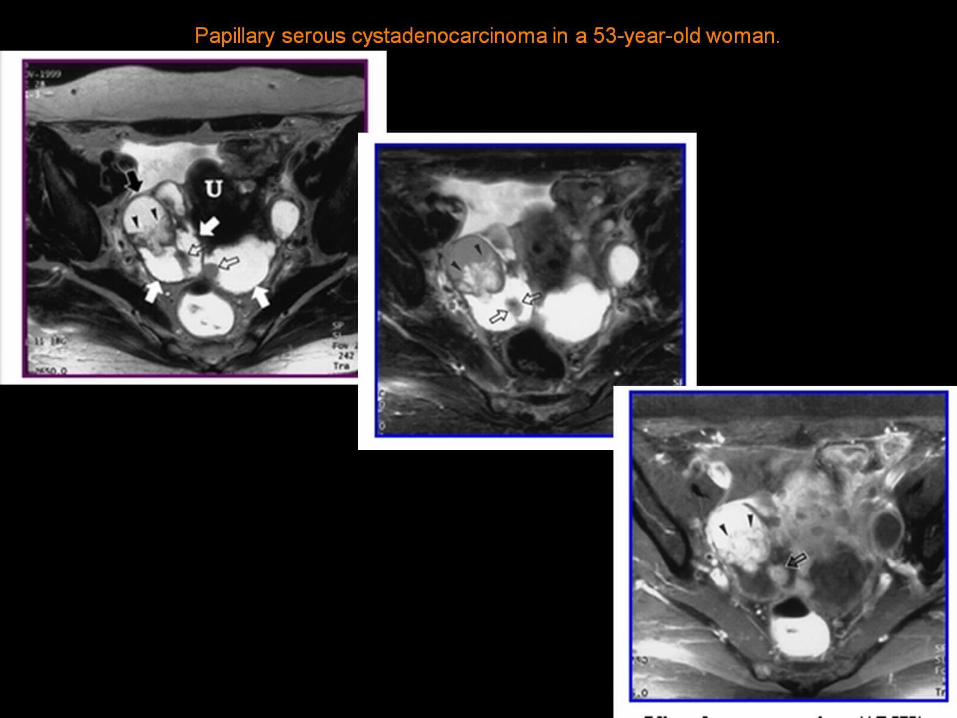
Papillary serous cystadenocarcinoma in a 53-year-old woman .

Endometrioid carcinoma of the ovary and endometrial carcinoma of the uterus in a 38-year-old woman .
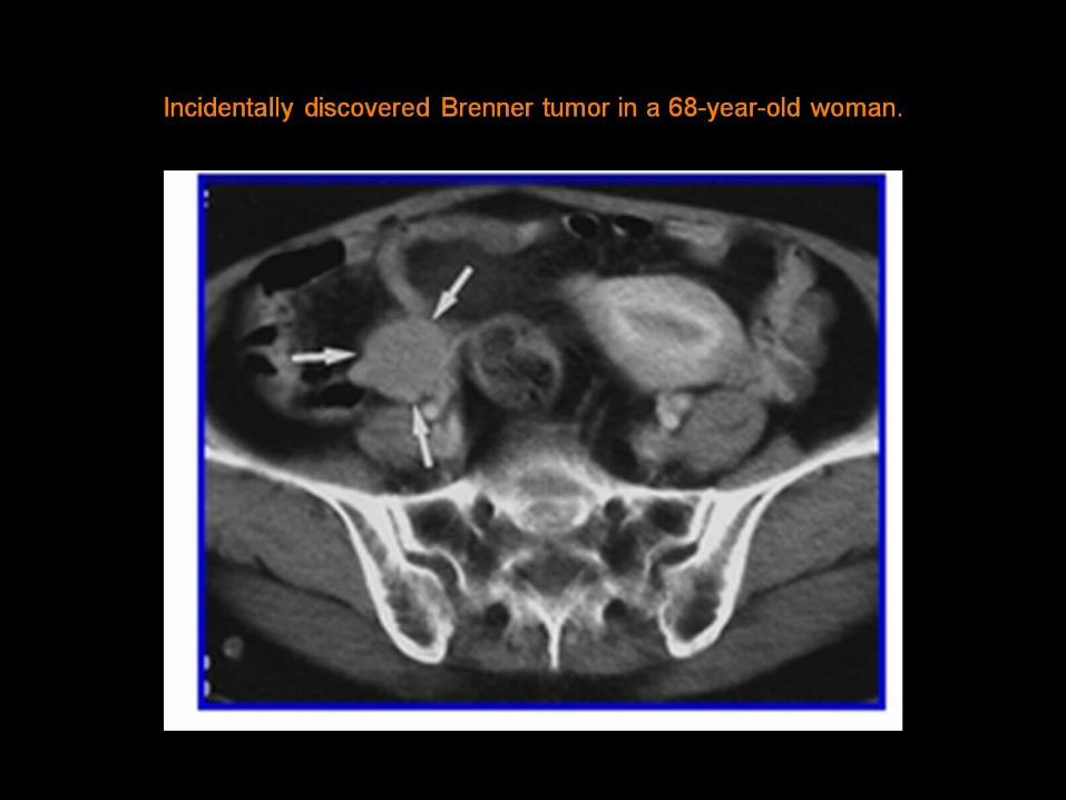
Incidentally discovered Brenner tumor in a 68-year-old woman .

Atypical mature teratoma with a purely cystic component in a 16-year-old girl .

Mature teratoma in a 22-year-old woman .
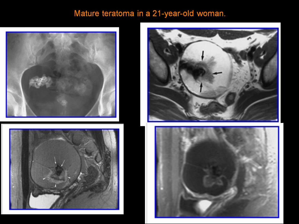
Mature teratoma in a 21-year-old woman .

Mature teratoma with a mainly fatty component in a 36-year-old woman .

Immature teratoma in a 23-year-old woman .

Ruptured immature teratoma in a 19-year-old woman .

Dysgerminoma in an 18-year-old woman .
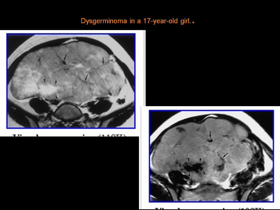
Dysgerminoma in a 17-year-old girl. .

Endodermal sinus tumor in a 29-year-old woman .

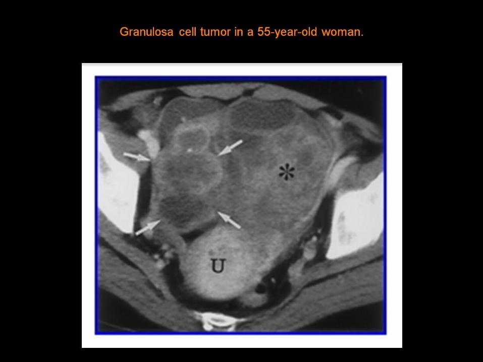
Granulosa cell tumor in a 55-year-old woman .

Granulosa cell tumor in a 71-year-old woman. (a) Sagittal turbo spin-echo T2-weighted MR image (b) Gadolinium-enhanced fat-suppressed FLASH T1-weighted MR image

Fibroma in a 53-year-old woman. (a) Conventional radiograph (b) Axial turbo spin-echo T1-weighted MR (c) On an axial turbo spin-echo T2-weighted MR

Fibrothecoma in a 46-year-old woman. (a) Axial turbo spin-echo T1 (b) On an axial turbo spin-echo T2 (c) Gadolinium-enhanced fat-suppressed T1

Subserosal leiomyoma with interface vessels in a 28-year-old woman. Sagittal turbo spin-echo T2

Sclerosing stromal tumor in a 28-year-old woman. Contrast-enhanced CT scan

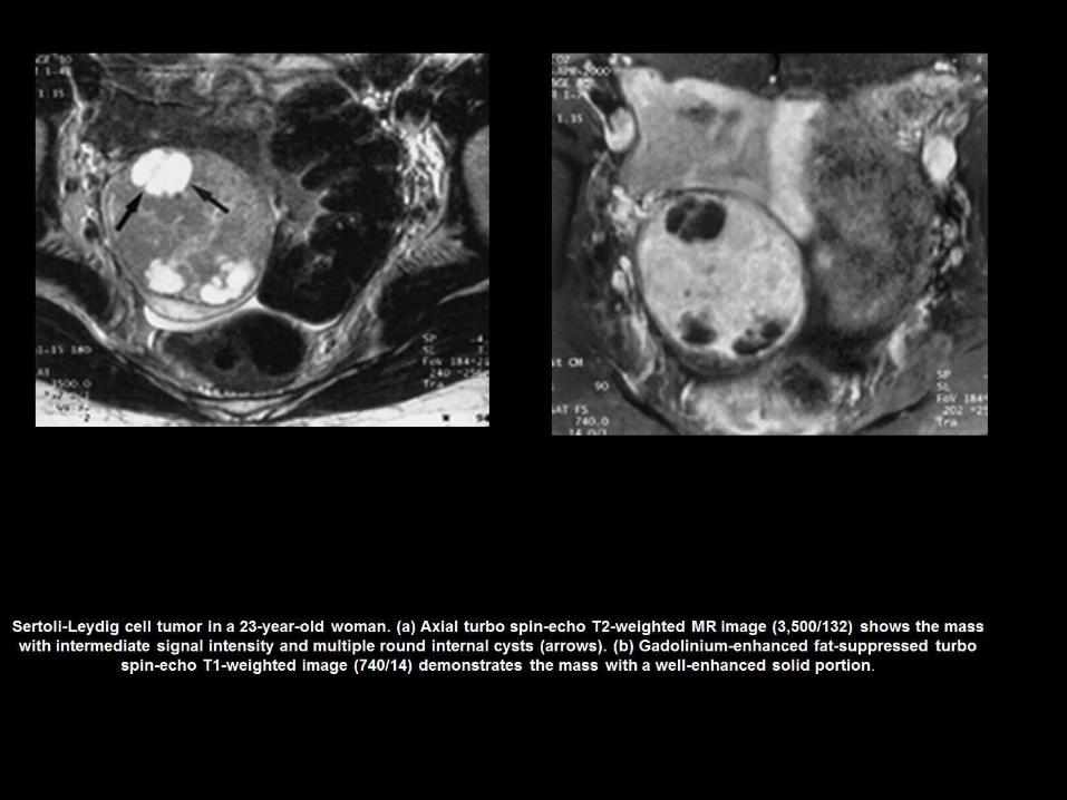
Sertoli-Leydig cell tumor in a 23-year-old woman. (a) Axial turbo spin-echo (b) Axial turbo spin-echo T2 (c) Gadolinium-enhanced fat-suppressed T1

Collision tumor (teratoma and mucinous cystadenoma) in a 46-year-old woman. (a) Contrast-enhanced CT scan b) Pelvic CT scan shows a multilocular cystic tumor (arrows) ,

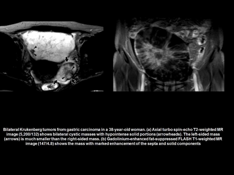
Bilateral Krukenberg tumors from gastric carcinoma in a 38-year-old woman. (a) Axial turbo spin-echo T2-weighted MR image (b) Gadolinium-enhanced fat-suppressed
FLASH T1-weighted MR


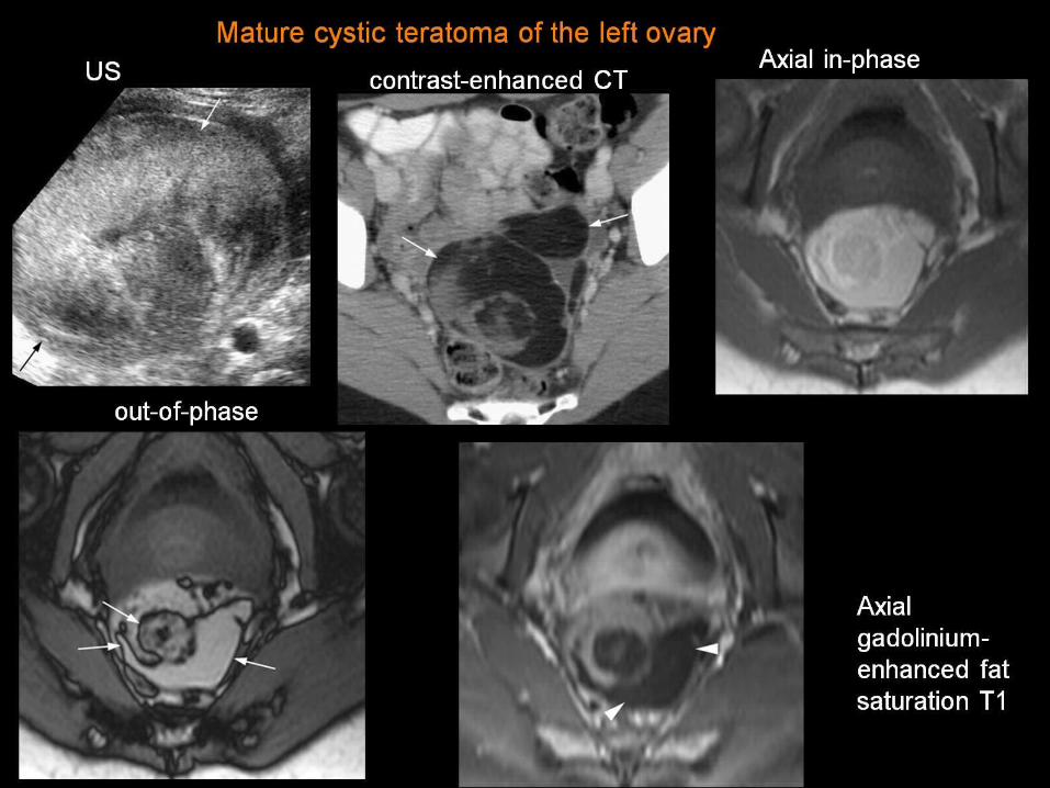
Mature cystic teratoma of the left ovary

Initial CT StudyOn computed tomography (CT) images, dilated, fluid-filled loops of small
bowel are evident. The transverse colon appears fairly normal

same patient before Post-Surgical CT Study

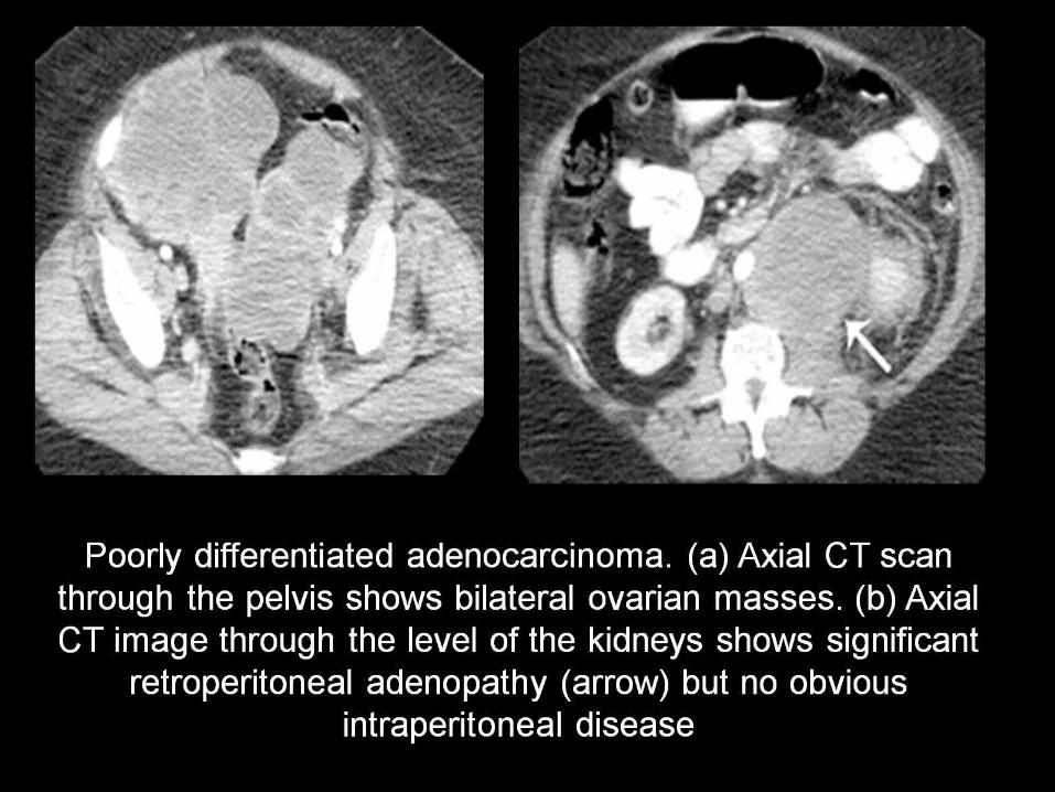
ROLE OF CT IN A PATIENT WITH OVARIAN MASS

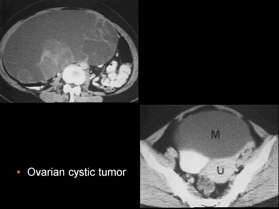
Ovarian tumour


Fig. 1. A 32-year-old woman with severe pelvic pain and a negative pregnancy test. Contrast-enhanced CT scan Ruptured Corpus Luteal Cyst

Fig. 2. A 32-year-old woman with severe pelvic pain and a negative pregnancy test.A. Non-enhanced CT scan of the pelvis
B. Contrast-enhanced CT shows that the thick wall of the cystic mass (arrow) is well enhanced, interruption is apparent (arrowheads

Fig. 3. A 34-year-old woman with lower abdominal pain and anemia.A. Non-enhanced CT scan of the pelvis hemoperitoneum.
B. Contrast-enhanced CT shows that the wall of the right adnexal lesion is well enhanced. In the cystic wall, suspicious interruptions (arrowheads) are apparent.

A 19-year-old woman with severe pelvic pain and a negative pregnancy test. At contrast-enhanced CT, the wall of the cystic mass (arrow) is well enhanced. Note the presence of high-attenuated fluid in the cul-de sac and dependent
portion of the cyst .
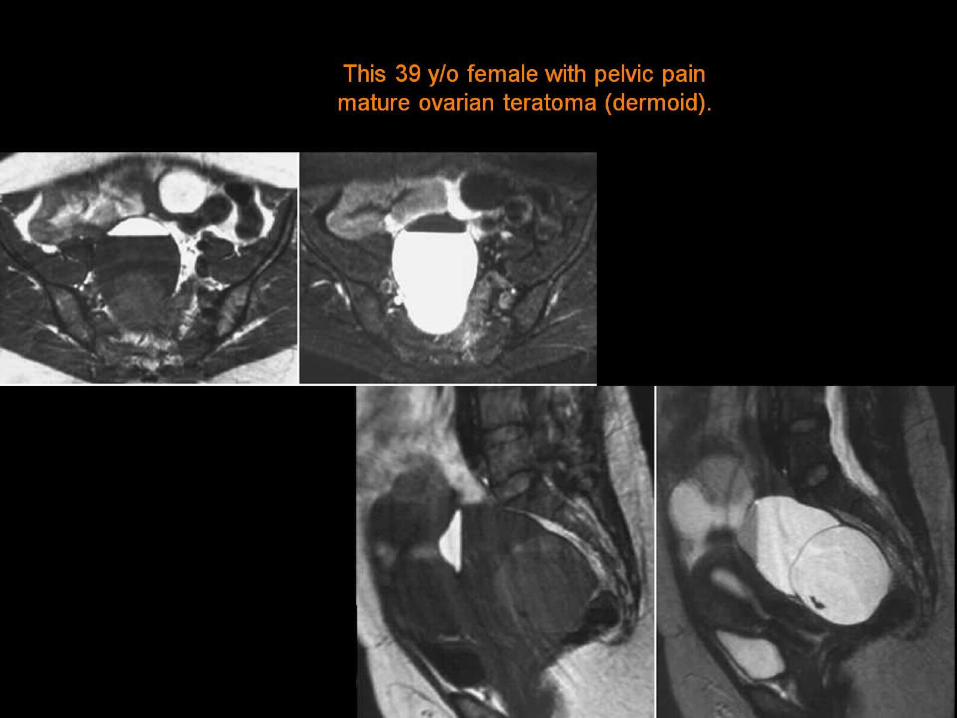
This 39 y/o female with pelvic pain mature ovarian teratoma (dermoid) .
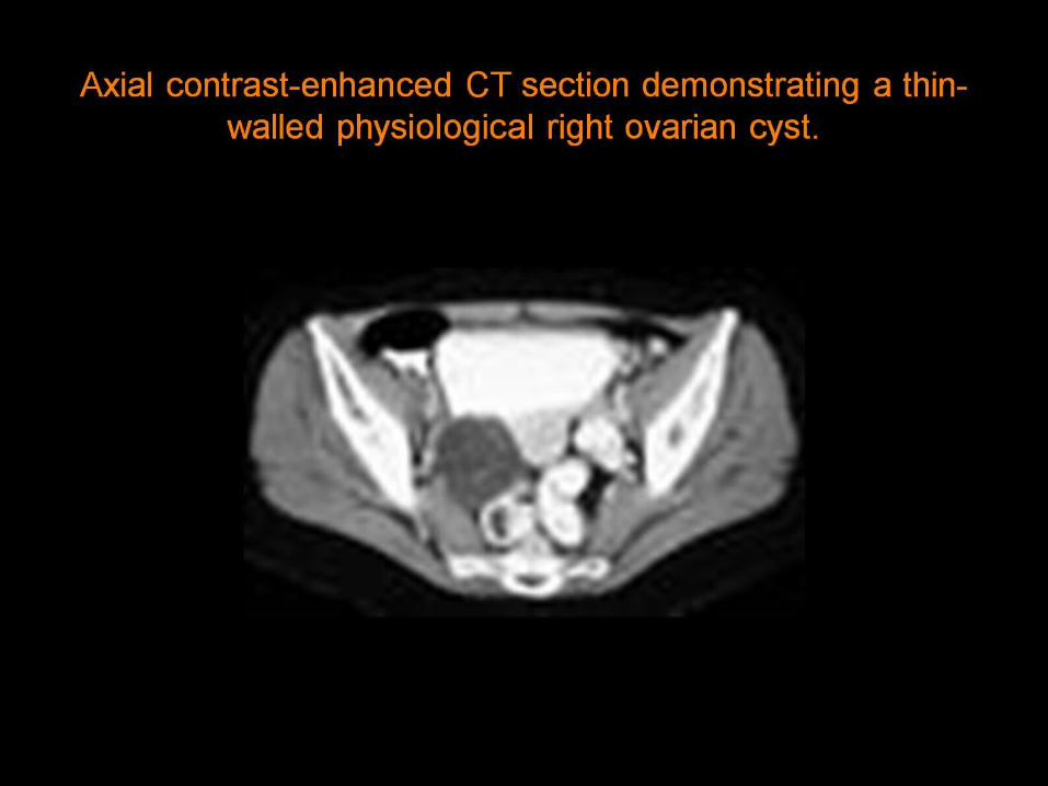
Axial contrast-enhanced CT section demonstrating a thin-walled physiological right ovarian cyst .

Axial contrast-enhanced CT section showing a right ovarian dysgerminoma (arrow) .


CT of the abdomen showing bilaterally enlarged cystic ovaries in a girl who had chronic hypothyroidism. b. CT of the pituitary fossa showing enlarged pituitary gland.
overstimulation of the ovaries secondary to hypertrophy of the adenohypophysis(
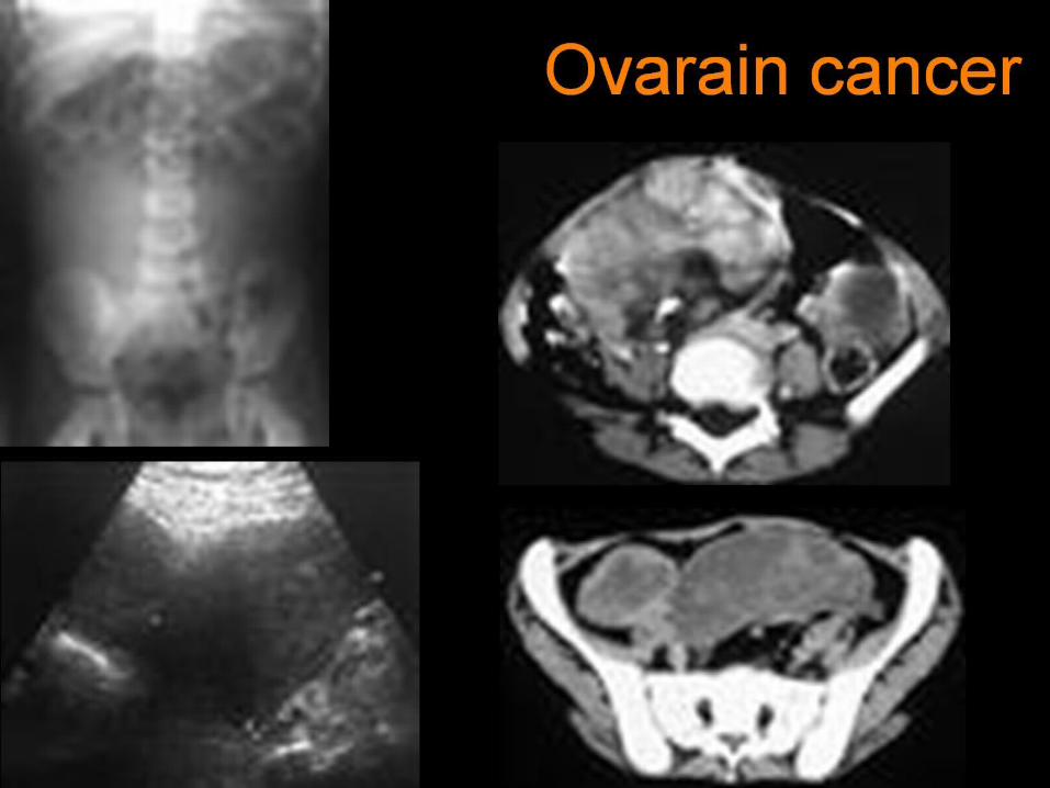
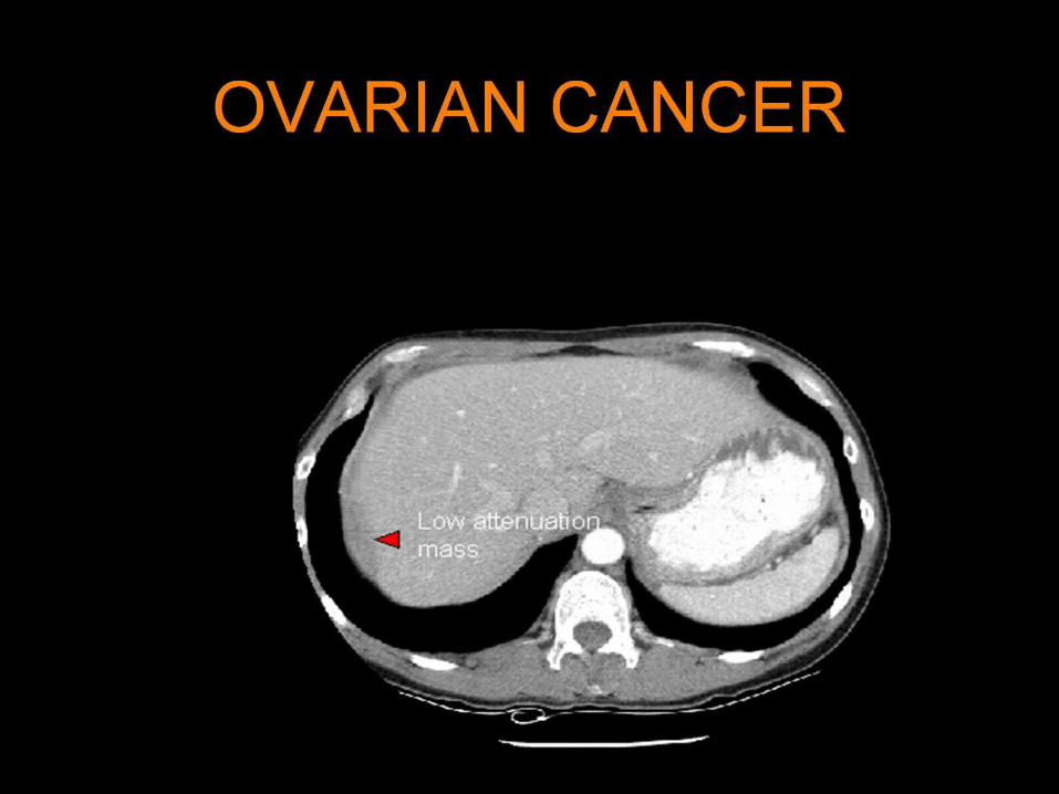
Ovarain cancer
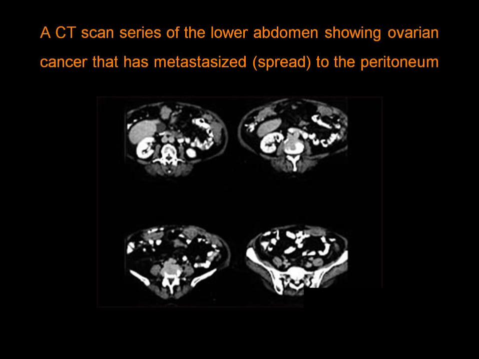
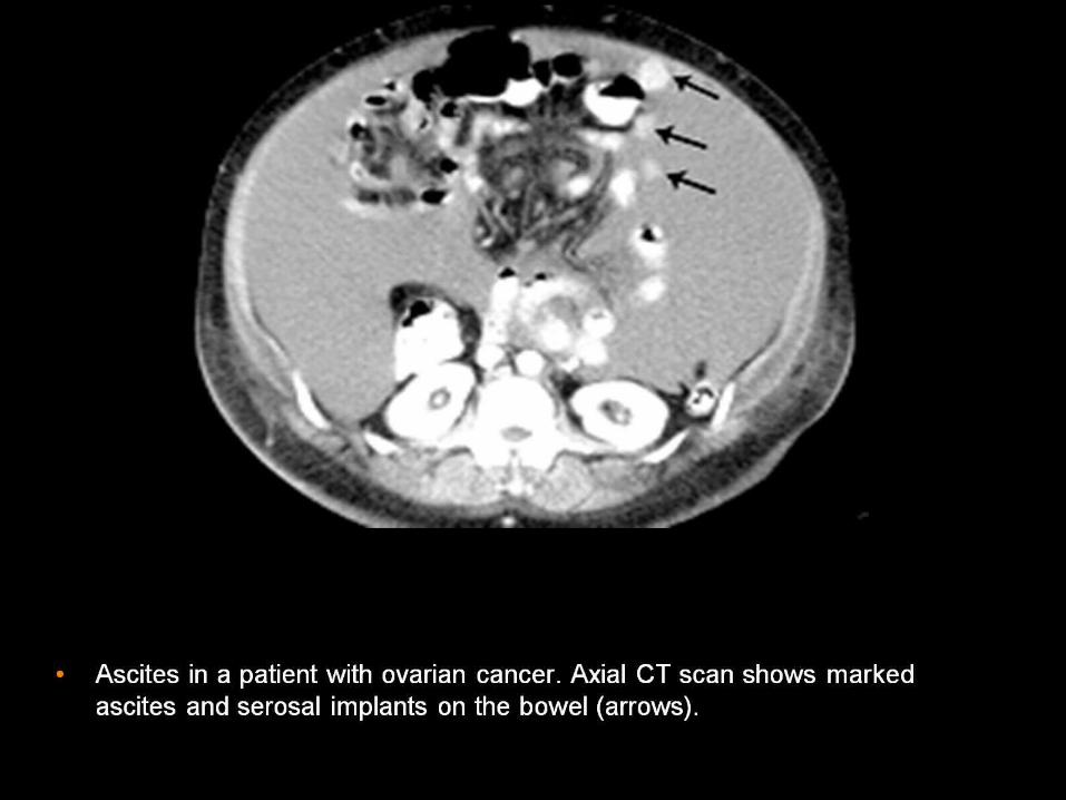
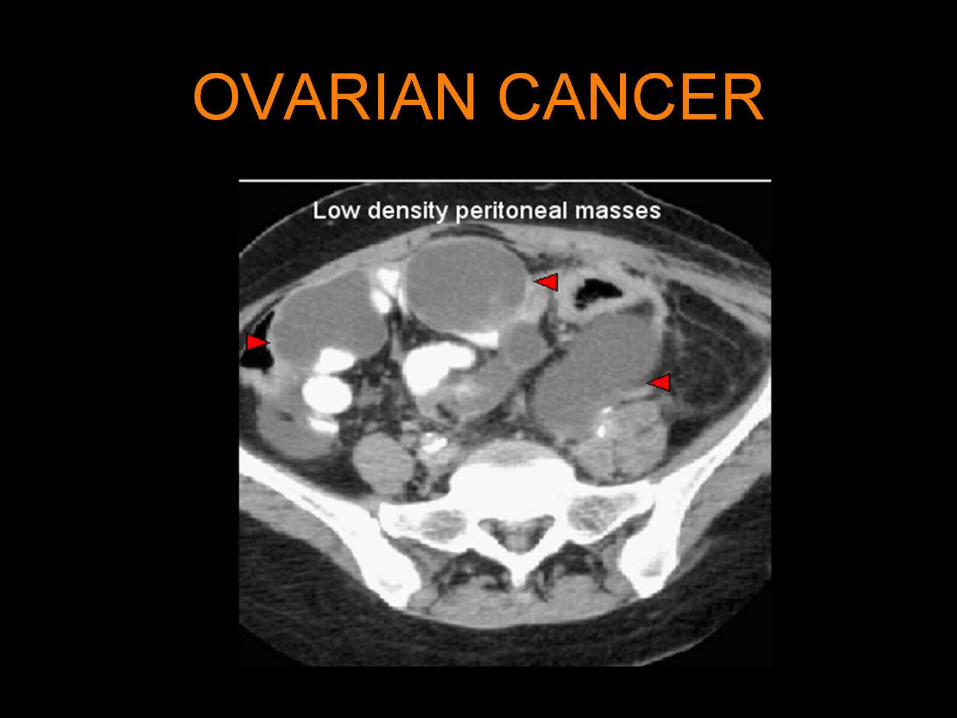
A CT scan series of the lower abdomen showing ovarian cancer
that has metastasized (spread) to the peritoneum


68-year-old woman with malignant transformation of left ovarian teratoma .

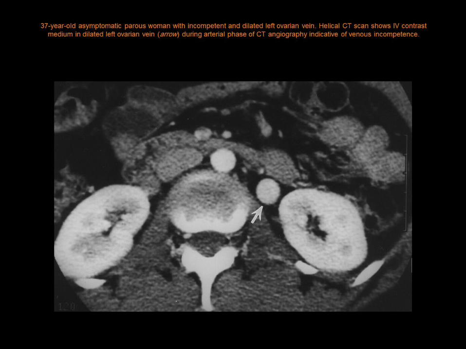
37-year-old asymptomatic parous woman with incompetent and dilated left ovarian vein. Helical CT scan shows IV contrast medium in dilated left ovarian vein (arrow) during arterial phase of CT angiography indicative of venous incompetence .

25-year-old asymptomatic parous woman with bilateral ovarian venous incompetence and dilatation. Helical CT scan obtained during arterial phase of CT
angiography shows retrograde filling of both right (large arrow) and left (small arrow) ovarian veins, which are dilated.

42-year-old woman with minimal reflux into nondilated ovarian vein. Helical CT obtained during arterial phase at cranial aspect of left ovarian vein shows contrast material in (A) left renal vein (open arrow) and in (B and C) most cranial portion of left ovarian vein (arrow). Six millimeters caudally to level of C (D), ovarian vein is less dense than on more cranial sections because of only
minimal reflux. Ovarian vein is not dilated (6 mm) and is located (B-D) lateral to lumbar vein (arrowhead) .
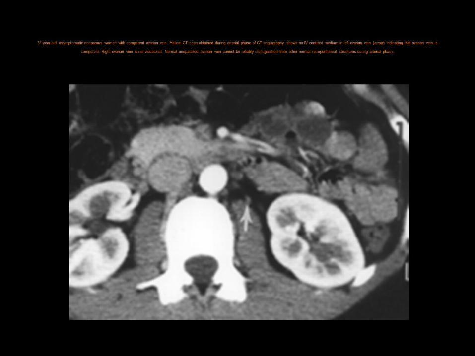
31-year-old asymptomatic nonparous woman with competent ovarian vein. Helical CT scan obtained during arterial phase of CT angiography shows no IV contrast medium in left ovarian vein (arrow) indicating that ovarian vein is competent. Right ovarian vein is
not visualized. Normal unopacified ovarian vein cannot be reliably distinguished from other normal retroperitoneal structures during arterial phase.
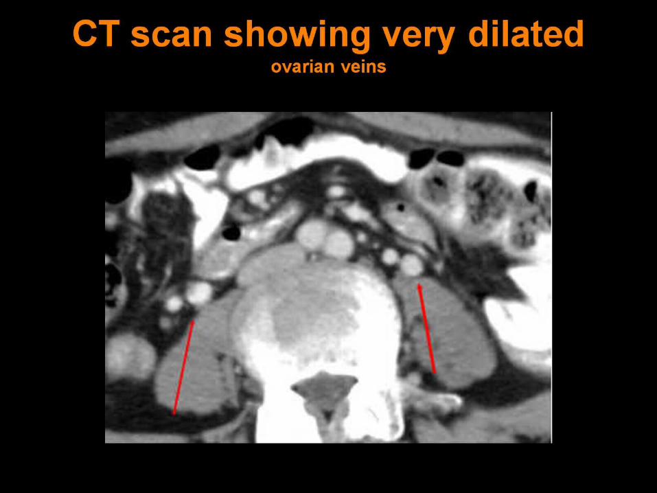
CT scan showing very dilated ovarian veins

CT scan showing pelvic varicosities, with a vein crossing anterior to the uterus to the right

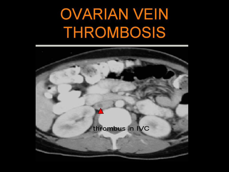
a) CT through the lower abdomen shows low attenuation thrombus distending the right ovarian vein. b) CT at the level of the kidneys shows thrombus extending into the IVC. c) CT shows a solid adnexal mass adjacent to the enlarged uterus .

Axial MRI through the lower abdomen shows high signal intensity thrombus filling the right ovarian vein .


Primary peritoneal echinococcosis masquerading as an ovarian cyst

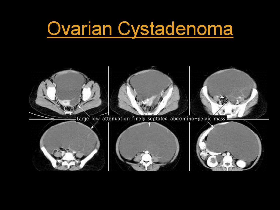
The CT scan showed a large 6 X 8 X 8 cm lobulated, right adenexal mass displacing the uterus anteriorly and to the left. Notice the collapsed rectum on image #3 & 4 adjacent to the left side of the
mass. Internal densities of the mass include soft tissue, calcium and fat (see CT scan below). Mature Cystic Teratoma (Dermoid Cyst)


Cystadenoma of the left ovary

Tumor implants- ovarian carcinoma

Ovarian dermoids

Ovarian carcinomatosis
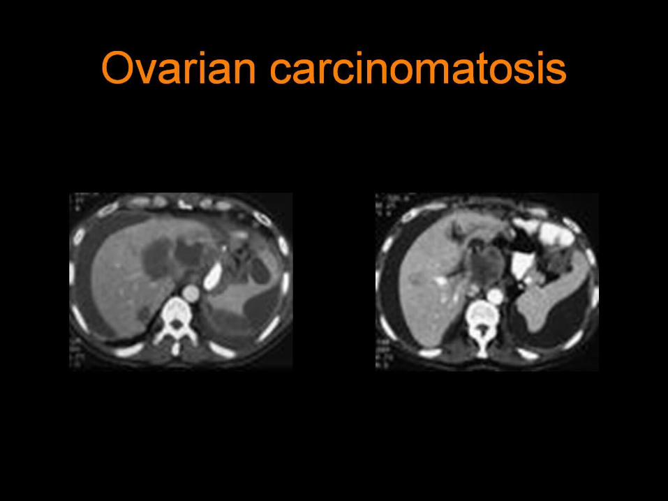
Ovarian carcinomatosis

invasive cervical cancer

invasive cervical cancer
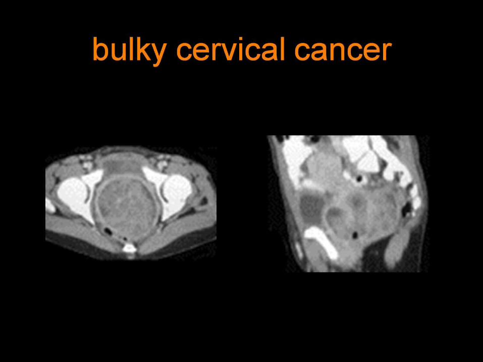
bulky cervical cancer
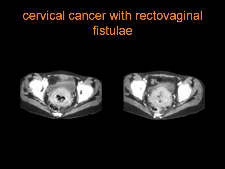
cervical cancer with rectovaginal fistulae

cervical stenosis due to cervical cancer

longitudinal image through the inferior vena cava shows a thrombus (arrows) with a freely floating upper margin.

Transverse image at the level of the right renal vein shows a thrombus (arrow) in the inferior vena cava

Color Doppler sonogram shows flow, encoded in blue (arrows), around the thrombus in the inferior vena cava
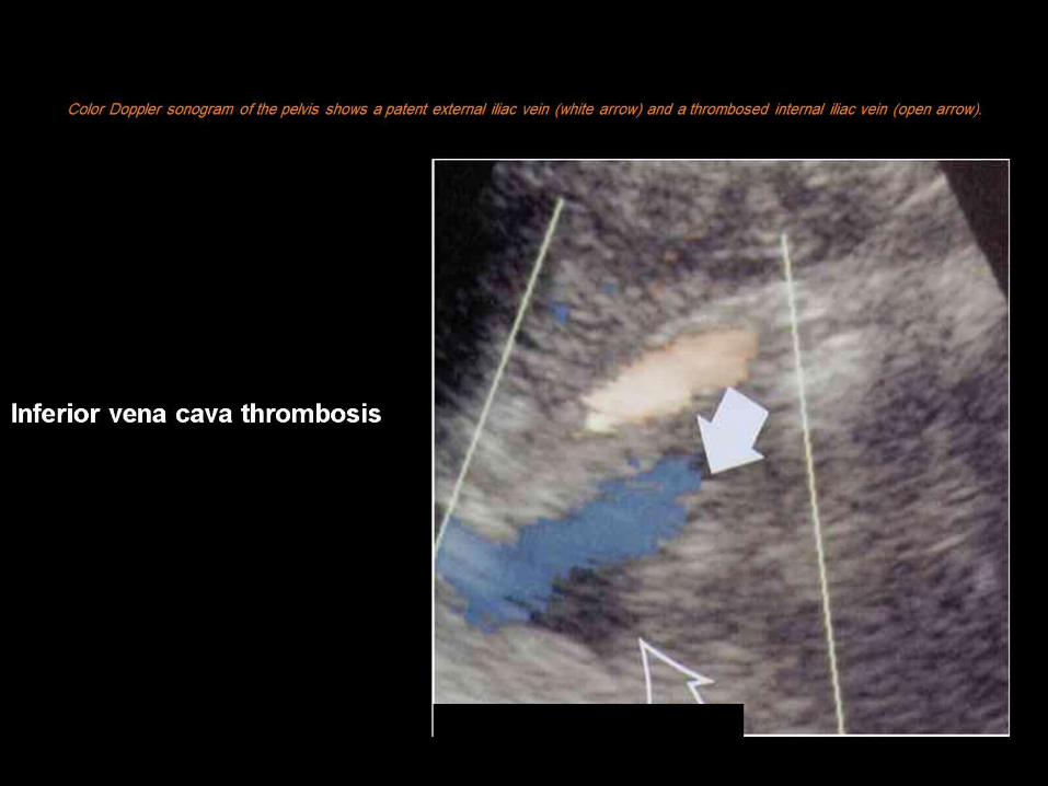
Color Doppler sonogram of the pelvis shows a patent external iliac vein (white arrow) and a thrombosed internal iliac vein (open arrow).

CT scan of the mid-abdomen shows thrombosis of the right ovarian vein (white arrow). The inferior vena cava is indicated by the black arrow. The metallic densities at the periphery of the cava are the edges of the Greenfield filter.

CT scan of the lower abdomen shows thrombus distending the right ovarian vein (arrow).

71 y/o female with abdominal pain ovarian fibroma with torsion pain

71 y/o female with abdominal pain Torsion of an ovarian fibroma

OVARIAN TOSION

FOLLICULAR CYST

FOLLICULAR CYST

OVARIAN VEIN THROMBOSIS

OVARIAN VEIN THROMBOSIS

OVARIAN VEIN THROMBOSIS

CT dermiod cyst

Fibriod cyst

Ovarian Cancer


OVARIAN CANCER

DERMIOD-TERATOMA

Benign cystadenoma

47-year-old woman with fever and right lower quadrant pain. Axial CT scan shows enlarged, nonenhancing, thrombosed right ovarian vein (arrows) with fat stranding (open arrows) in right lower quadrant adjacent to
normal appendix (A) .


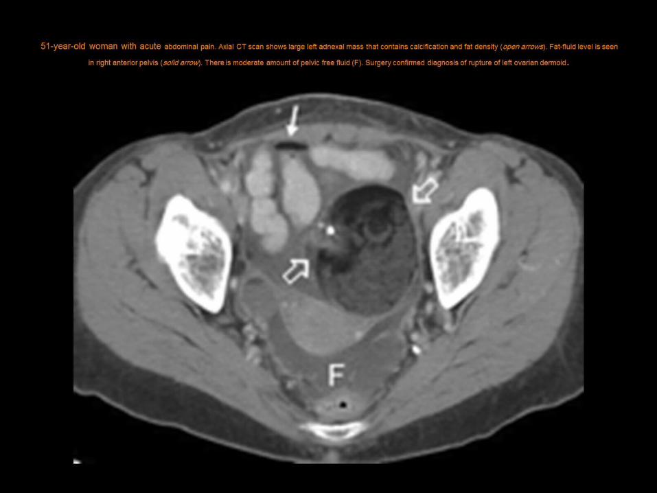
51-year-old woman with acute abdominal pain. Axial CT scan shows large left adnexal mass that contains calcification and fat density (open arrows). Fat-fluid level is seen in right anterior pelvis (solid arrow). There is moderate amount of pelvic free fluid (F).
Surgery confirmed diagnosis of rupture of left ovarian dermoid .

41-year-old woman with acute right lower quadrant (RLQ) pain and clinically suspected acute appendicitis. Axial CT scan shows large necrotic
leiomyoma (arrows) arising from right side of uterus (U). Minimal fat stranding is noted in RLQ adjacent to leiomyoma.

28-year-old woman with sudden-onset right lower quadrant (RLQ) pain. Axial CT scan shows fat stranding (arrows) predominating in RLQ posterior to
cecum (C) and appendix (A) and heterogeneous mass in upper pelvis representing enlarged ovary (O).

39-year-old woman with acute right lower quadrant pain. Axial CT scan shows minimal wall thickening of terminal ileum (asterisks) entering
cecum (C).

37-year-old woman with acute abdominal pain and clinical suspicion of perforated appendicitis. Axial CT scan shows fluid collection in right lower quadrant with attenuation of 77 H, consistent with blood (arrows). High-attenuation foci (arrowheads) are located to right of the uterus (U),
indicative of active bleeding. Rupture of ectopic pregnancy in right fallopian tube was confirmed surgically

47-year-old woman with fever and right lower quadrant pain. Axial CT scan shows enlarged, nonenhancing, thrombosed right ovarian vein (arrows) with fat stranding (open arrows) in right lower quadrant adjacent to normal appendix (A) .
51-year-old woman with acute abdominal pain. Axial CT scan shows large left adnexal mass that contains calcification and fat density (open arrows). Fat-fluid level is seen in right anterior
pelvis (solid arrow). There is moderate amount of pelvic free fluid (F). Surgery confirmed diagnosis of rupture of left ovarian dermoid .

41-year-old woman with acute right lower quadrant (RLQ) pain and clinically suspected acute appendicitis. Axial CT scan shows large necrotic leiomyoma (arrows) arising
from right side of uterus (U). Minimal fat stranding is noted in RLQ adjacent to leiomyoma.


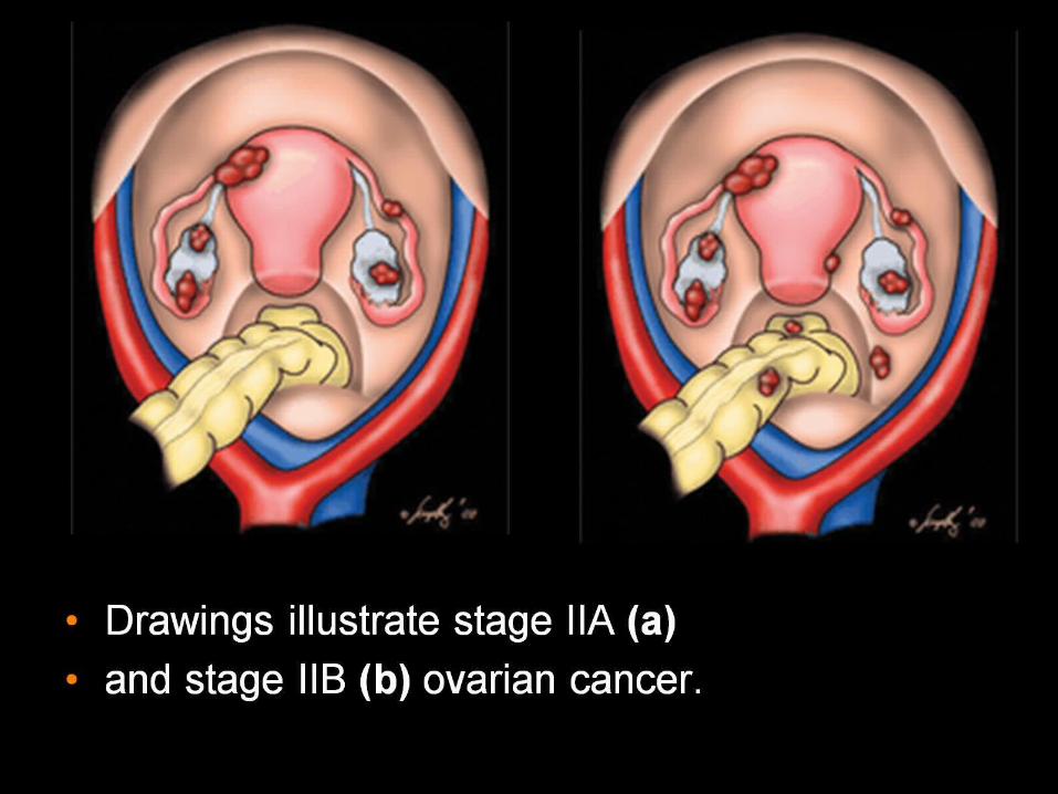
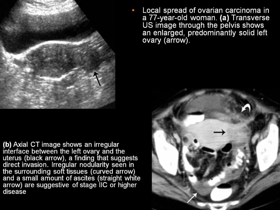
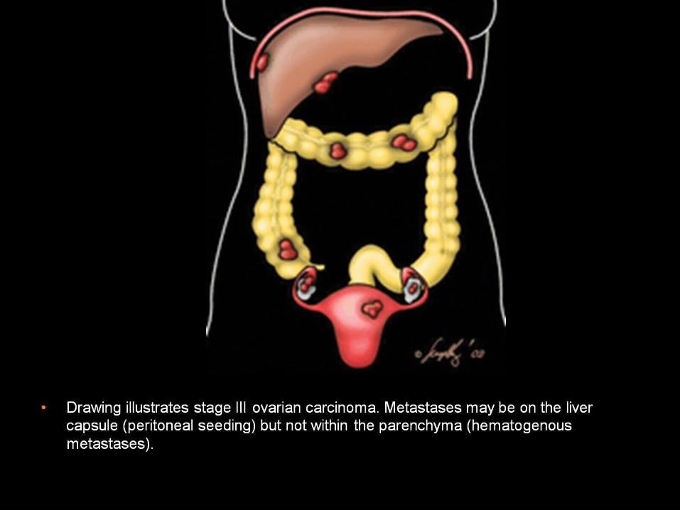
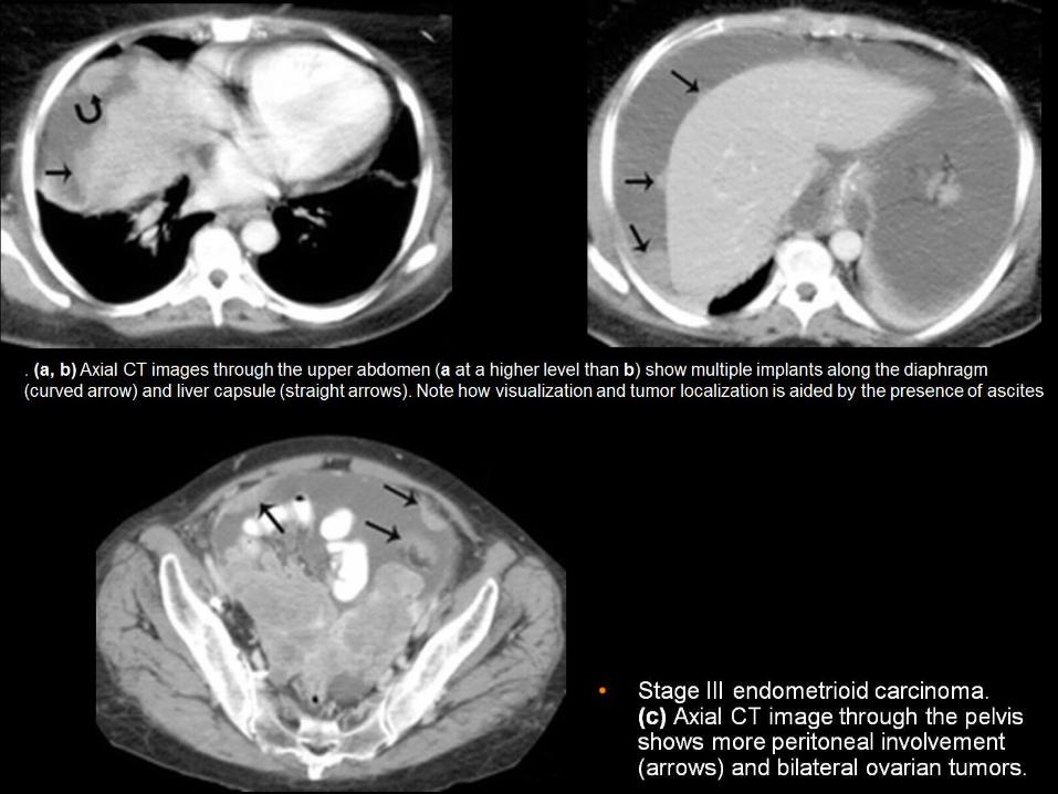
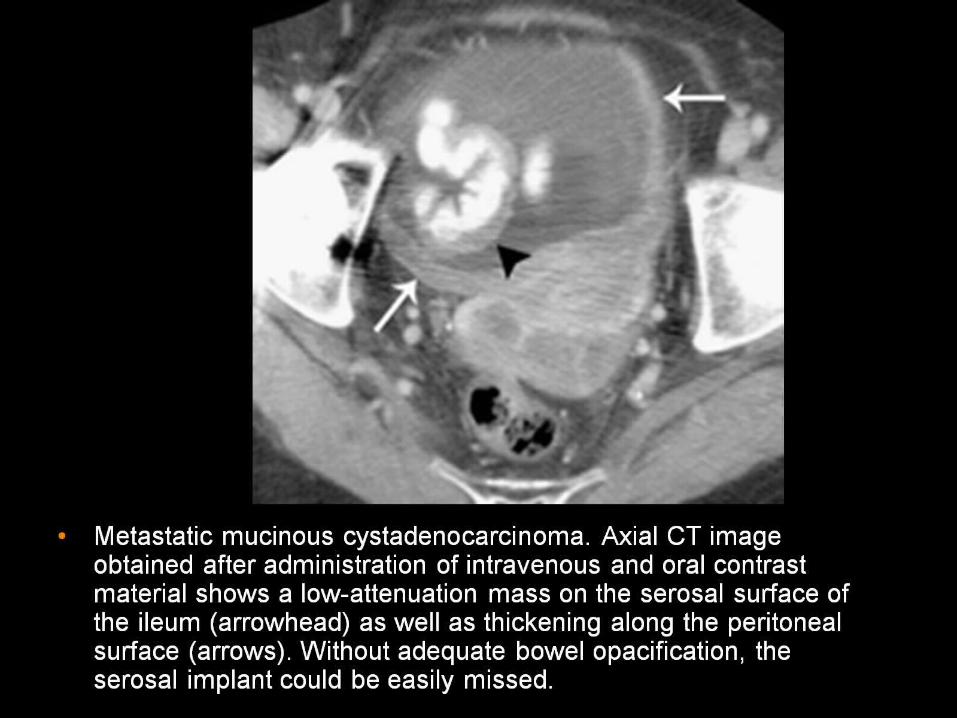
FIGO Staging of Ovarian Carcinoma


OVARIAN VEIN THROMBOSIS
OVARIAN VEIN THROMBOSIS

OVARIAN VEIN THROMBOSIS

OVARIAN VEIN THROMBOSIS

HYDROSALPNIX

Adnexal mass reveals fluid debris level consistent with hydrosalpnix


Ovarian Cancer

Ultrasound: Ovarian cancer


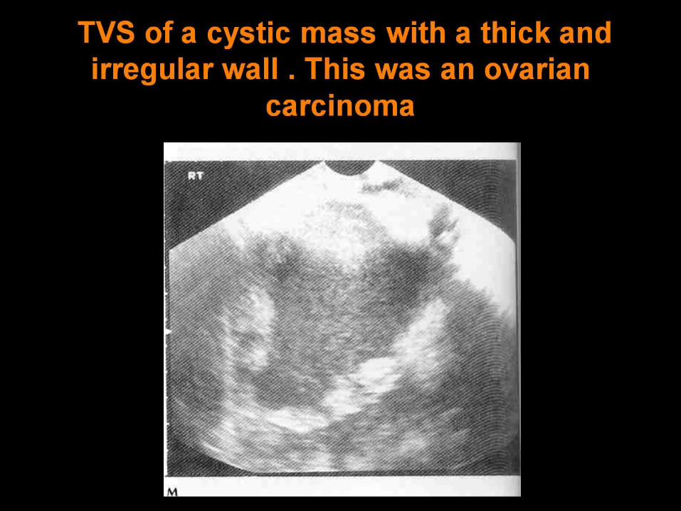
TVS of a cystic mass with a thick and irregular wall . This was an ovarian carcinoma

Papillary pro-jections ( arrow ) were found within this malignant teratoma


Malignant lesion


Ovarian cancer

Serous cystadenocarcinoma

Malignant lesion
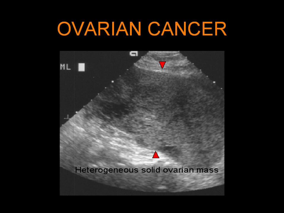
OVARIAN CANCER

OVARIAN CANCER

OVARIAN CANCER

OVARIAN CANCER
OVARIAN CANCER
Endometrial Cancer


Transabdominal sonogram of the cyst in Picture 7 demonstrating a large, complex, cystic mass with septations. Color Doppler image shows
vascularity within the septations. Red and blue colors show blood flow towards and away from the transducer. The resistive index was low.
Histology reported a mucinous cystadenocarcinoma of low malignant potential

OVARIAN MALIGNANCY
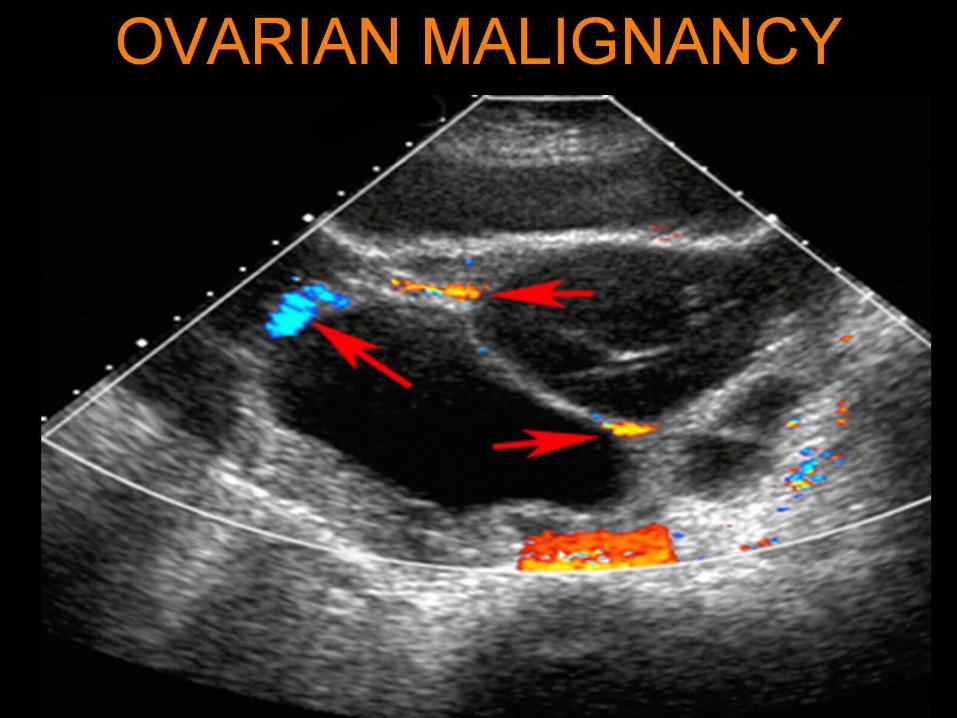
OVARIAN MALIGNANCY

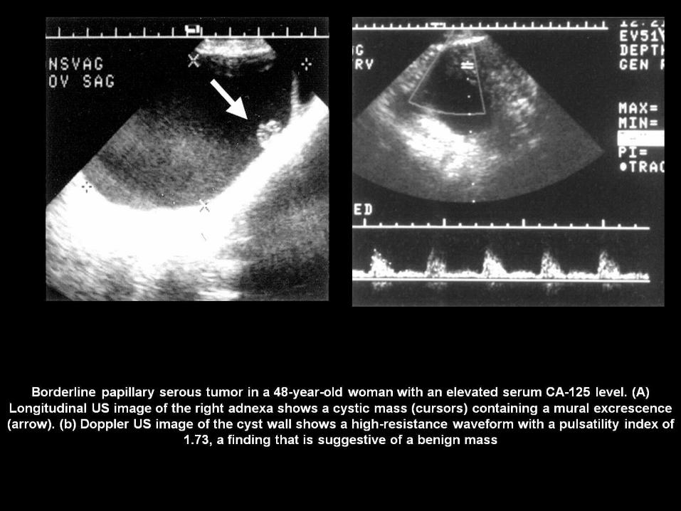
Borderline serous tumour
OVARIAN CANCER

OVARIAN CANCER