Embed Size (px)
Citation preview

Bio-Bank & Personalized MedicinePrimary cell models – phenotypic assays
Research & Clinical Support Services
A joint venture with
23-Oct-15 1

The ‘Essence’ of our Enterprise
23 October 2015 2
Vision: To be a premier global biotech that employs human translational platforms for the discovery & development of novel diagnostics, biomarkers & drugs with better clinical outcomes
Mission:• Build a systematic archive of ethically consented,
anonymized human samples & associated data for developing novel biomarkers & diagnostics
• Leverage primary human tissues & stem cells for disease modeling, target & drug discovery with higher rates of clinical success
• Use of tissues for R&D, NOT for regenerative purpose
Saarum Sciences & Sapien Biosciences were initially set-up as two separate entities. We are in the process of merging both operations into Sapien Biosciences for better synergy

23-Oct-15 Introduction to Saarum-Sapien 3
Data Associated With Our Samples
Current Inventory: >0.1 M Samples; Projected Inventory (in ~3 years): ~0.5-1M Samples
Sample diversity & associated data at Sapien

23-Oct-15 Introduction to Saarum-Sapien 4
Sample formats available
Viable/Live Cell Types Available
Sample Formats Available
Fresh: Live/snap-frozen

R&D Infrastructure available to Sapien
23-Oct-15 Introduction to Sapien Biosciences 5

PBMCs, B/T cells, CD34+, HUVECs, fibroblasts etc.
In vitro cell-based assay systems, models
6
Breast Cancer, Glioma, Head & Neck
cancers etc.
ID & validation of drug targets & Companion Dx
Breast Cancer, Glioma, Prostate
Cancer etc.
Lead-optimization of novel cancer-active
molecules
Breast cancer model with in-built readouts
Screening drugs for metastasis & cancer
stem cells
Cytokine release, Chemoattraction,
Migration etc.
Phenotypic screening of novel drugs
Current Offerings
Applications
Patient samples and associated data
OncoBloc™ OncoPrime™ MetBlock™ TruCell™ TruScreen™
Fixed & frozen sample collections
Patient-derived live cancer cells
Novel model of metastasis (EMT)
Patient/Normal cells, derivatives
Disease-relevant functional assays
Platform
Our translational research products and platforms

23-Oct-15
Ou
r G
liom
a co
llect
ion
Calcein (0.5 uM) PI (0.5 uM) Merged
10 X 10 X 10 X
10 X 10 X 10 X

23-Oct-15
Glioma Patients Samples and data
107 4242
Total no. of Glioma cases n = 149
FFPE cases
Fresh frozen
Grade* No.
I 2
II 19
III 7
IV 13
Gender
Male 22
Female 20
Year wise distribution of 42 cases
No of cases in each year
Availability of FFPE blocks for the no of cases
2013 18 17
2014 23 21
2015 1 1

23-Oct-15
Available data set with the cases
Patient Demographics
1 Gender
2 Race
3 Nationality
4 Age
Surgery details
1 Date of Surgery
2 Surgical Procedure
3 Surgical Margin (if any)
4 Location
Chemotherapy details
1 Date of Chemotherapy
2 Date of Chemotherapy Ended
3 Drug Name
4 Number of Cycle
5 Location
Radiotherapy details
1 Date of Radiotherapy
2 Date of Radiotherapy Ended
3 Radiation volume, fraction, site
Outcome details
1 Date of Last Contact
2 Vital Status
3 Cancer Status
4 Cause of death
5 Death Status
6 Recurrence, if any and type

23-Oct-15
Age distribution, Recurrence, samples in culture
13
15
7
8
5
3
No of Patients n = 42
0 to 20 yrs
20 to 30 yrs
30 to 40 yrs
40 to 50 yrs
50 to 60 yrs
60 to 70 yrs
70 and Above yrs
Tissue samples = 42
Data available = 37
Newly diagnosed cases = 30
Recurred cases = 7
Recurred samples with old
blocks = 2

23-Oct-15
Treatment Information for the 7 Recurred Cases at onset and during recurrence respectively
Treatment No of Patients
Only Surgery 1
Surgery + Adj. CT + RT 3
Surgery + Con. CT + RT 3
1
3
3
No of Patients
Only Surgery
Surgery + Adj. CT+ RT
Surgery + Con.CT + RT
Treatment No of Patients
Only Surgery 1
Surgery + RT 1
Surgery + Con. CT + RT 5
1
1
5
No of Patients
Only Surgery
Surgery + RT
Surgery + Con. CT +RT

23-Oct-15
Recurrent samples – details
SB IDs Age Sex DiagnosisType of Glioma
Site of tumour
Type of SurgeryChemotherapy: Drugs/Regimen/
No of Cycles
Concurrent Chemotherapy
Radiation Therapy:
Type/Dose/Fractions
Is Serum Available?
SB00008528 54 FRt Frontotemporal
glioma
AstrocytomaGrade II
Rt Frontotemporal
Rt Frontotemporal Craniotomy and
excision of tumourCT not given
Cap Temozolomide -
100 mg
Ext : 5580 cGY/31 frs
No
SB00006077 35 FLt Temporal
glioma
Anaplastic Astrocytoma
Grade II
Lt Temporal region of
brain
Lt Temporal Craniotomy in Lt
insular regionCT not given
Concurrent CT not given
Ext : 3600 cGy/20 frs
No
SB00005972 45 FRt Insular
glioma
Anaplastic Astrocytoma
Grade II
Rt Insular region of
brain
Rt Insular Craniotomy and
Parietal excision of tumour
Adj : Cap Temozolomide x 5
days for cycles
Concurrent CT not given
Ext : 5580 cGY/31 frs
No
SB IDs Type of Tumour for RecurrenceSite of
RecurrenceType of Surgery at
Recurrence
Concurrent Chemotherapy : Drugs/ Regimen (Treatment
at Recurrence)
Radiation Therapy: Type/Dose/Fractions(Treatment at Recurrence)
Is Serum Available?
SB00008528 Glioblastoma Multiforme, Gr - IVRt Frontotemporal
Rt Frontotemporal Craniotomy and
excision of frontal lesion
Cap Temozolomide - 120 mg during RT
Ext : 5800 cGY/ 329 frs No
SB00006077 Anaplastic Astrocytoma Gr - III
Lt Frontotemporal region of
brain
Lt Fronto-temporal Craniotomy and Ant Temporal lobectomy
Concurrent CT not given Ext : 5040 cGY/ 28 frs No
SB00005972 Anaplastic Astrocytoma Gr - IIIRt Insular region of
brain
Redo Craniotomy and gross toal
excision of tumour
Cap Temozolomide - 120 mg during RT
Ext : Dose PH- I- 4500 cGY/ 25 frs and PH-II - 5580
cGy/31 frsNo
Ori
gin
al T
um
or
Re
curr
en
t Tu
mo
r

23-Oct-15
Glioma primary cultures - ~ 20 patient samples cultured
No Sapien ID Pathology Age (Sex) Serum Plasma DNA FFPE
#1 SB.0000305 Gemistocytic astrocytoma Grade II 58 (M) Yes No No Yes
#2 SB.00000474 Anaplastic Oligodendroglioma WHO Grade III 49 (M) No Yes No Yes
#3 SB.0006129 Anaplastic Astrocytoma WHO Grade III 32 (M) No No Yes Yes
#4 SB.0000295 Anaplastic Astrocytoma, WHO Grade III 36 (F) Yes No No Yes
#5 SB.0006077 Anaplastic Astrocytoma WHO Grade III 35 (F) No No Yes (*RT) Yes (*RT)
#6 SB.0005972 Anaplastic Astrocytoma, WHO Grade III 45 (F) No No No6 *OT + 2
*RT
#7 SB.0008528 Glioblastoma Multiforme, WHO Grade IV 54 (F) No No NoYes
(*RT)
#8 SB.0005980 Glioblastoma Multiforme, WHO Grade IV 56 (M) No Yes No Yes
#9 SB.0000298 Glioblastoma Multiforme, WHO Grade IV 35 (F) No No No Yes
#10 SB. 0000038 Glioblastoma Multiforme WHO Grade IV 58 (M) No No No Yes
#11 SB.0000085 Glioblastoma Multiforme, WHO Grade IV 54 (F) Yes Yes Yes Yes
#12 SB.0000144 High Grade Glioma 38 (F) No Yes Yes No
Note : Glioma samples marked with YELLOW are recurrent patient samples.*OT – Original Tumor, *RT – Recurrent Tumor

Cell lines are poor model of cancer leading to drug failures
CD133 +ve cells : 35%
CD133 +ve cells : 1.5 %
CD133 +ve cells : 11%
CD133 +ve cells : 2%
Monolayer culture (2D)
Neurospheres(3D)
U87MG
SB.30648Patient derived
culture
Observation: Flow cytometry detection of cancer stem cells (CSCs) by CD133-PE antibody staining in U87MG cells and glioma patient-derived cells inboth monolayer and neurosphere cultures. The percent CD 133 positive cells for 2D cultures were 2% (U87MG) and 11% (SB.30648). Whereas percentCD133 positive cells for 3D cultures were 1.5% (U87MG) and 35% (SB.30648).
The low percent of CD133 positive cells for U87MG in 3D cultures were also showed by Vacas-Oleas et al (vol 2 , issue 1, 2013) published in OpenAccess Scientific reports.
U87MG
SB.30648Patient derived
culture

23-Oct-15
Glioma cultures in 2D - GBM (SB.8528) & U87MG Cell Line
SB.8528
100 µm
2D SB.8528
100 µm
2D
Day 4 Day 6
U87MG
200 µm
2D
Day 10
U87MG
200 µm
2D
Day 15
Rate of Proliferation
SB.8528 28 h
U87MG 24 h

23-Oct-15
Characterization of Glioma cultures: Localization of Beta-III Tubulin
Culture: High Grade Glioma (SB-144)Antibody Staining: Beta-Tubulin
A B
Observations: Beta-III-tubulin is majorconstituent of microtubules. In theabove figure beta-III-tubulinimmunofluorescence staining isobserved on cytoskeleton of most cells.
Primary Ab: Mouse Anti-Tubulin, beta III (Millipore)Ab Concentration; 250ug/mlAb dilution used: 1:100
Secondary Ab: Goat Anti-Mouse IgGAlexaFluor 488(Green) (Molecular Probes)Ab Dilution: 1:1000
DA
PI
20 X
No 1°
Ab
Introduction to Sapien and Saarum

23-Oct-15
Localization of Glial Fibrillary Acidic Protein by Immunofluorescence
Culture: Anaplastic Astrocytoma (SB.6129)Antibody Staining: GFAP
A B
Primary Ab: PurifedMouse Anti-GFAP (BD Inc)Ab dilution 1:100
Secondary Ab: Goat Anti-Mouse IgGAlexaFluor 488 (Green) (Molecular Probes)Ab Dilution; 1:1000
DA
PI
No 1°
Ab
Observations: GFAP is
an intermediate
filament protein present
in astrocytes. GFAP is
co-localized in
cytoskeleton of the cells.

Glioma cultures - 3D - High grade Glioma (SB.144) & U87MG
SB.144 SB.144
10X 10X
10X 10X
3D U87MGU87MG 3D
3D 3D
We have successfully cultured
high grade gliomas (SB.144
shown here) and U87MG cells in
serum free media on non-treated
plates for 10 to 15 days to form 3
Dimensional spheres known as
neurospheres. Such neurospheres
better represent human tumor
biology in vitro and can be used
for drug screening.
CSC evaluation is underway

Fluorescent staining of Anaplastic astrocytoma neuro-spheres (SB.6129) by Calcein-AM & PI
Calcein (0.5 uM) PI (0.5 uM) Merged
10 X 10 X 10 X
Calcein (1 uM)
10 X
PI (0.5 uM)
10 X
Merged
10 X
Observation: Anaplastic astrocytoma (SB.6129) cells were cultured in non-treated 6 cm dishes in serum-
free medium as spheroid cultures (neurospheres) for 7 days. Then, neurospheres from Passage 2 were
stained with Calcein-AM and Propidium Iodide to detect the live (Green, Calcein-AM) and dead (Red, PI)
cells within neurospheres. Image shows that more than 95% of cells in neurospheres were alive.

H & E staining of SB.6129 neurospheres as Paraffin embedded spheroids
Observation: Anaplastic astrocytoma cells (SB.6129) were cultured in non-treated 6 cm dishes in serum-
free medium as spheroid cultures (neurospheres) for 7 days. Then, egg albumin cell blocks were made
with these neurospheres, followed by formalin fixation and processing for paraffin block preparation.
Image above is an H&E stained section from the block showing a group of tumor cells with well preserved
nuclei and cytoplasm. Dead cells stain black. Only 4-5 cells appear to be dead in the entire neurosphere
confirming >95% cells being viable, as also shown by calcein staining in the previous slide.
Arrow indicates
dead cells stained
black
Comparison of CD 133 +ve cells (cancer stem cells) between monolayer (2D) vs Neurospheres (3D) cultures in GBM cell line (U87MG) & Desmoplastic infantile Ganglioglioma, Grade-I (SB.30648)
CD133 +ve cells : 35%
CD133 +ve cells : 1.5 %
CD133 +ve cells : 11%
CD133 +ve cells : 0.3%
CD133 +ve cells : 2%
CD133 +ve cells : 0.6 %
Monolayer culture (2D)
Neurospheres(3D)
U87MGU87MG
SB.30648SB.30648
40 X
U87MG
U87MG
2D
100 X
3D
100 X
SB.306482D
100 X
SB.306483D
Observation: Flow cytometry detection of cancer stem cells (CSCs) by CD133-PE antibody staining in U87MG cells and glioma patient-derived cells in both monolayer andneurosphere cultures. The percent CD 133 positive cells for 2D cultures were 2% (U87MG) and 11% (SB.30648). Whereas, percent CD133 positive cells for 3D cultures were 1.5%(U87MG) and 35% (SB.30648). The low percent of CD133 positive cells for U87MG in 3D cultures were also showed by Vacas-Oleas et al (vol 2 , issue 1, 2013) published in OpenAccess Scientific reports.
Unstained cells Unstained cells

Titrating a known Cytotoxic Drug in 3D Glioma cultures
Ab
sorb
ance
@ 4
50
nm
Bendamustine conc (uM)
Dose Response Curve
0.00
0.50
1.00
1.50
2.00
2.50
3.00
3.50
Observation: U87MG and one high grade glioma cell culture was
treated with Bendamustine at the concentrations indicated above. Dose
response curves were used to calculate IC50 values; the calculated IC50
value for both cell types was ~300 uM.

Dose-response of Compound B in Anaplastic Astrocytoma Neurospheres
• Compound B concentrations tested were 468 nM, 938 nM, 1.8 uM, 3.75 uM, 7.5 uM, 15 uM & 30 uM.
• Drug treatment was performed for 3 days (72 h) in triplicate.
• After incubation, WST-1 was added to samples and incubated for 3 h.
• The absorbance was read at 420 nm & 630 nm.
Observation: Treatment of primary anaplastic astrocytoma neurospheres with Compound
B demonstrated significant inhibition of viability, with IC50 between 0.9-1.9uM.
0
0.1
0.2
0.3
0.4
Ab
sorb
ance
(4
20
-6
30
) n
m
Drug Concentration (uM)
SB.6129 neurospheres treated with Compound B
SB - 6129 Sample SD
Cells alone 0.36 0.01
468 nM 0.27 0.07
938 nM 0.18 0.05
1.88 uM 0.19 0.03
3.75 uM 0.12 0.04
7.5 uM 0.09 0.03
15 uM 0.1 0.06
30 uM 0.06 0

Migration assay for Anaplastic astrocytoma (SB.6129)
DMSO 0 h DMSO
40 X
DMSO
0 h Benda (300 uM)
40 X
48 h24 h
24 h 48 h
Benda (300 uM)
Benda(300 uM)
40 X 40 X
40 X40 X

Migration assay for Anaplastic astrocytoma (SB.6129)
Observation: Treatment with Bendamustine (300 µM) effectively prevented the migration of anaplastic astrocytoma (SB.6129) cells at 24h & 48h. DMSO control showed filling of created open wound in 48 h.
98.33
47.65
93.18
5.94
96.88
DMSO DMSO BENDAMUSTINE DMSO BENDAMUSTINE
0 H 24 H 48 HP
erce
nta
ge O
pen
Wo
un
d A
rea
Migration Assay1. Anaplastic astrocytoma (SB.6129) cells were plated in 6-well plate for wound healing assay (migration assay).
2. Cells were grown to confluency in 6-well plate in their regular growth medium. Cell cultures were scratched with a 200 µl sterile pipette tip and washed with PBS to remove detached cells.
3. Cells were treated with 300 µM Bendamustine(duplicate) in parallel with 0.25% DMSO as control.
4. Images were acquired at 0h, 24h & 48h time points.
5. For automated image analysis, the acquired image data set was analyzed with the TScratch software.

Other Biomarker & Drug discovery platforms & case studies
• FFPE’s preserve
TIL signature and can be studied at molecular level
• Study specific TIL type(s) impacting your compounds clinical outcome
60 %
• In house data showing positive correlation between amount of TIL and prognosis in TNBC & Her2 positive breast cancer
Sample/Disease Diversity
• A diverse collection of disease specific FFPE
• Suitable for discovery & validation of novel biomarkers & drug targets
OncoBloc™

Other Biomarker & Drug discovery platforms & case studies
• Physiological relevant
primary cells : availablefrozen or in culture, ready
to be shipped & also custom collection
TruCell™
Breast
Glioma
Prostate
Cervical
Leukemia
Lymphoma
Hematological cancer
-Blood cells : PBMC, T/B cells, Neutrophils
healthy & Patients
-Skin Cells- Keratinocytes,
Melanocytes, Fibroblasts,
-Tumor associated fibroblast
-Synoviocytes
Cytotoxic effect of compound on B-cellsisolated from NHL patient
Neutrophil (from healthy donor) activation assay
0
50
100
150
0.1 uM 1 uM 10 uM No ActPerc
ent M
PO A
ctiv
ity
MPO assay for 0.1 million cells per well
fMLP, 2h fMLP, 4h fMLP, 16 h
Ca
nce
r P
rim
ary
Ce
lls
Concentration of Cytarabine (µM)
Apoptotic cells (%)
DMSO control 4.5
0.1 6.8
1 11.0
10 15.3
Other Biomarker & Drug discovery platforms & case studies
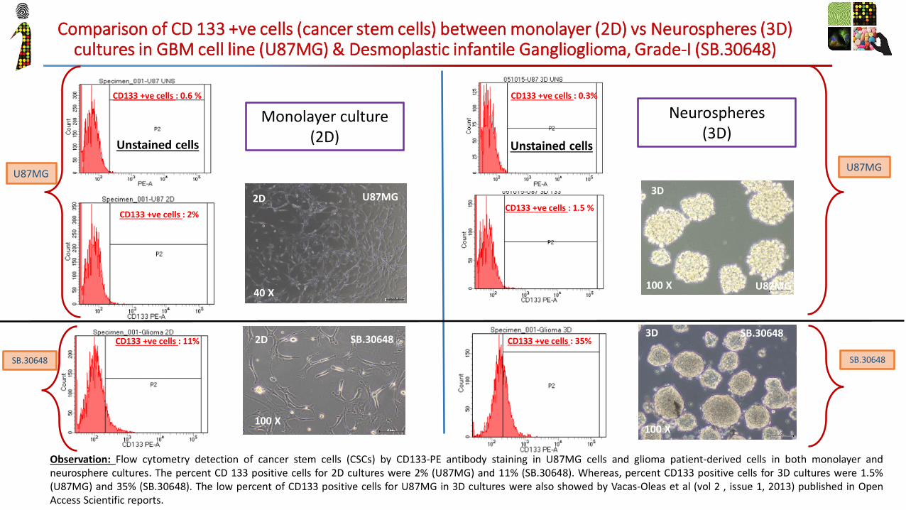
• Suitable to study underlying biology
• For phenotypic screening& optimization of novel inhibitors of cancer metastasis
MetBlock™
EPCAM & CK8/18 – Epithelial marker Vimentin & SMA – mesenchymal marker Engineered Models
• Some early evidence on the Epithelial to mesenchymal transition in our patented system.
MetBlockTM

Other Biomarker & Drug discovery platforms & case studies
• Custom models using patient
cells to provide physiological assay systems
• Suitable for screening compounds for better clinical outcome & to study action on true disease phenotype.
TruScreen™ IL-17 inhibition in PBMC’s from COPD patient
Skin organ ex-vivo culture
-20.0
0.0
20.0
40.0
60.0
80.0
100.0
120.0
1nM 3nM 10nM 30nM 100nM 300nM 1uM 10uM
Perc
enta
ge i
nhib
itio
n
Concentration of X
Inihibition of IL-17 expression by compound X
Patient 1
Patient 2
Patient 3
Day 0 Day 3
Day 5 Day 7
H&E staining of skin tissue after culturing for specified number of days. Suggests epidermis is intact and skin is maintained in good condition as also indicated by LDH

23-Oct-15
Effe
OncoPrime™
3D culture High grade Glioma Neurospheres
• Cancer patient-derived primary
cell panel with true clinical diversity & heterogeneity
• In-vitro phenotypic screening/ anti-proliferative activity of compound on panel of diverse cancer types
Ou
r B
rea
st
Can
cer
Pa
nel
Cell viability assay with some S.O.C
Other Biomarker & Drug discovery platforms & case studies

23-Oct-15
Sapien Biosciences Private Limited, Saarum sciences Private Limited,
1st Floor, AIMSR Bldg. Apollo Health City,Jubilee Hills, Hyderabad-500096
India
www.sapienbio.comwww.saarum.com
Phone: +91 7032647554 Contact us at: [email protected]
Phenotypic Screening Biomarker Discovery Custom Assays Primary Cells FFPE
ANY QUESTIONS ?
Contact us