Embed Size (px)
Citation preview
COMPLICATIONS OF SINUSITIS
Introduction
Surgical Anatomy
Routes of Spread
Risk Factors
Classification
Clinical Features amp Management
RHINOSINUSITIS
bull Definition - Group of disorders charcterised by inflammation of lining of nasal cavity
bull Symptoms of
ndash Nasal congestion
ndash Rhinorrhoea
ndash Sneezing
ndash Itching
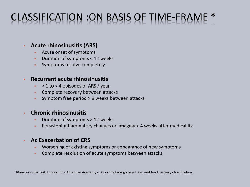
CLASSIFICATION ON BASIS OF TIME-FRAME
Acute rhinosinusitis (ARS) Acute onset of symptoms Duration of symptoms lt 12 weeks Symptoms resolve completely
Recurrent acute rhinosinusitis gt 1 to lt 4 episodes of ARS year Complete recovery between attacks Symptom free period gt 8 weeks between attacks
Chronic rhinosinusitis Duration of symptoms gt 12 weeks Persistent inflammatory changes on imaging gt 4 weeks after medical Rx
Ac Exacerbation of CRS Worsening of existing symptoms or appearance of new symptoms Complete resolution of acute symptoms between attacks
Rhino sinusitis Task Force of the American Academy of Otorhinolaryngology- Head and Neck Surgery classification
CLASSIFICATION OF COMPLICATIONS ACUTE
Local Orbital Intracranial Bony Dental
Systemic
Toxic shock syndrome Septicaemia
CHRONIC Mucocoele Pyocoele
OTHERS Polyarthritis Tenosynovitis OME
INTRODUCTION
Complications are said to arise when infection spreadsinto or beyond the wall of the sinusrsquo
Preantibiotic era frequent 17 died of meningitis 20 blinded 1
Now a rarity 3 mortality2
10 blindness3 once complications develops
1 Gamble Archives of Ophthalmology 1933 10483-497 2 Schramm Curtin and Kennerdell Laryngoscope 1982 3 Patt and Manning Otolary Head Neck Surgery 1991 104789-95
SURGICAL ANATOMY
Maxillary sinus
Birth 7ndash8 times 4ndash6 mm
Adult 31ndash32 times 18ndash20 mm
Volume (adult) 15 mL
Biphasic growth
invasion into the alveolar process following eruption of permanent dentition
SURGICAL ANATOMY
Maxillary Sinus
Relatively symmetrical
Rarely absent
Roof forms orbital floor traversed by infra orbital
canalmay be dehiscent
Posterior edge contributes to infraorbital fissure
Inferiorly floor encroached by dentition
SURGICAL ANATOMY
Maxillary Bone amp Sinus
Posterior surface ( Infratemporal surface)
Convex
Grooved by post sup alveolar n
Inferiorly bears the maxillary tuberosity attachment of Medial Pterygoid ms
Medial nasal surface
Contains large defect maxillary hiatus completed by bones amp mucous memb natural ostia
SURGICAL ANATOMY
Frontal Bone amp Sinus
Forms Forehead amp Orbital roof (thin+- dehiscence)
Also forms roof of ethmoid sinus
Sinus
28 2717 mm
Variable pneumatization variable shape amp size
Drains into frontal recess
Absent in 1
Usually paramedian intersinus septa
Partially dehiscent in 9
SURGICAL ANATOMY
Frontal Bone amp Sinus Relations-
Inf Orbit
Ethmoid labyrinth
Nasal cavity
Sup Ant Cranial Fossa
Olfactory niche bulb tract
Med Cribriform plate
SURGICAL ANATOMY
Ethmoid Bone amp Sinus
2 ethmoid labyrinth laterally constitute orbital plate (L Papyracea) extremely thin +- dehiscent
Perpendicular plate of ethmoid in between
Intervening cribriform plate amp crista gallifenestrations olfactory filaments ethmoidalvessels amp nerve dural prolongations traverse
Ethmoid Bone amp Sinus
Relations-
Superiorly ACF amp Frontal bone
Laterally Orbit
Posteromedially Sphenoid
Posterolaterally Optic N
Medially Nasal Cavity
SURGICAL ANATOMY
SURGICAL ANATOMY
Sphenoid Bone amp Sinus
Largest bone of skull base divides ACF amp MCF
Adult 20 times 22 times 16 mm volume 75 mL
Body with variable pneumatization Conchal presellar sellar mixed
Sinus divided by paramedian septum
May be incomplete
Completely absent in 1
SURGICAL ANATOMY
ORBITAL APEX AND SUPERIOR ORBITAL FISSURE RIGHT SIDE
SURGICAL ANATOMY Sphenoid Bone amp Sinus Relations-
Ant Post ethmoidal cells Post Occipital bone
Basilar A amp Brain Stem Lat Cavernous Sinus
ICA amp Sympathetic plx Abducen N CN III IV V1 V2
Inf Roof of nasopharynx Sup Olf tract Optic chiasma
Pituitary gland Frontal Lobe
SURGICAL ANATOMY CAVERNOUS SINUS
Largest venous sinus In MCF on either side of body ofsphenoid
Divided into number of spacescaverns by trabeculae
2 cm long X 1 cm wide
Structures on lateral wall
CN III IV V1 V2
Structures throrsquo centre
ICA + Sympathetic plx
Abducen N
CAVERNOUS SINUS RELATIONS- Sup
Optic tract and chiasma
Olfactory tract
ICA
Inf
Foramen lacerum
Med
Pituitary gland
Sphenoidal air cells
Lat
Temporal lobe
Ant
Sup orb fissure amp Orbital apex
Post
Apex of petrous temporal bone
Crus cerebri of midbrain
Temporal lobe
CAVERNOUS SINUS INCOMING CHANNELS
Orbit
Sup ophthalmic vn
Inf ophthalmic vn amp br
Central retinal vn
Brain
Supf Middle Cerebral Vn
Inf Cerebral Vn
Meninges
Spheno-parietal sinus
Middle Meningeal Vn
CAVERNOUS SINUS COMMUNICATIONS
Trans sinus throrsquo Sup petrosal sinus
IJV throrsquo Inf petrosal sinus
Pterygoid plx of vn throrsquo Emissary vn
Facial vn throrsquo Sup ophthalmic vn
Communicate with each other throrsquo Intercavernoussinus amp Basilar plx of vn
SURGICAL ANATOMY ORBITAL SEPTUM
The orbicularis oculi (O) overlies theorbital septum (S)retains the orbitalfat pads (F) within the orbit
The septum fuses with the maxillaryperiosteum (P) inferiorly and thetarsus (T) superiorly
The septum is perforated by thevessels and nerves which pass fromthe orbital cavity to the face and scalp
The eyelids are richly supplied withblood
SURGICAL ANATOMY ORBIT
ROUTES OF SPREAD
ROUTES OF SPREAD Local
Natural dehiscence or weakness of surrounding bone When natural routes blocked
Massive osteolysis in acute infection
Lamina papyracea infraorbital canal suture lines
Associated thromboplebitis Diploic veins of Breschet Frontal amp Sphenoid
At peak vascularity in adolescent
Valveless veins between sinus amp orbit
Roots of 2nd premolar amp molars
Distant Hematogenous spread Rare
RISK FACTORS
Patient factors More common in young 85 under 20 yrs
Immunocompromised amp Diabetes
Abnormal mucociliary clearance Chronicity of disease
Allergy Chronicity of disease
Local anatomical variations
Patient compliance to treatment
Pathogenic factors URTI Viremia rarely encephalitis
Invasive fungal rhinosinusitis Mucor
Staph aureus Brain abscess
Treatment factors Inappropiate amp Inadequate antibiotic therapy
BACTERIOLOGY
ORBITAL
Aerobes
Staphylococcus aureus
Haemophilus influenza
Strept pneumoniae
Moraxella catarrhalis
Streptococcus milleri
Streptococcus pyogenes
Anaerobes
INTRACRANIAL
Aerobes
Staphylococcus aureus
Strept Pneumoniae
Haemophilus influenza
Streptococcus sp
Pseudomonas aeruginosa
Klebsiella sp
Anaerobes
A ORBITAL COMPLICATION
Pre-antibiotic era 17-20 died of meningitis or had permanent blindness
Commonly due to ethmoiditis in young and frontal sinusitis in adult
Higher frequency during winter amp spring
A ORBITAL COMPLICATIONS
Hubert 1937
Inflammatory edema of eyelids
Subperiosteal abscess with
Edema of eyelids or
Spread of pus to lids
Abscess of orbital tissues
Orbital cellulitis ndash Mild to severe
Cavernous sinus thrombosis
Smith amp Spencer 1948
A ORBITAL COMPLICATIONSCHANDLER LANGENBRUNNER STEVENS 1970
1 Preseptal cellulitis
2 Orbital cellulitis without
abscess
3 Orbital cellulitis with
subextraperiosteal abscess
4 Orbital cellulitis with
intraperiosteal abscess
5 Cavernous sinus thrombosis
A ORBITAL COMPLICATIONS STAGE 1
Oedema of lids
Painless non-tender
No visual loss
Globe unaffected
No restricted extra-
ocular movements
A ORBITAL COMPLICATIONS STAGE 2
Orbital cellulitis without abscess
Diffuse edema of adipose tissue
Proptosis Chemosis Edema
Associated Pain
+- Dilated pupil
+- Visual loss
+- Ophthalmoplegia
+- Afferent pupillary defect
A ORBITAL COMPLICATIONS STAGE 3
Orbital cellulitis with sub extra-periosteal abscess
Proptosis
Globe displaced inferolaterally
Decreased EOM
Vision decreased
Onidi cells optic nerve vulnerable
A ORBITAL COMPLICATIONS STAGE 3
A ORBITAL COMPLICATIONS STAGE 4
Orbital cellulits with intra-periostealabscess
Severe proptosis and chemosis
Fixed pupil
Severe globe displacement
Rapid fixation of EOM
Opthalmoplegia
Visual loss due to optic neuropathy(13)
ldquoOrbital Apex Syndromerdquo
Ophthalmoplegia amp Dilated pupil
Paraesthesia in distribution of maxilary amp ophthalmic division of trigeminal n
Blindness amp Temporal headache
A ORBITAL COMPLICATIONS STAGE 5 Cavernous Sinus Thrombosis
Bright 1831
Uncommon no incidence data
Typically affects young adults
Pathophysiology
Bacterial growth
Induces thrombosis
Thrombus good growth medium
More bacterial growth
Thrombophlebitis extend posteriorly
Orbit to Cavernous sinus
Contralateral Cavernous sinus bilateral eye symptoms
Then Intracranially
A ORBITAL COMPLICATIONS STAGE 5 Fatal prior to antibiotic era (pre-1940s) Mortality estimate 14-79 Morbidity estimate 50
Cranial neuropathies amp Visual loss
Clinical features Onset 1-21 days (Avg 5-6 days) Progressive amp bilateral eye symptom Proptosis and fixation of eye ball Bilateral orbital apex syndrome Meningitis Systemic featutes
High grade fever amp headache Tachycardia hypotension
A ORBITAL COMPLICATIONS STAGE 5
A ORBITAL COMPLICATIONS STAGE 5
Complications
Intracranial infection
Meningitis
Encephalitis
Abscess
Pituitary insufficiency
Hemorrhagic infarction
Death
A ORBITAL COMPLICATIONS MANAGEMENT
History
General ENT examination
Complete neurological examination
Rigid endoscopy of nose
Swabs
A ORBITAL COMPLICATIONS MANAGEMENT
A ORBITAL COMPLICATIONS MANAGEMENT
Other investigations CBC Blood biochemistry Blood cultures Lumbar puncture Neuroimaging (CT MRI)
Expansion of cavernous sinuses Convex bowing of lateral wall Abnormal filling defects Dilation of superior ophthalmic vein Dural enhancement of cavernous sinus border
A ORBITAL COMPLICATIONS MANAGEMENT
Empiric high dose IV antibiotics
Third generation cephalosporin
Anti-staphylococcal penicillin
Metronidazole
Continued treatment with IV antibiotics for at least 2 wks after apparent clinical resolution
Steroids controversial (except if pituitary insufficiency)
Anticoagulation in CST
No consensus for use despite theoretical rationale
Risks include systemic and intracranial bleeding
A ORBITAL COMPLICATIONS LONG TERM SEQUELAE
Permanent visual loss
Ophthalmoplegia
Exposure Keratitis amp Ulceration
Other ocular changes Uveitis
Choroiditis
Glaucoma
Iris prolapse
Rupture of the globe
B INTRACRANIAL COMPLICATIONS
Less common than orbital complications
Both can coexist
More common in adolescent
amp young adults
Male preponderance
Includes
Meningitis Encephalitis
Subdural Abscess(23) gt Frontal Lobe(4) abscess gt Extradural Abscess (1)
Cavernous Sinus Thrombosis
B INTRACRANIAL COMPLICATIONS CLINICAL PRESENTATION
Thrombophlebitis
Septic thrombophlebitismultiple abscesses formation
Associated thrombosis of Cavernous sinus amp Sup Sagittal sinus
Usually gt10yrs Uncommon in infants
Presentation Acute or Chronic
Fever leucocytosis and headache
Seizures rigidity and focal neurological signs
Features of Increased ICP
Features of meningitis amp encephalitis
B INTRACRANIAL COMPLICATIONS MANAGEMENT
High index of suspicion
History
ENT amp Full neurological examination
If abscess suspected CECT (or MRI)
Fundoscopy Lumbar puncture
IV antibiotics (Cefuroxime+Flagyl) ndash 4-6weeks
Serial CT
Steroids ndash controversial
B INTRACRANIAL COMPLICATIONS MANAGEMENT
Surgery
Treat complication amp sinusitis
Drain Extradural abscess via approach to frontal
sinuses
Neurosurgical assistance
Burr holes For extradural abscess solitary abscess
Formal craniotomy
B INTRACRANIAL COMPLICATIONS
Prognosis
Mortality 15-43 despite antibiotics
Incidence increases with age
Multiple subdural abscess with coritcal thrombosis carries worst prognosis
Morbidity Permanent in 40
Convulsions
Hemiparesis
Early treatment better outcome
C BONY COMPLICATIONS POTTrsquoS PUFFY TUMOUR
Described by Sir Percival Pott in 1970 Frontal bone is diploic with marrow cavity Sinusitis Osteomyelitis of frontal bone
Anteriorly Forehead Potts Puffy tumour Posteriorly Subdural abscess
Presentation Fluctuant swelling +- Pain
Causative Org Staph amp Strepto- amp Anaerobes Investigation
Blood investigation + CECT
Treatment Medical IV Antibiotics Surgical Bilateral coronal incision +Bone debridement
C BONY COMPLICATIONS
POTTrsquoS PUFFY TUMOUR
D DENTAL COMPLICATIONS
Common with Maxillary Sinusitis
Close association of dentition with floor of sinus
Acute sinusitis Dental pain
Dental abscess Mistaken for Sinusitis
May coexist
E SYSTEMIC COMPLICATIONS
Toxic Shock Syndrome
Rare Potentially Fatal
Frequently associated with Staph aureus Streptococcus
Features of Toxaemia Fever Hypotension Rash amp MODS
Septicaemia
Hematogenous spread
Blood culture
Features of SIRS
Shock
MODS
F CHRONIC COMPLICATIONS MUCOCOELE Definition
Epithelial lined mucus containing sac completely filling thesinus and capable of expansion
1820 Langenback lsquoHydatidesrsquo
1896 Rollet lsquomucocoelersquo
Incidence 4 of case of unilateral proptosis
Most common sites F(65)gt E(25)gt M(10)gt S
Age grp 40-70 yrs
lt 5 bilateral or multiloculated53
F CHRONIC COMPLICATIONS MUCOCOELE Theories Chronic rinosinusitis Increased osteolysis
Active Bone resorption amp formation
Pressure erosion
Cystic degeneration of seromucimous glands
54
F CHRONIC COMPLICATIONS MUCOCOELE
Initial ophthalmic referral
Clinical features Proptosis
Diplopia
Displacement of globe
Limited ocular movement
Visual impairment
Mass
Endoscopy
55
F CHRONIC COMPLICATIONS MUCOCOELE
56
ImagingLoss of Scalloping of frontal sinus
F MUCOCOELE CECT PNS
FRONTAL MUCOCOELE BILOCULATED ETHMOID MUCOCOELE
57
Homogenous smooth walled mass expanding the sinus
F MUCOCOELE CULTURE OF ASPIRATE
Mixed infection
Staph aureus
Alpha hemolytic Streptococci
Heamophillus sp
GNB
Anaerobic bacteria
58
F MUCOCOELE TREATMENT
Surgery is the treatment
Goals
Eradication of disease
Minimal morbidity
Prevention of recurrence
59
F MUCOCOELE TREATMENT
Approach
Endoscopic drainage
Type I-III
External approach
Osteoplastic flap
+- sinus cavity obliteration
Combined approach
60
THANK YOU
Introduction
Surgical Anatomy
Routes of Spread
Risk Factors
Classification
Clinical Features amp Management
RHINOSINUSITIS
bull Definition - Group of disorders charcterised by inflammation of lining of nasal cavity
bull Symptoms of
ndash Nasal congestion
ndash Rhinorrhoea
ndash Sneezing
ndash Itching
CLASSIFICATION ON BASIS OF TIME-FRAME
Acute rhinosinusitis (ARS) Acute onset of symptoms Duration of symptoms lt 12 weeks Symptoms resolve completely
Recurrent acute rhinosinusitis gt 1 to lt 4 episodes of ARS year Complete recovery between attacks Symptom free period gt 8 weeks between attacks
Chronic rhinosinusitis Duration of symptoms gt 12 weeks Persistent inflammatory changes on imaging gt 4 weeks after medical Rx
Ac Exacerbation of CRS Worsening of existing symptoms or appearance of new symptoms Complete resolution of acute symptoms between attacks
Rhino sinusitis Task Force of the American Academy of Otorhinolaryngology- Head and Neck Surgery classification
CLASSIFICATION OF COMPLICATIONS ACUTE
Local Orbital Intracranial Bony Dental
Systemic
Toxic shock syndrome Septicaemia
CHRONIC Mucocoele Pyocoele
OTHERS Polyarthritis Tenosynovitis OME
INTRODUCTION
Complications are said to arise when infection spreadsinto or beyond the wall of the sinusrsquo
Preantibiotic era frequent 17 died of meningitis 20 blinded 1
Now a rarity 3 mortality2
10 blindness3 once complications develops
1 Gamble Archives of Ophthalmology 1933 10483-497 2 Schramm Curtin and Kennerdell Laryngoscope 1982 3 Patt and Manning Otolary Head Neck Surgery 1991 104789-95
SURGICAL ANATOMY
Maxillary sinus
Birth 7ndash8 times 4ndash6 mm
Adult 31ndash32 times 18ndash20 mm
Volume (adult) 15 mL
Biphasic growth
invasion into the alveolar process following eruption of permanent dentition
SURGICAL ANATOMY
Maxillary Sinus
Relatively symmetrical
Rarely absent
Roof forms orbital floor traversed by infra orbital
canalmay be dehiscent
Posterior edge contributes to infraorbital fissure
Inferiorly floor encroached by dentition
SURGICAL ANATOMY
Maxillary Bone amp Sinus
Posterior surface ( Infratemporal surface)
Convex
Grooved by post sup alveolar n
Inferiorly bears the maxillary tuberosity attachment of Medial Pterygoid ms
Medial nasal surface
Contains large defect maxillary hiatus completed by bones amp mucous memb natural ostia
SURGICAL ANATOMY
Frontal Bone amp Sinus
Forms Forehead amp Orbital roof (thin+- dehiscence)
Also forms roof of ethmoid sinus
Sinus
28 2717 mm
Variable pneumatization variable shape amp size
Drains into frontal recess
Absent in 1
Usually paramedian intersinus septa
Partially dehiscent in 9
SURGICAL ANATOMY
Frontal Bone amp Sinus Relations-
Inf Orbit
Ethmoid labyrinth
Nasal cavity
Sup Ant Cranial Fossa
Olfactory niche bulb tract
Med Cribriform plate
SURGICAL ANATOMY
Ethmoid Bone amp Sinus
2 ethmoid labyrinth laterally constitute orbital plate (L Papyracea) extremely thin +- dehiscent
Perpendicular plate of ethmoid in between
Intervening cribriform plate amp crista gallifenestrations olfactory filaments ethmoidalvessels amp nerve dural prolongations traverse
Ethmoid Bone amp Sinus
Relations-
Superiorly ACF amp Frontal bone
Laterally Orbit
Posteromedially Sphenoid
Posterolaterally Optic N
Medially Nasal Cavity
SURGICAL ANATOMY
SURGICAL ANATOMY
Sphenoid Bone amp Sinus
Largest bone of skull base divides ACF amp MCF
Adult 20 times 22 times 16 mm volume 75 mL
Body with variable pneumatization Conchal presellar sellar mixed
Sinus divided by paramedian septum
May be incomplete
Completely absent in 1
SURGICAL ANATOMY
ORBITAL APEX AND SUPERIOR ORBITAL FISSURE RIGHT SIDE
SURGICAL ANATOMY Sphenoid Bone amp Sinus Relations-
Ant Post ethmoidal cells Post Occipital bone
Basilar A amp Brain Stem Lat Cavernous Sinus
ICA amp Sympathetic plx Abducen N CN III IV V1 V2
Inf Roof of nasopharynx Sup Olf tract Optic chiasma
Pituitary gland Frontal Lobe
SURGICAL ANATOMY CAVERNOUS SINUS
Largest venous sinus In MCF on either side of body ofsphenoid
Divided into number of spacescaverns by trabeculae
2 cm long X 1 cm wide
Structures on lateral wall
CN III IV V1 V2
Structures throrsquo centre
ICA + Sympathetic plx
Abducen N
CAVERNOUS SINUS RELATIONS- Sup
Optic tract and chiasma
Olfactory tract
ICA
Inf
Foramen lacerum
Med
Pituitary gland
Sphenoidal air cells
Lat
Temporal lobe
Ant
Sup orb fissure amp Orbital apex
Post
Apex of petrous temporal bone
Crus cerebri of midbrain
Temporal lobe
CAVERNOUS SINUS INCOMING CHANNELS
Orbit
Sup ophthalmic vn
Inf ophthalmic vn amp br
Central retinal vn
Brain
Supf Middle Cerebral Vn
Inf Cerebral Vn
Meninges
Spheno-parietal sinus
Middle Meningeal Vn
CAVERNOUS SINUS COMMUNICATIONS
Trans sinus throrsquo Sup petrosal sinus
IJV throrsquo Inf petrosal sinus
Pterygoid plx of vn throrsquo Emissary vn
Facial vn throrsquo Sup ophthalmic vn
Communicate with each other throrsquo Intercavernoussinus amp Basilar plx of vn
SURGICAL ANATOMY ORBITAL SEPTUM
The orbicularis oculi (O) overlies theorbital septum (S)retains the orbitalfat pads (F) within the orbit
The septum fuses with the maxillaryperiosteum (P) inferiorly and thetarsus (T) superiorly
The septum is perforated by thevessels and nerves which pass fromthe orbital cavity to the face and scalp
The eyelids are richly supplied withblood
SURGICAL ANATOMY ORBIT
ROUTES OF SPREAD
ROUTES OF SPREAD Local
Natural dehiscence or weakness of surrounding bone When natural routes blocked
Massive osteolysis in acute infection
Lamina papyracea infraorbital canal suture lines
Associated thromboplebitis Diploic veins of Breschet Frontal amp Sphenoid
At peak vascularity in adolescent
Valveless veins between sinus amp orbit
Roots of 2nd premolar amp molars
Distant Hematogenous spread Rare
RISK FACTORS
Patient factors More common in young 85 under 20 yrs
Immunocompromised amp Diabetes
Abnormal mucociliary clearance Chronicity of disease
Allergy Chronicity of disease
Local anatomical variations
Patient compliance to treatment
Pathogenic factors URTI Viremia rarely encephalitis
Invasive fungal rhinosinusitis Mucor
Staph aureus Brain abscess
Treatment factors Inappropiate amp Inadequate antibiotic therapy
BACTERIOLOGY
ORBITAL
Aerobes
Staphylococcus aureus
Haemophilus influenza
Strept pneumoniae
Moraxella catarrhalis
Streptococcus milleri
Streptococcus pyogenes
Anaerobes
INTRACRANIAL
Aerobes
Staphylococcus aureus
Strept Pneumoniae
Haemophilus influenza
Streptococcus sp
Pseudomonas aeruginosa
Klebsiella sp
Anaerobes
A ORBITAL COMPLICATION
Pre-antibiotic era 17-20 died of meningitis or had permanent blindness
Commonly due to ethmoiditis in young and frontal sinusitis in adult
Higher frequency during winter amp spring
A ORBITAL COMPLICATIONS
Hubert 1937
Inflammatory edema of eyelids
Subperiosteal abscess with
Edema of eyelids or
Spread of pus to lids
Abscess of orbital tissues
Orbital cellulitis ndash Mild to severe
Cavernous sinus thrombosis
Smith amp Spencer 1948
A ORBITAL COMPLICATIONSCHANDLER LANGENBRUNNER STEVENS 1970
1 Preseptal cellulitis
2 Orbital cellulitis without
abscess
3 Orbital cellulitis with
subextraperiosteal abscess
4 Orbital cellulitis with
intraperiosteal abscess
5 Cavernous sinus thrombosis
A ORBITAL COMPLICATIONS STAGE 1
Oedema of lids
Painless non-tender
No visual loss
Globe unaffected
No restricted extra-
ocular movements
A ORBITAL COMPLICATIONS STAGE 2
Orbital cellulitis without abscess
Diffuse edema of adipose tissue
Proptosis Chemosis Edema
Associated Pain
+- Dilated pupil
+- Visual loss
+- Ophthalmoplegia
+- Afferent pupillary defect
A ORBITAL COMPLICATIONS STAGE 3
Orbital cellulitis with sub extra-periosteal abscess
Proptosis
Globe displaced inferolaterally
Decreased EOM
Vision decreased
Onidi cells optic nerve vulnerable
A ORBITAL COMPLICATIONS STAGE 3
A ORBITAL COMPLICATIONS STAGE 4
Orbital cellulits with intra-periostealabscess
Severe proptosis and chemosis
Fixed pupil
Severe globe displacement
Rapid fixation of EOM
Opthalmoplegia
Visual loss due to optic neuropathy(13)
ldquoOrbital Apex Syndromerdquo
Ophthalmoplegia amp Dilated pupil
Paraesthesia in distribution of maxilary amp ophthalmic division of trigeminal n
Blindness amp Temporal headache
A ORBITAL COMPLICATIONS STAGE 5 Cavernous Sinus Thrombosis
Bright 1831
Uncommon no incidence data
Typically affects young adults
Pathophysiology
Bacterial growth
Induces thrombosis
Thrombus good growth medium
More bacterial growth
Thrombophlebitis extend posteriorly
Orbit to Cavernous sinus
Contralateral Cavernous sinus bilateral eye symptoms
Then Intracranially
A ORBITAL COMPLICATIONS STAGE 5 Fatal prior to antibiotic era (pre-1940s) Mortality estimate 14-79 Morbidity estimate 50
Cranial neuropathies amp Visual loss
Clinical features Onset 1-21 days (Avg 5-6 days) Progressive amp bilateral eye symptom Proptosis and fixation of eye ball Bilateral orbital apex syndrome Meningitis Systemic featutes
High grade fever amp headache Tachycardia hypotension
A ORBITAL COMPLICATIONS STAGE 5
A ORBITAL COMPLICATIONS STAGE 5
Complications
Intracranial infection
Meningitis
Encephalitis
Abscess
Pituitary insufficiency
Hemorrhagic infarction
Death
A ORBITAL COMPLICATIONS MANAGEMENT
History
General ENT examination
Complete neurological examination
Rigid endoscopy of nose
Swabs
A ORBITAL COMPLICATIONS MANAGEMENT
A ORBITAL COMPLICATIONS MANAGEMENT
Other investigations CBC Blood biochemistry Blood cultures Lumbar puncture Neuroimaging (CT MRI)
Expansion of cavernous sinuses Convex bowing of lateral wall Abnormal filling defects Dilation of superior ophthalmic vein Dural enhancement of cavernous sinus border
A ORBITAL COMPLICATIONS MANAGEMENT
Empiric high dose IV antibiotics
Third generation cephalosporin
Anti-staphylococcal penicillin
Metronidazole
Continued treatment with IV antibiotics for at least 2 wks after apparent clinical resolution
Steroids controversial (except if pituitary insufficiency)
Anticoagulation in CST
No consensus for use despite theoretical rationale
Risks include systemic and intracranial bleeding
A ORBITAL COMPLICATIONS LONG TERM SEQUELAE
Permanent visual loss
Ophthalmoplegia
Exposure Keratitis amp Ulceration
Other ocular changes Uveitis
Choroiditis
Glaucoma
Iris prolapse
Rupture of the globe
B INTRACRANIAL COMPLICATIONS
Less common than orbital complications
Both can coexist
More common in adolescent
amp young adults
Male preponderance
Includes
Meningitis Encephalitis
Subdural Abscess(23) gt Frontal Lobe(4) abscess gt Extradural Abscess (1)
Cavernous Sinus Thrombosis
B INTRACRANIAL COMPLICATIONS CLINICAL PRESENTATION
Thrombophlebitis
Septic thrombophlebitismultiple abscesses formation
Associated thrombosis of Cavernous sinus amp Sup Sagittal sinus
Usually gt10yrs Uncommon in infants
Presentation Acute or Chronic
Fever leucocytosis and headache
Seizures rigidity and focal neurological signs
Features of Increased ICP
Features of meningitis amp encephalitis
B INTRACRANIAL COMPLICATIONS MANAGEMENT
High index of suspicion
History
ENT amp Full neurological examination
If abscess suspected CECT (or MRI)
Fundoscopy Lumbar puncture
IV antibiotics (Cefuroxime+Flagyl) ndash 4-6weeks
Serial CT
Steroids ndash controversial
B INTRACRANIAL COMPLICATIONS MANAGEMENT
Surgery
Treat complication amp sinusitis
Drain Extradural abscess via approach to frontal
sinuses
Neurosurgical assistance
Burr holes For extradural abscess solitary abscess
Formal craniotomy
B INTRACRANIAL COMPLICATIONS
Prognosis
Mortality 15-43 despite antibiotics
Incidence increases with age
Multiple subdural abscess with coritcal thrombosis carries worst prognosis
Morbidity Permanent in 40
Convulsions
Hemiparesis
Early treatment better outcome
C BONY COMPLICATIONS POTTrsquoS PUFFY TUMOUR
Described by Sir Percival Pott in 1970 Frontal bone is diploic with marrow cavity Sinusitis Osteomyelitis of frontal bone
Anteriorly Forehead Potts Puffy tumour Posteriorly Subdural abscess
Presentation Fluctuant swelling +- Pain
Causative Org Staph amp Strepto- amp Anaerobes Investigation
Blood investigation + CECT
Treatment Medical IV Antibiotics Surgical Bilateral coronal incision +Bone debridement
C BONY COMPLICATIONS
POTTrsquoS PUFFY TUMOUR
D DENTAL COMPLICATIONS
Common with Maxillary Sinusitis
Close association of dentition with floor of sinus
Acute sinusitis Dental pain
Dental abscess Mistaken for Sinusitis
May coexist
E SYSTEMIC COMPLICATIONS
Toxic Shock Syndrome
Rare Potentially Fatal
Frequently associated with Staph aureus Streptococcus
Features of Toxaemia Fever Hypotension Rash amp MODS
Septicaemia
Hematogenous spread
Blood culture
Features of SIRS
Shock
MODS
F CHRONIC COMPLICATIONS MUCOCOELE Definition
Epithelial lined mucus containing sac completely filling thesinus and capable of expansion
1820 Langenback lsquoHydatidesrsquo
1896 Rollet lsquomucocoelersquo
Incidence 4 of case of unilateral proptosis
Most common sites F(65)gt E(25)gt M(10)gt S
Age grp 40-70 yrs
lt 5 bilateral or multiloculated53
F CHRONIC COMPLICATIONS MUCOCOELE Theories Chronic rinosinusitis Increased osteolysis
Active Bone resorption amp formation
Pressure erosion
Cystic degeneration of seromucimous glands
54
F CHRONIC COMPLICATIONS MUCOCOELE
Initial ophthalmic referral
Clinical features Proptosis
Diplopia
Displacement of globe
Limited ocular movement
Visual impairment
Mass
Endoscopy
55
F CHRONIC COMPLICATIONS MUCOCOELE
56
ImagingLoss of Scalloping of frontal sinus
F MUCOCOELE CECT PNS
FRONTAL MUCOCOELE BILOCULATED ETHMOID MUCOCOELE
57
Homogenous smooth walled mass expanding the sinus
F MUCOCOELE CULTURE OF ASPIRATE
Mixed infection
Staph aureus
Alpha hemolytic Streptococci
Heamophillus sp
GNB
Anaerobic bacteria
58
F MUCOCOELE TREATMENT
Surgery is the treatment
Goals
Eradication of disease
Minimal morbidity
Prevention of recurrence
59
F MUCOCOELE TREATMENT
Approach
Endoscopic drainage
Type I-III
External approach
Osteoplastic flap
+- sinus cavity obliteration
Combined approach
60
THANK YOU

SURGICAL ANATOMY
Maxillary Sinus
Relatively symmetrical
Rarely absent
Roof forms orbital floor traversed by infra orbital
canalmay be dehiscent
Posterior edge contributes to infraorbital fissure
Inferiorly floor encroached by dentition
SURGICAL ANATOMY
Maxillary Bone amp Sinus
Posterior surface ( Infratemporal surface)
Convex
Grooved by post sup alveolar n
Inferiorly bears the maxillary tuberosity attachment of Medial Pterygoid ms
Medial nasal surface
Contains large defect maxillary hiatus completed by bones amp mucous memb natural ostia
SURGICAL ANATOMY
Frontal Bone amp Sinus
Forms Forehead amp Orbital roof (thin+- dehiscence)
Also forms roof of ethmoid sinus
Sinus
28 2717 mm
Variable pneumatization variable shape amp size
Drains into frontal recess
Absent in 1
Usually paramedian intersinus septa
Partially dehiscent in 9
SURGICAL ANATOMY
Frontal Bone amp Sinus Relations-
Inf Orbit
Ethmoid labyrinth
Nasal cavity
Sup Ant Cranial Fossa
Olfactory niche bulb tract
Med Cribriform plate
SURGICAL ANATOMY
Ethmoid Bone amp Sinus
2 ethmoid labyrinth laterally constitute orbital plate (L Papyracea) extremely thin +- dehiscent
Perpendicular plate of ethmoid in between
Intervening cribriform plate amp crista gallifenestrations olfactory filaments ethmoidalvessels amp nerve dural prolongations traverse
Ethmoid Bone amp Sinus
Relations-
Superiorly ACF amp Frontal bone
Laterally Orbit
Posteromedially Sphenoid
Posterolaterally Optic N
Medially Nasal Cavity
SURGICAL ANATOMY
SURGICAL ANATOMY
Sphenoid Bone amp Sinus
Largest bone of skull base divides ACF amp MCF
Adult 20 times 22 times 16 mm volume 75 mL
Body with variable pneumatization Conchal presellar sellar mixed
Sinus divided by paramedian septum
May be incomplete
Completely absent in 1
SURGICAL ANATOMY
ORBITAL APEX AND SUPERIOR ORBITAL FISSURE RIGHT SIDE
SURGICAL ANATOMY Sphenoid Bone amp Sinus Relations-
Ant Post ethmoidal cells Post Occipital bone
Basilar A amp Brain Stem Lat Cavernous Sinus
ICA amp Sympathetic plx Abducen N CN III IV V1 V2
Inf Roof of nasopharynx Sup Olf tract Optic chiasma
Pituitary gland Frontal Lobe
SURGICAL ANATOMY CAVERNOUS SINUS
Largest venous sinus In MCF on either side of body ofsphenoid
Divided into number of spacescaverns by trabeculae
2 cm long X 1 cm wide
Structures on lateral wall
CN III IV V1 V2
Structures throrsquo centre
ICA + Sympathetic plx
Abducen N
CAVERNOUS SINUS RELATIONS- Sup
Optic tract and chiasma
Olfactory tract
ICA
Inf
Foramen lacerum
Med
Pituitary gland
Sphenoidal air cells
Lat
Temporal lobe
Ant
Sup orb fissure amp Orbital apex
Post
Apex of petrous temporal bone
Crus cerebri of midbrain
Temporal lobe
CAVERNOUS SINUS INCOMING CHANNELS
Orbit
Sup ophthalmic vn
Inf ophthalmic vn amp br
Central retinal vn
Brain
Supf Middle Cerebral Vn
Inf Cerebral Vn
Meninges
Spheno-parietal sinus
Middle Meningeal Vn
CAVERNOUS SINUS COMMUNICATIONS
Trans sinus throrsquo Sup petrosal sinus
IJV throrsquo Inf petrosal sinus
Pterygoid plx of vn throrsquo Emissary vn
Facial vn throrsquo Sup ophthalmic vn
Communicate with each other throrsquo Intercavernoussinus amp Basilar plx of vn
SURGICAL ANATOMY ORBITAL SEPTUM
The orbicularis oculi (O) overlies theorbital septum (S)retains the orbitalfat pads (F) within the orbit
The septum fuses with the maxillaryperiosteum (P) inferiorly and thetarsus (T) superiorly
The septum is perforated by thevessels and nerves which pass fromthe orbital cavity to the face and scalp
The eyelids are richly supplied withblood
SURGICAL ANATOMY ORBIT
ROUTES OF SPREAD
ROUTES OF SPREAD Local
Natural dehiscence or weakness of surrounding bone When natural routes blocked
Massive osteolysis in acute infection
Lamina papyracea infraorbital canal suture lines
Associated thromboplebitis Diploic veins of Breschet Frontal amp Sphenoid
At peak vascularity in adolescent
Valveless veins between sinus amp orbit
Roots of 2nd premolar amp molars
Distant Hematogenous spread Rare
RISK FACTORS
Patient factors More common in young 85 under 20 yrs
Immunocompromised amp Diabetes
Abnormal mucociliary clearance Chronicity of disease
Allergy Chronicity of disease
Local anatomical variations
Patient compliance to treatment
Pathogenic factors URTI Viremia rarely encephalitis
Invasive fungal rhinosinusitis Mucor
Staph aureus Brain abscess
Treatment factors Inappropiate amp Inadequate antibiotic therapy
BACTERIOLOGY
ORBITAL
Aerobes
Staphylococcus aureus
Haemophilus influenza
Strept pneumoniae
Moraxella catarrhalis
Streptococcus milleri
Streptococcus pyogenes
Anaerobes
INTRACRANIAL
Aerobes
Staphylococcus aureus
Strept Pneumoniae
Haemophilus influenza
Streptococcus sp
Pseudomonas aeruginosa
Klebsiella sp
Anaerobes
A ORBITAL COMPLICATION
Pre-antibiotic era 17-20 died of meningitis or had permanent blindness
Commonly due to ethmoiditis in young and frontal sinusitis in adult
Higher frequency during winter amp spring
A ORBITAL COMPLICATIONS
Hubert 1937
Inflammatory edema of eyelids
Subperiosteal abscess with
Edema of eyelids or
Spread of pus to lids
Abscess of orbital tissues
Orbital cellulitis ndash Mild to severe
Cavernous sinus thrombosis
Smith amp Spencer 1948
A ORBITAL COMPLICATIONSCHANDLER LANGENBRUNNER STEVENS 1970
1 Preseptal cellulitis
2 Orbital cellulitis without
abscess
3 Orbital cellulitis with
subextraperiosteal abscess
4 Orbital cellulitis with
intraperiosteal abscess
5 Cavernous sinus thrombosis
A ORBITAL COMPLICATIONS STAGE 1
Oedema of lids
Painless non-tender
No visual loss
Globe unaffected
No restricted extra-
ocular movements
A ORBITAL COMPLICATIONS STAGE 2
Orbital cellulitis without abscess
Diffuse edema of adipose tissue
Proptosis Chemosis Edema
Associated Pain
+- Dilated pupil
+- Visual loss
+- Ophthalmoplegia
+- Afferent pupillary defect
A ORBITAL COMPLICATIONS STAGE 3
Orbital cellulitis with sub extra-periosteal abscess
Proptosis
Globe displaced inferolaterally
Decreased EOM
Vision decreased
Onidi cells optic nerve vulnerable
A ORBITAL COMPLICATIONS STAGE 3
A ORBITAL COMPLICATIONS STAGE 4
Orbital cellulits with intra-periostealabscess
Severe proptosis and chemosis
Fixed pupil
Severe globe displacement
Rapid fixation of EOM
Opthalmoplegia
Visual loss due to optic neuropathy(13)
ldquoOrbital Apex Syndromerdquo
Ophthalmoplegia amp Dilated pupil
Paraesthesia in distribution of maxilary amp ophthalmic division of trigeminal n
Blindness amp Temporal headache
A ORBITAL COMPLICATIONS STAGE 5 Cavernous Sinus Thrombosis
Bright 1831
Uncommon no incidence data
Typically affects young adults
Pathophysiology
Bacterial growth
Induces thrombosis
Thrombus good growth medium
More bacterial growth
Thrombophlebitis extend posteriorly
Orbit to Cavernous sinus
Contralateral Cavernous sinus bilateral eye symptoms
Then Intracranially
A ORBITAL COMPLICATIONS STAGE 5 Fatal prior to antibiotic era (pre-1940s) Mortality estimate 14-79 Morbidity estimate 50
Cranial neuropathies amp Visual loss
Clinical features Onset 1-21 days (Avg 5-6 days) Progressive amp bilateral eye symptom Proptosis and fixation of eye ball Bilateral orbital apex syndrome Meningitis Systemic featutes
High grade fever amp headache Tachycardia hypotension
A ORBITAL COMPLICATIONS STAGE 5
A ORBITAL COMPLICATIONS STAGE 5
Complications
Intracranial infection
Meningitis
Encephalitis
Abscess
Pituitary insufficiency
Hemorrhagic infarction
Death
A ORBITAL COMPLICATIONS MANAGEMENT
History
General ENT examination
Complete neurological examination
Rigid endoscopy of nose
Swabs
A ORBITAL COMPLICATIONS MANAGEMENT
A ORBITAL COMPLICATIONS MANAGEMENT
Other investigations CBC Blood biochemistry Blood cultures Lumbar puncture Neuroimaging (CT MRI)
Expansion of cavernous sinuses Convex bowing of lateral wall Abnormal filling defects Dilation of superior ophthalmic vein Dural enhancement of cavernous sinus border
A ORBITAL COMPLICATIONS MANAGEMENT
Empiric high dose IV antibiotics
Third generation cephalosporin
Anti-staphylococcal penicillin
Metronidazole
Continued treatment with IV antibiotics for at least 2 wks after apparent clinical resolution
Steroids controversial (except if pituitary insufficiency)
Anticoagulation in CST
No consensus for use despite theoretical rationale
Risks include systemic and intracranial bleeding
A ORBITAL COMPLICATIONS LONG TERM SEQUELAE
Permanent visual loss
Ophthalmoplegia
Exposure Keratitis amp Ulceration
Other ocular changes Uveitis
Choroiditis
Glaucoma
Iris prolapse
Rupture of the globe
B INTRACRANIAL COMPLICATIONS
Less common than orbital complications
Both can coexist
More common in adolescent
amp young adults
Male preponderance
Includes
Meningitis Encephalitis
Subdural Abscess(23) gt Frontal Lobe(4) abscess gt Extradural Abscess (1)
Cavernous Sinus Thrombosis
B INTRACRANIAL COMPLICATIONS CLINICAL PRESENTATION
Thrombophlebitis
Septic thrombophlebitismultiple abscesses formation
Associated thrombosis of Cavernous sinus amp Sup Sagittal sinus
Usually gt10yrs Uncommon in infants
Presentation Acute or Chronic
Fever leucocytosis and headache
Seizures rigidity and focal neurological signs
Features of Increased ICP
Features of meningitis amp encephalitis
B INTRACRANIAL COMPLICATIONS MANAGEMENT
High index of suspicion
History
ENT amp Full neurological examination
If abscess suspected CECT (or MRI)
Fundoscopy Lumbar puncture
IV antibiotics (Cefuroxime+Flagyl) ndash 4-6weeks
Serial CT
Steroids ndash controversial
B INTRACRANIAL COMPLICATIONS MANAGEMENT
Surgery
Treat complication amp sinusitis
Drain Extradural abscess via approach to frontal
sinuses
Neurosurgical assistance
Burr holes For extradural abscess solitary abscess
Formal craniotomy
B INTRACRANIAL COMPLICATIONS
Prognosis
Mortality 15-43 despite antibiotics
Incidence increases with age
Multiple subdural abscess with coritcal thrombosis carries worst prognosis
Morbidity Permanent in 40
Convulsions
Hemiparesis
Early treatment better outcome
C BONY COMPLICATIONS POTTrsquoS PUFFY TUMOUR
Described by Sir Percival Pott in 1970 Frontal bone is diploic with marrow cavity Sinusitis Osteomyelitis of frontal bone
Anteriorly Forehead Potts Puffy tumour Posteriorly Subdural abscess
Presentation Fluctuant swelling +- Pain
Causative Org Staph amp Strepto- amp Anaerobes Investigation
Blood investigation + CECT
Treatment Medical IV Antibiotics Surgical Bilateral coronal incision +Bone debridement
C BONY COMPLICATIONS
POTTrsquoS PUFFY TUMOUR
D DENTAL COMPLICATIONS
Common with Maxillary Sinusitis
Close association of dentition with floor of sinus
Acute sinusitis Dental pain
Dental abscess Mistaken for Sinusitis
May coexist
E SYSTEMIC COMPLICATIONS
Toxic Shock Syndrome
Rare Potentially Fatal
Frequently associated with Staph aureus Streptococcus
Features of Toxaemia Fever Hypotension Rash amp MODS
Septicaemia
Hematogenous spread
Blood culture
Features of SIRS
Shock
MODS
F CHRONIC COMPLICATIONS MUCOCOELE Definition
Epithelial lined mucus containing sac completely filling thesinus and capable of expansion
1820 Langenback lsquoHydatidesrsquo
1896 Rollet lsquomucocoelersquo
Incidence 4 of case of unilateral proptosis
Most common sites F(65)gt E(25)gt M(10)gt S
Age grp 40-70 yrs
lt 5 bilateral or multiloculated53
F CHRONIC COMPLICATIONS MUCOCOELE Theories Chronic rinosinusitis Increased osteolysis
Active Bone resorption amp formation
Pressure erosion
Cystic degeneration of seromucimous glands
54
F CHRONIC COMPLICATIONS MUCOCOELE
Initial ophthalmic referral
Clinical features Proptosis
Diplopia
Displacement of globe
Limited ocular movement
Visual impairment
Mass
Endoscopy
55
F CHRONIC COMPLICATIONS MUCOCOELE
56
ImagingLoss of Scalloping of frontal sinus
F MUCOCOELE CECT PNS
FRONTAL MUCOCOELE BILOCULATED ETHMOID MUCOCOELE
57
Homogenous smooth walled mass expanding the sinus
F MUCOCOELE CULTURE OF ASPIRATE
Mixed infection
Staph aureus
Alpha hemolytic Streptococci
Heamophillus sp
GNB
Anaerobic bacteria
58
F MUCOCOELE TREATMENT
Surgery is the treatment
Goals
Eradication of disease
Minimal morbidity
Prevention of recurrence
59
F MUCOCOELE TREATMENT
Approach
Endoscopic drainage
Type I-III
External approach
Osteoplastic flap
+- sinus cavity obliteration
Combined approach
60
THANK YOU
SURGICAL ANATOMY
Maxillary Bone amp Sinus
Posterior surface ( Infratemporal surface)
Convex
Grooved by post sup alveolar n
Inferiorly bears the maxillary tuberosity attachment of Medial Pterygoid ms
Medial nasal surface
Contains large defect maxillary hiatus completed by bones amp mucous memb natural ostia
SURGICAL ANATOMY
Frontal Bone amp Sinus
Forms Forehead amp Orbital roof (thin+- dehiscence)
Also forms roof of ethmoid sinus
Sinus
28 2717 mm
Variable pneumatization variable shape amp size
Drains into frontal recess
Absent in 1
Usually paramedian intersinus septa
Partially dehiscent in 9
SURGICAL ANATOMY
Frontal Bone amp Sinus Relations-
Inf Orbit
Ethmoid labyrinth
Nasal cavity
Sup Ant Cranial Fossa
Olfactory niche bulb tract
Med Cribriform plate
SURGICAL ANATOMY
Ethmoid Bone amp Sinus
2 ethmoid labyrinth laterally constitute orbital plate (L Papyracea) extremely thin +- dehiscent
Perpendicular plate of ethmoid in between
Intervening cribriform plate amp crista gallifenestrations olfactory filaments ethmoidalvessels amp nerve dural prolongations traverse
Ethmoid Bone amp Sinus
Relations-
Superiorly ACF amp Frontal bone
Laterally Orbit
Posteromedially Sphenoid
Posterolaterally Optic N
Medially Nasal Cavity
SURGICAL ANATOMY
SURGICAL ANATOMY
Sphenoid Bone amp Sinus
Largest bone of skull base divides ACF amp MCF
Adult 20 times 22 times 16 mm volume 75 mL
Body with variable pneumatization Conchal presellar sellar mixed
Sinus divided by paramedian septum
May be incomplete
Completely absent in 1
SURGICAL ANATOMY
ORBITAL APEX AND SUPERIOR ORBITAL FISSURE RIGHT SIDE
SURGICAL ANATOMY Sphenoid Bone amp Sinus Relations-
Ant Post ethmoidal cells Post Occipital bone
Basilar A amp Brain Stem Lat Cavernous Sinus
ICA amp Sympathetic plx Abducen N CN III IV V1 V2
Inf Roof of nasopharynx Sup Olf tract Optic chiasma
Pituitary gland Frontal Lobe
SURGICAL ANATOMY CAVERNOUS SINUS
Largest venous sinus In MCF on either side of body ofsphenoid
Divided into number of spacescaverns by trabeculae
2 cm long X 1 cm wide
Structures on lateral wall
CN III IV V1 V2
Structures throrsquo centre
ICA + Sympathetic plx
Abducen N
CAVERNOUS SINUS RELATIONS- Sup
Optic tract and chiasma
Olfactory tract
ICA
Inf
Foramen lacerum
Med
Pituitary gland
Sphenoidal air cells
Lat
Temporal lobe
Ant
Sup orb fissure amp Orbital apex
Post
Apex of petrous temporal bone
Crus cerebri of midbrain
Temporal lobe
CAVERNOUS SINUS INCOMING CHANNELS
Orbit
Sup ophthalmic vn
Inf ophthalmic vn amp br
Central retinal vn
Brain
Supf Middle Cerebral Vn
Inf Cerebral Vn
Meninges
Spheno-parietal sinus
Middle Meningeal Vn
CAVERNOUS SINUS COMMUNICATIONS
Trans sinus throrsquo Sup petrosal sinus
IJV throrsquo Inf petrosal sinus
Pterygoid plx of vn throrsquo Emissary vn
Facial vn throrsquo Sup ophthalmic vn
Communicate with each other throrsquo Intercavernoussinus amp Basilar plx of vn
SURGICAL ANATOMY ORBITAL SEPTUM
The orbicularis oculi (O) overlies theorbital septum (S)retains the orbitalfat pads (F) within the orbit
The septum fuses with the maxillaryperiosteum (P) inferiorly and thetarsus (T) superiorly
The septum is perforated by thevessels and nerves which pass fromthe orbital cavity to the face and scalp
The eyelids are richly supplied withblood
SURGICAL ANATOMY ORBIT
ROUTES OF SPREAD
ROUTES OF SPREAD Local
Natural dehiscence or weakness of surrounding bone When natural routes blocked
Massive osteolysis in acute infection
Lamina papyracea infraorbital canal suture lines
Associated thromboplebitis Diploic veins of Breschet Frontal amp Sphenoid
At peak vascularity in adolescent
Valveless veins between sinus amp orbit
Roots of 2nd premolar amp molars
Distant Hematogenous spread Rare
RISK FACTORS
Patient factors More common in young 85 under 20 yrs
Immunocompromised amp Diabetes
Abnormal mucociliary clearance Chronicity of disease
Allergy Chronicity of disease
Local anatomical variations
Patient compliance to treatment
Pathogenic factors URTI Viremia rarely encephalitis
Invasive fungal rhinosinusitis Mucor
Staph aureus Brain abscess
Treatment factors Inappropiate amp Inadequate antibiotic therapy
BACTERIOLOGY
ORBITAL
Aerobes
Staphylococcus aureus
Haemophilus influenza
Strept pneumoniae
Moraxella catarrhalis
Streptococcus milleri
Streptococcus pyogenes
Anaerobes
INTRACRANIAL
Aerobes
Staphylococcus aureus
Strept Pneumoniae
Haemophilus influenza
Streptococcus sp
Pseudomonas aeruginosa
Klebsiella sp
Anaerobes
A ORBITAL COMPLICATION
Pre-antibiotic era 17-20 died of meningitis or had permanent blindness
Commonly due to ethmoiditis in young and frontal sinusitis in adult
Higher frequency during winter amp spring
A ORBITAL COMPLICATIONS
Hubert 1937
Inflammatory edema of eyelids
Subperiosteal abscess with
Edema of eyelids or
Spread of pus to lids
Abscess of orbital tissues
Orbital cellulitis ndash Mild to severe
Cavernous sinus thrombosis
Smith amp Spencer 1948
A ORBITAL COMPLICATIONSCHANDLER LANGENBRUNNER STEVENS 1970
1 Preseptal cellulitis
2 Orbital cellulitis without
abscess
3 Orbital cellulitis with
subextraperiosteal abscess
4 Orbital cellulitis with
intraperiosteal abscess
5 Cavernous sinus thrombosis
A ORBITAL COMPLICATIONS STAGE 1
Oedema of lids
Painless non-tender
No visual loss
Globe unaffected
No restricted extra-
ocular movements
A ORBITAL COMPLICATIONS STAGE 2
Orbital cellulitis without abscess
Diffuse edema of adipose tissue
Proptosis Chemosis Edema
Associated Pain
+- Dilated pupil
+- Visual loss
+- Ophthalmoplegia
+- Afferent pupillary defect
A ORBITAL COMPLICATIONS STAGE 3
Orbital cellulitis with sub extra-periosteal abscess
Proptosis
Globe displaced inferolaterally
Decreased EOM
Vision decreased
Onidi cells optic nerve vulnerable
A ORBITAL COMPLICATIONS STAGE 3
A ORBITAL COMPLICATIONS STAGE 4
Orbital cellulits with intra-periostealabscess
Severe proptosis and chemosis
Fixed pupil
Severe globe displacement
Rapid fixation of EOM
Opthalmoplegia
Visual loss due to optic neuropathy(13)
ldquoOrbital Apex Syndromerdquo
Ophthalmoplegia amp Dilated pupil
Paraesthesia in distribution of maxilary amp ophthalmic division of trigeminal n
Blindness amp Temporal headache
A ORBITAL COMPLICATIONS STAGE 5 Cavernous Sinus Thrombosis
Bright 1831
Uncommon no incidence data
Typically affects young adults
Pathophysiology
Bacterial growth
Induces thrombosis
Thrombus good growth medium
More bacterial growth
Thrombophlebitis extend posteriorly
Orbit to Cavernous sinus
Contralateral Cavernous sinus bilateral eye symptoms
Then Intracranially
A ORBITAL COMPLICATIONS STAGE 5 Fatal prior to antibiotic era (pre-1940s) Mortality estimate 14-79 Morbidity estimate 50
Cranial neuropathies amp Visual loss
Clinical features Onset 1-21 days (Avg 5-6 days) Progressive amp bilateral eye symptom Proptosis and fixation of eye ball Bilateral orbital apex syndrome Meningitis Systemic featutes
High grade fever amp headache Tachycardia hypotension
A ORBITAL COMPLICATIONS STAGE 5
A ORBITAL COMPLICATIONS STAGE 5
Complications
Intracranial infection
Meningitis
Encephalitis
Abscess
Pituitary insufficiency
Hemorrhagic infarction
Death
A ORBITAL COMPLICATIONS MANAGEMENT
History
General ENT examination
Complete neurological examination
Rigid endoscopy of nose
Swabs
A ORBITAL COMPLICATIONS MANAGEMENT
A ORBITAL COMPLICATIONS MANAGEMENT
Other investigations CBC Blood biochemistry Blood cultures Lumbar puncture Neuroimaging (CT MRI)
Expansion of cavernous sinuses Convex bowing of lateral wall Abnormal filling defects Dilation of superior ophthalmic vein Dural enhancement of cavernous sinus border
A ORBITAL COMPLICATIONS MANAGEMENT
Empiric high dose IV antibiotics
Third generation cephalosporin
Anti-staphylococcal penicillin
Metronidazole
Continued treatment with IV antibiotics for at least 2 wks after apparent clinical resolution
Steroids controversial (except if pituitary insufficiency)
Anticoagulation in CST
No consensus for use despite theoretical rationale
Risks include systemic and intracranial bleeding
A ORBITAL COMPLICATIONS LONG TERM SEQUELAE
Permanent visual loss
Ophthalmoplegia
Exposure Keratitis amp Ulceration
Other ocular changes Uveitis
Choroiditis
Glaucoma
Iris prolapse
Rupture of the globe
B INTRACRANIAL COMPLICATIONS
Less common than orbital complications
Both can coexist
More common in adolescent
amp young adults
Male preponderance
Includes
Meningitis Encephalitis
Subdural Abscess(23) gt Frontal Lobe(4) abscess gt Extradural Abscess (1)
Cavernous Sinus Thrombosis
B INTRACRANIAL COMPLICATIONS CLINICAL PRESENTATION
Thrombophlebitis
Septic thrombophlebitismultiple abscesses formation
Associated thrombosis of Cavernous sinus amp Sup Sagittal sinus
Usually gt10yrs Uncommon in infants
Presentation Acute or Chronic
Fever leucocytosis and headache
Seizures rigidity and focal neurological signs
Features of Increased ICP
Features of meningitis amp encephalitis
B INTRACRANIAL COMPLICATIONS MANAGEMENT
High index of suspicion
History
ENT amp Full neurological examination
If abscess suspected CECT (or MRI)
Fundoscopy Lumbar puncture
IV antibiotics (Cefuroxime+Flagyl) ndash 4-6weeks
Serial CT
Steroids ndash controversial
B INTRACRANIAL COMPLICATIONS MANAGEMENT
Surgery
Treat complication amp sinusitis
Drain Extradural abscess via approach to frontal
sinuses
Neurosurgical assistance
Burr holes For extradural abscess solitary abscess
Formal craniotomy
B INTRACRANIAL COMPLICATIONS
Prognosis
Mortality 15-43 despite antibiotics
Incidence increases with age
Multiple subdural abscess with coritcal thrombosis carries worst prognosis
Morbidity Permanent in 40
Convulsions
Hemiparesis
Early treatment better outcome
C BONY COMPLICATIONS POTTrsquoS PUFFY TUMOUR
Described by Sir Percival Pott in 1970 Frontal bone is diploic with marrow cavity Sinusitis Osteomyelitis of frontal bone
Anteriorly Forehead Potts Puffy tumour Posteriorly Subdural abscess
Presentation Fluctuant swelling +- Pain
Causative Org Staph amp Strepto- amp Anaerobes Investigation
Blood investigation + CECT
Treatment Medical IV Antibiotics Surgical Bilateral coronal incision +Bone debridement
C BONY COMPLICATIONS
POTTrsquoS PUFFY TUMOUR
D DENTAL COMPLICATIONS
Common with Maxillary Sinusitis
Close association of dentition with floor of sinus
Acute sinusitis Dental pain
Dental abscess Mistaken for Sinusitis
May coexist
E SYSTEMIC COMPLICATIONS
Toxic Shock Syndrome
Rare Potentially Fatal
Frequently associated with Staph aureus Streptococcus
Features of Toxaemia Fever Hypotension Rash amp MODS
Septicaemia
Hematogenous spread
Blood culture
Features of SIRS
Shock
MODS
F CHRONIC COMPLICATIONS MUCOCOELE Definition
Epithelial lined mucus containing sac completely filling thesinus and capable of expansion
1820 Langenback lsquoHydatidesrsquo
1896 Rollet lsquomucocoelersquo
Incidence 4 of case of unilateral proptosis
Most common sites F(65)gt E(25)gt M(10)gt S
Age grp 40-70 yrs
lt 5 bilateral or multiloculated53
F CHRONIC COMPLICATIONS MUCOCOELE Theories Chronic rinosinusitis Increased osteolysis
Active Bone resorption amp formation
Pressure erosion
Cystic degeneration of seromucimous glands
54
F CHRONIC COMPLICATIONS MUCOCOELE
Initial ophthalmic referral
Clinical features Proptosis
Diplopia
Displacement of globe
Limited ocular movement
Visual impairment
Mass
Endoscopy
55
F CHRONIC COMPLICATIONS MUCOCOELE
56
ImagingLoss of Scalloping of frontal sinus
F MUCOCOELE CECT PNS
FRONTAL MUCOCOELE BILOCULATED ETHMOID MUCOCOELE
57
Homogenous smooth walled mass expanding the sinus
F MUCOCOELE CULTURE OF ASPIRATE
Mixed infection
Staph aureus
Alpha hemolytic Streptococci
Heamophillus sp
GNB
Anaerobic bacteria
58
F MUCOCOELE TREATMENT
Surgery is the treatment
Goals
Eradication of disease
Minimal morbidity
Prevention of recurrence
59
F MUCOCOELE TREATMENT
Approach
Endoscopic drainage
Type I-III
External approach
Osteoplastic flap
+- sinus cavity obliteration
Combined approach
60
THANK YOU

SURGICAL ANATOMY
Frontal Bone amp Sinus
Forms Forehead amp Orbital roof (thin+- dehiscence)
Also forms roof of ethmoid sinus
Sinus
28 2717 mm
Variable pneumatization variable shape amp size
Drains into frontal recess
Absent in 1
Usually paramedian intersinus septa
Partially dehiscent in 9
SURGICAL ANATOMY
Frontal Bone amp Sinus Relations-
Inf Orbit
Ethmoid labyrinth
Nasal cavity
Sup Ant Cranial Fossa
Olfactory niche bulb tract
Med Cribriform plate
SURGICAL ANATOMY
Ethmoid Bone amp Sinus
2 ethmoid labyrinth laterally constitute orbital plate (L Papyracea) extremely thin +- dehiscent
Perpendicular plate of ethmoid in between
Intervening cribriform plate amp crista gallifenestrations olfactory filaments ethmoidalvessels amp nerve dural prolongations traverse
Ethmoid Bone amp Sinus
Relations-
Superiorly ACF amp Frontal bone
Laterally Orbit
Posteromedially Sphenoid
Posterolaterally Optic N
Medially Nasal Cavity
SURGICAL ANATOMY
SURGICAL ANATOMY
Sphenoid Bone amp Sinus
Largest bone of skull base divides ACF amp MCF
Adult 20 times 22 times 16 mm volume 75 mL
Body with variable pneumatization Conchal presellar sellar mixed
Sinus divided by paramedian septum
May be incomplete
Completely absent in 1
SURGICAL ANATOMY
ORBITAL APEX AND SUPERIOR ORBITAL FISSURE RIGHT SIDE
SURGICAL ANATOMY Sphenoid Bone amp Sinus Relations-
Ant Post ethmoidal cells Post Occipital bone
Basilar A amp Brain Stem Lat Cavernous Sinus
ICA amp Sympathetic plx Abducen N CN III IV V1 V2
Inf Roof of nasopharynx Sup Olf tract Optic chiasma
Pituitary gland Frontal Lobe
SURGICAL ANATOMY CAVERNOUS SINUS
Largest venous sinus In MCF on either side of body ofsphenoid
Divided into number of spacescaverns by trabeculae
2 cm long X 1 cm wide
Structures on lateral wall
CN III IV V1 V2
Structures throrsquo centre
ICA + Sympathetic plx
Abducen N
CAVERNOUS SINUS RELATIONS- Sup
Optic tract and chiasma
Olfactory tract
ICA
Inf
Foramen lacerum
Med
Pituitary gland
Sphenoidal air cells
Lat
Temporal lobe
Ant
Sup orb fissure amp Orbital apex
Post
Apex of petrous temporal bone
Crus cerebri of midbrain
Temporal lobe
CAVERNOUS SINUS INCOMING CHANNELS
Orbit
Sup ophthalmic vn
Inf ophthalmic vn amp br
Central retinal vn
Brain
Supf Middle Cerebral Vn
Inf Cerebral Vn
Meninges
Spheno-parietal sinus
Middle Meningeal Vn
CAVERNOUS SINUS COMMUNICATIONS
Trans sinus throrsquo Sup petrosal sinus
IJV throrsquo Inf petrosal sinus
Pterygoid plx of vn throrsquo Emissary vn
Facial vn throrsquo Sup ophthalmic vn
Communicate with each other throrsquo Intercavernoussinus amp Basilar plx of vn
SURGICAL ANATOMY ORBITAL SEPTUM
The orbicularis oculi (O) overlies theorbital septum (S)retains the orbitalfat pads (F) within the orbit
The septum fuses with the maxillaryperiosteum (P) inferiorly and thetarsus (T) superiorly
The septum is perforated by thevessels and nerves which pass fromthe orbital cavity to the face and scalp
The eyelids are richly supplied withblood
SURGICAL ANATOMY ORBIT
ROUTES OF SPREAD
ROUTES OF SPREAD Local
Natural dehiscence or weakness of surrounding bone When natural routes blocked
Massive osteolysis in acute infection
Lamina papyracea infraorbital canal suture lines
Associated thromboplebitis Diploic veins of Breschet Frontal amp Sphenoid
At peak vascularity in adolescent
Valveless veins between sinus amp orbit
Roots of 2nd premolar amp molars
Distant Hematogenous spread Rare
RISK FACTORS
Patient factors More common in young 85 under 20 yrs
Immunocompromised amp Diabetes
Abnormal mucociliary clearance Chronicity of disease
Allergy Chronicity of disease
Local anatomical variations
Patient compliance to treatment
Pathogenic factors URTI Viremia rarely encephalitis
Invasive fungal rhinosinusitis Mucor
Staph aureus Brain abscess
Treatment factors Inappropiate amp Inadequate antibiotic therapy
BACTERIOLOGY
ORBITAL
Aerobes
Staphylococcus aureus
Haemophilus influenza
Strept pneumoniae
Moraxella catarrhalis
Streptococcus milleri
Streptococcus pyogenes
Anaerobes
INTRACRANIAL
Aerobes
Staphylococcus aureus
Strept Pneumoniae
Haemophilus influenza
Streptococcus sp
Pseudomonas aeruginosa
Klebsiella sp
Anaerobes
A ORBITAL COMPLICATION
Pre-antibiotic era 17-20 died of meningitis or had permanent blindness
Commonly due to ethmoiditis in young and frontal sinusitis in adult
Higher frequency during winter amp spring
A ORBITAL COMPLICATIONS
Hubert 1937
Inflammatory edema of eyelids
Subperiosteal abscess with
Edema of eyelids or
Spread of pus to lids
Abscess of orbital tissues
Orbital cellulitis ndash Mild to severe
Cavernous sinus thrombosis
Smith amp Spencer 1948
A ORBITAL COMPLICATIONSCHANDLER LANGENBRUNNER STEVENS 1970
1 Preseptal cellulitis
2 Orbital cellulitis without
abscess
3 Orbital cellulitis with
subextraperiosteal abscess
4 Orbital cellulitis with
intraperiosteal abscess
5 Cavernous sinus thrombosis
A ORBITAL COMPLICATIONS STAGE 1
Oedema of lids
Painless non-tender
No visual loss
Globe unaffected
No restricted extra-
ocular movements
A ORBITAL COMPLICATIONS STAGE 2
Orbital cellulitis without abscess
Diffuse edema of adipose tissue
Proptosis Chemosis Edema
Associated Pain
+- Dilated pupil
+- Visual loss
+- Ophthalmoplegia
+- Afferent pupillary defect
A ORBITAL COMPLICATIONS STAGE 3
Orbital cellulitis with sub extra-periosteal abscess
Proptosis
Globe displaced inferolaterally
Decreased EOM
Vision decreased
Onidi cells optic nerve vulnerable
A ORBITAL COMPLICATIONS STAGE 3
A ORBITAL COMPLICATIONS STAGE 4
Orbital cellulits with intra-periostealabscess
Severe proptosis and chemosis
Fixed pupil
Severe globe displacement
Rapid fixation of EOM
Opthalmoplegia
Visual loss due to optic neuropathy(13)
ldquoOrbital Apex Syndromerdquo
Ophthalmoplegia amp Dilated pupil
Paraesthesia in distribution of maxilary amp ophthalmic division of trigeminal n
Blindness amp Temporal headache
A ORBITAL COMPLICATIONS STAGE 5 Cavernous Sinus Thrombosis
Bright 1831
Uncommon no incidence data
Typically affects young adults
Pathophysiology
Bacterial growth
Induces thrombosis
Thrombus good growth medium
More bacterial growth
Thrombophlebitis extend posteriorly
Orbit to Cavernous sinus
Contralateral Cavernous sinus bilateral eye symptoms
Then Intracranially
A ORBITAL COMPLICATIONS STAGE 5 Fatal prior to antibiotic era (pre-1940s) Mortality estimate 14-79 Morbidity estimate 50
Cranial neuropathies amp Visual loss
Clinical features Onset 1-21 days (Avg 5-6 days) Progressive amp bilateral eye symptom Proptosis and fixation of eye ball Bilateral orbital apex syndrome Meningitis Systemic featutes
High grade fever amp headache Tachycardia hypotension
A ORBITAL COMPLICATIONS STAGE 5
A ORBITAL COMPLICATIONS STAGE 5
Complications
Intracranial infection
Meningitis
Encephalitis
Abscess
Pituitary insufficiency
Hemorrhagic infarction
Death
A ORBITAL COMPLICATIONS MANAGEMENT
History
General ENT examination
Complete neurological examination
Rigid endoscopy of nose
Swabs
A ORBITAL COMPLICATIONS MANAGEMENT
A ORBITAL COMPLICATIONS MANAGEMENT
Other investigations CBC Blood biochemistry Blood cultures Lumbar puncture Neuroimaging (CT MRI)
Expansion of cavernous sinuses Convex bowing of lateral wall Abnormal filling defects Dilation of superior ophthalmic vein Dural enhancement of cavernous sinus border
A ORBITAL COMPLICATIONS MANAGEMENT
Empiric high dose IV antibiotics
Third generation cephalosporin
Anti-staphylococcal penicillin
Metronidazole
Continued treatment with IV antibiotics for at least 2 wks after apparent clinical resolution
Steroids controversial (except if pituitary insufficiency)
Anticoagulation in CST
No consensus for use despite theoretical rationale
Risks include systemic and intracranial bleeding
A ORBITAL COMPLICATIONS LONG TERM SEQUELAE
Permanent visual loss
Ophthalmoplegia
Exposure Keratitis amp Ulceration
Other ocular changes Uveitis
Choroiditis
Glaucoma
Iris prolapse
Rupture of the globe
B INTRACRANIAL COMPLICATIONS
Less common than orbital complications
Both can coexist
More common in adolescent
amp young adults
Male preponderance
Includes
Meningitis Encephalitis
Subdural Abscess(23) gt Frontal Lobe(4) abscess gt Extradural Abscess (1)
Cavernous Sinus Thrombosis
B INTRACRANIAL COMPLICATIONS CLINICAL PRESENTATION
Thrombophlebitis
Septic thrombophlebitismultiple abscesses formation
Associated thrombosis of Cavernous sinus amp Sup Sagittal sinus
Usually gt10yrs Uncommon in infants
Presentation Acute or Chronic
Fever leucocytosis and headache
Seizures rigidity and focal neurological signs
Features of Increased ICP
Features of meningitis amp encephalitis
B INTRACRANIAL COMPLICATIONS MANAGEMENT
High index of suspicion
History
ENT amp Full neurological examination
If abscess suspected CECT (or MRI)
Fundoscopy Lumbar puncture
IV antibiotics (Cefuroxime+Flagyl) ndash 4-6weeks
Serial CT
Steroids ndash controversial
B INTRACRANIAL COMPLICATIONS MANAGEMENT
Surgery
Treat complication amp sinusitis
Drain Extradural abscess via approach to frontal
sinuses
Neurosurgical assistance
Burr holes For extradural abscess solitary abscess
Formal craniotomy
B INTRACRANIAL COMPLICATIONS
Prognosis
Mortality 15-43 despite antibiotics
Incidence increases with age
Multiple subdural abscess with coritcal thrombosis carries worst prognosis
Morbidity Permanent in 40
Convulsions
Hemiparesis
Early treatment better outcome
C BONY COMPLICATIONS POTTrsquoS PUFFY TUMOUR
Described by Sir Percival Pott in 1970 Frontal bone is diploic with marrow cavity Sinusitis Osteomyelitis of frontal bone
Anteriorly Forehead Potts Puffy tumour Posteriorly Subdural abscess
Presentation Fluctuant swelling +- Pain
Causative Org Staph amp Strepto- amp Anaerobes Investigation
Blood investigation + CECT
Treatment Medical IV Antibiotics Surgical Bilateral coronal incision +Bone debridement
C BONY COMPLICATIONS
POTTrsquoS PUFFY TUMOUR
D DENTAL COMPLICATIONS
Common with Maxillary Sinusitis
Close association of dentition with floor of sinus
Acute sinusitis Dental pain
Dental abscess Mistaken for Sinusitis
May coexist
E SYSTEMIC COMPLICATIONS
Toxic Shock Syndrome
Rare Potentially Fatal
Frequently associated with Staph aureus Streptococcus
Features of Toxaemia Fever Hypotension Rash amp MODS
Septicaemia
Hematogenous spread
Blood culture
Features of SIRS
Shock
MODS
F CHRONIC COMPLICATIONS MUCOCOELE Definition
Epithelial lined mucus containing sac completely filling thesinus and capable of expansion
1820 Langenback lsquoHydatidesrsquo
1896 Rollet lsquomucocoelersquo
Incidence 4 of case of unilateral proptosis
Most common sites F(65)gt E(25)gt M(10)gt S
Age grp 40-70 yrs
lt 5 bilateral or multiloculated53
F CHRONIC COMPLICATIONS MUCOCOELE Theories Chronic rinosinusitis Increased osteolysis
Active Bone resorption amp formation
Pressure erosion
Cystic degeneration of seromucimous glands
54
F CHRONIC COMPLICATIONS MUCOCOELE
Initial ophthalmic referral
Clinical features Proptosis
Diplopia
Displacement of globe
Limited ocular movement
Visual impairment
Mass
Endoscopy
55
F CHRONIC COMPLICATIONS MUCOCOELE
56
ImagingLoss of Scalloping of frontal sinus
F MUCOCOELE CECT PNS
FRONTAL MUCOCOELE BILOCULATED ETHMOID MUCOCOELE
57
Homogenous smooth walled mass expanding the sinus
F MUCOCOELE CULTURE OF ASPIRATE
Mixed infection
Staph aureus
Alpha hemolytic Streptococci
Heamophillus sp
GNB
Anaerobic bacteria
58
F MUCOCOELE TREATMENT
Surgery is the treatment
Goals
Eradication of disease
Minimal morbidity
Prevention of recurrence
59
F MUCOCOELE TREATMENT
Approach
Endoscopic drainage
Type I-III
External approach
Osteoplastic flap
+- sinus cavity obliteration
Combined approach
60
THANK YOU
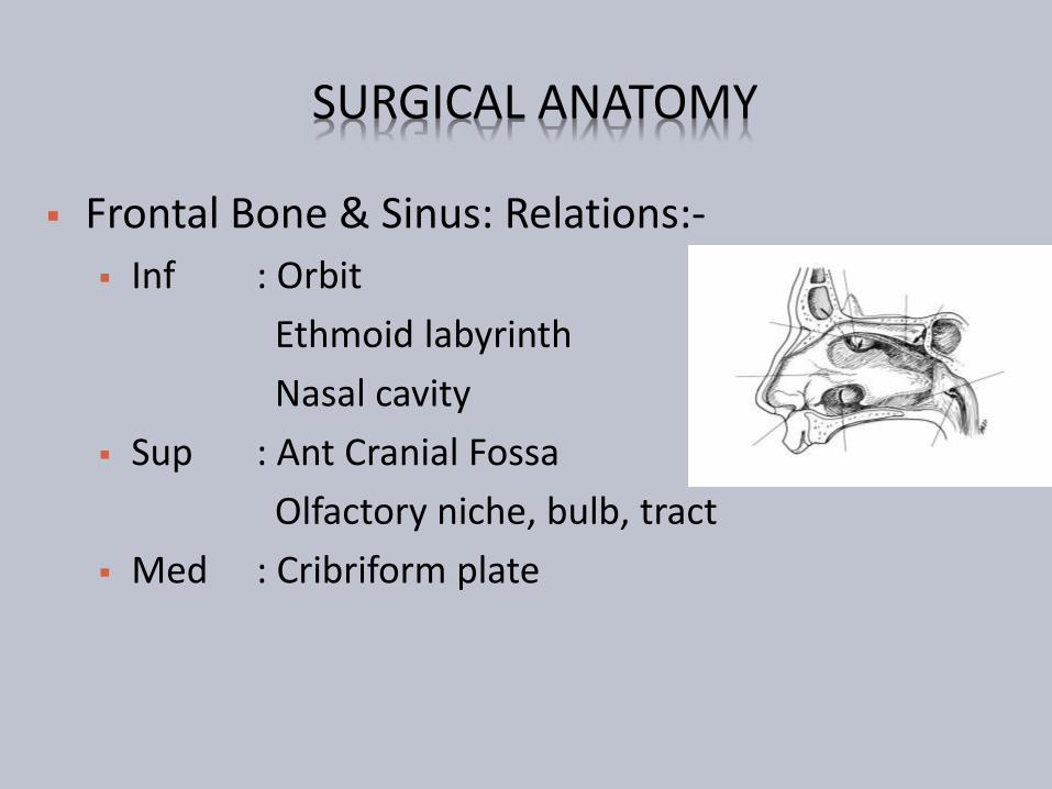
SURGICAL ANATOMY
Ethmoid Bone amp Sinus
2 ethmoid labyrinth laterally constitute orbital plate (L Papyracea) extremely thin +- dehiscent
Perpendicular plate of ethmoid in between
Intervening cribriform plate amp crista gallifenestrations olfactory filaments ethmoidalvessels amp nerve dural prolongations traverse
Ethmoid Bone amp Sinus
Relations-
Superiorly ACF amp Frontal bone
Laterally Orbit
Posteromedially Sphenoid
Posterolaterally Optic N
Medially Nasal Cavity
SURGICAL ANATOMY
SURGICAL ANATOMY
Sphenoid Bone amp Sinus
Largest bone of skull base divides ACF amp MCF
Adult 20 times 22 times 16 mm volume 75 mL
Body with variable pneumatization Conchal presellar sellar mixed
Sinus divided by paramedian septum
May be incomplete
Completely absent in 1
SURGICAL ANATOMY
ORBITAL APEX AND SUPERIOR ORBITAL FISSURE RIGHT SIDE
SURGICAL ANATOMY Sphenoid Bone amp Sinus Relations-
Ant Post ethmoidal cells Post Occipital bone
Basilar A amp Brain Stem Lat Cavernous Sinus
ICA amp Sympathetic plx Abducen N CN III IV V1 V2
Inf Roof of nasopharynx Sup Olf tract Optic chiasma
Pituitary gland Frontal Lobe
SURGICAL ANATOMY CAVERNOUS SINUS
Largest venous sinus In MCF on either side of body ofsphenoid
Divided into number of spacescaverns by trabeculae
2 cm long X 1 cm wide
Structures on lateral wall
CN III IV V1 V2
Structures throrsquo centre
ICA + Sympathetic plx
Abducen N
CAVERNOUS SINUS RELATIONS- Sup
Optic tract and chiasma
Olfactory tract
ICA
Inf
Foramen lacerum
Med
Pituitary gland
Sphenoidal air cells
Lat
Temporal lobe
Ant
Sup orb fissure amp Orbital apex
Post
Apex of petrous temporal bone
Crus cerebri of midbrain
Temporal lobe
CAVERNOUS SINUS INCOMING CHANNELS
Orbit
Sup ophthalmic vn
Inf ophthalmic vn amp br
Central retinal vn
Brain
Supf Middle Cerebral Vn
Inf Cerebral Vn
Meninges
Spheno-parietal sinus
Middle Meningeal Vn
CAVERNOUS SINUS COMMUNICATIONS
Trans sinus throrsquo Sup petrosal sinus
IJV throrsquo Inf petrosal sinus
Pterygoid plx of vn throrsquo Emissary vn
Facial vn throrsquo Sup ophthalmic vn
Communicate with each other throrsquo Intercavernoussinus amp Basilar plx of vn
SURGICAL ANATOMY ORBITAL SEPTUM
The orbicularis oculi (O) overlies theorbital septum (S)retains the orbitalfat pads (F) within the orbit
The septum fuses with the maxillaryperiosteum (P) inferiorly and thetarsus (T) superiorly
The septum is perforated by thevessels and nerves which pass fromthe orbital cavity to the face and scalp
The eyelids are richly supplied withblood
SURGICAL ANATOMY ORBIT
ROUTES OF SPREAD
ROUTES OF SPREAD Local
Natural dehiscence or weakness of surrounding bone When natural routes blocked
Massive osteolysis in acute infection
Lamina papyracea infraorbital canal suture lines
Associated thromboplebitis Diploic veins of Breschet Frontal amp Sphenoid
At peak vascularity in adolescent
Valveless veins between sinus amp orbit
Roots of 2nd premolar amp molars
Distant Hematogenous spread Rare
RISK FACTORS
Patient factors More common in young 85 under 20 yrs
Immunocompromised amp Diabetes
Abnormal mucociliary clearance Chronicity of disease
Allergy Chronicity of disease
Local anatomical variations
Patient compliance to treatment
Pathogenic factors URTI Viremia rarely encephalitis
Invasive fungal rhinosinusitis Mucor
Staph aureus Brain abscess
Treatment factors Inappropiate amp Inadequate antibiotic therapy
BACTERIOLOGY
ORBITAL
Aerobes
Staphylococcus aureus
Haemophilus influenza
Strept pneumoniae
Moraxella catarrhalis
Streptococcus milleri
Streptococcus pyogenes
Anaerobes
INTRACRANIAL
Aerobes
Staphylococcus aureus
Strept Pneumoniae
Haemophilus influenza
Streptococcus sp
Pseudomonas aeruginosa
Klebsiella sp
Anaerobes
A ORBITAL COMPLICATION
Pre-antibiotic era 17-20 died of meningitis or had permanent blindness
Commonly due to ethmoiditis in young and frontal sinusitis in adult
Higher frequency during winter amp spring
A ORBITAL COMPLICATIONS
Hubert 1937
Inflammatory edema of eyelids
Subperiosteal abscess with
Edema of eyelids or
Spread of pus to lids
Abscess of orbital tissues
Orbital cellulitis ndash Mild to severe
Cavernous sinus thrombosis
Smith amp Spencer 1948
A ORBITAL COMPLICATIONSCHANDLER LANGENBRUNNER STEVENS 1970
1 Preseptal cellulitis
2 Orbital cellulitis without
abscess
3 Orbital cellulitis with
subextraperiosteal abscess
4 Orbital cellulitis with
intraperiosteal abscess
5 Cavernous sinus thrombosis
A ORBITAL COMPLICATIONS STAGE 1
Oedema of lids
Painless non-tender
No visual loss
Globe unaffected
No restricted extra-
ocular movements
A ORBITAL COMPLICATIONS STAGE 2
Orbital cellulitis without abscess
Diffuse edema of adipose tissue
Proptosis Chemosis Edema
Associated Pain
+- Dilated pupil
+- Visual loss
+- Ophthalmoplegia
+- Afferent pupillary defect
A ORBITAL COMPLICATIONS STAGE 3
Orbital cellulitis with sub extra-periosteal abscess
Proptosis
Globe displaced inferolaterally
Decreased EOM
Vision decreased
Onidi cells optic nerve vulnerable
A ORBITAL COMPLICATIONS STAGE 3
A ORBITAL COMPLICATIONS STAGE 4
Orbital cellulits with intra-periostealabscess
Severe proptosis and chemosis
Fixed pupil
Severe globe displacement
Rapid fixation of EOM
Opthalmoplegia
Visual loss due to optic neuropathy(13)
ldquoOrbital Apex Syndromerdquo
Ophthalmoplegia amp Dilated pupil
Paraesthesia in distribution of maxilary amp ophthalmic division of trigeminal n
Blindness amp Temporal headache
A ORBITAL COMPLICATIONS STAGE 5 Cavernous Sinus Thrombosis
Bright 1831
Uncommon no incidence data
Typically affects young adults
Pathophysiology
Bacterial growth
Induces thrombosis
Thrombus good growth medium
More bacterial growth
Thrombophlebitis extend posteriorly
Orbit to Cavernous sinus
Contralateral Cavernous sinus bilateral eye symptoms
Then Intracranially
A ORBITAL COMPLICATIONS STAGE 5 Fatal prior to antibiotic era (pre-1940s) Mortality estimate 14-79 Morbidity estimate 50
Cranial neuropathies amp Visual loss
Clinical features Onset 1-21 days (Avg 5-6 days) Progressive amp bilateral eye symptom Proptosis and fixation of eye ball Bilateral orbital apex syndrome Meningitis Systemic featutes
High grade fever amp headache Tachycardia hypotension
A ORBITAL COMPLICATIONS STAGE 5
A ORBITAL COMPLICATIONS STAGE 5
Complications
Intracranial infection
Meningitis
Encephalitis
Abscess
Pituitary insufficiency
Hemorrhagic infarction
Death
A ORBITAL COMPLICATIONS MANAGEMENT
History
General ENT examination
Complete neurological examination
Rigid endoscopy of nose
Swabs
A ORBITAL COMPLICATIONS MANAGEMENT
A ORBITAL COMPLICATIONS MANAGEMENT
Other investigations CBC Blood biochemistry Blood cultures Lumbar puncture Neuroimaging (CT MRI)
Expansion of cavernous sinuses Convex bowing of lateral wall Abnormal filling defects Dilation of superior ophthalmic vein Dural enhancement of cavernous sinus border
A ORBITAL COMPLICATIONS MANAGEMENT
Empiric high dose IV antibiotics
Third generation cephalosporin
Anti-staphylococcal penicillin
Metronidazole
Continued treatment with IV antibiotics for at least 2 wks after apparent clinical resolution
Steroids controversial (except if pituitary insufficiency)
Anticoagulation in CST
No consensus for use despite theoretical rationale
Risks include systemic and intracranial bleeding
A ORBITAL COMPLICATIONS LONG TERM SEQUELAE
Permanent visual loss
Ophthalmoplegia
Exposure Keratitis amp Ulceration
Other ocular changes Uveitis
Choroiditis
Glaucoma
Iris prolapse
Rupture of the globe
B INTRACRANIAL COMPLICATIONS
Less common than orbital complications
Both can coexist
More common in adolescent
amp young adults
Male preponderance
Includes
Meningitis Encephalitis
Subdural Abscess(23) gt Frontal Lobe(4) abscess gt Extradural Abscess (1)
Cavernous Sinus Thrombosis
B INTRACRANIAL COMPLICATIONS CLINICAL PRESENTATION
Thrombophlebitis
Septic thrombophlebitismultiple abscesses formation
Associated thrombosis of Cavernous sinus amp Sup Sagittal sinus
Usually gt10yrs Uncommon in infants
Presentation Acute or Chronic
Fever leucocytosis and headache
Seizures rigidity and focal neurological signs
Features of Increased ICP
Features of meningitis amp encephalitis
B INTRACRANIAL COMPLICATIONS MANAGEMENT
High index of suspicion
History
ENT amp Full neurological examination
If abscess suspected CECT (or MRI)
Fundoscopy Lumbar puncture
IV antibiotics (Cefuroxime+Flagyl) ndash 4-6weeks
Serial CT
Steroids ndash controversial
B INTRACRANIAL COMPLICATIONS MANAGEMENT
Surgery
Treat complication amp sinusitis
Drain Extradural abscess via approach to frontal
sinuses
Neurosurgical assistance
Burr holes For extradural abscess solitary abscess
Formal craniotomy
B INTRACRANIAL COMPLICATIONS
Prognosis
Mortality 15-43 despite antibiotics
Incidence increases with age
Multiple subdural abscess with coritcal thrombosis carries worst prognosis
Morbidity Permanent in 40
Convulsions
Hemiparesis
Early treatment better outcome
C BONY COMPLICATIONS POTTrsquoS PUFFY TUMOUR
Described by Sir Percival Pott in 1970 Frontal bone is diploic with marrow cavity Sinusitis Osteomyelitis of frontal bone
Anteriorly Forehead Potts Puffy tumour Posteriorly Subdural abscess
Presentation Fluctuant swelling +- Pain
Causative Org Staph amp Strepto- amp Anaerobes Investigation
Blood investigation + CECT
Treatment Medical IV Antibiotics Surgical Bilateral coronal incision +Bone debridement
C BONY COMPLICATIONS
POTTrsquoS PUFFY TUMOUR
D DENTAL COMPLICATIONS
Common with Maxillary Sinusitis
Close association of dentition with floor of sinus
Acute sinusitis Dental pain
Dental abscess Mistaken for Sinusitis
May coexist
E SYSTEMIC COMPLICATIONS
Toxic Shock Syndrome
Rare Potentially Fatal
Frequently associated with Staph aureus Streptococcus
Features of Toxaemia Fever Hypotension Rash amp MODS
Septicaemia
Hematogenous spread
Blood culture
Features of SIRS
Shock
MODS
F CHRONIC COMPLICATIONS MUCOCOELE Definition
Epithelial lined mucus containing sac completely filling thesinus and capable of expansion
1820 Langenback lsquoHydatidesrsquo
1896 Rollet lsquomucocoelersquo
Incidence 4 of case of unilateral proptosis
Most common sites F(65)gt E(25)gt M(10)gt S
Age grp 40-70 yrs
lt 5 bilateral or multiloculated53
F CHRONIC COMPLICATIONS MUCOCOELE Theories Chronic rinosinusitis Increased osteolysis
Active Bone resorption amp formation
Pressure erosion
Cystic degeneration of seromucimous glands
54
F CHRONIC COMPLICATIONS MUCOCOELE
Initial ophthalmic referral
Clinical features Proptosis
Diplopia
Displacement of globe
Limited ocular movement
Visual impairment
Mass
Endoscopy
55
F CHRONIC COMPLICATIONS MUCOCOELE
56
ImagingLoss of Scalloping of frontal sinus
F MUCOCOELE CECT PNS
FRONTAL MUCOCOELE BILOCULATED ETHMOID MUCOCOELE
57
Homogenous smooth walled mass expanding the sinus
F MUCOCOELE CULTURE OF ASPIRATE
Mixed infection
Staph aureus
Alpha hemolytic Streptococci
Heamophillus sp
GNB
Anaerobic bacteria
58
F MUCOCOELE TREATMENT
Surgery is the treatment
Goals
Eradication of disease
Minimal morbidity
Prevention of recurrence
59
F MUCOCOELE TREATMENT
Approach
Endoscopic drainage
Type I-III
External approach
Osteoplastic flap
+- sinus cavity obliteration
Combined approach
60
THANK YOU
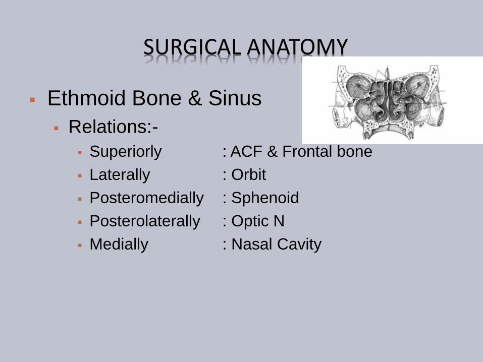

SURGICAL ANATOMY
Sphenoid Bone amp Sinus
Largest bone of skull base divides ACF amp MCF
Adult 20 times 22 times 16 mm volume 75 mL
Body with variable pneumatization Conchal presellar sellar mixed
Sinus divided by paramedian septum
May be incomplete
Completely absent in 1
SURGICAL ANATOMY
ORBITAL APEX AND SUPERIOR ORBITAL FISSURE RIGHT SIDE
SURGICAL ANATOMY Sphenoid Bone amp Sinus Relations-
Ant Post ethmoidal cells Post Occipital bone
Basilar A amp Brain Stem Lat Cavernous Sinus
ICA amp Sympathetic plx Abducen N CN III IV V1 V2
Inf Roof of nasopharynx Sup Olf tract Optic chiasma
Pituitary gland Frontal Lobe
SURGICAL ANATOMY CAVERNOUS SINUS
Largest venous sinus In MCF on either side of body ofsphenoid
Divided into number of spacescaverns by trabeculae
2 cm long X 1 cm wide
Structures on lateral wall
CN III IV V1 V2
Structures throrsquo centre
ICA + Sympathetic plx
Abducen N
CAVERNOUS SINUS RELATIONS- Sup
Optic tract and chiasma
Olfactory tract
ICA
Inf
Foramen lacerum
Med
Pituitary gland
Sphenoidal air cells
Lat
Temporal lobe
Ant
Sup orb fissure amp Orbital apex
Post
Apex of petrous temporal bone
Crus cerebri of midbrain
Temporal lobe
CAVERNOUS SINUS INCOMING CHANNELS
Orbit
Sup ophthalmic vn
Inf ophthalmic vn amp br
Central retinal vn
Brain
Supf Middle Cerebral Vn
Inf Cerebral Vn
Meninges
Spheno-parietal sinus
Middle Meningeal Vn
CAVERNOUS SINUS COMMUNICATIONS
Trans sinus throrsquo Sup petrosal sinus
IJV throrsquo Inf petrosal sinus
Pterygoid plx of vn throrsquo Emissary vn
Facial vn throrsquo Sup ophthalmic vn
Communicate with each other throrsquo Intercavernoussinus amp Basilar plx of vn
SURGICAL ANATOMY ORBITAL SEPTUM
The orbicularis oculi (O) overlies theorbital septum (S)retains the orbitalfat pads (F) within the orbit
The septum fuses with the maxillaryperiosteum (P) inferiorly and thetarsus (T) superiorly
The septum is perforated by thevessels and nerves which pass fromthe orbital cavity to the face and scalp
The eyelids are richly supplied withblood
SURGICAL ANATOMY ORBIT
ROUTES OF SPREAD
ROUTES OF SPREAD Local
Natural dehiscence or weakness of surrounding bone When natural routes blocked
Massive osteolysis in acute infection
Lamina papyracea infraorbital canal suture lines
Associated thromboplebitis Diploic veins of Breschet Frontal amp Sphenoid
At peak vascularity in adolescent
Valveless veins between sinus amp orbit
Roots of 2nd premolar amp molars
Distant Hematogenous spread Rare
RISK FACTORS
Patient factors More common in young 85 under 20 yrs
Immunocompromised amp Diabetes
Abnormal mucociliary clearance Chronicity of disease
Allergy Chronicity of disease
Local anatomical variations
Patient compliance to treatment
Pathogenic factors URTI Viremia rarely encephalitis
Invasive fungal rhinosinusitis Mucor
Staph aureus Brain abscess
Treatment factors Inappropiate amp Inadequate antibiotic therapy
BACTERIOLOGY
ORBITAL
Aerobes
Staphylococcus aureus
Haemophilus influenza
Strept pneumoniae
Moraxella catarrhalis
Streptococcus milleri
Streptococcus pyogenes
Anaerobes
INTRACRANIAL
Aerobes
Staphylococcus aureus
Strept Pneumoniae
Haemophilus influenza
Streptococcus sp
Pseudomonas aeruginosa
Klebsiella sp
Anaerobes
A ORBITAL COMPLICATION
Pre-antibiotic era 17-20 died of meningitis or had permanent blindness
Commonly due to ethmoiditis in young and frontal sinusitis in adult
Higher frequency during winter amp spring
A ORBITAL COMPLICATIONS
Hubert 1937
Inflammatory edema of eyelids
Subperiosteal abscess with
Edema of eyelids or
Spread of pus to lids
Abscess of orbital tissues
Orbital cellulitis ndash Mild to severe
Cavernous sinus thrombosis
Smith amp Spencer 1948
A ORBITAL COMPLICATIONSCHANDLER LANGENBRUNNER STEVENS 1970
1 Preseptal cellulitis
2 Orbital cellulitis without
abscess
3 Orbital cellulitis with
subextraperiosteal abscess
4 Orbital cellulitis with
intraperiosteal abscess
5 Cavernous sinus thrombosis
A ORBITAL COMPLICATIONS STAGE 1
Oedema of lids
Painless non-tender
No visual loss
Globe unaffected
No restricted extra-
ocular movements
A ORBITAL COMPLICATIONS STAGE 2
Orbital cellulitis without abscess
Diffuse edema of adipose tissue
Proptosis Chemosis Edema
Associated Pain
+- Dilated pupil
+- Visual loss
+- Ophthalmoplegia
+- Afferent pupillary defect
A ORBITAL COMPLICATIONS STAGE 3
Orbital cellulitis with sub extra-periosteal abscess
Proptosis
Globe displaced inferolaterally
Decreased EOM
Vision decreased
Onidi cells optic nerve vulnerable
A ORBITAL COMPLICATIONS STAGE 3
A ORBITAL COMPLICATIONS STAGE 4
Orbital cellulits with intra-periostealabscess
Severe proptosis and chemosis
Fixed pupil
Severe globe displacement
Rapid fixation of EOM
Opthalmoplegia
Visual loss due to optic neuropathy(13)
ldquoOrbital Apex Syndromerdquo
Ophthalmoplegia amp Dilated pupil
Paraesthesia in distribution of maxilary amp ophthalmic division of trigeminal n
Blindness amp Temporal headache
A ORBITAL COMPLICATIONS STAGE 5 Cavernous Sinus Thrombosis
Bright 1831
Uncommon no incidence data
Typically affects young adults
Pathophysiology
Bacterial growth
Induces thrombosis
Thrombus good growth medium
More bacterial growth
Thrombophlebitis extend posteriorly
Orbit to Cavernous sinus
Contralateral Cavernous sinus bilateral eye symptoms
Then Intracranially
A ORBITAL COMPLICATIONS STAGE 5 Fatal prior to antibiotic era (pre-1940s) Mortality estimate 14-79 Morbidity estimate 50
Cranial neuropathies amp Visual loss
Clinical features Onset 1-21 days (Avg 5-6 days) Progressive amp bilateral eye symptom Proptosis and fixation of eye ball Bilateral orbital apex syndrome Meningitis Systemic featutes
High grade fever amp headache Tachycardia hypotension
A ORBITAL COMPLICATIONS STAGE 5
A ORBITAL COMPLICATIONS STAGE 5
Complications
Intracranial infection
Meningitis
Encephalitis
Abscess
Pituitary insufficiency
Hemorrhagic infarction
Death
A ORBITAL COMPLICATIONS MANAGEMENT
History
General ENT examination
Complete neurological examination
Rigid endoscopy of nose
Swabs
A ORBITAL COMPLICATIONS MANAGEMENT
A ORBITAL COMPLICATIONS MANAGEMENT
Other investigations CBC Blood biochemistry Blood cultures Lumbar puncture Neuroimaging (CT MRI)
Expansion of cavernous sinuses Convex bowing of lateral wall Abnormal filling defects Dilation of superior ophthalmic vein Dural enhancement of cavernous sinus border
A ORBITAL COMPLICATIONS MANAGEMENT
Empiric high dose IV antibiotics
Third generation cephalosporin
Anti-staphylococcal penicillin
Metronidazole
Continued treatment with IV antibiotics for at least 2 wks after apparent clinical resolution
Steroids controversial (except if pituitary insufficiency)
Anticoagulation in CST
No consensus for use despite theoretical rationale
Risks include systemic and intracranial bleeding
A ORBITAL COMPLICATIONS LONG TERM SEQUELAE
Permanent visual loss
Ophthalmoplegia
Exposure Keratitis amp Ulceration
Other ocular changes Uveitis
Choroiditis
Glaucoma
Iris prolapse
Rupture of the globe
B INTRACRANIAL COMPLICATIONS
Less common than orbital complications
Both can coexist
More common in adolescent
amp young adults
Male preponderance
Includes
Meningitis Encephalitis
Subdural Abscess(23) gt Frontal Lobe(4) abscess gt Extradural Abscess (1)
Cavernous Sinus Thrombosis
B INTRACRANIAL COMPLICATIONS CLINICAL PRESENTATION
Thrombophlebitis
Septic thrombophlebitismultiple abscesses formation
Associated thrombosis of Cavernous sinus amp Sup Sagittal sinus
Usually gt10yrs Uncommon in infants
Presentation Acute or Chronic
Fever leucocytosis and headache
Seizures rigidity and focal neurological signs
Features of Increased ICP
Features of meningitis amp encephalitis
B INTRACRANIAL COMPLICATIONS MANAGEMENT
High index of suspicion
History
ENT amp Full neurological examination
If abscess suspected CECT (or MRI)
Fundoscopy Lumbar puncture
IV antibiotics (Cefuroxime+Flagyl) ndash 4-6weeks
Serial CT
Steroids ndash controversial
B INTRACRANIAL COMPLICATIONS MANAGEMENT
Surgery
Treat complication amp sinusitis
Drain Extradural abscess via approach to frontal
sinuses
Neurosurgical assistance
Burr holes For extradural abscess solitary abscess
Formal craniotomy
B INTRACRANIAL COMPLICATIONS
Prognosis
Mortality 15-43 despite antibiotics
Incidence increases with age
Multiple subdural abscess with coritcal thrombosis carries worst prognosis
Morbidity Permanent in 40
Convulsions
Hemiparesis
Early treatment better outcome
C BONY COMPLICATIONS POTTrsquoS PUFFY TUMOUR
Described by Sir Percival Pott in 1970 Frontal bone is diploic with marrow cavity Sinusitis Osteomyelitis of frontal bone
Anteriorly Forehead Potts Puffy tumour Posteriorly Subdural abscess
Presentation Fluctuant swelling +- Pain
Causative Org Staph amp Strepto- amp Anaerobes Investigation
Blood investigation + CECT
Treatment Medical IV Antibiotics Surgical Bilateral coronal incision +Bone debridement
C BONY COMPLICATIONS
POTTrsquoS PUFFY TUMOUR
D DENTAL COMPLICATIONS
Common with Maxillary Sinusitis
Close association of dentition with floor of sinus
Acute sinusitis Dental pain
Dental abscess Mistaken for Sinusitis
May coexist
E SYSTEMIC COMPLICATIONS
Toxic Shock Syndrome
Rare Potentially Fatal
Frequently associated with Staph aureus Streptococcus
Features of Toxaemia Fever Hypotension Rash amp MODS
Septicaemia
Hematogenous spread
Blood culture
Features of SIRS
Shock
MODS
F CHRONIC COMPLICATIONS MUCOCOELE Definition
Epithelial lined mucus containing sac completely filling thesinus and capable of expansion
1820 Langenback lsquoHydatidesrsquo
1896 Rollet lsquomucocoelersquo
Incidence 4 of case of unilateral proptosis
Most common sites F(65)gt E(25)gt M(10)gt S
Age grp 40-70 yrs
lt 5 bilateral or multiloculated53
F CHRONIC COMPLICATIONS MUCOCOELE Theories Chronic rinosinusitis Increased osteolysis
Active Bone resorption amp formation
Pressure erosion
Cystic degeneration of seromucimous glands
54
F CHRONIC COMPLICATIONS MUCOCOELE
Initial ophthalmic referral
Clinical features Proptosis
Diplopia
Displacement of globe
Limited ocular movement
Visual impairment
Mass
Endoscopy
55
F CHRONIC COMPLICATIONS MUCOCOELE
56
ImagingLoss of Scalloping of frontal sinus
F MUCOCOELE CECT PNS
FRONTAL MUCOCOELE BILOCULATED ETHMOID MUCOCOELE
57
Homogenous smooth walled mass expanding the sinus
F MUCOCOELE CULTURE OF ASPIRATE
Mixed infection
Staph aureus
Alpha hemolytic Streptococci
Heamophillus sp
GNB
Anaerobic bacteria
58
F MUCOCOELE TREATMENT
Surgery is the treatment
Goals
Eradication of disease
Minimal morbidity
Prevention of recurrence
59
F MUCOCOELE TREATMENT
Approach
Endoscopic drainage
Type I-III
External approach
Osteoplastic flap
+- sinus cavity obliteration
Combined approach
60
THANK YOU
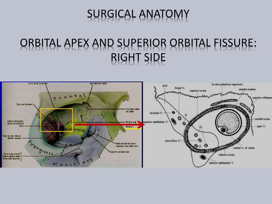
SURGICAL ANATOMY
ORBITAL APEX AND SUPERIOR ORBITAL FISSURE RIGHT SIDE
SURGICAL ANATOMY Sphenoid Bone amp Sinus Relations-
Ant Post ethmoidal cells Post Occipital bone
Basilar A amp Brain Stem Lat Cavernous Sinus
ICA amp Sympathetic plx Abducen N CN III IV V1 V2
Inf Roof of nasopharynx Sup Olf tract Optic chiasma
Pituitary gland Frontal Lobe
SURGICAL ANATOMY CAVERNOUS SINUS
Largest venous sinus In MCF on either side of body ofsphenoid
Divided into number of spacescaverns by trabeculae
2 cm long X 1 cm wide
Structures on lateral wall
CN III IV V1 V2
Structures throrsquo centre
ICA + Sympathetic plx
Abducen N
CAVERNOUS SINUS RELATIONS- Sup
Optic tract and chiasma
Olfactory tract
ICA
Inf
Foramen lacerum
Med
Pituitary gland
Sphenoidal air cells
Lat
Temporal lobe
Ant
Sup orb fissure amp Orbital apex
Post
Apex of petrous temporal bone
Crus cerebri of midbrain
Temporal lobe
CAVERNOUS SINUS INCOMING CHANNELS
Orbit
Sup ophthalmic vn
Inf ophthalmic vn amp br
Central retinal vn
Brain
Supf Middle Cerebral Vn
Inf Cerebral Vn
Meninges
Spheno-parietal sinus
Middle Meningeal Vn
CAVERNOUS SINUS COMMUNICATIONS
Trans sinus throrsquo Sup petrosal sinus
IJV throrsquo Inf petrosal sinus
Pterygoid plx of vn throrsquo Emissary vn
Facial vn throrsquo Sup ophthalmic vn
Communicate with each other throrsquo Intercavernoussinus amp Basilar plx of vn
SURGICAL ANATOMY ORBITAL SEPTUM
The orbicularis oculi (O) overlies theorbital septum (S)retains the orbitalfat pads (F) within the orbit
The septum fuses with the maxillaryperiosteum (P) inferiorly and thetarsus (T) superiorly
The septum is perforated by thevessels and nerves which pass fromthe orbital cavity to the face and scalp
The eyelids are richly supplied withblood
SURGICAL ANATOMY ORBIT
ROUTES OF SPREAD
ROUTES OF SPREAD Local
Natural dehiscence or weakness of surrounding bone When natural routes blocked
Massive osteolysis in acute infection
Lamina papyracea infraorbital canal suture lines
Associated thromboplebitis Diploic veins of Breschet Frontal amp Sphenoid
At peak vascularity in adolescent
Valveless veins between sinus amp orbit
Roots of 2nd premolar amp molars
Distant Hematogenous spread Rare
RISK FACTORS
Patient factors More common in young 85 under 20 yrs
Immunocompromised amp Diabetes
Abnormal mucociliary clearance Chronicity of disease
Allergy Chronicity of disease
Local anatomical variations
Patient compliance to treatment
Pathogenic factors URTI Viremia rarely encephalitis
Invasive fungal rhinosinusitis Mucor
Staph aureus Brain abscess
Treatment factors Inappropiate amp Inadequate antibiotic therapy
BACTERIOLOGY
ORBITAL
Aerobes
Staphylococcus aureus
Haemophilus influenza
Strept pneumoniae
Moraxella catarrhalis
Streptococcus milleri
Streptococcus pyogenes
Anaerobes
INTRACRANIAL
Aerobes
Staphylococcus aureus
Strept Pneumoniae
Haemophilus influenza
Streptococcus sp
Pseudomonas aeruginosa
Klebsiella sp
Anaerobes
A ORBITAL COMPLICATION
Pre-antibiotic era 17-20 died of meningitis or had permanent blindness
Commonly due to ethmoiditis in young and frontal sinusitis in adult
Higher frequency during winter amp spring
A ORBITAL COMPLICATIONS
Hubert 1937
Inflammatory edema of eyelids
Subperiosteal abscess with
Edema of eyelids or
Spread of pus to lids
Abscess of orbital tissues
Orbital cellulitis ndash Mild to severe
Cavernous sinus thrombosis
Smith amp Spencer 1948
A ORBITAL COMPLICATIONSCHANDLER LANGENBRUNNER STEVENS 1970
1 Preseptal cellulitis
2 Orbital cellulitis without
abscess
3 Orbital cellulitis with
subextraperiosteal abscess
4 Orbital cellulitis with
intraperiosteal abscess
5 Cavernous sinus thrombosis
A ORBITAL COMPLICATIONS STAGE 1
Oedema of lids
Painless non-tender
No visual loss
Globe unaffected
No restricted extra-
ocular movements
A ORBITAL COMPLICATIONS STAGE 2
Orbital cellulitis without abscess
Diffuse edema of adipose tissue
Proptosis Chemosis Edema
Associated Pain
+- Dilated pupil
+- Visual loss
+- Ophthalmoplegia
+- Afferent pupillary defect
A ORBITAL COMPLICATIONS STAGE 3
Orbital cellulitis with sub extra-periosteal abscess
Proptosis
Globe displaced inferolaterally
Decreased EOM
Vision decreased
Onidi cells optic nerve vulnerable
A ORBITAL COMPLICATIONS STAGE 3
A ORBITAL COMPLICATIONS STAGE 4
Orbital cellulits with intra-periostealabscess
Severe proptosis and chemosis
Fixed pupil
Severe globe displacement
Rapid fixation of EOM
Opthalmoplegia
Visual loss due to optic neuropathy(13)
ldquoOrbital Apex Syndromerdquo
Ophthalmoplegia amp Dilated pupil
Paraesthesia in distribution of maxilary amp ophthalmic division of trigeminal n
Blindness amp Temporal headache
A ORBITAL COMPLICATIONS STAGE 5 Cavernous Sinus Thrombosis
Bright 1831
Uncommon no incidence data
Typically affects young adults
Pathophysiology
Bacterial growth
Induces thrombosis
Thrombus good growth medium
More bacterial growth
Thrombophlebitis extend posteriorly
Orbit to Cavernous sinus
Contralateral Cavernous sinus bilateral eye symptoms
Then Intracranially
A ORBITAL COMPLICATIONS STAGE 5 Fatal prior to antibiotic era (pre-1940s) Mortality estimate 14-79 Morbidity estimate 50
Cranial neuropathies amp Visual loss
Clinical features Onset 1-21 days (Avg 5-6 days) Progressive amp bilateral eye symptom Proptosis and fixation of eye ball Bilateral orbital apex syndrome Meningitis Systemic featutes
High grade fever amp headache Tachycardia hypotension
A ORBITAL COMPLICATIONS STAGE 5
A ORBITAL COMPLICATIONS STAGE 5
Complications
Intracranial infection
Meningitis
Encephalitis
Abscess
Pituitary insufficiency
Hemorrhagic infarction
Death
A ORBITAL COMPLICATIONS MANAGEMENT
History
General ENT examination
Complete neurological examination
Rigid endoscopy of nose
Swabs
A ORBITAL COMPLICATIONS MANAGEMENT
A ORBITAL COMPLICATIONS MANAGEMENT
Other investigations CBC Blood biochemistry Blood cultures Lumbar puncture Neuroimaging (CT MRI)
Expansion of cavernous sinuses Convex bowing of lateral wall Abnormal filling defects Dilation of superior ophthalmic vein Dural enhancement of cavernous sinus border
A ORBITAL COMPLICATIONS MANAGEMENT
Empiric high dose IV antibiotics
Third generation cephalosporin
Anti-staphylococcal penicillin
Metronidazole
Continued treatment with IV antibiotics for at least 2 wks after apparent clinical resolution
Steroids controversial (except if pituitary insufficiency)
Anticoagulation in CST
No consensus for use despite theoretical rationale
Risks include systemic and intracranial bleeding
A ORBITAL COMPLICATIONS LONG TERM SEQUELAE
Permanent visual loss
Ophthalmoplegia
Exposure Keratitis amp Ulceration
Other ocular changes Uveitis
Choroiditis
Glaucoma
Iris prolapse
Rupture of the globe
B INTRACRANIAL COMPLICATIONS
Less common than orbital complications
Both can coexist
More common in adolescent
amp young adults
Male preponderance
Includes
Meningitis Encephalitis
Subdural Abscess(23) gt Frontal Lobe(4) abscess gt Extradural Abscess (1)
Cavernous Sinus Thrombosis
B INTRACRANIAL COMPLICATIONS CLINICAL PRESENTATION
Thrombophlebitis
Septic thrombophlebitismultiple abscesses formation
Associated thrombosis of Cavernous sinus amp Sup Sagittal sinus
Usually gt10yrs Uncommon in infants
Presentation Acute or Chronic
Fever leucocytosis and headache
Seizures rigidity and focal neurological signs
Features of Increased ICP
Features of meningitis amp encephalitis
B INTRACRANIAL COMPLICATIONS MANAGEMENT
High index of suspicion
History
ENT amp Full neurological examination
If abscess suspected CECT (or MRI)
Fundoscopy Lumbar puncture
IV antibiotics (Cefuroxime+Flagyl) ndash 4-6weeks
Serial CT
Steroids ndash controversial
B INTRACRANIAL COMPLICATIONS MANAGEMENT
Surgery
Treat complication amp sinusitis
Drain Extradural abscess via approach to frontal
sinuses
Neurosurgical assistance
Burr holes For extradural abscess solitary abscess
Formal craniotomy
B INTRACRANIAL COMPLICATIONS
Prognosis
Mortality 15-43 despite antibiotics
Incidence increases with age
Multiple subdural abscess with coritcal thrombosis carries worst prognosis
Morbidity Permanent in 40
Convulsions
Hemiparesis
Early treatment better outcome
C BONY COMPLICATIONS POTTrsquoS PUFFY TUMOUR
Described by Sir Percival Pott in 1970 Frontal bone is diploic with marrow cavity Sinusitis Osteomyelitis of frontal bone
Anteriorly Forehead Potts Puffy tumour Posteriorly Subdural abscess
Presentation Fluctuant swelling +- Pain
Causative Org Staph amp Strepto- amp Anaerobes Investigation
Blood investigation + CECT
Treatment Medical IV Antibiotics Surgical Bilateral coronal incision +Bone debridement
C BONY COMPLICATIONS
POTTrsquoS PUFFY TUMOUR
D DENTAL COMPLICATIONS
Common with Maxillary Sinusitis
Close association of dentition with floor of sinus
Acute sinusitis Dental pain
Dental abscess Mistaken for Sinusitis
May coexist
E SYSTEMIC COMPLICATIONS
Toxic Shock Syndrome
Rare Potentially Fatal
Frequently associated with Staph aureus Streptococcus
Features of Toxaemia Fever Hypotension Rash amp MODS
Septicaemia
Hematogenous spread
Blood culture
Features of SIRS
Shock
MODS
F CHRONIC COMPLICATIONS MUCOCOELE Definition
Epithelial lined mucus containing sac completely filling thesinus and capable of expansion
1820 Langenback lsquoHydatidesrsquo
1896 Rollet lsquomucocoelersquo
Incidence 4 of case of unilateral proptosis
Most common sites F(65)gt E(25)gt M(10)gt S
Age grp 40-70 yrs
lt 5 bilateral or multiloculated53
F CHRONIC COMPLICATIONS MUCOCOELE Theories Chronic rinosinusitis Increased osteolysis
Active Bone resorption amp formation
Pressure erosion
Cystic degeneration of seromucimous glands
54
F CHRONIC COMPLICATIONS MUCOCOELE
Initial ophthalmic referral
Clinical features Proptosis
Diplopia
Displacement of globe
Limited ocular movement
Visual impairment
Mass
Endoscopy
55
F CHRONIC COMPLICATIONS MUCOCOELE
56
ImagingLoss of Scalloping of frontal sinus
F MUCOCOELE CECT PNS
FRONTAL MUCOCOELE BILOCULATED ETHMOID MUCOCOELE
57
Homogenous smooth walled mass expanding the sinus
F MUCOCOELE CULTURE OF ASPIRATE
Mixed infection
Staph aureus
Alpha hemolytic Streptococci
Heamophillus sp
GNB
Anaerobic bacteria
58
F MUCOCOELE TREATMENT
Surgery is the treatment
Goals
Eradication of disease
Minimal morbidity
Prevention of recurrence
59
F MUCOCOELE TREATMENT
Approach
Endoscopic drainage
Type I-III
External approach
Osteoplastic flap
+- sinus cavity obliteration
Combined approach
60
THANK YOU
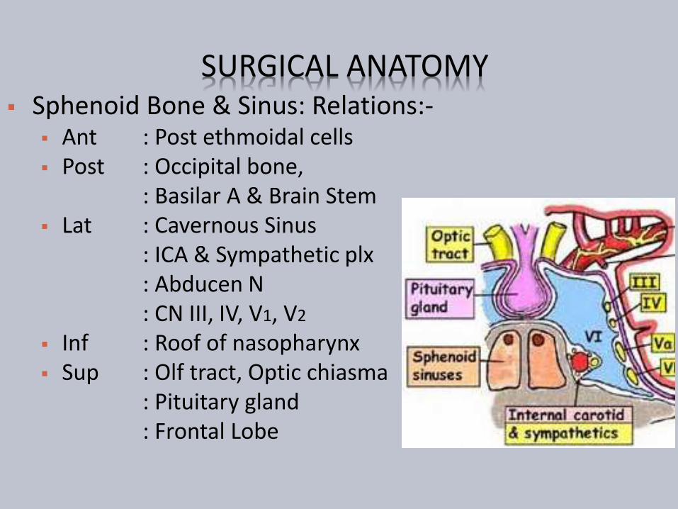
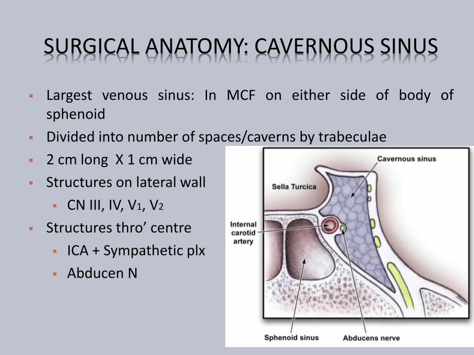
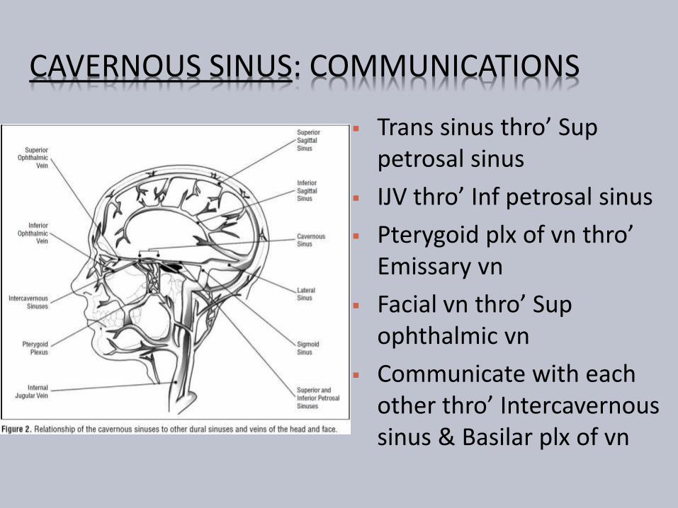
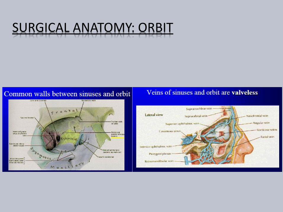
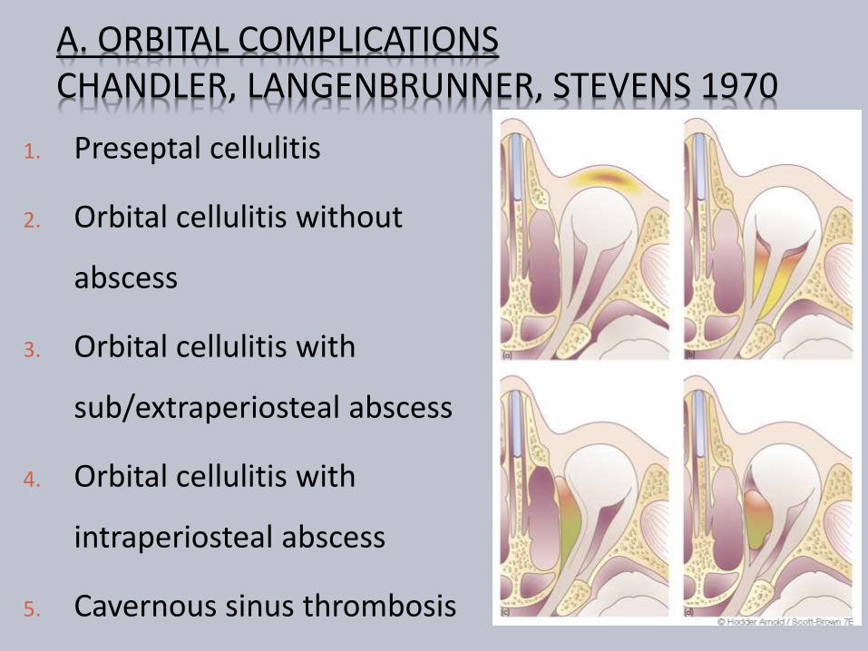
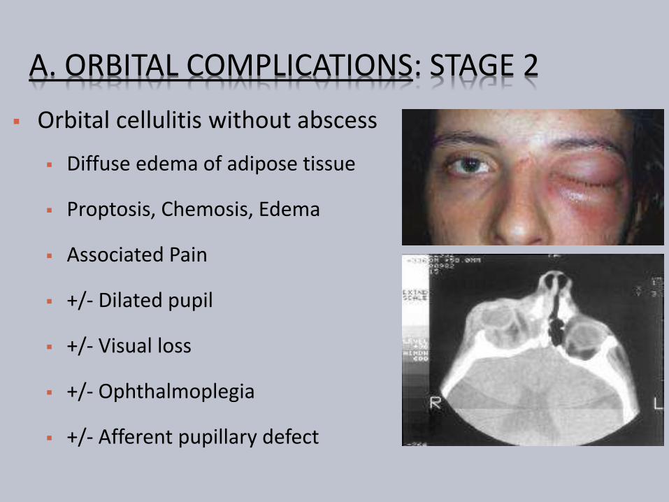
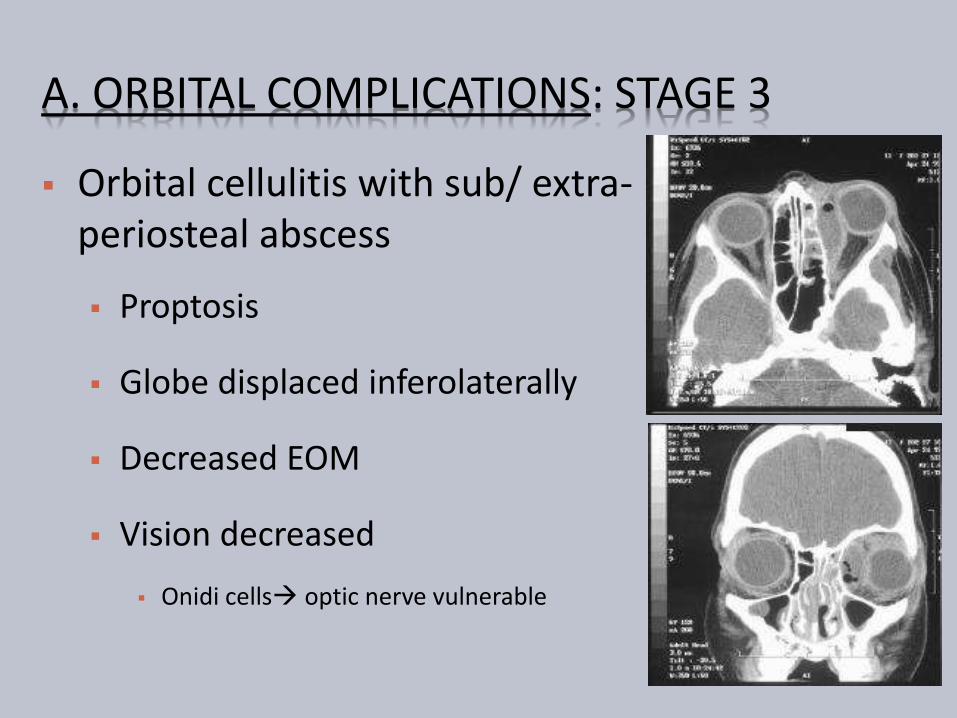
A ORBITAL COMPLICATIONS STAGE 3
Orbital cellulitis with sub extra-periosteal abscess
Proptosis
Globe displaced inferolaterally
Decreased EOM
Vision decreased
Onidi cells optic nerve vulnerable
A ORBITAL COMPLICATIONS STAGE 3
A ORBITAL COMPLICATIONS STAGE 4
Orbital cellulits with intra-periostealabscess
Severe proptosis and chemosis
Fixed pupil
Severe globe displacement
Rapid fixation of EOM
Opthalmoplegia
Visual loss due to optic neuropathy(13)
ldquoOrbital Apex Syndromerdquo
Ophthalmoplegia amp Dilated pupil
Paraesthesia in distribution of maxilary amp ophthalmic division of trigeminal n
Blindness amp Temporal headache
A ORBITAL COMPLICATIONS STAGE 5 Cavernous Sinus Thrombosis
Bright 1831
Uncommon no incidence data
Typically affects young adults
Pathophysiology
Bacterial growth
Induces thrombosis
Thrombus good growth medium
More bacterial growth
Thrombophlebitis extend posteriorly
Orbit to Cavernous sinus
Contralateral Cavernous sinus bilateral eye symptoms
Then Intracranially
A ORBITAL COMPLICATIONS STAGE 5 Fatal prior to antibiotic era (pre-1940s) Mortality estimate 14-79 Morbidity estimate 50
Cranial neuropathies amp Visual loss
Clinical features Onset 1-21 days (Avg 5-6 days) Progressive amp bilateral eye symptom Proptosis and fixation of eye ball Bilateral orbital apex syndrome Meningitis Systemic featutes
High grade fever amp headache Tachycardia hypotension
A ORBITAL COMPLICATIONS STAGE 5
A ORBITAL COMPLICATIONS STAGE 5
Complications
Intracranial infection
Meningitis
Encephalitis
Abscess
Pituitary insufficiency
Hemorrhagic infarction
Death
A ORBITAL COMPLICATIONS MANAGEMENT
History
General ENT examination
Complete neurological examination
Rigid endoscopy of nose
Swabs
A ORBITAL COMPLICATIONS MANAGEMENT
A ORBITAL COMPLICATIONS MANAGEMENT
Other investigations CBC Blood biochemistry Blood cultures Lumbar puncture Neuroimaging (CT MRI)
Expansion of cavernous sinuses Convex bowing of lateral wall Abnormal filling defects Dilation of superior ophthalmic vein Dural enhancement of cavernous sinus border
A ORBITAL COMPLICATIONS MANAGEMENT
Empiric high dose IV antibiotics
Third generation cephalosporin
Anti-staphylococcal penicillin
Metronidazole
Continued treatment with IV antibiotics for at least 2 wks after apparent clinical resolution
Steroids controversial (except if pituitary insufficiency)
Anticoagulation in CST
No consensus for use despite theoretical rationale
Risks include systemic and intracranial bleeding
A ORBITAL COMPLICATIONS LONG TERM SEQUELAE
Permanent visual loss
Ophthalmoplegia
Exposure Keratitis amp Ulceration
Other ocular changes Uveitis
Choroiditis
Glaucoma
Iris prolapse
Rupture of the globe
B INTRACRANIAL COMPLICATIONS
Less common than orbital complications
Both can coexist
More common in adolescent
amp young adults
Male preponderance
Includes
Meningitis Encephalitis
Subdural Abscess(23) gt Frontal Lobe(4) abscess gt Extradural Abscess (1)
Cavernous Sinus Thrombosis
B INTRACRANIAL COMPLICATIONS CLINICAL PRESENTATION
Thrombophlebitis
Septic thrombophlebitismultiple abscesses formation
Associated thrombosis of Cavernous sinus amp Sup Sagittal sinus
Usually gt10yrs Uncommon in infants
Presentation Acute or Chronic
Fever leucocytosis and headache
Seizures rigidity and focal neurological signs
Features of Increased ICP
Features of meningitis amp encephalitis
B INTRACRANIAL COMPLICATIONS MANAGEMENT
High index of suspicion
History
ENT amp Full neurological examination
If abscess suspected CECT (or MRI)
Fundoscopy Lumbar puncture
IV antibiotics (Cefuroxime+Flagyl) ndash 4-6weeks
Serial CT
Steroids ndash controversial
B INTRACRANIAL COMPLICATIONS MANAGEMENT
Surgery
Treat complication amp sinusitis
Drain Extradural abscess via approach to frontal
sinuses
Neurosurgical assistance
Burr holes For extradural abscess solitary abscess
Formal craniotomy
B INTRACRANIAL COMPLICATIONS
Prognosis
Mortality 15-43 despite antibiotics
Incidence increases with age
Multiple subdural abscess with coritcal thrombosis carries worst prognosis
Morbidity Permanent in 40
Convulsions
Hemiparesis
Early treatment better outcome
C BONY COMPLICATIONS POTTrsquoS PUFFY TUMOUR
Described by Sir Percival Pott in 1970 Frontal bone is diploic with marrow cavity Sinusitis Osteomyelitis of frontal bone
Anteriorly Forehead Potts Puffy tumour Posteriorly Subdural abscess
Presentation Fluctuant swelling +- Pain
Causative Org Staph amp Strepto- amp Anaerobes Investigation
Blood investigation + CECT
Treatment Medical IV Antibiotics Surgical Bilateral coronal incision +Bone debridement
C BONY COMPLICATIONS
POTTrsquoS PUFFY TUMOUR
D DENTAL COMPLICATIONS
Common with Maxillary Sinusitis
Close association of dentition with floor of sinus
Acute sinusitis Dental pain
Dental abscess Mistaken for Sinusitis
May coexist
E SYSTEMIC COMPLICATIONS
Toxic Shock Syndrome
Rare Potentially Fatal
Frequently associated with Staph aureus Streptococcus
Features of Toxaemia Fever Hypotension Rash amp MODS
Septicaemia
Hematogenous spread
Blood culture
Features of SIRS
Shock
MODS
F CHRONIC COMPLICATIONS MUCOCOELE Definition
Epithelial lined mucus containing sac completely filling thesinus and capable of expansion
1820 Langenback lsquoHydatidesrsquo
1896 Rollet lsquomucocoelersquo
Incidence 4 of case of unilateral proptosis
Most common sites F(65)gt E(25)gt M(10)gt S
Age grp 40-70 yrs
lt 5 bilateral or multiloculated53
F CHRONIC COMPLICATIONS MUCOCOELE Theories Chronic rinosinusitis Increased osteolysis
Active Bone resorption amp formation
Pressure erosion
Cystic degeneration of seromucimous glands
54
F CHRONIC COMPLICATIONS MUCOCOELE
Initial ophthalmic referral
Clinical features Proptosis
Diplopia
Displacement of globe
Limited ocular movement
Visual impairment
Mass
Endoscopy
55
F CHRONIC COMPLICATIONS MUCOCOELE
56
ImagingLoss of Scalloping of frontal sinus
F MUCOCOELE CECT PNS
FRONTAL MUCOCOELE BILOCULATED ETHMOID MUCOCOELE
57
Homogenous smooth walled mass expanding the sinus
F MUCOCOELE CULTURE OF ASPIRATE
Mixed infection
Staph aureus
Alpha hemolytic Streptococci
Heamophillus sp
GNB
Anaerobic bacteria
58
F MUCOCOELE TREATMENT
Surgery is the treatment
Goals
Eradication of disease
Minimal morbidity
Prevention of recurrence
59
F MUCOCOELE TREATMENT
Approach
Endoscopic drainage
Type I-III
External approach
Osteoplastic flap
+- sinus cavity obliteration
Combined approach
60
THANK YOU
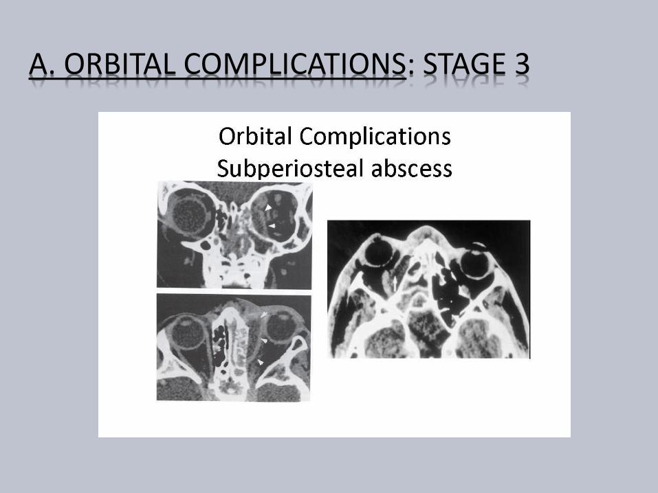
A ORBITAL COMPLICATIONS STAGE 3
A ORBITAL COMPLICATIONS STAGE 4
Orbital cellulits with intra-periostealabscess
Severe proptosis and chemosis
Fixed pupil
Severe globe displacement
Rapid fixation of EOM
Opthalmoplegia
Visual loss due to optic neuropathy(13)
ldquoOrbital Apex Syndromerdquo
Ophthalmoplegia amp Dilated pupil
Paraesthesia in distribution of maxilary amp ophthalmic division of trigeminal n
Blindness amp Temporal headache
A ORBITAL COMPLICATIONS STAGE 5 Cavernous Sinus Thrombosis
Bright 1831
Uncommon no incidence data
Typically affects young adults
Pathophysiology
Bacterial growth
Induces thrombosis
Thrombus good growth medium
More bacterial growth
Thrombophlebitis extend posteriorly
Orbit to Cavernous sinus
Contralateral Cavernous sinus bilateral eye symptoms
Then Intracranially
A ORBITAL COMPLICATIONS STAGE 5 Fatal prior to antibiotic era (pre-1940s) Mortality estimate 14-79 Morbidity estimate 50
Cranial neuropathies amp Visual loss
Clinical features Onset 1-21 days (Avg 5-6 days) Progressive amp bilateral eye symptom Proptosis and fixation of eye ball Bilateral orbital apex syndrome Meningitis Systemic featutes
High grade fever amp headache Tachycardia hypotension
A ORBITAL COMPLICATIONS STAGE 5
A ORBITAL COMPLICATIONS STAGE 5
Complications
Intracranial infection
Meningitis
Encephalitis
Abscess
Pituitary insufficiency
Hemorrhagic infarction
Death
A ORBITAL COMPLICATIONS MANAGEMENT
History
General ENT examination
Complete neurological examination
Rigid endoscopy of nose
Swabs
A ORBITAL COMPLICATIONS MANAGEMENT
A ORBITAL COMPLICATIONS MANAGEMENT
Other investigations CBC Blood biochemistry Blood cultures Lumbar puncture Neuroimaging (CT MRI)
Expansion of cavernous sinuses Convex bowing of lateral wall Abnormal filling defects Dilation of superior ophthalmic vein Dural enhancement of cavernous sinus border
A ORBITAL COMPLICATIONS MANAGEMENT
Empiric high dose IV antibiotics
Third generation cephalosporin
Anti-staphylococcal penicillin
Metronidazole
Continued treatment with IV antibiotics for at least 2 wks after apparent clinical resolution
Steroids controversial (except if pituitary insufficiency)
Anticoagulation in CST
No consensus for use despite theoretical rationale
Risks include systemic and intracranial bleeding
A ORBITAL COMPLICATIONS LONG TERM SEQUELAE
Permanent visual loss
Ophthalmoplegia
Exposure Keratitis amp Ulceration
Other ocular changes Uveitis
Choroiditis
Glaucoma
Iris prolapse
Rupture of the globe
B INTRACRANIAL COMPLICATIONS
Less common than orbital complications
Both can coexist
More common in adolescent
amp young adults
Male preponderance
Includes
Meningitis Encephalitis
Subdural Abscess(23) gt Frontal Lobe(4) abscess gt Extradural Abscess (1)
Cavernous Sinus Thrombosis
B INTRACRANIAL COMPLICATIONS CLINICAL PRESENTATION
Thrombophlebitis
Septic thrombophlebitismultiple abscesses formation
Associated thrombosis of Cavernous sinus amp Sup Sagittal sinus
Usually gt10yrs Uncommon in infants
Presentation Acute or Chronic
Fever leucocytosis and headache
Seizures rigidity and focal neurological signs
Features of Increased ICP
Features of meningitis amp encephalitis
B INTRACRANIAL COMPLICATIONS MANAGEMENT
High index of suspicion
History
ENT amp Full neurological examination
If abscess suspected CECT (or MRI)
Fundoscopy Lumbar puncture
IV antibiotics (Cefuroxime+Flagyl) ndash 4-6weeks
Serial CT
Steroids ndash controversial
B INTRACRANIAL COMPLICATIONS MANAGEMENT
Surgery
Treat complication amp sinusitis
Drain Extradural abscess via approach to frontal
sinuses
Neurosurgical assistance
Burr holes For extradural abscess solitary abscess
Formal craniotomy
B INTRACRANIAL COMPLICATIONS
Prognosis
Mortality 15-43 despite antibiotics
Incidence increases with age
Multiple subdural abscess with coritcal thrombosis carries worst prognosis
Morbidity Permanent in 40
Convulsions
Hemiparesis
Early treatment better outcome
C BONY COMPLICATIONS POTTrsquoS PUFFY TUMOUR
Described by Sir Percival Pott in 1970 Frontal bone is diploic with marrow cavity Sinusitis Osteomyelitis of frontal bone
Anteriorly Forehead Potts Puffy tumour Posteriorly Subdural abscess
Presentation Fluctuant swelling +- Pain
Causative Org Staph amp Strepto- amp Anaerobes Investigation
Blood investigation + CECT
Treatment Medical IV Antibiotics Surgical Bilateral coronal incision +Bone debridement
C BONY COMPLICATIONS
POTTrsquoS PUFFY TUMOUR
D DENTAL COMPLICATIONS
Common with Maxillary Sinusitis
Close association of dentition with floor of sinus
Acute sinusitis Dental pain
Dental abscess Mistaken for Sinusitis
May coexist
E SYSTEMIC COMPLICATIONS
Toxic Shock Syndrome
Rare Potentially Fatal
Frequently associated with Staph aureus Streptococcus
Features of Toxaemia Fever Hypotension Rash amp MODS
Septicaemia
Hematogenous spread
Blood culture
Features of SIRS
Shock
MODS
F CHRONIC COMPLICATIONS MUCOCOELE Definition
Epithelial lined mucus containing sac completely filling thesinus and capable of expansion
1820 Langenback lsquoHydatidesrsquo
1896 Rollet lsquomucocoelersquo
Incidence 4 of case of unilateral proptosis
Most common sites F(65)gt E(25)gt M(10)gt S
Age grp 40-70 yrs
lt 5 bilateral or multiloculated53
F CHRONIC COMPLICATIONS MUCOCOELE Theories Chronic rinosinusitis Increased osteolysis
Active Bone resorption amp formation
Pressure erosion
Cystic degeneration of seromucimous glands
54
F CHRONIC COMPLICATIONS MUCOCOELE
Initial ophthalmic referral
Clinical features Proptosis
Diplopia
Displacement of globe
Limited ocular movement
Visual impairment
Mass
Endoscopy
55
F CHRONIC COMPLICATIONS MUCOCOELE
56
ImagingLoss of Scalloping of frontal sinus
F MUCOCOELE CECT PNS
FRONTAL MUCOCOELE BILOCULATED ETHMOID MUCOCOELE
57
Homogenous smooth walled mass expanding the sinus
F MUCOCOELE CULTURE OF ASPIRATE
Mixed infection
Staph aureus
Alpha hemolytic Streptococci
Heamophillus sp
GNB
Anaerobic bacteria
58
F MUCOCOELE TREATMENT
Surgery is the treatment
Goals
Eradication of disease
Minimal morbidity
Prevention of recurrence
59
F MUCOCOELE TREATMENT
Approach
Endoscopic drainage
Type I-III
External approach
Osteoplastic flap
+- sinus cavity obliteration
Combined approach
60
THANK YOU
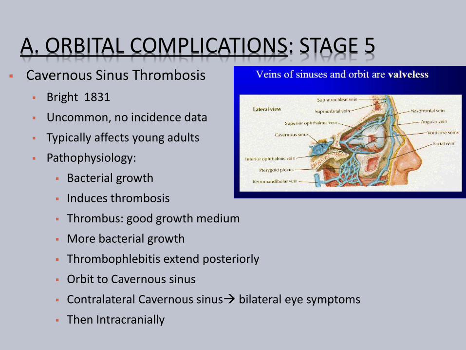
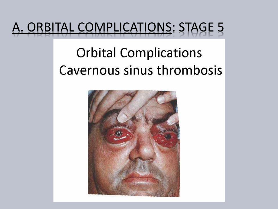
A ORBITAL COMPLICATIONS STAGE 5
A ORBITAL COMPLICATIONS STAGE 5
Complications
Intracranial infection
Meningitis
Encephalitis
Abscess
Pituitary insufficiency
Hemorrhagic infarction
Death
A ORBITAL COMPLICATIONS MANAGEMENT
History
General ENT examination
Complete neurological examination
Rigid endoscopy of nose
Swabs
A ORBITAL COMPLICATIONS MANAGEMENT
A ORBITAL COMPLICATIONS MANAGEMENT
Other investigations CBC Blood biochemistry Blood cultures Lumbar puncture Neuroimaging (CT MRI)
Expansion of cavernous sinuses Convex bowing of lateral wall Abnormal filling defects Dilation of superior ophthalmic vein Dural enhancement of cavernous sinus border
A ORBITAL COMPLICATIONS MANAGEMENT
Empiric high dose IV antibiotics
Third generation cephalosporin
Anti-staphylococcal penicillin
Metronidazole
Continued treatment with IV antibiotics for at least 2 wks after apparent clinical resolution
Steroids controversial (except if pituitary insufficiency)
Anticoagulation in CST
No consensus for use despite theoretical rationale
Risks include systemic and intracranial bleeding
A ORBITAL COMPLICATIONS LONG TERM SEQUELAE
Permanent visual loss
Ophthalmoplegia
Exposure Keratitis amp Ulceration
Other ocular changes Uveitis
Choroiditis
Glaucoma
Iris prolapse
Rupture of the globe
B INTRACRANIAL COMPLICATIONS
Less common than orbital complications
Both can coexist
More common in adolescent
amp young adults
Male preponderance
Includes
Meningitis Encephalitis
Subdural Abscess(23) gt Frontal Lobe(4) abscess gt Extradural Abscess (1)
Cavernous Sinus Thrombosis
B INTRACRANIAL COMPLICATIONS CLINICAL PRESENTATION
Thrombophlebitis
Septic thrombophlebitismultiple abscesses formation
Associated thrombosis of Cavernous sinus amp Sup Sagittal sinus
Usually gt10yrs Uncommon in infants
Presentation Acute or Chronic
Fever leucocytosis and headache
Seizures rigidity and focal neurological signs
Features of Increased ICP
Features of meningitis amp encephalitis
B INTRACRANIAL COMPLICATIONS MANAGEMENT
High index of suspicion
History
ENT amp Full neurological examination
If abscess suspected CECT (or MRI)
Fundoscopy Lumbar puncture
IV antibiotics (Cefuroxime+Flagyl) ndash 4-6weeks
Serial CT
Steroids ndash controversial
B INTRACRANIAL COMPLICATIONS MANAGEMENT
Surgery
Treat complication amp sinusitis
Drain Extradural abscess via approach to frontal
sinuses
Neurosurgical assistance
Burr holes For extradural abscess solitary abscess
Formal craniotomy
B INTRACRANIAL COMPLICATIONS
Prognosis
Mortality 15-43 despite antibiotics
Incidence increases with age
Multiple subdural abscess with coritcal thrombosis carries worst prognosis
Morbidity Permanent in 40
Convulsions
Hemiparesis
Early treatment better outcome
C BONY COMPLICATIONS POTTrsquoS PUFFY TUMOUR
Described by Sir Percival Pott in 1970 Frontal bone is diploic with marrow cavity Sinusitis Osteomyelitis of frontal bone
Anteriorly Forehead Potts Puffy tumour Posteriorly Subdural abscess
Presentation Fluctuant swelling +- Pain
Causative Org Staph amp Strepto- amp Anaerobes Investigation
Blood investigation + CECT
Treatment Medical IV Antibiotics Surgical Bilateral coronal incision +Bone debridement
C BONY COMPLICATIONS
POTTrsquoS PUFFY TUMOUR
D DENTAL COMPLICATIONS
Common with Maxillary Sinusitis
Close association of dentition with floor of sinus
Acute sinusitis Dental pain
Dental abscess Mistaken for Sinusitis
May coexist
E SYSTEMIC COMPLICATIONS
Toxic Shock Syndrome
Rare Potentially Fatal
Frequently associated with Staph aureus Streptococcus
Features of Toxaemia Fever Hypotension Rash amp MODS
Septicaemia
Hematogenous spread
Blood culture
Features of SIRS
Shock
MODS
F CHRONIC COMPLICATIONS MUCOCOELE Definition
Epithelial lined mucus containing sac completely filling thesinus and capable of expansion
1820 Langenback lsquoHydatidesrsquo
1896 Rollet lsquomucocoelersquo
Incidence 4 of case of unilateral proptosis
Most common sites F(65)gt E(25)gt M(10)gt S
Age grp 40-70 yrs
lt 5 bilateral or multiloculated53
F CHRONIC COMPLICATIONS MUCOCOELE Theories Chronic rinosinusitis Increased osteolysis
Active Bone resorption amp formation
Pressure erosion
Cystic degeneration of seromucimous glands
54
F CHRONIC COMPLICATIONS MUCOCOELE
Initial ophthalmic referral
Clinical features Proptosis
Diplopia
Displacement of globe
Limited ocular movement
Visual impairment
Mass
Endoscopy
55
F CHRONIC COMPLICATIONS MUCOCOELE
56
ImagingLoss of Scalloping of frontal sinus
F MUCOCOELE CECT PNS
FRONTAL MUCOCOELE BILOCULATED ETHMOID MUCOCOELE
57
Homogenous smooth walled mass expanding the sinus
F MUCOCOELE CULTURE OF ASPIRATE
Mixed infection
Staph aureus
Alpha hemolytic Streptococci
Heamophillus sp
GNB
Anaerobic bacteria
58
F MUCOCOELE TREATMENT
Surgery is the treatment
Goals
Eradication of disease
Minimal morbidity
Prevention of recurrence
59
F MUCOCOELE TREATMENT
Approach
Endoscopic drainage
Type I-III
External approach
Osteoplastic flap
+- sinus cavity obliteration
Combined approach
60
THANK YOU
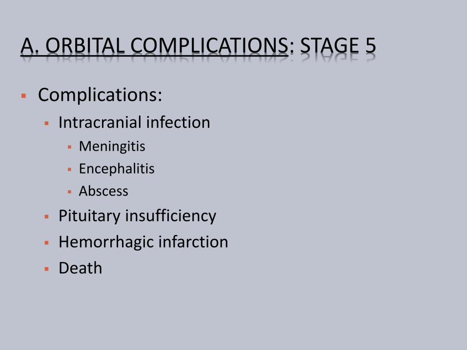
A ORBITAL COMPLICATIONS STAGE 5
Complications
Intracranial infection
Meningitis
Encephalitis
Abscess
Pituitary insufficiency
Hemorrhagic infarction
Death
A ORBITAL COMPLICATIONS MANAGEMENT
History
General ENT examination
Complete neurological examination
Rigid endoscopy of nose
Swabs
A ORBITAL COMPLICATIONS MANAGEMENT
A ORBITAL COMPLICATIONS MANAGEMENT
Other investigations CBC Blood biochemistry Blood cultures Lumbar puncture Neuroimaging (CT MRI)
Expansion of cavernous sinuses Convex bowing of lateral wall Abnormal filling defects Dilation of superior ophthalmic vein Dural enhancement of cavernous sinus border
A ORBITAL COMPLICATIONS MANAGEMENT
Empiric high dose IV antibiotics
Third generation cephalosporin
Anti-staphylococcal penicillin
Metronidazole
Continued treatment with IV antibiotics for at least 2 wks after apparent clinical resolution
Steroids controversial (except if pituitary insufficiency)
Anticoagulation in CST
No consensus for use despite theoretical rationale
Risks include systemic and intracranial bleeding
A ORBITAL COMPLICATIONS LONG TERM SEQUELAE
Permanent visual loss
Ophthalmoplegia
Exposure Keratitis amp Ulceration
Other ocular changes Uveitis
Choroiditis
Glaucoma
Iris prolapse
Rupture of the globe
B INTRACRANIAL COMPLICATIONS
Less common than orbital complications
Both can coexist
More common in adolescent
amp young adults
Male preponderance
Includes
Meningitis Encephalitis
Subdural Abscess(23) gt Frontal Lobe(4) abscess gt Extradural Abscess (1)
Cavernous Sinus Thrombosis
B INTRACRANIAL COMPLICATIONS CLINICAL PRESENTATION
Thrombophlebitis
Septic thrombophlebitismultiple abscesses formation
Associated thrombosis of Cavernous sinus amp Sup Sagittal sinus
Usually gt10yrs Uncommon in infants
Presentation Acute or Chronic
Fever leucocytosis and headache
Seizures rigidity and focal neurological signs
Features of Increased ICP
Features of meningitis amp encephalitis
B INTRACRANIAL COMPLICATIONS MANAGEMENT
High index of suspicion
History
ENT amp Full neurological examination
If abscess suspected CECT (or MRI)
Fundoscopy Lumbar puncture
IV antibiotics (Cefuroxime+Flagyl) ndash 4-6weeks
Serial CT
Steroids ndash controversial
B INTRACRANIAL COMPLICATIONS MANAGEMENT
Surgery
Treat complication amp sinusitis
Drain Extradural abscess via approach to frontal
sinuses
Neurosurgical assistance
Burr holes For extradural abscess solitary abscess
Formal craniotomy
B INTRACRANIAL COMPLICATIONS
Prognosis
Mortality 15-43 despite antibiotics
Incidence increases with age
Multiple subdural abscess with coritcal thrombosis carries worst prognosis
Morbidity Permanent in 40
Convulsions
Hemiparesis
Early treatment better outcome
C BONY COMPLICATIONS POTTrsquoS PUFFY TUMOUR
Described by Sir Percival Pott in 1970 Frontal bone is diploic with marrow cavity Sinusitis Osteomyelitis of frontal bone
Anteriorly Forehead Potts Puffy tumour Posteriorly Subdural abscess
Presentation Fluctuant swelling +- Pain
Causative Org Staph amp Strepto- amp Anaerobes Investigation
Blood investigation + CECT
Treatment Medical IV Antibiotics Surgical Bilateral coronal incision +Bone debridement
C BONY COMPLICATIONS
POTTrsquoS PUFFY TUMOUR
D DENTAL COMPLICATIONS
Common with Maxillary Sinusitis
Close association of dentition with floor of sinus
Acute sinusitis Dental pain
Dental abscess Mistaken for Sinusitis
May coexist
E SYSTEMIC COMPLICATIONS
Toxic Shock Syndrome
Rare Potentially Fatal
Frequently associated with Staph aureus Streptococcus
Features of Toxaemia Fever Hypotension Rash amp MODS
Septicaemia
Hematogenous spread
Blood culture
Features of SIRS
Shock
MODS
F CHRONIC COMPLICATIONS MUCOCOELE Definition
Epithelial lined mucus containing sac completely filling thesinus and capable of expansion
1820 Langenback lsquoHydatidesrsquo
1896 Rollet lsquomucocoelersquo
Incidence 4 of case of unilateral proptosis
Most common sites F(65)gt E(25)gt M(10)gt S
Age grp 40-70 yrs
lt 5 bilateral or multiloculated53
F CHRONIC COMPLICATIONS MUCOCOELE Theories Chronic rinosinusitis Increased osteolysis
Active Bone resorption amp formation
Pressure erosion
Cystic degeneration of seromucimous glands
54
F CHRONIC COMPLICATIONS MUCOCOELE
Initial ophthalmic referral
Clinical features Proptosis
Diplopia
Displacement of globe
Limited ocular movement
Visual impairment
Mass
Endoscopy
55
F CHRONIC COMPLICATIONS MUCOCOELE
56
ImagingLoss of Scalloping of frontal sinus
F MUCOCOELE CECT PNS
FRONTAL MUCOCOELE BILOCULATED ETHMOID MUCOCOELE
57
Homogenous smooth walled mass expanding the sinus
F MUCOCOELE CULTURE OF ASPIRATE
Mixed infection
Staph aureus
Alpha hemolytic Streptococci
Heamophillus sp
GNB
Anaerobic bacteria
58
F MUCOCOELE TREATMENT
Surgery is the treatment
Goals
Eradication of disease
Minimal morbidity
Prevention of recurrence
59
F MUCOCOELE TREATMENT
Approach
Endoscopic drainage
Type I-III
External approach
Osteoplastic flap
+- sinus cavity obliteration
Combined approach
60
THANK YOU

A ORBITAL COMPLICATIONS MANAGEMENT
History
General ENT examination
Complete neurological examination
Rigid endoscopy of nose
Swabs
A ORBITAL COMPLICATIONS MANAGEMENT
A ORBITAL COMPLICATIONS MANAGEMENT
Other investigations CBC Blood biochemistry Blood cultures Lumbar puncture Neuroimaging (CT MRI)
Expansion of cavernous sinuses Convex bowing of lateral wall Abnormal filling defects Dilation of superior ophthalmic vein Dural enhancement of cavernous sinus border
A ORBITAL COMPLICATIONS MANAGEMENT
Empiric high dose IV antibiotics
Third generation cephalosporin
Anti-staphylococcal penicillin
Metronidazole
Continued treatment with IV antibiotics for at least 2 wks after apparent clinical resolution
Steroids controversial (except if pituitary insufficiency)
Anticoagulation in CST
No consensus for use despite theoretical rationale
Risks include systemic and intracranial bleeding
A ORBITAL COMPLICATIONS LONG TERM SEQUELAE
Permanent visual loss
Ophthalmoplegia
Exposure Keratitis amp Ulceration
Other ocular changes Uveitis
Choroiditis
Glaucoma
Iris prolapse
Rupture of the globe
B INTRACRANIAL COMPLICATIONS
Less common than orbital complications
Both can coexist
More common in adolescent
amp young adults
Male preponderance
Includes
Meningitis Encephalitis
Subdural Abscess(23) gt Frontal Lobe(4) abscess gt Extradural Abscess (1)
Cavernous Sinus Thrombosis
B INTRACRANIAL COMPLICATIONS CLINICAL PRESENTATION
Thrombophlebitis
Septic thrombophlebitismultiple abscesses formation
Associated thrombosis of Cavernous sinus amp Sup Sagittal sinus
Usually gt10yrs Uncommon in infants
Presentation Acute or Chronic
Fever leucocytosis and headache
Seizures rigidity and focal neurological signs
Features of Increased ICP
Features of meningitis amp encephalitis
B INTRACRANIAL COMPLICATIONS MANAGEMENT
High index of suspicion
History
ENT amp Full neurological examination
If abscess suspected CECT (or MRI)
Fundoscopy Lumbar puncture
IV antibiotics (Cefuroxime+Flagyl) ndash 4-6weeks
Serial CT
Steroids ndash controversial
B INTRACRANIAL COMPLICATIONS MANAGEMENT
Surgery
Treat complication amp sinusitis
Drain Extradural abscess via approach to frontal
sinuses
Neurosurgical assistance
Burr holes For extradural abscess solitary abscess
Formal craniotomy
B INTRACRANIAL COMPLICATIONS
Prognosis
Mortality 15-43 despite antibiotics
Incidence increases with age
Multiple subdural abscess with coritcal thrombosis carries worst prognosis
Morbidity Permanent in 40
Convulsions
Hemiparesis
Early treatment better outcome
C BONY COMPLICATIONS POTTrsquoS PUFFY TUMOUR
Described by Sir Percival Pott in 1970 Frontal bone is diploic with marrow cavity Sinusitis Osteomyelitis of frontal bone
Anteriorly Forehead Potts Puffy tumour Posteriorly Subdural abscess
Presentation Fluctuant swelling +- Pain
Causative Org Staph amp Strepto- amp Anaerobes Investigation
Blood investigation + CECT
Treatment Medical IV Antibiotics Surgical Bilateral coronal incision +Bone debridement
C BONY COMPLICATIONS
POTTrsquoS PUFFY TUMOUR
D DENTAL COMPLICATIONS
Common with Maxillary Sinusitis
Close association of dentition with floor of sinus
Acute sinusitis Dental pain
Dental abscess Mistaken for Sinusitis
May coexist
E SYSTEMIC COMPLICATIONS
Toxic Shock Syndrome
Rare Potentially Fatal
Frequently associated with Staph aureus Streptococcus
Features of Toxaemia Fever Hypotension Rash amp MODS
Septicaemia
Hematogenous spread
Blood culture
Features of SIRS
Shock
MODS
F CHRONIC COMPLICATIONS MUCOCOELE Definition
Epithelial lined mucus containing sac completely filling thesinus and capable of expansion
1820 Langenback lsquoHydatidesrsquo
1896 Rollet lsquomucocoelersquo
Incidence 4 of case of unilateral proptosis
Most common sites F(65)gt E(25)gt M(10)gt S
Age grp 40-70 yrs
lt 5 bilateral or multiloculated53
F CHRONIC COMPLICATIONS MUCOCOELE Theories Chronic rinosinusitis Increased osteolysis
Active Bone resorption amp formation
Pressure erosion
Cystic degeneration of seromucimous glands
54
F CHRONIC COMPLICATIONS MUCOCOELE
Initial ophthalmic referral
Clinical features Proptosis
Diplopia
Displacement of globe
Limited ocular movement
Visual impairment
Mass
Endoscopy
55
F CHRONIC COMPLICATIONS MUCOCOELE
56
ImagingLoss of Scalloping of frontal sinus
F MUCOCOELE CECT PNS
FRONTAL MUCOCOELE BILOCULATED ETHMOID MUCOCOELE
57
Homogenous smooth walled mass expanding the sinus
F MUCOCOELE CULTURE OF ASPIRATE
Mixed infection
Staph aureus
Alpha hemolytic Streptococci
Heamophillus sp
GNB
Anaerobic bacteria
58
F MUCOCOELE TREATMENT
Surgery is the treatment
Goals
Eradication of disease
Minimal morbidity
Prevention of recurrence
59
F MUCOCOELE TREATMENT
Approach
Endoscopic drainage
Type I-III
External approach
Osteoplastic flap
+- sinus cavity obliteration
Combined approach
60
THANK YOU
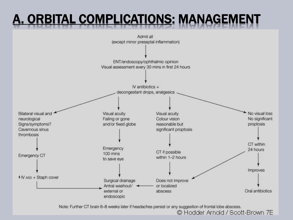
A ORBITAL COMPLICATIONS MANAGEMENT
A ORBITAL COMPLICATIONS MANAGEMENT
Other investigations CBC Blood biochemistry Blood cultures Lumbar puncture Neuroimaging (CT MRI)
Expansion of cavernous sinuses Convex bowing of lateral wall Abnormal filling defects Dilation of superior ophthalmic vein Dural enhancement of cavernous sinus border
A ORBITAL COMPLICATIONS MANAGEMENT
Empiric high dose IV antibiotics
Third generation cephalosporin
Anti-staphylococcal penicillin
Metronidazole
Continued treatment with IV antibiotics for at least 2 wks after apparent clinical resolution
Steroids controversial (except if pituitary insufficiency)
Anticoagulation in CST
No consensus for use despite theoretical rationale
Risks include systemic and intracranial bleeding
A ORBITAL COMPLICATIONS LONG TERM SEQUELAE
Permanent visual loss
Ophthalmoplegia
Exposure Keratitis amp Ulceration
Other ocular changes Uveitis
Choroiditis
Glaucoma
Iris prolapse
Rupture of the globe
B INTRACRANIAL COMPLICATIONS
Less common than orbital complications
Both can coexist
More common in adolescent
amp young adults
Male preponderance
Includes
Meningitis Encephalitis
Subdural Abscess(23) gt Frontal Lobe(4) abscess gt Extradural Abscess (1)
Cavernous Sinus Thrombosis
B INTRACRANIAL COMPLICATIONS CLINICAL PRESENTATION
Thrombophlebitis
Septic thrombophlebitismultiple abscesses formation
Associated thrombosis of Cavernous sinus amp Sup Sagittal sinus
Usually gt10yrs Uncommon in infants
Presentation Acute or Chronic
Fever leucocytosis and headache
Seizures rigidity and focal neurological signs
Features of Increased ICP
Features of meningitis amp encephalitis
B INTRACRANIAL COMPLICATIONS MANAGEMENT
High index of suspicion
History
ENT amp Full neurological examination
If abscess suspected CECT (or MRI)
Fundoscopy Lumbar puncture
IV antibiotics (Cefuroxime+Flagyl) ndash 4-6weeks
Serial CT
Steroids ndash controversial
B INTRACRANIAL COMPLICATIONS MANAGEMENT
Surgery
Treat complication amp sinusitis
Drain Extradural abscess via approach to frontal
sinuses
Neurosurgical assistance
Burr holes For extradural abscess solitary abscess
Formal craniotomy
B INTRACRANIAL COMPLICATIONS
Prognosis
Mortality 15-43 despite antibiotics
Incidence increases with age
Multiple subdural abscess with coritcal thrombosis carries worst prognosis
Morbidity Permanent in 40
Convulsions
Hemiparesis
Early treatment better outcome
C BONY COMPLICATIONS POTTrsquoS PUFFY TUMOUR
Described by Sir Percival Pott in 1970 Frontal bone is diploic with marrow cavity Sinusitis Osteomyelitis of frontal bone
Anteriorly Forehead Potts Puffy tumour Posteriorly Subdural abscess
Presentation Fluctuant swelling +- Pain
Causative Org Staph amp Strepto- amp Anaerobes Investigation
Blood investigation + CECT
Treatment Medical IV Antibiotics Surgical Bilateral coronal incision +Bone debridement
C BONY COMPLICATIONS
POTTrsquoS PUFFY TUMOUR
D DENTAL COMPLICATIONS
Common with Maxillary Sinusitis
Close association of dentition with floor of sinus
Acute sinusitis Dental pain
Dental abscess Mistaken for Sinusitis
May coexist
E SYSTEMIC COMPLICATIONS
Toxic Shock Syndrome
Rare Potentially Fatal
Frequently associated with Staph aureus Streptococcus
Features of Toxaemia Fever Hypotension Rash amp MODS
Septicaemia
Hematogenous spread
Blood culture
Features of SIRS
Shock
MODS
F CHRONIC COMPLICATIONS MUCOCOELE Definition
Epithelial lined mucus containing sac completely filling thesinus and capable of expansion
1820 Langenback lsquoHydatidesrsquo
1896 Rollet lsquomucocoelersquo
Incidence 4 of case of unilateral proptosis
Most common sites F(65)gt E(25)gt M(10)gt S
Age grp 40-70 yrs
lt 5 bilateral or multiloculated53
F CHRONIC COMPLICATIONS MUCOCOELE Theories Chronic rinosinusitis Increased osteolysis
Active Bone resorption amp formation
Pressure erosion
Cystic degeneration of seromucimous glands
54
F CHRONIC COMPLICATIONS MUCOCOELE
Initial ophthalmic referral
Clinical features Proptosis
Diplopia
Displacement of globe
Limited ocular movement
Visual impairment
Mass
Endoscopy
55
F CHRONIC COMPLICATIONS MUCOCOELE
56
ImagingLoss of Scalloping of frontal sinus
F MUCOCOELE CECT PNS
FRONTAL MUCOCOELE BILOCULATED ETHMOID MUCOCOELE
57
Homogenous smooth walled mass expanding the sinus
F MUCOCOELE CULTURE OF ASPIRATE
Mixed infection
Staph aureus
Alpha hemolytic Streptococci
Heamophillus sp
GNB
Anaerobic bacteria
58
F MUCOCOELE TREATMENT
Surgery is the treatment
Goals
Eradication of disease
Minimal morbidity
Prevention of recurrence
59
F MUCOCOELE TREATMENT
Approach
Endoscopic drainage
Type I-III
External approach
Osteoplastic flap
+- sinus cavity obliteration
Combined approach
60
THANK YOU
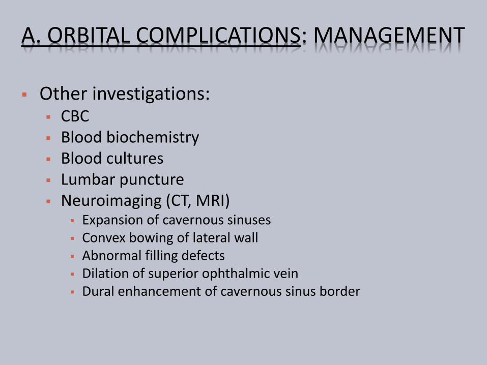
A ORBITAL COMPLICATIONS MANAGEMENT
Other investigations CBC Blood biochemistry Blood cultures Lumbar puncture Neuroimaging (CT MRI)
Expansion of cavernous sinuses Convex bowing of lateral wall Abnormal filling defects Dilation of superior ophthalmic vein Dural enhancement of cavernous sinus border
A ORBITAL COMPLICATIONS MANAGEMENT
Empiric high dose IV antibiotics
Third generation cephalosporin
Anti-staphylococcal penicillin
Metronidazole
Continued treatment with IV antibiotics for at least 2 wks after apparent clinical resolution
Steroids controversial (except if pituitary insufficiency)
Anticoagulation in CST
No consensus for use despite theoretical rationale
Risks include systemic and intracranial bleeding
A ORBITAL COMPLICATIONS LONG TERM SEQUELAE
Permanent visual loss
Ophthalmoplegia
Exposure Keratitis amp Ulceration
Other ocular changes Uveitis
Choroiditis
Glaucoma
Iris prolapse
Rupture of the globe
B INTRACRANIAL COMPLICATIONS
Less common than orbital complications
Both can coexist
More common in adolescent
amp young adults
Male preponderance
Includes
Meningitis Encephalitis
Subdural Abscess(23) gt Frontal Lobe(4) abscess gt Extradural Abscess (1)
Cavernous Sinus Thrombosis
B INTRACRANIAL COMPLICATIONS CLINICAL PRESENTATION
Thrombophlebitis
Septic thrombophlebitismultiple abscesses formation
Associated thrombosis of Cavernous sinus amp Sup Sagittal sinus
Usually gt10yrs Uncommon in infants
Presentation Acute or Chronic
Fever leucocytosis and headache
Seizures rigidity and focal neurological signs
Features of Increased ICP
Features of meningitis amp encephalitis
B INTRACRANIAL COMPLICATIONS MANAGEMENT
High index of suspicion
History
ENT amp Full neurological examination
If abscess suspected CECT (or MRI)
Fundoscopy Lumbar puncture
IV antibiotics (Cefuroxime+Flagyl) ndash 4-6weeks
Serial CT
Steroids ndash controversial
B INTRACRANIAL COMPLICATIONS MANAGEMENT
Surgery
Treat complication amp sinusitis
Drain Extradural abscess via approach to frontal
sinuses
Neurosurgical assistance
Burr holes For extradural abscess solitary abscess
Formal craniotomy
B INTRACRANIAL COMPLICATIONS
Prognosis
Mortality 15-43 despite antibiotics
Incidence increases with age
Multiple subdural abscess with coritcal thrombosis carries worst prognosis
Morbidity Permanent in 40
Convulsions
Hemiparesis
Early treatment better outcome
C BONY COMPLICATIONS POTTrsquoS PUFFY TUMOUR
Described by Sir Percival Pott in 1970 Frontal bone is diploic with marrow cavity Sinusitis Osteomyelitis of frontal bone
Anteriorly Forehead Potts Puffy tumour Posteriorly Subdural abscess
Presentation Fluctuant swelling +- Pain
Causative Org Staph amp Strepto- amp Anaerobes Investigation
Blood investigation + CECT
Treatment Medical IV Antibiotics Surgical Bilateral coronal incision +Bone debridement
C BONY COMPLICATIONS
POTTrsquoS PUFFY TUMOUR
D DENTAL COMPLICATIONS
Common with Maxillary Sinusitis
Close association of dentition with floor of sinus
Acute sinusitis Dental pain
Dental abscess Mistaken for Sinusitis
May coexist
E SYSTEMIC COMPLICATIONS
Toxic Shock Syndrome
Rare Potentially Fatal
Frequently associated with Staph aureus Streptococcus
Features of Toxaemia Fever Hypotension Rash amp MODS
Septicaemia
Hematogenous spread
Blood culture
Features of SIRS
Shock
MODS
F CHRONIC COMPLICATIONS MUCOCOELE Definition
Epithelial lined mucus containing sac completely filling thesinus and capable of expansion
1820 Langenback lsquoHydatidesrsquo
1896 Rollet lsquomucocoelersquo
Incidence 4 of case of unilateral proptosis
Most common sites F(65)gt E(25)gt M(10)gt S
Age grp 40-70 yrs
lt 5 bilateral or multiloculated53
F CHRONIC COMPLICATIONS MUCOCOELE Theories Chronic rinosinusitis Increased osteolysis
Active Bone resorption amp formation
Pressure erosion
Cystic degeneration of seromucimous glands
54
F CHRONIC COMPLICATIONS MUCOCOELE
Initial ophthalmic referral
Clinical features Proptosis
Diplopia
Displacement of globe
Limited ocular movement
Visual impairment
Mass
Endoscopy
55
F CHRONIC COMPLICATIONS MUCOCOELE
56
ImagingLoss of Scalloping of frontal sinus
F MUCOCOELE CECT PNS
FRONTAL MUCOCOELE BILOCULATED ETHMOID MUCOCOELE
57
Homogenous smooth walled mass expanding the sinus
F MUCOCOELE CULTURE OF ASPIRATE
Mixed infection
Staph aureus
Alpha hemolytic Streptococci
Heamophillus sp
GNB
Anaerobic bacteria
58
F MUCOCOELE TREATMENT
Surgery is the treatment
Goals
Eradication of disease
Minimal morbidity
Prevention of recurrence
59
F MUCOCOELE TREATMENT
Approach
Endoscopic drainage
Type I-III
External approach
Osteoplastic flap
+- sinus cavity obliteration
Combined approach
60
THANK YOU

B INTRACRANIAL COMPLICATIONS
Less common than orbital complications
Both can coexist
More common in adolescent
amp young adults
Male preponderance
Includes
Meningitis Encephalitis
Subdural Abscess(23) gt Frontal Lobe(4) abscess gt Extradural Abscess (1)
Cavernous Sinus Thrombosis
B INTRACRANIAL COMPLICATIONS CLINICAL PRESENTATION
Thrombophlebitis
Septic thrombophlebitismultiple abscesses formation
Associated thrombosis of Cavernous sinus amp Sup Sagittal sinus
Usually gt10yrs Uncommon in infants
Presentation Acute or Chronic
Fever leucocytosis and headache
Seizures rigidity and focal neurological signs
Features of Increased ICP
Features of meningitis amp encephalitis
B INTRACRANIAL COMPLICATIONS MANAGEMENT
High index of suspicion
History
ENT amp Full neurological examination
If abscess suspected CECT (or MRI)
Fundoscopy Lumbar puncture
IV antibiotics (Cefuroxime+Flagyl) ndash 4-6weeks
Serial CT
Steroids ndash controversial
B INTRACRANIAL COMPLICATIONS MANAGEMENT
Surgery
Treat complication amp sinusitis
Drain Extradural abscess via approach to frontal
sinuses
Neurosurgical assistance
Burr holes For extradural abscess solitary abscess
Formal craniotomy
B INTRACRANIAL COMPLICATIONS
Prognosis
Mortality 15-43 despite antibiotics
Incidence increases with age
Multiple subdural abscess with coritcal thrombosis carries worst prognosis
Morbidity Permanent in 40
Convulsions
Hemiparesis
Early treatment better outcome
C BONY COMPLICATIONS POTTrsquoS PUFFY TUMOUR
Described by Sir Percival Pott in 1970 Frontal bone is diploic with marrow cavity Sinusitis Osteomyelitis of frontal bone
Anteriorly Forehead Potts Puffy tumour Posteriorly Subdural abscess
Presentation Fluctuant swelling +- Pain
Causative Org Staph amp Strepto- amp Anaerobes Investigation
Blood investigation + CECT
Treatment Medical IV Antibiotics Surgical Bilateral coronal incision +Bone debridement
C BONY COMPLICATIONS
POTTrsquoS PUFFY TUMOUR
D DENTAL COMPLICATIONS
Common with Maxillary Sinusitis
Close association of dentition with floor of sinus
Acute sinusitis Dental pain
Dental abscess Mistaken for Sinusitis
May coexist
E SYSTEMIC COMPLICATIONS
Toxic Shock Syndrome
Rare Potentially Fatal
Frequently associated with Staph aureus Streptococcus
Features of Toxaemia Fever Hypotension Rash amp MODS
Septicaemia
Hematogenous spread
Blood culture
Features of SIRS
Shock
MODS
F CHRONIC COMPLICATIONS MUCOCOELE Definition
Epithelial lined mucus containing sac completely filling thesinus and capable of expansion
1820 Langenback lsquoHydatidesrsquo
1896 Rollet lsquomucocoelersquo
Incidence 4 of case of unilateral proptosis
Most common sites F(65)gt E(25)gt M(10)gt S
Age grp 40-70 yrs
lt 5 bilateral or multiloculated53
F CHRONIC COMPLICATIONS MUCOCOELE Theories Chronic rinosinusitis Increased osteolysis
Active Bone resorption amp formation
Pressure erosion
Cystic degeneration of seromucimous glands
54
F CHRONIC COMPLICATIONS MUCOCOELE
Initial ophthalmic referral
Clinical features Proptosis
Diplopia
Displacement of globe
Limited ocular movement
Visual impairment
Mass
Endoscopy
55
F CHRONIC COMPLICATIONS MUCOCOELE
56
ImagingLoss of Scalloping of frontal sinus
F MUCOCOELE CECT PNS
FRONTAL MUCOCOELE BILOCULATED ETHMOID MUCOCOELE
57
Homogenous smooth walled mass expanding the sinus
F MUCOCOELE CULTURE OF ASPIRATE
Mixed infection
Staph aureus
Alpha hemolytic Streptococci
Heamophillus sp
GNB
Anaerobic bacteria
58
F MUCOCOELE TREATMENT
Surgery is the treatment
Goals
Eradication of disease
Minimal morbidity
Prevention of recurrence
59
F MUCOCOELE TREATMENT
Approach
Endoscopic drainage
Type I-III
External approach
Osteoplastic flap
+- sinus cavity obliteration
Combined approach
60
THANK YOU

B INTRACRANIAL COMPLICATIONS MANAGEMENT
Surgery
Treat complication amp sinusitis
Drain Extradural abscess via approach to frontal
sinuses
Neurosurgical assistance
Burr holes For extradural abscess solitary abscess
Formal craniotomy
B INTRACRANIAL COMPLICATIONS
Prognosis
Mortality 15-43 despite antibiotics
Incidence increases with age
Multiple subdural abscess with coritcal thrombosis carries worst prognosis
Morbidity Permanent in 40
Convulsions
Hemiparesis
Early treatment better outcome
C BONY COMPLICATIONS POTTrsquoS PUFFY TUMOUR
Described by Sir Percival Pott in 1970 Frontal bone is diploic with marrow cavity Sinusitis Osteomyelitis of frontal bone
Anteriorly Forehead Potts Puffy tumour Posteriorly Subdural abscess
Presentation Fluctuant swelling +- Pain
Causative Org Staph amp Strepto- amp Anaerobes Investigation
Blood investigation + CECT
Treatment Medical IV Antibiotics Surgical Bilateral coronal incision +Bone debridement
C BONY COMPLICATIONS
POTTrsquoS PUFFY TUMOUR
D DENTAL COMPLICATIONS
Common with Maxillary Sinusitis
Close association of dentition with floor of sinus
Acute sinusitis Dental pain
Dental abscess Mistaken for Sinusitis
May coexist
E SYSTEMIC COMPLICATIONS
Toxic Shock Syndrome
Rare Potentially Fatal
Frequently associated with Staph aureus Streptococcus
Features of Toxaemia Fever Hypotension Rash amp MODS
Septicaemia
Hematogenous spread
Blood culture
Features of SIRS
Shock
MODS
F CHRONIC COMPLICATIONS MUCOCOELE Definition
Epithelial lined mucus containing sac completely filling thesinus and capable of expansion
1820 Langenback lsquoHydatidesrsquo
1896 Rollet lsquomucocoelersquo
Incidence 4 of case of unilateral proptosis
Most common sites F(65)gt E(25)gt M(10)gt S
Age grp 40-70 yrs
lt 5 bilateral or multiloculated53
F CHRONIC COMPLICATIONS MUCOCOELE Theories Chronic rinosinusitis Increased osteolysis
Active Bone resorption amp formation
Pressure erosion
Cystic degeneration of seromucimous glands
54
F CHRONIC COMPLICATIONS MUCOCOELE
Initial ophthalmic referral
Clinical features Proptosis
Diplopia
Displacement of globe
Limited ocular movement
Visual impairment
Mass
Endoscopy
55
F CHRONIC COMPLICATIONS MUCOCOELE
56
ImagingLoss of Scalloping of frontal sinus
F MUCOCOELE CECT PNS
FRONTAL MUCOCOELE BILOCULATED ETHMOID MUCOCOELE
57
Homogenous smooth walled mass expanding the sinus
F MUCOCOELE CULTURE OF ASPIRATE
Mixed infection
Staph aureus
Alpha hemolytic Streptococci
Heamophillus sp
GNB
Anaerobic bacteria
58
F MUCOCOELE TREATMENT
Surgery is the treatment
Goals
Eradication of disease
Minimal morbidity
Prevention of recurrence
59
F MUCOCOELE TREATMENT
Approach
Endoscopic drainage
Type I-III
External approach
Osteoplastic flap
+- sinus cavity obliteration
Combined approach
60
THANK YOU
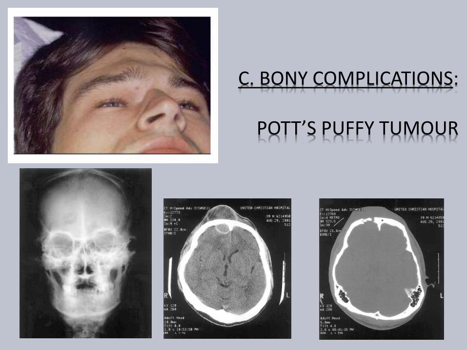
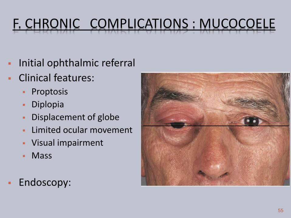
F CHRONIC COMPLICATIONS MUCOCOELE
56
ImagingLoss of Scalloping of frontal sinus
F MUCOCOELE CECT PNS
FRONTAL MUCOCOELE BILOCULATED ETHMOID MUCOCOELE
57
Homogenous smooth walled mass expanding the sinus
F MUCOCOELE CULTURE OF ASPIRATE
Mixed infection
Staph aureus
Alpha hemolytic Streptococci
Heamophillus sp
GNB
Anaerobic bacteria
58
F MUCOCOELE TREATMENT
Surgery is the treatment
Goals
Eradication of disease
Minimal morbidity
Prevention of recurrence
59
F MUCOCOELE TREATMENT
Approach
Endoscopic drainage
Type I-III
External approach
Osteoplastic flap
+- sinus cavity obliteration
Combined approach
60
THANK YOU
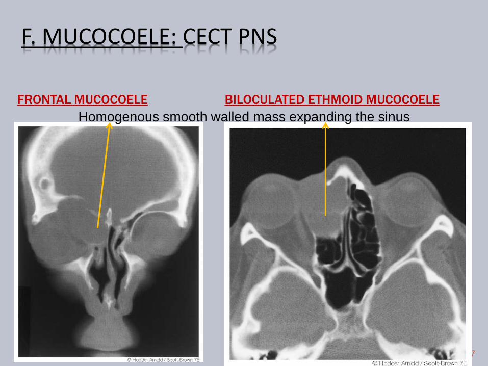

F MUCOCOELE TREATMENT
Surgery is the treatment
Goals
Eradication of disease
Minimal morbidity
Prevention of recurrence
59
F MUCOCOELE TREATMENT
Approach
Endoscopic drainage
Type I-III
External approach
Osteoplastic flap
+- sinus cavity obliteration
Combined approach
60
THANK YOU

F MUCOCOELE TREATMENT
Approach
Endoscopic drainage
Type I-III
External approach
Osteoplastic flap
+- sinus cavity obliteration
Combined approach
60
THANK YOU

THANK YOU