Embed Size (px)
Citation preview


Cellular immunity against viruses

Antigen-specific recognition of virus and virus-infected cells
1. B cell/Antibody mediated immunity2. Antigen processing and presentation
through class I and class II MHC molecules
3. Recognition of antigen/MHC and infected targets by virus-specific T cells

Figure 9-1Antibody mediated mechanisms of pathogen immunity

Figure 9-1 part 1 of 2Specific naïve B cells bind virus or viral proteins, internalize, process, and present to CD4+ T cells. The CD4 T cells provide signals and cytokines that stimulate the B cells class switch and differentiate into antibody secreting cells.
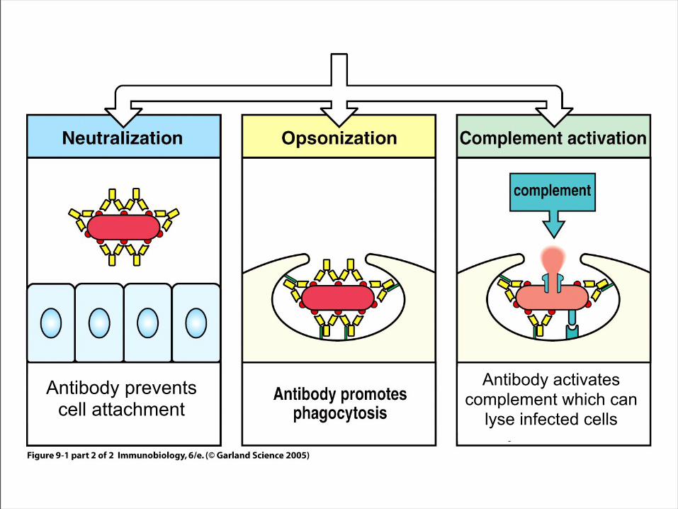
Figure 9-1 part 2 of 2
Antibody prevents cell attachment
Antibody activates complement which can
lyse infected cells

Figure 9-3Cognate antigen presentation and help between
virus-specific B cells and CD4 T cells
Presentation of specific antigen is 10,000x more efficient than non-specific Ag
CD40/CD40L provides both activation and survival signals, initiates Ig class switching. Cytokines like Il2 and IL4 promote B cell proliferation and class switching.

Figure 9-6CD4 helper T cells polarize their secretory machinery towards target B cells
Directed secretion of cytokine

Figure 9-25Antiviral mechanisms of antibodies:
Virus neutralization
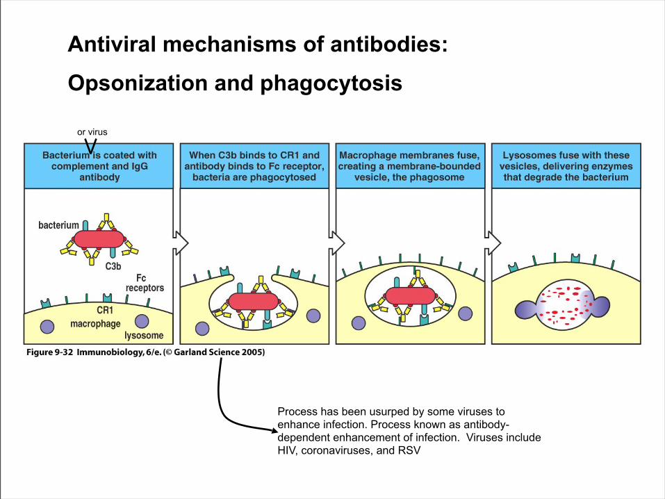
Figure 9-32Antiviral mechanisms of antibodies:
Opsonization and phagocytosis
Process has been usurped by some viruses to enhance infection. Process known as antibody-dependent enhancement of infection. Viruses include HIV, coronaviruses, and RSV
or virus

Figure 9-34Antiviral mechanisms of antibodies:
Antibody Dependent Cellular Cytotoxicity (ADCC)

Immune pressure causes:
• Antigenic Drift– Accumulation of
point mutations• Antigenic Shift
– Exchange of whole RNA gene segment
• Affects antibody mediated immunity
Evading antiviral antibodies
Influenza
Picornavirus

Figure 11-2

The receptor binding site of rhinovirus is hidden from neutralizing antibodies
“Canyon” containing the ICAM-1 binding site hidden from antibodies
Antibody binding sites in pink
Virus can alter its antigenicity without affecting target cell specificity
Avoiding antiviral antibodies

Immune selection of antigenic variants
• Antigenic drift– Occurs in a variety of
enveloped and non-enveloped viruses
– Reflects point mutations in the neutralizing epitopes of capsid or envelope proteins
• Antigenic shift– Unique to influenza– Reflects re-
assortment of envelope protein genes from different viruses
– Can quickly result in virus epidemics

Recognition of viral antigens

Figure 5-2
MHC presents antigenic peptides from the intracellular and extracellular environments
Lytic and Non-lytic viruses
Few viruses Lytic viruses
The lifestyle of the pathogen dictates the mechanism of immunity

Figure 5-6 part 1 of 2Class I MHC antigen processing
TAP: Transporter of Antigenic Peptides

Figure 5-6 part 2 of 2Class I MHC antigen processing
There are 2 forms of the proteosome: constitutive and immunoproteosome

Class I MHC antigen processing

Figure 5-3 part 1 of 2

Figure 5-3 part 2 of 2
View from the ER View from the side
TAP 1 and TAP 2 peptide transporters

Figure 5-4The structure of the
proteosome

The Immunoproteosome
PA28 proteosome activator is induced by IFN-γ.
PA28 alters the efficiency and pattern of peptides generated by the proteosome during an immune responseDr. Alice Sijts
a. The hexamer or heptamer rings of PA28 interact with the ends of the proteosome, composed of α subunits (pink) and β subunits (blue). Within this region is the α-annulus(green) that is normally blocked by other parts of the α subunits (red). B-C. Close up view of the α-annulus without PA28 bound (b), and with PA28 bound (c) . PA28 opens the α-annulus by changing its conformation .
w/o PA28
w/ PA28

Class II MHC antigen processing
**Endosomal vesicles may also be generated internally
within the cell

Figure 5-10 part 1 of 2Loading of Class II MHC with antigenic peptide

Figure 5-10 part 2 of 2Loading of Class II MHC with antigenic peptide

MHC:CLIPDM facilitatespeptide exchange
“MHC:Peptide” represents the end product of an exchange reaction within endosomal compartments
Export
DM:catalyst of peptide exchange
CLIP:peptide fragment remnant of invariant chain Peptide
One parameter that controls the immunodominance of peptides in CD4 T cell responses: DM editing
endosome

DM promotes loading/editing of antigenicpeptide within antigen presenting cells
+DM
peptide repertoire without DM
peptide repertoire with DM
Peptides offered to circulating CD4
cells by APC
Globular antigenic protein
Peptides that are generated and can bind to
the MHC molecule
CLIP
Editing
Dr. Andrea Sant

MHC Class II antigen processing

Dendritic cells
Professional antigen
presenting cells of the immune
system

Figure 8-14
Dendritic cells must mature before they can present antigen to activate naïve T cells
B7.1=CD80B7.2=CD86

Figure 2-17Regulation of naïve T cell activation by the DC
2-signal hypothesis

Figure 5-17
MHC and T cell receptor must match

Figure 8-13 part 1 of 2Once activated by appropriate antigen/MHC plus co–stimulation, a CTL can seek out & kill infected targets
Only signal 1 is required

The adaptive response takes time to develop

Figure 8-31Effector mechanisms elicited by different T cell
subsets

Interferon-γ• Enhances antigen processing
– Immunoproteosome subunits, class I MHC– Induces HLA-DM
• Enhances antigen presentation• Enhances lymphocyte adhesion
– Upregulates ICAM-1• Promotes Th1 differentiation• Promotes IL2 secretion• Activates macrophage• Inhibits viral gene expression

TNF-α

Next lecture:T cell mediated immunity
against viruses