MINISTRY OF HEALTH CARE VITEBSK STATE MEDICAL UNIVERSITY
HOSPITAL THERAPY DEPARTMENT
LECTURE NOTES BY
INTERNAL DISEASES
Compiled byM.R. KONOREV, MD, PhD, Dr.Sci
FOR FOREIGN STUDENTS OF 5 COURSES OF MEDICAL FACULTY
Vitebsk, El “VSMU”
2008
9

УДК-616.1/.4=20(042.3/.4) -ББК 54.1я73
К 64
Рецензенты:заведующий кафедрой пропедевтики внутренних болезней Витебского государственного медицинского университета, доктор медицинских наук Л.М. Немцов,профессор кафедры терапии Белорусской медицинской академии последипломного образования, доктор медицинских наук И.И. Бураков
К 64 Lecture notes by internal diseases for foreign students of 5 courses of medical faculty (Курс лекций по внутренним болезням (на английском языке) для иностранных студентов 5 курса лечебного факультета / М.Р. Конорев. - Витебск: ВГМУ, 2008. - 158 с.
ISBN 978-985-466-261-9
Курс лекций по внутренним болезням (на английском языке) для иностранных студентов 5 курса лечебного факультета составлен в соответствии с типовой учебной программой по внутренним болезням и ВПТ утвержденной Министерством Здравоохранения Республики Беларусь в 1997 г., и рабочей учебной программой по внутренним болезням и ВПТ для студентов лечебного факультета, утвержденной УО «ВГОДНМУ» 02.09.2002 г.
Предназначается для студентов факультетов подготовки иностранных граждан по специальности «Лечебное дело», а также для преподавателей медицинских ВУЗов, клинических ординаторов, аспирантов.
Рекомендовано к печати центральным учебно-методическим Советом Витебского государственного медицинского университета протокол № 2, от 26.03.2008 г!
Конорев М.Р.
УДК 616.1/.4=20(042.3/.4) ББК 54.1я73
© Конорев М.Р., 2008© УО «Витебский государственный ордена Дружбы
народов медицинский университет», 2008ISBN 978-985-466-261-9
3
CONTENTSPP
ABBREVIATIONS 5GASTROENTEROLOGY 6IRRITABLE BOWEL SYNDROME (IBS) 6CHRONIC DIARRHEA ЮCONSTIPATION 10INFLAMMATORY BOWEL DISEASE (IBD) 10CROHN'S DISEASE 12ULCERATIVE COLITIS (UC) 15FATTY LIVER 17CONGENITAL HYPERBILIRUBINAEMIAS (NON-HAEMOLYTIC) 18GILBERT SYNDROME (UNCONJUGATED HYPERBILIRUBINEMIA) 18DUBIN-JOHNSON SYNDROME (CONJUGATED HYPERBILIRUBINEMIA) 20 ROTOR SYNDROME (CONJUGATED HYPERBILIRUBINEMIA) 21HEMOCHROMATOSIS 22WILSON DISEASE 25CHOLELITHIASIS 28MEDICINE OF ACCIDENTS 33ACUTE RADIATION SICKNESS 3 3CHRONIC RADIATION SICKNESS 38DISEASES OF INTERNAL BODIES ASSOCIATED WITH BULLET 39WOUNDS AND EXPLOSIONSRHEUMATOLOGY 41SYSTEMIC LUPUS ERYTHEMATOSUS (SLE) 41ANTIPHOSPHOLIPID ANTIBODY SYNDROME (APS) 44SCLERODERMA/PROGRESSIVE SYSTEMIC SCLEROSIS (PSS) 45POLYMYOSITIS (PMyDERMATOMYOSITIS (DM) 49RHEUMATOID ARTHRITIS (RA) 53OSTEOARTHRITIS (OA) 57GOUT 59SEROPOSITIVE RHEUMATIC DISEASES: VASCULITIDES 63WEGENER’S GRANULOMATOSIS (WG) 64CHURG-STRAUSS SYNDROME 66POLYARTERITIS NODOSA (PAN) 68GIANT CELL ARTERITIS (TEMPORAL ARTERITIS) 70POLYMYALGIA RHEUMATICA 72TAKAYASU ARTERITIS 74CARDIOLOGY 77CARDIOMYOPATHIES 77
4
DILATED CARDIOMYOPATHY (DCM) 77HYPERTROPHIC CARDIOMYOPATHY (HCM) 82RESTRICTIVE CARDIOMYOPATHY (RCM) 85AORTIC STENOSIS (AS) 87AORTIC REGURGITATION (AR) 90MITRAL STENOSIS (MS) 95MITRAL REGURGITATION (MR) 98TRICUSPID VALVE DISEASE 100AORTIC COARCTATION 102PATENT DUCTUS ARTERIOSUS 103PATENT FORAMEN OVALE 106VENTRICULAR SEPTAL DEFECT 108HEMATOLOGY 111HEMOLYTIC ANEMIAS 111THALASSEMIAS: ALPHA THALASSEMIA 111THALASSEMIAS: BETA THALASSEMIA MINOR 113THALASSEMIAS: BETA THALASSEMIA MAJOR 113HEMOGLOBINOPATHIES: SICKLE CELL ANEMIA 114GLUCOSE-6-PHOSPHATE DEHYDROGENASE DEFICIENCY 117HEMATOLOGIC MALIGNANCIES 119ACUTE MYELOID LEUKEMIA (AML) 119ACUTE LYMPHOBLASTIC LEUKEMIA (ALL) 123CHRONIC LYMPHOCYTIC LEUKEMIA (CLL) 126CHRONIC MYELOGENOUS (GRANULOCYTIC) LEUKEMIA (CML) 129POLYCYTHEMIA RUBRA VERA (PRV) 13 3PLASMA CELL MYELOMA (MULTIPLE MYELOMA) 136HEMORRHAGIC DIATHESIS 139IDIOPATHIC (AUTOIMMUNE) THROMBOCYTOPENIC PURPURA (ITP) 139 HEMOPHILIA A, HEMOPHILIA В (CHRISTMAS DISEASE) 143THE LITERATURE 146GASTROENTEROLOGY 146RHEUMATOLOGY 147CARDIOLOGY 151HEMATOLOGY 154
5
ABBREVIATIONS
(A fib) atrial fibrillation (ASD) atrial septal defect (AVR) aortic valve replacement (CAD) coronary artery disease (CABG) coronary artery bypass graft (CHF) congestive heart failure (CO) cardiac output (DCM) dilated cardiomyopathy (EDM) early diastolic murmur (EF) ejection fraction (FVIII) factor VIII (FIX) factor IX(HA) Hemophilia A(HB) Hemophilia В(HCM) hypertrophic cardiomyopathy (HTN) hypertension(ICD) implantable cardioverter defibrillator(IE) infective endocarditis(INR) international normalized ratio(LAE) left atrial enlargement(LBBB) left bundle branch block(LVEDP) left ventricular end-diastolic pressure(LVEF) left ventricular systolic ejection fraction(LVH) left ventricle hypertrophy(MCH) mean corpuscular hemoglobin(MCV) mean corpuscular volume(MV) mitral valve(PAP) pulmonary artery pressure(PMI) point of maximal intensity(PND) paroxysmal nocturnal dyspnea(PT) prothrombin time(PTT) activated partial thromboplastin time(RAE) right atrial enlargement(RBCs) red blood cells(RVH) right ventricle hypertrophy(SEM) systolic ejection murmur(SV) systolic volume(51) first heart sound(52) second heart sound (S4) fourth heart sound (TV) tricuspid valve(VT) ventricular tachycardia
6
GASTROENTEROLOGY
IRRITABLE BOWEL SYNDROME (IBS)
Introduction. Irritable bowel syndrome (IBS) is a main functional gastrointestinal (GI) disorder characterized by abdominal pain and altered bowel habits in the absence o f specific and unique organic pathology.
Synonyms: Irritable bowel disease (IBD), functional bowel disease, irritable colon, mucous colitis, nervous bowel, spastic bowel, spastic colitis.
Definition. Traditionally, IBS is a diagnosis of exclusion. No specific motility or structural correlates have been consistently demonstrated, so IBS remains a clinically defined illness.
The Rome III Criteria for the diagnosis of IBS require that patients must have the following continuous or recurrent symptoms for at least 3 months (12 weeks) over0.5 year (6 months):
Abdominal pain or discomfort characterized by the following: relieved by defecation, associated with a change in stool frequency, associated with a change in stool consistency.
Supporting symptoms include the following: altered stool frequency, altered stool form, altered stool passage, mucorrhea, abdominal bloating or subjective distention.
Pathophysiology. Traditional theories regarding pathophysiology may be visualized as a 3-part complex of altered GI motility, visceral hyperalgesia, and psychopathology. Recently, microscopic inflammation has been documented in some patients. This concept is groundbreaking in that IBS had previously been considered to have no demonstrable pathologic alterations. Both colonic inflammation and small bowel inflammation have been discovered in a subset of patients with IBS as well as in patients with inception of IBS after infectious enteritis (postinfectious IBS). Risk factors for developing postinfectious IBS include female gender, longer duration of illness, the type of pathogen involved, an absence of vomiting during the infectious illness, and young age.
Epidemiolody.Incidence. -. Population-based studies in the US and Western Europe estimate the
prevalence of IBS at 10-20% and the incidence of IBS at 1-2% per year. Of people with IBS, approximately 10-20% seeks medical care. An estimated 20-50% of gastroenterology referrals relate to this symptom complex. Incidence is markedly different among others countries.
Mortality/Morbidity. IBS does not increase mortality or the risk of inflammatory bowel disease or cancer. The principal associated physical morbidities include abdominal pain and lifestyle modifications secondary to altered bowel habits. Work absenteeism resulting in lost wages is more frequent in patients with IBS.
Race. American and European cultures demonstrate similar frequencies of IBS across racial and ethnic lines. However, within the United States, survey questionnaires indicate a lower prevalence in Hispanics in Texas and Asians in California. Populations of Asia and Africa may have a lower prevalence.
7
Sex. In Western countries, women are 2-3 times more likel; to develop IBS than men, although males represent 70-80% of patients with IBS in the Indian subcontinent.
Age. Patients often retrospectively note the onset of abdominal pain and altered bowel habits in childhood. Approximately 50% of people with IBS report symptoms beginning before they were aged 35 years. The development of symptoms in people older than 40 years does not exclude IBS but should prompt a closer search for an underlying organic etiology.
Classification. There are two clinical variants of IBS: diarrhea-predominant IBS and constipation-predominant IBS.
Clinical.History: A meticulous history is the key to diagnosis. The Rome Criteria provide
the construct upon which questions are based (see Definition). Symptoms consistent with IBS include the following:
Altered bowel habitsConstipation variably results in complaints of hard stools of narrow caliber,
painful or infrequent defecation. Constipation is frequency of defecation less than three times a week.
Diarrhea usually is described as small volumes of loose stool, with urgency or frequent defecation. Diarrhea is frequency of defecation more than three times a day.
Postprandial urgency is common.Abdominal painPain frequently is diffuse without radiation. Common sites i f pain include the
lower abdomen, specifically the left lower quadrant.Meals may precipitate pain, and defecation commonly relieves pain.Abdominal distentionPatients frequently report increased amounts of bloating and gas.People with IBS may manifest increasing abdominal circumference throughout
the day, as assessed by CT scan.Clear or white mucorrhea of a noninflammatory etiology is commonly reported.Noncolonic and extraintestinal symptomsEpidemiologic associations with dyspepsia, heartburn, nausea, vomiting, sexual
dysfunction, and urinary frequency and urgency have been noted.Symptoms may worsen in the perimenstrual period.Fibromyalgia is a common comorbidity.Stressor-related symptoms may be revealed with careful questioning.Alarm or “Red Flags” symptoms. Inconsistent symptoms must alert the physi
cian to the possibility of an organic pathology. Symptoms not consistent with IBS include the following: 1. Onset in middle age or older. 2. Acute symptoms: IBS is defined by chronicity. 3. Progressive symptoms. 4. Nocturnal symploms. 5. Anorexia or weight loss. 6. Fever. 7. Rectal bleeding. 8. Painless diarrhea. 9. Steatorrhea. 10. Lactose intolerance. 11. Gluten intolerance. 12. Anemia. 13. Leukocytosis. 14. High erythrocyte sedimentation rate.
Physical.The patient has an overall healthy appearance.
8
The patient may be tense or anxious.The patient may present with sigmoid tenderness or a palpable sigmoid cord.Lab Studies.Hematologic studies to consider in all patients include the following:CBC count with differential: Screen for anemia, inflammation, and infection.Hemoccult: Screen for GI bleeding.Microbiologic studies directed for all patients include the following stool ex
aminations:Ova and parasites: Consider obtaining specimens for Giardia antigen as well.Enteric pathogensLeukocytesClostridium difficile toxinThe following selected studies are directed by history:Lactose tolerance test: Screen for lactose intolerance.Thyroid function tests: Screen for hyperthyroidism or hypothyroic ism.Serum calcium: Screen for hyperparathyroidism.Erythrocyte sedimentation rate or C-reactive protein: This is a nonspecific
screening test for inflammation.Serologies or small bowel biopsy for celiac disease: Consider in diarrhea-
predominant IBS.Imaging Studies.The following selected studies are directed by history:Upper GI barium study with small bowel follow-through: Screen for tumor, in
flammation, obstruction, and Crohn disease.Gallbladder ultrasonography: Consider this test if the patient has recurrent dys
pepsia or characteristic postprandial pain.Abdominal CT scan: Screen for tumors, obstruction, and pancreatic disease.Other Tests: Anal manometry may reveal spastic response to rectal distention or
other problems.Endoscopy directed for all patients includes flexible sigmoidoscopy to deter
mine inflammation or distal obstruction.The following selected studies are directed by history:Esophagogastroduodenoscopy with possible biopsy - Indicated for a patient with
persistent dyspepsia or if weight loss or symptoms suggest malabsorption or if celiac disease is a concern.
Colonoscopy - Indicated for patients with warning signs such as bleeding; anemia; chronic diarrhea; older age; history of colon polyps; cancer in the patient or first-degree relatives; or constitutional symptoms such as weight loss or anorexia.
Histologic Findings: New research suggests that neuronal degeneration and myenteric plexus lymphocytosis may exist in the proximal jejunum. Additionally, colonic lymphocytosis and enteroendocrine cell hyperplasia has been demonstrated in some patients.
T reatment.Diet: Fiber supplementation may improve symptoms of constipation and diar
rhea. Individualize the treatment because few patients experience exacerbated bloat-
9
ing and distention with high-fiber diets. The data regarding the effectiveness of fiber are controversial because 40-70% of patients improve with placebo.
Judicious water (1.5-2.0 liters/day) intake in patients who predominantly experience constipation is recommended.
Caffeine avoidance may limit anxiety and symptom exacerbation.Legume avoidance may decrease abdominal bloating.Activity: No limitation is recommended.Drugs:Diarrhea-predominant IBSAntidiarrheals - Are nonabsorbable synthetic opioids. They prolong GI transit
time and decrease secretion via peripheral p-opioid receptors. They reduce visceral nociception via afferent pathway inhibition.
Loperamide (Imodium) - 2-4 mg PO on reception (2-12 mg/day). Supporting dose is selected so that the frequency of defecation is 1-2 times a day.
Constipation-predominant IBSProkinetics - Are promotility agents, proposed for use with constipation-
predominant symptoms.Tegaserod (Zelnorm) - Used for the short-term treatment of women with IBS
when constipation is the predominant symptom. A selective partial agonist of the se- rotonin-4 (5HT4) receptor and possesses GI prokinetic activity. Tegaserod has FDA approval for use in both men and women with chronic constipation but only in women with constipation-predominant IBS. From 2007 year Zelnorm (tegaserod) will no longer be sold in the US because of side effect concerns.
Tegaserod (Zelnorm) - Women: 6 mg PO bid for 4-6 wk; in patients who respond to treatment, an additional 4-6 wk of therapy may be considered. Men: Not established.
Bulk-forming laxatives - These products are made of natural and semisynthetic hydrophilic polysaccharides and cellulose derivatives that dissolve or swell in the intestinal fluid, forming emollient gels that facilitate passage of intestinal contents and stimulate peristalsis.
Plantaginis ovatae semen (Mucofalk) - 5 g PO 2-4 times a day (to stir a powder in a glass of water before reception).
Lactulose - 20-60 ml/d PO (the dose is selected individually).Decrease of abdominal distention (adsorption of gases in intestine)Simethicone (Espumisan) - 40 mg PO 3 times a day.Decrease of abdominal pain by adsorption of gases in intestine,Anticholinergics - Are antispasmodics that inhibit intestinal smooth-muscle de
polarization at the muscarinic receptor.Hyoscine butylbromide - 10-20 mg PO 3-4 times a day at abdominal pain.Selective spasmolyticsMebeverine (Duspatalin) - 200 mg PO 2 times a day for 4-6 wk before 20
minutes prior to meal.Pinaverium bromide (Dicetel) - 50 mg PO 3-4 times a da;.Otilonium bromide (Spasmomen) - 20-40 mg PO 2-3 times a day prior to
meal.
10
Tricyclic antidepressants - Provide antidepressive and analgesic properties. Various agents have efficacy; much research has concentrated on imipramine and amitriptyline.
Imipramine (Tofranil) - 10-100 mg/d PO; start low and titrate as necessary.Amitriptyline (Elavil) - 10-100 mg/d PO; start low and titrate as necessary.Prognosis. IBS is a chronic relapsing disorder characterized by recurrent symp
toms of variable severity; however, life expectancy remains similar to that of the general population.
CHRONIC DIARRHEA
Definition. Chronic diarrhea is passage of frequent (more than three times a day) unformed stools (more than 200 mL of stool water for the last 24'hours) of more than 14 days (2 weeks) duration.
Etiology / Classification of chronic diarrhea:1. Inflammatory (Ulcerative colitis (UC), Crohn's disease, malignancy: lym
phoma, adenocarcinoma).2. Osmotic (ingestion, lactose intolerance, medications, laxatives).3. Maldigestion and Malabsorption (pancreatic insufficiency, bile salt defi
ciency, celiac sprue, Whipple's disease, bowel resection).4. Secretory (bacterial enterotoxins, secretagogues - VIP, gastrin, carcinoid).5. Functional (Irritable Bowel Syndrome - IBS).
CONSTIPATION
Definition. Constipation is passage of infrequent (less than three times a week), or hard stools with straining (stool water less than 50 mL/day).
Etiology. Most commonly functional gut motility changes due to lack of fiber in diet; change of diet, or poorly.
Organic causes (most common medication side effect (antidepressants, codeine), left sided colon cancer (consider in older patients), metaboli ;, diabetes melli- tus, hypothyroidism, hypercalcemia, neurological, intestinal pseudo-obstruction, Parkinson's disease, multiple sclerosis (MS), collagen vascular disease, scleroderma, amyloid).
INFLAMMATORY BOWEL DISEASE (IBD)
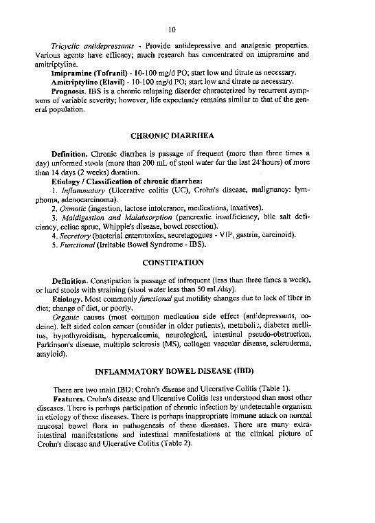
There are two main IBD: Crohn's disease and Ulcerative Colitis (Table 1).Features. Crohn's disease and Ulcerative Colitis less understood than most other
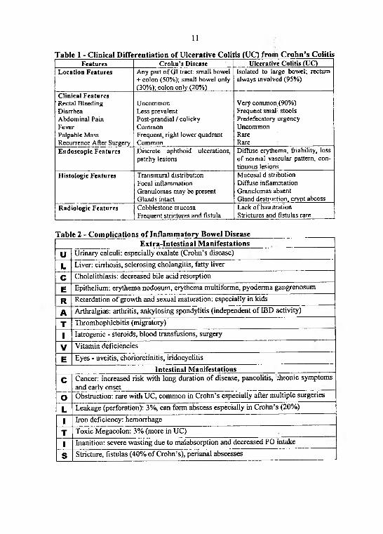
diseases. There is perhaps participation of chronic infection by undetectable organism in etiology of these diseases. There is perhaps inappropriate immune attack on normal mucosal bowel flora in pathogenesis of these diseases. There are many extra- intestinal manifestations and intestinal manifestations at the clinical picture of Crohn's disease and Ulcerative Colitis (Table 2).
11
Table 1 - Clinical Differentiation of Ulcerative Colitis (UC) from Crohn’s ColitisF e a tu r e s C r o h n ’s D is e a s e U lc e r a t iv e C o lit i s (U C )
L o c a t io n F e a tu r e s A n y p a r t o f G I tra c t: s m a ll b o w e l + c o lo n (5 0 % ) ; s m a l l b o w e l o n ly ( 3 0 % ) ; c o lo n o n ly (2 0 % )
Is o la te d to la rg e b o w e l; r e c tu m a lw a y s in v o lv e d (9 5 % )
C lin ic a l F e a tu r e s' R e c ta l B le e d in g D ia r rh e a A b d o m in a l P a in F e v e rP a lp a b le M a s s R e c u r re n c e A f te r S u rg e ry
U n c o m m o n L e ss p re v a le n t P o s t -p ra n d ia l / c o l ic k y C o m m o nF re q u e n t , r ig h t lo w e r q u a d ra n t C o m m o n
V e ry c o m m o n (9 0 % )F re q u e n t s m a l i s to o lsP re d e fe c a to ry u rg e n c yU n c o m m o nR a reR a re
E n d o s c o p ic F e a tu r e s D is c re te a p h th o id u lc e ra t io n s , p a tc h y le s io n s
D if fu s e e ry th e m a , f r ia b il i ty , lo s s o f n o rm a l v a s c u la r p a t te rn , c o n t in u o u s le s io n s
H is t o lo g ic F e a tu r e s T ra n s m u ra l d is tr ib u tio n F o c a l in f la m m a tio n G ra n u lo m a s m a y b e p re s e n t G la n d s in ta c t
M u c o s a l d s tr ib u t io n D if fu s e in f la m m a tio n G ra n u lo m a s a b s e n t G la n d d e s tru c t io n , c r y p t a b c e s s
R a d io lo g ic F e a tu r e s C o b b le s to n e m u c o s a F re q u e n t s tr ic tu re s a n d f is tu la
L a c k o f h a u i t r a t io n S tr ic tu re s a n d f is tu la s ra re
Table 2 - Complications of Inflammatory Bowel DiseaseExtra-Intestinal Manifestations
U Urinary calculi: especially oxalate (Crohn’s disease)
L Liver: cirrhosis, sclerosing cholangitis, fatty liver
C Cholelithiasis: decreased bile acid resorption
E Epithelium: erythema nodosum, eiythema multiforme, pyoderma gangrenosum
RA
Retardation of growth and sexual maturation: especially in kidsArthralgias: arthritis, ankylosing spondylitis (independent of IBD activity)
T Thrombophlebitis (migratory)
1 Iatrogenic - steroids, blood transfusions, surgery
V Vitamin deficiencies
E Eyes - uveitis, chorioretinitis, iridocyclitisIntestinal Manifestations
C Cancer: increased risk with long duration of disease, pancolitis, Aronie symptoms and early onset
О Obstruction: rare with UC, common in Crohn’s especially after multiple surgeries
L Leakage (perforation): 3%, can form abscess especially in Crohn’s (20%)
1 Iron deficiency: hemorrhage
T Toxic Megacolon: 3% (more in UC)
1 Inanition: severe wasting due to malabsorption and decreased PO intake
s Stricture, fistulas (40% of Crohn’s), perianal abscesses
12
CROHN'S DISEASE
Introduction. Crohn's disease is chronic inflammatory disorder affecting the small intestine and/or large intestine. In 1932, Crohn, Ginzberg, aid Oppenheimer described this disease and noted its localization to segments of the ileum. It was later pointed out that the disease may involve any part of the GI tract. •
Synonyms: regional enteritis, granulomatous enteritis, regional ileitis, terminal ileitis.
Definition. Crohn's disease is an idiopathic, chronic, transmural inflammatory process of the bowel that often leads to fibrosis and obstructive symptoms, which can affect any part of the GI tract from the mouth to the anus. Most cases involve the small bowel, particularly the terminal ileum.
Pathophysiology. Crohn's disease may affect any part of GI tract from mouth to anus. It is transmural inflammation with “skip" lesions. Crohn's disease associated with granulomas and deep Assuring / aphthous ulcerations and strictures. It is linear ulcers leading to mucosal islands and “cobble-stoning ". The deep fissures with risk of perforation into contiguous viscera lead to fistulae and abscesses. The enteric fistu- lae may communicate with skin, bladder, vagina, and other parts of bowel. Granulomas are found in 50% of surgical specimens and 15% of mucosa biopsies.
Epidemiolody.Frequency: The prevalence of Crohn's disease is approximately 7 cases per
100,000 population in USA. The prevalence of Crohn's disease is reportedly lower in southern European countries, South Africa, and Australia (approximately 0.9-3.1 cases per 100,000 population) and is even lower in Asian and South American countries (approximately 0.5-0.08 cases per 100,000 population).
Mortality/Morbidity: Studies have estimated ranges from no increased risk to up to a 5-fold increased risk of death.
Race: This condition is seemingly more common in whites than in blacks or Asians. A 2- to 4-fold increase in the prevalence of Crohn's disease has been found among the Jewish population in the United States, Europe, and South Africa compared to other ethnic groups.
Sex: The male-to-female ratio is 1.1-1.8:1.Age: The onset of Crohn's disease has a bimodal distribution: onset between the
ages of 15-30 years, second peak between the ages of 60-80 years. Most cases begin before age 30 years.
Clinical: Signs and symptoms most often presents as recurrent episodes of mild diarrhea (more common with involvement of colon), abdominal pain, fever and ileitis. Ileitis may present with post-prandial pain, vomiting, right lower quadrant (RLQ) mass, acute appendicitis. Fistulas, fissures, abscesses are common. There are two variants of clinical picture of Crohn's disease: slowly progressing and fulminant course. Extra-intestinal manifestations are more common with colonic involvement.
Physical: Physical findings may typically reveal right lower quadrant tenderness. Perianal manifestations: skin tags, fistulae, abscesses, and scarring. Extraintes- tinal manifestations: erythema nodosum, pyoderma gangrenosum, uveitis or episcleritis. A peripheral arthritis involving the large joints may also be present.
13
Lab Studies: Indicate the presence of inflammatory activity: anemia (chronic inflammation, chronic blood loss, and iron and vitamin B,2 malabsorption), leukocytosis (chronic inflammation, abscess, or steroid treatment), hypoalbuminemia, hypo- cholesterolemia, hypocalcemia, hypomagnesemia, and hypoprothrombinemia (malabsorption), increase of C-reactive protein (acute inflammation), increase the erythrocyte sedimentation rate (activity of Crohn’s colitis).
Stool samples should be tested for routine pathogens, ova, parasites, and Clostridium difficile toxin.
The serologic tests: 1. Antibodies to the yeast Saccharomyces cerevisiae (ie, anti-5 cerevisiae antibodies [ASCA]) are more commonly found in Crohn’s disease. 2. Perinuclear antineutrophil cytoplasmic antibodies (p-ANCA), and myeloperoxidase antigen, are more commonly found in ulcerative colitis (UC). These tests are only recommended as an adjunct to clinical diagnosis, as the test results are not specific and have been found to be positive in other bowel diseases.
Imaging Studies: Barium contrast studies are useful in evaluating features such as rigidity, pseudodiverticula, fistulization, and submucosal edema. CT scan is helpful in assessing extramural complications such as fistulae, abscesses, and hepatobiliary and renal complications. MRI can be superior to CT scanning in demonstrating pelvic lesions. Ultrasound is helpful in differentiating tubo-ovarian pathology. Radionucleotide scans may be helpful in assessing the severity and extent of the disease in patients who are too ill to undergo colonoscopy or barium studies.
Colonoscopy is useful in obtaining biopsy tissue. Upper endoscopy with biopsy is helpful in differentiating Crohn disease from peptic ulcer disease in patients with upper GI tract symptoms. ■
Histologic Findings: Transmural involvement with nonceseating granulomas and patchy skip lesions are seen in about 50% of cases. Lymphoid aggregates may also be seen throughout the bowel wall.
Treatment.Diet: elemental diets help remit acute Crolm's disease but are not palatable. The
diet should be balanced. Patients with extensive small bowel involvement need electrolyte, mineral and vitamin supplements.
Drugs. Most uncomplicated cases can be managed medically.Anti-inflammatory agents. Reduce inflammation by acting on host responses.Mesalazine - 3-4 g/d PO divided bid/tid.Sulfasalazine - 3-6 g/d PO divided bid/tid.Corticosteroids. - Exert both anti-inflammatory and immunosuppressant effects.Prednisone - 40-60 mg/d PO divided bid/qid; once in remission, slowly taper by
5-10 mg ql-2wk (but use only if symptoms are severe).Budesonide - 9 mg (3 X 3-mg cap) PO qd for 8 wk. Budesonide has less side ef
fects than prednisone.Immunosuppressives agents. Used chiefly as steroid-sparing agents. Interferes
with purine metabolism and inhibits synthesis of DNA, RNA, and proteins. It may decrease proliferation of immune cells, which results in lower autc immune activity.
Azathioprine - 1.5-2 mg/kg/d PO/IV.Methotrexate - 25 mg/week IM with concomitant lowering of prednisone dose;
14
once response achieved may switch to PO therapy; folic acid at dose of 1 mg/d should be given during treatment.
Antibiotics. Treatment of bacterial infections that may be associated with the underlying disease processes.
Metronidazole - 1 g/d PO divided bid/qid for 30-60 d. Imidazole ring-based antibiotic active against various anaerobic bacteria and protozoa, properties.
Ciprofloxacin - 500 mg PO bid (1 g/d). Fluoroquinolone with activity against pseudomonads, streptococci, MRSA, Staphylococcus epidermidis, and most gramnegative organisms, but no activity against anaerobes.
Immunomodulators - Interfere with development of immunolog.cal responses.Infliximab - Chimeric IgGlk monoclonal antibody that neutralizes cytokine
TNF-alpha and inhibits its binding to TNF-alpha receptor. Reduces infiltration of inflammatory cells and TNF-alpha production in inflamed areas.
Infliximab 5 mg/kg IV as single infusion over 2 h For fistulatir.g Crohn disease, an induction and maintenance regimen may be required: 5 mg/kg IV infusion at 0, 2, and 6 wk as induction regimen, then 5 mg/kg q6wk for maintenance IV infusion must be administered over at least 2 h; must use infusion set with in-line, sterile, nonpyro- genic, low-protein-binding filter (pore size <1.2 micrometers).
Chronic diarrhea: loperamide (2-4 mg), diphenoxylate with atropine (1 tab), and tincture of opium (8-15 gtt). Such agents may be administered up to 4 times daily. Use with caution to patients with active colitis (risk of developing toxic megacolon).
Patients with terminal ileitis (less than 100 cm of terminal ileum) cannot absorb bile acids, which can lead to secretory diarrhea in the colon. These patients benefit from bile acid sequestrants (cholestyramine (2-4 g], colestipol [5 g] bid/tid). It is a bile salt binding resin. Patients with terminal ileitis (more than 100 cm of ileum) have defective bile salt absorption and develop steatorrhea. These patients benefit from a low-fat diet and medium-chain triglyceride preparations. Bile sequestrants exacerbate this type of diarrhea.
Diarrhea may also develop because of bacterial overgrowth, short-bowel syndrome, and lactase deficiency.
Abdominal pain may be reduced with propantheline (0.125 mg), dicyclomine (10-20 mg), or hyoscyamine (0.125 mg). These drugs should not be used if a bowel obstruction is considered possible.
Surgical treatment. Surgery generally reserved for complications such as fistu- lae, obstruction, abscess, perforation, bleeding, malignancy and rarely for medically refractory disease.
Prognosis. Although Crohn disease is chronic with recurrent relapses, appropriate medical and surgical therapy helps patients to have a reasonable quality of life. The mortality rate increases with the duration of the disease, and GI tract cancer is the leading cause of disease-related death.
15
ULCERATIVE COLITIS (UC)
Introduction. Ulcerative colitis is inflammatory disease affecting colonic mucosa from rectum to cecum. Usually UC is chronic disease characterized by rectal bleeding and diarrhea, and prone to remissions and exacerbations. Often a lifelong illness, the condition has profound emotional and social impact on the affected individual. .
Synonyms: continuous idiopathic inflammation of the colonic or rectal mucosa.Definition. Ulcerative colitis is an idiopathic, chronic, recurrent, superficial, dif
fuse inflammatory disease of the colon or rectal mucosa. Most cases involve the rectum.
Pathophysiology. Ulcerative colitis can involve any portion of lower bowel from rectum only (proctitis) to entire colon (pancolitis). It is inflammation diffuse and confined to mucosa. The rectum is involved in more than 95% of cases. Some authorities believe that the rectum is always involved in an untreated patient. Partial healing may occur in a patient treated with topical therapy, creating diagnostic confusion.
Epidemiolody.Frequency: In the USA the prevalence rate is 35-100 cases per 100,000 people.
Prevalence rates may be lower in South America, Asia, and Africa.Race: Ulcerative colitis occurs more frequently in white people. The incidence
of ulcerative colitis is reported to be 2-4 times higher in Jewish people. However, recent population studies in North America do not completely support this assertion.
Sex: Ulcerative colitis affects 30% more females than males.Age: The onset of UC has a bimodai distribution: onset between the ages of 15-
25 years, second peak between the ages of 55-65 years. Most cases (2/3) begin before age 30 years, although it can occur in people of any age.
Clinical. Signs and symptoms most often presents as diar hea, rectal bleeding, abdominal cramps/pain (especially with defecation), tenesmus, urgency, incontinence, systemic symptoms (fever, anorexia, weight loss, fatigue), extra-intestinal manifestations (synovitis, ankylosing spondylitis (HLA-B27), sicroiliitis, erythema nodosum, pyoderma gangrenosum, aphthous stomatitis, episcleritis, iritis, primary sclerosing cholangitis, uric acid renal stones. There are two variants of clinical picture of ulcerative colitis: progressing with exacerbations and remissions (95 A of cases) and fulminant course (5% of cases).
Physical. Physical findings may typically reveal mild fever, tachycardia, dehydration, malnutrition, abdominal tenderness, blood on digital rectal examination.
Lab Studies. Anemia (hemoglobin less than 14 g/dL in males and less than 12 g/dL in females), thrombocytosis (platelet count more than 350,000/L), elevated sedimentation rate (variable reference ranges, usually 0-33 mm/h) and elevated C- reactive protein (more than 100 mcg/L; both of the last findings correlate with disease activity) hypoalbuminemia (albumin less than 3.5 g/dL), hypokalemia (potassium less than 3.5 mEq/L), hypomagnesemia (magnesium less than 1.5 mg/dL), elevated alkaline phosphatase (more than 125 U/L suggests primary sclerosing cholangitis - usually more than 3 times the upper limit of the reference range).
16
Imaging Studies. A plain abdominal radiograph is useful in evaluating features such as perforation, obstruction, or ileus. Barium enemas (not during acute phase or relapse) may show a narrow, tubular, shortened colon with loss of haustral folds, pseudopolyps, and small ulcers. A radionuclide scan may be useful in acute fulminant colitis when colonoscopy or barium enemas are contraindicated. Sigmoidoscopy can provide the diagnosis of colitis. Findings on colonoscopy with biopsy (contraindicated in severe exacerbation) confirm a diagnosis. Stool studies are useful to exclude other causes.
Histologic Findings. Ulcerative colitis is characterized by a uniform inflammatory reaction in the colonic mucosa, without intervening areas of normal mucosa.
Treatment.Drugs.Anti-inflammatory agents. Reduce inflammation.Mesalazine - 4 g/d PO divided bid/tid.Sulfasalazine - 1-4 g/d PO divided bid/tid.Corticosteroids. - Used in severe active cases for induction of remission.Methylprednisolone - 80 mg IV q8h; dose may vary.Prednisone - 40-60 mg PO for 10-14 d, then taper off over 8-12 wk, using sul
fasalazine or mesalamine as maintenance therapy.Immunosuppressives agents. Inhibit activity of the immune system.Azathioprine - 1.5-2.5 mg/kg/d POCyclosporine - 4 mg/kg/d IV infusion, 2-3 mg/kg/d in elderly patients or in pa
tients with renal dysfunction.Antibiotics. Ciprofloxacin and metronidazole usually are administered on an
empiric basis in patients with severe colitis in addition to steroids.Ciprofloxacin - 500 mg PO bid (1 g/d), 400 mg IV bid.Metronidazole - Loading dose: 15 mg/kg (or 1 g for 70-kg adult) IV over 1 h.
Maintenance dose: 6 h following loading dose, infuse 7.5 mg/kg (or 500 mg for 70- kg adult) over 1 h q6-8h; not to exceed 4 g/d properties.
Tumor necrosis factor (TNF) inhibitors - TNF is but one of many cytokines involved in the inflammatory cascade that may contribute to symptoms.
Infliximab - Neutralizes cytokine TNF-alpha and inhibits its binding to TNF- alpha receptor. Indicated for moderate-to-severe active ulcerative colitis in patients who have experienced inadequate response to conventional therapy.
Infliximab 5 mg/kg IV infusion at 0, 2, and 6 wk as induction regimen, then 5 mg/kg q8wk for maintenance IV infusion must be administered over at least 2 h; must use infusion set with in-line, sterile, nonpyrogenic, low-protein-binding filter (pore size <1.2 micrometers).
Surgical treatment. Surgery generally reserved for complications such as toxic megacolon, bleeding, pre-cancerous and for fulminant cases, and medically refractory disease.
Prognosis: Most cases are controlled with medical therapies, with the patient experiencing lifelong exacerbations and remissions. In more severe cases, surgery results in a cure.
17
FATTY LIVER
Introduction. Fatty liver disease can range from fatty liver alone (steatosis) to fatty liver associated with inflammation (steatohepatitis). This condition can occur with the use of alcohol (alcohol-related fatty liver) or in the absence of alcohol (nonalcoholic fatty liver disease - NAFLD)..Fatty change in the liver results from excessive accumulation of lipids within hepatocytes.
Synonyms: steatosis, hepatic steatosis, steatohepatitis, alcohol-related fatty liver, alcoholic steatohepatitis, nonalcoholic fatty liver disease, nonalcoholic steatohepatitis (NASH), liver fibrosis, drug-induced fatty liver.
Definition. Fatty liver (steatosis) is infringement o f a hepatic metabolism as a result of influence of various etiological factors resulting in increased the accumulation of triglycerides and other fats in liver cells. Steatohepatitis is fatty liver associated with hepatic inflammation and liver cell death.
Etiology. Alcohol, diabetes, obesity, jejuno-ileal bypass, hyperlipidemic states, drugs (methotrexate, tetracycline, amiodarone, valproic acid), fatty liver of pregnancy.
Pathophysiology. Potential pathophysiological mechanisms include: 1. Decreased mitochondrial fatty acid beta-oxidation. 2. Increased endogenous fatty acid synthesis or enhanced delivery of fatty acids to the liver. 3. Deficient incorporation or export of triglycerides as very-low density lipoprotein.
Epidemiolody.Frequency: Steatosis affects approximately 25% of the general population. Stea
tohepatitis may be related to alcohol-induced hepatic damage or may be unrelated to alcohol (NASH). NASH has been detected in 1.2-9% of patients undergoing routine liver biopsy. NAFLD is found in over 80% of patients who are obese.
Mortality/Morbiciity: Steatosis is a benign condition, and progression is very rare. Steatohepatitis may progress to liver fibrosis and cirrhosis and may result in liver-related morbidity and mortality.
Sex: 50% of patients with steatosis are females.Age: Fatty liver occurs in all age groups.Clinical. Part of the patients with fatty liver is asymptomatic. More than 50% of
patients with fatty liver or NASH report persistent fatigue, malaise, or upper abdominal discomfort. ;
Physical. Hepatomegaly is common. Rapid fulminant liver failure may present in patients with drug-induced fatty liver. Skeletal muscle wasting, cardiomyopathy, pancreatitis, or peripheral neuropathy may be present in patients who abuse alcohol.
Lab Studies. Aspartate aminotransferase (AST) or alanihe aminotransferase (ALT) level may be an elevated. In some patients with fatty liver or NASH the AST and ALT may be normal. Alkaline phosphatase can be elevated in some patients with NASH. Hyperlipidemia may be present. Before the diagnosis of NASH can be made, viral markers should be tested and viral infection excluded.
Imaging Studies. Ultrasound (the liver is hyperechogenic or bright, steatosis is detected only when more than 30% fatty change is present), laparoscopy (a spotty yellow appearance when a fatty change of more than 30% is present without fibrosis
18
and a diffuse yellow appearance when a similar fatty change is present with fibrosis), liver biopsy (histopathological examination are required to establish the diagnosis).
Histologic Findings: The diagnosis of fatty liver or NASH can be established only with a liver biopsy. Specific histologic findings include (1) steatosis, which usually is macrovesicular but may be microvesicular or mixed; (2) inflammatory infiltrates consist of mixed neutrophilic and mononuclear cells; portal infiltrates usually are not seen (unlike in hepatitis C); (3) ballooning degeneration; and (4) fibrosis. The first 3 findings are used to calculate the NAFLD Activity Score (0-8). The stage of disease is determined by the NAFLD Activity Score and the amount of fibrosis.
Treatment.Diet: Abstinence from alcohol (alcohol-related fatty liver), restriction in food of
rapidly absorbed carbohydrates with a high protein-to-calorie ratio (nonalcoholic fatty liver disease). Weight loss should be gradual, moderate, and controlled. Abrupt weight loss and gain may be associated with progression of the disease.
Activity: cardiovascular fitness and weight training.Drugs. No proven medical therapy is available.Prognosis. Steatosis may be reversible with weight loss and/or stopping alcohol
use. Of patients with steatohepatitis, 10% will progress to fibrosis and cirrhosis.
CONGENITAL HYPERBILIRUBINAEMIAS (NON-HAEMOLYTIC)
GILBERT SYNDROME (UNCONJUGATED HYPERBILIRUBINEMIA)
Introduction. Augustine Gilbert and Pierre Lereboullet first described Gilbert syndrome in 1901. It is the most common inherited cause of unconjugated hyperbilirubinemia.
Synonyms: constitutional hepatic dysfunction, constitutional' hyperbilirubinemia, familial nonhemolytic jaundice, hereditary nonhemolytic bilirubinemia, low- grade chronic hyperbilirubinemia.
Definition. Gilbert syndrome is characterized by intermittent jaundice and/or mild unconjugated hyperbilirubinemia (less than 50 micromoI/L) in the absence of hemolysis or underlying liver disease.
Pathophysiology. Unconjugated hyperbilirubinemia in Gilbert syndrome has long been recognized as due to underactivity of the conjugating enzyme system bilirubin-uridine diphosphate glucuronyl transferase (bilirubin-UGT). Bilirubin-UGT is responsible for conjugating bilirubin into bilirubin monoglucuronides and diglu- curonides and is located primarily in the endoplasmic reticulum of hepatocytes. A breakthrough in understanding the genetic basis o f Gilbert syndrome was achieved in 1995, when abnormalities in the TATAA region of the promoter were identified. Presently, whether reduced bilirubin-UGT activity results from a reduced number of enzyme molecules or a qualitative enzyme defect is unknown.
Epidemiolody.Frequency: The incidence of Gilbert syndrome is 3-7% of the population.Mortality/Morbidity: Gilbert syndrome is a benign condition with no associated
19
morbidity or mortality.Race: Gilbert syndrome is not restricted to any ethnic group and occurs in all
races.Sex: Population studies show that Gilbert syndrome occurs predominately in
men, with a male-to-female ratio ranging from 2:1-7:1.Age: Gilbert syndrome usually is diagnosed around puberty. In older patients,
the diagnosis usually is made when unconjugated hyperbilirubinemia is noted on routine blood tests.
Clinical. Gilbert syndrome may be precipitated by dehydration, fasting, menstrual periods, or stress, such as an intercurrent illness or vigorous exercise. Patients may complain of vague abdominal discomfort and general fatigue for which no cause is found. These episodes resolve spontaneously, and no treatment is required except for supportive care.
Physical. The liver changes are absent.Lab Studies. CBC (including reticulocyte count and blood smear) is normal.
This is a useful screening test to exclude hemolysis. Lactate dehydrogenase is normal (levels are elevated in hemolysis). Standard liver function test results are normal with the exception of unconjugated hyperbilirubinemia. In some cases increase in serum alkaline phosphatase is present.
Imaging Studies. Imaging studies are not required to confirm a diagnosis of Gilbert syndrome.
Additional tests rarely are required because a diagnosis of Gilbert syndrome can be made in the presence of unconjugated hyperbilirubinemia noted on several occasions; normal results on CBC, reticulocyte count, and blood smear; normal liver function test results; and absence of other disease processes.
Phenobarbital test: Phenobarbital and other enzyme inducers of the bilirubin- UGT system will normalize plasma bilirubin in patients with Gilbert syndrome. Steroids also can reduce plasma bilirubin levels in Gilbert syndrome by increasing hepatic uptake and storage of bilirubin.
Histologic Findings: The liver is normal histologically, except for occasional accumulation of a lipofuscinlike pigment around the terminal hepatic venules. Liver biopsies are not performed routinely and rarely are necessary.
Treatment. Inpatient care is not required.Diet: Diet is normal.Activity: No activity restrictions are necessary.Drugs: Various medications have been used experimentally , to reduce the hy
perbilirubinemia of Gilbert syndrome. For example, phenobarbital and glutethimide activate hepatic bilirubin-UGT activity, while tin-protoporphyrin inhibits heme oxygenase to reduce bilirubin levels. In light of the benign and inconsequential nature of this condition, the use of medications in Gilbert syndrome in clinical practice is unjustified.
The most important aspect of treatment once the diagnosis is established is reassurance. Patients with Gilbert syndrome should be informed of its benign nature and that hyperbilirubinemia is not associated with increased morbidity. Its excellent prognosis is associated with normal life expectancy, which must be made perfectly clear
20
to the patient.Prevention: Avoid known risk factors for precipitating hyperbilirubinemia (de
hydration, fasting).Prognosis. Gilbert syndrome is a common and benign condition. It has an excel
lent prognosis, and those who have it can lead a normal lifestyle.
DUBIN-JOHNSON SYNDROME (CONJUGATED HYPERBILIRUBINEMIA)
Introduction. Dubin-Johnson syndrome (DJS) is a type of hereditary conjugated hyperbilirubinemia that was first described independently in 1954 by Dubin and Johnson and by Sprinz and Nelson.
Synonyms: DJS, conjugated hyperbilirubinemia, chronic idiopathic jaundice.Definition. Dubin-Johnson syndrome is characterized by nonpruritic jaundice
with conjugated hyperbilirubinemia (35-85 micromol/L) and the accumulation of hepatocellular pigment in the absence of hemolysis or underlying liver disease.
Pathophysiology. DJS is an autosomal recessive disorder that is caused by a mutation in the gene responsible for the human canalicular multispecific organic anion transporter (cMOAT) protein. This protein mediates ATP-dependent transport of certain organic anions across the canalicular membrane of the hepatocyte. A defect in the cMOAT protein results in impaired hepatobiliary transport of non-bile salt organic anions and is thought to be responsible for the conjugated hyperbilirubinemia and for the accumulation of hepatocellular pigment.
Epidemiolody.Frequency: DJS is a rare disorder.Race: DJS has been described in all nationalities, ethnic backgrounds, and races.
Prevalence reportedly is highest among Iranian Jews (1:300).Age: Patients with DJS tend to develop nonpruritic jaundice during their teen
years.Clinical. The first symptoms nonpruritic jaundice is appeared at the teen years.
Most patients are asymptomatic. In some cases the patients have nonspecific right upper quadrant pain. Subclinical cases can become evident during pregnancy or following the initiation of oral contraceptives. The part of the patients has a family history of jaundice in an autosomal recessive pattern.
Physical. Physical examination findings are generally normal, with the exception of the presence of jaundice and possible hepatosplenomegaly. Hyperbilirubinemia and clinical icterus can be worsened by intercurrent illnesses, by drugs that can decrease hepatic excretion of organic anions (for example, oral contraceptives), and by pregnancy.
Lab Studies. The diagnosis of DJS can be confirmed by demonstrating an increase in the ratio of urinary coproporphyrin I to coproporphyrin III. This finding is a pathognomonic feature of DJS. Laboratory studies reveal conjugated hyperbilirubinemia, with total bilirubin levels in the 35- to 85 micromol/L range. Results of other laboratory tests, including liver enzymes, serum albumin, and hematologic studies (complete blood count, reticulocyte count, protlirombin time) are normal.
21
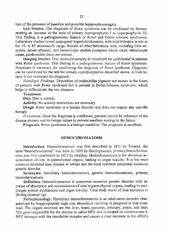
Im aging Studies. Oral cholecystography fails to visualize the gallbladder in patients with DJS. These findings can be mistaken for evidence o f gallbladder disease if the patient presents with abdominal pain and may result in an unnecessary cholecystectomy. Procedures are not necessary to confirm the diagnosis o f DJS. Diagnosis can be confirmed by the test for urinary coproporphyrins described above. A liver biopsy is not necessary for diagnosis. Patients may be noted to have a dark liver during routine surgeries (cholecystectomy), prompting biopsy.
Histologic Findings: Deposition o f melaninlike pigment occurs in the livers o f patients with DJS but not in Rotor syndrome, which helps to differentiate the two diseases. Macroscopically, the pigment can cause the liver to appear dark or almost black. Microscopically, there is accumulation o f coarsely granular pigment, most pronounced in the centrilobular zones. No associated scarring, hepatocellular necrosis, or distortion o f zonal architecture is present. The amount o f pigment can vary among patients and within an individual. Certain diseases (viral hepatitis) can cause the pigment to disappear. The pigment reaccumulates slowly once the acute process is resolved.
T reatm ent.Diet: Diet is normal.Activity: No activity restrictions are necessary.Drugs: DJS is a benign disorder and does not require any specific therapy. In
the past, patients were treated with phenobarbital, which was primarily used to reduce serum bilirubin levels. This is no longer recommended.
Prevention: Once the diagnosis is confirmed, patients should be informed of the disease process and its benign nature to prevent needless workup in the future.
Prognosis. DJS is a benign condition. The prognosis is excellent.
R O T O R SYNDROME (CONJUGATED HYPERBILIRUBINEM IA)
Introduction. Rotor syndrome is a type o f hereditary conjugated hyperbilirubinemia that was first described in 1948 by Rotor. v
Synonyms: conjugated hyperbilirubinemia, chronic idiopathic jaundice.Definition. Rotor syndrome is characterized by nonpruritic jaundice with conju
gated hyperbilirubinemia (35-85 micromol/L) without the accumulation of hepatocellular pigment in the absence o f hemolysis or underlying liver disease.
Pathophysiology. Rotor syndrome is possibly autosomal dominant disorder. There are defects in bilirubin handling in the liver.
Epidemiolody.Frequency: Rotor syndrome is more rare disorder than Dubin-Johnson syn
drome.Clinical. The first symptoms nonpruritic jaundice is appeared at the teen years.
Most patients are asymptomatic. In some cases the patients have nonspecific right upper quadrant pain. The part o f the patients has a family history of jaundice in an autosomal dominant pattern.
Physical. Physical examination findings are generally normal, with the excep
22
tion of the presence o f jaundice and possible hepatosplenomegaly.Lab Studies. The diagnosis o f Rotor syndrome can be confirmed by demon
strating an increase in the ratio o f urinary coproporphyrin I to coproporphyrin III. This finding is a pathognomonic feature o f Rotor and Dubin-Johnson syndromes. Laboratory studies reveal conjugated hyperbilirubinemia, with total bilirubin levels in the 35- to 85 micromol/L range. Results o f other laboratory tests, including liver enzymes, serum albumin, and hematologic studies (complete blood count, reticulocyte count, prothrombin time) are normal.
Im aging Studies. Oral cholecystography is visualized the gallbladder in patients with Rotor syndrome. This finding is a pathognomonic feature o f Rotor syndrome. Procedure is necessary for confirming the diagnosis o f Rotor syndrome. Diagnosis can be confirmed by the test for urinary coproporphyrins described above. A liver biopsy is not necessary for diagnosis.
Histologic Findings: Deposition o f melaninlike pigment not occurs in the livers of patients with Rotor syndrome but is present in Dubin-Johnson syndrome, which helps to differentiate the two diseases.
T reatm ent.Diet: Diet is normal.Activity: No activity restrictions are necessary.Drugs: Rotor syndrome is a benign disorder and does not require any specific
therapy.Prevention: Once the diagnosis is confirmed, patients should be informed o f the
disease process and its benign nature to prevent needless workup in the future.Prognosis. Rotor syndrome is a benign condition. The prognosis is excellent.
HEM OCHROM ATOSIS
Introduction. Hemochromatosis was first described in 1871 by Troisier, the term “hemochromatosis” was done in 1889 by Reclinghausen, primary hemochromatosis was first established in 1922 by Sheldon. Hemochromatosis is the abnormal accumulation of iron in parenchymal organs, leading to organ toxicity. It is the most common inherited liver disease in whites and the most common autosomal recessive genetic disorder.
Synonyms: hereditary hemochromatosis, genetic hemochromatosis, primary hemochromatosis.
Definition. Hemochromatosis is autosomal recessive genetic disorder with increase of absorption and accumulation o f iron in parenchymal organs, leading to multiorgan system dysfunction and organ toxicity. Total body stores o f iron increases to 20-40g (normal lg).
Pathophysiology. Hereditary hemochromatosis is an adult-onset disorder characterized by inappropriately high iron absorption resulting in progressive iron overload. The organs involved are the liver, heart, pancreas, pituitary, joints, and skin. The gene responsible for the disease is called HFE and is located on chromosome 6. HFE interacts with the transferrin receptor and causes a clear decrease in the affinity
23
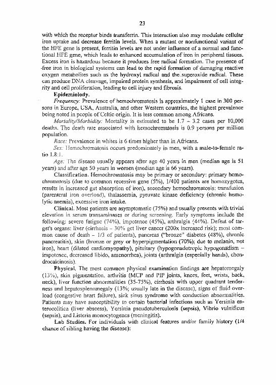
with which the receptor binds transferrin. This interaction also may modulate cellular iron uptake and decrease ferritin levels. When a mutant or nonfunctional variant of the HFE gene is present, ferritin levels are not under influence of a normal and functional HFE gene, which leads to enhanced accumulation of iron in peripheral tissues. Excess iron is hazardous because it produces free radical formation. The presence of •free iron in biological systems can lead to the rapid formation of damaging reactive oxygen metabolites such as the hydroxyl radical and the. superoxide radical. These can produce DNA cleavage, impaired protein synthesis, and impairment of cell integrity and cell proliferation, leading to cell injury and fibrosis.
Epidemiolody.Frequency: Prevalence of hemochromatosis is approximately 1 case in 300 per
sons in Europe, USA, Australia, and other Western countries, the highest prevalence being noted in people of Celtic origin. It is less common among Africans.
Mortality/Morbidity: Mortality is estimated to be 1.7 - 3.2 cases per 10,000 deaths. The death rate associated with hemochromatosis is 0.9 persons per million population.
Race: Prevalence in whites is 6 times higher than in Africans.Sex: Hemochromatosis occurs predominately in men, with a male-to-female ra
tio 1.8:1.Age: The disease usually appears after age 40 years in men (median age is 51
years) and after age 50 years in women (median age is 66 years).Classification. Hemochromatosis may be primary or secondary: primary hemo
chromatosis (due to common recessive gene (5%), 1/400 patients are homozygotes, results in increased gut absorption of iron), secondary hemochromatosis: transfusion (parenteral iron overload), thalassemia, pyruvate kinase deficiency (chronic hemolytic anemia), excessive iron intake.
Clinical. Most patients are asymptomatic (75%) and usually presents with trivial elevation in serum transaminases or during screening. Early symptoms include the following: severe fatigue (74%), impotence (45%), arthralgia (44%). Defeat of target's organs: liver (cirrhosis - 30% get liver cancer (200x increased risk); most common cause of death - 1/3 of patients), pancreas (“bronze” diabetes (48%), chronic pancreatitis), skin (bronze or grey or hyperpigmentation (70%); due to melanin, not iron), heart (dilated cardiomyopathy), pituitary (hypogonadotropic hypogonadism - impotence, decreased libido, amenorrhea), joints (arthralgia (especially hands), chon- drocalcinosis).
Physical. The most common physical examination findings are hepatomegaly (13%), skin pigmentation, arthritis (MCP and PIP joints, knees, feet, wrists, back, neck), liver function abnormalities (35-75%), cirrhosis with upper quadrant tenderness and hepatosplenomegaly (13%; usually late in the disease), signs of fluid overload (congestive heart failure), sick sinus syndrome with conduction abnormalities. Patients may have susceptibility to certain bacterial infections such as Yersinia en- terocolitica (liver abscess), Yersinia pseudotuberculosis (sepsis), Vibrio vulnificus (sepsis), and Listeria monocytogenes (meningitis).
Lab Studies. For individuals with clinical features and/or family history (1/4 chance of sibling having the disease):
24
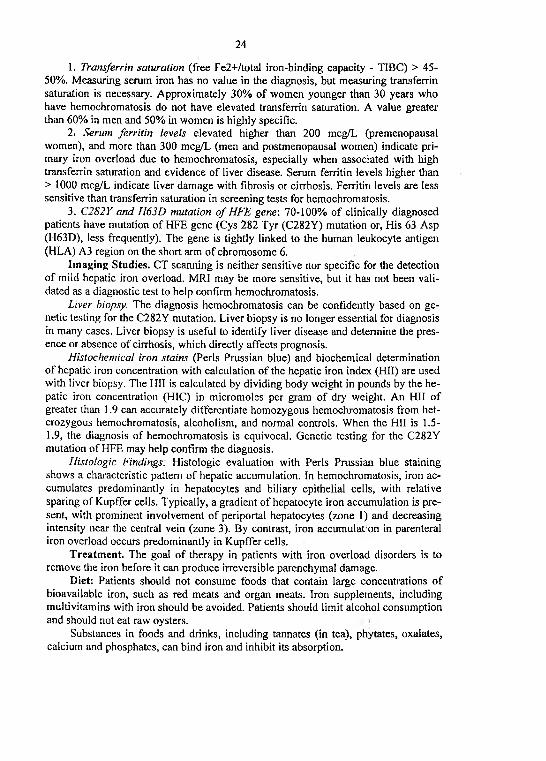
1. Transferrin saturation (free Fe2+/total iron-binding capacity - TIBC) > 45- 50%. Measuring serum iron has no value in the diagnosis, but measuring transferrin saturation is necessary. Approximately 30% of women younger than 30 years who have hemochromatosis do not have elevated transferrin saturation. A value greater than 60% in men and 50% in women is highly specific.
2. Serum ferritin levels elevated higher than 200 mcg/L (premenopausal women), and more than 300 mcg/L (men and postmenopausal women) indicate primary iron overload due to hemochromatosis, especially when associated with high transferrin saturation and evidence of liver disease. Serum ferritin levels higher than > 1000 mcg/L indicate liver damage with fibrosis or cirrhosis. Ferritin levels are less sensitive than transferrin saturation in screening tests for hemochromatosis.
3. C282Y and H63D mutation o f HFE gene: 70-100% of clinically diagnosed patients have mutation of HFE gene (Cys 282 Tyr (C282Y) mutation or, His 63 Asp (H63D), less frequently). The gene is tightly linked to the human leukocyte antigen (HLA) A3 region on the short arm of chromosome 6.
Imaging Studies. CT scanning is neither sensitive nor specific for the detection of mild hepatic iron overload. MRI may be more sensitive, but it has not been validated as a diagnostic test to help confirm hemochromatosis.
Liver biopsy. The diagnosis hemochromatosis can be confidently based on genetic testing for the C282Y mutation. Liver biopsy is no longer essential for diagnosis in many cases. Liver biopsy is useful to identify liver disease and determine the presence or absence of cirrhosis, which directly affects prognosis.
Hislochemical iron stains (Peris Prussian blue) and biochemical determination of hepatic iron concentration with calculation of the hepatic iron index (НП) are used with liver biopsy. The HII is calculated by dividing body weight in pounds by the hepatic iron concentration (HIC) in micromoles per gram of dry weight. An HII of greater than 1.9 can accurately differentiate homozygous hemochromatosis from heterozygous hemochromatosis, alcoholism, and normal controls. When the HII is 1.5- 1.9, the diagnosis of hemochromatosis is equivocal. Genetic testing for the C282Y mutation of HFE may help confirm the diagnosis.
Histologic Findings: Histologic evaluation with Peris Prussian blue staining shows a characteristic pattern of hepatic accumulation. In hemochromatosis, iron accumulates predominantly in hepatocytes and biliary epithelial cells, with relative sparing of Kupffer cells. Typically, a gradient of hepatocyte iron accumulation is present, with prominent involvement of periportal hepatocytes (zone 1) and decreasing intensity near the central vein (zone 3). By contrast, iron accumulation in parenteral iron overload occurs predominantly in Kupffer cells.
Treatment. The goal of therapy in patients with iron overload disorders is to remove the iron before it can produce irreversible parenchymal damage.
Diet: Patients should not consume foods that contain large concentrations of bioavailable iron, such as red meats and organ meats. Iron supplements, including multivitamins with iron should be avoided. Patients should limit alcohol consumption and should not eat raw oysters.
Substances in foods and drinks, including tannates (in tea), phytates, oxalates, calcium and phosphates, can bind iron and inhibit its absorption.
25
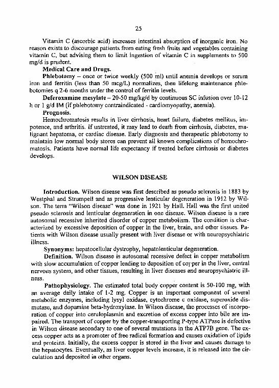
Vitamin C (ascorbic acid) increases intestinal absorption o f inorganic iron. No reason exists to discourage patients from eating fresh fruits and vegetables containing vitamin C, but advising them to limit ingestion o f vitamin C in supplements to 500 mg/d is prudent.
Medical Care and Drugs.Phlebotomy - once or twice weekly (500 ml) until anemia develops or serum
iron and ferritin (less than 50 mcg/L) normalizes, then lifelong maintenance phlebotomies q 2-6 months under the control o f ferritin levels.
Deferoxamine mesylate - 20-50 mg/kg/d by continuous SC infusion over 10-12 h or 1 g/d IM (if phlebotomy contraindicated - cardiomyopathy, anemia).
Prognosis.Hemochromatosis results in liver cirrhosis, heart failure, diabetes mellitus, im
potence, and arthritis. If untreated, it may lead to death from cirrhosis, diabetes, malignant hepatoma, or cardiac disease. Early diagnosis and therapeutic phlebotomy to maintain low normal body stores can prevent all known complications of hemochromatosis. Patients have normal life expectancy if treated before cirrhosis or diabetes develops.
WILSON DISEASE
Introduction. Wilson disease was first described as pseudo sclerosis in 1883 by Westphal and Strumpell and as progressive lenticular degeneration in 1912 by Wilson. The term “Wilson disease” was done in 1921 by Hall. Hall was the first united pseudo sclerosis and lenticular degeneration in one disease. Wilson disease is a rare autosomal recessive inherited disorder o f copper metabolism. The condition is characterized by excessive deposition o f copper in the liver, brain, and other tissues. Patients with Wilson disease usually present with liver disease or with neuropsychiatric illness.
Synonyms: hepatocellular dystrophy, hepatolenticular degeneration.Definition. Wilson disease is autosomal recessive defect in copper metabolism
with slow accumulation o f copper leading to deposition o f copper in the liver, central nervous system, and other tissues, resulting in liver diseases and neuropsychiatric illness.
Pathophysiology. The estimated total body copper content is 50-100 mg, with an average daily intake o f 1-2 mg. Copper is an important component o f several metabolic enzymes, including lysyl oxidase, cytochrome c oxidase, superoxide dis- mutase, and dopamine beta-hydroxylase. In Wilson disease, the processes of incorporation of copper into ceruloplasmin and excretion o f excess copper into bile are impaired. The transport o f copper by the copper-transporting P-type ATPase is defective in Wilson disease secondary to one o f several mutations in the ATP7B gene. The excess copper acts as a promoter o f free radical formation and causes oxidation of lipids and proteins. Initially, the excess copper is stored in the liver and causes damage to the hepatocytes. Eventually, as liver copper levels increase, it is released into the circulation and deposited in other organs.
26
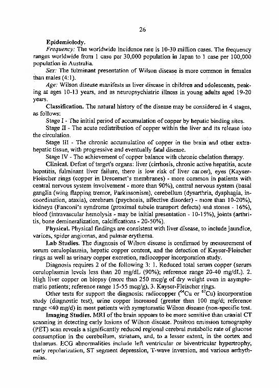
Epidemiolody.Frequency: The worldwide incidence rate is 10-30 million cases. The frequency
ranges worldwide from 1 case per 30,000 population in Japan to 1 case per 100,000 population in Australia.
Sex: The fulminant presentation of Wilson disease is more common in females than males (4:1).
Age: Wilson disease manifests as liver disease in children and adolescents, peaking at ages 10-13 years, and as neuropsychiatric illness in young adults aged 19-20 years.
Classification. The natural history of the disease may be considered in 4 stages, as follows:
Stage I - The initial period o f accumulation o f copper by hepatic binding sites.Stage II - The acute redistribution o f copper within the liver arid its release into
the circulation.Stage III - The chronic accumulation o f copper in the brain and other extra-
hepatic tissue, with progressive and eventually fatal disease.Stage IV - The achievement o f copper balance with chronic chelation therapy.Clinical. Defeat o f target's organs: liver (cirrhosis, chronic active hepatitis, acute
hepatitis, fulminant liver failure, there is low risk of liver car eer), eyes (Kayser- Fleischer rings (copper in Descemet’s membranes) - more common in patients with central nervous system involvement - more than 90%), central nervous system (basal ganglia (wing flapping tremor, Parkinsonism), cerebellum (dysarthria, dysphagia, incoordination, ataxia), cerebrum (psychosis, affective disorder) - more than 10-20%), kidneys (Fanconi’s syndrome (proximal tubule transport defects) and stones - 16%), blood (intravascular hemolysis - may be initial presentation - 10-15%), joints (arthritis, bone demineralization, calcifications - 20-50%).
Physical. Physical findings are consistent with liver disease, to include jaundice, varices, spider angiomas, and palmar erythema.
L ab Studies. The diagnosis o f Wilson disease is confirmed by measurement of serum ceruloplasmin, hepatic copper content, and the detection o f Kayser-Fleischer rings as well as urinary copper excretion, radiocopper incorporation study.
Diagnosis requires 2 of the following 3: 1. Reduced total serum copper (serum ceruloplasmin levels less than 20 mg/dL (90%); reference range 20-40 mg/dL). 2. High liver copper on biopsy (more than 250 meg/g of dry weight even in asymptomatic patients; reference range 15-55 meg/g), 3. Kayser-Fleischer rings.
Other tests for support the diagnosis: radiocopper (MCu or 67Cu) incorporation study (diagnostic test), urine copper increased (greater than 100 mg/d; reference range <40 mg/d) in most patients with symptomatic Wilson disease (non-specific test.
Im aging Studies. MRI o f the brain appears to be more sensitive than cranial CT scanning in detecting early lesions of Wilson disease. Positron emission tomography (PET) scan reveals a significantly reduced regional cerebral metabolic rate of glucose consumption in the cerebellum, striatum, and, to a lesser extent, in the cortex and thalamus. ECG abnormalities include left ventricular or biventricular hypertrophy, early repolarization, ST segment depression, T-wave inversion, and various arrhythmias.
27
Abdominal imaging: CT scan, MRI, ultrasound, and nuclear medicine studies o f the liver have been uninformative, with findings neither specific nor sensitive for Wilson disease.
Treatm ent.Diet: Patients should generally avoid eating foods with,a high copper content
such as liver, chocolate, nuts, mushrooms, legumes, and shellf.sh (especially lobster). Drinking water from atypical sources (eg, well water) should be analyzed for copper content and replaced with purified water if the copper content is greater than 0.2 parts per million.
Drugs: The mainstay o f therapy for Wilson disease is the use of chelating agents and medications that block copper absorption from the GI tract.
Chelating agents bind excess copper.Tetrathiom olybdate is being used under the investigational new drug approval
o f the US Food and Drug Administration at the University o f Michigan as an initial treatment for those who present with neurologic or psychiatric manifestations. This drug works as both a chelating agent and as an inhibitor o f copper absorption from the GI tract. Doze: 120-200 mg/d PO.
Penicillamine - Forms soluble complexes with metals excreted in urine. Initial doze: 1.5-2 g/d PO, Maintenance doze: 750 mg to 1 g/d PO 30 min ac, must be administered with pyridoxine 25 mg/d PO.
Trientine - Effective oral chelator used to induce cupriuresis. Useful for patients who cannot tolerate penicillamine. Indicated in Wilson disease if initial presentation is hepatic. Should be administered with zinc. Doze: 250-500 mg PO tid ac.
Nutrients are essential to normal growth and development.Zinc - Cofactor for >70 types o f enzymes. Approved for patients initially treated
with a chelating agent. Should be used for maintenance after initial therapy. Decreased absorption o f copper in diet and enterohepatic circulation.
D im ercaprol - For refractory cases o f Wilson disease not responding to first- or second-line treatment. Doze: 3-5 mg/kg IM q4h.
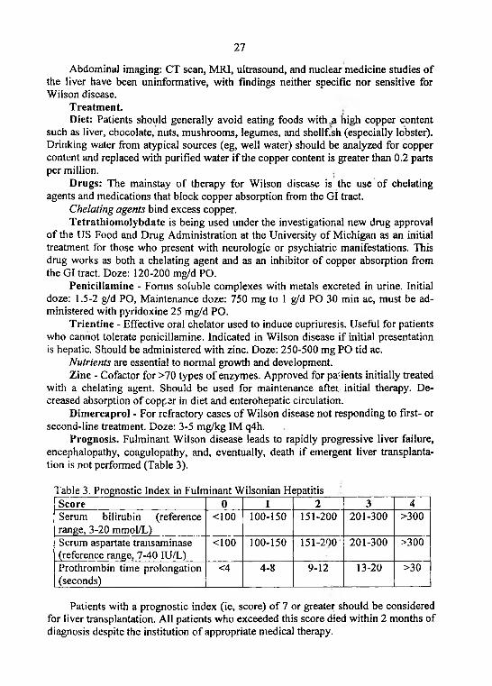
Prognosis. Fulminant Wilson disease leads to rapidly progressive liver failure, encephalopathy, coagulopathy, and, eventually, death if emergent liver transplantation is not performed (Table 3).
Table 3. Prognostic Index in Fulminant Wilsonian HepatitisScore 0 1 2 3 4Serum bilirubin (reference range, 3-20 mmol/L)
<100 100-150 151-200 201-300 >300
Serum aspartate transaminase (reference range, 7-40 IU/L)
<100 100-150 151-200 201-300 >300
Prothrombin time prolongation (seconds)
<4 4-8 9-12 13-20 >30
Patients with a prognostic index (ie, score) o f 7 or greater should be considered for liver transplantation. All patients who exceeded this score died within 2 months of diagnosis despite the institution o f appropriate medical therapy.
28
Prognosis after liver transplantation is relatively good. In a study involving 55 patients with Wilson disease who underwent hepatic transplantation, the 1-year survival rate was 79% and the overall survival rate was 72% at 3 months to 20 years.
CHOLELITHIASIS
Introduction. Gallstones are concretions that form in the biliary tract, usually in the gallbladder. Their development is insidious, and they may remain asymptomatic for decades. Migration of gallstones may lead to occlusion o f the biliary and pancreatic ducts, causing pain (biliary colic) and producing acute complications, such as acute cholecystitis, ascending cholangitis, or acute pancreatitis. Chronic gallstone disease (cholecystolithiasis) may lead to fibrosis and loss of functio n o f the gallbladder and predisposes to gallbladder cancer. Excision o f the gallbladder (cholecystectomy) to cure gallstone disease is among the most frequently performed abdominal surgical procedures.
Synonyms: gallstone disease, choledocholithiasis, cholecystolithiasis.Definition. Cholelithiasis is disease which arises at formation o f gallstones in
the biliary tract and is shown by symptoms of biliary colic at gallsranes’s migration and occlusion of the biliary and pancreatic ducts with producing acute complications, such as acute cholecystitis, ascending cholangitis, or acute pancreatitis.
Pathophysiology. Formation of gallstones arises in that case if substances in bile are present in concentrations that approach the limits o f solubility. When bile is concentrated in the gallbladder, it can become supersaturated with these substances, which then precipitate from solution as microscopic crystals. The crystals are trapped in gallbladder mucus, producing gallbladder sludge. Over time, the crystals grow, aggregate, and fuse to form macroscopic stones. Occlusion o f the ducts by sludge and stones produces the biliary colic and complications of gallstone disease. Cholesterol and calcium bilirubinate, as two main substances, are involved in gallstone formation.
Cholesterol gallstones. More than 80% of gallstones contain cholesterol as their major component. If the bile is supersaturated with cholesterol crystals may form. Thus, the main factors that determine whether cholesterol gallstones will form are as follows: the amount o f cholesterol secreted by liver cells, relative to lecithin and bile salts, and the extent o f concentration and stasis o f bile in the gallbladder.
Pigm ent gallstones. Black pigment stones represent 10-20% o f gallstones. In situations o f high heme turnover, such as chronic hemolysis or cirrhosis, unconjugated bilirubin may be present in bile at higher than normal concentrations. Calcium bilirubinate may then crystallize from solution and eventually form stones. Over time, various oxidations cause the bilirubin precipitates to take on a je t black color, and stones formed in this manner are termed black pigment stones.
Brown pigment stones are fairly common in some parts of Southeast Asia. Bile normally is sterile, but, in some unusual circumstances it may become colonized with bacteria. The bacteria hydrolyze conjugated bilirubin, and the resulting increase in unconjugated bilirubin may lead to precipitation o f calcium bilirubinate crystals. Bacterial hydrolysis o f lecithin leads to the release o f fatty acids, w lich complex with
29
calcium and precipitate from solution. The resulting concretions have a claylike consistency and are termed brown pigment stones. Unlike cholesterol or black pigment stones, which form almost exclusively in the gallbladder, brown pigment stones often form de novo in the bile ducts.
Mixed gallstones. Cholesterol gallstones may become colonized with bacteria and can elicit gallbladder mucosal inflammation. Lytic enzymes from bacteria and leukocytes hydrolyze bilirubin conjugates and fatty acids. As a result, over time, cholesterol stones may accumulate a substantial proportion of calcium bilirubinate and other calcium salts, producing mixed gallstones. Large stones may develop a surface rim o f calcium resembling an eggshell that may be visible on plain x-ray films.
Epidemiolody.Frequency: The prevalence o f cholelithiasis in Western countries and United
States in women increases by about 1% per year; in men - about 0.5% per year, but it appears to be somewhat lower in Asia and Africa. The lifetime risk o f developing gallstones in Caucasians is 50% for women and 30% for men.
Mortality/Morbidity: Gallstone disease is responsible for about 10,000 deaths per year in the United States. About 7000 deaths are attributable to acute gallstone complications, such as acute pancreatitis.
Race: Caucasians, Mexicans, and Native Americans have a relatively high prevalence o f gallstones. Gallstone disease is less common in Asians and Africans.
Sex: Women are more likely to develop cholesterol gallstones than men, especially during their reproductive years (the excess risk is 2-3:1). The estrogen increases biliary cholesterol secretion. Pigment gallstones affect men and women equally.
Age: Gallstones continue to form throughout adult life, and the prevalence is greatest at advanced age.
Classification. Gallbladder disease has 4 stages:1. The lithogenic state, in which conditions favor gallstone formation;2. Asymptomatic gallstones (the carrier o f gallstones);3. Episodes o f biliary colic (actually gallbladder disease);4. Complicated cholelithiasis.Clinical. Gallstones may be present in the gallbladder for decades without caus
ing symptoms or complications. In patients with asymptomatic gallstones discovered incidentally, the likelihood of developing symptoms or complications is 1-2% per year. Pain termed biliary colic occurs when gallstones fortuitously impact in the cystic duct during a gallbladder contraction, increasing gallbladder wall tension. In most cases, the stone dislodges, the obstruction is relieved after 30-90 minutes following relaxation o f the gallbladder, and the pain resolves. Episodes o f biliary colic are sporadic and unpredictable. The patient localizes the pain to the epigastrium or right upper quadrant and may describe radiation to the right scapular tip. The pain is constant and is not relieved by emesis, antacids, defecation, or positional changes. It may be accompanied by nausea and vomiting. Complications o f gallbladder stones are acute cholecystitis with perforation and pericholecystic abscess, chronic cholecystitis, gallbladder adenocarcinoma, cholecystoenteric fistula, gallstone ileus.
Physical. Patients with the lithogenic state or asymptomatic gallstones have no
30
abnormal findings on physical examination. Patients with biliary colic and especially in acute cholecystitis may experience tenderness to palpation over the gallbladder and when the patient inhale while the examiner maintains steady pressure below the right costal margin (Murphy sign). Localized rebound tenderness, guarding, or rigidity may occur with pericholecystic inflammation. Choledocholithiasis with obstruction o f the common bile duct produces cutaneous and scleral icterus that may evolves over hours to days. Patients with ascending cholangitis have severe right upper quadrant tenderness with jaundice and fever (Charcot triad). Acute gallstone pancreatitis is often characterized by epigastric tenderness. In severe cases, retroperitoneal hemorrhage may produce ecchymoses o f the flanks and periumbilical ecchymoses (Cullen sign and Grey-Turner sign).
Risk factors o f cholesterol gallstones (F pentade): Fat (obesity), Female (women), Fertile (multiple pregnancies), Fair (blond hair), Forty (after 40 years). Other risk factors o f cholesterol gallstones are gallbladder stasis (high spinal cord injuries, prolonged fasting with total parenteral nutrition, rapid weight loss associated with severe caloric and fat restriction such as diet and gastric bypass surgery), drugs (estrogens, clofibrate and other fibrate hypolipidemic drugs, somatostatin analogs), heredity (about 25% o f the predisposition to cholesterol gallstones appears to be he- reditary).
Black pigment gallstones occur in individuals with high heme turnover (sickle cell anemia, hereditary spherocytosis, and beta thalassemia, about half o f all cirrhotic patients have pigment gallstones). In most cases, no risk factor can be identified.
Risk factors o f brown pigment gallstones include colonization of bile with bacteria and intraductal stasis (postsurgical biliary strictures or choledochal cysts).
L ab Studies. Patients with uncomplicated cholelithiasis or simple biliary colic typically have normal laboratory test results. Acute cholecystitis is associated with polymorphonuclear leukocytosis, mild elevations of liver enzymes (in severe cases). Choledocholithiasis with acute common bile duct obstruction initially produces an acute increase in the level o f liver transaminases (alanine and aspartate aminotransferases), followed within hours by a rising serum bilirubin level.
Im aging Studies. Upright and supine abdominal radiographs are helpful in establishing a diagnosis o f gallstone disease. Black pigment or mixed gallstones may contain sufficient calcium to appear radiopaque on plain x-ray films. Calcification in the gallbladder wall (the so called porcelain gallbladder) is indicative o f severe chronic cholecystitis. Ultrasound is the most sensitive, specific, noninvasive, and inexpensive test for the detection o f all types o f gallstones. Gallstones appear as echo- genic foci in the gallbladder. They move freely with positional changes and cast an acoustic shadow. In acute cholecystitis, ultrasound may demonstrate edema o f the gallbladder wall and pericholecystic fluid. Computed tomography (CT) is less sensitive than ultrasound for the detection o f gallbladder stones. Magnetic resonance imaging (MRI) with magnetic resonance cholangiopancreatography (MRCP) has emerged as an excellent imaging study for noninvasive identification o f gallstones anywhere in the biliary tract, including the common bile duct. Endoscopic retrograde cholangiopancreatography (ERCP) permits x-ray imaging of the bile ducts. Today, ERCP is usually performed in conjunction with endoscopic retrograde sphinctero-
31
tomy and gallstone extraction. Endoscopic ultrasound (EUS) is also an accurate and relatively noninvasive technique to identify stones in the distal common bile duct.
Treatm ent.Diet: Little evidence suggests that dietary composition affects the natural history
o f gallstone disease in humans. Obese patients who undertake aggressive weight loss programs or undergo bariatric surgery are at risk to develop gallstones; short-term prophylaxis with ursodeoxycholic acid should be considered.
T herapy of non-radiopaque (radiolucent) stones (cholesterol gallstones):1. Oral litholysis with ursodeoxycholic acid. Indications: stones diameter < 1 .5
cm, functioning gallbladder without gallstone complications and common bile duct stone. In humans, long-term administration o f ursodeoxycholic acid reduces cholesterol saturation o f bile, both by reducing liver cholesterol secretion and by reducing the detergent effect o f bile salts in the gallbladder (thereby preserving vesicles that have a high cholesterol carrying capacity). Desaturation o f bile prevents crystals from forming and, in fact, may allow gradual extraction o f cholesterol from existing stones.
Ursodeoxycholic acid is a natural bile salt o f bears. It is a weak detergent. Doze: 10 mg/kg/daily PO for 6-24 months (500 mg or 2 capsules daily at weight o f a body up to 60 kg; 750 mg or 3 capsules daily at weight o f a body up to 80 kg; 1000 mg or 4 capsules daily at weight o f a body up to 100 kg; 1250 mg or 5 capsules daily at weight o f a body over 100 kg;).
2. Extracorporeal shock-wave lithotripsy (ESWL) with adjuvant oral litholysis. Indications: 1 -3 stones (total diameter < 3cm), stones diameter < 1 cm per stone (up to 3 stones) or 1 stone up to 2 cm, functioning gallbladder without gallstone complications and common bile duct stone. ESWL with oral litholysis are associated with an 80% stone clearance rate. About 20% o f patients experience stone recurrence.
3. Contact litholysis or direct dissolution o f gallbladder stones using methyl-tert- butyl ether (MTBE). Indications: solitary or multiple stones without dependence on stone size or number, functioning gallbladder without gallstone complications and common bile duct stone. About 80-90% o f patients treated with contact litholysis experience stone clearance, with a recurrence rate o f 30-50%. Contact litholysis is an invasive method that should only be considered in exceptional cases.
T herapy of radiopaque stones (pigment and calcified gallstones):1. The traditional (open) cholecystectomy. Indications: gallstone complications,
common bile duct stone, suspicion o f bile duct carcinoma. Removal o f the gallbladder (cholecystectomy) is the treatment o f choice for symptomatic cholelithiasis. At the time o f cholecystectomy, the surgeon can explore the common bile duct and remove common bile duct stones. Following cholecystectomy, a few individuals experience recurrent pain resembling biliary colic. The term postcholecystectomy syndrom e is sometimes used for this condition. Many patients with postcholecystectomy syndrome have long-term functional pain that was originally misdiagnosed as being o f biliary origin. Persistence of symptoms following cholecystectomy is unsurprising. Some individuals with postcholecystectomy syndrome have an underlying motility disorder o f the sphincter o f Oddi, termed biliary dyskinesia, in which the sphincter fails to relax normally following ingestion o f a meal. The diagnosis can be estab
32
lished in specialized centers by endoscopic biliary manometry. In established cases of biliary dyskinesia, endoscopic retrograde sphincterotomy usually is effective in relieving the symptoms.
2. Laparoscopic cholecystectomy. Indications: biliary colics secondary to cholelithiasis, patient request.
Because the natural history of gallstones is generally benign, cholecystectomy is not required for patients with asymptomatic gallstones. Cholecystectomy for asymptomatic gallstones may be indicated under certain circumstances:,patients with large gallstones greater than 2 cm in diameter, with nonfunctional or calcified (porcelain) gallbladder and who are at high risk of gallbladder carcinoma, with spinal cord injuries or sensory neuropathies affecting the abdomen, proposed recipients o f organ transplants (other than the liver), with sickle cell anemia.
Prognosis of cholelithiasis depends on the presence and severity o f complications. O f all patients who refuse surgery or are unfit to undergo surgery, 45% remain asymptomatic from cholelithiasis, while 55% experience varying degrees o f complications.
33
MEDICINE OF ACCIDENTS
ACUTE RADIATION SICKNESS
Introduction. Causes o f radiation sickness may be accidental or intentional. Excessive exposure to ionizing radiation occurs following strategic nuclear explosions and accidents in nuclear power plants. Routine cases o f exposure to ionizing radiation are x-ray devices.
Synonyms: acute and chronic radiation exposure.Definition. Acute radiation sickness - a condition arising with irradiation o f all
body or its part penetrating radiation (X-rays, gamma rays or; neutrons) in dosage above 1 Gray (GY).
Pathophysiology. Ionizing radiation is either penetrating (X-rays, gamma rays or neutrons) or non-penetrating (alpha and beta particles). Penetrating radiation affects the whole body, while non-penetrating radiation affect only the skin. All radiation effects depend on the type o f radiation, the distribution of dose and the dose rate.
K inds of ionizing radiations: corpuscular and electromagnetic.C orpuscular radiations - a stream of nuclear and subnuclear particles, with the
certain weight, a charge and changing speed.The easy charged particles are electrons, positrons.The heavy charged particles are protons, alpha particles.Neutral particles are neutrons.At nuclear explosion a basis o f corpuscular radiation alpha particles make, beta
particles and neutrons.A lpha particles are positively charged nucleus o f helium consisting o f two pro
tons and two neutrons. They are formed at disintegration of radioactive substances (radium, uranium and thorium). Have high ionizing activity (a double positive charge), are well absorbed by substance and, therefore, have small penetrating ability. In air run o f alpha particles are equal 8-10 cm, in water and tissues o f an organism the 100-th shares o f millimeter.
Beta particles are electrons with a negative charge and positrons with a positive charge. In air run o f beta particles makes from tens centimeters up to several meters, in tissues o f an organism - 0.2-0.5 cm.
N eutrons - particles with weight 1.0089 nuclear units and a zero charge. They take o ff from nucleus o f atoms at some nuclear reactions. Neutrons not having a charge, will easily penetrate into atoms, cooperate with a nucleus by elastic and not elastic impacts, radiating capture and nuclear reaction.
Most dangerous o f electromagnetic radiations are X-rays and gam ma rays.E lectrom agnetic radiations are distributed in vacuum with speed o f light, will
consist o f the periodic electric and magnetic fluctuations having different length of a wave. Short-wave radiations have high frequency of fluctuations, energy and penetrating ability. To them concern X-rays and gamma rays. They are let out as balls of fire (quantum, photons) and measured in electron volts.
Sources of X-rays: tubes of x-ray devices, betatrons, linear accelerators.Sources of gamma rays (gamma quantum): nuclear reactions, disintegration o f
34
radioactive substances.X-ray and gamma rays, having small length of a wave and the big energy, will
deeply penetrate into fabrics o f an organism (for water solutions and a human tissue on tens centimeters). X-ray and gamma rays at influence on substance, in part or completely transfer the energy to electron (photo effect), and after impact change direction the movement (effect of quantum dispersion). As a result o f two effects are formed fast-flying elecrtons that results in ionization of molecules o f substance. Rigid gamma rays and ultra rigid X-rays at collision with a nucleus of atom can disappear with formation of pair from electron and a positron.
Excessive exposure to ionizing radiation occurs following accidents in hospitals, industry, nuclear power plants and strategic nuclear explosions.
Action of ionizing radiation:1. Direct action of ionizing radiation on radiosensitive organic substances o f
cells and tissue o f an organism with development o f radiochemical reactions. According to the theory o f "target" damage and destruction o f the irradiated cell comes only after damage o f a radiosensitive part or cellular "target". Basic cellular "targets": nu- cleoproteins and key cellular enzymes.
2. Indirect action consist that ionizing radiation results to destroy o f water, and to formation o f various super active free radicals which change structure of organic molecules and result in damage or destruction of a cell.
Radiation dosage is measured in joules per kilogram (J kg'1); 1 J kg'1 is also known as one gray (lGy). This is equivalent to 100 rads or 250 roentgens. Radiation differs in the density o f ionization it causes. Therefore a doze-equivalent called a sieved (Sv) is used.
Epidemiolody. Sources o f ionizing radiation are natural and artificial.Natural sources: 1. External irradiation: (space irradiation - 300 microSievert
(Sv) per year on a sea level; earth radiation - 300-600 microSievert per year - radioactive К40, PB87 on the planet Earth). 2. Internal irradiation: radioactive gas radon - a product o f disintegration U238, T232.
Artificial sources: 1. Used by medicine - 20% from a natural radioactive irradiation. 2. Nuclear explosions - 0,8 % from a natural background (now). 3. Atomic engineering - work o f the atomic power stations without accidents: 0,04 - 0,05% from a natural background (now). 4. Professional irradiation: workers o f the nuclear industry, medical staff (X-ray cabinets), medical staff o f resorts with radonie baths, pilots, miners. 5. Household sources: TVs, clock with luminous dial.
Classification of acute radiation sickness:I. The bone m arrow form (a dosage o f an irradiation 1-10 Gy; Table 4):On a degree: I. Mild (1-2 Gy); II. Moderate (2-4 Gy); III. Severe (4-6 Gy); IV.
Very severe (6-10 Gy)On the periods: 1. Initial (the period o f the common initial reaction); 2. Latent;
3. Clinical; 4. Convalescence.II. The intestinal form (10-20 Gy)Ш . The toxic form (20-80 Gy)IV. The cerebral (brain) form (80-100 Gy)
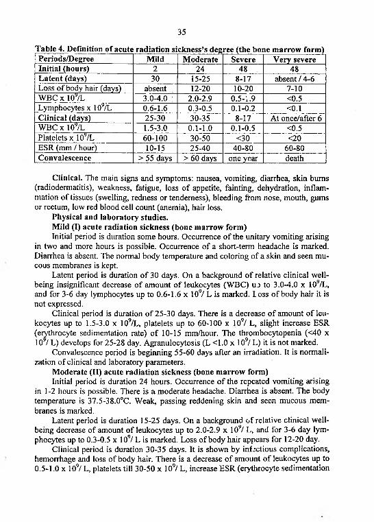
35
Table 4. Definition of acute radiation sickness’s degree (the bone marrow form)Periods/Degree Mild Moderate Severe Very severeInitial (hours) 2 24 48 48Latent (days) 30 15-25 8-17 absent / 4-6Loss of body hair (days) absent 12-20 10-20 7-10WBC x 109/L 3.0-4.0 ; 2.0-2.9 0.5-1.9 <0.5Lymphocytes x 1OTL 0.6-1.6 0.3-0.5 0.1-0.2 <0.1Clinical (days) 25-30 30-35 8-17 At once/after 6WBC X 107l 1.5-3.0 0 . 1- 1.0 0.1-0.5 <0.5Platelets x 109/L 60-100 30-50 <30 <20ESR (mm / hour) 10-15 25-40 40-80 60-80Convalescence > 5 5 days > 60 days one year death
Clinical. The main signs and symptoms: nausea, vomiting, diarrhea, skin bums (radiodermatitis), weakness, fatigue, loss o f appetite, fainting, dehydration, inflammation o f tissues (swelling, redness or tenderness), bleeding from nose, mouth, gums or rectum, low red blood cell count (anemia), hair loss.
Physical and laboratory studies.Mild (I) acute radiation sickness (bone marrow form)Initial period is duration some hours. Occurrence o f the unitary vomiting arising
in two and more hours is possible. Occurrence o f a short-term headache is marked. Diarrhea is absent. The normal body temperature and coloring o f a skin and seen mucous membranes is kept.
Latent period is duration o f 30 days. On a background o f relative clinical wellbeing insignificant decrease o f amount o f leukocytes (WBC) uo to 3.0-4.0 x 10⁹/L, and for 3-6 day lymphocytes up to 0.6-1.6 x 10⁹/ L is marked. l oss o f body hair it is not expressed.
Clinical period is duration o f 25-30 days. There is a decrease o f amount o f leukocytes up to 1.5-3.0 x 10⁹/L, platelets up to 60-100 x 109/ L, slight increase ESR (erythrocyte sedimentation rate) o f 10-15 mm/hour. The thrombocytopenia (<40 x 107 L) develops for 25-28 day. Agranulocytosis (L <1.0 x 10⁹/L ) it is not marked.
Convalescence period is beginning 55-60 days after an irradiation. It is normalization of clinical and laboratory parameters.
Moderate (II) acute radiation sickness (bone marrow form)Initial period is duration 24 hours. Occurrence o f the repeated vomiting arising
in 1-2 hours is possible. There is a moderate headache. Diarrhea is absent. The body temperature is 37.5-38.0°C. Weak, passing reddening skin and seen mucous membranes is marked.
Latent period is duration 15-25 days. On a background o f relative clinical wellbeing decrease of amount o f leukocytes up to 2.0-2.9 x 10⁹/ L, and for 3-6 day lymphocytes up to 0.3-0.5 x 10⁹/ L is marked. Loss o f body hair appears for 12-20 day.
Clinical period is duration 30-35 days. It is shown by infectious complications, hemorrhage and loss o f body hair. There is a decrease o f amount o f leukocytes up to 0.5-1.0 x 10⁹/ L, platelets till 30-50 x IQ9/ L, increase ESR (erythrocyte sedimentation
36
rate) up to 25-40 mm / hour. The thrombocytopenia (<40 x 109/ L) develops for 17- 24 day, agranulocytosis (L <1.0 x 10⁹/ L) - for 20-30 day.
Convalescence period is beginning 60 days after an irradiation. It is normalization o f clinical and laboratory parameters.
Severe (III) acute radiation sickness (bone m arrow form)Initial period is duration 2 days. It is characterized by the repeated vomiting aris
ing in 0.5-1 hour, the expressed headache. Occurrence diarrhea is possible. The body temperature is 37.5-38.0°C. Moderate reddening skin and seen mucous membranes is marked.
Latent period is duration 8-17 days. On a background o f relative improvement o f the common state o f health decrease of amount o f leukocytes up to 0.5-1.9 x 10⁹/L, and for 3-6 day lymphocytes up to 0.1-0.2 x 10⁹/L is marked. Los;' o f body hair appears for 10-20 day.
Clinical period come after 8-17 days. It is shown by the general intoxication, fever and intestinal syndrome and hypotonic. There is a sharp decrease o f amount o f leukocytes up to 0.1-0.5 x 10⁹/L, platelets up to 30 x 109/L, increase ESR (erythrocyte sedimentation rate) up to 40-80 mm/hour. The thrombocytopenia (<40 x 109/L) develops for 10-16 day, agranulocytosis (L <1.0 x 10⁹/L) - for 8-20 day.
Convalescence period lasts one year. Normalization o f clinical and laboratory parameters occurs for a long time. At part o f the patients clinical recovery does not occur, and the complications incompatible with life are marked. The development tumors, leukocytopenia and thrombocytopenia are marked too.
Very severe (III) acute radiation sickness (bone m arrow form)Initial period is duration 2 days. It is characterized by the unrestrained vomiting
arising in 5-20 seconds, the strong headache, and the confused consciousness. Frequently arises o f diarrhea. The body temperature is more 38°C. Expressed reddening skin and seen mucous membranes is marked.
Latent period is absent or comes within the first 6 days. Sharp decrease of amount o f leucocytes up to 0 .5xl09/L, lymphocytes up to 0.1xl09/L is marked for 3-6 day. Loss of body hair appears for 7-10 day.
Clinical period comes at once or after 6 day. It is shown by the expressed general intoxication, fever, and intestinal syndrome, and hypotonic. There is a sharp decrease of amount o f leucocytes below 0.5xl09/L or leucopenia has not time to develop, platelets up to 20x109/L or the thrombocytopenia has not time to develop, increase ESR up to 60-80 mm/hour. The thrombocytopenia (<40 x 109/л) develops by 10 day, agranulocytosis (L <1.0 x 10⁹/L) - for 6-8 day.
At absence of medical aid all patients perish. With medical aid at 50% o f patients clinical recovery does not occur, and complications incompatible with life are marked. Further at other 50% o f persons during many years the residual phenomena are kept and there are remote somatic and genetic consequences.
The intestinal form of acute radiation sickness arises at dosage o f an irradiation 10-20 Gy. It is shown destroy of blood tissue and intestines! with development radiating enterocolitis. Symptoms expressed токсемии (a fever, pains in muscles and joints, progressing general weakness, headaches) are marked. The death comes from a stop of heart activity on 2-3 week o f disease.
37
The toxic form of acute radiation sickness arises at dosage o f an irradiation 20-80 Gy. It is shown encephalopathy with vascular reaction and a hypostasis o f a brain. It is accompanied sharp cardiovascular insufficiency. At the phenomena of coma struck perish for 4-8 day.
The cerebral (brain) form of acute radiation sickness arises at dosage of an irradiation 80-100 Gy. It is shown by defeat o f cells o f a brain and brain vessels with development o f heavy neurologic frustration. It is accompanied by occurrence of vomiting with time loss o f consciousness in 20-30 minutes after an irradiation with the subsequent psychomotor excitation, clonic and tonic spasms, frustration of breath. The death comes from a paralysis o f breath at the first o'clock or the first 2-3 day.
T reatm ent o f acute radiation sickness.Preventive maintenance o f vomiting: motilium 10-20 mg PO 3 times a day. At
persistent vomiting appoint IV 30-50 ml o f 10% o f a solution o f chloride o f sodium. During o f 5-6 hours the patients should not accept a liquid.
At loss o f water enter a physiological solution from 500 ml and is higher, depending on a degree of loss o f water. Instead o f a physiological solution probably introduction o f a salt solution o f other structure (1000 ml of distilled water, 5.0 g chloride o f sodium, 4.0 g a hydrocarbonate o f sodium, 1.0 г chloride Kali).
Medical tactics during other periods o f acute radiation sickness is defined with a degree o f oppression blood tissue system. A thrombocytopenia, leukopenia, agranulocytosis and decrease o f immunity result in development hemorrhage syndrome, anemia, infectious complications, with hit in blood o f toxic products and microbes. Accumulation in blood o f peroxides and abnormal peptides, as a result o f radiating influence, also result in intoxication.
Treatment o f hemorrhage syndrome: thrombocytes concentrate (2.5-4.5xl09 cells) 2-4 times a week; antihaemophilic plasma (0.25 L) in a day; trasilol (300 000 - 500 000 U).
Increase o f immunity: antibodies o f the directed action (antistaphylococcal, anti- coli) or immune plasma.
Treatment o f leuko(neutro)penia (0.5 x 10⁹/L) and infectious complications with septic current: leukocytes concentrate (up to 20 x 109 cells), compatible on systems ABO, a Rhesus factor, at long application, on other systems of groups of blood and HLA.
Treatment o f anemia: erythrocytes concentrate, erythrocytes weight or washed o f erythrocytes (1-3 times a week).
Desintoxication therapy: a physiological solution (200-400 ml in a day).Treatment o f an intestinal syndrome: see treatment o f loss the water.Treatment o f gastrointestinal tract inflammation: decontamination o f intestines
with use o f not soaked up antibiotics (gentamycin, lincomycin) iri a combination with co-trimoxazole and metronidazole.
Dietetic therapy: mixes o f amino acids (up to 1 L in day), dispersion fatty, 5%- 20 % solutions o f glucose, and vitamins o f group С, P, B.
38
CHRONIC RADIATION SICKNESS
Definition. Chronic radiation sickness - a condition arising at long (months and years), regular, the general irradiation in small dozes exceeding maximum permissible dozes (1.2-1.8 Gy within 2-3 years).
Classification. On a degree: I. Mild; II. Moderate; III. Severe.-Clinical, physical and laboratory studies.M ild (I) chronic radiation sicknessComplaints to a headache, the general weakness, fast fatigue, infringement of
dream (drowsiness in the afternoon and a sleeplessness at night), bad appetite, locks, dyspepsia which frequently is not connected to reception o f the food, unpleasant sensations in the field of heart.
Leukopenia (3-4 x 10⁹/L), decrease platelets up to the bottom border o f norm. In a bone brain it is marked braking o f maturing myeloid cells. On the part o f internal bodies the passing hypotonic; infringement o f function of a stomach and endocrinal glands is diagnosed.
M oderate (II) chronic radiation sicknessComplaints to a headache which is hardly stopped by usual means, a constant,
the expressed general weakness, fast fatigue, sharp infringement o f dream, absence of appetite, loss o f weight, amplification o f pains in the field o f heart and a stomach, the increased fragility o f capillaries. In some cases there is an infringement thermoregulation.
Anemia (2.0-3.5xl012/L), leucopenia (1.5-2.0xl07L) with lymphopenia, decrease platelets up to 100xl09/L is marked. In bone marrow oppression of all blood lines o f cells is marked. On the part o f internal bodies the hypotonic, trophic frustration, a dystrophy of myocardium, oppression o f function o f a stomach, an intestines and endocrinal glands is diagnosed. Infectious complications are possible.
Severe (III) chronic radiation sicknessAnemia (<2.0xI012/L), leucopenia (<1.0xl09/L), agranulocytosis, decrease
platelets till 20-50xl09/L are observed. There are very low level o f myelocariocytes (<1 ,0x109/L) in a bone marrow. On the part o f internal bodies the hypotonic, the deep trophic frustration, hemorrhage syndrome, plural infectious complications with possible development of a sepsis is diagnosed.
The forecast o f this stage of disease is very serious.T reatm ent o f mild and m oderate chronic radiation sicknessPreparations of calcium, phosphorus, tincture of a ginseng.The drugs normalizing nervous system: preparations o f bromine, tincture o f a
valeriana, various tranquilizers.The drugs stimulating blood system: vitamins Bg, B n, a folic acid, anabolic
hormones (retabolil).T reatm ent of severe chronic radiation sickness is as acute radiation sickness.Prognosis. The remote consequences o f action o f ionizing radiation are the in
crease risk of cancer and leukemia, genetic defects, decrease o f life expectancies.
39
DISEASES O F INTERNAL BODIES ASSOCIATED W ITH BULLET W OUNDS AND EXPLOSIONS
Introduction. There are general and local changes in reply to a trauma in an organism. The main role is given allergic, nervous - reflex and infection mechanisms, hypoxia, loss blood. Under o f these influences the critically wounded patient transforms to the patient.
Definitions.T raum atic shock is nervous - reflex reaction o f an organism associate with a
trauma. Traumatic shock is stimulating excitation, and then braking nervous system with oppression o f the vital functions (hypoventilation, hypercapnia, acidosis, hypo- proteinemia, hyperglycemia).
P uru len t fever is arising at inflammation development in a wound associated with products o f tissue disintegration, microbes and toxins. It is reaction o f an organism to purulent processes in a wound.
Toxic fever is arising only with wound toxins infiltration in the organism (a version purulent fever).
Clinical: a persistent fever, inflammatory-dystrophic changes o f internal bodies.T reatm en t - surgical (sanitation o f a wound).Definition.W ound sepsis - the general infectious disease caused by the local center puru
lent wound infection, but lost with it connection (difference from purulent fever).Clinical: high fever and leucocytes, sweat, anemia, pneumonia, inflammation o f
kidneys and heart.Definition.T raum atic exhaustion - the heavy dystrophic process arising at a long suppura
tion at wounded patient (purulent fever and wound sepsis).Clinical: loss o f weight, an atrophy o f all bodies and systems, amyloidosis, hy
postases, anemia.Conditions of development: long purulent necrosis process in an organism.
Pathology of internal bodies a t patients with bullet wounds
Pulm onary pathology a t bullet’s w ound: 1) pulmonitis; 2) pleurisy (plevritis); 3) pneumonia; 4) pulmonary hemorrhages; 5) purulent pulmonary diseases.
Pulm onitis, plevritis and pulm onary hem orrhages.Clinical: pains in breasts, cough, hemoptysis, fever.X-ray picture: pulmonary blackouts.Complications o f pulmonary hemorrhage: pneumonia, atelectasis, pulmonary
abscess or gangrene.Pneum onia and puru len t pulm onary diseases.Reflux pneumonia develops at wounds o f maxillofacial area (microbes activa
tion, distribution o f an infection on bronchial tubes), at wounded patients in a unconsciousness.
40
Hypostatic pneumonia develops at long motionless position o f the patient (decrease of pulmonary protective properties, insufficient pulmonary ventilation).
Atelectasis pneumonia develops similarly reflux pneumonia with obstruction of bronchial tubes and formation atelectasis. Small atelectasis clinically is not distinguished. Complications large atelectasis: acute pulmonary insufficiency with development o f large pneumonia.
The Toxic-septic pneumonia develops at blood distribution of an infection. One of early displays wound sepsis. Clinical: the small and plural centers inclined to merge and development o f pulmonary abscess.
C ardiovascular pathology associated with bullet wounds.Allocate four forms of the closed trauma of heart: concussion, a bruise o f heart,
traumatic break o f heart, posttraumatic myocardial infarction.Concussion is characterized by fast development short and not heavy clinic and
electrocardiographs changes. Clinical: heart pain, a tachycardia, аритмия, infringement heart conductivity and repolarization. Duration o f clinical displays is some minutes or hours.
The bruise of heart is characterized by slow, gradual development of clinical (heart pain, angina, cardiac arrhythmias, expansion of borders o f heart, a short murmur, soft heart sounds, rare development of acute or coronary insufficiency).
T raum atic b reak of heart is the heaviest form of the closed trauma with high mortality.
Posttraum atic m yocardial infarction.Clinical: there is pain form (70%), not Q variants of damage, mainly a forward
wall left ventricular.Digestion pathology at bullet’s wound: peptic ulcer, hepatitis, dyspepsia.Kidneys pathology a t bullet’s wound: nephritis, pyelonephritis with transition
in urosepsis and amyloidosis.Blood system pathology at bullet’s wound: acute post hemorrhage anemia.
Pathology of internal bodies a t patients with explosive defeats
Bruise and concussion of heart (see cardiovascular pathology associated with bullet’s wound).
Bruise of lungs: a chest pain, cough with allocation o f foamy blood, sputum with blood.
Bruise of a liver: pains in the field of a liver, pains in a stomach, a nausea, vomiting, symptoms of an internal bleeding, morbidity of a stomach at palpation, peritoneal symptoms, rise o f AST and ALT levels.
Bruise of a stom ach and intestines develops as an acute stomach and gastro enteric frustration.
Diagnostics o f bruises o f internal bodies is extremely complicated at life and averages 29.7 %.
41
RHEUMATOLOGY
SYSTEMIC LUPUS ERYTHEMATOSUS (SLE)
Introduction. Systemic lupus erythematosus (SLE) is a chronic, multisystem, inflammatory disease that can affect every organ system of the body. SLE is protean in its manifestations with cerebral and renal disease as the most serious problems.
Synonyms: SLE, lupus, multisystem inflammatory disease.Definition. Systemic lupus erythematosus is a chronic disorder characterized by
inflammation in several organ systems and the production of autoantibodies that participate in immunologically-mediated tissue injury, peripheral polyarthritis with symmetric involvement o f small and large joints w ithout joint erosion.
Pathophysiology. There are four possible etiological factors: altered immunity, heredity, infection, and drugs. Altered immunity mechanism is the development of autoantibodies associated with a defect in apoptosis that causes increased cell death and a disturbance in immune tolerance with altered regulating mechanism. Autoanti- bodyes causing tissue damage by cytotoxic effects or Ag-Ab complexes. In active SLE, this process has been confirmed based on the presence of complexes o f nuclear antigens such as DNA, immunoglobulins, and complement proteins at these sites. Antinuclear antibodies (ANAs) are present in the serum in virtually all patients with active SLE, and antibodies to native double-stranded DNA (dsDNA) are relatively specific for the diagnosis o f SLE. Heredity. Approximately 10% patients with SLE have positive family history (increased frequency of HLA B8, DR3 in Caucasians and HLA DR2 in Japanese lupus patients). Role of estrogen is: established too. Prepubertal and postmenopausal women have similar incidence to men. Men who develop lupus have a higher concentration o f estrogenic metabolites. Infection. Viruses are nonspecific stimulant o f immune response. Drugs that increase the risk of SLE development are: anticonvulsants (dilantin, phenobarbital), methyldopa, antihypertensives (hydralazine), antiarrhythmics (procainamide). Anti-histone antibodies are commonly seen in drug-induced lupus.
Epidemiolody.Frequency: Worldwide, the prevalence o f SLE is variable, from 12 cases per
100,000 population in Britain to 39 cases per 100,000 population in Sweden.Mortality/Morbidity: The natural history o f SLE varies from relatively benign
disease to rapidly progressive and even fatal disease. Patients with isolated skin and musculoskeletal involvement have higher survival rates than those with renal and central nervous system (CNS) disease. Despite improvements in overall survival rates, patients with SLE still have a death rate that is 3 times that o f the general population. There is bimodal mortality pattern: early (within 2 years: active SLE, active nephritis, infection secondary to steroid use) and late (> 10 years: inactive SLE, inactive nephritis, atherosclerosis possibly secondary to long-term steroid use).
Race: Worldwide, different races appear to have varying rates o f disease. However, because of different prevalence rates among people of the .ame race in different geographical locations, a clear conclusion cannot yet be drawn.
42
Sex: SLE occurs predominantly in women o f childbearing age, suggesting a role for hormonal factors in the pathogenesis o f the disease. A hormonal influence hypothesis is supported by the 6- to 10- fold higher incidence of SLE in women of reproductive age and a higher prevalence o f SLE in men with Klinefelter disease. Additionally, pregnancy and administration o f exogenous estrogen often precipitate exacerbations o f SLE. Men at all ages have a risk o f disease similar to that o f women who are prepubertal or postmenopausal.
Age: A correlation between age and incidence o f SLE mirrors peak years o f female sex hormone production. Disease incidence is highest among women aged 14- 64 years. Males do not have an age-related peak in incidence.
Classification. There are three variants o f lupus: acute, subacute, and chronic.Clinical-immunological variants.Neonatal lupus erythematosus (NLE) (rare):- Due to transfer o f maternal anti-Ro and/or anti-La antibodies through placenta;- Shortly after birth, infants develop typical discoid lesions with exposure to UV
light;- Very rare to develop SLE later in life;- Anti-Ro positive mothers with SLE -1-5% risk o f developing NLE;- Transient complications: fetal thrombocytopenia, rash, and rarely congenital
heart block;- Neonates may require pacemaker.Late-onset SLE:- Presents at age > 50;- Higher incidence of interstitial lung disease;- Less neuropsychiatric and renal involvement.Subacute cutaneous SLE:- photosensitive rash;- Ro positive, ANA negative.Clinical and physical. Nonspecific fatigue, fever, arthralgia and weight loss are
the most frequent symptoms. Arthralgia, myalgia, and arthritis represent the most common presenting joints symptoms in SLE. Small joints o f the .rands, wrists, and knees without joint erosion are involved most frequently. Malar rash (erythematous rash over the cheeks and nasal bridge), photosensitive rash (any unusual rash or symptom exacerbation after sun exposure) and discoid lesions (plaquelike lesions with follicular plugging and scarring) are the most common presenting skin symptoms in SLE. Pleurisy with pleuritic chest pain with or without pleural effusions is the most common pulmonary manifestation of SLE. Pericarditis that manifests as chest pain is the most common cardiac manifestation o f SLE. The kidney is the most commonly involved visceral organ in SLE. History of multiple cytopenias such as leuco- penia, lymphopenia, anemia, or thrombocytopenia may suggest SLE, among other etiologies.
Other associated features:- Skin manifestations: urticaria, livedo reticularis, bullae, panmculitis, alopecia;- Vasculitic lesions: periungual telangiectasia, Raynaud’s;- Eye manifestations: conjunctivitis, episcleritis, keratoconjunctivitis;
43
- Neuropsychiatric: personality disorders, depression, psychoses.Lab Studies.Diagnostic criteria. Many patients have constitutional symptoms (fatigue,
weight loss, fever) at the time of presentation. Person is diagnosed with SLE if any 4 or more o f the 11 criteria are present serially or simultaneously. The presence o f 4 o f the 11 criteria holds a sensitivity o f 85% and a specificity o f 95%.
“4,7,11” rule - 4 out o f 11 criteria (4 lab, 7 clinical) for diagnosis SLE.Clinical criteria:1. Malar rash (classic “butterfly rash”; no scarring involved sincebasement
membrane intact).2. Discoid rash (may cause scarring).3. Photosensitivity.4. Oral/nasal ulcers (usually painless).5. Arthritis (non-erosive, symmetric; involving 2 or more small or large periph
eral joints).6. Serositis (pleurisy, pericarditis, peritonitis).7. Neurologic disorders (headache, seizures, psychosis, neuropathy).L aboratory criteria:8. Renal disorders (proteinuria > 0.5 g/day, cellular casts (RBC, Hb, granular,
tubular or mixed).9. Blood disorders (hemolytic anemia, leukopenia, lymphopenia, thrombocyto
penia).10. Immunologic disorders (positive Lupus erythematosus (LE) cells prepara
tion, anti-double-stranded DNA antibodies (anti-dsDNA Ab), anti-Smith (Sm) antibodies (anti-Sm Ab), antiphospholipid antibodies (anticardiolipin immunoglobulin G [IgG] or immunoglobulin M [IgM] or lupus anticoagulant), biologic false-positive serologic test results for syphilis).
11. Antinuclear antibodies (ANA) - most sensitive test. Higher titers o f the test generally more specific (>1:160).
The following is the American College of Rheumatology diagnostic criteria in SLE, presented in the “SOAP BRAIN M D ” acronym: Serositis, Oral/nasal ulcers, Arthritis, Photosensitivity, Blood disorders, Renal disorders, Antinuclear antibodies, Immunologic disorders, Neurologic disorder, Malar rash, Discoid rash.
Im aging Studies. Joint radiography. The most common radiographic changes include periarticular osteopenia, soft tissue swelling and the absence o f erosions. Chest radiography and chest CT scans can be used to monitor interstitial lung disease and to assess for pneumonitis, pulmonary emboli, and alveolar hemorrhage. Echocardiography is used to assess for pericardial effusion, pulmonary hypertension, or verrucous Libman-Sacks endocarditis. Renal biopsy confirms the presence o f lupus nephritis.
T reatm ent.Diet: No diet-based treatment o f SLE has been proven effective.Activity: Patients should be reminded that activity may need to be modified as
tolerated. Specifically, stress such as physical illness may precipitate SLE flares. Additionally, persons with SLE should wear sunscreen and protective clothing or avoid
44
sun exposure to limit photosensitive rash or disease flares (patient education - sunblock, avoid UV light and estrogens).
Nonsteroidal anti-inflammatory drugs (NSAIDs). These agents provide symptomatic relief for arthralgias, arthritis, fever, and mild serositis (pleurisy, pericarditis).
Diclofenac (nonselective COX-1 and COX-2 inhibitors) - Up to 100-150 mg/d PO; not to exceed total daily dose o f 200 mg
Nimesulide (selective COX-2 inhibitors) - 100 mg PO 2 times a day.Meloxicam (selective COX-2 inhibitors) - 7.5-15 mg/d PO.Antimalarials. They are useful to prevent and treat lupus skin rashes, constitu
tional symptoms, arthralgias, and arthritis. They also help to prevent lupus flares and have been associated with reduced morbidity and mortality in SLE.
Hydroxychloroquine (Plaquenil) - 200 mg PO qd/bid.Immunosuppressant agents. These agents act as immunosuppressives and cyto
toxic and anti-inflammatory agents.M ethotrexate - 7.5-25 mg PO/IM qwk (for managing arthritis, serositis, cuta
neous, and constitutional symptoms. Blocks purine synthesis a id AICAR, thus increasing anti-inflammatory adenosine concentration at sites o f inflammation).
Cyclophosphamide - 500-750 mg/m2 IV qmo (used for immunosuppression in cases of serious SLE organ involvement, especially severe CNS involvement, vasculitis, and lupus nephritis).
Azathioprine - 1 mg/kg/d PO for 6-8 wk, increase by 0.5 mg/kg q4wk until response or until dose reaches 2.5 mg/kg/d (immunosuppressant and less toxic alternative to cyclophosphamide and as steroid-sparing agent in nonrenal disease).
M ycophenolate - titrate to 1 g PO bid (useful for maintenance in lupus nephritis and other serious lupus cases).
Corticosteroids. These agents are used predominately for anti-inflammatory activity and as immunosuppressants. Preparations include oral, intravenous, topical, and intraarticular injections.
M ethylprednisolone (M edrol, Solu-M edrol) - 1 g/d IV for 3 d (used for acute organ-threatening exacerbations).
Prednisone - 5-60 mg/d PO qd or divided bid/qid; taper over week(s) as symptoms resolve (low-dose oral prednisone can be used for milder SLE, but more severe involvement necessitates high doses of oral or intravenous therapy).
Prognosis. Prognosis for SLE is highly variable. Renal and CNS involvement tend to be associated with a poorer prognosis.
ANTIPHOSPHOLIPID ANTIBODY SYNDROME (APS)
Definition. Antiphospholipid antibody syndrome (APS) is multisystem vasculo- pathy manifested by recurrent thromboembolic events, spontaneous abortions, and thrombocytopenia. The circulating autoantibodies (antiphospholipid antibody and lupus anticoagulant) interfere with coagulation cascade.
Classification. There are primary and secondary APS. Secondary APS develops in: SLE, other connective tissue diseases, malignancy, drugs (hydralazine, procaina
45
mide, phenytoin, interferon, quinidine), infections (HIV, hepatitis С, ТВ, infectious mononucleosis). C atastrophic APS associated with fatal condition with sepsis, respiratory distress syndrome, malignant hypertension, multiorgan infarction and transfusion dependent thrombotic thrombocytopenic purpura.
Clinical. Primary manifestation is venous (DVT, PE, renal and retinal vein thrombosis) or arterial (stroke, TIA, multi-infarct dementia, chorea, myocardial infarction, valvular incompetence, limb ischemia) occlusion, recurrent spontaneous abortions, thrombocytopenia, hemolytic anemia, neutropenia, skin symptoms (livedo reticularis (classical lesion), purpura, leg ulcers, and gangrene), serology disorders (lupus anticoagulant or anticardiolipin antibody positive on 2 occasions, at least 8 weeks apart).
T reatm ent of throm bosis is lifelong anticoagulation wkh warfarin (target INR 2.5-3.5), recurren t fetal loss (aspirin, heparin, + /- steroids), catastrophic APS (high-dose steroids, anticoagulation, cyclophosphamide, plasmapheresis).
SCLERODERM A/PROGRESSIVE SYSTEM IC SCLEROSIS (PSS)
Introduction. Scleroderma is derived from the Greek words skleros (hard or indurated) and derma (skin). Hippocrates first described this condition as thickened skin. Carlo Curzio (1752) offered the first detailed description o f this condition when a patient presented with hard skin, which he described as woodlike or containing a dry hide. In 1836, Giovambattista Fantonetti applied the term scleroderma to a patient's condition. He applied the term to describe a patient with dark, leatherlike skin who exhibited a loss o f range of joint motion that resulted from skin tightening. Robert H. Goetz first described in detail the concept o f scleroderma as a systemic disease in 1945; he introduced the term progressive systemic sclerosis to emphasize the systemic and often progressive nature o f the disease.
Synonyms: scleroderma, systemic sclerosis, progressive systemic sclerosis, proximal scleroderma, diffuse cutaneous scleroderma, limited cutaneous scleroderma, diffuse systemic scleroderma.
Definition. Scleroderma is generalized, systemic disorder o f connective tissue characterized by fibrosis, chronic inflammatory infiltration and degenerative changes in blood vessels, visceral organs and skin, and humoral and cellular immune alterations. Clinical hallmarks o f PSS are tight skin and Raynaud’s phenomenon. The diagnosis made on clinical grounds.
Pathophysiology. The symptoms o f scleroderma result from inflammation and progressive tissue fibrosis and occlusion of the microvasculature by excessive production and deposition o f types I and П1 collagens. The levels o f other macromolecules found in the connective tissue (glycosaminoglycans, tenascin, fibronectin) are also increased. The vascular alterations show a predilection for affecting the small arteries and arterioles. Vascular dysfunction is one o f the earliest alterations o f systemic sclerosis and may represent the initiating event in its pathogenesis. Severe alterations in small blood vessels o f skin and internal organs, including fibrosis and perivascular
46
cellular infiltration with activated T cells, are almost always present in systemic sclerosis.
Epidemiolody.Frequency: systemic sclerosis is found worldwide.Mortality/Morbidity: Pulmonary hypertension and scleroderma renal crisis are
the most frequent causes of mortality. Survival averages 12 years from diagnosis and correlates best with the clinical disease subtype (diffuse cutaneous vs limited cutaneous) and extent o f organ involvement. The limited cutaneous subset carries a 10-year survival rate o f 71%. The diffuse cutaneous subset carries a 10-year survival rate o f 21%. Pulmonary hypertension is a major prognostic factor for survival.
Race: Systemic sclerosis affects individuals o f all races. The risk o f systemic sclerosis in blacks is slightly higher than in whites; in young black women, the risk is 10 times higher.
Sex: The risk o f systemic sclerosis is 3-9 times higher in women than in men.Age: The peak onset occurs in individuals aged 30-50 years.Possible etiological factors o f PSS: silica exposure, epoxy resins, aromatic hy
drocarbons; PSS-like: polyvinyl chloride, toxic oil syndrome, contaminated L- tryptophan (eosinophilia myalgia syndrome).
Classification. There are two forms (localized and generalized) of scleroderma.Forms o f Scleroderma:Localized form (no involvement of internal organs) - mostly children and young
adults: a) morphea (hard oval patches on the skin), b) linear (line o f thickened skin).Generalized form (systemic sclerosis): a) limited systemic sclerosis (skin sclero
sis restricted to hands, face, neck, 3rd to 4th decade, pulmonary hypertension common and/or CR EST syndrom e - Calcinosis, Raynaud’s, Esophageal dysmotility, Sclerodactyly, Telangiectasia); b) diffuse systemic sclerosis (widespread skin disease (proximal to wrist, can involve trunk), early visceral involvement - renal, pulmonary).
Clinical and physical.Skin: bilateral symmetrical swelling of fingers, hands and feet leading to skin
tightening, initial phase characterized by painless pitting edema, which on resolution leaves thick, tight skin, characteristic face (mask-like facies, beak nose, radial perioral furrows), other skin changes (atrophy, ulcerations, hypo- and hyperpigmentation, matt telangiectasias, calcinosis, periungual erythema, pruritus).
Raynaud’s phenomenon: clinically presents as episodes (minutes to hours) of blanching and/or cyanosis o f digits followed by erythema, tingling and pain, due to vasospasm and structural disease o f blood vessels following cold exposure or emotional stress, if severe, can result in infarction of tissue at fingertips, may be digital pitting scars, frank gangrene or autoamputation of the fingers or toes, scleroderma is the most common cause of secondary Raynaud’s phenomenon.
GI tract (90%): becomes a rigid tube leading to decreased motility, distal esophageal hypomotility (dysphagia in substemal region), loss o f lower esophageal sphincter function (gastric reflux, ulcerations and strictures), small bowel hypomotility (bacterial overgrowth, diarrhea, bloating, cramping, malabsorption, weight loss), large bowel hypomotility (infrequent cause of constipation), pathognomonic radio- graphic finding on barium contrast studies are large bowel wide mouth diverticula.
47
Kidneys: “scleroderma renal crisis” (10-15%) may lead to malignant arterial hypertension, oliguria and microangiopathic hemolytic anemia, mild proteinuria, creatinine elevation and/or hypertension are more common.
Lungs: interstitial fibrosis, pulmonary hypertension, pleurisy, and pleural effusions.
Heart: left ventricular dysfunction, pericarditis, arrhythmias, pericardial effusion.
Musculoskeletal: polyarthralgias and sometimes frank polyarthritis affecting both small and large joints, bones resorbed with subcutaneous calcifications (calcinosis), “resorption o f distal tufts” (radiological finding), proximal weakness secondary to disuse/atrophy/low grade inflammatory myopathy.
Endocrine: may have hypothyroidism.L ab Studies.Diagnostic criteria - 1 major or 2 or more minor o f the following: major crite
rion: proximal scleroderma; minor criteria: sclerodactyly, digital pitting scars or loss o f substance from the finger pad, bibasilar pulmonary fibrosis
Serology: 1. Antinuclear antibodies are present in about 95% o f the patients, usually with a speckled or homogenous pattern. A nucleola. pattern, although less common, is more specific for systemic sclerosis. 2. Topoisomerase I antibodies (anti- topoisomerase 1, formerly Scl-70) are present in approximately 30% o f patients with diffuse disease (absent in limited disease) and are associated with pulmonary fibrosis.3. Anticentromere antibodies are present in about 60-90% o f patients with limited disease and are rare in patients with diffuse disease. Anti-centromere favours diagnosis o f CREST. 4. Current studies report new autoantibodies in systemic sclerosis that may play a role in its pathogenesis; these autoantibodies include antiendothelial cell (AECA), anti-fibrillin (FBN1), anti-matrix metalloproteinase (MMP)-1 and anti- MMP-3, and anti-platelet-derived growth factor receptor (PDGFr).
Im aging Studies. CT scan is required to evaluate pulmonary fibrosis. Chest radiography is a very insensitive imaging procedure that shows only late findings of pulmonary fibrosis, such as increased interstitial markings. Extremity radiography should be performed to reveal calcinosis and resorption of the distal tufts o f the digits. Conduct echocardiography to evaluate the patient's pulmonary artery pressure and to assess septal fibrosis or pericardial effusions. Perform esophagraphy to document esophageal dysmotility and incompetent LES.
Treatm ent.Diet. Instruct the patient to avoid large does o f vitamin C (>1000 mg/d) because
it stimulates collagen formation and may enhance its deposition.Activity. Ensure that the patient maintains a core body temperature to try to
minimize the Raynaud phenomenon (education about precautionary measures - avoid cold). Assist the patient in avoiding contamination o f any skin wound caused by ischemic lesions or calcinosis. Digital ulcers must be kept clean and dry. Instruct the patient to perform continuous physical and occupational therapy to maintain range of motion and to minimize or delay contractures.
48
Drugs.Glucocorticosteroids. These agents are used to treat inflammatory complications
(myositis, pneumonitis).Prednisone - 2.5-5 mg PO qam initially, titrate upward pm to control symp
toms, gradually decrease to maintain at lowest possible dose; doses >40 mg/d can increase risk of adrenal crisis. Immunosuppressant for treatment o f autoimmune disorders. Prednisone is inactive and must be metabolized to prednisolone. Metabolism may be impaired in patients with liver disease.
Immunosuppressive agents. These agents inhibit key steps in immune reactions.Azathioprine (Im uran) - 50-150 mg/d PO qam. Antagonizes purine metabo
lism and inhibits synthesis of DNA, RNA, and proteins.M ethotrexate - 7.5-25 mg/wk PO/IV/IM/SC; adjust dose gradually to attain sat
isfactory response. Antimetabolite that inhibits DNA synthesis and cell reproduction in malignant cells. May suppress immune system. Satisfactory response observed in 3-6 wk following administration.
Cyclophosphamide - 50-150 mg/d PO single am dose; fluid intake is important (2-3 L/d); empty bladder hs. As an alkylating agent, the mechanism of action o f the active metabolites may involve cross-linking o f DNA, which may interfere with growth of normal and neoplastic cells.
Mycophenolate Mofetil - 1-1.5 g PO bid. Used to help limit collagen formation. Has cytostatic effects on lymphocytes.
Chelating agents. These agents may improve certain aspects o f the disease (skin thickening).
Penicillamine - 250-1500 mg/d PO on empty stomach divided bid/tid. Mechanisms responsible for the formation o f collagen are unknown.
Endothelin Receptor Antagonist. These agents bind to endothelin receptor present in endothelium and vascular smooth muscle. The effect can result in vasodilation.
Bosentan - 62.5 mg PO bid for 4 wk, then increase to 125 mg PO bid. Dual endothelin A and В receptor antagonist for treatment o f pulmonary arterial hypertension. Decreases both pulmonary and systemic vascular resistance and increases cardiac output without increasing heart rate.
Am brisentan - 5 mg PO qd initially; may increase to 10 mg PO qd if 5 mg/d tolerated; do not chew, crush, or split tab. Endothelin receptor antagonist indicated for pulmonary arterial hypertension. Inhibits vessel constriction ; and elevation of blood pressure by competitively binding to endothelin-1 receptors ETA and ETB in endothelium and vascular smooth muscle. This leads to significant increase in cardiac index associated with significant reduction in pulmonary artery pressure, pulmonary vascular resistance, and mean right atrial pressure.
Phosphodiesterase Type 5 Inhibitor, Peripheral Vasodilator. These agents may increase vasodilation in the pulmonary vascular bed.
Sildenafil - 20 mg PO tid; adjust dose in liver or renal failure. Promotes selective smooth muscle relaxation in lung vasculature possibly by inhibiting PDE-5. This results in subsequent reduction o f blood pressure in pulmonary arteries and increase in cardiac output.
49
Symptomatic treatment. Raynaud’s can be treated with calcium channel blockers, prazosin, prostaglandin derivatives such as prostaglandin Ei, dipyridamole, aspirin, and topical nitrates. GI symptoms may be treated with antacids, H2 blockers, proton pump inhibitors, prokinetic agents, octreotide, smaller meals, and laxatives. Small bowel bacterial overgrowth can be treated with broad-spectrum antibiotics (tetracycline, metronidazole). Renal disorders are best prevented and treated with ACE inhibitors. Arthralgias can be treated with acetaminophen and nonsteroidal antiinflammatory drugs (NSAIDs).
Prognosis. For patients with limited involvement, 10-year survival rates are roughly 60-70%. For patients with diffuse disease, 10-year survival rates are 20%. Factors that imply a more severe prognosis are as follows: youth, african descent, rapid progression of skin symptoms, extent o f skin involvement, anemia, elevated erythrocyte sedimentation rate (ESR).
POLYM YOSITIS (PM )/DERM ATOM YOSITIS (DM)
Introduction. Idiopathic inflammatory myopathy such as polymyositis / derma- tomyositis is characterized by proximal limb and neck weakness, sometimes associated with muscle pain. Early symptom is difficulty lifting head off pillow.
Synonyms: polymyositis, PM, idiopathic inflammatory myopathy, dermatomy- ositis, DM, inclusion body myositis, IBM.
Definition.Polymyositis (PM) is an idiopathic inflammatory myopathy with symmetric
proximal muscle weakness, elevated skeletal muscle enzyme levels, and characteristic electromyography (EMG) and muscle biopsy findings.
Dermatomyositis (DM) is an idiopathic inflammatory myc pathy (clinically similar to PM) associated with characteristic dermatologic manifestations.
Inclusion body myositis (IBM) is a slowly progressive idiopathic inflammatory myopathy generally found in older males with characteristic pathological findings.
Pathophysiology.Polymyositis (PM )/Dermatomyositis (DM): PM is CD8 cell-mediated muscle
necrosis, DM is В cell and CD4 immune complex-mediated perifasicular vasculitis, DM has characteristic dermatological features, found in children and adults, (F>M), PM found in adults.
DM /PM Associated with M alignancy: increased risk o f malignancy in females, age > 50, DM > PM, normal CK, refractory disease, 2.4-6.5 fold increased risk o f underlying malignancy usually in internal organ (ovarian, stomach, prostate, nonmelanoma skin cancer), cancers typical to that population.
Inclusion Body Myositis: age > 40, slowly progressive, vacuoles in cells on biopsy, M > F, suspect when patient unresponsive to treatment.
Epidemiolody.Frequency: Idiopathic inflammatory myopathies are relatively rare diseases, and
incidence ranges from 0.5-8.4 cases per million populations. A lower incidence among Japanese persons has been observed.
50
Mortality/Morbidity: Most patients with PM respond favorably to immunosuppressive therapy but may require lifelong treatment. Five-year survival rates have been estimated at more than 80%. Causes o f death include severe muscle weakness, pulmonary involvement, cardiac involvement, associated malignancy, and complications o f immunosuppressive therapy, especially infection.
Race: In the United States, an increased incidence in the black population has been observed.
Sex: PM affects women more frequently than men (2:1 ratio); IBM affects men twice as frequently as women.
Age: PM usually affects adults older than 20 years, and especially people aged 45-60 years. PM rarely affects children, unlike DM. The age of onset o f PM with another collagen vascular disease is related to the associated condition. Eighty percent o f patients with IBM are older than 50 years at onset.
Classification of idiopathic inflammatory myopathies (by Bohan and Peter):I - Primary idiopathic PM.II - Primary idiopathic DM.III - PM or DM associated with malignancy.IV - Childhood PM or DM.V - PM or DM associated with another connective-tissue disease.VI - Inclusion body myositis (ШМ).VII - Miscellaneous (eg, eosinophilic myositis, myositis ossificans, focal myosi
tis, giant cell myositis).Clinical and physical. Progressive symmetrical proximal muscle weakness
(shoulder and hip) that develops over weeks to months with an increase in muscle enzyme levels increased (CK, aldolase, LDH, transaminases), dermatological involvement (mainly seen in DM): Gottron’s papules (pink-violaceous, flat-topped papules overlying the dorsal surface o f the interphalangeal joints) and Gottron’s sign (erythematous smooth or scaly patches over the dorsal interphalangeal or metacarpophalangeal joints, elbows, knees, or medial malleoli) are pathognomonic o f dermatomy- ositis (occurs in 70% o f patients), heliotrope (purple) rash over the eyelids; usually with edema, “shawl sign” (erythematous rash over neck, upper chest, and shoulders), cardiac involvement: dysrhythmias, congestive heart failure, conduction defect, ventricular hypertrophy, pericarditis, GI involvement: oropharyngeal and lower esophageal dysphagia, reflux, pulmonary involvement: weakness of respiratory muscles, intrinsic lung pathology, aspiration, constitutional manifestations: PM is a systemic disease, and patients can present with the following symptoms: morning stiffness, fatigue, anorexia, fever (associated with anti-synthetase antibodies such as anti-Jo-1), weight loss.
Diagnostic criteria:1. Progressive symmetric proximal muscle weakness.2. Muscle enzyme levels: increased CK, aldolase, LDH, transaminases (AST,
ALT). . . .. .3. EMG: short polyphasic motor units, high frequency repetitive discharge, m-
sertional irritability.
51
4. Muscle biopsy: segmental fibre necrosis, basophilic regeneration, perivascularinflammation and atrophy. . -
5. Cutaneous eruption typical o f dermatomyositis (required for diagnosis ot
^ D e f in i te PMZDM: fulfill 4 criteria, Probable PMZDM: fulfill 3 criteria, Possible PMZDM: fulfill 2 criteria.
L ab Studies. ,Serum creatine kinase (CK) levels are usually elevated from 5-50 times the
normal value in cases o f PM. CK levels are usually minimally elevated or within reference ranges in patients with IBM. Other muscle enzymes may be elevated: lactic dehydrogenase, aspartate aminotransferase, alanine aminotransferase, aldolase.
Nonspecific markers o f inflammation include the following: CBC count may show leukocytosis or thrombocytosis, elevated erythrocyte sedimentation rate or C- reactive protein occurs in 50% o f patients with PM. Antinuclear antibody assay findings are positive in a third o f patients with PM and in only 15% o f patients with IBM.
Myositis-specific antibodies are associated with PM.Antisynthetase antibodies, such as anti-Jo-1 antibodies, are, associated with cer
tain clinical features. Patients with antisynthetase syndrome demonstrate idiopathic inflammatory myopathy, interstitial lung disease, arthritis, Raynaud phenomenon, fever, and mechanic's hands. .
Antibodies to signal recognition particles (SRPs) are present in about 4 /о of patients with PM and are associated with acute onset o f severe weakness, increased incidence o f cardiac involvement, and higher mortality rates.
Im aging Studies. Muscle-imaging techniques such as MRI and ultrasonography may be useful to document and localize the extent o f muscle involvement. Chest radiographs and high-resolution CT scans o f the chest are helpful for evaluation of interstitial lung disease. Barium swallow studies are helpful for evaluation o f dysphagiaor dysphonia. .
Malignancy surveillance: detailed history and physical (breast, pelvic and rectal exam), CXR, abdominal and pelvic ultrasound, stool occult fc lood, pap smear, mammogram.
T reatm ent. _Diet: Patients may benefit from a high-protein diet. Histamine 2 receptor an
tagonists, proton pump inhibitors, and/or prokinetic agents may be useful in patients with esophageal reflux and dysmotility. Prescribe calcium with vitamin D supplementation and oral bisphosphonates for osteoporosis prophylaxis.
Activity: Once acute inflammation is under control, the rehabilitation program should include active range-of-motion exercises and isometric contractions of the muscle groups. With improvement in muscle strength, patients should perform isotonic exercises with light resistance. Encourage patients to do 15-30 minutes o f aerobic exercise when the disease is inactive.
D rugs. The goals o f pharmacotherapy are to reduce morbidity and to preventcomplications.
52
Corticosteroids (1-2 mg/kg/day and slow taper). Inhibit the inflammatory process by multiple mechanisms, including inhibiting proinflammatory cytokine production, monocyte/macrophage function, and angiogenesis.
Prednisone - 1 mg/kg/d PO for 4-8 wk until CK findings return to normal limits. Initially administered in divided doses; taper gradually to maintain control o f disease activity. Anti-inflammatory and immunosuppressive agent used in the treatment o f autoimmune disorders; may decrease inflammation by reversing increased capillary permeability and suppressing neutrophilic activity. Also stabilizes lysosomal membrane and suppresses lymphocytes, reducing cytokine and antibody production.
Immunosuppressants. Immunosuppressive agents may be of benefit for patients whose conditions have not responded to steroids or for patients unable to tolerate prednisone.
M ethotrexate - 7.5 mg/wk PO/SC given as single dose; increase weekly dose by 2.5-5 mg, depending on clinical response and toxicity; not to exceed a dose of 25 mg/wk; may also be administered IV. Unknown mechanism of action in treatment of chronic inflammatory diseases; may affect immune function, inc luding inhibition of production of proinflammatory cytokines.
Azathioprine (Im uran) - starting dose of 1 mg/kg/d PO for 4-8 wk; increase by0.5 mg/kg qmo, depending on clinical and hematologic response and toxicity up to 2.5-3 mg/kg/d. Purine analog that inhibits synthesis of DNA, RNA, and proteins. Azathioprine may decrease proliferation of immune cells, which results in lower immunological activity.
Im m une globulin, intravenous (Sandoglobulin) - 1-2 g/kg IV over 2 d, given qmo for 6 mo. Neutralizes circulating myelin antibodies through anti-idiotypic antibodies; down-regulates proinflammatory cytokines, including IFN-gamma; blocks Fc receptors on macrophages; suppresses helper T and В lymphocytes and augments suppressor T lymphocytes. Exact mechanism of action in treatment of DM is unknown.
Chloram bucil (Leukeran) - 0.1-0.2 mg/kg/d PO; average maintenance dose is 2-4 mg/d. Alkylates and cross-links strands of DNA, inhibiting DNA replication and RNA transcription.
Cyclophosphamide - 1-3 mg/kg/d PO; may be given as pulse therapy at 500- 1000 mg/m /т о IV. Chemically related to nitrogen mustards. As an alkylating agent, the mechanism o f action of the active metabolites may involve cross-linking of DNA, which may interfere with growth of normal cells such as lymphocytes and neoplastic cells.
Cyclosporine - 3-5 mg/kg/d PO divided bid. Cyclic polypeptide that suppresses cell-mediated immune reactions such as delayed hypersensitivity and, to a lesser extent, humoral immunity, allograft rejection, experimental allergic encephalomyelitis, and graft vs host disease for a variety o f organs. Selectively inhibits transcription of IL-2, predominately in helper lymphocytes.
Tumor necrosis factor inhibitors may be used in refractory cases of PM that have failed to respond to conventional therapy with steroids.
E tanercept - 25 mg SC twice a week. Binds specifically to TNF and blocks its interaction with cell surface TNF receptors, rendering TNF biologically inactive.
53
Inflixim ab (Remicade) - 3 mg/kg IV as an induction regimen at 0, 2, and 6 wk; repeat q2mo thereafter. Binds to soluble and transmembranous forms o f TNF-alpha, rendering TNF biologically inactive. >■
Prognosis. Most patients' conditions respond well to treatment, although residual weakness is common. Osteoporosis, a common complication of chronic corticosteroid therapy, may cause significant morbidity. Poor prognostic factors include the following: older age, female sex, African American race, interstitial lung disease, presence o f anti-Jo-1 (lung disease) and anti-SRP antibodies (severe muscle disease, cardiac involvement), associated malignancy, delayed or inadequate treatment, dysphagia, dysphonia, cardiac and pulmonary involvement.
RHEUM ATOID A RTH RITIS (RA)
Introduction . Rheumatoid arthritis causes joint destruction and thus often leads to considerable morbidity and mortality. Constitutional symptoms, including fatigue, malaise, and morning stiffness, are common. Characterized by a number o f extra- articular involvement o f organs such as the skin, heart, lungs, and eyes.
Synonyms: RA, systemic inflammatory disease.Definition. Rheumatoid arthritis (RA) is systemic inflammatory disease charac
terized by chronic, symmetric, erosive synovitis o f mainly peripheral joints (wrists, MCP joints and MTP joints).
Pathophysiology.Hallmark of RA is hypertrophy of the synovial membrane outgrowth o f granula
tion tissue (pannus) into and over the articular surface results in destruction o f articular cartilage and subchondral bone. Initiating event unknown, but appears to involve antigenic stimulation o f susceptible T cells. Stimulation of T cells results in В and T cell proliferation, angiogenesis, accumulation o f inflammatory cells in the synovium, synovial cell proliferation, and development o f rapidly growing pannus. All pathways lead to destructive erosions with IL-1, IL-6 and TNF playing major roles.
Two theories which attempt to explain chronic remissions and exacerbations seen in RA.
1. Sequestered Ag: During inflammation, ICs are deposited at cartilage-bone junction, which is an avascular area —* ICs remain free o f reticulo-endothelial system but are released as further cartilage breaks down —> triggering cascade.
2. Molecular mimicry: Cartilage damage —* altered configuration o f cartilage resembles the offending agents’ —* triggering cascade.
Epidemiolody.Frequency: The worldwide incidence o f RA is approximately 3 cases per 10,000
population, and the prevalence rate is approximately 1%.Mortality/Morbidity: RA is associated with significant morbidity, disability, and
mortality. Spontaneous clinical remission is uncommon (approximately 5-10%). After 5 years o f disease, approximately 33% o f patients will not be working; after 10 years, approximately half will have substantial functional disaoility. Poor prognostic factors include persistent synovitis, early erosive disease, extra-articular findings (in
54
eluding subcutaneous rheumatoid nodules), positive serum RF findings, family history o f RA, male sex, and advanced age.
Race: RA affects all populations, although a few groups have much higher prevalence rates (5-6% in some Native American groups) and some have lower rates (black persons from the Caribbean region).
Sex: Females are 2-3 times more likely to develop RA than males.Age: The frequency of RA increases with age and peaks in persons aged 35-50
years. Nevertheless, the disease is observed in both elderly persons and children.Classification.X-ray stages of RA:Stage / (early RA): no destructive changes observed upon roentgenographic ex
amination, radiographic evidence of osteoporosis possible.Stage II (moderate progression): radiographic evidence of periarticular osteopo
rosis with or without slight subchondral bone destruction (single erosions in the hand’s joints).
Stage III (severe progression): radiographic evidence o f cartilage and bone destruction in addition to periarticular osteoporosis (plural erosions in the hand’s joints), joint deformity (eg, subluxation, ulnar deviation, hyperexteision) without fibrous or bony ankylosis.
Stage IV (terminal progression): bony ankylosis + criteria of stage III.Functional capacity classification of RAClass I: no restrictions.Class II: moderate restriction; able to perform normal activities.Class III: marked restriction; can’t perform activities o f usual occupation / self-
care.Class IV: incapacitation, confinement to wheelchair.Clinical and physical. The American College of Rheumatology developed the
following criteria for the classification o f RA.Diagnostic criteria ofRA (ARA, 1987)1. Morning stiffness (> 1 hour) for > 6 weeks.2. Arthritis o f 3 or more joint areas (commonly involved joints include PIP,
MCP, wrist, elbow, knee, ankle, MTP) for > 6 weeks.3. Arthritis in at least 1 of: MCP, PEP, or wrist for > 6 weeks.4. Symmetric arthritis for > 6 weeks.5. Rheumatoid nodules.6. Serum RF - found in 60-70% o f RA patients and in 5% of healthy persons.7. X-ray changes: erosions or periarticular osteopenia, most likely to see earliest
changes at the ulnar styloid, at the 1st and 2nd MCP, PIP joints.A patient can be classified as having RA if 4 o f 7 criteria are present. Criteria 1-
4 must be present for at least 6 weeks, and a physician must observe criteria 2-5. These criteria are intended as a guideline for classification of patients, often for research purposes. They do not absolutely confirm or exclude a diagnosis o f RA in a particular patient, especially in those with early arthritis.
Complication o f chronic synovitis. Joint deformities: swan neck (hyperextension of PIP, flexion of DIP), boutouniere (fixed flexion contracture of PIP, extended DIP),
55
ulnar deviation o f MCP; radial deviation o f wrist joint, hammer toes (subluxation of heads o f MTP, foreshortening o f extensor tendons), flexion contractures, atlanto-axial and subaxial subluxation, compression o f carpal tunnel (thenar atrophy, tingling of thumb, index finger and middle finger), anemia o f chronic disease, high ESR, hypergammaglobulinemia, early mortality. ,
Extra-articular features (EAF) o f RA can be classified in terms o f the underlying process which is either a vasculitis or a lymphocytic infiltrate. Vasculitis, episcleritis, nodules, periungual infarction, skin ulcers, neuropathy. Lymphocytic infiltration. Sjogren’s syndrome (dry eyes and mouth), pulmonary fibrosis, Hashimoto s thyroiditis, pleural effusion/pleurisy/lung nodules (Caplan’s syndrome), pericardi- tis/myocarditis/valvular disease, hepatosplenomegaly (Felty’s syndrome: neutropenia, RA, splenomegaly).
Remission o f RA.1. Duration o f morning stiffness not exceeding 15 minutes.2. No fatigue.3. No joint pain.4. No joint tenderness or pain with motion.5. No soft tissue swelling in joints or tendon sheaths.6. ESR o f less than 30 mm/h for a female or less than 20 mm/h for a male.Five or more o f the following conditions present for at least 2 consecutive
months.L ab Studies. No pathognomonic test is available to help confirm the diagnosis
o f RA; instead, the diagnosis is made using clinical, laboratory, and imaging features. Markers o f inflammation, such as ESR and CRP, are associated with disease activity, additionally, the CRP value over time correlates with radiographic progression. Hematologic parameters include a CBC count and synovial fluid analysis.
Complete blood cell count. Anemia o f chronic disease is common and correlates with disease activity; it improves with successful therapy. Hypochromic anemia suggests blood loss, commonly from the GI tract (associated with NSAIDs). Anemia may also be related to disease-modifying antirheumatic drug (DMARD) therapy. Thrombocytosis is common and is also associated with disease activity. Thrombocytopenia may be a rare adverse event o f therapy and may occur in patients with Felty syndrome. Leukocytosis may occur but is usually mild. Leukopenia may be a consequence o f therapy or a component o f Felty syndrome, which may then respond to DMARD therapy.
Synovial flu id analysis. An inflammatory synovial fluid (WBC count >2000/pL) is present with counts generally from 5,000-50,000/p.L.
Immunologic parameters include RF, antinuclear antibodies, and, possibly, other newer antibodies (anti-RA33, anti-ССР). Rheumatoid factor (RF) is present in approximately 60-80% o f patients with RA over the course o f their disease but is present in fewer than 40% of patients with early RA. RF values fluctuate somewhat with disease activity, although high-titered RF generally remains present even in patients with drug-induced remissions. Antinuclear antibodies are present in approximate у 40% o f patients with RA, but test results for antibodies to most nuclear antibody subsets are negative. Newer antibodies (anti-RA33, anti-ССР): Recent studies o f anti-
56
ССР antibodies suggest a sensitivity and specificity equal to or belter than those of RF, with an increased frequency of positive results in early RA. The, presence o f both anti-ССР antibodies and RF is highly specific for RA. Additionally, anti-ССР antibodies, as do RF, indicate a worse prognosis.
Im aging Studies. Radiographs: Note that erosions may be. present in the feet, even in the absence o f pain and in the absence of erosions in the hands. Extremities - Hands, wrists, knees, feet, elbows, shoulders, hips, cervical spine. MRI: early recognition of erosions based on MRI images has been sufficiently validated. Sonography: This allows recognition of effusions in joints that are not easily accessible (eg, hip joints, shoulder joints in obese patients) and cysts (Baker cysts). Sonography may be used as an office-based procedure. Bone scanning: Findings may help to distinguish inflammatory from noninflammatory changes in patients with minimal swelling. Densitometry: Findings are useful for helping diagnose changes in bone mineral density indicative of osteoporosis.
T reatm ent.Management o f RA: control inflammation, relieve pain and stiffness, maintain
function and lifestyle, prevent joint damage.Nonpharmacologic. Education is important in helping patients to understand
their disease and to learn how to cope with its consequences. Physiotherapy and physical therapy are initiated to help improve and sustain range of motion, increase muscle strength, and reduce pain. Orthopedic measures include reconstructive and replacement-type surgical measures.
Medical therapy o f RA. Key is early diagnosis and early intervention with Disease Modifying AntiRheumatic Drugs (DMARDs).
Disease Modifying Antirheumatic DrugsM ethotrexate - 7.5-25 mg PO/IV/IM/SC qwk. Unknown mechanism of action
in treatment o f inflammatory reactions. It is ameliorates symptoms of inflammation (pain, swelling, stiffness). Gradually adjust dose to attain satisfactory response.
Sulfasalazine - Initial: 1 g PO tid/qid. Maintenance: 2 g/d PO in divided doses. Acts locally to decrease inflammatory response and systemically inhibits prostaglandin synthesis.
Leflunomide (Arava) - Initial: 100 mg PO qd for 3 d. Maintenance dose: 10-20 mg/d PO. First new DMARD approved in more than 10 years. Blocks autoimmune antibodies and reduces inflammation. Studies indicate that it reduces symptoms, possibly better than MTX, and may even slow progression o f RA. Use with caution in renal insufficiency.
Inflixim ab (Remicade) - 3 mg/kg IV at weeks 0, 2, and 6; then q4-8wk, usually with MTX; some patients require higher doses - 4-5 mg/kg. Cf'.meric IgGlk monoclonal antibody that neutralizes cytokine TNF-alpha and inhibits its binding to TNF- alpha receptor. Infliximab reduces infiltration o f inflammatory cells and TNF-alpha production in inflamed areas. Used in patients who have inadequate response to MTX monotherapy.
Nonsteroidal anti-inflammatory drugs.It is Symptom Modifying AntiRheumatic Drugs (SMARDs). NSAIDs provide
symptom control such as decreasing joint pain, tenderness, and morning stiffness.
57
NSAIDs inhibits inflammatory reactions and pain by decreasing prostaglandin synthesis. Do not alter natural history of RA.
Diclofenac (nonselective COX-1 and COX-2 inhibitors) - Up to 100-150 mg/d PO may help relieve persistent night pain or morning stiffness; not to exceed total daily dose of 200 mg
Nim esulide (selective COX-2 inhibitors) - 100 mg PO 2 times a day.Meloxicam (selective COX-2 inhibitors) - 7.5-15 mg/d PO.GlucocorticoidsGlucocorticoids are potent anti-inflammatory drugs and are commonly used in
patients with RA to bridge the time until DMARDs are effective. Doses o f up to 10 mg o f prednisone per day are typically used, but some patients, may require higher doses.
Prognosis. Life expectancy for patients with RA is shortened by 5-10 years, although those who respond to therapy may have lower mortality rates. The overall mortality rate for patients with RA is reportedly 2.5 times that o f the general population. Much of the excess mortality derives from infection, vasculitis, and poor nutrition. Mortality from cancer is unchanged.
OSTEOARTHRITIS (OA)
Introduction . Osteoarthritis (OA) is the most common articular disease worldwide, affecting millions individuals. Its high prevalence entails significant costs to society. Direct costs include physician visits, medications, an i surgical intervention. Indirect costs include such items as time lost from work.
Synonyms: osteoarthrosis, OA.Definition. Traditionally, osteoarthritis has been considered a disease o f articu
lar cartilage. The current concept holds that OA involves the entire joint organ, including the subchondral bone and synovium. Therefore, the term degenerative joint disease is no longer appropriate when referring to osteoarthritis. Markers o f osteoarthritis: Cartilage is grossly affected. Focal ulcerations eventually lead to cartilage loss. Subchondral bone formation occurs as well, with development o f bony osteophytes.
Pathophysiology. There are three stages of pathogenesis:Stage 1: Proteolytic breakdown o f the cartilage matrix occurs. Chondrocyte me
tabolism is affected, leading to an increased production o f enzymes that destroy the cartilage matrix.
Stage 2: This stage involves the fibrillation and erosion o f the cartilage surface, with a subsequent release o f proteoglycan and collagen fragments into the synovial fluid.
Stage 3: The breakdown products o f cartilage induce a chronic inflammatory response in the synovium.
Epidemiolody.Frequency: OA is the most common articular disease. Estimates vary among
different populations.
58
Mortality/Morbidity: Disease progression characteristically is slow, occurring over several years or decades. Pain is usually the initial and principal source o f morbidity in OA. The patient can become progressively more inactive leading to morbidities related to decreasing physical activity including potential weight gain.
Race: Different prevalences have been cited for different ethnic groups. African American women appear to have a greater prevalence of knee OA than other groups.
Sex: Equivalent prevalence occurs in men and women aged 45-55 years. After age 55, prevalence becomes greater in women. DIP and PIP joint involvement resulting in Heberden's and Bouchard's nodes is more common in women.!
Age: OA can be defined epidemiologically (using radiographic criteria) or clinically (radiographs plus clinical symptoms). Radiographic criteria indicate that OA occurs in 30% of affected individuals aged 45-65 years and in more than 80% by their eighth decade o f life, although most are asymptomatic.
Classification. There are two variants o f OA: primary (idiopatic) OA (most common, etiology unknown, likely genetic predisposition) and secondary OA (post- traumatic or mechanical, post-inflammatory (RA) or infections, et al.).
Clinical. Initially, symptomatic patients have pain during activity, which can be relieved by rest and may respond to simple analgesics. Morning stiffness in the joint usually lasts for less than half an hour. Stiffness at times of rest may develop. Joints may become unstable with disease progression; therefore, the pain can become more prominent (even at times o f rest) and may not respond to medications.
Physical. Sings and symptoms localized to affected joints (not a systemic disease). Therefore physical examination findings are mostly limited to the affected joints. Malalignment with a bony enlargement (depending on the disease severity) may occur. In most cases, erythema or warmth over the joint does not occur; however, an effusion may be present. Limitation o f joint motion or muscle atrophy around a more severely affected joint may occur.
The joints predominantly involved are weight bearing and include the knees, hips, cervical and lumbosacral spine, and feet. Other commonly affected joints include the distal interphalangeal (DIP; Heberden nodes=osteophytes —» enlargement of joints) and proximal interphalangeal (PIP; Bouchard nodes) jointe o f the hands.
Lab Studies. No specific laboratory abnormalities are associated with OA. The acute-phase reactants and erythrocyte sedimentation rate are not elevated. Synovial fluid analysis usually indicates a white cell count less than 2000 per mm3 with a mononuclear predominance.
Imaging Studies.Radiography. Conduct imaging studies of the affected joint. There are four clas
sic finding: Asymmetric joint space narrowing, Subchondral sclerosis (“seagull sign’ - whiter than normal area on each side o f bone), Osteophytes, Subchondral cyst formation (Geode formation or intraosseous cysts). Roentgenographic findings are often poor predictors o f the degree of symptomatology in a particular patient.
Arthrocentesis o f the affected joint can help exclude inflammatory arthritis, infection, or crystal arthropathy.
59
Histologic Findings: Histologically, the earliest changes occur in the cartilage. Proteoglycan staining is diminished, and, eventually, irregularity o f the articular surface with clefts and erosions occurs.
T reatm ent.Diet: A diet to achieve some degree of weight loss may be beneficial.Activity: OA may severely hinder the patient's ability to work or even to per
form daily living activities, depending on the joints involved and the degree of involvement.
Nonpharmacologic interventions are the cornerstones o f OA therapy and include patient education, temperature modalities, weight loss, Rest/low-impact exercise, physical therapy, occupational therapy, and joint unloading in certain joints (knee, hip).
Drugs:Nonsteroidal anti-inflammatory drugs (NSAIDs). Have analgesic, anti
inflammatory, and antipyretic activities. Used for the relief o f OA pain. Mechanism o f action NSAIDs is nonselective inhibition o f cyclooxygenases 1 and 2, resulting in reduced synthesis o f prostaglandins and thromboxanes. Patients at high risk for GI toxicity may consider adding a proton pump inhibitor to the regimen or substituting a СОХ-2-specific inhibitor for the NSAID.
Diclofenac (nonselective COX-1 and COX-2 inhibitors) - Up to 100-150 mg/d PO; not to exceed total daily dose o f 200 mg.
Nimesulide (selective for COX-2 receptors, compared to traditional NSAIDs) - 100 mg PO 2 times a day.
Meloxicam (selective for COX-2 receptors, compared to traditional NSAIDs) - 7.5-15 mg/d PO.
Celecoxib (СОХ-2-specific inhibitor) - 100 mg PO bid or 200 mg PO qd. At therapeutic concentrations, COX-2 (inducible by cytokines at sites o f inflammation such as the joints) is inhibited and COX-1 isoenzyme (present in platelets and GI tract) is spared; therefore, in nonaspirin users, incidence o f GI toxicity, such as endoscopic peptic ulcers, bleeding ulcers, perforations, and obstructions, is decreased when compared to nonselective NSAIDs.
SYmptomatic Slow Acting Drugs fo r OA (SYSADOA).Alflutop - 1ml (10 mg) IM every day (during 10 days) after thatC hondroitin sulfate (Structum ) - 750 mg PO 2 times a day (3 weeks), then
500 mg PO 2 times a day (up to 4 weeks).Prognosis depends on joints involved and severity. No proven disease/structure-
modifying drugs for OA currently exist, and thus, the medication-based regimen is directed at symptom relief.
GOUT
In troduction. Gout is crystal-induced arthropathy associated with derangement in purine metabolism resulting in hyperuricemia, monosodium urate crystal deposits in tissues (tophi), synovium (microtophi) and recurrent episodes o f acute arthritis.
60
Synonyms: gout, chronic tophaceous gout, tophaceous gout, gouty arthritis, podagra, pseudogout, primary gout, secondary gout, acute gout, chronic gout.
Definition.Gout is a primary, common disorder o f uric acid metabolism that can lead to re
current episodes of joint inflammation, tissue deposition of uric acid crystals, and joint destruction if left untreated.
Podagra is not synonymous with gout. Podagra is secondary gout or pseudog- out. Podagra can be observed in patients with pseudogout, sarcoidosis, gonococcal arthritis, psoriatic arthritis, and reactive arthritis. In other side the term “Podagra”, if we say about gouty arthritis, is inflammation of the first metatarsophalangeal joint (one o f the symptoms o f gout).
A definitive diagnosis can be made using joint aspiration and synovial fluid analysis. t
Pathophysiology. Gout is caused by excess stores o f uric acid that accumulate in tissues, including the synovium. Sources of uric acid are the diet and endogenous. Synthesis of uric acid: hypoxanthine —* xanthine —* uric acid (bofti steps catalyzed by xanthine oxidase). Due to dietary excess, overproduction o f urate (<10% o f cases) or relative undersecretion of urate (> 90% o f cases) exist. Clinically, gout flares can be triggered by fluxes in uric acid levels or by microtrauma, each of which can lead to shedding o f uncoated crystals. When bare areas o f the urate crysta s are exposed, they can bind immunoglobulin G (IgG). Such binding stimulates neutrophils to engulf and phagocytose the crystals. Once this occurs, there is a brisk influx of neutrophils associated with elevated levels of interleukin-1, interleukin-6, interleukin-8, tumor necrosis factor-alpha, prostaglandin E2, leukotriene B4, kinins, C5a, and other inflammatory mediators. It is also conceivable is that recoating o f uric acid crystals by apo E or apo В could lead to the spontaneous resolution o f attacks.
Epidemiolody.Fi equency: Gout has a worldwide distribution; regional differences may reflect
environmental, dietary, and genetic influences.Mortality/Morbidity: Gout is associated with considerable morbidity. Untreated
chronic tophaceous gout can develop and lead to severe joint destruction. Hyperuricemia is associated with increased all-cause mortality. This is due to diseases associated with gout, such as insulin resistance, type 2 diabetes mellitus, abdominal obesity, hypercholesterolemia, and hypertension.
Race. Blacks have a slightly higher prevalence compared to whites.Sex. The prevalence for men is 13.6 cases per 1000 men, and the prevalence for
women is 6.4 cases per 1000 women.Age: Gout is rare in premenopausal women or in men younger than 30 years.
Uric acid levels are elevated for 20 years before the onset o f gout. Tophi may be clinically detected approximately 10 years after the first attack o f gout.
Classification. There are two main variants o f hyperuricemia: primary (genetic) and secondary.
Primary or genetic hyperuricemia: Mostly due to idiopathic . renal undersecretion (90%). Also primary hyperuricemia may be associated with idiopathic overproduction or abnormal enzyme production/function.
61
Secondary hyperuricemia:Undersecretion: renal failure, drugs (diuretics, ASA, ethanol, cyclosporine,
levodopa, ethambutol, vitamin В 12, nicotinic acid), conditions (sarcoidosis, hypothyroidism, hyperparathyroidism, preeclampsia/eclampsia).
Overproduction: increased nucleic acid turnover: hemolysis, myeloproliferative and limphoproliferative diseases, psoriasis, exercise, ethanol, obesity.
To pay attention!Majority o f people with hyperuricemia do not have gout, and normal or low uric
acid levels do not rule out gout.Sudden changes in uric acid levels, temperature and pH are more important than
actual levels.Common precipitants: alcohol use, dietary excess, dehydration (e.g. thiazide and
loop diuretics), trauma, illness, surgery, tumour lysis syndrome.Other associated conditions: hypertension, obesity, diabetes, starvation.Clinical.Acute gouty arthritis: Painful, usually involving lower extremities (first MTP
joint), precipitation o f urate crystals in the joint space, involvement o f big toe = “podagra”, attack will subside on its own within several days to weeks and may or may not recur.
Tophi: Urate deposits in cartilage, tendons, bursae, soft tissues, and synovial membranes. Common sites: first MTP, ear helix, olecranon bursae, fingers, toes, pressure points. Painless, but may limit joint mobility. Tophi tend to develop after 10 years in untreated patients who develop chronic gouty arthritis.
Kidney: Gouty nephropathy, uric acid calculi. 'Physical. During an acute attack, examine all joints to determine if the patient's
arthritis is monoarticular or polyarticular. Involved joints show all the signs of inflammation: swelling, warmth, erythema, and tenderness. The erythema over the joint can resemble cellulitis, and the skin may desquamate as the attack subsides. The joint capsule becomes quickly swollen, resulting in a loss of range o f motion o f the involved joint. During an acute gout attack, patients can have a fever, particularly if it is an attack o f polyarticular gout. Look for sites of infection that could have potentially seeded the joint and caused an infectious arthritis that can’ resemble or coexist with acute gouty arthritis. The presence o f tophi suggests long-standing hyperuricemia.
L ab Studies.Diagnostic criteria o f gout:Synovial fluid: Need to demonstrate crystals o f monosodium urate in joint aspi
rate. Negatively birefringent, needle-shaped or toothpicks-shaped crystals within the WBC of synovial fluid under polarizing lens present in > 90% rif aspirates.
Serum uric acid: Serum urate is usually raised > 0.36 (for men), > 0.42 (for women) mmol/L.
Other laboratory studies: Blood chemistry: serum creatinine (renal function), glucose (diabetes mellitus), liver enzymes (liver function). CBC count: WBC count can be elevated in patients during the acute gouty attack, particularly if it is polyarticular. Lipids: Hypertriglyceridemia and low high-density lipoproteins are associated
62
with gout. Urinalysis: Patients with gout have a high incidence of renal stones; therefore, patients may have or may have had hematuria.
Imaging Studies. Plain radiographs: The most common radiographic finding early in the disease is soft-tissue swelling or a normal radiograph. Haziness suggestive of tophi can be seen in late gout, and tophi may calcify. Erosions that are not typical o f rheumatoid arthritis may suggest gout. Erosions with maintenance o f the joint space, without periarticular osteopenia, outside the joint capsule, with overhanging edges, with sclerotic borders (“cookie-cutter” or “punched-out” borders), asymmetrically among the joints, associated with distal joints, especially in the lower extremities.
Histologic Findings: Tophi have been found in all tissues except the brain. But only the ghosts o f uric acid crystals may be seen if formalin is used. Alcohol-fixed tissue is best for identification of uric acid crystals.
T reatm ent. There are 3 stages in the management o f gout: (1) treating the acute attack, (2) providing prophylaxis to prevent acute flares, and (3) lowering excess stores o f uric acid to prevent flares of gouty arthritis and to prevent tissue deposition o f uric crystals.
M anagem ent: diet advice and medical therapy.Activity: Patients should avoid using the inflamed joint during the acute attack.
Otherwise, they s hould be active.T reatm ent of Acute G out: The goal o f therapy is to rapidly reduce the pain and
swelling.Nonsteroidal anti-inflammatory drugs. NSAIDs are the drugs of choice to treat
acute inflammation of gout in patients who can safely take these medications.Diclofenac (nonselective COX-1 and COX-2 inhibitors) - Up to 100-150 mg/d
PO; not to exceed total daily dose o f 200 mg.Nimesulide (selective for COX-2 receptors, compared to traditional NSAIDs) -
100 mg PO 2 times a day.Meloxicam (selective for COX-2 receptors, compared to traditional NSAIDs) -
7.5-15 mg/dPO.Anti-inflammatory agents.Colchicine - lm g immediately, then 0.5 mg every 6-12 hours, but this causes
diarrhea (a classic treatment but now rarely indicated). Inhibits microtubules and thereby may inhibit phagocytosis, neutrophil mobility, and chemotaxis. Also may inhibit generation o f prostaglandins.
Allopurinol can worsen an acute attack (therefore do not start during acute flare).
T reatm ent of C hronic Gout: The goal o f therapy is to lower serum uric acid levels to approximately to 0.36 mmoi/L (target level).
Diet: low-cholesterol, low-fat diet, avoid alcohol, especially beer, which is high in purines, avoid foods with high purine content (visceral meats, sardines, shellfish, beans, peas), avoid drugs with hyperuricemic effects (pyrazinamids, ethambutol, thiazide). This can reduce serum urate by 15%.
Xanthine oxidase inhibitors (Antihyperuricemic Drugs) prevent the generation of uric acid and thereby reduce the tissue stores o f uric acid.
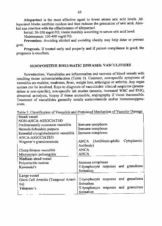
63
Allopurinol is the most effective agent to lower serum uric acid levels. Al- lopurinol blocks xanthine oxidase and thus reduces the generation o f uric acid. Alcohol can interfere with the effectiveness o f allopurinol.
Initial: 50-100 mg/d PO; titrate monthly according to serum uric acid level.Maintenance: 100-400 mg/d PO.Prevention: Avoiding alcohol and avoiding obesity may help deter or prevent
gout. . , ,Prognosis. If treated early and properly and if patient compliance is good, the
prognosis is excellent.
SEROPOSITIVE RHEUMATIC DISEASES: VASCULITIDES
Introduction . Vasculitides are inflammation and necrosis o f blood vessels with resulting tissue ischemia/infarction (Table 5). Common, non-specific symptoms of vasculitis are malaise, weakness, fever, weight loss, arthralgias or arthritis. Any organ system can be involved. Keys to diagnosis o f vasculitides: clinical suspicion (presentation is non-specific), non-specific lab studies (anemia, increased WBC and ESR), abnormal urinalysis, biopsy if tissue accessible, angiography if tissue inaccessible. Treatment o f vasculitides generally entails corticosteroids and/or immunosuppressives.
Table 5. Classification o f Vasculitis and Postulated Mechanism of Vascular DamageSmall vesselNON-ANCA-ASSOCIATED Predominantly cutaneous vasculitis Henoch-Schonlein purpura Essential cryoglobulinemic vasculitis ANCA-ASSOCIATED Wegener’s granulomatosis
Churg-Strauss vasculitis Microscopic polyangiitis
Immune complexes Immune complexes Immune complexes
ANCA (AntiNeutrophilic Cytoplasmic Antibody)ANCAANCA
M edium-sized vesselPolyarteritis nodosa Kawasaki’s
Immune complexesT-lymphocyte response and granuloma formation
L arge vesselGiant Cell Arteritis (Temporal Arteritis)Takayasu’s
T-lymphocyte response and granuloma formationT-lymphocyte response and granuloma formation
64
W EGENER’S GRANULOMATOSIS (WG)
Introduction. Wegener granulomatosis initially reported by Klinger in 1931, the condition was later described in more detail by Wegener. In 1954, Goodman and Churg provided the definitive description of WG with their identification of a triad of pathological features (necrotizing angiitis, necrotizing otitis media and sinusitis, necrotizing glomerulonephritis).
Synonyms: WG, Wegener's granulomatosis, Wegener's disease, Wegener disease, systemic necrotizing angiitis, necrotizing granulomatous inflammation o f the respiratory tract, necrotizing glomerulonephritis, necrotizing granulomatous pulmonary inflammation.
Definition. Wegener granulomatosis (WG) is a systemic vasculitis with granulomatous inflammation o f small and medium arteries, veins, arterioles, and, occasionally, large arteries of upper and lower respiratory tract and kidneys. There are triad o f pathological features, including systemic necrotizing angiitis, necrotizing granulomatous inflammation o f the respiratory tract, and necrotizing glomerulonephritis.
Pathophysiology. The recent discovery of antibodies to cytoplasmic antigens within neutrophils (antineutrophilic cytoplasmic antibody [ANCA], designated C- ANCA for diffuse cytoplasmic staining and P-ANCA for perinuclear staining by immunofluorescence) in most patients with WG raises the possibility o f humoral autoimmunity. Tissue injury in WG is a result of interaction between the inflammatory cascade and the pathogenic immune response to the neutrophil granule proteins.
Epidemiolody.Frequency: A population-based study reported a prevalence of 8.5 cases per mil
lion populations in Norfolk, England.Mortality/Morbidity: WG is associated with a very high (>90%) mortality rate if
it remains untreated. With aggressive therapy, mortality rates improve.Race: WG affects persons of all races.Sex: WG has a slight male predominance.Age: WG has been described in persons of all ages, from young children to eld
erly adults, most common in middle age.Classification. There is ELK (Ears, nose, and throat or upper respiratory tract;
Lungs; and Kidneys) classification. The broad spectrum o f organ involvement o f WG has been described as ears, nose, and throat or upper respiratory tract (E); lungs (L); and kidneys (K); ie, the ELK, classification. This classification system suggests that within the spectrum of WG, patients may have involvement o f any o f the ELK sites singularly or in combination. Under the ELK system, any typical manifestation in E (ears, nose, and throat), L (lungs), or К (kidneys) associated with a typical histopa- thology or positive C-ANCA findings qualifies for the diagnosis of WG.
Clinical and Physical. There are following clinical manifestation of WG: systemic features (malaise, fever, weakness, weight loss), upper respiratory tract (sinusitis or rhinitis, nasoseptal perforation, saddle nose deformity, otitis media, and extension into the orbit with proptosis), lungs (cough, hemoptysis, tracheobronchial erosion, pneumonitis, cavity formation), kidney (segmental necrotizing glomeru-
65
lonephritis, vasculitis rarely seen), skin (14%; purpuric rash over the lower extremities), eye and orbit (29%; red or swollen eyes), joints (usually arthralgias, but up to 25% o f patients may have arthritis with swelling and pain o f the joints without erosions and deformations).
Usually Wegener granulomatosis transform from inflammatory prodrome (serous otitis media and sinusitis) to full blown vasculitic syndrome.
L ab and Im aging Studies.The American College o f Rheumatology has established the following criteria
for the diagnosis o f WG (1990). These criteria were developed before ANCA testing was in widespread use as a diagnostic test for WG. A patient is diagnosed with WG if at least 2 o f these 4 criteria are present. The presence of any 2 or. more o f these criteria yields a sensitivity o f 88% and a specificity o f 92%.
Diagnostic criteria.1. Nasal or oral inflammation (oral ulcers or purulent or bloody nasal discharge).2. Abnormal chest radiograph findings (nodules, fixed in f ltrates, or cavities).3. Urinary sediment (Microhematuria (>5 red blood cells per high-power field)
or red blood cell casts in urine sediment).4. Biopsy o f involved tissue (artery or arteriole show granulomas within the wall
o f an artery or in the perivascular or extravascular area, lungs s 'io w granulomas , kidneys show necrotizing segmental glomerulonephritis).
Other tests include specific (ANCA; c-ANCA > p-ANCA) and general (anemia, leukocytosis, thrombocytosis (>400,000/pL), elevated ESR and C-reactive protein level).
T reatm ent. The 3 phases o f treatment o f WG are induction o f remission, maintenance of remission, and treatment o f relapse.
Drugs.For induction therapy, prednisone and cyclophosphamide are started; prednisone
is tapered over 2-3 months, and cyclophosphamide is continued for 6-12 months following disease remission. Azathioprine or methotrexate has been used with some success in patients who do not tolerate cyclophosphamide therapy.
Corticosteroids have anti-inflammatory properties and cause profound and varied metabolic effects.
Prednisone - 1-1.5 mg/kg PO qd for 4-6 wk; taper over 6 wk as symptoms resolve. Used as an immunosuppressant in the treatment o f autoimmune disorders and vasculitis.
Alkylating agents have improved prognosis of patients with WG. Medication is initiated with corticosteroids but is continued for at least 12 months following disease remission.
Cyclophospham ide - 2 mg/kg/day PO initially; not to exceed 200 mg/day. As an alkylating agent, mechanism o f action o f active metabolites may involve cross- linking o f DNA, which may interfere with growth of normal and neoplastic cells. Aim to reduce and maintain WBC count to 4000-7000/pL.
Immunosuppressive agents use in patients who experience adverse effects with cyclophosphamide. Generally started at lower dose and gradually increased over time. Onset o f effect generally takes several weeks; therefore, use in severe disease
66
not advised. Patients also may be placed on maintenance therapy with azathioprine following induction therapy with cyclophosphamide.
Azathioprine - 2 mg/kg/day PO as single dose; not to exceed 200 mg/day. Inhibits mitosis and cellular metabolism by antagonizing purine metabolism and inhibiting synthesis o f DNA, RNA, and proteins. Effects may decrease proliferation of immune cells and result in lower autoimmune activity.
M ethotrexate - 0.3 mg/kg/wk PO/IM usual dose; 15-20 mg/wk average dose. Unknown mechanism of action in treatment of inflammatory reactions; may affect immune function, It is ameliorates o f symptoms of inflammation (pain, swelling, stiffness). Adjust dose gradually to attain satisfactory response.
Prognosis. The overall mortality rate is 13-36% at 8-10 years. Mortality is associated with age older than 60 years, the development of end-stage renal failure, and an initial creatinine value o f greater than 2.26 mg/dL.
CHURG-STRAUSS SYNDROME
Introduction. In 1951, Churg and Strauss first described the syndrome in 13 patients who had asthma, eosinophilia, granulomatous inflammation, necrotizing systemic vasculitis, and necrotizing glomerulonephritis. Churg-Strauss syndrome, Wegener granulomatosis (WG), and microscopic polyangiitis are 3 closely related vasculitic syndromes that affect medium- and small-sized vessels and are associated with antibodies to neutrophil cytoplasmic antigens (ANCAs).
Synonyms: Churg-Strauss syndrome, CSS, allergic granulomatosis angiitis, Churg-Strauss-like syndrome, CSS-like syndrome.
Definition. Churg-Strauss syndrome (CSS), or allergic granulomatous angiitis, is a rare systemic vasculitic syndrome with granulomatous inflammation of small- and medium-sized vessels, hypereosinophilia and eosinophilic tissue infiltration. There are triads of pathological features, including allergic rhinitis, asthma, and systemic vasculitis. Other manifestations include coronary arteritis, myocarditis, and neuropathy. CSS is associated with antibodies to neutrophil cytoplasmic antigens (ANCAs).
Pathophysiology. The cause of allergic angiitis and granulomatosis is not known. No data have been reported regarding the role o f immune complexes or cell- mediated mechanisms in this disease, although autoimmunity is evident with the presence of hypergammaglobulinemia, increased levels o f immunoglobulin E (IgE), rheumatoid factor, and ANCA.
Epidemiolody.Frequency: Incidence is approximately 2.5 cases per 100,000 adults per year.Mortality/Morbidity: The principal causes of morbidity and mortality are myo
carditis and myocardial infarction secondary to coronary arteritis. With treatment, the 1-year survival rate is 90% and the 5-year survival rate is 62%.
Sex: Males are affected slightly more frequently than females.Age: The age at onset varies from 15-70 years, with a mean of approximately 38
years. The mean age at diagnosis is around 50 years.
67
Clinical and Physical.Churg-Strauss syndrome has 3 phases: 1. Allergic rhinitis and asthma. 2. Eosi
nophilic infiltrative disease (eosinophilic pneumonia or gastroenteritis). 3. Systemic medium- and small-vessel vasculitis with granulomatous inflammation.
There are following clinical manifestation o f CSS: constitutional symptoms (malaise, fatigue, weight loss (70%), fever (57%), myalgias (52%), flulike symptoms), allergic rhinitis (common symptom), asthma symptoms (97%), paranasal sinusitis (61%), pulmonary symptoms (37%; including cough and hemoptysis), arthralgias (40%), skin manifestations (49%; purpura, skin nodules, urticarial rash, necrotic bullae, digital ischemia, livedo reticularis), cardiac manifestations (heart failure, myocarditis, pericarditis, constrictive pericarditis, and myocardial infarction), renal manifestations (hypertension, uremia, renal failure), gastrointestinal symptoms (abdominal pain (59%), diarrhea (33%), and GI bleeding (18%), peripheral neuropathy (mononeuritis multiplex - 77%).
L ab Studies.Hematology: eosinophilia (>10% eosinophils or 5000-9000 eosinophils/pL),
anemia, elevated erythrocyte sedimentation rate (ESR) and C-reactive protein (CRP) levels.
Renal tests: elevated serum BUN and creatinine levels in cases o f renal involvement, abnormal urine sediment, proteinuria, microscopic hematuria, and RBC casts.
Immunological tests: antineutrophil cytoplasmic antibodi es (70% o f patients are perinuclear-ANCA (p-ANCA) - positive), increased serum IgE levels, hypergammaglobulinemia, positive results for rheumatoid factor at low titer.
Im aging Studies.Chest radiograph: pulmonary opacities can be found in 26-77% o f cases, and
films demonstrate no abnormalities in approximately 25% of patients.Other imaging studies are indicated for the complications o f the disease and
specific organ-system involvement, including abdominal CT scan for pancreatitis, coronary angiography for myocardial ischemia and infarction, and echocardiography for congestive heart failure (CHF).
Biopsy: I f local organ involvement exists, obtaining a biopsy o f that organ is most helpful in confirming the diagnosis. If no localizing finding exists, obtaining nerve or muscle biopsy may be considered.
Histologic Findings: The characteristic pathologic changes, found especially in the lung, include small necrotizing granulomas, as well as necrotizing vasculitis involving small arteries and venules. The granulomas are composed o f a central eosinophilic core surrounded radially by macrophages and epithelioid giant cells.
The American College o f Rheumatology (ACR) has proposed 6 criteria for the diagnosis o f Churg-Strauss syndrome. The presence o f 4 or more criteria yields a sensitivity o f 85% and a specificity o f 99.7%.
Diagnostic criteria.1. Asthma (wheezing, expiratory rhonchi).2. Eosinophilia (more than 10% in peripheral blood).3. Paranasal sinusitis.
68
4. Pulmonary infiltrates (may be transient).5. Histological proof o f vasculitis with extravascular eosinophils6. Mononeuritis multiplex or polyneuropathy.Treatm ent.Diet: Adjust nutritional requirements according to the particular clinical situa
tion (renal failure, heart failure).Drugs.Glucocorticoids alone are usually adequate for the treatment o f Churg-Strauss
syndrome. Cytotoxic drugs are necessary in fewer than 20% of patients.Corticosteroids. These agents have anti-inflammatory properties and cause pro
found and varied metabolic effects. Corticosteroids modify the immune response to diverse stimuli.
Prednisone - 0.5-1.5 mg/kg/d or 40-60 mg/d PO, taper over 6-12 wk as symptoms resolve; long-term low doses may be required indefinitely. Prednisone is immunosuppressant for treatment o f autoimmune disorders. Oral prednisone is usually sufficient to control disease activity.
Cytotoxic agents. These agents inhibit cell growth and proliferation. They are reserved for cases resistant to corticosteroids.
Cyclophosphamide - 1-2 mg/kg/d PO, may also be administered in pulsed doses of 500-1000 mg/m2 IV qmo. Dose is adjusted according to clinical response, hematologic response, and toxicity. Cyclophosphamide is chemically related to nitrogen mustards. As an alkylating agent, the mechanism of action of the active metabolites may involve cross-linking o f DNA, which interferes with growth o f rapidly proliferating cells.
Prognosis. Without treatment, the 5-year survival rate is about 25%. With treatment, the 1-year survival rate is 90% and the 5-year survival rate is 62%. Causes of death include the following: cardiac failure, myocardial infarction, or both (most common cause), renal failure, cerebral hemorrhage, gastrointestinal bleeding, status asthmaticus.
POLYARTERITIS NODOSA (PAN)
Introduction. Kussmaul and Maier first described polyarteritis nodosa in 1866 after observing areas of focal inflammatory exudations that gave rise to palpable nodules along the course of the arteries in a patient with advanced disease. For many years, PAN was used as a generic term to describe any type o f systemic vasculitis.
Synonyms: PAN, c-PAN, classic polyarteritis nodosa, hepatitis В-related PAN, HBV-related PAN, non-HBV-related PAN.
Definition. Classic polyarteritis nodosa (PAN or c-PAN) is a systemic vasculitis characterized by focal panmural necrotizing inflammatory lesions in small- and medium-sized arteries resulting in thrombosis, aneurysm or dilatation at lesion site with involve one or many organ systems: most commonly affects joints, kidneys, peripheral nerves, the gut, skin. Healed lesions show proliferation of fibrous tissue and endothelial cells that may lead to luminal occlusion.
69
Pathophysiology. The pathogenesis o f c-PAN is unknown. Evidence for immune complex-induced disease is confined to HBV-related PAN. c-PAN is not associated with antineutrophil cytoplasmic antibodies (ANCA).
Epidemiolody.Frequency: PAN is a rare disease, with an estimated annual incidence ranging
from 4.6 cases per 1,000,000 population in England to 77 cases per 1,000,000 population in a population of Alaskan Eskimos hyperendemic for hepatitis B.
Mortality/Morbidity: Untreated, the prognosis for c-PAN is very poor, with a 5- year survival rate o f 13% or less. A regimen o f GCs and the cytotoxic agent cyclophosphamide (CY) achieved a 5-year survival rate o f 82% in retrospective studies, but this added benefit of CY has not been demonstrated in prospective studies.
Race: PAN has been diagnosed in people o f all racial groups.Sex: PAN affects men and women equally.Age: PAN has been diagnosed in patients o f any age; however, it is predomi
nantly observed in patients aged 40-60 years.Classification. Classic polyarteritis nodosa is sometimes associated with viral
infections, especially hepatis В virus (HBV). c-PAN is thus further subclassified as either HBV- or non-HBV-related PAN, which is a clinically important distinction that affects treatment.
Clinical and physical. PAN is generally an acute multisystem disease with a relatively short prodrome (from weeks to months). Constitutional features including fever, weight loss, and arthralgia or arthritis are common. There are following clinical manifestation of PAN: peripheral nervous system (50-70%; peripheral neuropathy with sudden pain, paresthesia, motor deficit, and mononeuritis multiplex), joints and muscles (myalgias (30-73%), arthralgia (50%), and arthritis (20%) usually early in course), skin (25-60%; palpable purpura, ulceration, livedo reticularis, and digital tip infarct), GI tract (34%; abdominal pain, hematemesis, melena, ischemic bowel, transaminase elevation), kidneys (30-60%; vascular nephropathy - aneurysmal dilatation (not glomerulonephritis), hypertension (25% o f patients), oliguric renal failure), heart (10-30%; coronary arteritis leading to congestive heart failure, but angina and myocardial infarction are rare).
L ab Studies. Certain laboratory findings, although nonspecific, may be helpful in the initial evaluation to understand the systemic nature of the disease. These laboratory findings include the following: elevated erythrocyte sedimentation rate (ESR) greater than 60 mm/h (78-89%), increased C-reactive protein, leukocytosis (45-75%), decreased serum albumin, normochromic anemia (34-79%), thrombocytosis, presence o f hepatitis В surface antigen (7-36%). Cryoglobulins, circulating immune complexes, may be observed in patients with HBV-related PAN but otherwise are not characteristic.
Im aging Studies. The diagnosis established by biopsy or angiography. The diagnosis o f PAN is made by first sampling accessible tissue by biopsy, preferably clinically abnormal tissue. The most accessible tissue sites for biopsy are skin, sural nerve, testes, and skeletal muscle. Histology reveals a focal necrotizing arteritis of generally mixed cellular infiltrate within the vessel wall. If clinically involved tissue is not accessible, then consider a visceral angiogram. Positive angiographic findings
70
include arterial saccular or fusiform aneurysms and narrowing or tapering o f the arteries.
T reatm ent.The initial management o f PAN includes high-dose GCs, usually administered
as pulse intravenous methylprednisolone (15 mg/kg per dose IV, repeated q24h for 1- 3 d). Subsequently, start daily oral prednisone at 1 mg/kg/d. As the patient's clinical status and ESR normalize, usually within 1 month, the prednisone dosage can be tapered progressively over a period of 9-12 months and then stopped. When prednisone is combined with cyclophosphamide, the steroid dose is tapered more rapidly to reduce the increased risk o f infection. Most patients with PAN require immunosuppressive therapy with cyclophosphamide in addition to GCs.
Drugs. The goals o f pharmacotherapy are to reduce morbidity and to prevent complications.
Corticosteroids have anti-inflammatory properties and cause profound and varied metabolic effects. They modify the body's immune response to diverse stimuli.
M ethylprednisolone -15 mg/kg per dose IV, repeat q24h for 1-3 days.Prednisone -1 mg/kg/d PO.Antineoplastic agents chemically related to nitrogen mustards. Cyclophos
phamide is used in conjunction with GCs, thus reducing the need for higher doses of corticosteroids.
Cyclophospham ide - 3-5 mg/kg IV bolus q2-4wk, 2-3 mg/kg/day PO.Prognosis. The 5 factors are associated with poor prognosis. It is 5-factor score
(FFS). The presence o f any or a combination of the following 5 factors is thought to predict increased mortality risk: 1. renal insufficiency (serum creatinine greater than or equal to 140 mmol/L (1.58 mg/dL); 2. proteinuria (more than 1 g/d); 3. GI tract involvement; 4. cardiomyopathy; and 5. CNS involvement. When the FFS is zero, the expected mortality rate at 5 years is 12%. When the FFS is 1, the mortality rate is 26%, and when the FFS is greater than or equal to 2, the mortality rate is 46%.
GIANT CELL ARTERITIS (TEM PORAL ARTERITIS)
Introduction . In the late 19th century, Jonathan Hutchinson reported on a man who had difficulty wearing a hat because o f his tender temporal arteries. Since then, the giant cell arteritis (temporal arteritis) clinical spectrum has been growing.
Synonyms: giant cell arteritis, giant-cell arteritis, temporal arteritis, ТА, GCA, cranial arteritis.
Definition. Temporal arteritis (giant cell arteritis (GCA) or cranial arteritis) is a systemic, inflammatory, vascular syndrome o f medium- and large-sized arteries, predominantly those originating from the aortic arch, the aorta itself and the cranial arteries. Ophthalmic posterior ciliary arteries most common involve. Therefore untreated GCA can lead to blindness (20-25%).
Pathophysiology. The etiology of giant cell arteritis is unknown, but the pathogenesis involves a chronic inflammatory process, predominantly o f large arteries, resulting in the elaboration of various cytokines (interferon-gamma (IFN-gamma),
71
transforming growth factor-beta (TGF-beta), interleukin (IL)-!1, IL-1 beta, and IL-6). Systemic manifestation is likely related to the inflammatory process and cytokine elaboration, while end organ involvement is related to vascular occlusion.
Epidemiolody.Frequency: The incidence o f giant cell arteritis is unknown.Mortality/Morbidity: Patients with giant cell arteritis are at risk for blindness.
Giant cell arteritis is a basis for aneurysms, dissections, stenotic lesions o f the aorta, and stenotic lesions o f the major branches o f the aorta.
Race: Ggiant cell arteritis is much less common in African Americans compared to white persons in USA.
Sex: Giant cell arteritis affects women twice as often as men.Age: Most patients with giant cell arteritis present over 50 years o f age, and
peak incidence occurs in patients aged 60-80 years.Clinical and physical. Constitutional symptoms including malaise, fatigue, fe
ver, night sweats, anorexia, weight loss. Poorly localized tenderness over the joints, especially the shoulders and hips are common. There are following clinical manifestation o f GCA: symptoms related to vasculitis involving branches o f the external carotid artery (temporal headaches and scalp tenderness - 50-75%; jaw claudication and pain (predominantly in the masseter muscles with chewing) - 50%), symptoms related to vasculitic involvement o f the ophthalmic artery and its branches (sudden, painless loss o f vision, diplopia and/or permanent visual loss - 20-50%), symptoms related to large artery involvement (pulseless disease, aortic aneurysm +/— rupture - 15%, aortic arch syndrome - rare). Polymyalgia rheumatica (proximal myalgia, constitutional symptoms, elevated ESR) occurs in 50-70% o f patients with giant cell arteritis.
L ab Studies.Blood studies: elevate ESR and C-reactive protein (CRP), normocytic normo
chromic anemia and thrombocytosis.Liver function tests: elevate the alkaline phosphatase level (33%). •Im aging Studies. Researchers have recently explored the role o f color duplex
ultrasonography in the diagnosis o f giant cell arteritis. Thoracic or abdominal ultrasound may be helpful for diagnosing and monitoring patients with aortic aneurysms.
Histologic findings o f temporal artery biopsy: An inflammatory infiltrate, predominantly o f mononuclear cells, usually involves the entire vessel wall (panarteritis). Fragmentation of the internal elastic lamina is characteristic. Fibrinoid necrosis is not a feature o f the lesion. Giant cells are commonly present, and they often seem to engulf parts o f the internal elastic lamina.
The diagnosis o f giant cell arteritis is made by clinical suspicion, increased ESR, increased CRP, temporal artery biopsy.
T reatm ent.Corticosteroid therapy for giant cell arteritis is started at high doses with gradual
tapering using clinical manifestations and the ESR level. Corticosteroid and immunosuppressive (if refractory in corticosteroids) therapy is highly effective in the treatment o f giant cell arteritis and in the prevention o f blindness and other vascular complications.
72
Drugs.Corticosteroids have anti-inflammatory properties and cause profound and var
ied metabolic effects. Corticosteroids modify the body's immune response to diverse stimuli.
Prednisone - 40-60 mg PO qd or divided bid or 1 mg/kg/day in divided doses until symptoms resolve. Intravenous solu-Medrol in high doses for acute visual loss or visual symptoms Attempt using minimal effective dose; do not stop abruptly; taper gradually.
Immunosuppressive agents may be anti-inflammatory for giant cell arteritis and result in steroid sparing in relatively resistant cases.
M ethotrexate - 7.5-25 mg PO or SC qwk.Prognosis. Visual damage is often irreversible. The average duration of treat
ment is 2 years; however, some patients require treatment for 5 years or more. Morbidity from steroid therapy is often worse than the underlying disease.
POLYMYALGIA RHEUMATICA
Introduction. In 1957, Barber described polymyalgia rheumatica (PMR) as an aching syndrome that was not associated with other defined rheumatic, infectious, or neoplastic disorders and that usually occurred in elderly patients with constitutional symptoms and an elevated erythrocyte sedimentation rate (ESR). Polymyalgia rheumatica is a relatively common clinical syndrome of unknown etiology. Approximately 15% of patients with polymyalgia rheumatica develop giant cell arteritis (GCA), and approximately 50% o f patients with giant cell arteritis have associated polymyalgia rheumatica.
Synonyms: polymyalgia rheumatica, PMR.Definition. Polymyalgia rheumatica (PMR) is characterized by proximal myal
gia (profound pain) of the hip and shoulder girdles with accompanying morning stiffness lasting for more than 1 hour.
Pathophysiology. The cause o f polymyalgia rheumatica is presently unknown. Patients with polymyalgia rheumatica often have elevated interleukin-2 (IL-2) and interleukin-6 (IL-6) levels. One hypothesis holds that in a genetically predisposed patient, an environmental factor, possibly a virus, causes monocyte activation, which helps determine the production of cytokines that induce manifestations characteristic o f polymyalgia rheumatica. Prevalence o f antibodies to adenovirus and respiratory syncytial virus was reportedly higher in patients with polymyalgia rheumatica. Occurrence in siblings suggests a genetic role in the pathophysiology of the disease.
Epidemiolody.Frequency varies according to country; highest rates occur in Northern Europe.
For example, in Italy, incidence is 12.7 cases per 100,000 persons, in the USA incidence is 52.5 cases per 100,000 persons. Prevalence is approximately 0.5-0.7%.
Mortality/Morbidity: With appropriate treatment, survival is similar to that of unaffected persons of the same age. Polymyalgia rheumatica is self-limited and often
73
remits in 1-3 years. Untreated patients, however, often feel unwell and have an impaired quality o f life.
Race: Polymyalgia rheumatica almost always affects whites; however, polymyalgia rheumatica may occur in African persons.
Sex: Polymyalgia rheumatica is twice as common in females (F:M = 2:1).Age: Incidence increases with advancing age. Polymyalgia rheumatica is rarely
encountered before the age o f 50 years (age o f onset typical у > 50 years). Median age at diagnosis is 72 years.
Clinical. Patients are often in good health prior to disease onset. Onset is abrupt in about 50% o f patients. There are following clinical manifestation o f polymyalgia rheumatica: constitutional symptoms prominent (fever, weight loss, malaise), morning stiffness o f proximal muscles and joints (neck, hip and shoulder girdles, thighs).
Physical. Physical examination reveals tender muscles but no weakness or atrophy. Morning stiffness for more than 1 hour, often more prolonged. Possible development o f arthralgia and myalgia up to 6 months after onset o f systemic complaints.
Lab Studies. Laboratory investigations often reveal mild normocytic, normochromic anemia, elevated ESR, CRP and platelets, normal creatine kinase (CK).
Im aging Studies. Radiographs o f the painful joints rarely show abnormalities such as osteopenia, joint space narrowing, or erosions.
Diagnostic criteria.1. Age 50 years or older at onset.2. Bilateral aching and morning stiffness for at least 1 month and involving at
least 2 o f 3 areas: neck or torso, shoulders or arms, hips or thighs.3. Increased erythrocyte sedimentation rate (ESR) 40 mm/h or greater.4. Prompt and lasting response o f symptoms to corticosteroids (15 mg/d).5. Must rule out infection, RA, SLE, PAN, malignancy, and giant cell arteritis.T reatm ent.Diet: Ensure adequate calcium and vitamin D intake with corticosteroid use.Activity: Generally, activity restriction is unnecessary.Drugs. The goals o f pharmacotherapy are to reduce morbidity and to prevent
complications.Oral corticosteroids are the first line of treatment. These agents cause profound
and varied metabolic effects. Exact mechanism o f action in polymyalgia rheumatica is not well-known, although the disease may be caused by general anti-inflammatory and immunomodulatory effects. In addition, corticosteroids down-regulate cytokine production.
Prednisone - 0.2-0.3 mg/kg/d PO (10-15 mg/d) initial; maintain effective dose for 2-4 wk after patient becomes asymptomatic; generally, effective dose can be lowered by 1-2.5 mg/d q2-4wk to find the minimum dose needed to maintain symptom suppression; once 10-mg dose is reached, taper by 1 mg/d dectSments q4wk.
Nonsteroidal anti-inflammatory drugs can be administered to some patients with mild symptoms; however, most patients require corticosteroids for total control of symptoms. NSAIDs may be helpful in later stages o f corticosteroid dosage tapering. NSAIDs generally have no effect on ESR.
74
Diclofenac (nonselective COX-1 and COX-2 inhibitors) - Up to 100-150 mg/d PO; not to exceed total daily dose o f 200 mg.
Nimesulide (selective for COX-2 receptors, compared to traditional NSAIDs) - 100 mg PO 2 times a day.
Meloxicam (selective for COX-2 receptors, compared to traditional NSAIDs) - 7.5-15 mg/d PO.
Prognosis. Polymyalgia rheumatica is usually self-limited. With prompt diagnosis and adequate therapy, the condition has an excellent prognosis.
TAKAYASU ARTERITIS
Introduction . Takayasu arteritis is a systemic disease that may have isolated, atypical, and catastrophic manifestations. Takayasu described the retinal changes of the disease in 1908, the same year that the association between the retinal changes and pulse deficit was reported. However, the disorder was not termed Takayasu's disease until 1954.
Synonyms: Takayasu arteritis, ТА, pulseless disease, Takayasu's disease, Takayasu's arteritis, Takayasu disease.
Definition. Takayasu arteritis (ТА) is a chronic, systemic, progressive, inflammatory, occlusive disease o f larger arteries, most often affecting the aorta and its branches. The pulmonary arteries can also be involved.
Pathophysiology. Cell-mediated autoimmunity appears to play an important role in the mechanism o f vascular injury. The early stage consists o f a continuous or patchy granulomatous inflammatory reaction involving macrophages, lymphocytes, and multinucleated giant cells.
Epidemiolody.Frequency: Although Takayasu arteritis has a worldwide distribution, it is ob
served more frequently in Asia and India than in Western Europe and North America. Worldwide incidence is estimated at 2.6 cases per million persons per year. The prevalence in Sweden is similar to that in the United States (ie, 2.6-6.4 persons per million populations). In the United Kingdom, the annual incidence is 0.15 cases per million persons.
Mortality/Morbidity: The mortality rate is to range between 2-35% over 5 years. Such disparity may reflect differences in access to care, definitions o f disease activity, and indications for treatment. The overall morbidity depends on the severity o f the lesions and their consequences (vascular complications such as aortic regurgitation, congestive heart failure, cerebrovascular events, myocardial infarction, aneurysm rupture, or renal failure).
Race: Takayasu arteritis is observed more frequently in patients o f Asian or Indian descent. Japanese patients with Takayasu arteritis have a higher incidence of aortic arch involvement. In contrast, series from India report higher incidences o f thoracic and abdominal involvement. In US patients with Takayasu arteritis, the most commonly involved vessels are the left subclavian, superior mesenteric, and abdominal aorta.
75
Sex: Approximately 80% o f patients are women.Age: Most patients are aged 4-63 years, with the mean age o f onset being ap
proximately 30 years. Fewer than 15% o f cases present in individuals older than 40 years.
Clinical. The presentation o f Takayasu arteritis is heterogeneous. Approximately 20% o f patients with Takayasu arteritis are clinically asymptomatic, with the disease being detected based on abnormal vascular findings upon examination. Most patients present with systemic and vascular symptoms. There are following clinical manifestation of Takayasu arteritis: constitutional symptoms (40%; malaise (40- 60%), fatigue, fever, weight loss (40-60%), arthralgias (40-60%), arthritis, myalgias), vascular features (60-70%; jaw and extremity claudication), neurological manifestations (60%; transient ischemic attacks, hemorrhagic or ischemic stroke, transient or permanent blindness, headaches, seizures, subclavian steal syndrome), cardiac fea tures (50%; aortic regurgitation, angina, myocardial infarction, myocarditis and cardiomyopathy, congestive heart failure (primary cause o f death), arrhythmias, sudden death), pulmonary features (40%; pulmonary hypertension (often asymptomatic), hemoptysis, pleuritis), renal manifestations (renovascular hypertension (most frequent), renal amyloidosis is rarely reported), dermatological manifestations (ulcerating nodular lesions, erythema nodosum, erythema induratum, pyoderma gangrenosum, erythema multiforme), gastrointestinal manifestations (rare; nausea, vomiting, diarrhea, abdominal pain, bleeding - due to mesenteric artery ischemia).
Physical. There are following physical examination of Takayasu arteritis: blood pressure (systolic blood pressure difference (>10 mm Hg) between arms, hypertension - 50% o f patients), peripheral pulses (absent or diminished pulses), palpation (vessel tenderness - carotidynia), auscultation (bruit over subclavian arteries or the abdominal aorta).
Lab Studies. The acute phase reactants and clinical parameters generally used to define active inflammatory disease do not universally reflect active blood vessel inflammation in Takayasu arteritis. The erythrocyte sedimentation rate is elevated in most but not all patients during active inflammatory disease.
Im aging Studies. Angiography can evaluate only the ap pearance o f the lumen and cannot differentiate between active and inactive lesions. Takayasu arteritis can be divided into 6 types based on angiographic involvement: Type I - Branches o f the aortic arch; Type Ila - Ascending aorta, aortic arch, and its branches; Type lib - Type Ila region plus thoracic descending aorta; Type III - Thoracic descending aorta, abdominal aorta, renal arteries, or a combination; Type IV - Abdominal aorta, renal arteries, or both; Type V - Entire aorta and its branches.
Biopsy o f medium- to large-sized vessels may be diagnostic in early stages of the disease. However, in the chronic phase, diagnosis by biopsy alone is inadequate. In contrast to other vasculitides, tissue biopsies play little to no role in the diagnosis o f Takayasu arteritis.
Histologic Findings: Takayasu arteritis is characterized by a special pattern of histopathological changes. The early stage consists o f a continuous or patchy granulomatous inflammatory reaction involving macrophages, lymphocytes, and multinu- cleated giant cells. Inflammation initially occurs in the vasa vasorum, with the artery
76
wall becoming irregularly thickened and the lumen becoming narrowed. Takayasu arteritis progresses to a sclerotic stage, with intimal and adventitial fibrosis and scarring o f the media.
Diagnostic criteria.The American College o f Rheumatology has established classification criteria
for Takayasu arteritis (3 of 6 criteria are necessary), as follows:1. Age of 40 years or younger at disease onset.2. Claudication of the extremities.3. Decreased pulsation of one or both brachial arteries.4. Difference o f at least 10 mm Hg in systolic blood pressure between arms.5. Bruit over one or both subclavian arteries or the abdominal aorta.6. Arteriographic narrowing or occlusion of the entire aorta, its primary
branches, or large arteries in the upper or lower extremities that is not due to arteriosclerosis, fibromuscular dysplasia, or other causes.
T reatm ent.D rugs include corticosteroids with or without cytotoxic agents.Corticosteroids are the mainstay of therapy for active Takay,isu arteritis.Prednisone - 1 mg/kg/day PO qd or divided bid/qid. Taper over 2 wk as symp
toms resolve.Cytotoxic agents are used for patients with steroid resistance or relapsing Taka
yasu arteritis. These agents are usually continued for one year after remission and are then tapered to discontinuation.
Azathioprine - 1-2 mg/kg PO qd in single or divided doses.M ethotrexate - 7.5-25 mg/week PO or IM.Cyclophosphamide - 2 mg/kg/d PO (should be reserved for patients with the
most severe and refractory disease states).Cyclosporin A - also has been used in steroid-resistant patients at initial doses
o f 5 mg/kg/d and then 2-3 mg/kg/d PO qd or divided bid for maintenance; if creatinine clearance increases by >30%, dosage must be lowered.
M ycophenolate mofetil - 2 g/d PO. It has been used in patients with Takayasu arteritis resistant to steroids and other immunosuppressant drugs. May be added if steroids and other immunosuppressant drugs are ineffective in achieving or maintaining remission.
Prognosis. The 15-year survival rate has been reported to be 90-95%. Approximately 20% of patients have a monophasic and self-limited disease. In others, Takayasu arteritis is progressive or relapsing/remitting and requires immunosuppressive treatment.
77
CARDIOLOGY
CARDIOMYOPATHIES
Definition. Idiopathic cardiomyopathies are intrinsic myocardial disease not secondary to coronary artery disease (CAD), valvular heart disease, congenital heart disease, hypertension (HTN) or pericardial disease. The diagnosis o f any o f the following mandates exclusion o f the above conditions: dilated cardiomyopathy (DCM), hypertrophic cardiomyopathy (HCM), restrictive cardiomyopathy (RCM).
Classification of cardiom yopathies:I. Idiopathic cardiomyopathies:1. Dilated (sporadic, genetic or familial).2. Hypertrophic (with or without asymmetric septal hypertrophy).3. Restrictive (nondilated, nonhypertrophic - Endomyocardial fibrosis, Loffler
endocarditis).II. Specific cardiomyopathies:1. Secondary to other cardiovascular disease (ischemia, hypertension, valvular
disease, tachycardia induced).2. Infectious (viral, rickettsial, bacterial, metazoal, protozoal, probable infectious
- Whipple disease, Lyme disease).3. Metabolic (endocrine diseases - hyperthyroidism, hypothyroidism, ac
romegaly, myxedema, hypoparathyroidism, hyperparathyroidism, diabetes mellitus; electrolyte imbalance - potassium, phosphate, magnesium, other; nutritional - thiamine deficiency (beriberi), protein deficiency, starvation, carnitine deficiency).
4. Toxic (drugs, poisons, foods, anesthetic gases, heavy metals, alcohol).5. Collagen vascular disease (systemic lupus erythematosus, rheumatoid arthri
tis, progressive systemic sclerosis, polymyositis).6. Infiltrative (hemochromatosis, amyloidosis, glycogen storage disease).7. Granulomatous (sarcoidosis).8. Physical agents (extreme temperatures, ionizing radiation, electric shock,
nonpenetrating thoracic injury).9. Neuromuscular disorders (muscular dystrophy - Limb-girdle (Erb dystrophy),
Duchenne dystrophy, fascioscapulohumeral (Landouzy-Dejerine dystrophy); Friedreich disease, myotonic dystrophy).
10. Primary cardiac tumor (myxoma).11. Senile.12. Peripartum.13. Immunological (postvaccination, serum sickness, transplant rejection).
DILATED CARDIOMYOPATHY (DCM)
Introduction. Cardiomyopathy is a complex disease process that can affect the heart of a person of any age. It is a common problem throughout the world. Cardiomyopathy is an important cause of morbidity and mortality among the world's aging
78
population. Cardiomyopathies have multiple etiologies. Persons with cardiomyopathy may have asymptomatic left ventricular systolic dysfunction, left ventricular diastolic dysfunction, or both.
Synonyms: dilated cardiomyopathy, DCM, dilatated cardiomyopathy, congestive cardiomyopathy.
Definition. Dilated cardiomyopathy (DCM) is condition in which the normal muscular function o f the myocardium has been altered by specific or multiple etiologies, with varying degrees o f physiologic compensation for that malfunction. Dilated cardiomyopathy (DCM) manifests hemodynamically as decreased cardiac output, increased pulmonary venous pressure, and enlargement o f left ventricular cavity size with little or no wall hypertrophy.
Etiology.1. Idiopathic (risk factors: male, black race, family history).2. Alcohol.3. Inflammatory (subsequent to myocarditis).4. Collagen vascular disease (SLE, PAN, dermatomyositis, progressive systemic
sclerosis).5. Infectious (post-viral (Coxsackie), Chagas disease, Lyme disease, Rickettsial
diseases, acute rheumatic fever).6. Neuromuscular disease (Duchenne muscular dystrophy, myotonic dystrophy,
Friedreich ataxia).7. Metabolic (uremia, nutritional deficiency such as thiamine, selenium, car
nitine).8. Endocrine (thyrotoxicosis, DM).9. Familial.10. Peripartum.11. Toxic: cocaine, heroine, glue sniffing, organic solvents12. Radiation induced.13. Drugs: chemotherapeutics (adriamycin).Pathophysiology. DCMs are associated with both systolic and diastolic dys
function. The decrease in systolic function o f the myocardium is by far the primary abnormality. This leads to an increase in the end-diastolic and end-systolic volumes with clinical manifestations (CHF).
Epidemiolody.Frequency: The true incidence of cardiomyopathies is unknown.Mortality/Morbidity: Cardiomyopathy is an important cause o f morbidity and
mortality among the world's aging population.Age: Cardiomyopathy is a complex disease process that can affect the heart o f a
person o f any age.Clinical. The clinical manifestations of dilated cardiomyopathy include CHF,
systemic or pulmonary emboli, arrhythmias, and sudden death (major cause o f mortality due to fatal arrhythmia).
Classic staging o f heart failure is based on the New York Heart Association (NYHA) system. The following is a new approach to the classification of heart failure:
79
Stage A (high risk for developing heart failure) - Hypertension, coronary artery disease, diabetes mellitus, family history o f cardiomyopathy.
Stage В (asymptomatic heart failure) - Previous myocardial infarction, left ventricular systolic dysfunction, asymptomatic valvular disease.
Stage C (symptomatic heart failure) - Structural heart disease, dyspnea, fatigue, reduced exercise tolerance.
Stage D (refractory end-stage heart failure) - Marked symptoms at rest despite maximal medical therapy, recurrent hospitalizations.
Physical. There are following physical examination of DCM.General inspection: cardiac cachexia, peripheral edema, cyanosis, and clubbing.Pulses: irregularly irregular rhythm (atrial fibrillation).Precordial palpation: palpate for heaves, shifted point o f maximal impulse, and
cardiomegaly (broad and displaced point o f maximal impulse, right ventricular heave).
Precordial auscultation: murmurs (with appropriate maneuvers), tachycardia, gallops (almost always present).
Percussion and auscultation o f the lungs: crackles (pulmonary rales) or signs of pleural effusion may be noted.
Percussion and palpation o f the liver: hepatomegaly (elevated venous pressure), hepatojugular reflux, ascites.
The level o f compensation (or decompensation) determines which signs are present.
Im aging Studies.12 lead ECG: ST-T wave abnormalities, poor R wave progression, conduction
defects (BBB), arrhythmias.Chest x-ray: global cardiomegaly (globular heart), signs o f CHF, pulmonary
vascular congestion (Kerley lines, bilateral pleural effusion - usually occurs first on the right side)
Echocardiography: 4-chamber enlargement, depressed ejection fraction, MR and TR secondary to cardiac dilatation.
Endomyocardial biopsy: not routine, used to diagnose infiltrative RCM and myocarditis, or to rule out a treatable cause.
Angiography: selected patients.T reatm ent.Diet: The importance o f patient education cannot be overemphasized, especially
regarding dietary restrictions. Dietary recommendations include sodium and water restrictions, abstinence from EtOH. An average diet contains 6 g/d o f salt. Avoiding extra table salt decreases this intake to 3 g/d. Patients with CHF should restrict their salt intake to less than 2-4 g/d. Fluid restriction is only necessary in very late stages o f the disease.
Activity: Encourage patients to exercise moderately because deconditioning is a very common cause o f dyspnea. Cardiac rehabilitation has been shown to improve patient outcomes.
Drugs. The goals o f pharmacotherapy are to reduce morbidity and to prevent complications.
80
Angiotensin-converting enzyme inhibitors are the current criterion standard in the treatment o f left ventricular dysfunction (in the absence o f contraindications to ACE inhibition).
Enalapril - 2.5 mg/d PO or 1.25 mg/d IV (titrate to maximum dose). ACE inhibitors reduce angiotensin II levels, causing a decrease in aldosterone secretion.
Angiotensin II receptor blockers are equally effective as ACE inhibitors in the treatment o f heart failure. Adverse effect profile is similar to that o f ACE inhibitors with regard to renal insufficiency or hyperkalemia. An added advantage o f angiotensin receptor blockers is that they do not cause potentiation of bradykinin and therefore do not cause cough.
V alsartan - 80 mg/d PO; may increase to 160 mg/d if needed (for patients unable to tolerate ACE inhibitors).
Beta-adrenergic blocking agents (beta-blockers) are both safe and effective in the treatment persons with any class o f heart failure.
Carvedilol - 3.125 mg PO qd/bid; titrate q2w kto 50 mg bid. Carvedilol blocks betar , alpha-, and beta2-adrenergic receptor sites, decreasing ad'energic-mediated myocyte damage.
M etoprolol - 6.25 mg PO (gradually increase to 50 mg bid or as tolerated).Bisoprolol - 2.5 mg/d PO (titrate to 10 mg/d PO).Metoprolol and bisoprolol are selective beta]-adrenergic receptor blockers that
decrease automaticity o f contractions. During IV administration o f metoprolol, carefully monitor blood pressure, heart rate, and ECG.
Aldosterone antagonists (Spironolactone) are complementary to standard therapy in modulating the RAAS because aldosterone levels remain elevated despite ACE inhibitor therapy. Spironolactone is currently indicated for treating patients with moderate-to-severe heart failure (NYHA class Ш-IV) in addition to ACE inhibitors, beta-blockers, diuretics, and digoxin.
Spironolactone - 12.5-25 mg/d PO (for management o f edema resulting from excessive aldosterone excretion). Spironolactone competes with aldosterone for receptor sites in distal renal tubules, increasing water excretion while retaining potassium and hydrogen ions.
Cardiac glycosides (Digoxin) therapy for heart failure has nc benefit on mortality rates. However, it does improve NYHA functional class, hemodynamics, symptoms, exercise capacity, and quality o f life and reduces hospitalizations for heart failure.
Digoxin - 0.125-0.25 mg/d PO or 0.125-0.25 mg IV. Digoxin is cardiac glycoside with direct inotropic effects in addition to indirect effects on the cardiovascular system. Digoxin acts directly on cardiac muscle, increasing myocardial systolic contractions. Indirect actions result in increased carotid sinus nerve activity and enhanced sympathetic withdrawal for any given increase in mean arterial pressure.
Diuretics reserved for congestive states and are not indicated for daily use for patients who are in NYHA functional class I or II (and even some class III patients). Agents such as metolazone, hydrochlorothiazide, and acetazolamide may used to augment loop diuretics.
81
Furosemide (Lasix) - 20-80 mg/d PO/IV/IM; titrate not to exceed 1 g/d. Fu- rosemide increases excretion of water by interfering with chloride-binding cotransport system, which, in turn, inhibits sodium and chloride reabsorption in ascending loop of Henie and distal renal tubule. Bioavailability of PO furosemide is 50%. If switch is made from IV to PO, an equivalent PO dose should be used. Doses vary depending on clinical condition.
Antiarrhythmics are useful in patients with supraventricular and nonsustained ventricular tachycardias. The class III antiarrhythmics amiodarone is favored in these patients for the treatment of supraventricular and ventricular dysrhythmias.
Amiodarone (Cordarone) is variable dosing. Amiodarone may inhibit AV conduction and sinus node function. Prolongs action potential and refractory period in myocardium and inhibits adrenergic stimulation.
The use of anticoagulants is restricted to patients in atrial fibrillation, patients with artificial valves, and patients with known mural thrombus.
W arfarin - 5-15 mg/d PO qd for 2-5 d; adjust dose to target INR of 2. Warfarin interferes with hepatic synthesis of vitamin K—dependent coagulation factors. Tailor dose to maintain INR in range of 2-3.
Surgical therapy.Heart transplantation.Absolute indications for heart transplantation are refractory cardiogenic shock,
dependence on intravenous inotropes for organ perfusion, peak VO2 less than 10 mL/kg/min with achievement of anaerobic threshold, severe ischemia not amenable to any intervention, symptomatic ventricular arrhythmias refractory to all therapies.
Relative indications for heart transplantation are peak V 02 of 11-14 mL/kg/min (or <50-55% predicted for age and sex) and major limitation of daily activity, recurrent instability of CHF not due to noncompliance or suboptimal medical therapy.
Left ventricular assist devices (LVAD). Portable electric left' ventricular assist devices have been proven as the standard of care when a bridge to transplantation is needed. Left ventricular assist devices are being evaluated as permanent implantations in patients who are not candidates for heart transplantation (ie, "destination therapy"). Current limitations include high infection rates and mechanical malfunction.
Volume reduction surgery (role remains unclear).Partial left ventriculectomy (Batista procedure). Reducing the left ventricular di
ameter (Laplace law) in patients with DCM is thought to improve ventricular function.
Prognosis depends on etiology. A higher mortality rate is associated with increased age, male sex, and severe CHF. Patients with overall CHF have 50% 5 year survival. Cause of death usually is connected with CHF or sudden death. Systemic emboli are significant source of morbidity. Patients with mild heart failure have significantly better prognoses, especially with optimal medical therapy.
82
HYPERTROPHIC CARDIOMYOPATHY (HCM)
Introduction. Hypertrophic cardiomyopathy (HCM) is a genetic disorder (> 100 mutations associated with development of autosomal dominant inheritance). The disorder has a variable presentation and carries a high incidence of sudden death.
Synonyms; hypertrophic cardiomyopathy (HCM), hypertrophi; obstructive cardiomyopathy, idiopathic hypertrophic subaortic stenosis (IHSS), muscular subaortic stenosis, asymmetric septal hypertrophy (ASH), myocardial hypertrophy.
Definition. Hypertrophic cardiomyopathy is defined as unexplained ventricular hypertrophy (not due to systemic HTN or AS). This hypertrophy can occur in any region of the left ventricle but frequently involves the interventricular septum, which results in an obstruction of flow through the left ventricular (LV) outflow tract. Therefore HCM can be separated into obstructive and nonobstructive types.
Pathophysiology. The molecular basis for HCM is defects in several of the genes encoding for the sarcomeric proteins, such as myosin heavy chain, actin, tropomyosin, and titin. Multiple mutations have been identified, with genotype-specific risks of mortality and degree of hypertrophy. Histopathologic features are myocardial fiber disarray, myocyte hypertrophy, and interstitial fibrosis.
Epidemiolody.Frequency: The overall prevalence of HCM is low and has been estimated to
occur in 0.05-0.2% of the population.Mortality/Morbidity:Sudden death: Most patients are asymptomatic. The first clinical manifestation
of the disease in such individuals may be sudden death (ventricular tachycardia or fibrillation).
Arrhythmia: Patients can have atrial fibrillation, atrial flutter, ventricular ectopy, ventricular tachycardia, and ventricular fibrillation. These patients are among the highest-risk group for ventricular fibrillation.
Heart failure: These patients have a high likelihood of recurrent heart failure resulting from both mitral regurgitation and profound diastolic dysfunction.
Sex: HCM is slightly more common in males than in females. HCM usually presents at a younger age in females.
Age: HCM has a bimodal peak of occurrence. The most common presentation is in the third decade of life, but it may present in persons of any age, from newborns to elderly individuals.
Classification. There are two variants of hypertrophic cardiomyopathy: hypertrophic obstructive cardiomyopathy (HOCM) (dynamic left ventricular outflow tract (LVOT) obstruction) and nonobstructive hypertrophic cardiomyopathy (decreased compliance and diastolic dysfunction - impaired filling).
Clinical. Hypertrophic cardiomyopathy may be asymptomatic for a long period of time. Symptoms can include sudden cardiac death (may be first manifestation), dyspnea, syncope and presyncope (LV outflow obstruction or arrhythmia), angina (may occur in the absence of detectable coronary atherosclerosis), palpitations, orthopnea, paroxysmal nocturnal dyspnea, congestive heart failure (CHF), and dizziness.
83
Physical. There are following physical examination of HCM.Pulses: rapid upstroke pulse, bifid pulse.Precordial palpation: Point of maximal intensity (PMI) - normal at 5th intra-
clavicular space (ICS) at midclavicular line (<10 cm from midline). PMI: localized, sustained, double impulse, ‘triple ripple’ (triple apical impulse).
Precordial auscultation: first heart sound (S^ is normal, second heart sound (S2) is normal or paradoxically split, fourth heart sound (S4) is present, systolic, harsh, diamond-shaped ejection murmur of mitral regurgitation is present (which is best heard between the apex and left sternal border, enhanced by squat-to standing or valsalva), diastolic decrescendo murmur of aortic regurgitation is heard in 10% of patients.
Lab Studies. No specific laboratory blood tests are required in the workup of HCM.
Imaging Studies.12 lead ECG: LVH, ST-T wave abnormalities, prominent Q waves or tall R
wave in V,.Echocardiography: LVH - asymmetric septal hypertrophy (roost common pres
entation), systolic anterior motion (SAM) of anterior MV leaflet, resting or dynamic ventricular outflow tract obstruction, MR (due to SAM and associated with left ventricular (LV) outflow tract obstruction), diastolic dysfunction, LAE.
Chest radiograph: The cardiac silhouette may range from normal to markedly increase. Left atrial enlargement frequently is observed (when significant mitral regurgitation is present).
Cardiac catheterization: increased LV end-diastolic press-ure, variable systolic gradient across LV outflow tract.
Adult first-degree relatives of patients with HCM should be screened (physical exam, ECG, 2D-ECHO) serially every 5 years.
Treatment.Diet: No special diet is required. The patient should avoid excessive weight
gain.Activity: avoid strenuous exercise, avoid factors which increase obstruction.Drugs. The purpose of pharmacologic therapy is to reduce the pressure gradient
across the LV outflow tract by reducing the inotropic state of the left ventricle, improving compliance of the left ventricle, and reducing diastolic dysfunction. To date, only one pharmacologic agent, amiodarone (Cordarone), has been shown to reduce the incidence of arrhythmogenic sudden cardiac death.
Beta-adrenergic blocking agents are reducing inotropic state o f left ventricle. Decrease myocardial oxygen consumption, thereby reducing myocardial ischemia potential. Decrease heart rate, thus reducing myocardial oxygen consumption and reducing myocardial ischemia potential.
Metoprolol - 25-100 mg PO bid. Metoprolol is first-line therapy in treatment of both obstructive and nonobstructive HCM. Dose titrated to heart rate between 50 and 60 bpm.
Atenolol - 25-100 mg/d in AM or divided bid. Atenolol selectively blocks betal receptors with little or no effect on beta2 types.
84
Sotalol - 80-320 mg PO bid. Sotalol is class III antiarrhythmic agent that blocks K+ channels, prolongs action potential duration, and lengthens QT interval. Sotalol is noncardiac selective beta-adrenergic blocker that may be helpful in the use of conversion from and suppression of atrial fibrillation and flutter.
Propranolol - 20-80 mg PO qid. Propranolol is class II antiarrhythmic nonselec- tive beta-adrenergic receptor blocker with membrane-stabilizing activity that decreases automaticity of contractions. Dose titrated to heart rate between 50-60 bpm.
Antiarrhythmics alter the electrophysiologic mechanisms responsible for arrhythmia. H
Disopyramide - 150-300 mg PO ql2h. Disopyramide decreases inotropic state of left ventricle. Disopyramide is decreased ventricular and supraventricular arrhythmias, diastolic dysfunction and increases LV compliance, thereby reducing the pressure gradient across the LV outflow tract. Disopyramide raises threshold for both atrial and ventricular ectopy.
Amiodarone (Cordarone) - 800-1600 mg PO qd or divided bid/tid for 2 wk, then 200-400 mg qd. Amiodarone is only agent proven to reduce the incidence and risk of cardiac sudden death, with or without obstruction to LV outflow. Amiodarone is very efficacious in converting atrial fibrillation and flutter to sinus rhythm and in suppressing the recurrence of these arrhythmias.
Calcium channel blockers are alternative to beta-blockers; they decrease inotropic state of the left ventricle, decrease gradient across the LV outflow tract, decrease diastolic dysfunction, and increase diastolic filling of the left ventricle by improving LV diastolic relaxation. Calcium channel blockers may have a better effect on exercise performance.
Verapamil - SR dosage form: 120-720 mg PO qd and IR dosage form: 80-240 mg PO tid. Verapamil is during depolarization, inhibits calcium ion from entering slow channels or voltage-sensitive areas of the vascular smooth muscle and myocardium. Verapamil is useful in patients with moderate-to-severe COPD. Use of short- acting calcium channel blockers is being discouraged because of numerous reports of adverse cardiac and hemodynamic events associated with their use, particularly in patients with known coronary artery disease. >
Natriuretic peptides are dilated veins and arteries.Nesiritide - 2 mcg/kg IV bolus over 60 s; follow by 0.01 mcg/kg/min continu
ous infusion; bolus volume (mL) = 0.33 x patient weight (kg); infusion flow rate of bolus (mL/h) = 0.1 x patient wt (kg). Nesiritide is recombinant DNA form of human В-type natriuretic peptides (hBNP), which dilate veins and arteries.
Antibiotics are used for infective endocarditis prophylaxis for patients with obstructive HCM (see aortic stenosis).
Surgical therapy is carried out for patients with drug-refractory symptoms. Following options of surgical therapy are exist.
Left ventricular myomectomy is used for patients with severe symptoms refractory to therapy.
Mitral valve replacement is reserved for those patients with severe mitral regurgitation due to systolic anterior movement of the mitral valve.
85
Transvenous catheter ablation of the septal region (catheter septal ablation) has been performed using selective arterial ethanol infusion to destroy myocardial tissue.
The implantable cardioverter defibrillator (ICD) has been used for the prevention of sudden arrhythmic death.
Prognosis. Mortality rate is 4% per year. Sudden death is the most common reason. This is a chronic illness with lifestyle restrictions.
Risk factors for sudden cardiac death: history of survived cardiac arrest, family history of multiple sudden deaths, syncope, sustained ventricular tachycardia (VT) or VT on ambulatory monitoring, marked ventricular hypertrophy.
Prevention of sudden death in high risk patients: amiodarone or implantable cardioverter defibrillator (ICD).
RESTRICTIVE CARDIOMYOPATHY (RCM)
Introduction. Restrictive cardiomyopathy (RCM) is a rare disease of the myocardium. RCM accounts for approximately 5% of all cases of primary heart muscle disease. This disease may be idiopathic or associated with other disease (eg amyloidosis). The disease course varies depending on the pathology, and treatment often is unsatisfactory.
Synonyms: restrictive cardiomyopathy, RCM, Loeffler endocarditis, chronic endomyocardial fibrosis, diastolic heart failure.
Definition. The World Health Organization (WHO) defines RCM as a myocardial disease characterized by restrictive filling and reduced diastolic volume of either or both ventricles with normal or near-normal systolic function and wall thickness. Increased interstitial fibrosis may be present.
Pathophysiology. There are two stages in the pathogenesis of restrictive cardiomyopathy. The first stage is associated with developing stiffness of the myocardium (six steps: infiltration of the myocardium —► increased stiffness of the myocardium —» decreased ventricular compliance —♦ diastolic dysfunction —» reduced left ventricular filling volume —► low cardiac output). The second stage is clinical presentation of restrictive cardiomyopathy (congestive heart failure with diastolic dysfunction predominates).
Based on pathology, RCM can be classified as obliterative (ie, thrombus-filled ventricles) or nonobliterative/idiopathic (progressive fibrosis of the myocardium without thrombus formation).
Epidemiolody. Loeffler endocarditis is common in the temperate zone, and chronic endomyocardial fibrosis is observed in the tropics.
Classification. There are two variants o f restrictive cardiomyopathy.Idiopathic restrictive cardiomyopathies: endomyocardial fibrosis (Africans),
eosinophilic - Loeffler's endocarditis or eosinophilic endomyocardial disease.Restrictive cardiomyopathies associated with other diseases: infiltrative (amy
loidosis (especially in primary amyloidosis associated with ligh. chain disease), sarcoidosis), non-infiltrative (scleroderma, idiopathic myocardial fibrosis), storage diseases (hemochromatosis (especially in DM, cirrhosis), Fabry's disease, glycogen stor-
86
age diseases), endomyocardial (radiation heart disease, pseudoxanthoma elasticum, and carcinoid syndrome associated TV or PV dysfunction).
Clinical. Complains of patients usually include gradually worsening shortness of breath, progressive exercise intolerance, and fatigue. Fatigue, weakness, and lethargy are results of the decreased stroke volume. Patients may have distention of the abdomen and bilateral swollen feet (right heart failure). Patients may complain of palpitations (atrial fibrillation), which are common in idiopathic RCM. One third of patients may present with thromboembolic complications. Patients may have a history of syncopal attacks.
Physical. There are following physical examination of RCM.General examination: Patients may be more comfortable in the sitting position.
Patients may have minimal-to-moderate ascites and pitting edema of the feet. In advanced cases, the liver may be palpable and pulsatile.
Pulses: The jugular venous pulse fails to fall during inspiration and actually may rise (Kussmaul sign). The pulse is low volume, consistent with decreased stroke volume.
Precordial auscultation: Heart sounds Sj and S2 are normal, with a normal S2
split. A loud diastolic filling sound (S3) is present. Murmurs due to mitral and tricuspid valve regurgitation may be heard.
Auscultation o f the lungs: Breath sounds are decreased due to pleural effusion. Crepitations due to left heart failure may be heard.
Laboratory Studies are performed to establish the severity of the disease and to monitor the patient. Complete blood count with peripheral smear helps establish eosinophilia. Blood gas analysis is performed to monitor hypoxia.
Imaging Studies.12 lead ECG: low QRS voltage, non-specific, diffuse ST-T wave changes (no
correspondence with vascular territory), + /- nonischemic Q waves.Chest x-ray: mild cardiac enlargement (usually show dilated atria and normal
ventricles).Echocardiography: normal pericardium, normal or only slightly decreased sys
tolic function, impaired ventricular filling and diastolic dysfunction.Cardiac catheterization: end-diastolic ventricular pressures.Treatm ent The goal of treatment in RCM is to reduce symptoms by lowering
elevated filling pressures without significantly reducing the cardiac output.Diuretics may improve symptoms of venous congestion through elimination of
retained fluid and preload reduction.Hydrochlorothiazide - 25-100 mg PO qd or in divided doses. Hydrochlorothi
azide inhibits reabsorption of sodium in distal tubules, causing increased excretion of sodium and water as well as potassium and hydrogen ions.
Furosemide (Lasix) - 20-80 mg/d PO/IV/IM; may repeat or increase after 6-8 h; not to exceed 600 mg/d (see dilated cardiomyopathy).
Nitrates -used to reduce preload in diastolic dysfunction.Nitroglycerin causes relaxation of vascular smooth muscle by stimulating intra
cellular cyclic guanosine monophosphate production. Nitroglycerin is available as lingual pump spray, sublingual tablets, patches, and ointments.
87
Spray: 1-2 sprays; dose may be repeated q3-5min as hemodynamics permit; not to exceed 3 sprays in 15 min.
Sublingual tablets: 0.3- to 0.6-mg tab, 1 tab SL, may repeat in 5 min; not to exceed 3 tablets in 15 min.
Patch: Apply 0.2-mg/h patch or 0.4-mg/h patch for 12-14 h/d; remove patch for 10-12 h/d.
Ointment (15 mg/in): Apply 1/2 in every am to chest wall; repeat in 6 h; may increase to 1 in, then to 2 in bid.
Cardiac glycosides used to treat atrial fibrillation and systolic dysfunction in RCM (see dilated cardiomyopathy).
Digoxin - loading dose: 0.75-1.5 mg PO over 12-24h; 0.25-1.5 mg IV over 12- 24h, maintenance dose: 0.23-0.5 mg/d PO.
The treatment of Loffler endocarditis consists of correctly identifying the condition before the end-stage fibrosis occurs; administration of corticosteroids, cytotoxic agents (eg, hydroxyurea), and interferon to suppress the intense eosinophilic infiltration o f the myocardium; and conventional heart failure medication.
Surgical therapy is carried out for patients with drug-refractory symptoms. Following options of surgical therapy are exist. Cardiac transplantation can be considered in patients with refractory symptoms in idiopathic or familial RCM. Loeffler endocarditis: Surgical therapy, with excision of the fibrotic endocardium and replacement of the mitral and tricuspid calves, is palliative in the fibrotic stage of the disease but may provide symptomatic improvement. The operative mortality rate is in the range of 15-25%.
Prognosis. The disease course varies depending on the pathology, and treatment often is unsatisfactory (generally poor prognosis).
AORTIC STENOSIS (AS)
Introduction. With the increase in age of population, disease processes in elderly individuals have become a major point of interest among health care professionals. Valvular aortic stenosis is no exception to this trend because senile degenerative AS currently is the leading indication for aortic valve replacement. Obviously, accurate diagnosis and efficient treatment of this clinical entity are becoming increasingly more important.
Synonyms: aortic stenosis, AS, valvular aortic stenosis, valvular AS, aorta stenosis, aortic valve obstruction.
Definition. Aortic stenosis (AS) is narrowed valve orifice (aortic valve area: normal = 3-4 cm2, severe AS = < 1.0 cm2, critical AS = < 0.75 cm') with outflow obstruction and fixed cardiac output.
Etiology.1. Congenital aortic stenosis (usually bicuspid): calcified degeneration or con
genital AS.2. Acquired aortic stenosis: degenerative calcified AS (most common, in other
words is "wear and tear") or rheumatic disease.
88
Pathophysiology. There are two stages in the pathogenesis of aortic stenosis. The first stage is associated with pressure overloaded of the left ventricle (four steps: increased left ventricle end-diastolic pressure —* concentric left ventricle hypertrophy —♦ subendocardial ischemia —► forward failure). The second stage is clinical presentation of aortic stenosis (left ventricle failure, pulmonary edema, congestive heart failure).
Epidemiolody.Frequency: Aortic sclerosis increases with age and is present in 29% of indi
viduals older than 65 years and in 37% of individuals older than 75 years. In elderly persons, the prevalence of aortic stenosis is between 2% and 9%.
Mortality/Morbidity: 40%-64%of patients with severe aortic stenosis (AS) treated medically survived 5 years, whereas the 10-year survival rate was 20%. Onset of angina and syncope is associated with an average survival of 2-3 years, whereas the onset of congestive heart failure (CHF) is associated with an average survival of 1-1.5 years. Asymptomatic patients, even with critical AS, have an excellent prognosis regarding survival, with an expected death rate of less than 1% per year; only 4% of sudden cardiac deaths in severe AS occur in asymptomatic patients.
Race: No racial predilection is associated with congenital or acquired AS.Age: Congenital valvular aortic stenosis usually occurs in children 1-15 years.
The main causes of acquired AS include rheumatic heart disease (usually occurs in school-aged children 5-15 years and young adults 20-30 years) and senile degenerative calcification (usually occurs in individuals older than 75 years).
Clinical. The classic symptom triad of AS includes Angina pectoris, Syncope, and Dyspnea (ASD rule), which most commonly manifest after the sixth decade of life (prognosis associated with onset).
Angina: due to concentric left ventricle hypertrophy and subendocardial ischemia (decreased subendocardial flow and increased myocardial 0 2 demand), may have limitation of normal activity or resting angina in tight AS (associated with < 5 year survival).
Syncope: due to fixed cardiac output or arrhythmia (< 3 year survival).Dyspnea (left ventricle failure): systolic + /- diastolic dysfunction, pulmonary
edema, may have orthopnea, if secondary.Right heart failure may have ascites, peripheral edema, congestive hepa
tomegaly (< 2 years survival).Physical. There are following physical examination of AS.Pulses: apical-carotid delay (a lag time may be present between the apical im
pulse and the carotid impulse), pulsus “parvus et tardus” (the carotid arterial pulse is small and rises slowly - decreased amplitude and delayed upstroke), narrow pulse pressure, brachial-radial delay, thrill over carotid.
Precordial palpation: point of maximal intensity (PMI) - sustained (LVH) +/— diffuse (displaced, late, with LV dilation), + /- palpable S4, systolic thrill in 2nd right intercostal space (RICS) + /- along left lower sternal bender (LLSB).
Precordial auscultation: Most sensitive physical finding is the classic cres- cendo-decrescendo systolic murmur (PMI: second intercostal space in the right upper sternal border), harsh, high-pitched, radiates to both carotid arteries, neck, musical
89
quality of murmur at apex (Gallavardin phenomenon) leading to its misinterpretation as a murmur of mitral regurgitation. Others of physical finding: + /- diastolic murmur of associated mild aortic regurgitation, S2 - soft S2, absent A2 component, paradoxical splitting (severe AS), ejection click, S4 — early in disease (increased LV compliance), S3 - only in late disease (if LV dilatation present).
Imaging Studies.12 lead ECG: left ventricle hypertrophy (LVH), + /- left bundle branch block
(LBBB), left atrial enlargement (LAE) or atrium fibrillation.Chest x-ray: post-stenotic aortic root dilatation, calcified valve, LVH + LAE,
CHF (develops later).Echocardiography (ECHO): test of choice for diagnosis and monitoring valvu
lar area and pressure gradient (assess severity of AS), LVH and LV function, shows leaflet abnormalities and "jet" flow across valve.
Cardiac catheterization is procedure for exclusion of CAD (especially before surgery in those with angina), diagnosis and monitoring valvular area and pressure gradient (for inconclusive ECHO), measuring the left ventricular end-diastolic volume (LVEDP) and cardiac output (normal unless associated LV dysfunction).
Treatment.Activity: Patients with mild AS can lead a normal life. In cases of moderate AS,
moderate-to-severe physical exertion and competitive sports should be avoided.Treatment of valvular AS is interventional. When intervention is not an option,
signs of heart failure must be treated with inotropic therapy, diuretics, and nitrates.Asymptomatic patients: follow for development of symptoms, serial echocardio
grams, avoid heavy exertion, infective endocarditis (IE) prophylaxis, and avoid nitrates, arterial vasodilators and ACEI in severe AS.
Drug therapy essentially is reserved for bacterial endocarditis prophylaxis.Antibiotics are used for endocarditis prophylaxis during dental/oral/respiratory
tract and gastrointestinal/gastrourinary (GI/GU) procedures.Amoxicillin - 2 g PO 1 h before the procedure; alternatively, 3 g PO 1 h before
procedure followed by 1.5 g 6 h after initial dose. Amoxicillin interferes with synthesis of cell wall mucopeptides during active multiplication, resulting in bactericidal activity against susceptible bacteria. Used as prophylaxis in minor procedures.
Ampicillin - 2 g PO/IV/IM 30 min prior to procedure and 1 g 6 h after first dose for prophylaxis in patients undergoing surgical procedures. Ampicillin coadministered with gentamicin for prophylaxis.
Clindamycin - 600 mg PO/IV 1 h prior to procedure and 150 mg PO/IV 6 h after first dose. Clindamycin is useful in patients allergic to penicillin who require antibiotic prophylaxis prior surgical procedures.
Vancomycin - surgical procedures: 1 g IV, infused over 1 h, one hour prior to the procedure, plus 1.5 mg/kg gentamicin infused over 1 h, one hour prior to surgery. Potent antibiotic directed against gram-positive organisms and active against Enterococcus species. Useful to treat septicemia and skin structure infections. Indicated for patients who cannot receive or have failed to respond to penicillins and cephalosporins or have infections with resistant staphylococci. Use creatinine clearance to adjust dose in patients diagnosed with renal impairment. Vancomycin used in con
90
junction with gentamicin for prophylaxis in penicillin-allergic patients undergoing surgical procedures.
Gentamicin - 1.5 mg/kg IV with 1-2 g ampicillin 30 min prior to procedure; not to exceed 80 mg. Aminoglycoside antibiotic for gram-negative coverage. Used in combination with both an agent against gram-positive organisms and one that covers anaerobes. Gentamicin used in conjunction with ampicillin or vancomycin for prophylaxis in surgical procedures.
Erythromycin - 1 g PO 1-2 h before procedure, followed by 500 mg 6 h after initial dose. Used for prophylaxis in penicillin-allergic patients undergoing surgical procedures.
Azithromycin or Clarithromycin is 500 mg PO 1 h before procedure. Azithromycin inhibits bacterial growth, possibly by blocking dissociation of peptidyl t-RNA from ribosomes, causing RNA-dependent protein synthesis to arrest.
Cefazolin is 1 g IV/IM within 30 min before procedure. It is the first-generation semisynthetic cephalosporin that arrests bacterial cell wall synthesis, inhibiting bacterial growth. Cefazolin is primarily active against skin flora, including S aureus.
Cephalexin or Cefadroxil - 2 g PO 1 h before procedure. First-generation cephalosporin that arrests bacterial growth by inhibiting bacterial cell wall synthesis. Bactericidal activity against rapidly growing organisms. Primary activity against skin flora; used for skin infections or prophylaxis in minor procedures.
Indications for surgery: onset of symptoms: angina, syncope, or dyspnea (CHF), progression of LV dysfunction, moderate AS if other cardiac surgery (coronary artery bypass graft - CABG) required.
Surgical options: AV replacement (excellent long-term results, procedure of choice), open or balloon valvuloplasty (children: repair possible if minimal disease; adults (rarely done): pregnancy, palliative in patients with comorbidity, or to stabilize patient awaiting AV replacement - 50% recurrence of AS in 6 months after valvuloplasty). Complications: low CO, bleeding, conduction block, stroke.
Prognosis. Asymptomatic patients have excellent survival (near normal). Symptomatic, untreated patients have a high mean mortality (5 years after onset of angina, < 3 years after onset of syncope; and < 2 years after onset of dyspnea/CHF). The most common fatal valvular lesion (early mortality/sudden death): ventricular dysrhythmias (likeliest cause of sudden death), sudden onset LV failure, IE, complete heart block.
AORTIC REGURGITATION (AR)
Introduction. Primary disease of the aortic valve leaflets, the wall of the aortic root, or both may cause aortic regurgitation (AR). With the decline in the incidence of syphilitic aortitis and rheumatic valvulitis in the second half of the 20th century, various aortic root disorders such as Marfan disease and degeneration of bicuspid aortic valves have become the most common causes of AR.
Synonyms: aortic regurgitation, AR, aortic insufficiency.
91
Definition. Aortic regurgitation is blood flow from aorta back into the left ventricle (diastolic run-off) with left ventricular (LV) volume overload.
Etiology.1. Supravalvular aortic regurgitation (aortic root disease with dilatation of as
cending aorta): atherosclerotic dilatation and aneurysm; cystic medial necrosis annu-■ loaortic ectasia (Marfan syndrome); dissecting aortic aneurysm; systemic hypertension (HTN); idiopathic Aortic root dilation; syphilis; connective tissue diseases (ankylosing spondylitis, psoriatic arthritis, Reiter syndrome, rheumatoid aortitis).
2. Valvular aortic regurgitation; congenital abnormalities (bicuspid AV, large VSD), connective tissue diseases (SLE, rheumatoid arthritis, etc ), rheumatic fever (+/- associated AS), IE; myxomatous degeneration; deterioration of prosthetic valve.
3. Acute aortic regurgitation: IE, aortic dissection, trauma, acute rheumatic fever, failed prosthetic valve.
Pathophysiology. There are two stages in the pathogenesis of aortic regurgitation. The first stage is associated with pressure overloaded of the left ventricle (seven steps: volume overload —► LV dilatation —> increased SV and more diastolic run-off with high SBP and low DBP (wide pulse pressure) —> increased wall tension (pressure overload) -» eccentric left ventricle hypertrophy with decreased coronary perfusion, —» subendocardial ischemia (increased myocardial 0 2 demand) —> forward failure). The second stage is clinical presentation of aortic regurgitation (left ventricle failure, congestive heart failure).
Epidemiolody.Frequency: In some studies, up to 8.5% of women and 13% of men were found
to have some degree of AR.Mortality/Morbidity: A long asymptomatic period with a relatively rapid down
hill course after the onset of cardiac symptoms characterizes the natural history of chronic AR. In asymptomatic patients with normal LV systolic function, the rate of progression to symptoms and/or LV dysfunction is less than 5% per year, and the rate of sudden death is less than 0.2% per year. For asymptomatic patients with LV systolic dysfunction, the rate of progression to cardiac symptoms is higher than 25% per year. In symptomatic patients, the mortality rate associated with angina is higher than 10% per year and, with congestive heart failure (CHF), is higher than 20% per year.
Race: Incidence of AR is similar across various racial populations.Sex: AR affects males and females equally.Age: Significant AR can be found in patients of any age; however, the age at
which AR becomes clinically significant varies based on etiology. If left untreated, significant cardiac symptoms commonly appear in the fifth decade of life and beyond, usually after considerable cardiomegaly and myocardial dysfunction have occurred.
Clinical. The classic symptoms of AR includes: dyspnea, orthopnea, paroxysmal nocturnal dyspnea (PND; occur late), fatigue and palpitations (arrhythmias or hyperdynamic circulation in significant AR). Other symptoms are syncope (uncommon symptom), angina (without coronary artery disease, only if severe AR).
Usually symptoms appear only after onset of LV failure (late in disease). Left atrium enlargement presents earlier onset of symptoms.
92
Physical. There are following physical examination of chronic AR.Pulses: increased volume, water-hammer, collapsing type (bounding and rap
idly collapsing).Bisferiens pulse - twice beating in systole; occurs inpresence of combined AS
and ARCorrigan's pulse is visible carotid pulse.Pistol-shot sounds - booming systolic and diastolic sounds heard over femoral
artery (without compression).Traubc’s sign - double sound heard with the stethoscope lightly applied over
the artery.De Musset's sign - head bobbing with each heartbeat due to increased pulse
pressure (PP often greater than 100 mm Hg, associated with a low diastolic pressure < 60 mm Hg).
Quincke's sign - light transmitted through the patient's fingertip shows capillary pulsations (nonspecific).
Duroziez’s sign (test) - light proximal (distal) compression of femoral artery produces systolic (diastolic) murmur over femoral artery.
Hill's sign (test) - femoral-brachial systolic blood pressure difference > 20 (greater differences correlate with more severe AR).
Other signs - pulsating uvula (Mueller), liver (Rosenbach), pupil (Gandolfi), or spleen (Gerhard).
Precordial palpation: heaving apex (hyperdynamic), diffuse, displaced inferi- orly and leftward point of maximal impulse (PMf) (volume overload).
Precordial auscultation: SI is soft in severe AR (early closure of MV), S2 is soft or absent (severe AR), may be loud if calcified, S3 gallop correlates with development of LV dysfunction in severe AR (early LV decompensation).
Early diastolic decrescendo murmur (EDM) - high-pitched, length correlates with severity, best heard adjacent to the sternum in the second to fourth left intercostal space with patient sitting, leaning forward on full expiration.
Systolic ejection murmur (SEM) (concomitant, physiologic, higi flow murmur) - in aortic area (common in moderate-to-severe AR).
Austin Flint murmur - a mid- and late-diastolic low-frequency murmur or rumble at apex, secondary to regurgitant jet on anterior MV leaflet (severe AR).
Most of these signs are absent of acute AR (SV not yet increased).Patient usually presents in CHF, tachycardia, soft SI, soft or absent S2, short
early diastolic murmur, preclosure of MV (ECHO).Lab Studies. No specific laboratory blood tests are required in the workup of
AR. However, serologic testing may be required when attempting to distinguish the various etiologies of AR.
Imaging Studies.12 lead ECG: left ventricle hypertrophy (LVH), left atrial enlargement (LAE).Chest x-ray: LV enlargement, LAE, aortic root dilatation.Eechocardiography: gold standard for diagnosis and assessment of severity of
AR, regurgitant jet from aorta into LV, association of aortic leaflet morphology, LV size, LVF, aortic root size, fluttering of anterior MV leaflet, Doppler most sensitive.
93
Radionuclide imaging should be used as follows: Radionuclide angiography findings can help determine the AR regurgitant fraction and the left-to-right ventricular stroke volume ratio. An accurate noninvasive assessment of the severity of AR can be determined if concomitant mitral regurgitation, tricuspid regurgitation, or pulmonary regurgitation is not present. Left-to-right ventricular stroke volume ratio o f 2 or more denotes severe AR. •
Cardiac catheterization: increased LV volume; CO normal or decreased (LV dysfunction); increased left ventricular end-diastolic volume (LVEDP). Coronary angiography indicated if age > 40.
Treatment.Diet: Place patients on a low-sodium diet with fluid restriction when CHF
symptoms appear.Activity: Asymptomatic patients with normal LV systolic function may partici
pate in all forms of normal daily physical activity, including mild forms of exercise and, in some cases, competitive athletics; however, isometric exercise (weight lifting) should be avoided. Patients with evidence of LV dysfunction or low cardiac reserve should not engage in vigorous sports or heavy exertion.
Asymptomatic patients: follow for development of symptoms, serial echocardiograms, infective endocarditis (IE) prophylaxis, + /- afterload reduction (nifedipine, angiotensin-converting enzyme (ACE) inhibitors).
Drugs.Antibiotics are used for endocarditis prophylaxis during dental/oral/respiratory
tract and gastrointestinal/gastrourinary (GI/GU) procedures (see aortic stenosis).Calcium channel blockers inhibit movement of calcium ions across the cell
membrane, depressing both impulse formation (automaticity) and conduction velocity.
Nifedipine - 10 mg PO bid initially, and then titrate to 20 mg PO bid. Nifedipine produces significant fall in arterial pressure, reduces LV volume and mass, increases EF, and delays need for AVR in asymptomatic patients with severe AR and normal LV systolic function. Effective vasodilator therapy requires adjustment of dosage to decrease arterial pressure.
Angiotensin-converting enzyme inhibitors are competitive inhibitors of angiotensin-converting enzyme (ACE). Reduce angiotensin II levels, decreasing aldosterone secretion.
Enalapril - 5 mg PO bid for 2 wk initially; if hemodynamically stable, increase to 10 mg PO bid for 2 wk, then to 20 mg PO bid maintenance. Enalapril produces a small increase in EF and significant decrease in LV volume and mass. Effective vasodilator therapy requires adjustment of dosage to achieve a decrease in arterial pressure.
Symptomatic patients: treat CHF (afterload reduction, Digoxin, and diuretics), acute AR (may stabilize with IV vasodilators before surgery).
Drugs.Cardiac glycosides inhibit sodium-potassium ATPase. Inhibition of the enzyme
leads to an increase in the intracellular concentration of sodium and calcium. Vagomimetic action leads to reduced activity of sympathetic nervous system.
94
Digoxin - <70 years and good renal function: 0.25 mg PO qd general initial dose. >70 years or impaired renal function: 0.125 mg PO qd general; initial dose. Severe renal impairment: 0.0625 mg general initial dose. 0.4-0.6 mg if rapid digitalization with IV loading is needed; produces detectable effect in 5-30 min; 0.1- to 0.3-mg additional doses may be administered cautiously at 6- to 8-h intervals until clinical evidence of an adequate effect. Pharmacologic consequences include an increase in the force and velocity of myocardial systolic contraction (positive inotropic action) and slowing of the heart rate and decreased conduction velocity through the AV node (vagomimetic effect). Use in patients with heart failure is associated with 25% reduction in the frequency of hospitalization for heart failure. Use is not associated with mortality benefit.
Diuretics increase urine flow. These agents are ion transport inhibitors that decrease the reabsorption of sodium at different sites in the nephron. Diuretics have major clinical uses in managing disorders involving abnormal fluid retention (edema) or in treating hypertension, in which their diuretic action causes decreased blood volume.
Furosemide (Lasix) - 20-80 mg PO administered as a single dose is usual initial dose; repeat or increase 6-8 h later if needed; dose may be titrated carefully up to 600 mg/d in patients with clinically severe edematous states; at higher doses, careful clinical observation and close laboratory monitoring are particularly important. Like torsemide and bumetanide, furosemide is a potent loop diuretic. Compared to all other classes of diuretics, these drugs have the highest efficacy in mobilizing sodium and chloride from the body. Loop diuretics inhibit the Na+, K+, and Cl- cotransport in the ascending limb of the loop of Henie. Furosemide and other loop diuretics are indicated in treatment of edema associated with CHF, cirrhosis of the liver, and renal disease, including nephrotic syndrome. Furosemide may be used to treat hypertension alone or in combination with other antihypertensive agents.
Indications for surgery: acute AR leading to LV failure (best treated surgically), chronic severe AR (generally operate prior to onset of irreversible LV dysfunction): symptomatic patients with chronic severe AR, progression of LV dilatation, consider if poor LV systolic ejection fraction (LVEF) (< 55%) at rest, or failure to increase EF with exercise (with serial radionuclide angiography (MUGA) assessment).
Surgical options: AV replacement (mechanical, bioprosthetic, homograft, or sometimes pulmonary autograft (Ross procedure) valve may be used), valve repair (rare in AR), subcommissural annuloplasty for annular dilatation.
Prognosis. .Severe acute AR. Only 10-30% patients live more than 1 year after diagnosis.Chronic AR. Asymptomatic patients with normal LV function have a mortality
rate of less than 0.2% per year. The rate of progression to symptoms and/or LV dysfunction is less than 5% per year. Patients with angina have a mortality rate of higher than 10% per year. Patients with CHF have a mortality rate of higher than 20% per year. Late complications: arrhythmias, CHF, IE.
95
MITRAL STENOSIS (MS)
Introduction. The main cause of mitral stenosis is rheumatic fever. Other etiologies are uncommon and include congenital mitral stenosis, such as parachute mitral valve or Lutembacher syndrome; rheumatologic conditions, specifically systemic lupus erythematosus and rheumatoid arthritis; inborn errors of-metabolism (Fabry disease, Hurler-Scheie syndrome); marked mitral annular calcification of the mitral valve; infective endocarditis with large vegetations (often fungal); and carcinoid syndrome. Sometimes, conditions such as left atrial myxoma can mimic mitral stenosis by obstructing outflow. Amyloid deposition in the mitral valve can also lead to mitral stenosis.
Synonyms: mitral stenosis, MS, mitral valve stenosis, MVS.Definition. Mitral stenosis is an obstruction to left ventricular inflow at the mi
tral valve (MV) level because of structural abnormality of the mitral valve apparatus (normal MV area = 4-6 cm2, hemodynamically significant MS with MV orifice < 2 cm2) with fixed CO.
Etiology.1. Congenital mitral stenosis (rare).2. Acquired mitral stenosis (rheumatic heart disease (RHD) is most common,
especially in developing nations; F > M).Pathophysiology. There are two stages in the pathogenesis of mitral stenosis.
The first stage is associated with left ventricular inlet obstruction (seven steps: MS with LV inlet obstruction -> fixed CO ->• left atrial enlargement (LAE) — increased LA pressure —» increased pulmonary vascular resistance —» increased right-sided pressure -* forward failure). The second stage is clinical presentation of mitral stenosis (right ventricle failure, right-sided congestive heart failure).
Epidemiolody.Frequency: The prevalence of rheumatic disease among persons in developed
nations (Europe, USA) is steadily declining (I case per 100,000 people). The prevalence is higher in developing nations. For example, in India, the prevalence is approximately 100-150 cases per 100,000 people.
Mortality/Morbidity: The 10-year survival rate for asymptomatic persons is approximately 80%, for patients with mild symptoms is approximately 60%, for patients who develop congestive heart failure is 15%.
Sex: For reasons not clearly known, mitral stenosis is more common in females than in males. Nearly two thirds of patients with mitral stenosis are female.
Age: The onset o f symptoms usually occurs between the third and fourth decade of life.
Clinical. The early symptoms of MS includes: dyspnea /ccugh only with exertion or during high output states (fever). It is increasing the gradient between the left atrium and the left ventricle with increased LA pressure and pulmonary congestion.
The late symptoms of MS are resting dyspnea, activity limitation, fatigue, low exercise tolerance, orthopnea, hemoptysis (the rupture of thin, dilated bronchial veins due to left atrial hypertension).
96
Complications of MS are recurrent pulmonary embolism (PE), pulmonary infections (bronchitis, pneumonia), LA thrombi follow by atrial fibrillation (15%; systemic emboli: brain, kidney, spleen, arm), orthopnea/paroxysmal nocturnal dyspnea (PND; increased venous return, increased LA pressure, pulmonary congestion), hoarseness (compression of the left recurrent laryngeal nerve by an enlarged left atrium against the pulmonary artery), persistent cough (compression of bronchi by the enlarged left atrium), palpitations (atrial fibrillation secondary to LAE).
Physical. There are following physical examination of chronic MS.General examination: mitral facies (mitral flush on the cheeks, pinched and blue
facies), hepatic enlargement/pulsation, ascites, peripheral edema (all secondary to tricuspid regurgitation and right ventricle failure).
Pulse: + /- irregularly irregular (atrial fibrillation), may be small volume.Precordial palpation: apex - inconspicuous LV (tapping apex; decreased left
ventricular filling), palpable pulmonary artery (PA) pulsation (in severe MS, pulmonary hypertension), palpable diastolic thrill at apex (sometimes, left lateral recumbent position).
The auscultatory findings characteristic of mitral stenosis are a loud first heart sound, an opening snap, and a diastolic rumble.
Precordial auscultation: loud first heart sound (SI; when valves are heavily calcified and not pliable no closure of MV - no SI), widely split the second heart sound (S2; the pulmonic component (P2) is loud secondary to pulmonary hypertension), the opening snap (OS) follows the second heart sound (lost if heavily calcified and not pliable, heard best in expiration at apex after S2), mid-diastolic murmur (low pitch, heard with bell, rumbling in character, heard best at apex, in the left lateral position and post-exercise).
A longer murmur and a shorter S2-OS interval correlate with worse MS. If pulmonary hypertension present - loud S2 (heard best at pulmonary artery), pulmonary regurgitation (Graham Steel murmur) associated murmurs: soft systolic apical murmur (MR), pansystolic murmur (tricuspid regurgitation).
Chest examination: crackles (pulmonary congestion).Lab Studies. Perform routine baseline tests such as CBC count, electrolyte
status, and renal and liver function tests.Imaging Studies.12 lead ECG: normal sinus rhythm / atrium fibrillation, left atrial enlargement
(P mitrale), right ventricle hypertrophy (RVH).Chest x-ray: LA enlargement (straightening of left heart border due to the large
left atrial appendage, double shadow (contour) in right cardiac silhouette double), pulmonary congestion or interstitial edema (Kerley lines), pulmonary hemosiderosis (diffuse nodularity) MV calcification.
Echocardiography is gold standard. Anatomic abnormalities of the mitral valve and left atrium are usually well defined (thickened calcified valve, fusion of leaflets, left atrial enlargement), floppier can estimate valvular area.
Cardiac catheterization / coronary angiography are carried out: when a discrepancy exists between clinical and echocardiographic findings, in patients in whom left
97
atrial myxoma must be excluded, who underwent mitral valvotomv and developed serious symptoms.
Treatment.Activity: avoid factors that increase LA pressure (tachycardia, fever, vigorous
exercise, etc.).Drugs. The goals of medical treatment are to reduce the recurrence of rheumatic
fever, provide prophylaxis for infective endocarditis, reduce the symptoms of pulmonary congestion (eg, orthopnea, paroxysmal nocturnal dyspnea), control the ventricular rate in patients with atrial fibrillation, and prevent thromboembolic complications.
Asymptomatic patients with mitral valve disease must receive secondary prophylaxis against beta-hemolytic streptococci (benzathine penicillin G 1.2 million U IM q3-4 wk) for their lifetime to prevent the recurrence of rheumatic fever.
Antibiotics are used for endocarditis prophylaxis during dental/oral/respiratory tract and gastrointestinal/gastrourinary (GI/GU) procedures (see aortic stenosis).
Control the ventricular rate in patients with atrial fibrillation by intravenous beta-blocker or calcium channel blocker therapy (diltiazem or erapamil) or digoxin, cardioversion (recent-onset atrial fibrillation, <6 month).
Beta-adrenergic blockers inhibit chronotropic, inotropic, and vasodilatory responses to beta-adrenergic stimulation.
Metoprolol - 100 mg/day PO qd or divided bid/tid initially- increase at 1-wk intervals pm, not to exceed total of 450 mg/day. Selective betal-adrenergic receptor blocker that decreases automaticity of contractions. During IV administration, carefully monitor blood pressure, heart rate, and ECG.
Calcium channel blockers inhibit movement of calcium ions across the cell membrane, depressing both impulse formation (automaticity) and conduction velocity.
Diltiazem - 60-120 mg PO bid not to exceed total o f 240 mg/day. During depolarization, inhibits calcium ions from entering slow channels and voltage-sensitive areas of vascular smooth muscle and myocardium.
Cardiac glycosides (Digoxin - see aortic regurgitation) inhibit sodium- potassium ATPase. Inhibition of the enzyme leads to an increase in the intracellular concentration of sodium and calcium. Vagomimetic action leads to reduced activity of sympathetic nervous system. These agents alter the electrophysiologic mechanisms responsible for arrhythmia.
Reduce the symptoms of pulmonary congestion (orthopnea, paroxysmal nocturnal dyspnea) by diuretics and rate control (beta-blockers). Dietary sodium restriction and nitrates that can decrease preload can be of additional use.
Diuretics (Furosemide - see aortic regurgitation) are used for treatment of pulmonary congestion. Treatment may improve symptoms of venous congestion through elimination of retained fluid and preload reduction.
Prevent thromboembolic complications by anticoagulation therapy (with atrial fibrillation receive anticoagulation for life, previous embolus - for at least 1 year, and for at least 3 weeks prior to cardioversion).
Anticoagulants prevent recurrent or ongoing thromboembolic occlusion of the vertebrobasilar circulation.
98
Warfarin - 5-15 mg/d PO qd for 2-5 d; adjust dose according to desired INR. Warfarin interferes with hepatic synthesis of vitamin К-dependent coagulation factors. Used for prophylaxis and treatment of venous thrombosis, pulmonary embolism, and thromboembolic disorders. Tailor dose to maintain an INR of 2-3.
Heparin - Initial dose: 40-170 U/kg IV. Maintenance infusion: 18 U/kg/h IV. Alternatively: 50 U/kg/h IV initially, followed by continuous infusion of 15-25 U/kg/h; increase dose by 5 U/kg/h q4h prn using aPTT results. Heparin augments activity of antithrombin III and prevents conversion of fibrinogen to fibrin. Does not actively lyse but is able to inhibit further thrombogenesis. Prevents reaccumulation of clot after spontaneous fibrinolysis.
Indications for surgery: MV area <1.0 cm2 with symptoms, NYHA class III or IV, worsening pulmonary hypertension, infective endocarditis, systemic embolization, unacceptable lifestyle limitations due to symptoms.
Surgical options: closed commisurotomy, balloon valvuloplasty (transthoracic echo (TTE) determines suitability for valvuloplasty and based on morphology of leaflets and subchordal apparatus), open commisurotomy (best procedure if valve amenable to repair), MV replacement (if immobile leaflets/heavy calcification, severe subvalvular disease, MR). Closed commisurotomy, balloon valvuloplasty, open commisurotomy are the "turn the clock back" - re-stenosis will develop.
Prognosis. Symptoms arise > 15-20 years after initial rheumatic involvement of the valve, followed by severe incapacitation (i.e. class IV NYHA symptoms) about 3 years later. Complications of atrial fibrillation: acute respiratory decompensation; systemic and cerebral embolization (often no evidence of residual atrial thrombus). Other complications: infective endocarditis, pulmonary hemorrhage, cardiac cachexia.
MITRAL REGURGITATION (MR)
Introduction. Mitral regurgitation (MR) is one of the most common cardiac valvular lesions, but affected persons may remain asymptomatic for many years. The usual etiologies are myxomatous degeneration, ruptured chordae tendineae, collagen- vascular disease, and rheumatic fever. Advances in MR management have resulted in earlier diagnoses, with timely surgical intervention.
Synonyms: mitral regurgitation, MR, mitral incompetence, mitral insufficiency, mitral valve regurgitation, mitral valve incompetence, mitral valve insufficiency.
Definition. Mitral regurgitation (MR) is characterized by an abnormal reversal of blood flow from the left ventricle to the left atrium across MV during systole.
Etiology. Mitral regurgitation is communicated with damage of one of four main elements of mitral valve.
1. Annulus: LV dilatation (CHF, DCM, myocarditis), mitral annular calcification; IE (abscess).
2. Leaflets: congenital (clefts), myxomatous degeneration (MVP, Marfan’s), IE, rheumatic heart disease, collagen vascular disease.
3. Chordae: trauma/tear, myxomatous degeneration, IE, acute MI.
99
4. Papillary muscles and LV wall: ischemia/infarction, rupture, aneurysm, hypertrophic cardiomyopathy (HCM).
Pathophysiology.There are two stages in the pathogenesis of mitral regurgitation. The first stage
is associated with gradually increase flow across MV into LA (six steps: chronic MR = gradually increase flow across MV (into LA) during systole —* progressive LAE —» decreased fraction of SV flows forward —* LV dilatation (to decrease SV and maintain CO) —> increased LV wall tension —► forward failure). The second stage is clinical presentation of mitral regurgitation (CHF).
Epidemiolody.Frequency: In areas other than the Western world, rheumatic heart disease is the
leading cause of MR.Morlality/Morbidity: Natural history studies of patients with rheumatic MR have
shown 5- and 10-year survival rates of 80% and 60%, respectively. Overall, the operative mortality rate associated with mitral valve replacement ranges from 5-12%. Independent risk factors for surgical intervention are emergent surgery, previous valve surgery, coronary artery disease, and age. The presence of ischemic MR or concomitant coronary artery disease raises the mortality rate to 16%. The operative mortality rate for mitral valve repair is lower than 5%.
Sex: MR is independently associated with female sex.Age: MR is independently associated advanced age.Clinical. "MR begets MR" is the mnemonic abbreviation "or understanding mi
tral regurgitation’s clinic. "MR begets MR" - MR causes LV dilatation which in turn leads to annulus dilatation increased MR.
Few symptoms initially - LAE generally can prevent an increase in PAP and the subsequent pulmonary edema.
The late symptoms of MR are dyspnea, PND/orthopnea, fatigue and lethargy.Physical. There are following physical examination of chronic MR.Pulse: quick and vigorous (unless LV failure).Precordial palpation: apex - displaced, hyperdynamic, enlarged due to LV dila
tation, + /- left parasternal lift (LA expands with MR), apical thrill.Precordial auscultation: SI normal, soft, or buried in murmur, S3 usually pre
sent, holosystolic murmur - at apex, usually radiates to the left axilla and sternal border, sometimes to base or back (posteriorly directed jet). MR murmur secondary to mitral valve prolapse (MVP) - usually mid-systolic. Papillary muscle dysfunction is typically a late systolic whoop or honk. Mid-diastolic rumble is increase flow across valve (often no MS). Atrial fibrillation, CHF, pulmonary HTN develop late.
Imaging Studies.12 lead ECG: LAE, left atrial delay (bifid P waves), possible LVH (increased
QRS voltage and ST-T wave changes in the lateral precordial leads).Chest x-ray: LVH, LAE, pulmonary venous HTN.Echocardiography: etiology (flail leaflets, vegetations, etc.), severity (regurgi
tant volume/fraction/orifice area), LV function (increased LV/LA size, ejection fraction), color flow mapping shows abnormal jet from LV to LA.
100
Cardiac catheterization helps to assess coronary arteries. Left Ventriculography confirms mitral valve regurgitation by demonstrating a flow of contrast into the left atrium (contrast fills LA to assess flow and chamber contours). LV end-diastolic and end-systolic dimensions can be measured and used to calculate the ejection fraction, LV mass, and regurgitant volume per beat into the left atrium. Catheterization may be used to assess global myocardial function along with the pulmonary capillary wedge pressure.
TreatmentMedical tactics:1. Asymptomatic MR - serial echocardiograms to monitor progress.2. IE prophylaxis - antibiotics (see aortic stenosis).3. Symptomatic MR - decreased preload (diuretics - see aortic regurgitation)
and decreased afterload (ACEI - see aortic regurgitation) for severe LV dysfunction and MR in poor surgical candidate.
4. If atrial fibrillation is encountered, digitalis therapy (digoxin - see aortic regurgitation) is considered.
5. The use of balloon counterpulsation should be considered as a preoperative measure.
Indications for surgical intervention: persistent symptoms (NYHA class II) despite optimal medical therapy, onset of LV dysfunction or increased LV volume or size, even if asymptomatic.
Surgical options: Mitral valve reconstruction with mitral annuloplasty, quadratic segmental resection, shortening of the elongated chordae, or posterior leaflet resection. If unable to repair MV the next step is mitral valve replacement with either a mechanical valve (requiring lifelong anticoagulation) or a bioprosthetic porcine valve.
Prognosis. Mechanical prosthetic valves have failure-free rates of approximately 98% per year. The 5-year survival rate is approximately 55-70% for mitral replacement and 75-85% for mitral valve repair.
TRICUSPID VALVE DISEASE
Introduction. Tricuspid valve dysfunction can result from structural and morphological alterations in the valve or from functional aberrations of the myocardium. The causes of pure tricuspid regurgitation are multiple, and this lesion is the fifth most frequently excised native cardiac valve in patients older than 15 years. Tricuspid stenosis is almost always rheumatic in origin and is generally accompanied by mitral stenosis.
Synonyms: Tricuspid valve disease, tricuspid regurgitation (TR), tricuspid stenosis (TS).
Definition. Tricuspid valve disease is characterized by an abnormal reversal of blood flow from the right ventricle to the right atrium across trxuspid valve (TV) during systole (tricuspid regurgitation) or/and narrowed valve orifice with outflow obstruction (tricuspid stenosis).
101
Etiology.Tricuspid stenosis (TS): rheumatic, congenital, carcinoid syndrome, fibroelasto
sis.Tricuspid regurgitation (TR): RV dilatation (commonest cause), IE (IV drug us
ers), rheumatic, carcinoid, tricuspid prolapse, trauma.Epidemiolody.Frequency: Incidence o f tricuspid regurgitation appears to be less than 1%. Tri
cuspid stenosis is found in approximately 3% o f the international population.Mortality/Morbidity: The morbidity and mortality o f the disease process are
secondary to the underlying cause. In rheumatic disease, mortality rates with treatment are less than 3%. Tricuspid regurgitation resulting from myocardial dysftinction or dilatation has a mortality rate o f up to 50% at 5 years. The mortality associated with tricuspid stenosis depends on the precipitating cause. The general mortality rate is approximately 5%.
Race: No race predilection is apparent.Sex: For tricuspid regurgitation no sex predilection is apparent. Tricuspid steno
sis is observed more commonly in women than in men, similar to mitral stenosis o f rheumatic origin.
Age: In patients older than 15 years, the most common form o f tricuspid regurgitation is rheumatic valvular disease. In the adult population, other predisposing factors, including carcinoid, bacterial endocarditis, and CHF, takes precedence. The frequency rate form o f tricuspid stenosis in the older population, due to secondary causes, ranges from 0.3-3.2%.
Clinical. Symptoms o f right heart failure: fatigue, pedal edema, abdominal pain (liver congestion), ascites, dyspnea (may reflect right heart forward failure).
Physical.Pulse: irregular if A fib and low volume.Precordial palpation for left parasternal lift (RV) in TRPrecordial auscultation: note - all right sided sounds are louder with inspiration,
except a pulmonary ejection click. TS: diastolic rumble in 4th left intercostal space (LICS). TR: holosystolic murmur along LLSB (Carvallo's murmur); may behave like an ejection murmur. RV S3 along LLSB (with inspiration).
Abdominal examination: hepatomegaly (congestion) with systolic pulsations from TR, edema and ascites (fluid retention).
Im aging Studies.12 lead ECG: Tricuspid stenosis - right atrial enlargement (RAE). Tricuspid re
gurgitation - right atrial enlargement, right ventricle hypertrophy (RVH), A fib.Chest x-ray: Tricuspid stenosis - dilatation o f RA without pulmonary artery
enlargement. Tricuspid regurgitation: enlargement o f right atrial and right ventricle.ECHO: diagnostic o f TS, TR.T reatm ent.Symptomatic tricuspid valve disease', preload reduction (diuretics and others -
see aortic regurgitation).Tricuspid valve surgery usually determined by need for other interventions (e.g.
MVR o f associated MS).
102
Prognosis in these patients is fair. If the cause of the regurgitation is infection, removal o f the valve generally cures the problem, provided that the inciting cause is removed (eg, poor dentition, illicit drug use). For patients with accompanying pulmonary hypertension or cardiac dilatation, the prognosis is directly associated with the prognosis for these problems. The prognosis is generally good if therapy is provided for tricuspid stenosis.
AORTIC COARCTATION
Introduction. Coarctation o f the aorta is a complex cardiac lesion that requires careful follow-up care through adulthood and pregnancy. Surgical repair has decreased the mortality rate from 65% to 35%.
Synonyms: juxtaductal coarctation, preductal coarctation, postductal coarctation, coarctated aortic segment, coarctation of the aorta, narrowing of the aorta.
Definition. Coarctation of the aorta is a narrowing of the aoita most commonly found just distal to the origin of the left subclavian artery.
Pathophysiology. The vascular malformation responsible for coarctation is a defect in the vessel media, giving rise to a prominent posterior infolding (the “posterior shelf’), which may extend around the entire circumference of the aorta. The lesion is often discrete but may be long, segmental, or tortuous in nature.
Epidemiolody.Frequency: This condition represents 5-10% of all congenital cardiac lesions.Race: Coarctation is 7 times more common in whites than Asian persons.Sex: Male-to-female predominance is 1.3-2:1.Age: Age at detection of coarctation of the aorta is dependent on severity o f ob
struction and coexistence of other lesions.Classification. Most patients with coarctation have juxtaductal coarctation.
Older terms such as preductal (infantile-type) or postductal (adult-type) are often misleading.
Clinical. Young patients may present with congestive heart failure, severe acidosis, or poor perfusion to the lower body. Adult patients are usually asymptomatic. They may present with hypertension, headache, nosebleed, leg cramps, muscle weakness, cold feet, or neurological changes.
Physical.Pulses: Abnormal differences in upper and lower extremity arterial pulses and
blood pressures are clinical hallmarks of coarctation of the aorta. Pulses distal to the obstruction are diminished and delayed. This may be appreciated best by simultaneous arm and leg pulse palpation.
Precordial auscultation: A continuous and/or late systolic murmur is best heard posteriorly over the thoracic spine.
Lab Studies. No specific laboratory tests are necessary.Imaging Studies.12 lead ECG: In older patients, long-standing coarctation of the aorta or a higher
gradient across the coarctation stimulates left ventricular hypertrophy.
103
Chest x-ray: In coarctation diagnosed early in life, chest radiograph shows cardiac enlargement and pulmonary venous congestion. In older children, chest radiograph findings are usually normal. Rib notching is observed as irregularities and scalloping on the undersurface o f the posterior ribs. This finding is observed more frequently in patients with significant gradient across coarctation o f long standing.
Barium esophagram: Barium esophagram shows the classic "E sign," representing compression from the dilated left subclavian artery and poststenotic dilatation of the descending aorta.
Echocardiography: In older patients, coarctation may be difficult to diagnose by surface echocardiography. For these patients, MRI, transesophageal echocardiography, or cardiac catheterization with angiogram may be necessary to make the diagnosis.
Magnetic resonance imaging: MRI is a sensitive test for location and extent of coarctation as well as involvement o f adjacent vessels and presence o f collaterals. However, it is expensive, time consuming, and not universally available. MRI is seldom used as a primary diagnostic tool. It is a useful tool for detecting and monitoring aneurysms and restenosis.
Cardiac catheterization: If the peak gradient across the coarctation is less than 20 mm Hg, the coarctation is mild. A gradient o f greater than 20 mm Hg across the coarctation is suggestive o f the need for intervention.
T reatm ent.Activity: Significant and prolonged isometric activities are contraindicated. The
risk of dissection, even in repaired coarctation, remains significant and may be increased with isometric activity.
Drugs: No specific medications are used to treat coarctation of the aorta because it is a mechanical obstruction.
Indications fo r surgical intervention: There are three specific indications for intervention in patients with coarctation o f the aorta: significant coarctation or recoarctation o f the aorta with long-standing hypertension, hemodynamically significant aortic stenosis, and female patient contemplating pregnancy.
Surgical options: There are three main types o f surgery to repair coarctation: resection o f the coarctation site and end-to-end anastomosis, patch aortoplasty, and catheter-based intervention (ballooning and/or stenting).
Prognosis. Patients who are not treated for coarctation of the aorta may reach the age of 35 years; fewer than 20% survive to age 50 years. If coarctation is repaired before the age o f 14 years, the 20-year survival rate is 91%. If coarctation is repaired after the age o f 14 years, the 20-year survival rate is 79%.
PATENT DUCTUS ARTERIOSUS
Introduction. Galen initially described the ductus arteriosus in the early first century. Harvey undertook further physiologic study in fetal circulation. Patent ductus arteriosus (PDA) is the result o f failure o f the fetal ductus arteriosus to constrict and close after birth.
104
Synonyms: patent ductus arteriosus, PDA, fetal ductus arteriosus.Definition. PDA is a persistent communication between the descending thoracic
aorta and the pulmonary artery that results from failure of normal physiological closure of the fetal ductus.
Pathophysiology. In the fetal circulation, the ductus arteriosus is necessary to divert blood flow from the high-resistance pulmonary vascular bed, which receives only 5-8% o f the right ventricular outflow, creating a right-to-left shunt. At birth, expansion o f the neonatal lungs is associated with an immediate fall in pulmonary vascular resistance. There are two stages of ductus arteriosus’s closure. Normal ductal constriction (first stage) begins at birth and reaches completion in 8-10 hours — 3 days. A second stage o f closure related to fibrous proliferation of the intima is complete in 2-3 weeks. Patency after 3 months is considered abnormal.
In normal birth weight and full-term neonates, the ductus arteriosus (DA) closes within 3 days after birth. The DA is patent for more than 3 days after birth in 80% of preterm neonates weighing less than 750 g and its persistent patency is associated with increased morbidity and mortality. In isolated PDA, signs and symptoms are consistent with left-to-right shunting. The shunt volume is determined by the size of the open communication and the pulmonary vascular resistance.
Epidemiolody.Frequency: PDA occurs with an incidence o f approximately 1 per 2000-2500
live births, comprising 5-10% of all congenital cardiac disease.Sex: Female-to-male predominance is 2:1.Age: PDA is common in premature infants and may add significantly to morbid
ity and mortality.Clinical. Signs and symptoms of PDA are the result o f left-to-right shunting and
are proportional to the magnitude of the blood flow through the ductus. Most patients with PDA present with a machinery murmur and are asymptomatic. Neonates and infants may present with signs of heart failure including tachypnea, diaphoresis, failure to thrive, inability to feed, and irritability. They may also have a history of frequent recurrent pulmonary infections. Adults whose PDA has gone undiagnosed may present with signs and symptoms o f heart failure, atrial arrhythmia, or even differential cyanosis limited to the lower extremities, indicating shunting of unoxygenated blood from the pulmonary to systemic circulation.
Physical. Patients typically present in good health, with normal respirations and heart rate.
Pulses: Widened pulse pressure and bounding peripheral pulses are frequently present (ductus is moderate or large).
Precordial auscultation: The continuous murmur with a machinery quality is typically loudest at the left upper and midstemal border. In patient, with severe heart failure and severe elevation of pulmonaiy vascular resistance, no audible murmur may be present.
Patients with large PDA can develop Eisenmenger pathophysiology and present with cyanosis because o f reversed shunting when pulmonary arterial pressures exceed systemic pressure.
105
Distinguishing between clinically significant and nonsignificant PDA is important. A clinically significant PDA is characterized by respiratory problems with ventilation difficulties, coupled with pulmonary congestion with tachycardia, bounding pulses, and metabolic acidosis. The left-to-right shunt leads to an increased risk of complications that include intraventricular hemorrhage, narcotizing enterocolitis, chronic lung disease, and death.
L ab Studies. No specific laboratory tests are necessary.Im aging Studies.12 lead ECG:Large PDA - Left ventricular hypertrophy and left atrial enlargement; with pul
monary hypertension, combined ventricular hypertrophy; when pulmonary vascular disease dominates, possible evidence o f only right ventricular hypertrophy.
Moderate PDA is usually left ventricular hypertrophy.Small PDA is usually ECG normal.Chest x-ray:Large patent ductus arteriosus (PDA) - Marked cardiomegaly with predominant
left atrial and left ventricular enlargement, marked enlargement o f the main pulmonary artery, and accentuated peripheral pulmonary vascular markings; prominent ascending aorta; increased pulmonary venous markings, interstitial edema, and pulmonary edema when left ventricular failure intervenes; possible PDA calcification in adults.
Moderate PDA is moderate cardiomegaly with prominence o f the left ventricle and signs o f left atrial enlargement; prominent main pulmonary artery and increased pulmonary vascular markings in the peripheral lung fields; prominent ascending aorta; possible PDA calcification in adults.
Small PDA is usually normal; possible slight prominence o f the main and peripheral pulmonary arteries.
Studies have shown that chest radiographs have limited predictive value in determining which infants will benefit from ligation.
Echocardiography: The aortic end o f the PDA is localized first and is then tracked back to the pulmonary artery. Precisely documenting the size, shape, and course o f the ductus is difficult. High velocity jets o f turbulent flow in the pulmonary artery can be reliably detected by color flow Doppler imaging. This technique is sensitive in detecting even the small PDA. Echocardiography provides important diagnostic information regarding associated congenital cardiovascular malformations.
Cardiac catheterization and angiography: Cardiac catheterization may be required for confirmation o f clinical diagnosis in children with pulrnonaiy hypertension and/or associated congenital cardiovascular malformations; response to pulmonary vasodilators can be important in planning operative intervention. Color-flow Doppler mapping is more sensitive than cardiac catheterization in detecting a small PDA.
The diagnosis is almost always based on careful clinical evaluation, including physical examination showing the characteristic murmur, typical electrocardiographic abnormalities, radiographic changes, and echocardiographic/Doppler findings.
T reatm ent.
106
Medical therapy: Infants with signs of failure may be treated initially with di- goxin and diuretic therapy, but interruption of the ductus is required for definitive treatment. Indomethacin has proven efficacious resulting in twice the spontaneous closure rate. Prophylactic indomethacin was also found to reduce the incidence o f severe grades o f intracranial hemorrhage. Side effects o f indomethacin include cerebral vasoconstriction. Prophylactic ibuprofen is also widely used. When compared with indomethacin, ibuprofen is associated with a lower risk of oliguria in preterm infants.
Surgical therapy: Surgery is the mainstay of treatment for PDA. If an infant has failed to thrive or has overt congestive heart failure, the ductus should be interrupted, regardless o f age and size. If the patient is asymptomatic, elective ligation and division can be carried out at approximately age 4 years when the risks of intubation are decreased and the child is more capable of understanding the procedure and process.
Prognosis. The prognosis is generally considered excellent in patients in whom the patent ductus arteriosus (PDA) is the only problem. Studies have shown that preterm babies with a gestational age of 30 weeks or younger had a 72% rate o f spontaneous closure of PDA. In addition, 28% of children with PDA who were conservatively treated (with prophylactic ibuprofen) reported a 94% closure rate. This rate compared well with rates reported in literature following medical treatment (80- 92%). In the adult patient, prognosis is more dependent on the condition of the pulmonary vasculature and the status of the myocardium if congestive cardiomyopathy was present prior to ductal closure. Patients with minimal or reactive pulmonary hypertension and limited myocardial changes may have a normal life expectancy.
PATENT FORAM EN OVALE
Introduction. Foramen ovale has been known since the time of Galen. In 1564, Leonardi Botali, an Italian surgeon, was the first to describe the presence o f foramen ovale at birth. In 1877, Cohnheim described paradoxical embolism in relation to patent foramen ovale.
Synonyms: patent foramen ovale, PFO.Definition. Patent foramen ovale is a flaplike opening between the atrial septa
primum and secundum at the location of the fossa ovalis that persists after age 1 year. In other words, patent foramen ovale is an anatomical interatrial communication with potential for right-to-left shunt.
Pathophysiology. In utero, the foramen ovale serves as a physiologic conduit for right-to-left shunting. Once after birth the pulmonary circulation is established, left atrial pressure increases, allowing functional closure o f the foramen ovale. This is followed by anatomical closure of the septum primum and septum secundum by the age o f 1 year. Patent foramen ovale is an anatomical interatrial communication that persists after age 1 year. In this case, any conditions that increase right atrial pressure more than left atrial pressure can induce paradoxical flow and may result in an embolic event. This reasoning has greatly altered the previous conception of patent foramen ovale and is changing current management of the condition.
107
Epidemiolody. Patent foramen ovale is detected in 10-15% of the population by contrast transthoracic echocardiography. Autopsy studies show a 27% prevalence of probe-patent foramen ovale.
Clinical. Most patients with isolated patent foramen ovale are asymptomatic. Patients may have a history of stroke or transient ischemic event o f undefined etiology-
Physical. No abnormal cardiac clinical findings are associated with isolated patent foramen ovale.
L ab Studies. No specific lab tests are necessary to diagnose patent foramen ovale.
Im aging Studies.Echocardiography: In some instances, patent foramen ovale is detectable with
color flow Doppler imaging. A small "flame" o f color signal may be seen in the middle region of the atrial septum.
Contrast echocardiography is usually required to detect small patent foramen ovale. After obtaining optimal visualization o f the atrial septum on transthoracic or transesophageal echocardiography, a bolus o f agitated saline is injected into an ante- cubital vein. Subsequently, microbubbles appear in the right atrium. The study is positive for patent foramen ovale if the microbubbles appear in the left atrium within 3 cardiac cycles o f their appearance in the right atrium. Valsa1 va maneuver increases right atrial pressure and facilitates right-to-left shunting.
Transesophageal contrast echocardiography provides superior visualization of the atrial septum and therefore is preferred to transthoracic contrast echocardiography for detecting patent foramen ovale. When clinically indicated, transesophageal contrast echocardiography is strongly recommended for patients whose findings on transthoracic echocardiography are negative.
12 lead ECG: No specific electrocardiographic findings are noted in patent foramen ovale.
T reatm ent.Medical Care: Most patients with a patent foramen ovale as an isolated finding
receive no special treatment. No consensus exists on treatment of patent foramen ovale in patients with transient ischemic attack or stroke.
When patent foramen ovale is associated with an otherwise unexplained neurologic event, traditional treatment has been antiplatelet (ie, aspirin) therapy alone in low-risk patients or combined with warfarin in high-risk individuals to prevent cryptogenic stroke. With administration of warfarin, the international normalized ratio (INR) is maintained at 2-3. Consultation with a neurologist is mandatory to direct this treatment.
Drugs. Antiplatelet therapy and anticoagulation with warfarin can be indicated to prevent recurrent systemic thromboembolism.
W arfarin - 5-15 mg/d PO qd for 2-5 d; adjust dose according to desired INR. Tailor dose to maintain an INR in the range o f 2 to 3. Warfarin interferes with hepatic synthesis o f vitamin К -dependent coagulation factors. Warfarin used for prophylaxis and treatment o f venous thrombosis, pulmonary embolism, and thromboembolic dis
108
orders. Precautions: do not switch brands after achieving therapeutic response; caution in active tuberculosis or diabetes.
Patent foramen ovale is not associated with an increased risk o f endocarditis. Antibiotic prophylaxis is not indicated.
Surgical Care: Surgical closure o f patent foramen ovale with double continuous suture has resulted in elimination o f residual shunt across the patent foramen ovale. Although relatively simple, surgical therapy is invasive, costly, and painful, and is associated with all the usual risks of cardiac surgery. Closure o f patent foramen ovale during cardiac catheterization is an emerging therapeutic option for patent foramen ovale.
Prognosis. Most patients with a patent foramen ovale as an isolated finding receive no particular treatment with good prognosis for life.
VENTRICULAR SEPTAL D EFECT
Introduction. VSDs result from a deficiency o f growth or a failure o f alignment or fusion o f component parts o f the ventricular septum. Incomplete closure of the interventricular foramen and failure o f the membranous part o f the interventricular septum to develop result from failure o f tissue to grow from the right side of the fused endocardial cushions and to fuse with the aorticopulmonary septum and muscular part o f the interventricular septum.
Synonyms: ventricular septal defect, VSD, Swiss cheese septum.Definition. A ventricular septal defect (VSD) is a congenital abnormal opening
in the ventricular septum that allows communication of blood between the left and right ventricles. Blood flow across the defect is typically left to right and depends on the size o f the defect and the pulmonary vascular resistance (PVR).
Pathophysiology. The VSD permits a left-to-right shunt to occur at the ventricular level. A left-to-right shunt at the ventricular level has 3 adverse hemodynamic consequences: (1) left ventricular (LV) volume overload, (2) increased pulmonary blood flow, and (3) compromise of systemic cardiac output.
Epidemiolody. VSDs rank first in frequency on all lists o f cardiac defects. They account for 25-40% o f all cardiac malformations at birth. International frequencies are approximately 1-2 cases per 1000 live births.
Clinical. The clinical picture and functional impairment o f VSDs primarily depend on the size of the defect, the status o f the pulmonary vasculature, and the degree o f shunting, and less on the location o f the VSD.
A small VSD usually causes no symptoms. Mild left-to-right shunting usually causes respiratory distress and mild tachypnea result from abnormal pulmonary compliance. Patients with moderately sized VSDs and decreased pu'monary compliance frequently have a history o f 1 or more episodes of pneumonia and/or upper respiratory tract infections. Infants with a large left-to-right shunt often have congestive heart failure and fail to gain weight.
Patients with VSDs complicated by pulmonary hypertension and reversed shunts (ie, Eisenmenger complex) may present with exertional dyspnea, chest pain, syncope,
109
hemoptysis, cyanosis, clubbing, and polycythemia. Bacterial endocarditis can develop regardless o f the size o f the VSD and is related to turbulent blood flow through the defect.
Physical.Precordial palpation: a systolic thrill can commonly be palpated in the region o f
the murmur along the lower left sternal border. A systolic thrill is less common with large VSDs than with moderate or small defects.
Precordial auscultation: wide splitting o f the S2 (varies with respiration), the pulmonic component is accentuated (large defects, with appreciable Ieft-to-right shunts), a harsh grade IV-VI holosystolic murmur (best heard along the left sternal border, louder at the third and fourth intercostal spaces, and widely transmitted over the precordium). A grade V-VI murmur may be associated with a very high-velocity flow through only a small, hemodynamically insignificant VSD. The murmur o f VSD does not radiate to the left axilla, as with mitral regurgitation, and does not increase in intensity with inspiration, as with tricuspid regurgitation.
L ab Studies. No laboratory tests are specific to the workup o f ventricular septal defects (VSDs).
Im aging Studies.12 lead ECG: ECG findings may suggest the severity o f VSD.ECG is normal (the VSD is small).ECG shows LV hypertrophy and RV hypertrophy may also be evident (the VSD
is moderate).ECG shows LV and RV hypertrophy (the VSD is large), equiphasic RS com
plexes in the midprecordial leads (Katz-Wachtel phenomenon).Chest x-ray:With a small left-to-right shunt, chest radiograph findings are usually normal.In patients with moderately sized defects, the cardiac silh Duette is enlarged (car-
diomegaly), with a prominent LV contour.In patients with large defects with high flow, chest radiographs show cardio-
megaly with a more globular cardiac silhouette because o f RV and LV enlargement, as well as left atrial and, occasionally, right atrial enlargement.
Echocardiography:A complete segmental 2-dimensional echocardiographic study can provide accu
rate information about the size, location, and number o f septal defects, as well as associated lesions. Studies have shown that real time 3-dimensional echocardiographic imaging o f muscular VSD can accurately present the exact shape and structure o f the defect. Such information can have significant impact on treatment strategies o f individual patients.
Color flow Doppler processing has been the most important advance in the detection o f VSD. Flow across the interventricular septum can be detected with greater ease compared with previous methods.
Cardiac catheterization:In patients with left-to-right shunts, oxygen saturation is increased in the right
ventricle compared with the right atrium because o f shunting o f highly oxygenated blood from the left ventricle into the right ventricle.
поТ reatment.Activity: Restricting the activities o f a child with an isolated VSD is rarely nec
essary.Medical Care: Patients with small VSDs do not require treatment because ap
proximately 80% o f such lesions heal spontaneously.The medical treatment o f infants with ventricular septal defect (VSD) is directed
at the control of congestive heart failure. The goals o f therapy are to relieve symptoms, to minimize frequency and severity o f respiratory infections, and to facilitate normal growth. ;
Medical management includes endocarditis antibiotic prophylaxis (see aortic stenosis) for all patients with VSDs. Respiratory infections require prompt evaluation and treatment.
Surgical Care: Small and moderate VSDs have a natural tendency to become smaller and eventually close. Surgery is not indicated for these defects.
For symptomatic patients or patients with larger VSDs, surgical closure is indicated. However, the timing of surgery varies. The ideal time to intervene is when the likelihood of spontaneous VSD closure is lowest and the risk of irreversible pulmonary vascular disease and ventricular dysfunction is minimized.
Prognosis. The course is variable depending on the size of the VSD. O f all VSDs noted at age 1 month, 80% close spontaneously. The highest closure rates are observed in the first year o f life and in patients with small defects. The natural history o f VSD indicates that 27% o f patients die by age 20 years, 53% by age 40 years, and 69% by age 60 years. For uncomplicated VSD repair, the operative mortality rate should approach 0%. The overall risk for VSD repair is less than 5%. The patient who undergoes VSD closure in childhood is usually asymptomatic and leads a normal life. The overall 25-year survival rate for all patients managed with medical or surgical therapy is 87%; mortality rates increase with the severity o f the VSD.
I l l
HEMATOLOGY
H EM O LY TIC ANEMIAS
Introduction. Hemolysis is the premature destruction o f erythrocytes, and it leads to hemolytic anemia when bone marrow activity cannot compensate for the erythrocyte loss. Clinical presentation depends on whether the onset o f hemolysis is gradual or abrupt and on the severity o f erythrocyte destruction. A patient with mild hemolysis may be asymptomatic. In more serious cases, the anemia can be life threatening, and patients can present with angina and cardiopulmonary decompensation. Clinical presentation also reflects the underlying cause for hemolysis. For example, sickle cell anemia is associated with a painful occlusive crisis.
Classification of hemolytic anemias:I. Hereditary causes (intrinsic):- Abnormal membrane (spherocytosis, elliptocytosis)- Abnormal enzymes (G6PD deficiency, pyruvate kinase deficiency)- Abnormal hemoglobin synthesis (thalassemias, hemoglobinopathies)II. Acquired causes (extrinsic): immune (hemolytic transfusion reaction, idio
pathic immune HA, drugs, cold agglutinins, secondary autoimmune HA), and non- immune (RBC fragmentation syndromes, paroxysmal nocturnal hemoglobinuria, liver disease, hypersplenism, march hemoglobinuria).
Hemoglobin S truc tu re and Production.. The alpha-globin genes are duplicated (4 a genes) on chromosome 16. The
betta-globin genes 2 p are located on chromosome 11. Heme group is located in centre with iron. Normal adult hemoglobin A (HbA) contains a tetramer of globin chains a 2/p 2 - Fetal hemoglobin, HbF (5 2) switches to adult forms HbA (P 2) and HbA2 (5 2) at 3-6 months o f life. HbA constitutes 97% of adult hemoglobin. HbA2 constitutes 3% o f adult hemoglobin.
THALASSEM IAS: ALPHA THALASSEM IA
Introduction. Alpha thalassemia is the most prevalent of all thalassemias. The imbalance o f alpha- and beta-globin chains creates the pathology in thalassemia. The condition results in a variety o f clinical syndromes depending on the degree o f alpha chain deletion. The alpha thalassemic genetic abnormality primarily affects Southeast Asian and Mediterranean populations.
Synonyms: alpha thalassemia minor, alpha thalassemia major.Definition. Alpha thalassemia is autosomal recessive disease with the deficient
or absent production o f alpha-globin chains synthesis.Pathophysiology. Normal adult hemoglobin contains a tetramer o f globin
chains a 2/p2. Two conditions exist in the red blood cell when the alpha-globin synthesis decreases or is absent altogether. Intracellular precipitation of unmatched beta chains form inclusion bodies, causing damage to red blood cell precursors in the mar
112
row and ineffective erythropoiesis. Diminished hemoglobinization of individual red cells results in hypochromia and microcytosis.
There are 4 grades o f severity o f alpha thalassemia (depending on the number of defective alpha genes): 1 - silent (genotype a-/aa), 2 - trait (genotype a -/a - or — /a a ) , 3 - HbH Disease (genotype a-/--; presents in adults due to excess chain production), 4 - Hb Bart’s (genotype ; hydrops fetalis, not compatible with life).
Epidcmiolody.Frequency: The highest frequency occurs in Southeast Asia and Africa, where
as many as 30% o f the general population have the condition. Other areas of increased incidence include Italy, the Middle East, Greece, North Africa, and the Mediterranean.
Mortality/Morbidity: Each o f the 4 genotypes has an individual clinical course. The most severe alpha thalassemia genotype, —I— (or hydrops fetalis), is not compatible with life and results in premature, pale, bloated infants that usually are stillborn or in severe respiratory and cardiogenic distress. Death usually occurs within hours after birth.
Race: The alpha thalassemic genetic abnormalities are common in people of Asian, African, and Mediterranean heritage.
Clinical and physical. Hemoglobin H disease (a-/—): chrm ic hemolytic anemia, splenomegaly (occurs by age 1 year, with progression to jaundice and hepa- tosplenomegaly). Skeletal changes due to expanded erythropoiesis in the marrow affect one third o f patients.
Lab Studies.Peripheral blood film: microcytes (mean corpuscular volume (MCV) is < 75
fL.), hypochromia (mean corpuscular hemoglobin (MCH) is < 24 pg.), occasional target cells and small misshapen red cells. Hemoglobin is < 110 g/L. Reticulocyte count is 5-10%. Brilliant cresyl blue stain demonstrates hemoglobin H inclusion bodies. Hb electrophoresis doesn’t diagnostic. DNA analysis is using alpha gene probe.
Bone marrow aspiration and biopsy are not helpful in establishing the precise diagnosis and are not indicated unless other confounding problems exist.
Im aging Studies. Imaging studies are not useful in these disorders.T reatm ent.Medical Care: Avoid iron supplementation. It contributes to iron overload and
does not affect hematologic values or cell morphology. Administer folate supplementation to provide adequate amounts o f the vitamin for increased utilization resulting from the hemolytic process and high bone marrow turnover rate. Provide prompt attention to infection, especially in children who have had a splenectomy. Administer blood transfusions only if necessary. If chronic transfusion is needed (hemoglobin H disease), iron chelation therapy should be considered to avoid iron overloading.
Surgical Care: Hemoglobin H disease. Perform a splenectomy if transfusion requirements are increasing. Surgical or orthodontic correction may be necessary to correct skeletal deformities o f the skull and maxilla due to erythroid hyperplasia.
Prognosis. The prognosis is excellent for silent carriers. Because hydrops fetalis is incompatible with life, hemoglobin H (a - /- ) is the most serious syndrome. The
113
overall survival for hemoglobin H disease is variable; however, it generally is very good. Many patients survive into adulthood. However, some patients have a more complicated course and may not do as well.
THALASSEM IAS: BETA THALASSEM IA M INOR
Definition. Beta thalassemia minor is defect in production o f Hb p that leads to microcytosis.
Epidemiolody. Beta thalassemia minor is common among people of Mediterranean and Asian descent.
Clinical. Clinical presentation depends on extent o f disease.Physical. Mild or no anemia is present. Spleen is possible palpable. Beta thalas
semia minor may be masked by Fe deficiency.L ab Studies.Peripheral blood film : microcytosis (MCV is < 70-75 fL.) + /- hypochromia
(MCH is < 24 pg.), target cells and increased poikilocytosis (“fish RBC”) may be present, basophilic stippling usually present. Hemoglobin is 90-140 g/L.
Hb electrophoresis: specific - Hb A2 increased to 2.5-5% (normal 1.5-3.5%) and non-specific - 50% have slight increase in HbF.
Im aging Studies. Imaging studies are not useful in these disorders.T reatm ent. Beta thalassemia minor not necessary to treat. Patient and family
should receive genetic counselling.
THALASSEM IAS: BETA THALASSEM IA M 4JO R
Definition. Beta thalassemia major is autosomal recessive disease with defect in production o f Hb p that leads to microcytosis and increase in HbF. Ineffective chain synthesis leading to ineffective erythropoiesis and hemolysis o f RBC.
Epidemiolody. Beta thalassemia major is common among people o f Mediterranean and Asian descent.
Clinical. Initial presentation o f beta thalassemia major is at 3-6 months due to replacement o f HbF by HbA. Severe anemia develops in the first year o f life. Jaundice and stunted growth and development (hypogonadal dwarf) are present.
Physical. Palpation o f abdomen: gross hepatosplenomegaly (extramedullary hematopoiesis).
L ab Studies.Peripheral blood film : hypochromic microcytosis, increased reticulocytes, baso
philic stippling, target cells. Hemoglobin is 40-60 g/L.Hb electrophoresis: Hb A - 0-10% (normal > 95%), Hb F - 90-100%.Im aging Studies.X-ray: skull x-ray has “hair-on-end” appearance (expanded marrow cavity),
pathological fractures common, gallstones (evidence o f increased Hb catabolism).
114
Treatm ent. The main ways o f treatment o f beta thalassemia major are transfusion, Fe chelation to prevent iron overload (e.g. desferal), and bone marrow transplant.
Prognosis. The course is variable depending on the complications of beta thalassemia. Death from untreated anemia (transfuse), infection (treat early), hemochromatosis (late, secondary to transfusions), usually 20-30 years old.
HEM OGLOBINOPATHIES: SICKLE CELL ANEMIA
Introduction. Sickle cell disease (SCD) and its variants are genetic disorders of mutant hemoglobins (Hb). The most common form found in North America is homozygous Hb S disease, first described by Herrick in 1910. Morbidity, frequency of crisis, degree of anemia, and the organ systems involved vary considerably from individual to individual.
Synonyms: hemoglobin SS disease, homozygous hemoglobin S disease, sickle cell disease, SCD.
Definition. Sickle cell disease (SCD) is autosomal recessive disorder of mutant hemoglobin S (Hb S) that leads to recurrent episodes o f sickling under deoxy conditions with vasoocclusive crises.
Pathophysiology. Hb S arises from a mutation substituting thymine for adenine in the sixth codon of the beta-chain gene, GAG to GTG. This causes coding of valine instead of glutamine in position 6 of the Hb beta chain. The resulting Hb has the physical properties o f forming polymers under deoxy conditions. It also exhibits changes in solubility and molecular stability. These properties ar? responsible for the profound clinical expressions of the sickling syndromes.
Under deoxy conditions, Hb S undergoes marked decrease in solubility, increased viscosity, and polymer formation at concentrations exceeding 30 g/dL. It forms a gel-like substance containing Hb crystals called tactoids. 1Ъе gel-like form of Hb is in equilibrium with its liquid-soluble form. A number of factors influence this equilibrium. Normal adult hemoglobin (Hb A) and fetal hemoglobin (Hb F) have an inhibitory effect on gelation.
At low p 0 2, deoxy Hb S polymerizes, leading to rigid crystal-like rods that distort membranes of red blood cells (RBCs) = SICKLES. The p 0 2 level at which sickling occurs is related to the precentage of Hb S present (in heterozygotes (Hb AS) sickling occurs at a p 0 2 of 40 mmHg, in homozygous (Hb SS), sickling occurs at a p 0 2 o f 80 mmHg). Sickling is aggravated by increased H+, increased C 0 2, increased 2,3-DPG, increased temperature and osmolality.
These physiological changes result in a disease with the following cardinal signs: 1) hemolytic anemia, 2) painful vasoocclusive crisis, and 3) multiple organ damage with microinfarcts, including heart, skeleton, spleen, and central nervous system.
Epidemiolody.Frequency: In several sections o f Africa, the prevalence of sickle cell trait (het
erozygote) is as high as 30%.
115
Mortality/Morbidity: SCD diagnosis is suggested with the typical clinical picture o f chronic hemolytic anemia and vasoocclusive crisis. Vasoocclusive crisis and chronic pain are associated with considerable economic loss and disability. Mortality is high, especially in the early childhood years. The leading cause of death is acute chest syndrome. The life expectancy o f patients with SCD is 42 years for males and 48 years for females. Median survival is approaching 50 years. In Africa, available mortality data are sporadic and incomplete. Many children are riot diagnosed, especially in rural areas, and death is often attributed to malaria or other comorbid conditions.
Race: SCD is present mostly in blacks. It also is found, with much less frequency, in eastern Mediterranean and Middle East populations.
Sex: The male-to-female ratio is 1:1. jAge: SCD is a lifelong condition. It first manifests in the second half o f the first
year o f life and persists for the entire lifespan.Clinical. The presenting symptoms o f SCD involve pain and chronic hemolytic
anemia.SCD usually manifests early in childhood. There is jaundice in the first year of
life. Retarded growth and development + /- skeletal changes are present. Spleen is enlarged in child and atrophic in adult. The following 3 prognostic factors have been identified as predictors o f an adverse outcome: 1) dactylitis in infants younger than 1 year, 2) Hb level o f less than 70 g/L, and 3) leukocytosis in the absence o f infection.
The most common clinical picture during adult life is vasoocclusive crisises (infarctions). For example acute chest syndrome (pulmonary infarct) associated with infection, such as parvovirus, leading to aplastic anemia, acidosis, dehydration, and hypoxia (Table 6). There is susceptibility to infections by encapsulated organisms due to hyposplenism. The crisis begins suddenly, sometimes as a consequence of infection or temperature change, such as an air-conditioned environment during a hot summer day. However, often, no precipitating cause can be identified.
Severe deep pain is present in the extremities, involving long bones. The abdomen is affected with severe pain resembling acute abdomen. The face also may be involved. Pain may be accompanied by fever, malaise, and leukocytosis. The person in crisis is in extreme discomfort. Approximately half the individuals with SCD experience vasoocclusive crisis. The crisis may last several hours to several days and terminate as abruptly as it began (Table 6).
Physical. Physical findings are not specific. Scleral icterus is present, and, upon ophthalmoscopic examination o f the conjunctiva with the +40 lens, abnormal or corkscrew-shaped blood vessels may be seen. The mucous membranes are pale. A systolic murmur may be heard over the entire precordium. In childhood, splenomegaly is present, although this is not present in adults due to autosplenectomy. In adulthood, leg ulcers may be found over the malleoli.
L ab Studies. Peripheral blood film : sickled cells. Pripheral blood smear: the presence o f sickled erythrocytes. Hb electrophoresis (confirmatory test): Hb S fraction > 80%.
Im aging Studies. Radiograph may demonstrate areas o f infarction for painful bones. Magnetic resonance imaging (MRI) demonstrates areas o f avascular necrosis
116
for the femoral and humeral heads and may distinguish between osteomyelitis and bony infarction for painful bones. Abdominal sonogram is useful to document spleen size and the presence o f biliary stones.
Table 6. Organs Affected by Vasoocclusive CrisisOrgan Problembrain seizures, hemiplegiaeye hemorrhage, blindnessliver infarcts, RUQ syndromelung chest syndrome (chest pain, fever, tachypnea, leukocytosis, and
pulmonary infiltrates)gallbladder stonesheart hyperdynamic flow murmursspleen enlarged (child); atrophic (adult)_______________________________kidney hematuria; loss o f renal concentrating abilityintestines acute abdomenplacenta stillbirthspenis priapismdigits dactylitisfemoral head aseptic necrosisbone infarction, infectionankle leg ulcers
Treatm ent.Diet. A general well-balanced diet is required. No restrictions are necessary.Activity. Although activity is unrestricted, patients may not be able to tolerate
vigorous exercise or exertion. Patients with avascular necrosis o f the femur may not be able to tolerate weightbearing and may be restricted to bed rest. Patients with chronic leg ulcers may need to restrict activity that involves raising the legs.
Drugs. The drugs used in treatment o f SCD include antimetabolites, analgesics, antibiotics, and vaccines (vaccination in childhood e.g. pneumococcus, meningococcus).
Antimetabolites administration results in increased production of Hb F, which inhibits sickling.
H ydroxyurea (Hydrea) - Initial doze is 10 mg/kg. After 6 weeks is 15 mg/kg. Goal: Maximally tolerated dose up to 35 mg/kg without signs of toxicity. Patient usually should start on 500 mg (1 tab/d PO) then increase to 1000 mg after 6-8 wk (2 tab/d PO); dose may be increased by 500 mg q6-8wk to a total dose of 35 mg/kg, ie, 4 tab/d (2000 mg). Adjust dose and monitor q2wk in beginning, following blood counts; monitoring eventually is monthly; count pills at each visit to monitor compliance. Hydroxyurea is inhibitor o f deoxynucleotide synthesis. Hydroxyurea affects DNA and to enhance production o f HbF. Presence o f HbF in the SS cells decreases polymerization and precipitation of HbS. Note: hydroxyurea is cytotoxic and may cause bone marrow suppression.
117
Opioid analgesics used to control acute crisis and chronic pain.M orphine sulfate - Initial doze is 0.05-0.08 mg/kg IV qlSm in until pain is con
trolled or oversedation occurs. Morphine sulfate is an opioid analgesic and interacts with endorphin receptors in the CNS.
Nonsteroidal analgesics add to effects o f opioids during painful crisis and allow use o f lower doses o f narcotics.
K etorolac - 30 mg IV q6h for a maximum o f 5 d. Ketorolac is IV NŚAID and very powerful analgesic. Ketorolac inhibits prostaglandin synthesis by decreasing activity o f the enzyme, cyclooxygenase, which results in decreased formation of prostaglandin precursors, which, in turn, results in reduced inflammation.
Vitamins. Folic acid replenishes depleted folate stores secondary to hemolysis and is necessary for erythropoiesis.
Folic acid - 1 mg/d PO. Folic acid is necessary for proper nucleotide metabolism. Folic acid is important cofactor for enzymes used in production of RBCs.
T reatm ent o f vasoocclusive crisis: oxygen, hydration (reduces viscosity), antimicrobials, correct acidosis, analgesics/narcotics (give enough), magnesium (inhibits potassium and water efflux from RBCs thereby preventing dehydration), exchange transfusion for CNS crisis.
Surgical care is limited to treating disease complications. Skin grafts can help heal chronic leg ulcers. Hip replacement or other orthopedic procedures can be used to treat avascular necrosis. Resistant priapism may require surgical draining o f the penile corpora. I f impotence occurs, insertion of a penile prosthesis may be considered. Cholecystectomy may be needed for gallstones, whether acute cholecystitis is present or not. Use either the classic abdominal incision or laparoscopy.
Prognosis. Because this is a lifelong disease, prognosis is guarded. The goal is to achieve a normal life span with minimal morbidity. As tnerapy improves, the prognosis also improves.
G LUCOSE-6-PHOSPHATE DEHYDROGENASE DEFICIENCY
Introduction. Glucose-6-phosphatase dehydrogenase (G-6-PD) deficiency is the most common disease-producing enzymopathy in humans. Inherited as an X- linked disorder, G-6-PD deficiency affects 400 million people worldwide. The disease is highly polymorphic, with more than 300 reported variants. It confers protection against malaria, which probably accounts for its high gene frequency.
Synonyms: G-6-PD deficiency, chronic nonspherocytic hemolytic anemia.Definition. Glucose-6-phosphatase dehydrogenase (G-6-PD) deficiency is
chronic nonspherocytic hemolytic anemia with rapid hemolysis o f red blood cells under action o f oxidative drugs.
Pathophysiology. G-6-PD deficiency is a genetic condition. The molecular basis for G-6-PD deficiency results from mutations in the G6PD locus at Xq28. The gene is 18 kilobases long with 13 exons, leading to an enzyme o f 515 amino acids. Most o f the mutations are single-base changes that result in an amino acid substitution. The G6PD enzyme catalyzes the oxidation o f glucose-6-phosphate to 6-
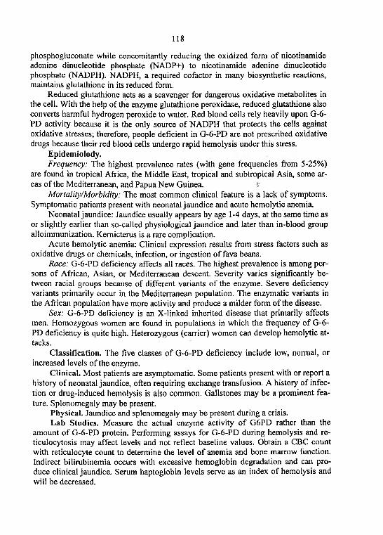
118
phosphogluconate while concomitantly reducing the oxidized form o f nicotinamide adenine dinucleotide phosphate (NADP+) to nicotinamide adenine dinucleotide phosphate (NADPH). NADPH, a required cofactor in many biosynthetic reactions, maintains glutathione in its reduced form.
Reduced glutathione acts as a scavenger for dangerous oxidative metabolites in the cell. With the help of the enzyme glutathione peroxidase, reduced glutathione also converts harmful hydrogen peroxide to water. Red blood cells rely heavily upon G-6- PD activity because it is the only source of NADPH that protects the cells against oxidative stresses; therefore, people deficient in G-6-PD are not prescribed oxidative drugs because their red blood cells undergo rapid hemolysis under this stress.
Epidemiolody.Frequency: The highest prevalence rates (with gene frequencies from 5-25%)
are found in tropical Africa, the Middle East, tropical and subtropical Asia, some areas of the Mediterranean, and Papua New Guinea. ć
Mortality/Morbidity: The most common clinical feature is a lack of symptoms. Symptomatic patients present with neonatal jaundice and acute hemolytic anemia.
Neonatal jaundice: Jaundice usually appears by age 1-4 days, at the same time as or slightly earlier than so-called physiological jaundice and later than in-blood group alloimmunization. Kemicterus is a rare complication.
Acute hemolytic anemia: Clinical expression results from stress factors such as oxidative drugs or chemicals, infection, or ingestion o f fava beans.
Race: G-6-PD deficiency affects all races. The highest prevalence is among persons o f African, Asian, or Mediterranean descent. Severity varies significantly between racial groups because of different variants o f the enzyme. Severe deficiency variants primarily occur in the Mediterranean population. The enzymatic variants in the African population have more activity and produce a milder form of the disease.
Sex: G-6-PD deficiency is an X-linked inherited disease that primarily affects men. Homozygous women are found in populations in which the frequency o f G-6- PD deficiency is quite high. Heterozygous (carrier) women can develop hemolytic attacks.
Classification. The five classes o f G-6-PD deficiency include low, normal, or increased levels o f the enzyme.
Clinical. Most patients are asymptomatic. Some patients present with or report a history o f neonatal jaundice, often requiring exchange transfusion. A history of infection or drug-induced hemolysis is also common. Gallstones may be a prominent feature. Splenomegaly may be present.
Physical. Jaundice and splenomegaly may be present during a crisis.Lab Studies. Measure the actual enzyme activity o f G6PD rather than the
amount o f G-6-PD protein. Performing assays for G-6-PD during hemolysis and re- ticulocytosis may affect levels and not reflect baseline values. Obtain a CBC count with reticulocyte count to determine the level o f anemia and bone marrow function. Indirect bilirubinemia occurs with excessive hemoglobin degradation and can produce clinical jaundice. Serum haptoglobin levels serve as an index of hemolysis and will be decreased.
119
Im aging Studies. Abdominal ultrasound may be useful in assessing for splenomegaly and gallstones.
Histologic Findings: Acute hemolysis from G-6-PD deficiency is associated with formation o f Heinz bodies, which consist o f denatured hemoglobin.
T reatm ent.Diet: Patients must avoid broad beans (ie, fava beans). Favism occurs only in
the Mediterranean variety o f G-6-PD deficiency. Avoid prescribing medications that can cause hemolytic anemia.
Activity: Curtailment o f physical activity may be necessary if severe anemia results from hemolysis.
D rugs prevention: Avoid oxidant drugs such as the antimalarial drugs primaquine, chloroquine, pamaquine, and pentaquine. Avoid nitrofurantoin. Avoid nalidixic acid, ciprofloxacin niridazole, norfloxacin, methylene blue, chloramphenicol, phenazopyridine, and vitamin К analogs. Avoid sulfonamides such as sulfanilamide, sulfamethoxypyridazine, sulfacetamide, sulfadimidine, sulfapyridine, sulfam- erazine, and sulfamethoxazole. Avoid exposure to certain chemicals such as those in mothballs. The following substances should also be avoided in G-6-PD deficiency: acetanilid, doxorubicin, isobutyl nitrite, naphthalene, phenylhydrazine, pyridium.
Prognosis. Most individuals with G-6-PD deficiency do not need treatment.
H EM A TO LO G IC M ALIGNANCIES
ACUTE M YELOID LEUKEM IA (AML)
Introduction . Acute myelogenous leukemia (AML) is a malignant disease of the bone marrow in which hematopoietic precursors are arrested in an early stage of development. Incidence increases with age. AML usually associated with exposure to benzene, radiation and alkylating agents.
Synonyms: AML, acute myelogenous leukemia, acute nonlymphoblastic leukemia, acute nonlymphocytic leukemia, acute non-lymphoblastic leukemia, acute non-lymphocytic leukemia.
Definition. Acute myeloid leukemia (AML) is a malignnnt disease of the bone marrow with failure of myeloid cell to differentiate beyond blast stage and clonal proliferation o f immature hematopoietic cells.
Pathophysiology. The underlying pathophysiology consists o f a maturational arrest o f bone marrow cells in the earliest stages of development (blasts). The mechanism of this arrest is under study, but in many cases, it involves the activation of abnormal genes through chromosomal translocations and other genetic abnormalities.
This developmental arrest results in 2 disease processes. First, the production of normal blood cells markedly decreases, which results in varying degrees o f anemia, thrombocytopenia, and neutropenia. Second, the rapid proliferation o f these cells, along with a reduction in their ability to undergo programmed cell death (apoptosis), results in their accumulation in the bone marrow, blood, and, frequently, the spleen and liver.
120
Uncontrolled growth of blasts in marrow leads to suppression of normal hematopoietic cells, appearance of blasts in peripheral blood, accumulation o f blasts in other sites, metabolic consequences o f a large tumour mass.
Note. Chronic myeloproliferative disorders and myelodysplastic syndromes can transform into AML.
Epidemiolody.Frequency: AML is more commonly diagnosed in developed countries.Mortality: In adults, treatment results are generally analyzed separately for
younger (18-60 y) and older (>60 y) patients. With current standard chemotherapy regimens, approximately 25-30% o f adults younger than 60 years survive longer than 5 years and are considered cured. Results in older patients are more disappointing, with fewer than 10% of patients surviving long-term.
Race: AML is more common in whites than in other populations.Sex: AML is more common in men than in women. The difference is even more
apparent in older patients.Age: Prevalence increases with age. The median age of onset is 65 years. How
ever, this disease affects all age groups.Classification.The older, more traditional, FAB classification is as follows:M0 - Undifferentiated leukemiaM l - Myeloblastic without differentiationМ2 - Myeloblastic with differentiationМ3 - PromyelocyticM4 - MyelomonocyticM4eo - Myelomonocytic with eosinophiliaM5 - Monoblastic leukemiaM5a - Monoblastic without differentiationM5b - Monocytic with differentiationM6 - ErythroleukemiaM7 - Megakaryoblastic leukemia
The newer WHO classification is as follows:A ML with recurrent genetic abnormalities:AML with t(8;21)(q22;q22), (AML 1/ЕТО)AML with abnormal bone marrow eosinophils and inv(16)(pl3q22) or
t(16;16)(pl3)(q22), (CBFB/MYH11)APL with t( 15; 17)(q22;q 12), (PML/RARa) and variants AML with 1 lq23 (MLL) abnormalities AML with multilineage dysplasia:Following myelodysplastic syndrome (MDS) or MDS/myebproliferative dis
ease (MPD)Without antecedent MDS or MDS/MPD but with dysplasia in at least 50% of
cells in 2 or more lineagesAML and MDS, therapy related:Alkylating agent or radiation-related type
121
Topoisomerase II inhibitor type OthersAML, not otherwise classified:AML, minimally differentiatedAML, without maturationAML, with maturationAcute myelomonocytic leukemiaAcute monoblastic or monocytic leukemiaAcute erythroid leukemiaAcute megakaryoblastic leukemiaAcute basophilic leukemiaAcute panmyelosis and myelofibrosisMyeloid sarcomaClinical. Patients present with symptoms resulting from bone marrow failure,
organ infiltration with leukemic cells, or both. The time course is variable.Symptoms o f bone marrow failure are related to anemia, neutropenia, and
thrombocytopenia (decrease in normal hematopoiesis).Anemia: pallor, weakness, fatigue, dizziness, dyspnea on exertion, anginal chest
pain in patients with coronary artery disease.Neutropenia: infections and fever, which may occur with or without specific
documentation o f an infection.Thrombocytopenia: purpura, mucosal bleeding associated with disseminated in
travascular coagulation (DIC). Potentially life-threatening sites, o f bleeding include the lungs, gastrointestinal tract, and the central nervous system.
The most common sites o f infiltration with leukemic cells include the spleen, liver, and gums.
Patients with splenomegaly note fullness in the left upper quadrant and early satiety. Patients with gum infiltration often present to their dentist first. Gingivitis due to neutropenia can cause swollen gums, and thrombocytopenia can cause the gums to bleed. Patients with markedly elevated WBC counts (>100,000 cells/pL) can present with symptoms o f leukostasis (ie, respiratory distress and altered mental status). Leu- kostasis is a medical emergency that requires immediate intervention. Patients with eyes infiltration can cause blurred vision and diplopia. Patients with accumulation of blast cells in marrow can present with symptoms o f skeletal pain, bony tenderness, especially sternum.
Physical. Physical signs o f anemia, including pallor and i cardiac flow murmur, are frequently present. Fever and other signs o f infection can occur, including lung findings o f pneumonia. Patients with thrombocytopenia usually demonstrate pete- chiae, particularly on the lower extremities. Petechiae are smali, often punctate, hemorrhagic rashes that are not palpable. Areas o f dermal bleeding or bruises (ie, ecchy- moses) that are large or present in several areas may indicate a coexistent coagulation disorder such as DIC. Purpura is characterized by flat bruises that are larger than petechiae but smaller than ecchymoses. Signs relating to organ infiltration with leukemic cells include hepatosplenomegaly and, to a lesser degree, lymphadenopathy. Occasionally, patients have skin rashes due to infiltration o f the skin with leukemic
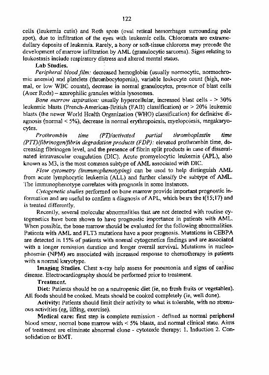
122
cells (leukemia cutis) and Roth spots (oval retinal hemorrhages surrounding pale spot), due to infiltration of the eyes with leukemic cells. Chloromata are extramedullary deposits of leukemia. Rarely, a bony or soft-tissue chloroma may precede the development o f marrow infiltration by AML (granulocytic sarcoma). Signs relating to leukostasis include respiratory distress and altered mental status.
L ab Studies.Peripheral blood film: decreased hemoglobin (usually normocytic, normochro
mic anemia) and platelets (thrombocytopenia), variable leukocyte count (high, normal, or low WBC counts), decrease in normal granulocytes, presence of blast cells (Auer Rods) - azurophilic granules within lysosomes.
Bone marrow aspiration: usually hypercellular, increased blast cells - > 30% leukemic blasts (French-American-British (FAB) classification) or > 20% leukemic blasts (the newer World Health Organization (WHO) classification) for definitive diagnosis (normal < 5%), decrease in normal erythropoiesis, myelopoiesis, megakaryocytes.
Prothrombin time (PT)/activated partial thromboplastin time (PTT)/fibrinogenJfibrin degradation products (FDP): elevated prothrombin time, decreasing fibrinogen level, and the presence o f fibrin split products in case o f disseminated intravascular coagulation (DIC). Acute promyelocytic leukemia (APL), also known as М3, is the most common subtype o f AML associated with DIC.
Flow cytometry (immunophenotyping) can be used to help distinguish AML from acute lymphocytic leukemia (ALL) and further classify the subtype of AML. The immunophenotype correlates with prognosis in some instances.
Cytogenetic studies performed on bone marrow provide important prognostic information and are useful to confirm a diagnosis o f APL, which bears the t(15;17) and is treated differently.
Recently, several molecular abnormalities that are not detected with routine cytogenetics have been shown to have prognostic importance in patients with AML. When possible, the bone marrow should be evaluated for the following abnormalities. Patients with AML and FLT3 mutations have a poor prognosis. Mutations in СЕВРА are detected in 15% o f patients with normal cytogenetics findings and are associated with a longer remission duration and longer overall survival. Mutations in nucleo- phosmin (NPM) are associated with increased response to chemotherapy in patients with a normal karyotype.
Im aging Studies. Chest x-ray help assess for pneumonia and signs of cardiac disease. Electrocardiography should be performed prior to treatment.
T reatm ent.Diet: Patients should be on a neutropenic diet (ie, no fresh fruits or vegetables).
All foods should be cooked. Meats should be cooked completely (ie, well done).Activity: Patients should limit their activity to what is tolerable, with no strenu
ous activities (eg, lifting, exercise).Medical care: first step is complete remission - defined as normal peripheral
blood smear, normal bone marrow with < 5% blasts, and normal clinical state. Aims o f treatment are eliminate abnormal clone - cytotoxic therapy: 1. Induction 2. Consolidation or BMT.

123
Induction therapy: The most common approach is called ”3 and 7,” which consists o f 3 days o f a 15- to 30-minute infusion o f an anthracycline (idarubicin or daun- orubicin), combined with 100 mg/m2 o f arabinosylcytosine (araC) as a 24-hour infusion daily for 7 days. Idarubicin is given at a dose of 12 mg/m2/d for 3 days, daun- orubicin at 45-60 mg/m2/d for 3 days. These regimens require adequate cardiac, hepatic, and renal function. Using these regimens, approximately 50% o f patients achieve remission with one course. Another 10-15% enters remission following a second course o f therapy.
Consolidation therapy: Treatment options for consolidation therapy include high-dose araC, autologous stem cell transplantation, or allogeneic stem cell transplantation.
Drugs. Antineoplastics are used for induction or consolidation therapy.Cytosine arabinoside, cytarabine (Cytosar-U) - 100 mg/m2/d IV as a 24-h
continuous infusion for 7 d 3 g/m2/d IV as a 3-h infusion bid on d 1, 3, and 5. Cytosine arabinoside is antimetabolite specific for cells in the S-phase o f the cell cycle. Cytosine arabinoside acts through inhibition of DNA polymerase and cytosine incorporation into DNA and RNA.
D aunorubicin - 45-60 mg/m2/d IV as a 15- to 30-min infusion for 3 d. Daun- orubicin is topoisomerase-II inhibitor. Daunorubicin inhibits DNA and RNA synthesis by intercalating between DNA base pairs.
Idarubicin (Idam ycin) - 12 mg/m2/d IV as a 15- to 30-min infusion for 3 d. Idarubicin is topoisomerase-II inhibitor. Idarubicin inhibits cell proliferation by inhibiting DNA and RNA polymerase.
Supportive care: prophylaxis against infection via regular control o f urine, feces, sputum, oropharynx, catheter sites, perianal area, antibiotics if fever and chest x-ray, prevention and treatment o f metabolic abnormalities.
Prognosis. Achievement o f first remission is 70-80% if 60 years old, and 50% if > 60 years old. Median survival is 12-24 months; 5 year survival is 40%. Statistics may be improved by BMT - 50-60% cure rate.
ACUTE LYM PHOBLASTIC LEUK EM IA (ALL)
Introduction . Acute lymphoblastic leukemia (ALL) develops from any lymphoid cell blocked at a particular stage of development. ALL may be distinguished from other malignant lymphoid disorders by the immunophenotype o f the cells, which is similar to B- or T-precursor cells. Immunochemistry, cytochemistry, and cytogenetic markers also may aid in categorizing the malignant lymphoid clone.
Synonyms: acute lymphoblastic leukemia, ALL.Definition. Acute lymphoblastic leukemia (ALL) is a malignant (clonal) disease
o f the bone marrow in which early lymphoid precursors proliferate and replace the normal hematopoietic cells o f the marrow.
Pathophysiology. The malignant cells o f ALL are lymphoid precursor cells (ie, lymphoblasts) that are arrested in an early stage o f development. This arrest is caused by an abnormal expression of genes, often as a result o f chromosomal translocations.
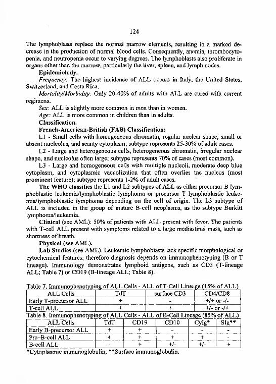
124
The lymphoblasts replace the normal marrow elements, resulting in a marked decrease in the production of normal blood cells. Consequently, anemia, thrombocytopenia, and neutropenia occur to varying degrees. The lymphoblasts also proliferate in organs other than the marrow, particularly the liver, spleen, and lymph nodes.
Epidemiolody.Frequency: The highest incidence of ALL occurs in Italy, the United States,
Switzerland, and Costa Rica.Mortality/Morbidity: Only 20-40% o f adults with ALL are cured with current
regimens.Sex: ALL is slightly more common in men than in women.Age: ALL is more common in children than in adults.Classification.French-Am erican-B ritish (FAB) Classification:LI - Small cells with homogeneous chromatin, regular nuclear shape, small or
absent nucleolus, and scanty cytoplasm; subtype represents 25-30% of adult cases.L2 - Large and heterogeneous cells, heterogeneous chromatin, irregular nuclear
shape, and nucleolus often large; subtype represents 70% of cases (most common).L3 - Large and homogeneous cells with multiple nucleoli, moderate deep blue
cytoplasm, and cytoplasmic vacuolization that often overlies the nucleus (most prominent feature); subtype represents 1-2% o f adult cases.
The W HO classifies the LI and L2 subtypes o f ALL as either precursor В lymphoblastic leukemia/Iymphoblastic lymphoma or precursor T lymphoblastic leuke- mia/lymphoblastic lymphoma depending on the cell o f origin. The L3 subtype of ALL is included in the group o f mature В-cell neoplasms, as the subtype Burkitt lymphoma/leukemia.
Clinical (see AML): 50% of patients with ALL present with fever. The patients with T-cell ALL present with symptoms related to a large mediastinal mass, such as shortness o f breath.
Physical (see AML).L ab Studies (see AML). Leukemic lymphoblasts lack specific morphological or
cytochemical features; therefore diagnosis depends on immunophenotyping (B or T lineage). Immunology demonstrates lymphoid antigens, such as CD3 (T-lineage ALL; Table 7) or CD19 (B-lineage ALL; Table 8).
Table 7. Immunophenotyping o f ALL Cells - ALL o f Т-Cell Lineage (15% of ALL)ALL Cells TdT surface CD3 CD4/CD8
Early T-precursor ALL + - +/+ or -/-T-cell ALL + + +/- or -/+
Table 8. Immunophenotyping o f ALL Cells - ALL o f В-Cell Lineage (85% o f ALL)ALL Cells TdT CD19 CD10 Cyig* Sig“
Early В-precursor ALL + + - -Pre-B-cell ALL + 4- + + -
В-cell ALL - + +/- +/- +‘ Cytoplasmic immunoglobulin; “ Surface immunoglobulin.
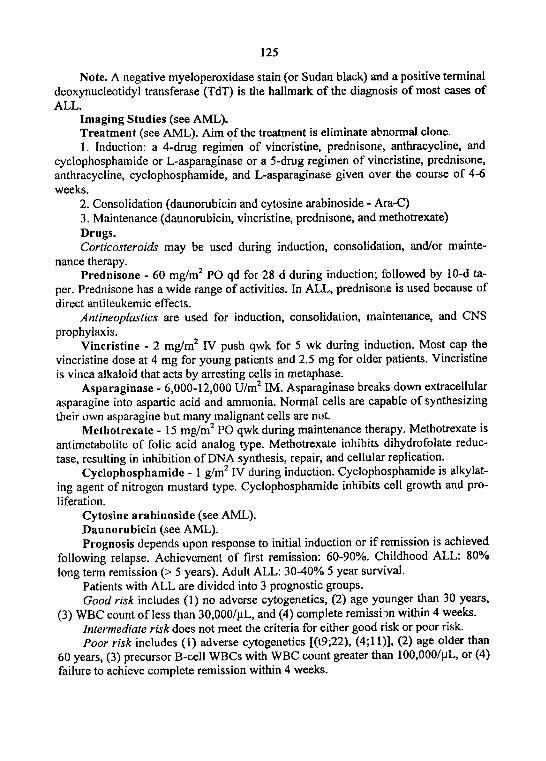
125
Note. A negative myeloperoxidase stain (or Sudan black) and a positive terminal deoxynucleotidyl transferase (TdT) is the hallmark o f the diagnosis o f most cases o f ALL.
Im aging Studies (see AML).T reatm ent (see AML). Aim o f the treatment is eliminate abnormal clone.1. Induction: a 4-drug regimen o f vincristine, prednisone, anthracycline, and
cyclophosphamide or L-asparaginase or a 5-drug regimen o f vincristine, prednisone, anthracycline, cyclophosphamide, and L-asparaginase given over the course o f 4-6 weeks.
2. Consolidation (daunorubicin and cytosine arabinoside - Ara-C)3. Maintenance (daunorubicin, vincristine, prednisone, and methotrexate)Drugs.Corticosteroids may be used during induction, consolidation, and/or mainte
nance therapy.Prednisone - 60 mg/m2 PO qd for 28 d during induction; followed by 10-d ta
per. Prednisone has a wide range o f activities. In ALL, prednisone is used because of direct antileukemic effects.
Antineoplastics are used for induction, consolidation, maintenance, and CNS prophylaxis.
V incristine - 2 mg/m2 IV push qwk for 5 wk during induction. Most cap the vincristine dose at 4 mg for young patients and 2.5 mg for older patients. Vincristine is vinca alkaloid that acts by arresting cells in metaphase.
Asparaginase - 6,000-12,000 U/m2 IM. Asparaginase breaks down extracellular asparagine into aspartic acid and ammonia. Normal cells are capable o f synthesizing their own asparagine but many malignant cells are not.
M ethotrexate - 15 mg/m2 PO qwk during maintenance therapy. Methotrexate is antimetabolite o f folic acid analog type. Methotrexate inhibits dihydrofolate reductase, resulting in inhibition o f DNA synthesis, repair, and cellular replication.
Cyclophospham ide - 1 g/m2 IV during induction. Cyclophosphamide is alkylating agent o f nitrogen mustard type. Cyclophosphamide inhibits cell growth and proliferation.
Cytosine arabinoside (see AML).D aunorubicin (see AML).Prognosis depends upon response to initial induction or if remission is achieved
following relapse. Achievement o f first remission: 60-90%. Childhood ALL: 80% long term remission (> 5 years). Adult ALL: 30-40% 5 year survival.
Patients with ALL are divided into 3 prognostic groups.Good risk includes (1) no adverse cytogenetics, (2) age younger than 30 years,
(3) WBC count o f less than 30,000/pL, and (4) complete remission within 4 weeks.Intermediate risk does not meet the criteria for either good risk or poor risk.Poor risk includes (1) adverse cytogenetics [(t9;22), (4;11)], (2) age older than
60 years, (3) precursor В-cell WBCs with WBC count greater than 100,000/[jL, or (4) failure to achieve complete remission within 4 weeks.
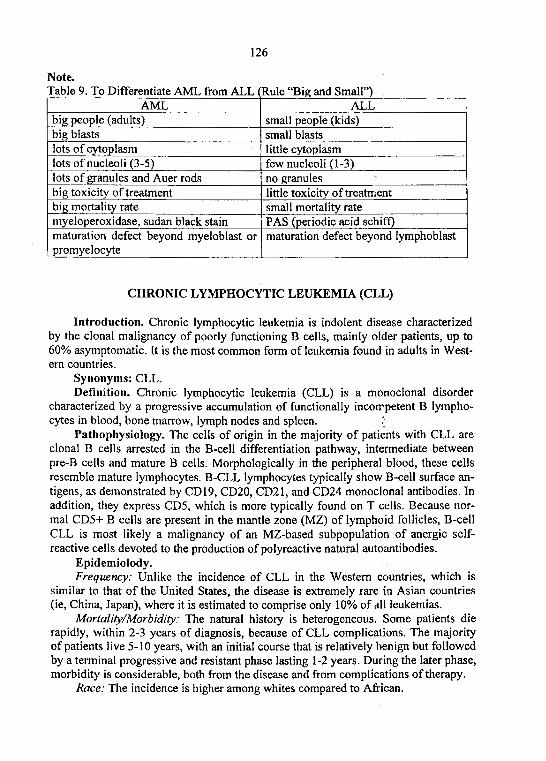
126
Note.Table 9. To Differentiate AML from ALL (Rule “Big and Small”)
AML ALLbig people (adults) small people (kids)big blasts small blastslots o f cytoplasm little cytoplasmlots o f nucleoli (3-5) few nucleoli (1-3)lots o f granules and Auer rods no granulesbig toxicity of treatment little toxicity of treatmentbig mortality rate small mortality ratemyeloperoxidase, Sudan black stain PAS (periodic acid schiff)maturation defect beyond myeloblast or promyelocyte
maturation defect beyond lymphoblast
CHRONIC LYM PHOCYTIC LEUKEM IA (CLL)
Introduction. Chronic lymphocytic leukemia is indolent disease characterized by the clonal malignancy of poorly functioning В cells, mainly older patients, up to 60% asymptomatic. It is the most common form of leukemia found in adults in Western countries.
Synonyms: CLL.Definition. Chronic lymphocytic leukemia (CLL) is a monoclonal disorder
characterized by a progressive accumulation o f functionally incompetent В lymphocytes in blood, bone marrow, lymph nodes and spleen. I
Pathophysiology. The cells o f origin in the majority o f patients with CLL are clonal В cells arrested in the В-cell differentiation pathway, intermediate between pre-B cells and mature В cells. Morphologically in the peripheral blood, these cells resemble mature lymphocytes. B-CLL lymphocytes typically show В-cell surface antigens, as demonstrated by CD19, CD20, CD21, and CD24 monoclonal antibodies. In addition, they express CD5, which is more typically found on T cells. Because normal CD5+ В cells are present in the mantle zone (MZ) of lymphoid follicles, B-cell CLL is most likely a malignancy of an MZ-based subpopulation of anergic self- reactive cells devoted to the production o f poly reactive natural autoantibodies.
Epidemiolody.Frequency: Unlike the incidence of CLL in the Western countries, which is
similar to that o f the United States, the disease is extremely rare in Asian countries (ie, China, Japan), where it is estimated to comprise only 10% of all leukemias.
Mortality/Morbidity: The natural history is heterogeneous. Some patients die rapidly, within 2-3 years o f diagnosis, because of CLL complications. The majority o f patients live 5-10 years, with an initial course that is relatively benign but followed by a terminal progressive and resistant phase lasting 1-2 years. During the later phase, morbidity is considerable, both from the disease and from complications of therapy.
Race: The incidence is higher among whites compared to African.
127
Sex: The incidence is higher in males than in females, with a male-to-female ratio o f 1.7:1.
Age: CLL is a disease that primarily affects elderly individuals, with the majority o f cases reported in individuals older than 55 years. The incidence continues to rise in those older than 55 years. Recently, individuals aged 35 years or younger are being diagnosed more frequently.
Classification. Two staging systems are in common use, the Rai-Sawitsky in the United States and the Binet in Europe. Neither is completely satisfactory, and both have been often modified. Because of its historical precedent and wide use, the Rai- Sawitsky system is described first, followed by the Binet. The International Workshop on Chronic lymphocytic Leukemia (IWCLL) system is listed last.
The Rai-Sawitsky staging system divides CLL into 5 Stages, 0-IV.Stage 0 is lymphocytosis in the blood and marrow only with a survival of
longer than 120 months.Stage 1 is lymphocytosis and adenopathy, with a survival o f 95 months.Stage II is lymphocytosis plus splenomegaly and/or hepatomegaly, with a sur
vival o f 72 months.Stage III is lymphocytosis plus anemia (hemoglobin <10 g), with a survival of
30 months.Stage IV is lymphocytosis plus thrombocytopenia (platelets <100,000), with a
survival o f 30 months.The Binet staging system uses 3 stages, A, B, and C.Stage A requires hemoglobin o f greater than or equal to 100 g/L, platelets
greater than or equal to 100 X 10-9, and fewer than 3 lymph node areas involved (Rai-Sawitsky stages 0 , 1, II). Survival is longer than 120 months.
Stage В requires hemoglobin and platelet levels as in stage A and 3 or more lymph node areas involved (Rai-Sawitsky stages I and II). Survival is 61 months.
Stage C is hemoglobin less than 100 g/L, platelets less than 100 X 10-9, or both (Rai-Sawitsky stages III and IV). Survival is 32 months.
The IWCLL has recommended integrating the Rai-Sawitsky and Binet systems as follows: A(0), A(I), A(II), B(I), B(II), C(III), and C(IV).
Clinical. Patients with CLL present with a wide range o f symptoms and signs at presentation. Onset is insidious, and it is not unusual for this disorder to be discovered incidentally after a blood cell count is performed for another reason.
Predisposition to repeated infections such as pneumonia, herpes simplex labialis, and herpes zoster, enlarged lymph nodes, early satiety and/or abdominal discomfort related to an enlarged spleen, mucocutaneous bleeding and/or petechiae secondary to thrombocytopenia, tiredness and fatigue secondary to anemia.
Complications o f CLL: bone marrow failure, bulky lymphadenopathy, hyper- splenism, immune hemolytic anemia, immune thrombocytopenia, hypogammaglo- binemia, monoclonal gammopathy (often IgM), hyperuricemia with treatment, transformation to histiocytic lymphoma.
Physical.General inspection and palpation: localized or generalized lymphadenopathy,
splenomegaly (30-40% o f cases), hepatomegaly (20% o f cases), petechiae, pallor.
128
L ab Studies.Periferal blood film : absolute lymphocytosis > 5.0 x 10⁹/L (usually > 10.0 x
109/L) with small and mature lyrriphocytes and smudge cells (artifacts due to damaged lymphocytes during the slide preparation).
Bone marrow aspiration: diffuse or focal infiltration o f marrow by lymphocytes.Peripheral blood flow cytometry: the presence of circulating clonal B-
lymphocytes expressing CD5, CD19, CD20(dim), CD 23, and an absence o f FMC-7 staining.
Im aging Studies.Liver/spleen scan may demonstrate splenomegaly.Computed tomography o f chest, abdomen, or pelvis generally is not required for
staging purposes. However, be careful to not miss lesions such as obstructive uropa- thy or airway obstruction that are caused by lymph node compression on organs or internal structures.
T reatm ent.M edical Care: At the time o f diagnosis, most patients do not need to be treated
with chemotherapy unless they have weight loss o f more than 10%, extreme fatigue, fever related to leukemia, night sweats, progressive marrow failure, autoimmune anemia or thrombocytopenia not responding to prednisone, progressive splenomegaly, massive lymphadenopathy, or progressive lymphocytosis. Progressive lymphocytosis is defined as an increase o f greater than 50% in 2 months or a doubling time o f less than 6 months.
Various combination regimens have shown improved response rates in several randomized trials but failed to show any survival advantage. Common combination regimens include chlorambucil and corticosteroids; cyclophosphamide, doxorubicin, and prednisone (CAP); cyclophosphamide, vincristine, and prednisone (CVP); and cyclophosphamide, doxorubicin, vincristine, and prednisone (CHOP; see ALL and AML).
Chlorambucil (alkylating agent) and fludarabine (antimetabolite) are commonly used in the treatment o f CLL. Purine analogs and, in particular, fludarabine are very active against CLL. Fludarabine produces remissions in a significant proportion of patients. It appears to induce apoptosis in malignant lymphocytes upon exposure.
Alkylating agents inhibit cell growth and proliferation.Chloram bucil (Leukeran) - 0.1-0.2 mg/kg/d PO or 3-6 m gW /d PO for 3-6
wk; adjust dose based on blood counts Chlorambucil is nitrogen mustard derivative with bifunctional alkylating activity, forms intrastrand crosslinks, interfering with DNA replication and RNA transcription and translation.
Nucleotide analogs (antimetabolites).F ludarabine - 25 mg/m2/d IV over 30 min qd for 5 d; repeat 5-d course q28d.
Adjust dose based on hematological or nonhematological toxicity. Fludarabine is nucleotide analog of vidarabine converted to 2-fluoro-ara-A that enters the cell and is phosphorylated to form active metabolite 2-fluoro-ara-ATP, which, inhibits DNA synthesis.
Antineoplastic agents are treating by inhibiting key factors responsible for neoplastic transformation o f cells.
129
Alem tuzum ab (Com path) - 3-10 mg IV over 2 h initially; titrate slowly to 30 mg and administer 3 times / wk for up to 12 wk if no adverse effects. Alemtuzumab is monoclonal antibody against CD52, an antigen found on В-cells, T-cells, and almost all CLL cells. Alemtuzumab binds to the CD52 receptor o f the lymphocytes, which slows the proliferation o f leukocytes.
Surgical C are: Refractory splenomegaly and pancytopenia is a common problem in patients with advanced CLL that occasionally necessitates splenectomy. Substantial improvements in hemoglobin and platelet counts are observed in up to 90% o f patients undergoing splenectomy. All patients who are to undergo splenectomy should be immunized at least a week in advance with Pneumovax and Haemophilus and Neisseria meningitides vaccines.
Prognosis. Patients with CLL have 9 year median survival, but vary greatly.
CH R O N IC M YELOGENOUS (GRANULOCYTIC) LEUK EM IA (CM L)
Introduction. Chronic myelogenous (granulocytic) leukemia is a myeloproliferative disorder with overproduction o f myeloid cells, erythoid cells and platelets in peripheral blood and marked myeloid hyperplasia in bone marrow.
Synonyms: chronic myelogenous leukemia, CML, chronic granulocytic leukemia, Philadelphia chromosome positive myeloproliferative disorder.
Definition. Chronic myelogenous leukemia (CML) is a myeloproliferative disorder characterized by increased proliferation o f the granulocytic cell line without the loss o f their capacity to differentiate. Consequently, the peripheral blood cell profile shows an increased number o f granulocytes and their immature precursors, including occasional blast cells.
Pathophysiology. CML is an acquired abnormality that involves the hematopoietic stem cell. It is characterized by a cytogenetic aberration consisting o f a reciprocal translocation between the long arms o f chromosomes 22 and 9; t(9;22). The translocation results in a shortened chromosome 22, an observation first described by Nowell and Hungerford and subsequently termed the Philadelphia (Ph) chromosome after the city o f discovery.
Epidemiolody.Frequency: Increased incidence was reported among individuals exposed to ra
diation in Nagasaki and Hiroshima after the dropping o f the atomic bomb.Mortality/Morbidity: Generally, 3 phases o f the disease are recognized. The me
dian survival o f patients using older forms o f therapy was 3-5 years. Most patients present in the chronic phase, characterized by splenomegaly and leukocytosis with generally few symptoms. After an average o f 3-5 years, the disease usually evolves into the blast crisis, which is marked by an increase in the bone marrow or peripheral blood blast count or by the development o f soft tissue or skin leukemic infiltrates. The manifestations of blast crisis are similar to those o f acute leukemia. In many patients, an accelerated phase occurs 3-6 months before the diagnosis o f blast crisis. Clinical features in this phase are intermediate between the chronic phase and blast crisis.
130
Age: In general, this disease occurs in the fourth and fifth decades o f life. Younger patients aged 20-29 years may be affected and may present with a more aggressive form, such as in accelerated phase or blast crisis. Uncommonly, CML may appear as a disease o f new onset in elderly individuals.
Clinical. Nonspecific symptoms o f tiredness, fatigue, and weight loss may occur long after the onset o f the disease (40% o f patients are asymptomatic).
The disease has 3 clinical phases, and it follows a typical course o f an initial chronic phase, during which the disease process is easily controlled; followed by a transitional and unstable course (accelerated phase); and, finally, a more aggressive course (blast crisis), which is usually fatal.
Cronic phase: normal bone marrow function, white blood cells differentiate and function normally. Patients often have symptoms related to enlargement o f the spleen (shoulder tip pain due to splenic infarction), liver, or both.
Acelerated (ransitional) phase: fever, marked increase in basophils (> 20%), peripheral blast cells (> 15%), promyelocytes (> 30%), and decrease in platelets less than 100,000 cells/pL, increased extramedullary hematopoiesis (unusual sites), transformation (disease similar to idiopathic myelofibrosis), pancytopenia secondary to marrow aplasia.
Acute phase (blast transformation or blast crisis): 2/3 develop a picture similar to AML, 1/3 develop a picture similar to ALL. Acute phase is unresponsive to remission induction but remission induction (return to chronic phase) achievable.
In some patients who present in the accelerated, or acute, leukemia phase o f the disease (skipping the chronic phase), bleeding, petechiae, and ecchymoses may be the prominent symptoms. In these situations, fever is usually associated with infections.
Physical. Splenomegaly is the most common physical finding in patients with CML. In more than half the patients with CML, the spleen extends more than 5 cm below the left costal margin at time o f discovery. The size o f the spleen correlates with the peripheral blood granulocyte counts, with the biggest spleens being observed in patients with high WBC counts. A very large spleen is usually a harbinger o f the transformation into an acute blast crisis form o f the disease. Hepatomegaly also occurs, although less commonly than splenomegaly. Hepatomegaly is usually part of the extramedullary hematopoiesis occurring in the spleen. Physical findings o f leu- kostasis and hyperviscosity can occur in some patients, with extraordinary elevation o f their WBC counts, exceeding 300,000-600,000 cells/pL. Upon funduscopy, the retina may show papilledema, venous obstruction, and hemorrhages.
Lab Studies.Peripheral blood film: a typical leukoerythroblastic blood picture, with circulat
ing immature cells from the bone marrow. Leukocytosis (20,000-60,000 cells/pL) with early myeloid precursors and hypogranular basophils are present. Eosinophils and basophils may be increased (more prominent during the transition to acute leukemia). A mild-to-moderate anemia is very common at diagnosis and is usually normochromic and normocytic. The platelet counts at diagnosis can be low, normal, or even increased in some patients (>1 million in some).
Myeloblasts, myelocytes, metamyelocytes, and nucleated red blood cells are commonly present in the blood smear, mimicking the findings in the bone marrow.
131
The presence of the different midstage progenitor cells diffe.entiates this condition from the acute myelogenous leukemias, in which a leukemic gap (maturation arrest) or hiatus exists that shows absence o f these cells.
Leukocyte alkaline phosphatase (LAP) is normal constituent o f secondary neutrophil granules. LAP is low or absent in most cells o f patients with CML (normal or increased in other chronic myeloproliferative diseases and reactive states).
Bone marrow is hypercellular: myeloid hyperplasia (eg, neutrophils, eosinophils, basophils) with a left shift, and increased megakaryocytes. Mild fibrosis is often seen in the reticulin stain.
Cytogenetic studies o f the bone marrow cells and even peripheral blood, should reveal the typical Philadelphia (P h i) chromosome (translocation between chromosomes 9 and 22). The c-abl proto-oncogene is translocated from chromosome 9 to “breakpoint cluster region” (bcr) o f chromosome 22 to produce bcr-c-abl fusion gene (messenger) RNA that encodes for a mutation protein with much greater tyrosine kinase activity compared with the normal protein. Detection of this fusion gene is a diagnostic test for CML (present in over 90% o f patients).
Im aging Studies. Typical hepatomegaly and splenomegaly may be imaged by using a liver/spleen scan. Most often, these are so obvious that radiological imaging is not necessary.
Histologic Findings: Diagnosis is based on the histopathologic findings in the peripheral blood and the Phi chromosome in the bone marrow cells.
T reatm ent.M edical Care: The 3-fold goals o f treatment o f CML have changed markedly in
the past 10 years; they are to achieve a hematologic remission (normal CBC count and physical examination, ie, no organomegaly), to achieve cytogenetic remission (normal chromosome returns with 0% Ph-positive cells), and, most recently, to achieve molecular remission (negative PCR result for the mutational BCR/ABL m- RNA). The latter is an attempt for cure and prolongation of patient survival.
Myelosuppressive agents control the underlying hyperproliferation of the myeloid elements. A myelosuppressive agent is necessary to bring down WBC counts and, occasionally, elevated platelet counts. Size of the spleen correlates with WBC counts, and it shrinks as WBC counts approach reference range. Also, intermediate and myeloblast cells disappear from the circulation.
H ydroxyurea (Hydrea) - initial dose: 30 mg/kg/d at an average o f 1000-1500 mg/d PO in 500-mg tabs. Hydroxyurea can be given at higher doses in patients with extremely high WBC counts (>300,000/p.L) and adjusted accordingly as counts fall and platelet counts drop; dose can be given as a single daily dc se or divided into 2-3 doses at higher dose ranges. Hydroxyurea (Hydrea) is inhibitor o f deoxynucleotide synthesis and DOC for inducing hematologic remission in CML. Less leukemogenic than alkylating agents such as busulfan, melphalan (Alkeran), or chlorambucil. Myelosuppressive effects last a few days to a week and are easier to control than with alkylating agents.
Busulfan (M yleran) - 4-8 mg/d PO; may administer up to 12 mg/d; maintenance dosing range is 1-4 mg/d to 2 mg/wk; discontinue regimen when WBC count reaches 10,000-20,000/j.iL; resume therapy when WBC reaches 50,000/pL. Busulfan
132
(Myleran) is potent cytotoxic drug which, at recommended dosage, causes profound myelosuppression. As alkylating agent, mechanism of action o f active metabolites may involve cross-linking o f DNA, which may interfere with growth of normal and neoplastic cells.
Tyrosine kinase inhibitors: Imatinib mesylate, or STI571, in oral formulation is an agent with strong tyrosine kinase inhibition activity of the BCR/ABL abnormality in all phases of CML.
Im atin ib mesylate (Gleevec): Chronic phase: 400 mg/d PO with food and large glass o f water; may increase to 600 mg/d if no severe adverse effects or severe non- leukemia-related neutropenia or thrombocytopenia. Accelerated phase or blast crisis: 600 mg/d PO with food and large glass o f water; may increase to 800 mg/d (400 mg bid) if no severe adverse effects or severe non-leukemia-related neutropenia or thrombocytopenia, disease continues to progress (any time), hematologic response is not satisfactory (after at least 3 mo treatment), or a loss o f previously achieved hematologic response occurs. Imatinib mesylate is specifically designed to inhibit tyrosine kinase activity of bcr-abl kinase in Ph-positive leukemic CML cell lines. Well absorbed after oral administration, with maximum concentrations achieved within 2-4 h. Elimination is primarily in feces in form o f metabolites.
Interferons: Alfa, beta, and gamma are the 3 types known to date. Alfa group has been found to inhibit propagation o f Ph-positive hematopoietic clone, allowing return o f normal cells in bone marrow.
In terferon alfa-2a (Roferon A) o r alfa-2b (In tron A) - Approximately 5 mil- lion/m2/d SC until complete cytogenetic remission (100% Ph-negative BM cells by FISH). Remission can occur within 1-2 у from onset o f therapy; individual maximally tolerated dose can be obtained by starting at 3 MIU or 1.5 MIU qd and increasing by 3 МШ/d qmo until tolerance or cytogenetic remission.
Interferon alfa-2a (Roferon A) or alfa-2b (Intron A): Both are recombinant alpha interferons with some minor amino acid differences but are considered equivalent modalities in treatment o f CML. Roferon A comes in single (3-, 6-, 9-, and 36-MIU strength) or multidose vials (9- or 18-MIU strength). Intron A comes in multidose pens of 18 MIU (delivers 3 MIU/dose), 30 MIU (5 MIU/dose), and 60 MIU (10 MIU/dose), with each pen good for 6 doses. Elderly patients who cannot tolerate adverse effects may be started at half the recommended starting dose.
Surgical C are: Splenectomy and splenic irradiation have been used in patients with large and painful spleens, usually in the late phase o f the disease. This is rarely needed in patients whose disease is well controlled.
Prognosis. There is classification o f patients into good-, intermediate-, or poor- risk groups, with an average survival o f 5-6 years, 3-4 years, and 2 years, respectively. Poor prognosis in patients with CML is associated with several clinical and laboratory factors, including older age, symptomatic presentation,;poor performance status, hepatomegaly, splenomegaly, negative Ph chromosome, anemia, thrombocytopenia, thrombocytosis, decreased megakaryocytes, basophilia, or myelofibrosis (increased reticulin or collagen). Several therapy-associated factors may indicate a poor prognosis in patients with CML, including longer time to hematologic remission with myelosuppression therapy, short duration o f remission, total dose o f hydroxyurea or
133
busulfan, or poor suppression o f Ph-positive cells by chemotherapy or interferon alfa therapy.
PO LY CY TH EM IA RUBRA VERA (PRV)
Introduction. The most prominent feature o f polycythemia rubra vera is an elevated absolute red blood cell mass because o f uncontrolled red blood cell production. This is accompanied by increased white blood cell (myeloid) and platelet (megakar- yocytic) production, which is due to an abnormal clone o f the hematopoietic stem cells with increased sensitivity to the different growth factors for maturation.
Synonyms: polycythemia vera, PV, plethora vera, primary polycythemia.Definition. Polycythemia rubra vera (PRV) is a stem cell disorder characterized
as a panhyperplastic (red and white blood cells, platelets), malignant, and neoplastic marrow disorder with overproduction o f erythroid cells.
Pathophysiology. Normal stem cells are present in the bone marrow o f patients with PV. Also present are abnormal clonal stem cells that interfere with or suppress normal stem cell growth and maturation. Evidence indicates that the etiology o f panmyelosis is unregulated neoplastic proliferation. The origin o f the stem cell transformation remains unknown.
Epidemiolody.Frequency: PRV is relatively rare, occurring in 0.6 persons per 10,000 popula
tions.Mortality/Morbidity: PRV is a chronic disease, and its natural history o f 1.5-3
years of median survival in the absence o f therapy has been extended to at least 10-20 years because o f new therapeutic tools. The major causes o f morbidity and mortality are as follows. Thrombosis has been reported in 15-60% of patients with PRV. It is the major cause o f death in 10-40% o f patients. Hemorrhagic complications occur in 15-35% o f patients and lead to death in 6-30% of these patients. Peptic ulcer disease is reported to be associated with PRV at a 3- to 5-fold higher rate than that o f the general population. Myelofibrosis and pancytopenia occur in 3-10% o f patients, usually late in the disease, which is considered the spent phase o f PRV. Acute leukemia or a myelodysplastic syndrome develops in 1.5% o f patients treated with phlebotomy alone.
Race: Originally, Jews were thought to have a higher predilection for PRV than persons o f other ethnic groups; however, many studies show that PRV occurs in persons o f all ethnic groups.
Sex: PRV has no sex predilection, although the Polycythemia Vera Study Group (PVSG) found that slightly more males are affected than females.
Age: The peak incidence o f PRV is age 50-70 years. However, it occurs in persons o f all age groups, including those in early adulthood and childhood, albeit rarely.
Classification. There are primary and secomdary polycythemia. Primary polycythemia is polycythemia rubra vera.
Causes o f secondary polycythemia: spurious (decrease in plasma volume), poor tissue oxygenation (high altitude, cyanotic congenital heart disease or pulmonary dis
134
ease, hemoglobinopathies with increased 0 2 affinity, carbon monoxide poisoning), local renal hypoxia (renal artery stenosis, renal cysts), ectopic production o f erythropoietin (uterine leiomyoma, cerebellar hemangioma, hepatocellular cancer, pheo- chromocytoma, renal cell cancer).
Clinical. Symptoms are often insidious in onset. They are often related to blood hyperviscosity secondary to a marked increase in the cellular elements o f blood, which impairs microcirculation.
Symptoms are secondary to high red cell mass and hyperviscosity: headache, dizziness, tinnitus, congestive heart failure, and thrombosis.
Symptoms are secondary to platelet abnormalities: cerebrovascular accident, myocardial infarction, phlebitis, bleeding, bruising.
Symptoms are secondary to high blood histamine (from basophils): pruritus, especially post-bath or shower, peptic ulcer.
Symptoms are secondary to high cell turnover: gout (due to hyperuricemia).Physical. Physical findings are due to manifestations of the myeloproliferative
process and excess of the cellular elements o f blood.General inspection: plethora or a ruddy complexion (increase in total red blood
cell mass). This manifests in the face, palms, naiJbeds, mucosa, and conjunctiva.Palpation o f abdomen: splenomegaly (present in 75% of patients at the time of
diagnosis), and hepatomegaly (present in approximately 30% of patients with PRV).Arterial (blood) pressure: hypertension.Complications: vascular complications (thrombosis, hemoThage), myeloid
metaplasia, acute leukemia.Lab Studies.Peripheral bloodfilm: increased cell counts in all cell lines in the myeloid series
(ie, red blood cells, white blood cells (60% of patients) > 12,000/pL (preferentially granulocytes), and platelets (50% o f patients) > 400,000/pL). Automated red blood cell counts and hematocrit values (including hemoglobin levels) may be deceptive with regard to the total red blood cell mass. However, patients with hemoglobin concentrations o f at least 20 g/dL or hematocrit values of at least 60% in males and 56% in females always have an elevated red blood cell mass.
Total red blood cell mass: in males, greater than or equal to 36 mL/kg; in females, greater than or equal to 32 mL/kg.
Biochemistry: hyperuricemia occurs in 40% of patients and reflects the high turnover rate o f bone marrow cells releasing DNA metabolites.
Leukocyte alkaline phosphatase: increased greater than 100 U/L (70% of patients).
Routine coagulation test results are normal, with a high turnover rate for fibrinogen. The prothrombin time and activated partial thromboplastin time may be ar- tifactually prolonged because of the erythrocytosis.
Bone marrow: Overall hypercellularity with expansion o f all cell lines with megakaryocytic proliferation and the presence of myelofibrosis can help diagnose PRV and MPD, but PRV patients may have normal bone marrow findings.
135
Cytogenetic studies: a clonal abnormality o f the bone marrow cells in 30% of patients who are not treated and in 50% o f patients who are treated with alkylating or myelosuppressive agents.
Im aging Studies. An enlarged spleen is often palpable and does not require any imaging studies. In some patients with posteriorly enlarged spleens or in those who are obese, ultrasonography or CT scanning may be able to detect an enlargement missed during the physical examination.
T reatm ent.Phlebotom y (if symptoms are due to eiythrocytosis alone and platelet count
normal or only slightly increased) - from daily or every other day removal o f 250- 500 mL o f whole blood (patients with severe plethora who have altered mentation or associated vascular compromise can be bled more vigorously) to twice a week (patients with hematocrit values of less than 70%). Phlebotomy or bloodletting has been the mainstay o f therapy for this disease process for a long time. The object is to remove excess cellular elements, mainly red blood cells, to improve the circulation of blood by lowering the blood viscosity.
Drugs: if symptoms systemic or secondary to splenic enlargement.Antimetabolites - H ydroxyurea (H ydrea) is a nonalkylatirg agent that inhibits
DNA synthesis and cell replication by blocking the enzyme ribonucleoside diphosphate reductase (see CML).
Imidazole quinazolines are demonstrated to have powerful antiaggregating effects on platelets and to cause thrombocytopenia.
A nagrelide hydrochloride (Agrylin) - 0.5 mg PO qid or 1 mg PO bid for 7 d. Primary activities is to lower platelet levels but shows slight decrease in mean hemoglobin and hematocrit while WBC counts maintained. Effective in PRV with elevated platelet counts. Adjust dosage to lowest effective dose to reduce and maintain platelet counts, WBC count, and hemoglobin levels within reference range.
Interferons: Recombinant interferon alfa is a biologic response modifier with myelosuppressive activity.
R ecom binant alfa-2a (Roferon) o r ajfa-2b (Intron) interferon - 1.5-3 million U SC 3 times / wk. Recombinant alfa-2a or alfa-2b interferon is protein product manufactured by recombinant DNA technology. Can lower counts and shrink enlarged spleens (see CML).
Note. In general, 32P should be reserved for patients older than 80 years or patients with comorbid conditions in whom life expectancy is less than 5-10 years and the convenience o f 32P dosing outweigh the substantial risks o f developing acute leukemia 5-15 years after 32P administration.
Surgical C are: Consider splenectomy in patients with painful splenomegaly or repeated episodes o f thrombosis causing splenic infarction.
Prognosis. PRV is a chronic disease, and its natural history of 1.5-3 years of median survival in the absence o f therapy has been extended to at least 10-20 years because o f new therapeutic tools.
136
PLASMA CELL M YELOM A (M ULTIPLE MYELOM A)4»
In troduction. First described in 1848, multiple myeloma is a disease characterized by a proliferation o f malignant plasma cells and a subsequent overabundance of monoclonal paraprotein. An intriguing feature o f this disease is that the antibodyforming cells (ie, plasma cells) are malignant and, therefore, may cause unusual manifestations.
Synonyms: myeloma, myeloma multiple, plasma cell myeloma, plasma cell leukemia.
Definition. Plasma cell myeloma is monoclonal malignancy of plasma cells engaged in the production o f a specific protein (paraprotein) characterized by replacement o f bone marrow and bone destruction. The protein produced is monoclonal i.e. one class o f heavy chains and one type o f light chains (“M” protein).
Pathophysiology. Multiple myeloma can cause a wide variety o f problems. The proliferation of plasma cells may interfere with the normal production o f blood cells, resulting in leukopenia, anemia, and thrombocytopenia. The cells may cause soft tissue masses (plasmacytomas) or lytic lesions in the skeleton. Feared complications of this malignancy are bone pain, hypercalcemia, and spinal cord compression. The aberrant antibodies that are produced lead to impaired humoral immunity, and patients have a high prevalence of infection, especially with encapsulated organisms. The overproduction o f these antibodies may lead to hyperviscosity, amyloidosis, and renal failure.
Epidemiolody.Frequency: 3 cases per 100,000.Mortality/Morbidity: Multiple myeloma affects the kidneys in several ways: di
rect tubular injury, amyloidosis, or involvement by plasmacytoma. Spinal cord compression is one o f the most severe adverse effects o f myeloma (20% of patients). A frequent complication o f multiple myeloma is pathologic fractures. Patients with myeloma commonly develop hypercalcemia.
Race: Multiple myeloma accounts for 1-2% o f the malignancies.Sex: The male-to-female ratio is 3:2.Age: The median age o f patients is 68 years for men and 70 years for women.Clinical. Presenting symptoms include bone pain (70%), tenderness, deformity,
pathologic fractures (93%), weakness, fatigue (due to anemia), weight loss, night sweats with advanced disease, abnormal bleeding (epistaxis, purpura), infection (often resulting from pneumococcal infection), hypercalcemia, spinal cord compression, renal failure.
Physical.General inspection: pallor, bone deformity, pathologic fractures, bone tender
ness, petechiae and purpura.Palpation o f abdomen: hepato/splenomegaly.L ab Studies.Peripheral blood film: rouleaux formation (aggregates o f RBC resembling
stacks o f coins), rare plasma cells, normocytic anemia, thrombocytopenia, leuco- penia.
137
Bone marrow: focal or diffuse increase in plasma cells, primitive plasma cells.Biochemistry: hypercalcemia (apathy, weakness, polydipsia, polyuria), increased
creatinine, increased ESR, narrow anion gap (myeloma protein is a cation).Electrophoresis: monoclonal protein on serum protein electrophoresis, heavy
chain and light chain types identified by serum immunoelectrophoresis, decreased normal immunoglobulins, urine electrophoresis (Bence-Jones protein, a light chain dimer).
Im aging Studies.X-ray: Perform a complete skeletal series at diagnosis, including the skull (a
very common site o f bone lesions in persons with multiple myeloma), the long bones (looking for impending fractures), and the spine. Diffuse osteopenia may suggest myelomatous involvement before discrete lytic lesions are apparent.
M RI scan: Findings from MRI scans of the vertebrae are often positive when plain radiographs are not. For this reason, evaluate symptomatic patients with MRI to obtain a clear view o f the spinal column and to assess the integrity o f the spinal cord.
Classic diagnostic triad (must show increased numbers o f atypical immature plasma cells): 1. greater than 10% abnormal plasma cells in bone marrow; 2. lytic bone lesions; 3. monoclonal protein spike in serum or urine.
T reatm ent.Diet: Patients with myeloma who are receiving bisphosphonate therapy should
include adequate calcium in their diet.Activity: Encourage patients with myeloma to be physically active, as appropri
ate to their individual bone status. Physical activity may help maintain bone strength.Drugs. Multiple myeloma is treated with several categories o f medications.
Chemotherapeutic agents are used to reduce the disease burden, and bisphosphonates are used to promote bone healing and to provide secondary prophylaxis against skeletal-related events (eg, hypercalcemia, bone fracture, spinal cord compression, need for radiation, need for surgery). In addition, erythropoietin is used to treat anemia, either alone or in conjunction with chemotherapy.
The regimen used most often is melphalan and prednisone (M and P). Both drugs are typically administered by mouth for 4-7 days; the cycle is repeated every 4- 6 weeks, depending on count recovery.
VAD is administered as a 4-day continuous intravenous infusion o f vincristine and doxorubicin (Adriamycin), with 4 daily oral doses o f dexamethasone (Decadron).
Chemotherapeutic agents.M elphalan (A lkeran) - 9 mg/m2 PO qd for 4 d; alternatively, 6 mg/m2 PO qd
for 7 d. Most widely used regimen is M and P. Melphalan inhibits mitosis by cross- linking DNA strands.
D oxorubicin (Adriam ycin) - 9 mg/m2/d IV continuous infusion on days 1-4 of VAD regimen. Adriamycin is part o f VAD. Inhibits topoisomerase II and produces free radicals, which may cause destruction o f DNA; these 2 events can, in turn, inhibit growth o f neoplastic cells.
V incristine (Oncovin) - 0.4 mg/d IV continuous infusion on days 1-4 o f VAD therapy regimen. Vincristine is part o f VAD therapy. Mechanism o f action is complex and includes depolymerization o f microtubules.
138
Corticosteroids have anti-inflammatory properties and cause profound and varied metabolic effects. Corticosteroids modify body's immune response to diverse stimuli.
Prednisone - 50 mg PO bid for 4 d; alternatively, 100 mg PO qd for 7 d. Most widely used regimen is M and P. Prednisone stabilizes lysosomal membranes and suppresses lymphocyte and antibody production.
Dexamethasone -40 mg/d PO on days 1-4, 9-12, and 17-20 of VAD therapy regimen. Dexamethasone is part o f VAD therapy. Many believe high-dose steroid component o f VAD accounts for much o f its efficacy. In some patients, high-dose dexamethasone alone may produce significant clinical responses. Dexamethasone stabilizes lysosomal membranes and suppresses lymphocyte and antibody production.
Bisphosphonates inhibits bone resorption via action on osteoclast or osteoclast precursors.
Pam idronate - 90 mg IV infusion over 2 h q3-4wk. Pamidronate inhibits normal and abnormal bone resorption. Pamidronate appears to inhibit bone resorption without inhibiting bone formation and mineralization. Newer drugs similar in structure and function are being studied and may have improved efficacy and greater convenience.
Zoledronic acid - 4 mg IV over at least 15 min qmo; hydrate patient prior to infusion; may repeat treatment if serum calcium does not return to desired level after 7 d. Zoledronic acid inhibits bone resorption, possibly by acting on osteoclasts or osteoclast precursors. Zoledronic acid is effective in treating hypercalcemia o f malignancy.
Colony-stimulating factors induce erythropoiesis.Epoetin alfa, erythropoietin is 10,000 U SC 3 times/wk; alternate schedules,
including 40,000U SC once/wk, are sometimes used. Erythropoietin stimulates division and differentiation of committed erythroid progenitor cells; induces release of reticulocytes from bone marrow into blood stream. Naturally occurring hormone produced by kidneys to stimulate bone marrow production of red blood cells. In patients with multiple myeloma, administration of exogenous erythropoietin may correct anemia, leading to a significant improvement in performance status and quality o f life.
Adjunctive therapy for myeloma includes radiation therapy to target areas of pain, impending pathologic fracture, or existing pathologic fracture.
Patients presenting with acute renal failure may benefit from plasmapheresis. Hydration (to maintain a urine output o f >3 L/d), management o f hypercalcemia, and avoidance of nephrotoxins (eg, intravenous contrast media, antibiotics) are also key factors.
Using the patient's own (ie, autologous) bone marrow or peripheral blood stem cells facilitates more intense antimyeloma therapy.
Prognosis. The prognosis for survival in unselected patients with multiple myeloma is 3 years (median survival 24-30 months).
139
H EM O RRH A G IC DIATHESIS
ID IO PA TH IC (AUTOIM M UNE) TH RO M BO CY TO PEN IC PURPURA (ITP)
Introduction . Idiopathic thrombocytopenic purpura (ITP) is a clinical syndrome with low number o f circulating platelets. In persons with ITP, platelets are coated with autoantibodies to platelet membrane antigens, resulting in splenic sequestration and phagocytosis by mononuclear macrophages. The resulting shortened life span of platelets in the circulation, together with incomplete compensation by increased platelet production by bone marrow megakaryocytes, results in a decreased platelet count.
Synonyms: idiopathic thrombocytopenic purpura, ITP, autoimmune thrombocytopenic purpura, primary thrombocytopenic purpura.
Definition. Idiopathic thrombocytopenic purpura (ITP) is a clinical syndrome in which a decreased number of circulating platelets (thrombocytopenia) manifests as a bleeding tendency, easy bruising (purpura), or extravasation of blood from capillaries into skin and mucous membranes (petechiae). No single laboratory result or clinical finding establishes a diagnosis o f ITP; it is a diagnosis o f exclusion (exclude other causes o f thrombocytopenia, such as leukemia, myelophthisic marrow infiltration, myelodysplasia, aplastic anemia, or adverse drug reactions).
Pathophysiology. An abnormal autoantibody, usually immunoglobulin G (IgG) with specificity for 1 or more platelet membrane glycoproteins (GPs), binds to circulating platelet membranes. Autoantibody-coated platelets induce Fc receptor- mediated phagocytosis by mononuclear macrophages, primarily but not exclusively in the spleen. The spleen is the key organ in the pathophysiology of ITP not only because platelet autoantibodies are formed in the white pulp but also because mononuclear macrophages in the red pulp destroy immunoglobulin-coated platelets. If bone marrow megakaryocytes cannot increase production and maintain a normal number o f circulating platelets, thrombocytopenia and purpura develop. Impaired throm- bopoiesis is attributed to failure of a compensatory increase in thrombopoietin and megakaryocyte apoptosis.
Epidemiolody.Frequency: The annual incidence o f chronic ITP is estimated to be 5.8-6.6 cases
per 100,000 persons, but these data are not from large population based studies.Mortality/Morbidity: The primary cause o f long-term morbidity and mortality is
hemorrhage. The most frequent cause o f death in association with ITP is spontaneous or accidental trauma-induced intracranial bleeding in patients whose platelet counts are less than 10 x 109/L (<10 x 103/|iL). This situation occurs in less than 1% of patients.
Sex: In children, the prevalence is the same among boys and girls. In adults, women are affected approximately 3 times more frequently than men.
Age: Children may be affected at any age, but the prevalence peaks in children aged 3-5 years. Adults may be affected at any age, but most cases are diagnosed in women aged 30-40 years. Onset in a patient older than 60 years is uncommon, and a search for other causes o f thrombocytopenia is warranted. The most likely causes in
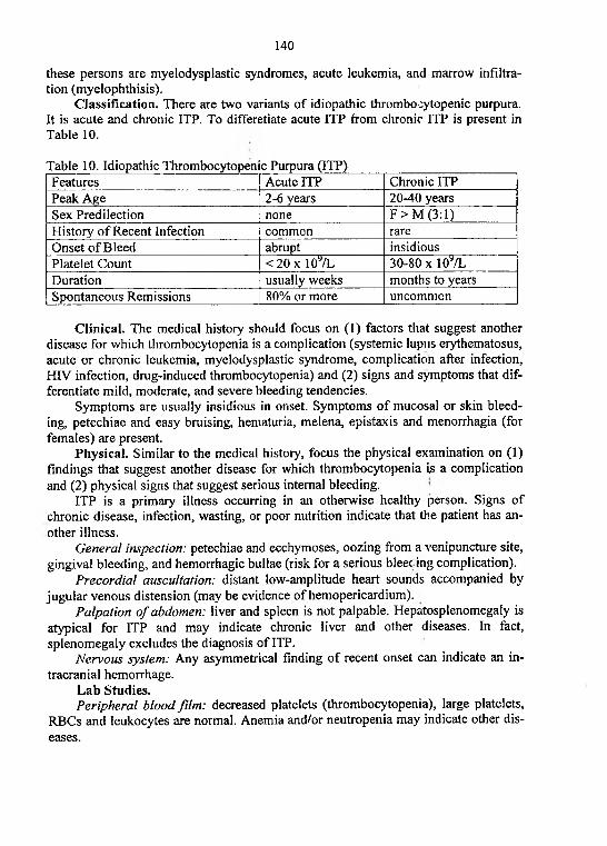
140
these persons are myelodysplastic syndromes, acute leukemia, and marrow infiltration (myelophthisis).
Classification. There are two variants o f idiopathic thrombocytopenic purpura. It is acute and chronic ITP. To differetiate acute ITP from chronic ITP is present in Table 10.
Table 10. Idiopathic Thrombocytopenic Purpura (ITP)Features Acute ITP Chronic ITPPeak Age 2-6 years 20-40 yearsSex Predilection none F > M (3:1)History o f Recent Infection common rareOnset o f Bleed abrupt insidiousPlatelet Count < 20 x 10y/L 30-80 x 10VLDuration usually weeks months to yearsSpontaneous Remissions 80% or more uncommon
Clinical. The medical history should focus on (1) factors that suggest another disease for which thrombocytopenia is a complication (systemic lupus erythematosus, acute or chronic leukemia, myelodysplastic syndrome, complication after infection, HIV infection, drug-induced thrombocytopenia) and (2) signs and symptoms that differentiate mild, moderate, and severe bleeding tendencies.
Symptoms are usually insidious in onset. Symptoms o f mucosal or skin bleeding, petechiae and easy bruising, hematuria, melena, epistaxis and menorrhagia (for females) are present.
Physical. Similar to the medical history, focus the physical examination on (1) findings that suggest another disease for which thrombocytopenia is a complication and (2) physical signs that suggest serious internal bleeding.
ITP is a primary illness occurring in an otherwise healthy person. Signs of chronic disease, infection, wasting, or poor nutrition indicate that the patient has another illness.
General inspection: petechiae and ecchymoses, oozing from a venipuncture site, gingival bleeding, and hemorrhagic bullae (risk for a serious bleec ing complication).
Precordial auscultation: distant low-amplitude heart sounds accompanied by jugular venous distension (may be evidence of hemopericardium).
Palpation o f abdomen: liver and spleen is not palpable. Hepatosplenomegaly is atypical for ГГР and may indicate chronic liver and other diseases. In fact, splenomegaly excludes the diagnosis o f ITP.
Nervous system: Any asymmetrical finding o f recent onset can indicate an intracranial hemorrhage.
Lab Studies.Peripheral blood film : decreased platelets (thrombocytopenia), large platelets,
RBCs and leukocytes are normal. Anemia and/or neutropenia may indicate other diseases.
141
Bone marrow: plentiful megakaryocytes (critical test to rule out other causes o f thrombocytopenia).
Coagulation test: increased bleeding time, prothrombin time (PT) and activated partial thromboplastin time (aPTT) is normal.
Test fo r antiplatelet antibodies: anti-platelet antibodies present in most.Helicobacter pylori testing: Studies from Italy and Japan indicate that many per
sons with ITP have H. pylori gastric infections and that eradication o f H. pylori results in increased platelet counts. In the United States and Spa n, the prevalence o f H. pylori infections does not appear to be increased in persons with ITP and eradication o f H. pylori has not increased platelet counts. Therefore, routine testing for H pylori infections in adults and children with ITP is not recommended.
Im aging Studies. CT scanning and MRI are relatively benign and useful nonin- vasive imaging studies that can be used to rule out other causes o f thrombocytopenia. However, they are not part o f the routine evaluation o f patients who may have ITP. Promptly perform CT or MRI when the medical history or physical findings suggest serious internal bleeding.
T reatm ent.Treatment o f ITP with mild bleeding tendencies is conservative (platelet count >
30,000, no mucosal bleeding).Treatment o f ITP with moderate bleeding tendencies is steroids (moderate dose,
then taper (80% responsive); platelet count < 20-30,000 or evidence o f mucosal bleeding).
Treatment o f ГГР with severe bleeding tendencies is steroids, splenectomy (if steroids fail), IV gamma globulin (if steroids and splenectomy fail or if rapid response is required), immunosuppressives (if steroids and IV gamma globulin fail), and danazol (for postmenopausal women).
Drugs.Corticosteroids are the treatment o f choice for initial management o f acute ITP.
Increase the platelet count by decreasing splenic uptake o f autoar.tibody-coated platelets and by decreasing synthesis o f autoantibody. Dosages must be tapered after a safe platelet count is achieved, and the drug is replaced with IV RhIG or IVIG to avoid serious complications o f chronic hypercorticism.
Prednisone - 1-2 mg/kg PO; if treatment not urgent or if patient at risk for adverse effects (eg, diabetes, hypertension, psychiatric illness), 0.25 mg/kg/d (30-40 mg/d) may be adequate. Oral corticosteroid used most frequently because o f relatively low cost, known adverse effects, and long-term clinical record. For aggressive treatment, may be combined with IV RhIG or IVIG. In emergency, replace PO prednisone with IV methylprednisolone.
M ethylprednisolone (Solu-M edrol) - 1 g/d IV. Methylprednisolone is for initial management o f severe bleeding tendency in ITP. IV recommended when most rapid and reliable treatment o f ITP required. In this situation, combine with IV RhIG in qualified Rh(D)-positive patients or IVIG in Rh(D)-negative patients or unqualified Rh(D)-positive patients.
142
Blood products are used to improve clinical and immunologic aspects o f the disease. They may decrease autoantibody production and increase solubilization and removal o f immune complexes.
IV RhIG - 50 mcg/kg IV single infusion; followed by 20-40 mcg/kg pm; in patients whose hemoglobin concentration >8 g/dL, off-label dose of 75 mcg/kg may increase efficacy without adverse effect. Specialized immunoglobulin product manufactured from pools o f plasma from Rh(D)-negative persons and alloimmunized to D blood group antigen. IV RhIG is subjected to anion-exchange column chromatography to permit IV infusion and solvent-detergent treatment and nanofiltration to reduce infectivity by lipid-enveloped viruses. Induces immune RBC hemolysis in Rh(D)-positive recipients, decreasing function o f mononuclear macrophages (reticuloendothelial blockade) and sparing immunoglobulin-coated platelets from splenic destruction.
IVIG (Sandoglobulin) - begin with 1 g/kg IV at starting rate o f 0.5 mL/kg/h (5% solution); not to exceed 4 mL/kg/h; if no adverse reactions, 10% solution may be started at 0.5 mL/kg/h and increased to 8 mL/kg/h; repeat q3-4wk when indicated by decreasing platelet count. Large dose of 1 g/kg induce decreased function o f mononuclear macrophages (reticuloendothelial blockade), sparing immunoglobulin-coated platelets from splenic destruction. IVIG is used with IV methylprednisolone to manage acute ITP in children. Decreased time to an increased platelet count compared with IV RhIG, but difference not appear to be clinically significant. Compared with IV RhIG, associated with more adverse effects, longer infusions, and increased cost, causing many hematologists to prefer IV RhIG as supplement to corticosteroids, at least for Rh(D)-positive patients.
Immunosuppressive antimetabolites are used in patients with ITP to reduce production o f abnormal autoantibody.
Azathioprine - 2 mg/kg/d PO or IV. Azathioprine may be effective in some patients with ITP who do not or no longer have response to corticosteroids, IV RhIG, or IVIG. Azathioprine may be used with prednisone to reduce dose of prednisone or as another PO medication to delay splenectomy.
The steroidogenic properties o f androgens may modulate immune system.Danazol - 200-600 mg/d PO; after several wk, may reduce to 50-100 mg/d to
decrease adverse effects. Danazol may impair clearance o f immunoglobulin-coated platelets and decreases autoantibody production. Danazol increased platelet counts in 40-50% of patients, particularly postmenopausal women.
Surgical Care: In persons with acute ITP, splenectomy usually results in rapid, complete, and lifelong clinical remission. In persons with chronic ITP, the results of splenectomy are typically less predictable than they are in patients with acute ITP. Platelet counts may not fully revert to normal values, and relapses are not uncommon.
Prognosis. Approximately 60-90% of adults with ITP respond with an increased platelet count after treatment with prednisone or prednisone and IV RhIG or IVIG. O f those adults who do not maintain an increased platelet count and require splenectomy, approximately two thirds have a sustained response and 10-15% have a partial response.
143
H EM O PH ILIA A, H EM O PH ILIA В (CHRISTM AS DISEASE)
Introduction. Hemophilia A (HA) is considered the classic form o f hemophilia, and hemophilia В (HB) is termed Christmas disease. HA is a consequence of a congenital deficiency o f factor VIII (FVIII), and HB is a consequence of a congenital deficiency o f factor IX (FIX). This deficiency results in insufficient generation of thrombin by FIXa and FVIIIa complex through the intrinsic pa thway o f the coagulation cascade.
Synonyms: hemophilia A, HA, hemophilia B, HB, Christmas disease.Definition. Hemophilia A (HA) is disease associated with congenital deficiency
o f factor VIII (FVIII). Hemophilia В (HB or Christmas disease) is disease associated with congenital deficiency of factor IX (FIX).
Pathophysiology. The genes for both FVIII (ie, HA) and FIX (ie, HB) are located on the long arm o f chromosome X. The gene for FVIII (F8C) is unusually large, representing 186 kb o f the X chromosome. It comprises 26 exons and 25 in- trons. Mature FVIII contains 2332 amino acids. Approximately 40% of cases of severe FVIII deficiency arise from a large inversion that disrupts the FVIII gene. Deletions, insertions, and point mutations account for the remaining 50-60% of HA defects.
The FIX gene (F9) has 34 kb composes 8 exons and 7 intervening sequences. The mature protein is composed o f 415 amino acids. Point mutations and deletions in the FIX gene are the most common causes o f HB.
The hallmark o f hemophilia is hemorrhage into the joints. This bleeding is painful and leads to long-term inflammation and deterioration o f t.ie joint, resulting in permanent deformities, misalignment, loss o f mobility, and extremities o f unequal lengths.
Approximately 30% of patients with severe HA develop alloantibody inhibitors that can neutralize FVIII. These inhibitors are typically immunoglobulin G (IgG), predominantly o f the IgG4 subclass that do not fix complements and do not result in the end-organ damage observed with circulating immune complexes.
Epidemiolody.Frequency: The worldwide incidence of HA is approximately 1 case per 5000
male individuals, with approximately one third o f affected individuals not having a family history. HB occurs in 1 case per 25,000 male individuals and represents one fourth of all patients with hemophilia. The prevalence o f HA varies with the reporting country, with a range o f 5.4-14.5 cases per 100,000 male indi"iduals. The prevalence o f HB varies from 0.9-3.2 cases per 100,000 male individuals.
Mortality/Morbidity: Life expectancy has increased from 11 years before the 1960s for patients who are severely affected to more than 50- 60 years by the early 1980s. The mortality rate for patients with hemophilia is twice that o f the healthy male population. For severe hemophilia, the rate is increased 4-6 times. If hepatitis and cirrhosis are excluded, the overall mortality rate o f patients with severe HA is 1.2 times that o f the healthy male population.
Race: HA and HB are observed in all ethnic and racial groups. The prevalence might be observed in the Chinese population.
144
Sex: Both forms of hemophilia are sex-linked coagulopathies because they are inherited as X-linked traits; therefore, the disease primarily affects male individuals. Female individuals who carry the affected genes usually do not have bleeding manifestations. Lyonized females (ie, those with unequal inactivation o f FVIII or FIX alleles and with hemizygosity o f all or part o f the X chromosome) may be symptomatic. Female patients may have clinical bleeding due to hemophilia if 1 o f 3 conditions is present: (1) extreme lyonization, (b) homozygosity for the hemophilia gene (eg, father with hemophilia and mother who is a carrier), or (3) Turner syndrome (XO) associated with the affected hemophilia gene (on average, on’y the X chromosome). Mild hemophilia may be more common in girls than previously recognized. In 1 study, 5 o f 55 patients with mild hemophilia (factor levels 5-50%) were girls.
Age: Approximately 25% of children and adolescents with hemophilia aged 6- 18 years have below-normal performance in regard to their cognitive skills and have more emotional and behavioral problems than others.
Classification. The classification o f the severity o f hemophilia has been based on either clinical bleeding symptoms or on plasma procoagulant levels, which are the most widely used criteria. Persons with less than 1% normal factor (<0.01 IU/mL) are considered to have severe hemophilia. Persons with 1-5% normal factor (0.01-0.05 IU/mL) are considered to have moderately severe hemophilia. Persons with more than 5% but less than 40% normal factor (>0.05 to <0.40 IU/mL) are considered to have mild hemophilia. Clinical bleeding symptom criteria have been used because patients with FVIII or FIX levels less than 1% occasionally have little or no spontaneous bleeding and appear to have clinically moderate or mild hemophilia. Furthermore, the reverse is true for patients with procoagulant activities o f 1-5%, who may present with symptoms o f clinically severe disease.
Clinical. Patients with hemophilia present patient's family h story and bleeding symptoms in anamnesis. Presenting symptoms include hemarthroses, hematomas, GI and genitourinary (GU) bleeding (90%), bleeding in response to trauma, such as intracranial hemorrhage following head injury (mild and moderate disease), and spontaneous bleeding (severe disease).
Physical. Direct the examination to identify signs related to spontaneous or, with minimal challenge, bleeding in the joints, muscles, and other soft tissues. Observe the patient's stature. Examine the weight-bearing joints and, in general, the large joints for deformities or ankylosis.
L ab Studies.Coagulation test: increased whole-blood clotting times, prolonged activated par
tial thromboplastin time (aPTT), normal bleeding time and INR (prothrombin time).Im aging Studies. Radiographs may show synovial hypertrophy, hemosiderin
deposition, fibrosis, and damage to cartilage that progress with subchondral bone cyst formation, which may occur in patients who are untreated or inadequately treated or in those with recurrent joint hemorrhages. Ultrasonography is useful in the evaluation o f joints affected by acute or chronic effusions. This technique is not helpful for evaluating the bone or cartilage. MRI is useful in the evaluation o f the cartilage, synovium, and joint space.
T reatm ent.
145
Activity: Patients with severe hemophilia can bleed from any anatomic site after negligible or minor trauma, or they may even bleed spontaneously. Any physical activity may trigger bleeding in soft tissues. Prophylactic factor replacement early in life may help prevent bleeding, as well as chronic arthritic and muscular damage and deformity.
D rugs. Various FVIII and F IX concentrates are now available to treat HA and HB. Recombinant FVIII and FIX are now commercially available. They have lowered the risk o f viral contamination than FVIII and FIX concentrates.
For dosage calculations, these general guidelines may be applied:F V III 1 U/kg increases FVIII plasma levels by 2%. The reaction half-time is 8-
12 hours.FIX 1 U/kg increases FIX plasma levels by 1%. The reaction half-time is 16
hours.Variations in responses related to patient or product parameters make determina
tions o f factor levels important. These determinations are performed immediately after infusions and thereafter to ensure an adequate response and maintenance levels.
Mild hemorrhages (ie, early hemarthrosis, epistaxis, gingival bleeding): Maintain an HA factor level o f 30% and an HB factor level o f 20%.
Major hemorrhages (ie, hemarthrosis or muscle bleeds with pain and swelling, prophylaxis after head trauma with negative findings on examination): Maintain an HA factor level o f 50% and an HB factor level o f 40%.
Life-threatening bleeds (ie, major trauma or surgery, advanced or recurrent hemarthrosis): Maintain an HA factor level o f 80-90% and an HB factor level o f 60- 80%. Plasma levels are maintained higher than 40-50% for a minimum o f 7-10 days.
Posterior pituitary hormones raise endogenous FVIII levels in mild HA. Posterior pituitary hormones increase as much as 3-fold from the baseline is observed, with peak responses at 30-60 minutes after infusion.
Desm opressin acetate (DDAVP) is 0.3 mcg/kg in 50 mL NS IV infusion over 15-30 min. Desmopressin acetate increases cellular permeability o f collecting ducts, resulting in renal reabsorption o f water. Tachyphylaxis may occur even after first dose, but drug can be effective again after several days.
Antifibrinolytic agents are used in oral surgery or bleeding. Their use should be avoided in cases o f genitourinary bleeding (ie, obstructive uropathy) and in combination with prothrombin complex concentrate (PCC).
A m inocaproic acid - 5 g POAV loading dose, then 12-16 g/d in divided doses; not to exceed 30 g in 24 h. Aminocaproic acid inhibits fibrinolysis by inhibiting plasminogen activator substances and, to a lesser degree, antiplasmin activity. Main problem is thrombi formed during treatment not lysed, and effectiveness uncertain. Has been used to prevent recurrence of subarachnoid hemorrhage;.
Prognosis. Prophylactic uses o f antihemophilic factors and early treatment with replacement therapy with factors that are safe from infections have dramatically improved the prognosis o f patients regarding morbidity and mot tab. ty due to severe hemophilia. Dramatic gains in life expectancy occurred during the era o f replacement therapy. The life expectancy rose from 11 years or less for patients with severe hemophilia before the 1960s to more than 50-60 years by the early: 1980s.
146
THE LITERATURE
GASTROENTEROLOGY
1. Behar, J. Functional gallbladder and sphincter of oddi disorders / J. Behar, E. Corazziari, M. Guelrud // Gastroenterology. - 2006. - Vol.130, №5. - P. 1498-1509.
2. Brown, M.O. Inflammatory bowel disease / M.O. Brown // Prim. Care. — 1999.-V ol.26, № 1 .-P . 141-170.
3. Camilleri, M. Irritable bowel syndrome. Diagnosis and treatment / M. Camil- leri, R.C. Spiller. - London: W.B. Saunders, 2002.
4. Drossman, D.A. The functional gastrointestinal disorders and the Rome III process / D.A. Drossman // Gastroenterology. - 2006. - Vol.130. - P. 1377-1390.
5. Friedman, L. Handbook of Liver Disease / L. Friedman, E. Keefee, editors. - Churchill Livingstone, 1998.
6. Friedman, S. Inflammatory bowel disease. / S. Friedman, R.S. Blumberg. - In: Braunwald E, ed. Harrison's Principles o f Internal Medicine. Vol 2. 15th ed. McGraw-Hill Professional Publishing, 2001. - P . 1679-1691.
7. Harrison, S.A. Pharmacologic management o f nonalcoholic fatty liver disease / S.A. Harrison, B.A. Neuschwander-Tetri // Clinics in Liver Disease. - 2004. - V ol.8 .-P .715-728.
8. Heaton, K. Irritable bowel syndrome / K. Heaton, W.G. Thompson. - Oxford: Health press, 1999.
9. Heuman, D.M. Pathogenesis and dissolution of gallstones / D.M. Heuman, E.L. Moore, Z.R. Vlahcevic. - In: Zakim D, Boyer TD, eds. Hepatology: A Textbook o f Liver Disease. 3rd ed. Philadelphia: WB Saunders; 1996. - P . 376-417.
10. Kleiner, D.E. Design and validation of a histological scoring system for nonalcoholic fatty liver disease / D.E. Kleiner, E.M. Brunt, M. Van Natta // Hepatology. -2 0 0 5 .-V o I .4 1 ,№ 6 .-P . 1313-1321. ■
11. Kombluth, A. Immunomodulators in Ulcertive Colitis / A. Kombluth. - Advanced Therapy of Inflammatory Bowel Disease, 2001. - P. 139-142.
12. Kombluth, A. Crohn's disease / A. Kombluth, D.B. Sactar, P. Salomon. - In: Sleisenger and Fordtran's Gastrointestinal and Liver Disease: Pathophysiol- ogy/Diagnosis/Management. Vol 2. 6th ed. WB Saunders Co, 1998. - P . 1708-1734.
13. Kumar, P. Clinical Medicine. Fourth Edition / P. Kumar, M. Clark. - London: W.B. Saunders, 1999.
14. Martin, D.J. Surgical versus endoscopic treatment o f bile duct stones / D.J. Martin, D.R. Vernon, J. Toouli // Cochrane Database Syst. Rev. — 2006. - CD003327.
15. Matteoni, C.A. Nonalcoholic fatty liver disease: a spectrum o f clinical and pathological severity / C.A. Matteoni, Z.M. Younossi, T. Gramlich // Gastroenterology. - 1999. - Vol. 116, №6. - P . 1413-1419.
16. Peppercorn, M.A. Clinical manifestations and diagnosis o f Crohn's disease in adults. / M.A. Peppercorn. - UpToDate, 2006,14.2. Available at: http: // www. up- todate.com / index.asp.
147
17. Portincasa, P. Cholesterol gallstone disease / P. Portincasa, A. Moschetta, G. Palasciano 11 Lancet. - 2006. - Vol. 15, №368 (9531). - P. 230-239.
18. Shaffer, E.A. Epidemiology and risk factors for gallstone disease: has the paradigm changed in the 21st century? / E.A. Shaffer // Curr. Gastroenterol. Rep. - 2005. - Vol.7, №2. - P. 132-140.
19. Spiller, R.C. Postinfectious irritable bowel syndrome / R.C. Spiller // Gastroenterology. - 2003. - Vol.124, №6. - P. 1662-1671.
20. Tierney, L.M. Crohn’s disease / L.M. Tierney. - In: Tierney LM, ed. Current Medical Diagnosis and Treatment 2001. 40th ed. McGraw-Hill Professional Publishing, 2001 .- P . 638-642.
RHEUMATOLOGY
21. Alexanderson, H. Exercise: an important component of treatment in the idiopathic inflammatory myopathies / H. Alexanderson // Curr. Rheum. Rep. - 2005 - Vol.7, №2. - P . 115-124.
22. Altman, R. Development o f criteria for the classification and reporting of osteoarthritis. Classification o f osteoarthritis o f the knee. Diagnostic and Therapeutic Criteria Committee o f the American Rheumatism Association / R. Altman, E. Asch, D. Bloch // Arthritis Rheum. - 1986. - Vol.29, №8. - P. 1039-1049.
23. Altman, R. The American College o f Rheumatology criteria for the classification and reporting of osteoarthritis o f the hand / R. Altman, G. Alarcon, D. Appel- rou th // Arthritis Rheum. - 1990. - Vol.33, № 11.- P . 1601-1610.
24. Altman, R. The American College of Rheumatology 'criteria for the classification and reporting o f osteoarthritis o f the hip / R. Altman, G. Alarcon, D. Appel- routh // Arthritis Rheum. - 1991. - Vol.34, №5. - P . 505-514.
25. Altman, R.D. Clinical features o f osteoarthritis / R.D. Altman, C.J. Lozada. - In: Hochberg, Silman, Smolen, Weinblatt, and Weismann, eds. Practical Rheumatology, 2004. - P . 503-510.
26. American College of Rheumatology Ad Hoc Committee on Clinical Guidelines: Guidelines for the management o f rheumatoid arthritis // Arthritis Rheum. - 1996. - Vol.39, №5. - P. 713-722.
27. American College o f Rheumatology Ad Hoc Committee on Clinical Guidelines: Guidelines for the management o f rheumatoid arthritis: 2002 Update // Arthritis Rheum. - 2002. - Vol.46, №2. - P. 328-346.
28. American Rheumatism Association Diagnostic and Therapeutic Criteria Committee. Preliminary criteria for the classification o f systemic sclerosis (scleroderma). Subcommittee for scleroderma criteria o f the American Rheumatism Association Diagnostic and Therapeutic Criteria Committee // Arthritis Rheum. - 1980. - Vol.23, №5. - P. 581-590.
29. A multicenter, prospective, randomized, double-blind, placebo-controlled trial o f corticosteroids and intravenous cyclophosphamide followed by oral azathio- prine for the treatment o f pulmonary fibrosis in scleroderma / R.K. Hoyles [et al.] // Arthritis Rheum. - 2006. - Vol.54, №12. - P. 3962-3970.
148
30. Arnett, F.C. Is scleroderma an autoantibody mediated disease? / F.C. Arnett // Curr. Opin. Rheumatol. - 2006. - Vol.18, №6. - P . 579-581.
31. Bieber, J.D. Gout: on the brink o f novel therapeutic options for an ancient disease / J.D. Bieber, R.A. Terkeltaub // Arthritis Rheum. — 2004. - Vol.50, №8. — P. 2400-2414.
32. Bohan, A. History and classification o f polymyositis and dermatomyositis / A. B ohan//C lin . Dermatol. - 1 9 8 8 .- Vol.6, №2. - P . 3-8.
33. Bongartz, T. Large-vessel involvement in giant cell arteritis / T. Bongartz, E.L. M atteson/ / Curr. Opin. Rheumatol. -20 0 6 . - Vol.18, №1. - P . 10-17.
34. Boyev, L.R. Efficacy o f unilateral versus bilateral temporal artery biopsies for the diagnosis o f giant cell arteritis / L.R. Boyev, N.R. Miller, W.R. Green // Am. J. Ophthalmol. - 1999. - Vol.128, №2. - P . 211-215.
35. Briani, C. Update on Idiopathic inflammatory myopathies / C. Briani, A. Doria, M.C. Dalakas // Autoimmunity. - 2006. - Vol.39, №3. - P. 161-170.
36. Campion, E.W. Asymptomatic hyperuricemia. Risks and consequences in the Normative Aging Study / E.W. Campion, R.J. Glynn, L.O. DeLabiy // Am. J. Med. - 1987. - Vol.82, №3. - P. 421-426.
37. Cepeda, E.J. Autoantibodies in systemic sclerosis and fibrosing syndromes: clinical indications and relevance / E.J. Cepeda, J.D. Reveille // Curr. Opin. Rheumatol. -2 0 0 4 . - Vol.16, №6. - P . 723-732.
38. Combined treatment o f giant-cell arteritis with methotrexate and prednisone, a randomized, double-blind, placebo-controlled trial / J.A. Jover [et al.] // Ann. Intern. Med. - 2001. - Vol.134, №2. - P. 106-114.
39. Conn, D.L. Polyarteritis / D.L. Conn // Rheum. Dis. Clin. North Am. - 1990. - Vol.16, J№ 2.-P . 341-362.
40. Dalakas, M.C. Polymyositis, dermatomyositis and inclusion-body myositis / M.C. Dalakas // N. Engl. J. Med. - 1991. - Vol.325, .№21. - P. 1487-1498.
41. Disease pattern in cranial and large-vessel giant cell arteritis / A. Brack [et al.] // Arthritis Rheum. - 1999. - Vol.42, Ne2. - P . 311-317.
42. Duna, G.F. Wegener's granulomatosis / G.F. Duna, C. Galperin, G.S. Hoffman // Rheum. Dis. Clin. North Am. - 1995. - Vol.21, №4. - P . 949-986.
43. Edworthy, S.M. Clinical Manifestations of Systemic Lupus Erythematosus / S.M. Edworthy. - In: Harris E.D., et al, Eds. Kelley's Textbook of Rheumatology, 7th Edition. Saunders, 2005. - P . 1201-1224.
44. Elkon, K.B. Systemic lupus erythematosus: autoantibodies in SLE / K.B. Elkon. - In: Klippel J.H., Dieppe P.A., eds. Rheumatology. 2nd; ed. S t Louis, Mo: Mosby, 1998.
45. Emery, P. Treatment o f rheumatoid arthritis / P. Emery // BMJ. - 2006. - Vol.332, №7534. - P. 152-155.
46. Eustace, J.A. Disease o f the month. The Churg Strauss Syndrome / J.A. Eustace, T. Nadasdy, M. Choi // J. Am. Soc. Nephrol. - 1999. - Vol.10, №9. — P. 2048- 2055.
47. Febuxostat compared with allopurinol in patients with hyperuricemia and gout / M.A. Becker [et al.] // N. Engl. J. Med. - 2005. - Vol.353, №23. - P. 2450- 2461.
149
48. Fox, G.N. Giant cell arteritis / G.N. Fox // CMAJ. - 2005. - Vol.173 №12 - P . 1490.
49. Frankel, S.K. Update in the diagnosis and management o f pulmonary vasculitis / S.K. Frankel, G.P. Cosgrove, A. Fischer // Chest. - 2006. - Vol.129, №2 - P 452-465.
50. Gatenby P.A. Vasculitis-diagnosis and treatment / P.A. Gatenby // Aust. N. Z. J. Med. - 1999. - Vol.29, №5. - P. 662-677.
51. Gladman, D.D. Systemic Lupus Erythematosus: Clinical Features / D.D. Gladman, M.B. Urowitz. — In: Klippel J.H., Dieppe P.A., eds. Rheumatology. 2nd ed. St. Louis, Mo: Mosby, 1998. - P. 7.1.1-7.1.18.
52. Gran, J.T. Current therapy o f polymyalgia rheumatica / J.T. Gran. - Scand. J. Rheumatol. - 1999. - Vol.28, №5. - P. 269-272.
53. Guillevin, L. Polyarteritis nodosa, microscopic polyangiitis, and Churg- Strauss syndrome: clinical aspects, neurologic manifestations, and treatment / L. Guillevin, F. Lhote, R. Gherardi // Neurol. Clin. - 1997. -Vol.15, № 4.-P. 865-886.
54. Guillevin, L. Treatment o f classic polyarteritis nodosa in 1999 / L. Guillevin // Nephrol. Dial. Transplant. - 1999. - Vol.14, №9. - P. 2077-2079.
55. Hahn, B.H. Management o f Systemic Lupus Erythematosus / B.H. Hahn. - In: Harris E.D., et al. Eds. Kelley's Textbook o f Rheumatology, 7th Edition Saunders, 2005.- P . 1225-1247.
56. Hahn, B.H. Pathogenesis o f systemic lupus erythematosus / B.H. Hahn, G.A. Karpouza, B.P. Tsao. - In: Harris ED, et al. Eds. Kelley's Textbook o f Rheumatology, 7th edition. Saunders, 2005. - P. 1174-1200.
57. Hellmich, B. Advances in the therapy o f Wegener’s granulomatosis / B. Hellmich, P. Lamprecht, W.L. Gross // Curr. Opin. Rheumatol. - 2006. - Vol.18, №1. - P. 25-32.
58. Hengstman, G. Anti-TNF-blockade with infliximab (Remicade) in polymyositis and dermatomyositis / G. Hengstman, F. Van den Housen, B. Van Engelen // Arthritis and Rheumatology. - 2000. - Vol.43, №9. - P. S 193.
59. Hepatic involvement in a patient with giant cell arteritis / 1. Mecklenburg [et al.] // Dig. Dis. Sci. -2 0 0 6 . - Vol.51, №1. - P . 39-40.
60. Herrick, A.L. Pathogenesis o f Raynaud's phenomenon / A.L. Herrick // Rheumatology (Oxford). - 2005. - Vol.44, №5. - P. 587-596.
61. Hicks, J.E. Rehabilitating patients with idiopathic inflammatory myopathy / J.E. Hicks // Musculoskel Med. - 1995. - №12. - P . 4-54.
62. Hochberg, M.C. Guidelines for the medical management o f osteoarthritis. Part I. Osteoarthritis o f the hip. American College o f Rheumatology / M.C. Hochberg, R.D. Altman, K.D. Brandt // Arthritis Rheum. - 1995. - Vol.38, №11. - P. 1535-1540.
63. Hogenmiller, M.S. An update on osteoarthritis therapeutics / M.S. Hogen- miller, C.J. L ozada// Curr. Opin. Rheumatol. -2 0 0 6 . - Vol.18, №3. - P . 256-260.
64. Hunder, G.G. Giant cell arteritis and polymyalgia rheumatica / G.G. Hunder. - In: Kelley's Textbook o f Rheumatology, 2001. - Vol.2. - P. 1155-1164.
65. Immunosuppressants and Immunomodulatory treatment for dermatomyositis and polymyositis / E.H. Choy [et al.] // Cochrane Database Syst. Rev. - 2005. - №3.
150
- CD 003643.66. Keogh, K.A. Churg-Strauss syndrome: clinical presentation, antineutrophil
cytoplasmic antibodies, and leukotriene receptor antagonists / K.A. Keogh, U. Specks // Am. J. Med. - 2003. - Vol.l 15, №4. - P. 284-290.
67. Kumar, A. Intravenous immunoglobulin: striving for appropriate use / A. Kumar, S.S. Teuber, М.Ё. Gershwin H Int. Arch. Allergy Immunol. - 2006. - V o l.l4 0 ,№ 3 .-P . 185-198.
68. Labbe, P. Epidemiology and optimal management o f polymyalgia rheu- matica / P. Labbe, P. Hardouin // Drugs Aging - 1998. - Vol.13, №2. - P. 109-118.
69. Lee, D.M. Clinical utility of the anti-ССР assay in patients with rheumatic diseases / D.M. Lee, P.H. Schur // Ann. Rheum. Dis. - 2003. - Vol.62, №9. - P. 870- 874.
70. Liossis, S.N. Mycophenolate mofetil as first-line treatment improves clinically evident early scleroderma lung disease / S.N. Liossis, A. Bounas, A.P. An- donopoulos. // Rheumatology (Oxford). - 2006. - Vol.45, №8. - P. 1005-1008.
71. Long-term survival of patients with Wegener's granulomatosis from the American College of Rheumatology Wegener's Granulomatosis Classification Criteria Cohort / E.L. Matteson [et al.] // Am. J. Med. - 1996. - Vol.101, №2. - P. 129- 134.
72. Lozada, C.J. Management o f osteoarthritis / C.J. Lozada. - In: Harris, Budd, Firestein, et al, eds. Kelley's Textbook o f Rheumatology. 7th ed., 2005. - P. 1528- 1540.
73. Mayes, M.D. Scleroderma epidemiology / M.D. Mayes. - Rheum. Dis. Clin. North. Am. - 2003. - Vol.29, №2. - P. 239-254.
74. Numano, F. Takayasu arteritis—beyond pulselessness / F. Numano, Y. Ko- bayashi // Intern. Med. - 1999. - Vol.38, №3. - P. 226-232.
75. Oddis, C.V. Idiopathic inflammatory myopathies: a treatment update / C.V. Oddis // Curr. Rheumatol. Rep. - 2003. - Vol.5, №6. - P. 431-436.
76. Olsen, N.J. New drugs for rheumatoid arthritis / N.J. Olsen, C.M. Stein // N. Engl. J. Med. -20 0 4 . - Vol.350, №21. - P . 2167-2179.
77. Pelletier, J.P. Etiopathogenesis o f Osteoarthritis / J.P. Pelletier, J. Martel- Pelletier, D.S. Howell. - In: Arthritis and Allied Conditions. 13th ed., 1997. - P. 1969-1984.
78. [Polymyalgia rheumatica and giant-cell arteritis] / A.V. Glazunov [et al.] // Klin. Med. (Mosk). -20 0 5 . - Vol.83, №11. - P . 39-45.
79. Polymyalgia rheumatica and giant-cell arteritis / C. Salvarani [et al.] // N. Engl. J. Med. -20 0 2 . - Vol.347, №4. - P . 261-271.
80. Puig J.G., Michan A.D., Jimenez M.L. Female gout. Clinical spectrum and uric acid metabolism / J.G. Puig, A.D. Michan, M.L. Jim enez// Arch. Intern. Med. — 1991. - Vol. 151, №4. - P. 726-732.
81. Salomonsson, S. Cytokines in Idiopathic Inflammatory Myopathies / S. Salomonsson, I.E. Lundberg // Autoimmunity. - 2006. - Vol.39, №3. - P. 177-190.
82. Salvarani, C. Polymyalgia rheumatica / C. Salvarani, P. Macchioni, L. Boiardi // Lancet. - 1997. - Vol.350, №9070. - P . 43-47.
83. Seo, P. Large-vessel vasculitis / P. Seo, J.H. Stone // Arthritis Rheum. —
151
2004. - Vol.51, №1. - P. 128-139.84. Steen, V.D. Case-control study o f corticosteroids and other drugs that either
precipitate or protect from the development o f scleroderma renal crisis / V.D. Steen, T.A. Jr. Medsger // Arthritis Rheum. - 1998. - Vol.41, №9. - P. 1613-1619.
85. Steroid therapy improves endothelial function in patients with biopsy-proven giant cell arteritis / C. Gonzalez-Juanatey [et al.] // J. Rheumatol. - 2006. - Vol 33 №1, — P.74-78.
86. Takayasu's arteritis: a cell-mediated large-vessel vasculitis / R. Rizzi [et al.] // Int. J. Clin. Lab. Res. - 1999. - Vol.29, №1. - P. 8-13.
87. Targoff, I.N. Update on myositis-specific and myositis-associated autoantibodies / I.N. Targoff // Curr. Opin. Rheumatol. - 2000. - Vol. 12, №6. - P. 475-481.
88. The American College o f Rheumatology 1990 criteria for the classification o f Churg-Strauss syndrome (allergic granulomatosis and angiitis» / A.T. Masi [et al.] // Arthritis Rheum. - 1990. - Vol.33, №8. - P . 1094-1100.
89. The effects o f vitamin C supplementation on serum concentrations o f uric acid: results o f a randomized controlled trial / H.Y. Huang [et al.] // Arthritis Rheum.- 2005. - Vol.52, №6. - P. 1843-1847.
90. Todd D.C., Cockcroft D.W. Prolonged survival in Churg-Strauss syndrome / D.C. Todd, D.W. Cockcroft // Ann. Allergy Asthma Immunol. - 2004. - Vol.92, №1.- P. 92-93.
91. White, B. Interstitial lung disease in scleroderma / B. White // Rheum. Dis. Clin. North. Am. - 2003. - Vol.29, №2. - P. 371-390.
92. Woywodt, A. Wegener's granulomatosis / A. Woywodt, M. Haubitz, H. Haller // Lancet. - 2006. - Vol.367, №9519. - P. 1362-1366.
CARDIOLOGY
93. ACC/AHA 2006 guidelines for the management o f patients with valvular heart disease: a report o f the American College of Cardiology/American Heart Association Task Force on Practice Guidelines / R.O. Bonow [et al.] // Circulation. - 2006. - Vol.114, №5. - P . e84-231.
94. Adams, H.P. Patent foramen ovale: paradoxical embolism and paradoxical data / H.P. Adams // Mayo Clin. Proc. - 2004. - Vol.79, №1. - P. 15-20.
95. Allegaert, K. Impact o f ibuprofen administration on renal drug clearance in the first weeks o f life / K. Allegaert, M. Rayyan, B.J. Anderson / Methods Find Exp. Clin. Pharmacol. - 2006. - Vol.28, №8. - P . 519-522.
96. Bekeredjian, R. Valvular heart disease: aortic regurgitation / R. Bekeredjian, P.A. Graybum // Circulation. -2 0 0 5 . - Vol.112, №1. - P . 125-134.
97. Bonow, R.O. Mitral stenosis / R.O. Bonow, E. Braunwald. — Braunwald's Heart Disease: A Textbook o f Cardiovascular Medicine. 7th ed. In: Valvular Heart. Chap 57. Philadelphia, Pa: WB Saunders, 2005. - 1553-1602.
98. Braunwald, E. Aortic Stenosis / E. Braunwald. - In: Braunwald E, ed. Heart disease: a textbook o f cardiovascular medicine. 4th ed. Philadelphia, Pa: WB Saunders, 1 9 9 2 ,-P. 1035-1043.
152
99. Braunwald, E. Heart Disease / E. Braunwald. - A Textbook o f Cardiovascular Medicine. 6th ed. WB Saunders Co, 2001. - P . 1530-1533.
100. Carabello, B.A. Management of valvular regurgitation / B.A. Carabello // Curr. Opin. Cardiol. - 1995. - Vol.10, №2. - P. 124-127.
101. Carabello, B.A. Mitral valve disease / B.A. Carabello // Curr. Probl. Cardiol. - 1993. - Vol.18, №7. - P. 423-478.
102. Carabello, B.A. Modem management o f mitrai stenosis / B.A. Carabello // Circulation. - 2005. - V ol.l 12, №3. - P. 432-437.
103. Coarctation o f the aorta before and after correction: the role o f cardiovascular MRI / E. Konen [et al.] // AJR Am. J. Roentgenol. - 2004. - Vol.182, №5. - P. 1333-1339.
104. Coarctation o f the aorta in an adult: problems of diagnos is and management / C. Varma [et al.] // Chest. - 2003. - Vol.123, №5. - P. 1749-1752.
105. Concurrent percutaneous valvuloplasty of mitral and tricuspid valve stenoses / S. ShariefF [et al.] // J. Invasive Cardiol. - 2005. - Vol.17, №6. - P. 340- 342.
106. Donnan, G.A. Patent foramen ovale and stroke: closure by further randomized trial is required! / G.A. Donnan, S.M. Davis // Stroke. - 2004. - Vol.35, №3. — P. 806.
107. Effect o f medical treatment in stroke patients with patent foramen ovale: patent foramen ovale in Cryptogenic Stroke Study / S. Homma [et al.] // Circulation.- 2002. - V ol.l05, №22. - P. 2625-2631.
108. Familial hypertrophic cardiomyopathy: from mutations to functional defects / G. Bonne [et al.] // Circ. Res. — 1998. — Vol.83, №6. —P. 580-593.
109. Fenster, M.S. Mitral regurgitation: an overview / M.S. Fenster, M.D. Feldman // Curr. Probl. Cardiol. - 1995. - Vol.20, №4. — P. 193-280.
110. Frater, R. Tricuspid insufficiency / R. Frater // J. Thorac. Cardiovasc. Surg.- 2001. - Vol.122, №3. - P. 427-429.
111. Fransson, S.G. The Botallo mystery / S.G. Fransson // Clin. Cardiol. - 1999. - Vol.22, №6. — P. 434-436.
112. Harlan, J.L. Coarctation o f the aorta in infants / J.L. Harlan, D.B. Doty, B. Brandt // J. Thorac. Cardiovasc. Surg. - 1984. - Vol.88, №6. - P. 1012-1019.
113. Homung, T.S. Interventions for aortic coarctation / T.S. Hornung, L.N. Benson, P.R. McLaughlin // Cardiol. Rev. - 2002. - Vol.10, №3. - P, 139-148.
114. Hughes, B.R. Aortic stenosis: is it simply a degenerative process or an active atherosclerotic process? / B.R. Hughes, G. Chahoud, J.L. M ehta// Clin. Cardiol.- 2005. - Vol.28, №3. - P. 111-114.
115. Isolated thrombus producing tricuspid stenosis: an unusual presentation in primary antiphospholipid syndrome / S. Mukhopadhyay [et al.] // Indian Heart. J. - 2004. - Vol.56, №1. - P . 61-63.
116. Isolated tricuspid stenosis / J.R. Morgan [et al.] // Circulation. - 1971. — Vol.44, № 4 .-P .7 2 9 -7 3 2 .
117. Kirklin, J.W. Patent ductus arteriosus / J.W. Kirklin, B. Barratt-Boyes. — In: Cardiac Surgery. 2nd ed. Churchill Livingstone, 1993. - P . 841-856.
118. Kushwaha, S.S. Restrictive cardiomyopathy / S.S. Ku hwaha, J.T. Fallon,
153
V. Fuster // N. Engl. J. Med. - 1997. - Vol.336, №4. - P. 267-276.119. Landzberg, M.J. Indications for the closure o f patent foramen ovale / M.J.
Landzberg, P. Khairy // Heart. - 2004. - Vol.90, №2. - P. 219-224.120. Long-term survival and functional results after aortic valve replacement in
asymptomatic patients with chronic severe aortic regurgitation and left ventricular dysfunction / R. Scognamiglio [et al.] // J. Am. Coll. Cardiol. - 2005. - Vol.45, №7. -P .1025-1030 .
121. Long-term vasodilator therapy in patients with severe aortic regurgitation / A. Evangelista [et al.] / N. Engl. J. Med. - 2005. - Vol.353, №13. - P. 1342-1349.
122. Long-term vasodilator therapy o f chronic aortic insufficiency. A randomized double-blinded, placebo-controlled clinical trial / B. Greenberg [et al] // Circulation. - 1988. - Vol.78, №1. - P. 92-103.
123. Maron, B.J. Hypertrophic cardiomyopathy / В J . Maron // Curr. Probl. Cardiol. - 1993. - Vol.18, №11. - P. 639-704.
124. Mahoney, L.T. Acyanotic congenital heart disease. Atrial and ventricular septal defects, atrioventricular canal, patent ductus arteriosus, pulmonic stenosis / L.T. M ahoney// Cardiol. Clin. - 1993. - V o l.ll, № 4 .- P . 603-616.
125. McMinn, T.R. Hereditary dilated cardiomyopathy / T.R. McMinn, J. Ross // Clin. Cardiol. - 1995. - Vol.18, №1. - P. 7-15.
126. Medical Genetics / L.B. Jorde [et al.]. - 2nd ed. Mosby, 1999. — P. 115-117.
127. Mestroni, L. Familial dilated cardiomyopathy: evidence for genetic and phenotypic heterogeneity. Heart Muscle Disease Study Group / L. Mestroni, C. Rocco, D. Gregori // J. Am. Coll. Cardiol. - 1999. - Vol.34, №1. - P . 181-190.
128. Nichols, D.G. Patent ductus arteriosus / D.G. Nichols. - In: Critical Heart Disease in Infants and Children. Mosby-Year, 1995. - P . 716-721.
129. Nifedipine in asymptomatic patients with severe aortic regurgitation and normal left ventricular function / R. Scognamiglio [et al.] // N. Engl. J. Med. — 1994. - V o l.3 3 1 ,№ ll .-P . 689-694.
130. Prevalence o f hypertrophic cardiomyopathy in a general population of young adults. Echocardiographic analysis o f 4111 subjects i*) the CARDIA Study. Coronary Artery Risk Development in (Young) Adults / B.J. Maron [et al.] // Circulation. - 1995. - Vol.92, №4. - P . 785-789.
131. Primary restrictive cardiomyopathy: clinical and pathologic characteristics / D. Katritsis [et al.] // J. Am. Coll. Cardiol. - 1991. - Vol.18, №5. - P . 1230-1235.
132. Rahimtoola, S.H. The year in valvular heart disease / S.H. Rahimtoola // J. Am. Coll. Cardiol.- 2 0 0 5 , - Vol.45, № 1 .-P . 111-122.
133. Prospective study o f asymptomatic valvular aortic stenosis. Clinical, echocardiographic, and exercise predictors o f outcome / C.M. Otto [ęt al.] // Circulation. - 1997. - Vol.95, №9. - P. 2262-2270.
134. Rothman, A. Coarctation o f the aorta: an update / A. Rothman // Curr. Probl. Pediatr. - 1998. - Vol.28, №2. - P. 33-60.
135. Shah, S.S. Ibuprofen for the prevention o f patent ductus arteriosus in preterm and/or low birth weight infants / S.S. Shah, A. Ohlsson // Cochrane Database Syst. Rev. - 2006. - CD004213.
154
136. The frequency of familial dilated cardiomyopathy in a series o f patients with idiopathic dilated cardiomyopathy / V.V. Michels [et al.] // N. Engl. J. Med. - 1992. - Vol.326, №2. - P. 77-82.
137. The natural history of adults with asymptomatic, hemodynamically significant aortic stenosis / P.A. Pellikka [et al.] // J. Am. Coll. Cardiol. - 1990. - Vol.15, № 5 .- P . 1012-1017. :
138. Tricuspid stenosis and regurgitation: Doppler and color flow echocardiography and cardiac catheterization findings / J.W. Ha [et al.] // Clin. Cardiol. - 2000. - Vol.23, №1. - P. 51-52.
139. Two-dimensional and color Doppler assessment o f ventricular septal defect o f congenital origin / F. Helmcke [et al.] // Am. J. Cardiol. - 1989. - Vol.63, №15. — P. 1112-1116.
140. Usefulness o f Indomethacin for Patent Ductus Arteriosus m Full-Term Infants / T. Takami [et al.] // Pediatr. Cardiol. - 2007.
141. Wyllie, J. Treatment o f patent ductus arteriosus / J. Wyllie // Semin. Neonatal. - 2003. - Vol.8, №6. - P. 425-432.
HEMATOLOGY
Accelerated and blastic phases of chronic myelogenous leukemia / F.J. Giles [et al.] // Hematol. Oncol. Clin. North. Am. -20 0 4 . - Vol.18, №3. - P . 753-774.
A double-blind placebo-controlled trial o f granulocyte colony-stimulating factor in elderly patients with previously untreated acute myeloid leukemia: a Southwest oncology group study (9031) / J.E. Godwin [et al.] // Blood. — 1998. - Vol.91, №10. -P .3607-3615 .
Adult acute lymphoblastic leukemia: a multicentric randomized trial testing bone marrow transplantation as postremission therapy. The French Group on Therapy for Adult Acute Lymphoblastic Leukemia / D. Fiere [et al.] // J. Clin. Oncol. - 1993. - Vol.l 1, № 10.- P . 1990-2001.
A prospective, randomized trial of autologous bone marrow transplantation and chemotherapy in multiple myeloma. Intergroupe Francais du Myelome / M. Attal [et al.] // N. Engl. J. Med. - 1996. - Vol.335, №2. - P. 91-97.
A randomized placebo-controlled phase П1 study o f granulocyte-macrophage colony-stimulating factor in adult patients (> 55 to 70 years o f age) with acute myelogenous leukemia: a study of the Eastern Cooperative Oncology Group (E1490) / J.M. Rowe [et al] // Blood. - 1995. - Vol.86, №2. - P. 457-462.
Arcasoy, M.O. Hematologic disorders and nonimmune hydrops fetalis / M.O. Arcasoy, P.G. Gallagher// Semin Perinatal. - 1995. - Vol.19, №6. - P . 502-515.
A unique clonal JAK2 mutation leading to constitutive signalling causes poly- cythaemia vera / C. James [et al.] // Nature. - 2005. — Vol.434, №7037. - P. 1144- 1148.
Autologous or allogeneic bone marrow transplantation compared with intensive chemotherapy in acute myelogenous leukemia. European Organization for Research and Treatment o f Cancer (EORTC) and the Gruppo Italiano Malattie Ematologiche
155
Malignę dell'Adulto / R.A. Zittoun [et al] // N. Engl. J. Med. - 1995. - Vol.332, №4 -P .2 1 7 -2 2 3 .
Balias, S.K. Complications o f sickle cell anemia in adults: guidelines for effective management / S.K. Balias // Cleve Clin. J. Med. Jan. - 19^9.. — Vol.66, №1 - P 48-58.
Balias, S.K. Management o f sickle pain / S.K. Balias // Curr. Opin. Hematol. M ar.- 1997.-V o l.4 , № 2 . - P. 104-111.
Bernini, L.F. Alpha-thalassaemia / L.F. Bernini, C.L. Harteveld // Baillieres Clin. Haematol. - 1998. - Vol.l 1, №1. - P. 53-90.
Beutler, E. Glucose-6-phosphate dehydrogenase deficiency / E. Beutler // N. Engl. J. Med. - 1991. - Vol.324, №3. - P. 169-174.
Beutler, E. G6PD deficiency / E. Beutler // Blood. - 1994. - Vol.84, №11 - P 3613-3636.
Boissel, N. Should adolescents with acute lymphoblastic leukemia be treated as old children or young adults? Comparison o f the French FRALLE-93 and LALA-94 trials /N . Boissel // J. Clin. Oncol. -2 0 0 3 . - Vol.21, №5. - P . 774-780.
Bookchin, R.M. Pathophysiology o f sickle cell anemia / R.M. Bookchin, V.L. Lew //H em atol. Oncol. Clin. North. Am. - 1996. - Vol.10, Na6. - P. 1241-1253.
Bunn, H.F. Hemoglobin: Molecular, Genetic and Clinical Aspects / H.F. Bunn, B.G. Forget. - Philadelphia, Pa: WB Saunders, 1986.
Bunn, H.F. Pathogenesis and treatment o f sickle cell disease / H.F. Bunn // N. Engl. J. Med. - 1997. - Vol.337, №11. - P. 762-769.
Caligaris-Cappio, F. New Insights into the Biology o f В-Chronic Lymphocytic Leukemia / F. Caligaris-Cappio. - American Society o f Hematology Education Program Book, 1999. - P. 249-254.
Castro, O. Management o f sickle cell disease: recent advances and controversies / O. Castro // Br. J. Haematol. - 1999. - V ol.l07, №1. - P. 2-11.
Chui, D.H. Hydrops fetalis caused by alpha-thalassemia: an emerging health care problem / D.H. Chui, J.S. Waye // Blood. - 1998. - Vol.91, №7. - P. 2213-2222.
Clinical Hematology / M.M. Wintrobe [et al.]. - Vol. 1. 10th ed. Baltimore, Md: Williams & Wilkins, 1999. - P. 1329-1398.
Congenital and acquired thrombocytopenia / D.B. Cines [et; al.]. - Hematology (Am Soc Hematol Educ Program), 2004. - P. 390-406.
Davies, S.C. Blood transfusion in sickle cell disease / S.C. Davies // Curr. Opin. Hematol. - 1996. - Vol.3, №6. - P. 485-491.
Definitions in hemophilia. Recommendation o f the scientific subcommittee on factor VIII and factor IX o f the scientific and standardization committee o f the International Society on Thrombosis and Haemostasis. Factor VII and Factor IX Subcommittee / G.C. White [et al.] // Thromb. Haemost. - 2001. - Vol.85, №3. - P. 560.
Dighiero, G. В-cell chronic lymphocytic leukemia: present status and future directions. French Cooperative Group on CLL / G. Dighiero, P. Travade, S. Chevret // Blood. - 1991. - Vol.78, №8. - P. 1901-1914.
DiMichele, D. Hemophilia. A new approach to an old disease / D. DiMichele, E J . Neufeld // Hematol. Oncol. Clin. North. Am. - 1998. - Vol.12, №6. - P. 1315- 1344.
156
Efficacy and safety of low-dose aspirin in polycythemia vera / R. Landolfi [et al.] //N . Engl. J. Med. - 2004. - Vol.350, № 2 .-P . 114-124.
From efficacy to safety: a Polycythemia Vera Study group report on hydroxyurea in patients with polycythemia vera / S.M. Fruchtman [et al.] // Semin Hematol. — 1 9 9 7 .- V o l.3 4 ,№ l.-P . 17-23.
Gebreyohanns, M. Sickle cell disease: primary stroke prevention / M. Gebreyo- hanns, R.J. Adams // CNS Spectr. Jun. - 2004. - Vol.9, №6. - P. 445-449.
Goldman, J.M. Chronic myeloid leukemia: current treatment options / J.M. Goldman, B J . Druker // Blood. -2 0 0 1 . - Vol.98, №7. — P. 2039-2042.
Han, X. Advances in the pathological diagnosis and biology of acute lymphoblastic leukemia / X. Han // Ann. Diagn. Pathol. - 2005. - Vol.9, №4. - P. 239- 257.
Helicobacter pylori eradication can induce platelet recovery in idiopathic thrombocytopenic purpura / G. Emilia [et al.] // Blood. - 2001. - Vol.97, №3. — P. 812-814.
Helicobacter pylori infection and idiopathic thrombocytopenic purpura / G. Emilia [et al.] // Br. J. Haematol. - 2002. - Vol.l 18, №4. - P. 1198-1199.
Hoyer, L.W. Hemophilia A / L.W. Hoyer // N. Engl. J. Med. - 1994. - Vol.330, № 1 , - P.38-47.
Idiopathic thrombocytopenic purpura: a practice guideline developed by explicit methods for the American Society o f Hematology / J.N. George [et al.] // Blood. - 1996.- V o l .8 8 ,№ l.-P . 3-40. i
Initial therapy for chronic myelogenous leukemia: playing thę odds / S.J. Lee [et a l.] / / J. Clin. Oncol. - 1998. - Vol. 16, № 9 .- P . 2897-2903.
Kantarjian, H.M. Chronic myelogenous leukemia / H.M. Kantarjian, M. Talpaz // Hematol. Oncol. Clin. North. Am. — 2004. — Vol.18, №3. — P. 715-716.
Kantaijian, H. Long-term follow-up results o f hyperfractionated cyclophosphamide, vincristine, doxorubicin, and dexamethasone (Hyper-CVAD), a doseintensive regimen, in adult acute lymphocytic leukemia / H. Kantarjian // Cancer. — 2004. - Vol.101, № 12.- P . 2788-2801.
Karpatkin, S. Autoimmune (idiopathic) thrombocytopenic purpura / S. Karpat- k in / / Lancet. — 1997. — Vol.349, №9064. — P. 1531-1536.
Karpatkin, S. Autoimmune thrombocytopenias / S. Karpatkin // Autoimmunity. - 2004 .-V ol.37 , № 4 .-P .3 6 3 -3 6 8 .
Keating, M.J. Improving the Complete Remission Rate in Chronic Lymphocytic Leukemia / M.J. Keating. - American Society of Hematology Education Program Book, 1999.- P . 262-269.
x. Kumar, P. Clinical Medicine. Fourth Edition / P. Kumar, M. Clark. - London: W.B. Saunders, 1999.
Lee, R. The thalassemias and related disorders: quantitative disorders o f hemoglobin synthesis / R. Lee, J. Foerster, J. Lukens. - In: Wintrobe's Clinical Hematology. Philadelphia, Pa: Lippincott, Williams, and Wilkins, 1999. - P . 1405-1448.
Laparoscopic splenectomy for idiopathic thrombocytopenic purpura (ITP) / D.E.Pace [et al.]/ / Surg. E n d o sc .-2 0 0 3 .- V o l.l7 ,№ l.-P .9 5 -9 8 . :
Lawrenz, D.R. Sickle cell disease: a review and update of current therapy / D.R.
157
Law renz// J. Oral Maxillofac. Surg. - 1999. - Vol.57, №2. - P . 171-178.Lozier, J.N. Clinical aspects and therapy o f hemophilia / J.N. Lozier, C.M.
Kessler. - In: R. Hoffman [et al.], eds. Hematology: Basic Principles and Practice. 3rd ed. Philadelphia, PA: Churchill Livingstone, 2000.
Management o f multiple myeloma and related-disorders: guidelines from the Italian Society o f Hematology (SIE), Italian Society o f Experimental Hematology (SEES) and Italian Group for Bone Marrow Transplantation (GITMO) / G. Barosi [et al.] // Haematologica. - 2004. - Vol.89, №6. - P. 717-741.
Management o f multiple myeloma: a systematic review and critical appraisal of published studies / A. Kumar [et al.] // Lancet Oncol. - 2003. - Vol.4, №5. - P. 293- 304.
Mancini, M. A comprehensive genetic classification o f adult acute lymphoblastic leukemia (ALL): analysis o f the GIMEMA 0496 protocol / M. Mancini // Blood. - 2005. - Vol.105, №9. - P. 3434-3441.
Micromedex: Healthcare series. Hemophilia. Available at:www.micromedex.com., 2005.
Multiple myeloma: VMCP/VBAP alternating combination chemotherapy is not superior to melphalan and prednisone even in high-risk patients / M. Boccadoro [et al.] // J. Clin. Oncol. - 1991. - Vol.9, №3. - P. 444-448.
Nordic Myeloma Study Group: Interferon-alpha 2b added to melphalan- prednisone for initial and maintenance therapy in multiple myeloma. A randomized, controlled trial. The Nordic Myeloma Study Group // Ann. Intern. Med. - 1996. - Vol.124, № 2 .-P .2 1 2 -2 2 2 .
Rai, K.R. Chronic lymphocytic leukemia / K.R. Rai, M. Keating. - Cancer Medicine, 1997. - P . 2697-2728.
Redaelli, A. A systematic literature review o f the clinical and epidemiological burden of acute lymphoblastic leukaemia (ALL) / A. Redaelli // Eur. J. Cancer Care. - 2005. - Vol.14, №1. - P . 53-62.
Sandler, S.G. Intravenous Rh immune globulin for treating immune thrombocytopenic purpura / S.G. Sandler // Curr. Opin. Hematol. - 2001. - Vol.8, №6. - P. 417- 420.
Sandler, S.G. Immune thrombocytopenic purpura - current management practices / S.G. Sandler, S.O. Tutuncuoglu // Expert Opin. Pharmacother. - 2004. - Vol.5, № 12 .- P . 2515-2527.
Schrier, S.L. Thalassemia: pathophysiology o f red cell changes / S.L. Schrier // Annu Rev. Med. - 1994. - Vol.45. - P . 211-218.
Sklar, G.E. Hemolysis as a potential complication o f acetaminophen overdose in a patient with glucose-6-phosphate dehydrogenase deficiency / G.E. Sklar // Pharmacotherapy. - 2002. - Vol.22, №5. - P. 656-658.
Steinberg, M.H. Management o f sickle cell disease / M.H. Steinberg // N. Engl. J. Med. - 1999. - Vol.340, №13. - P. 1021-1030.
Steinberg, M.H. Review: sickle cell disease: present and future treatment / M.H. Steinberg // Am. J. Med. Sci. - 1996. - Vol.312, №4. — P. 166-174.
Streiff, M.B. The diagnosis and management o f polycythemia vera in the era since the Polycythemia Vera Study Group: a survey o f American Society o f Hema

158
tology members’ practice patterns / M.B. Streiff, B. Smith, J.L. Spivak // Blood. - 2002 .-V ol.99 , № 4 ,- P. 1144-1149.
Stroke prevention trial in sickle cell anemia / R.J. Adams [et al.] // Control Clin. Trials. - 1998. - Vol.19, №1. - P. 110-129.
Stuart, M.J. Sickle-cell disease / M.J. Stuart, R.L. Nagel // Lancet. - 2004. - Vol.364, № 9442.- P . 1343-1360.
The efficacy and safety of В-cell depletion with anti-CD20 monoclonal antibody in adults with chronic immune thrombocytopenic purpura / N. Cooper [et al.] // Br. J. Haematol. -20 0 4 . - Vol. 125, № 2 .-P . 232-239.
The potential role o f thrombopoietin in idiopathic thrombocytopenic purpura / A. von dem Borne [et al.] // Blood Rev. -2002 . - Vol. 16, №1. - P . 57-59.
Therapy for chronic myelogenous leukemia with unrelated donor bone marrow transplantation: results in 102 cases / P.B. McGlave [et al.] // Blood. - 1990. - Vol.75, №8. - P. 1728-1732.
Vardiman, J.W. The World Health Organization (WHO) classification of the myeloid neoplasms / J.W. Vardiman, N.L. Harris, R.D. Brunning // Blood. - 2002. - Vol. 100, №7. - P . 2292-2302.
Varterasian, M.Ł. Biologic and clinical advances in multiple myeloma. / M.L. Varterasian // Oncology (Huntingt). - 1995. - Vol.9, №5. - P. 417-430.
Vasoactive factors in sickle cell disease: in vitro evidence for endothelin-1- mediated vasoconstriction / S. Ergul [et al.] // Am. J. Hematol. - 2004. - Vol.76, №3. - P . 245-251.
Young, G. Prophylactic recombinant factor V ila in haemophilia patients with inhibitors / G. Young, M. McDaniel, D.J. Nugent // Haemophilia. - 2005 May. - V o l . l l ,№ 3 .- P . 203-207.
Zwiebel, J.A. Chronic lymphocytic leukemia: staging and prognostic factors / J.A. Zwiebel, B.D. Cheson// Semin Oncol. - 1998. - Vol.25, №1. - ? . 42-59.
159
Учебное издание Конорев Марат Русланович
Курс лекций по внутренним болезням (на английском языке) для иностранных студентов 5 курса лечебного факультета
Редактор Г.И. Юпатов Технический редактор И.А. Борисов Компьютерная верстка М.Р. Конорев
Корректор М.Р. Конорев
Подписано в печать 2 O O tft.Формат бумаги 64x84 1/16 Бумага типографская №2.
Гарнитура ТАЙМС. Уел. печ. листов 9.2*1. Уч.-изд. л 6 ',9 / ■Тираж3 0 0 экз. Заказ № У<?3 ■
Издатель и полиграфическое исполнение У О «Витебский государственный медицинский университет»
ЛИ № 232 от 30.04.2004.
Отпечатано на ризографе в Витебском государственном медицинском университете.210602, Витебск, Фрунзе, 27
Тел. (8-0212) 261966
Recommended


