
J. exp. Biol. (1976), 65, 725-736 725With 3 figures
Printed in Great Britain
CALCIUM CHANNEL STABILITY MEASURED BY GRADUALLOSS OF EXCITABILITY IN PAWN MUTANTS OF
PARAMECIUM AURELIA
BY STANLEY J. SCHEIN
Department of Molecular Biology, Albert Einstein College of Medicine,Bronx, New York 10461 and the Marine Biological Laboratory,
Woods Hole, Massachusetts 02543
{Received 2 July 1976)
SUMMARY
Mutants of Paramecium aurelia that are unable to reverse swimmingdirection are called pawns. They lack the inward ionic (calcium) currentrequired for the upstroke of the electrically excitable membrane response.By following the progressive loss of reversal response and excitability incells that are suddenly changed from a heterozygous (wild-type) state to ahomozygous mutant state, an estimate of the stability and mean lifetime of thecalcium channel has been obtained. During rapid growth, channel dilution dueto division occurred, but no channel decay was observed. Under conditionsof slow growth, decay could also be observed; channel lifetime was found tobe from 5 to 8 days.
INTRODUCTION
The nature of the events responsible for the action potential in excitable membraneshas been investigated primarily with electrophysiological techniques. Recently,Kung and his colleagues (Kung, 1971; Chang et al. 1974) and I (Schein, 1976) havealso applied genetic methods to the investigation of membrane excitability in Para-mecium aurelia. Wild-type paramecia can reverse ciliary beat (and thus swimmingdirection) in response to various stimuli — tactile, chemical and thermal. The reversalmechanism, which has been shown to be sensitive to calcium concentration (Naitoh &Kaneko, 1972) is activated by the rapid influx of calcium which is responsible for therapid rise of the membrane potential during the (graded) action potential. Behaviouralmutants that are unable to reverse swimming direction have been isolated and areknown as pawns. They have been shown to have a defective calcium conductancemechanism (Schein, Bennett & Katz, 1976). The mutants have been classifiedgenetically into three genes, two of which (pioA and pwB) are represented by manyalleles and are the subject of this report.
To investigate further the nature and stability of the Ca-channel and the relation-ship of the pawn gene-products to it, the gradual expression of the pawn phenotypehas been followed (behaviourally and electrophysiologically) in cells converted fromheterozygous (WTjpw - phenotypically wild type) to homozygous pawn by autogamy.A delay in the full-fledged phenotypic expression of the genotype, called phenotypic

726 s.j
X
pw pw
pw +
; : ; :
pw piv
or
. SCHEIN
Pi (pawn) x
Pi (wild type)
+ +Conjugation
phenotypically
wild type
Autogamy
+ +
F, (homozygous)
i become pawn
i become wild type
Fig. i. Genetics of P. aurelia. When a (homozygous) pawn is crossed with the wild type, theresulting F1 is heterozygous (pio/ + ). Since the pw mutation is recessive, the resulting pheno-type of the Ft is wild type. After about 20 divisions, starvation will induce autogamy in the Flteach individual giving rise to either a homozygous pawn or a homozygous wild type.
lag, was clearly described in bacteria by Davis (1948). His study, using penicillinselection of auxotrophs, led him to suggest that the wild-type enzyme had to be dilutedout by growth before the auxotrophic genotype could be expressed. In Paramedumaurelia Sonneborn & Lynch (1934) demonstrated a similar phenomenon which hecalled 'cytoplasmic lag'. Two genetically marked individuals of opposite mating typecan be mated; each conjugant produces two identical haploid germ nuclei, one ofwhich is exchanged with the partner. The result is two genetically identical hetero-zygous Fx ex-conjugants (Fig. 1). Some divisions later, the Fx can be induced toundergo autogamy, a process similar to conjugation except that the two identicalhaploid germ nuclei fuse and the F2 product becomes homozygous at all loci. Thereare thus two stages at which a sudden change in genotype (comparable to bacterialmutation) can occur.
Sonneborn coined the phrase 'cytoplasmic lag' to describe the phenotypic trans-formation of a pair of genetically identical ex-conjugant Fx cultures arising from amating of two parent strains differing in size and rate of growth. Each F1 cultureresembled for several divisions the parent from which it arose until, eventually, thetwo cultures became identical.
This paper describes the behaviour and electrical excitability of the homozygousex-autogamous (F^) progeny of a culture of F^s heterozygous for a pawn mutation.Firstly, phenotypic lag is quantitatively demonstrated for the pawn character, andsecondly, that phenomenon is exploited to give an estimate of calcium channelhalf-life.

Pawn mutants of P. aurelia 727
MATERIALS AND METHODS
Culture conditions and mating procedure
Cells are grown and mated as in Sonneborn (1970) with one exception. The growthmedium was made from cerophyl (5 g/1) rye powder and buffered with 5 mM-
,̂ 5 mM-NajHPO4. For more details, see Schein (1976).
Cell strains
The two pawn mutants used in this study (jnoA^ig) and pwB(ioo)) have beendescribed previously (Schein, 1976). Both mutants are extreme pawns. They show noreversal behaviour in the test situations described below, and electrophysiologicalresults demonstrate the nearly complete absence of inward (calcium) current (Scheinet al. 1976).
The spinner (sp) trait is used as a marker. The wild-type reversal response changesthe direction of the ciliary power stroke by 1800 (see Tamm, Sonneborn & Dippell(1975) for a more detailed description). The sp mutant acts as though the change isonly 900; instead of swimming backward, it spins in place. The sp trait is easilyelicited by exposure to test solution S (see below), which causes long, vigorous, fre-quent reversal responses. The sp trait is unrelated to the pawn trait. A sp cell respondsto behavioural stimuli as vigorously as the wild type, and its electrical properties arealso no different from the wild type (Schein, unpublished data).
Constructing the F^s
Each of the homozygous pawns (pwA(4ig)lpwA(4ig) and pwB(ioo)lpwB(ioo))were mated with a normally excitable marked strain (spjsp). The ex-conjugants weregrown separately through 12 divisions in 2 ml tube cultures and then tested. Theproducts of a successful mating (the Fx) were then easily identified as phenotypicallywildtype(genotypically/>«?.<4(4/o)/ + , +/sp orpwB(ioo)[ +, +(sp). Several clones ofeach F1 were made; these were grown through 14 more divisions in 5 ml tubecultures and then allowed to starve for 3 days, starvation inducing autogamy.
Autogamy and cell staging
288 individual starved Fx were cloned in three 96-well plates (Tissue Culture Plate,Microtest II, Falcon no. 3040, and Lids, Falcon no. 3041). Refeeding initiated growthfollowing autogamy; each slot had sufficient medium (0-2 ml) to support 10 generationsof growth. At various times (18 h, 22 h, 24 h,. . .) the contents of wells were checkedfor the number of cells. These cells were also tested at the same time.
Slow growth - room temperature
1-5 ml of starved F1 were added to 1-4 ml medium; this allowed a single divisionfollowing autogamy. Two days later and every other day thereafter, a small amount ofmedium was added (1-5, 1-5, 2, 2, 2, 2, 4, 4 and 4 ml).
47 KXB65

728 S. J. SCHEIN
Response tosolution P
Response tosolution S
Summary ofreversalbehaviour
Table i.
i
Paralysed within
Vigorous reversalresponse
Normal (wildtype)
Levels of reversal behaviour
Type
a 3
Swims very Swims slowlyslowly
Vigorous reversal Weak reversalresponse response; •
insufficient for asp determination
Slightly reduced Considerablyreduced
4
Swims normally
No reversalresponse
Absent (extremepawn)
• The spinners do not, of course, reverse; however, the reversal response may be vigorous.
Table 2. Phenotypic lag in pwB(ioo)
Cells/clone2
48
i6||32
Numberoi clones
tested1 0 4
4 i2 0
4432
i^umDcr oispinners*S6J2 1
1 0
14/23H8/18
Behavioural phenotypef
Type 251(18,22, 24, 27 h)§18(27, 31 h)10 (41, 48 h)
2 (48 h)O
Type 30
0
0
21 (48, 53 h)0
Type 40
0
0
0
14 (53 I
• Spinning (or not spinning) is observed following transfer of individual paramecia to solution S.t See Table 1 for summary of classification scheme.X The spinner genotype can be distinguished by the 2-cell stage, but only with difficulty. By the 4-cell
stage and beyond, the determination becomes easy.§ All of the 2-cell stage pawn determinations were checked after the cultures had grown to more
than 64 cells. Of the 104 2-cell cultures tested, 1 pawn culture was mistaken for a wild type and 1 wildtype culture was mistaken for a pawn.
|| Not all 16-and 32-cell cultures had exactly 16 or 32 cells. More than 8 was considered 16; morethan 16 was considered 32.
If By this stage, none of the pawns could be classified as to the spinner trait, so column 4 now showsthe number of spinner clones over the total number of clones which could be classified with respect tothe trait.
Slow growth - 12 °C15 ml of starved Fx were added to 1-5 ml medium. Two days later the cells were
placed in a refrigerator (12 + 1 °C) but otherwise treated in the same manner as theroom temperature culture.
Behavioural test solutions
Not only does Ba2+ pass through the Ca-channel (Naitoh & Eckert, 1968), but it isalso thought to block delayed rectification K-channels as well and affect repolariza-tion (Grundfest, 1961).
Test solution S (for stimulation: 1 mM-NaHjPOa, 1 mM-Na2HPO4,2 mM-Na citrate,1-5 mM-CaCl2, 2-0 mM-BaCla) induces frequent reversals in the wild type.
Within 15 s of transfer to test solution P (for paralysing: 1 mM-NaHgPO^, 1 mM-
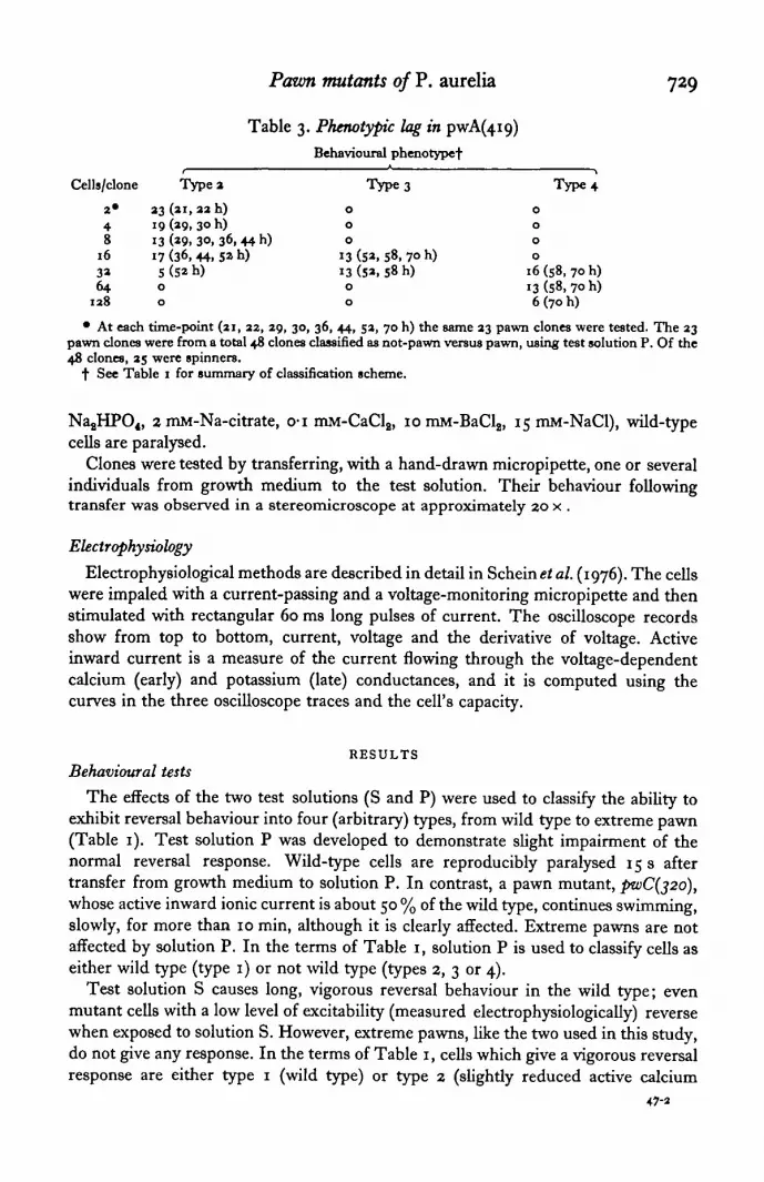
Pawn mutants of P. aurelia 729
Table 3. Phenotypic lag in pwA(4io,)Behavioural phenotypef
Is/clone
2 *
48
16
3364
138
Type a
23 (ai, 22 h)19 (29, 30 h)13 (ao, 30, 36, 44 h)17 (36, 44. 5» h)5 (5a h)0
0
Type3
0
0
0
13 (5a, 58, 7° h)13 (5a, 58 h)
0
0
Type4
0
0
0
0
16 (58, 70 h)13 (58, 70 h)6(7oh)
• At each time-point (21, 22, 29, 30, 36, 44, 52, 70 h) the same 23 pawn clones were tested. The 23pawn clones were from a total 48 clones classified as not-pawn versus pawn, using test solution P. Of the48 clones, 25 were spinners.
•f See Table 1 for summary of classification scheme.
,̂ 2 mM-Na-citrate, o-i mM-CaCl2, 10 mM-BaCl2, 15 mM-NaCl), wild-typecells are paralysed.
Clones were tested by transferring, with a hand-drawn micropipette, one or severalindividuals from growth medium to the test solution. Their behaviour followingtransfer was observed in a stereomicroscope at approximately 20 x .
Electrophysiology
Electrophysiological methods are described in detail in Schein et al. (1976). The cellswere impaled with a current-passing and a voltage-monitoring micropipette and thenstimulated with rectangular 60 ms long pulses of current. The oscilloscope recordsshow from top to bottom, current, voltage and the derivative of voltage. Activeinward current is a measure of the current flowing through the voltage-dependentcalcium (early) and potassium (late) conductances, and it is computed using thecurves in the three oscilloscope traces and the cell's capacity.
RESULTSBehavioural tests
The effects of the two test solutions (S and P) were used to classify the ability toexhibit reversal behaviour into four (arbitrary) types, from wild type to extreme pawn(Table 1). Test solution P was developed to demonstrate slight impairment of thenormal reversal response. Wild-type cells are reproducibly paralysed 15 s aftertransfer from growth medium to solution P. In contrast, a pawn mutant, pwC(j2o),whose active inward ionic current is about 50 % of the wild type, continues swimming,slowly, for more than 10 min, although it is clearly affected. Extreme pawns are notaffected by solution P. In the terms of Table 1, solution P is used to classify cells aseither wild type (type 1) or not wild type (types 2, 3 or 4).
Test solution S causes long, vigorous reversal behaviour in the wild type; evenmutant cells with a low level of excitability (measured electrophysiologically) reversewhen exposed to solution S. However, extreme pawns, like the two used in this study,do not give any response. In the terms of Table 1, cells which give a vigorous reversalresponse are either type 1 (wild type) or type 2 (slightly reduced active calcium
47-2

730 S. J. SCHEIN
conductance). Test solution S was originally used to distinguish cells carrying thespinner (sp) trait from those not carrying the trait. Cells which do give a response,but one which is insufficient for a sp versus non-sp determination, are type 3(excitability considerably reduced); cells which give no response at all are type 4(extremely inexcitable, like the extreme pwAfag) and pioB(ioo)).
Behavioural demonstration of phenotypic lag
Table 2 shows the combined results from two experiments which followed theexpression of the pwB(ioo) phenotype. At each time listed, from 16 to 32 fresh cloneswere tested. Autogamy should produce a random segregation of the unlinked pw andsp genes. Of the clones tested, the number of sp clones and the total number of pwclones were both about one-half, as expected.
After just a single division (cells at the '2-cell stage' - 18-25 n post-autogamy) thebehaviour of the pawn paramecia (type 2, and see Table 2, fourth footnote) could bedistinguished from their non-pawn sibs by the use of solution P. However, the 2-cellstage pawn paramecia were clearly very excitable; their swimming was markedlyslowed by solution P and they responded vigorously with reversals in solution S. Asstated above, this may. be compared with paralysis of the wild type within 15 s and noeffect on the fully expressed pwB(ioo).
With the cells exhibiting type 2 behaviour, it was also observed that the 4-, 8- and16-cell stage paramecia showed increasing resistance to solution P, although this is notindicated in the table.
When there were 16 cells per clone (4 divisions post-autogamy) the reversal res-ponse elicited by solution S was sufficiently weak so that it was impossible to dis-tinguish spinner from non-spinner pawns (type 3). By the 32-cell stage, the cells hadbecome extreme pawns (type 4). They gave no reversal response at all in solution S.
A similar experiment was performed using pwA(/fig). In this case, the same 23pawn clones were re-tested at each time-point. The results, shown in Table 3, werenearly identical to the results of Table 2. Although synchrony was not as good, it isclear that one could detect the pawn genotype at the 2-cell stage. Also, the cellsreached type 3 by the 16-cell stage and type 4 (extreme pawns) by the 32-cell stage.
Electrophysiological demonstration of phenotypic lag
To correlate the phenomenon demonstrated by the behavioural experiments withmembrane excitability, electrophysiological measurements were made on the Fx
(pwB(ioo)l + ) and post-autogamous pwB(ioo) homozygotes at 30 h (4-cell stage) andat 40 h (8-cell stage). These measurements may be compared both with those from thehomozygous wild type and the pwB(ioo)lpwB(ioo).
The most informative trace in each oscilloscope record (Fig. 2) is the lowest trace,dVjdt. The discussion of the computation of active inward current in Schein et al.
Fig. 2. The electrical response of (A) Fx( + /B(ioo)), (B) B(ioo) at 30 h post-autogamy, at the4-cell stage and (C) B(ioo) at 40 h post-autogamy, at the 8-cell stage. The changes in potential(middle traces) in a cell are produced by applied currents (top traces). The bottom trace in eachpicture is the time derivative of voltage. The applied (stimulus) current increases from left toright, in the depolarizing (upper row of pictures) and hyperpolarizing (lower row of pictures)directions. Note especially the derivative traces with the arrows.

Pavm mutants of P. aurelia
B
A
r
731
_| 1 nA
/ 4 "\jlOmV
10 ms
1 nA
I 10 mV
10 ms
I 1 nA
l 10 mV
10 ms
|-l nA
l 10 mV
10 ms
Fig. 2. For legend see opposite.
_|1 nA|10mV
y10 ms

732 S. J. SCHEEN
20
10
0020 40
0-50
025
000
pwB(100)1 9 7 4-cell stage
1-52
20 40
2-0
10
no
B
" ft•
- I
Ft
106AO-65
A 0 ' 2 5
( + /pwB(100))
014
20 40
0-50
0-25
000
DpwB(lOO)
8-cell stage
20 40
Time (ms)
Fig. 3. The computed active inward current, obtained as in Fig. 3 of Schein et al. (1976), of (A)the wild type at 0-48, 0-57, 0-69 and 1-18 nA applied current, (B) the F t ( + /B(ioo)) at 0-14,0-25, 0-65 and :o6 nA, (C) the pwB(iao) at the 4-cell stage at 1-52, 17s, i-8S and 197 nA,and for (D) thepwB(ioo) at the 8-cell stage at 1 -32,1 -66,1 93 and a-o6 nA. In each plot, the left-most curve was chosen because the peak active inward current is very close to the maximumfor that cell.
(1976) makes it clear that comparison of the height of the first peak (due to passivecharging of the membrane capacitance) with the second peak gives an easily visual-ized, nearly quantitative estimate of inward calcium current.
The oscilloscope records of Fig. 2A-C, particularly the derivative traces, show theprogressive decrease in active inward current as one proceeds from the F-^ (Fig. 2 A) topawns (identified by their behavioural response) at the 4- and 8-cell stages (Fig. 2Band 2C) to the fully expressed pawn phenotype (Fig. 9 of Schein et al. 1976).
These records also show that the wild type behaviour of the i^-heterozygote isreflected in its electrical properties, which are fully wild type. Records of the wildtype are shown in Fig. 4 of Schein et al. (1976).
The data in Fig. 2 can be used to generate a measure of active calcium conductance,called active inward current (Schein et al. 1976). Fig. 3 shows representative tracingsof active inward current from each of the recording sessions from which the picturesin Fig. 2 were taken; the ordinate in the 4- and 8-cell stage parts of the figure isexpanded fourfold. The active inward current approaches a maximum (as described inSchein et al. (1976)) at 2-5 nA in the Ft (approximately equal to 2-8 nA in a wild-type cell), 0-67 nA by the 4-cell stage (0-50 nA in another 4-cell stage individual), and0-37 nA by the 8-cell stage. The fully-expressed mutant has essentially zero calciumconductance. These results demonstrate nearly quantitative agreement with a simplehalving of active calcium conductance with each division.
Additionally, Fig. 9 of Schein et al. (1976) shows results from the homozygous

Pawn mutants of P. aurelia 733
Table 4. Half-life of the calcium channel
Time(days)
I I
I I
2 02 0
I II I
2O2O
Con-ditions
RTirC o l d "RTCold
RTCold
RTCold
Growth*
8x6x
10 xII X
8x4 X
16 x7-5 x
Num-1 r
uer 01cellstested
90603030
3030
3060
Spinners/testablef
24/S417/367/187/i3
8/178/18
8/17H/33
Behaviouralphenotypej
Type Type '2 3
0 360 240 00 0
pwB(ioo)0 134 12
0 00 0
Type4
0
0
1 2
17
00
1327
Cell state (x) at whichrapidly grown cells
WOLLIQ demonsudiethe behaviouralphenotype§
16 < x < 3216 s; 1 < 3232 *S x32 < 1
16 aS x < 32x < 16
16 a; x < 3232 «S 132 *3 x
Half-life (days) ||
S
5
S '4 *
•5 -5 «
c Tti Tt
r«T «
i Tt
; T
TtTi
i II
; 8i 12s 13
a II
= 2O
S 9
• Cultures were started from 6700 individuals; they were recounted at the time of the test.f Spinning (or not spinning) is observed following transfer of individual paramecia to solution S.j See Table 1 for summary of classification scheme.§ Type 2 behaviour is exhibited by rapidly grown cells from the 2-cell stage to less than the 16-cell
stage. Type 3 behaviour is exhibited by rapidly grown cells from the 16-cell stage to less than the 32-cellstage. Type 4 behaviour is exhibited by cells which are at the 32-cell stage or beyond.
|| T is the time required for 50% decay. Given the cell stage (say, the 16-cell stage) appropriate torapidly grown cells exhibiting type 3 behaviour, then a slowly grown cell which exhibits Type 3behaviour at the 8-cell stage must have lost excitability by a factor of \ in addition to the J due todilution. If the cell had reached the 8-cell stage at 11 days post-autogamy, then the channel half-life wouldbe equal to 11 days. A simple equation utilizes this data to give
T = (days of growth)/
[(dilution factor suggested by cell stage (1) at which rapidly grown cells exhibit this type of behaviour)"
(actual increase in the number of cells during slow growth)
In the example T = n/log, (16/8) = 11 days.
% RT: room temperature growth. See Materials and Methods.• • Cold: see Materials and Methods.
pzo(Bioo)lpwB(ioo) strain. It has normal passive electrical properties as well as normaldelayed rectification; however, anomalous rectification (the 'relaxation' of the voltagewhen the membrane potential is hyperpolarized to — 40 mV and beyond) is not seenuntil the cell is hyperpolarized to about —70 mM. A review of Fig. 2B and 2 C showsthat the effect of the mutation on anomalous rectification can be seen by the 4- and8-cell stages.
Phenotypic lag under conditions of slow growth
The electrophysiological results suggest that a simple halving of the number ofwild-type channels occurs with each division. If there were rapid decay of channels,then a much more rapid decrease in excitability would have occurred. For example, ifthe half-life of a channel were 8 h (1 normal division time), then excitability woulddecrease by a factor of four with each division, and cells at the 4- and 8-cell stagewould have fa and ̂ the wild-type active inward current. Since the results in theabove section indicate that loss of channels agrees with a model incorporating onlydilution, it was hoped that by allowing the same number of divisions in a much longer

734 S. J. SCHEIN
period, the effect of decay could be observed. Cells were therefore grown as describedin Slow growth in Materials and Methods.
Cells were tested behaviourally (Table 4) at 11 days (2-3 divisions) and at 20 days(3-4 divisions). Normal segregation of the spinner and pawn genes demonstratenormal autogamy. The progress of the expression of the behavioural phenotype fromtype 2 to type 4 is similar for both genes. For example, at the 8-cell stage, after 11days at room temperature, slowly grown pwAfoig) and pwB(i00) both behave as type3 cells. The data presented in Tables 2 and 3 show that the phenotype of the normallygrovmpwA^fig) and pwB(ioo) mutants appears as type 3 between the 16- and 32-cellstage. If after 11 days, an 8-cell stage paramecium behaves like a 16- to 32-cell stageparamecium grown rapidly, then the effect of channel loss was to decrease excitabilityadditionally by ^ to £ in those 11 days. The lifetime (T) is therefore between 11 and5'5 days. Similar limits are implied by each of the rows of Table 4 and these limits havebeen used to estimate the lifetime (T). The range of T is from 5-5 to 8 days in the
experiment and from 5-5 to 9 days in the pwB(i00) experiments.
DISCUSSIONThe stability of the Ca-channel
Autogamy in a heterozygous paramecium leads to a sudden change in the genotypeof the organism, a change comparable to mutation. Mutation in bacteria may not bereflected in phenotype until the concentration of the affected macromolecule isdiluted out by cell division. The steady loss of the reversal response of homozygouspawns in the divisions which follow autogamy of both pwAj + and pwBj + hetero-zygotes has been demonstrated using two behavioural tests (Tables 2 and 3). As earlyas the 2-cell stage (1 division post-autogamy) the pawn homozygotes could be dis-tinguished from their wild-type sibs by exposure to solution P. By the 16-cell stage (4divisions) the pawns still responded to the very stimulating solution S, but the responsewas short in duration and infrequent. By the 32-cell stage (5 divisions) the pawns nolonger showed any reversal, even with the most sensitive test stimulation.
The behaviour of the Ft heterozygote appears wild type. The electrophysiologicalmeasurements confirmed that impression and demonstrated the lack of a gene-dosageeffect in the heterozygote.
The gradual loss of excitability following autogamy, indicated by behavioural tests,is paralleled by a loss of membrane excitability measured electrophysiologically (Figs.2 and 3). The decrease in active inward ionic current, one-quarter at the 4-cell stageand one-eighth at the 8-cell stage, agrees with a simple dilution hypothesis anddemonstrates the stable nature of the calcium channel structure.
In addition, the electrophysiological data presented here, using rapidly growing cul-tures, reveal that channel half-life is certainly more than 40 h. The behavioural data,using slowly grown cultures, permit an estimate of from 5 to 8 days for channel half-life. The model which is presented in Schein et al. (1976), based on electrophysiologicaldata, and in Schein (1976), based on genetic and behavioural data, implies that thepawn genes' products directly affect or are themselves structural components of thecalcium channel. The experiments presented in this paper are entirely consistentwith such a scheme.

Pawn mutants of P. aurelia 735
The Ca-channel is monomeric or stably polymeric
The detection of non-zero active inward current in even the extreme pawn mutants,coupled with the knowledge that they were produced by nitrosoguanidine muta-genesis, suggests that the mutations are point mutations (Schein et al. (1976)) and thatmutant channels are indeed synthesized. The data here suggest in addition that thenewly synthesized mutant channels do not poison the old wild-type channels, andimply that the channel is monomeric or stably polymeric.
Agreement with other dataFurther support for the correlation between behavioural classification and excita-
bility, and thus for the dilution hypothesis and the measurement of half-life, is foundin Schein et al. (1976). pwC(j2o), which this report would classify as showing State 2behaviour, appropriate for 2-cell stage paramecia, has about \ (40 %) the wild-typeactive calcium conductance. pwA(2i4), which this report would classify as showingState 3 behaviour, appropriate for 16- to 3 2-cell stage paramecia, has 5 % the wild-typeactive calcium conductance.
Berger (1976) has also studied phenotypic lag in the expression of thepwA pheno-type; our results are in close agreement. Following autogamy and 4 days of starvation,growth through a median of 3-3 divisions was required before he observed the pwAphenotype. His threshold of detection of pawn behaviour corresponds approximatelywith what I have called State 3 behaviour. In my study, four divisions were required.He found that a single (+ IpwA) macronuclear fragment was sufficient to confer weakreversal behaviour (corresponding again to what I have called State 3 behaviour) on thehomozygous (pwA/ptvA) exautogamous cell. Under the conditions of his experiment,the single fragment would be expected to increase its original DNA content by 1-5(Berger, personal communication). The 'old genes' are therefore 1*5 x (1/35) or 5 %of the total. My results show that state 3 behaviour is observed when the active inwardcalcium currents are 5 % of the wild type.
Contribution of old' genes
Berger (1974) has also shown that the old macronuclear genes are not immediatelydestroyed. Instead, the old macronucleus is transformed into about 35 macronuclearfragments, which are gradually destroyed during starvation following the induction ofautogamy. The longer the starvation, the more fragments are destroyed for the pur-pose of recycling DNA precursors. The 'rapidly grown cells' used in this study wereallowed to starve for 3 days which, according to Fig. 2 of Berger's report (1974),leaves about 9 of the original 35 fragments. In addition, only half of the macronuclearfragment genes from the heterozygous F1 ancestor are wild type at the pawn loci.Thus, prior to the first division of the new homozygous pawn F2, only \ x ^ or •£• ofthe old wild-type genes are present. A separate report by Berger (1973) demonstratesthat the rate of RNA synthesis per unit of DNA is the same in old macronuclear frag-ments as in the new macronucleus. Thus, the contribution of 'old wild-type genes',which should be ^ of the total at the 2-cell stage and halving with each division, toactive calcium conductance, which is | at the 2-cell stage and halving with eachdivision, appears to be insignificant.

736 S. J. SCHEIN
It could be argued that the ' old Fx genes' are disproportionately active. In that case,the electrophysiological results still permit use of the behavioural tests as a quantitativeassay for excitability. The application of the quantitative behavioural test to the slowlygrowing cells remains valid, as does the 5- to 8-day half-life. Furthermore, the ' slowlygrown cells' were subjected to the same 3 days of starvation, which was then followedby an additional 11 days of underfeeding and starvation. Berger's (1974) data onlyextend to 8 days of starvation, at which time recycling pressure has left an average 1*5(of the original 35) macronuclear fragments, left to be distributed among the 4, 8,16,. . . descendants. These data strengthen the conclusion that 'old Fx genes' playlittle or no role in this example of phenotypic lag.
I would like to thank Dr Charles David and Dr M. V. L. Bennett of the AlbertEinstein College of Medicine for advice and encouragement during this work. I wouldalso like to thank Dr George M. Katz and Mr Sidney Steinberg of the College ofPhysicians and Surgeons, Columbia University, for the use of the current-damp andfor giving generously of their time and energy with problems of an electrical nature.Also, I would like to thank Dr James Berger, who was kind enough to read the manu-script critically.
REFERENCES
BHRGER, J. D. (1973). Nuclear differentiation and nucleic acid synthesis in well-fed erconjugants ofParamecium aurelia. Chromosoma 43, 247-68.
BERGER, J. D. (1974). Selective autolysis of nuclei as a source of DNA precursors in Paratnecium aureliaexconjugants. J. Protozool. ax, 145-52.
BERGER, J. D. (1976). Gene expression and phenotypic change in Paratnecium tetraaurelia exconjugants.Genet. Res. 27, 123-34.
CHANG, S.-Y., VAN HOUTEN, J., ROBLES, L. J., Lui, S. S. & KUNG, C. (1974). An extensive behaviouraland genetic analysis of the pawn mutants in Paramecium aurelia. Genet. Res. 33, 165- 73.
DAVIS, B. D. (1948). Isolation of biochemically deficient mutants of bacteria by penicillin. J. Am. Chem.Soc. 70, 4267.
GRUNDFEST, H. (1961). Ionic mechanisms in electrogenesis. Ann. New York Acad. Set. 94, 405-57.KUNG, C. (1971). Genetic mutants with altered system of excitation in Paramecium aurelia. II. Muta-
genesis, screening and genetic analysis of the mutants. Genetics 69, 29-45.KUNG, C. & ECKERT, R. (1972). Genetic modification of electric properties in an excitable membrane.
Proc. natn. Acad. Set. XJ.SjA. 69, 93-7.NAITOH, Y. & ECKERT, R. (1968). Electrical properties of Paramecium caudatum: all-or-none electro-
genesis. Z. vergl. Physiologie 61, 453-72.NAITOH, Y., ECKERT, R. & FRIEDMAN, K. (1972). A regenerative calcium response in Paramecium. J'. exp.
Biol. 56, 667-81.NAITOH, Y. & KANEKO, H. (1972). ATP-reactivated triton-extracted models of paramecium: modifi-
cation of ciliary movement by calcium ions. Science, 176, 523—4.SCHEIN, S. J. (1976). Nonbehavioral selection for pawns, mutants of Paramecium aurelia with de-
creased excitability. Genetics 84, (in press).SCHEIN, S. J., BENNETT, M. V. L. & KATZ, G. M. (1976). Altered calcium conductance in pawns,
behavioral mutants of Paramecium aurelia. J. exp. Biol. 65, 699—724.SONNEBORN, T. M. (1970). Methods in Paramecium research. In Methods in Cell Physiology, vol. 4 (ed.
D. M. Prescott), pp. 241-339. New York: Academic Press.SONNEBORN, T. M. & LYNCH, R. S. (1934). Hybridization and segregation in Paramecium aurelia.
J. exp. Zool. 67, 1-72.TAMM, S. L., SONNEBORN, T. M. & DIPPELL, R. V. (1975). The role of cortical orientation in the con-
trol of the direction of ciliary beat in Paramecium. J. Cell. Biol. 64, 98-112.
Recommended

![[Comparative description of macronuclear electrophoretic karyotypes of Paramecium primaurelia and Paramecium novaurelia sibling species]](https://img.dokumen.tips/doc/110x75/6344f0a538eecfb33a065292/comparative-description-of-macronuclear-electrophoretic-karyotypes-of-paramecium.jpg)


