Embed Size (px)
Citation preview

248 Nanomed J, Vol. 1, No. 4, Summer 2014
Received: Jan. 28, 2014; Accepted: Mar. 12, 2014
Vol. 1, No. 4, Summer 2014, page 248-257
Received: Apr. 22, 2014; Accepted: Jul. 12, 2014
Vol. 1, No. 5, Autumn 2014, page 298-301
Online ISSN 2322-5904
http://nmj.mums.ac.ir
Original Research
The acute liver injury in rat caused by gold nanoparticles
Monir Doudi1, Mahbubeh Setorki
2*
1Department of Microbiology, Falavarjan Branch, Islamic Azad University, Isfahan, Iran
2Department of Biology, Izeh Branch, Islamic Azad University, Izeh, Iran
Abstract
Objective(s): Gold nanoparticles (GNPs) command a great deal of attention for biomedical
applications nowadays. The data about the degree of toxicity and the accumulation of gold
nanoparticles in-vivo is not enough to judge.
Materials and Methods: A total of 32 healthy male Wistar rats were randomly divided into 4
including: three GNP-treated and one control group. Groups 1, 2 and 3 received 0.5 cc of a
solution containing 5, 10, and 100 ppm Au daily via intraperitoneal (IP) injection for 7 days,
respectively. The control group was treated with 0.5 cc normal saline with same procedure.
Then, several biochemical parameters such as serum glutamate oxaloacetat transaminase
(SGOT) and serum glutamate pyrvate transaminase (SGPT) were evaluated at 2, 7 and 14
days after the last injection. After 14 days, all the rats were sacrificed and liver, lung tissues
were separated and evaluated.
Results: SGOT two days after intervention was significantly greater in the group 2 than the
control group. In liver histological assessment, in group 1, basophils were observed around
the central veins, in group 2 fading and no observation of central veins was seen, and in
group 3 hepatic damage was noticed. The lung histological results showed severe vascular
hyperemia in group 1, air sacs damage in group 2, and complete air sacs destruction in group
3.
Conclusion: The results showed extreme changes in the histopathology of lung and liver
tissues caused by spherical nanogold with 5-10 nm size in all of three treatment groups.
Keywords: Fibrous, Gold nanoparticle, Hypertrophy
*Corresponding Author: Mahbubeh Setorki; Department of Biology, Izeh Branch, Islamic Azad University,
Izeh, Iran.
Email:[email protected]
Acute liver injury caused by gold nanoparticles
Nanomed J, Vol. 1, No. 4, Summer 2014 249
Original Research (font 12)
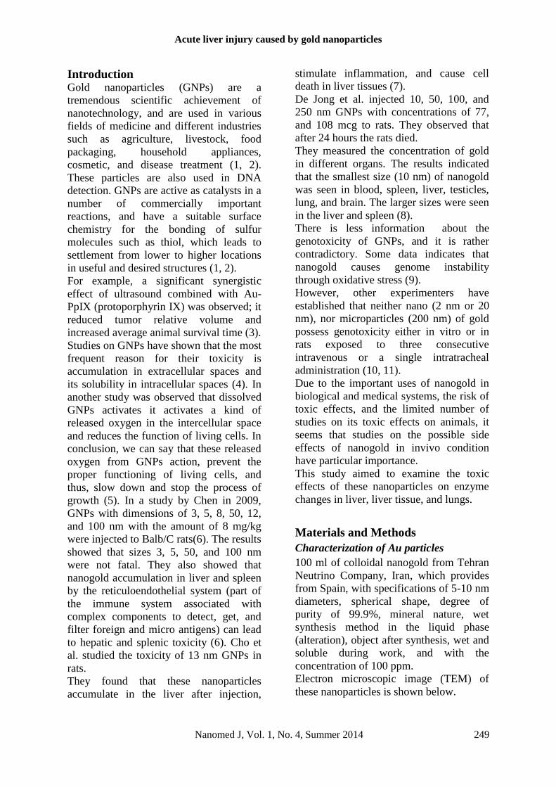
Introduction Gold nanoparticles (GNPs) are a
tremendous scientific achievement of
nanotechnology, and are used in various
fields of medicine and different industries
such as agriculture, livestock, food
packaging, household appliances,
cosmetic, and disease treatment (1, 2).
These particles are also used in DNA
detection. GNPs are active as catalysts in a
number of commercially important
reactions, and have a suitable surface
chemistry for the bonding of sulfur
molecules such as thiol, which leads to
settlement from lower to higher locations
in useful and desired structures (1, 2).
For example, a significant synergistic
effect of ultrasound combined with Au-
PpIX (protoporphyrin IX) was observed; it
reduced tumor relative volume and
increased average animal survival time (3).
Studies on GNPs have shown that the most
frequent reason for their toxicity is
accumulation in extracellular spaces and
its solubility in intracellular spaces (4). In
another study was observed that dissolved
GNPs activates it activates a kind of
released oxygen in the intercellular space
and reduces the function of living cells. In
conclusion, we can say that these released
oxygen from GNPs action, prevent the
proper functioning of living cells, and
thus, slow down and stop the process of
growth (5). In a study by Chen in 2009,
GNPs with dimensions of 3, 5, 8, 50, 12,
and 100 nm with the amount of 8 mg/kg
were injected to Balb/C rats(6). The results
showed that sizes 3, 5, 50, and 100 nm
were not fatal. They also showed that
nanogold accumulation in liver and spleen
by the reticuloendothelial system (part of
the immune system associated with
complex components to detect, get, and
filter foreign and micro antigens) can lead
to hepatic and splenic toxicity (6). Cho et
al. studied the toxicity of 13 nm GNPs in
rats.
They found that these nanoparticles
accumulate in the liver after injection,
stimulate inflammation, and cause cell
death in liver tissues (7).
De Jong et al. injected 10, 50, 100, and
250 nm GNPs with concentrations of 77,
and 108 mcg to rats. They observed that
after 24 hours the rats died.
They measured the concentration of gold
in different organs. The results indicated
that the smallest size (10 nm) of nanogold
was seen in blood, spleen, liver, testicles,
lung, and brain. The larger sizes were seen
in the liver and spleen (8).
There is less information about the
genotoxicity of GNPs, and it is rather
contradictory. Some data indicates that
nanogold causes genome instability
through oxidative stress (9).
However, other experimenters have
established that neither nano (2 nm or 20
nm), nor microparticles (200 nm) of gold
possess genotoxicity either in vitro or in
rats exposed to three consecutive
intravenous or a single intratracheal
administration (10, 11).
Due to the important uses of nanogold in
biological and medical systems, the risk of
toxic effects, and the limited number of
studies on its toxic effects on animals, it
seems that studies on the possible side
effects of nanogold in invivo condition
have particular importance.
This study aimed to examine the toxic
effects of these nanoparticles on enzyme
changes in liver, liver tissue, and lungs.
Materials and Methods
Characterization of Au particles
100 ml of colloidal nanogold from Tehran
Neutrino Company, Iran, which provides
from Spain, with specifications of 5-10 nm
diameters, spherical shape, degree of
purity of 99.9%, mineral nature, wet
synthesis method in the liquid phase
(alteration), object after synthesis, wet and
soluble during work, and with the
concentration of 100 ppm.
Electron microscopic image (TEM) of
these nanoparticles is shown below.

Doudi M, et al
250 Nanomed J, Vol. 1, No. 4, Summer 2014
Figure 1. Electron microscopic image (TEM) of
nanoparticles, 5-10 nm diameter, spherical shape,
99.9% degree of purity, mineral nature, and wet
synthesis method in the liquid phase (alteration).
Animals, drug administration and
samples collecting A total of 32 healthy male Wistar rats
obtained from the Animal Center of
Shahrekord University, Iran. All animal
handling and manipulation procedures
were performed according to the guideline
of the Animal Welfare Act and the
experimental protocols were approved by
the Office of Research Ethics Committee
at University of Shahrekord.
The rats were nearly of the same age (12
weeks old) and weighing 225 ± 25 g. The
rats were maintained on standard
laboratory rodent diet pellets and were
housed in humidity and temperature-
controlled vent-ilated cages on a 12 hour
day/night cycle. After two weeks of
accommodation to the animal room, they
were semi-randomly distributed into 4
groups of 8. Groups 1, 2 and 3 received a
daily dose of solution containing 5, 10, and
100 ppm nanogold, respectively, via
intraperitoneal (IP) injection for 7 days.
Animals in group 4, the control group,
received 0.5 cc normal saline with same
procedure. The effects of GNPs on serum
biochemical levels were evaluated at 2, 7
and 14 days after the last injection. Blood
samples were collected from the eye vein
by removing the eyeball quickly at time
points. All animals (at 14th day) were
anesthetized by diethylether and sacrificed.
Serum was collected by centrifuging blood
at 2,500 rpm for 10 minutes.
The tissues such as liver and lung were
autopsied.
Fractions of tissues were kept in 10% (v/v)
formalin for immediate histopathological
examination.
Biochemical analysis of liver function
Whole blood was centrifuged twice at
3000 rpm for 10 minutes in order to
separate serum. Using a biochemical
autoanalyzer (Hitachi Automatic Analyzer
902, Roche, Germany), serum biochemical
analysis was carried out. To evaluate the
liver function, the levels of serum
glutamate oxaloacetat transaminase
(SGOT) and serum glutamate pyrvate
transaminase (SGPT) were measured.
Histopathological examination
Histological observations were performed
according to the standard laboratory
procedures. Rats (four rat/treatment group)
at the end of day 14th were dissected for
histology. A small piece of lung and liver
fixed in 10% (v/v) formalin was embedded
in a paraffin block, sliced into 5 μm
thicknesses and then placed onto glass
slides. The section was stained with
Hematoxylin–Eosin (HE) and examined
by light microscopy.
Statistical analysis
All data were analyzed using the statistical
package for social sciences (version 19,
SPSS Inc., Chicago, IL) software and were
summarized and expressed as mean and
standard deviation (mean ± SD).
Multivariate analysis of variance (MAN-
OVA) model was the used, using serum
values at 2, 7 and 14 day after intervention
as dependant variables. We compared the
groups and the baseline value of serum
was controlled.
We used Wilk’s lambda or Roy Largest
Root for total differences between the
groups; P-value of each dependent variable
was reported in last row of tables; and the
Tukey was used for paired comparisons.
Acute liver injury caused by gold nanoparticles
Nanomed J, Vol. 1, No. 4, Summer 2014 251
Original Research (font 12)
Less than 0.05 P-values were considered
significant.
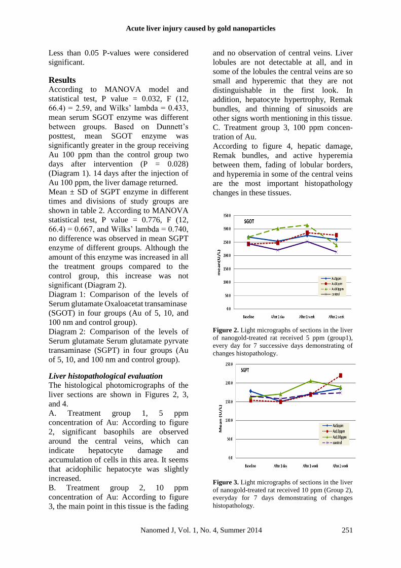
Results According to MANOVA model and
statistical test, P value = 0.032, F (12,
66.4) = 2.59, and Wilks’ lambda = 0.433,
mean serum SGOT enzyme was different
between groups. Based on Dunnett’s
posttest, mean SGOT enzyme was
significantly greater in the group receiving
Au 100 ppm than the control group two
days after intervention (P = 0.028)
(Diagram 1). 14 days after the injection of
Au 100 ppm, the liver damage returned.
Mean ± SD of SGPT enzyme in different
times and divisions of study groups are
shown in table 2. According to MANOVA
statistical test, P value = 0.776, F (12,
66.4) = 0.667, and Wilks’ lambda = 0.740,
no difference was observed in mean SGPT
enzyme of different groups. Although the
amount of this enzyme was increased in all
the treatment groups compared to the
control group, this increase was not
significant (Diagram 2).
Diagram 1: Comparison of the levels of
Serum glutamate Oxaloacetat transaminase
(SGOT) in four groups (Au of 5, 10, and
100 nm and control group).
Diagram 2: Comparison of the levels of
Serum glutamate Serum glutamate pyrvate
transaminase (SGPT) in four groups (Au
of 5, 10, and 100 nm and control group).
Liver histopathological evaluation The histological photomicrographs of the
liver sections are shown in Figures 2, 3,
and 4.
A. Treatment group 1, 5 ppm
concentration of Au: According to figure
2, significant basophils are observed
around the central veins, which can
indicate hepatocyte damage and
accumulation of cells in this area. It seems
that acidophilic hepatocyte was slightly
increased.
B. Treatment group 2, 10 ppm
concentration of Au: According to figure
3, the main point in this tissue is the fading
and no observation of central veins. Liver
lobules are not detectable at all, and in
some of the lobules the central veins are so
small and hyperemic that they are not
distinguishable in the first look. In
addition, hepatocyte hypertrophy, Remak
bundles, and thinning of sinusoids are
other signs worth mentioning in this tissue.
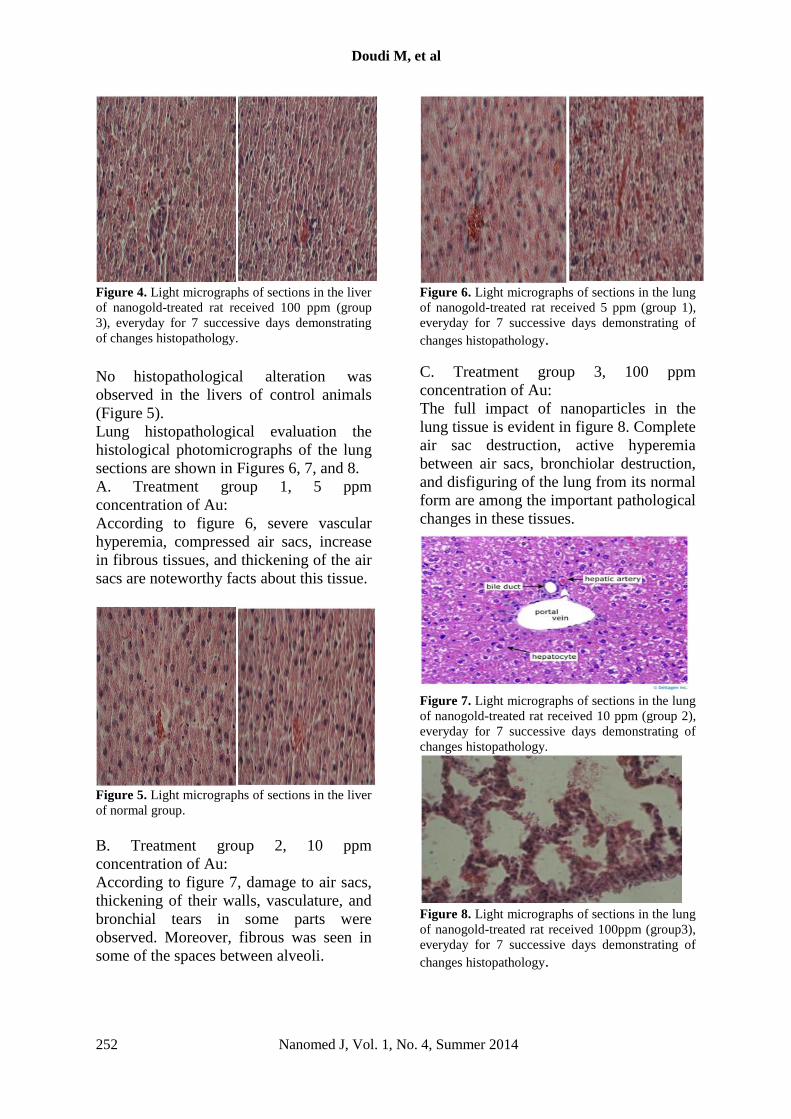
C. Treatment group 3, 100 ppm concen-
tration of Au.
According to figure 4, hepatic damage,
Remak bundles, and active hyperemia
between them, fading of lobular borders,
and hyperemia in some of the central veins
are the most important histopathology
changes in these tissues.
Figure 2. Light micrographs of sections in the liver
of nanogold-treated rat received 5 ppm (group1),
every day for 7 successive days demonstrating of
changes histopathology.
Figure 3. Light micrographs of sections in the liver
of nanogold-treated rat received 10 ppm (Group 2),
everyday for 7 days demonstrating of changes
histopathology.
Doudi M, et al
252 Nanomed J, Vol. 1, No. 4, Summer 2014
Figure 4. Light micrographs of sections in the liver
of nanogold-treated rat received 100 ppm (group
3), everyday for 7 successive days demonstrating
of changes histopathology.
No histopathological alteration was
observed in the livers of control animals
(Figure 5).
Lung histopathological evaluation the
histological photomicrographs of the lung
sections are shown in Figures 6, 7, and 8.
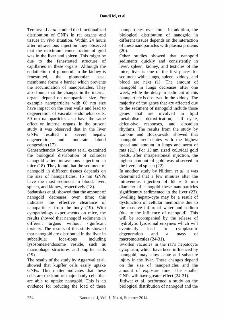
A. Treatment group 1, 5 ppm
concentration of Au:
According to figure 6, severe vascular
hyperemia, compressed air sacs, increase
in fibrous tissues, and thickening of the air
sacs are noteworthy facts about this tissue.
Figure 5. Light micrographs of sections in the liver
of normal group.
B. Treatment group 2, 10 ppm
concentration of Au:
According to figure 7, damage to air sacs,
thickening of their walls, vasculature, and
bronchial tears in some parts were
observed. Moreover, fibrous was seen in
some of the spaces between alveoli.
Figure 6. Light micrographs of sections in the lung
of nanogold-treated rat received 5 ppm (group 1),
everyday for 7 successive days demonstrating of
changes histopathology.
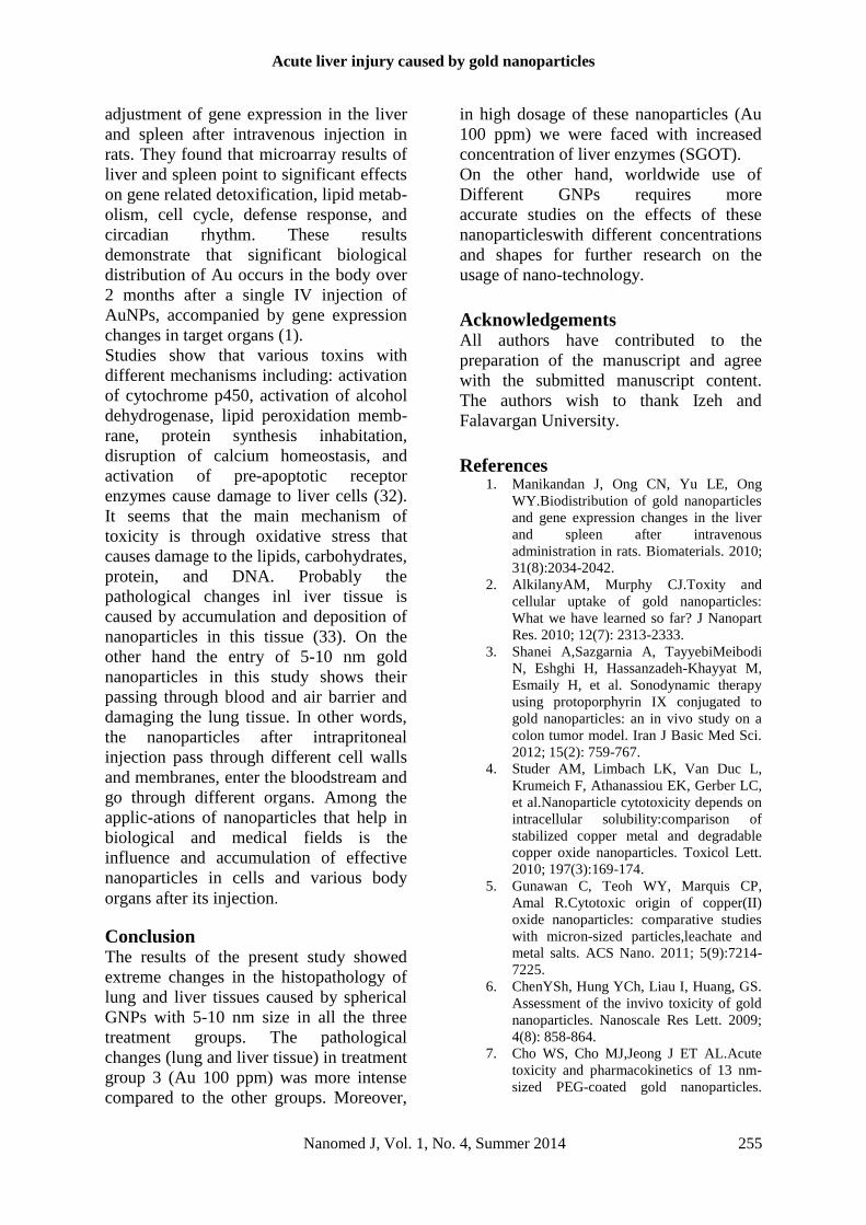
C. Treatment group 3, 100 ppm
concentration of Au:
The full impact of nanoparticles in the
lung tissue is evident in figure 8. Complete
air sac destruction, active hyperemia
between air sacs, bronchiolar destruction,
and disfiguring of the lung from its normal
form are among the important pathological
changes in these tissues.
Figure 7. Light micrographs of sections in the lung
of nanogold-treated rat received 10 ppm (group 2),
everyday for 7 successive days demonstrating of
changes histopathology.
Figure 8. Light micrographs of sections in the lung
of nanogold-treated rat received 100ppm (group3),
everyday for 7 successive days demonstrating of
changes histopathology.

Acute liver injury caused by gold nanoparticles
Nanomed J, Vol. 1, No. 4, Summer 2014 253
Original Research (font 12)
No histopathological alteration was
observed in the lungs of control animals
(Figure 9).
Discussion The aim of many studies in previous
decades was on the understanding of
interaction between different types of
nanoparticles and cells, such as: function,
size, form, and the chemical level of
nanoparticles (12).
While gold (bulk) seems to be ‘safe’, but
using nanogold requires experiments, for
compatibility and its great impact on the
environment (if these nanoparticles
are
Figure9. Light micrographs of sections in the lung
of normal.
in large amounts for in vivo usage)
(13,14). GNPs can be considered as
extraordinary molecular carriers for the
targeting, intracellular trafficking and
delivery of a huge array of biomolecules
including DNA, RNA, proteins, peptides,
drugs, genes and other molecules of
therapeutic significance.
A number of researchers have studied the
cellular uptake and cytotoxicity of these
nanoparticles. The important issue is
paying attention to the difference between
cytotoxicity and cell damage. The
nanoparticles which have less cytotoxicity
or do not show any cytotoxicity (with
standard tests) may still have the ability to
cause severe cell damage. Cytotoxicity is
also dependent on the type of used cell.
However it is thought that the uptake and
toxicity of nanoparticles are controllable
and can be manipulated.
The results of this study showed that mean
SGOT enzyme two days after intervention
in the group receiving concentration of Au
100 ppm was significantly greater than the
control group (P = 0.028). Fourteen days
after the injection of Au 100 ppm the liver
damage returned. Also between the mean
SGPT enzymes of different groups no
difference was observed (although the
amount of this enzyme increased in all
groups compared to control group but this
increase was not significant).
Liver histological findings in the group
receiving concentration of Au 5 ppm:
basophils observed around the central
veins (which indicate the hepatic damage
and the accumulation of cells in this area),
in the group receiving concentration of Au
10 ppm: fading and no observation of
central veins, liver lobules not being
detectable at all, the lobules of the central
veins being small and hyperemic,
hepatocyte hyper-trophy, Remak bundles,
and in the group receiving concentration of
Au 100 ppm: , hepatic damage, and Remak
bundles, active hyperemia between them,
fading of lobular borders, and hyperemia
in some of the central veins were
observed.
The lung histological results showed:
severe vascular hyperemia, compressed air
sacs, increase of fibrous tissues, and
thickening of the air sacs (treatment group
1), air sacs damage, thickening of their
walls, vascu-lature, bronchial tears in some
parts, and fibrous in some of the spaces
between the alveoli (treatment group 2),
complete air sacs destructions, active
hyperemia between them, bronchiolar
destruction, and disfiguring of the lung
from its normal form (treatment group 3).
There are various reports regarding the
normal toxicity of these nanoparticles that
depend on different modifications of these
nanoparticles, surface functional
attachment, form, size, and the diameter of
these particles (15, 16).
Doudi M, et al
254 Nanomed J, Vol. 1, No. 4, Summer 2014
Terentyukl et al. studied the functionalized
distribution of GNPs in rat organs and
tissues in vivo situation. Within 24 hours
after intravenous injection they observed
that the maximum concentration of gold
was in the liver and spleen. This might be
due to the fenestrated structure of
capillaries in these organs. Although the
endothelium of glomeruli in the kidney is
fenestrated, the glomerular basal
membrane forms a barrier which prevents
the accumulation of nanoparticles. They
also found that the changes in the internal
organs depend on nanoparticle size. For
example nanoparticles with 60 nm size
have impact on the vein walls and lead to
degeneration of vascular endothelial cells.
50 nm nanoparticles also have the same
effect on internal organs. In the present
study it was observed that in the liver
GNPs resulted in severe hepatic
degeneration and moderate blood
congestion (17).
Ganeshchandra Sonavanea et al. examined
the biological distribution of colloidal
nanogold after intravenous injection in
mice (18). They found that the sediment of
nanogold in different tissues depends on
the size of nanoparticles. 15 nm GNPs
have the most sediment in blood, liver,
spleen, and kidney, respectively (18).
Sadauskas et al. showed that the amount of
nanogold decreases over time; this
indicates the effective clearance of
nanoparticles from the body (19). With
cytopathology experi-ments on mice, the
results showed that nanogold sediments in
different organs without significant
toxicity. The results of this study showed
that nanogold are distributed in the liver in
subcellular loca-tions including
lysosomes/endosome vesicle, such as
macrophage structures and kupffer cells
(19).
The results of the study by Aggarwal et al.
showed that kupffer cells easily uptake
GNPs. This matter indicates that these
cells are the kind of major body cells that
are able to uptake nanogold. This is an
evidence for reducing the load of these
nanoparticles over time. In addition, the
biological distribution of nanogold in
different tissues depends on the interaction
of these nanoparticles with plasma proteins
(20).
Other studies showed that nanogold
sediments quickly and consistently in
liver, spleen, kidney, and testicles of the
mice; liver is one of the first places for
sediment while lungs, spleen, kidney, and
blood are next (1). The amount of
nanogold in lungs decreases after one
week, while the delay in sediment of this
nanoparticle is observed in the kidney. The
majority of the genes that are affected due
to the sediment of nanogold include those
genes that are involved in lipid
metabolism, detoxification, cell cycle,
defen-sive responses, and circadian
rhythms. The results from the study by
Lanone and Boczkowski showed that
nanogold precip-itates with the highest
speed and amount in lungs and aorta of
rats (21). For 13 nm sized colloidal gold
beads, after intraperitoneal injection, the
highest amount of gold was observed in
the liver and spleen (22).
In another study by Niidom et al. it was
determined that a few minutes after the
intravenous injection of 65 ± 5 mm
diameter of nanogold these nanoparticles
significantly sedimented in the liver (23).
Swelling hepato-cyte may be a result of
dysfunction of cellular membrane due to
the massive influx of water and sodium
(due to the influence of nanogold). This
will be accompanied by the release of
hydrolytic lysosomal enzymes which will
eventually lead to cytoplasmic
degeneration and a mass of
macromolecules (24-31).
Swollen vacuoles in the rat’s hepatocyte
cytoplasm, which have been influenced by
nanogold, may show acute and subacute
injury in the liver. These changes depend
on the size of nanoparticles and the
amount of exposure time. The smaller
GNPs will have greater effect (24-31).
Jittiwat et al. performed a study on the
biological distribution of nanogold and the
Acute liver injury caused by gold nanoparticles
Nanomed J, Vol. 1, No. 4, Summer 2014 255
Original Research (font 12)
adjustment of gene expression in the liver
and spleen after intravenous injection in
rats. They found that microarray results of
liver and spleen point to significant effects
on gene related detoxification, lipid metab-
olism, cell cycle, defense response, and
circadian rhythm. These results
demonstrate that significant biological
distribution of Au occurs in the body over
2 months after a single IV injection of
AuNPs, accompanied by gene expression
changes in target organs (1).
Studies show that various toxins with
different mechanisms including: activation
of cytochrome p450, activation of alcohol
dehydrogenase, lipid peroxidation memb-
rane, protein synthesis inhabitation,
disruption of calcium homeostasis, and
activation of pre-apoptotic receptor
enzymes cause damage to liver cells (32).
It seems that the main mechanism of
toxicity is through oxidative stress that
causes damage to the lipids, carbohydrates,
protein, and DNA. Probably the
pathological changes inl iver tissue is
caused by accumulation and deposition of
nanoparticles in this tissue (33). On the
other hand the entry of 5-10 nm gold
nanoparticles in this study shows their
passing through blood and air barrier and
damaging the lung tissue. In other words,
the nanoparticles after intrapritoneal
injection pass through different cell walls
and membranes, enter the bloodstream and
go through different organs. Among the
applic-ations of nanoparticles that help in
biological and medical fields is the
influence and accumulation of effective
nanoparticles in cells and various body
organs after its injection.
Conclusion The results of the present study showed
extreme changes in the histopathology of
lung and liver tissues caused by spherical
GNPs with 5-10 nm size in all the three
treatment groups. The pathological
changes (lung and liver tissue) in treatment
group 3 (Au 100 ppm) was more intense
compared to the other groups. Moreover,
in high dosage of these nanoparticles (Au
100 ppm) we were faced with increased
concentration of liver enzymes (SGOT).
On the other hand, worldwide use of
Different GNPs requires more
accurate studies on the effects of these
nanoparticleswith different concentrations
and shapes for further research on the
usage of nano-technology.
Acknowledgements All authors have contributed to the
preparation of the manuscript and agree
with the submitted manuscript content.
The authors wish to thank Izeh and
Falavargan University.
References
1. Manikandan J, Ong CN, Yu LE, Ong
WY.Biodistribution of gold nanoparticles
and gene expression changes in the liver
and spleen after intravenous
administration in rats. Biomaterials. 2010;
31(8):2034-2042.
2. AlkilanyAM, Murphy CJ.Toxity and
cellular uptake of gold nanoparticles:
What we have learned so far? J Nanopart
Res. 2010; 12(7): 2313-2333.
3. Shanei A,Sazgarnia A, TayyebiMeibodi
N, Eshghi H, Hassanzadeh-Khayyat M,
Esmaily H, et al. Sonodynamic therapy
using protoporphyrin IX conjugated to
gold nanoparticles: an in vivo study on a
colon tumor model. Iran J Basic Med Sci.
2012; 15(2): 759-767.
4. Studer AM, Limbach LK, Van Duc L,
Krumeich F, Athanassiou EK, Gerber LC,
et al.Nanoparticle cytotoxicity depends on
intracellular solubility:comparison of
stabilized copper metal and degradable
copper oxide nanoparticles. Toxicol Lett.
2010; 197(3):169-174.
5. Gunawan C, Teoh WY, Marquis CP,
Amal R.Cytotoxic origin of copper(II)
oxide nanoparticles: comparative studies
with micron-sized particles,leachate and
metal salts. ACS Nano. 2011; 5(9):7214-
7225.
6. ChenYSh, Hung YCh, Liau I, Huang, GS.
Assessment of the invivo toxicity of gold
nanoparticles. Nanoscale Res Lett. 2009;
4(8): 858-864.
7. Cho WS, Cho MJ,Jeong J ET AL.Acute
toxicity and pharmacokinetics of 13 nm-
sized PEG-coated gold nanoparticles.

Doudi M, et al
256 Nanomed J, Vol. 1, No. 4, Summer 2014
ToxicolApplPharmacol. 2009; 236(1):16-
24.
8. De Jong WH, Hagens WI, Krystek P,
Burger MC, Sips AJ, Geertsma
RE.Particle size-dependent organ
distribution of gold nanoparticles after
intravenous administration. Biomaterials.
2008; 29(12):1912-1919.
9. Choi SY, Jeong S, Jang SH, Park J, Park
JH, Ock KS, et al. In vitro toxicity of
serum protein-adsorbed citrate-reduced
gold nanoparticles in human lung
adenocarcinoma cells. ToxicolIn Vitro.
2012; 26(2):229-237.
10. Trickler WJ, Lantz SM, Murdock RC,
Schrand AM, Robinson BL, Newport GD,
et al. Brain microvessel endothelial cells
responses to gold nanoparticles: In vitro
pro-inflammatory mediators and
permeability. Nanotoxicology. 2011;
5(4):479-492.
11. Li JJ, Lo SL, Ng CT, Gurung RL, Hartono
D, Hande MP, et al. Genomic instability
of gold nanoparticle treated human lung
fibroblast cells. Biomaterials.
2011;32(23):5515-5523.
12. Lewinski N, Colvin V, Drezek R.
Cytotoxicity of nanoparticles. Small.
2008; 4(1):26-49.
13. Colvin, V. The Potential environmental
impact of engineered nanomaterials. Nat
Biotechnol. 2003; 21: 1166-1170.
14. Shukla R, Bansal V, Chaudhary M, Basu
A, Bhonde RR, Sastry M.
Biocompatibility of gold nanoparticles and
their endocytotic fate inside the cellular
compartment: a microscopic overview.
Langmuir. 2005; 21(23): 10644-10654.
15. Takahashi H, Niidome Y, Niidome T,
Kaneko K, Kawasak H, Yamada S.
Modification of gold nanorods using
phosphatidylcholine to reduce
cytotoxicity. Langmuir. 2006; 22(1): 2-5.
16. Pan Y, Neuss S, Leifert A, Fischler M,
Wen F, Simon U, et al. Size-dependent
cytotoxicity of gold nanoparticles. Small.
2007; 3(11): 1941-1949.
17. TerentyuklGS,Maslyakova GN,
Suleymanova LV, KhlebtsovBN, Kogan
BY,Akchurin GG, et al. Circulation and
distribution of gold nanoparticles and
induced alterations of tissue morphology
at intravenous particle delivery. J
Biophotonics. 2009; 2(5): 292-302.
18. Sonavane G, Tomoda K, Makino
K.Biodistribution of colloidal gold
nanoparticles after intravenous
administration: effect of particle size.
Colloids Surf B Biointerfaces. 2008;
66(2): 274-280.
19. Sadauskas E, Danscher G, Stoltenberg M,
Vogel U, Larsen A, Wallin H.
Protracted
elimination of gold nanoparticles from
mouse liver. Nanomedicine. 2009; 5(2):
162-169.
20. Aggarwal P, Hall JB, McLeland
CB,Dobrovolskaia MA, McNeil SE.
Nanoparticle interaction with plasma
proteins as it relates to particle
biodistribution, biocompatibility and
therapeutic efficacy. Adv Drug Deliv Rev.
2009; 61(6): 428-437.
21. Lanone S, Boczkowski J. Biomedical
applications and potential health risks of
nanomaterials: molecular mechanisms.
CurrMol Med. 2006; 6(6): 651-663.
22. Hillyer JF, Albrecht RM. Correlative
instrumental neutron activation analysis,
light microscopy, transmission electron
microscopy, and X-ray microanalysis for
qualitative and quantitative detection of
colloidal gold spheres in biological
specimens.MicroscMicroanal. 1999; 4(5):
481-490.
23. Niidome T, Yamagata M, Okamoto Y,
Akiyama Y, Takahashi H, Kawano T, et
al. PEG-modified gold nanorods with a
stealth character for in vivo application. J
Control Release. 2006; 114(3): 343-347.
24. Abdelhalim MAK, Jarrar BM. Gold
nanoparticles administration induced
prominent inflammatory, central vein
intima disruption, fatty change and
Kupffer cells hyperplasia. Lipids Health
Dis.2011; 10: 133.
25. Abdelhalim MAK, Jarrar BM. Gold
nanoparticles induced cloudy swelling to
hydropic degeneration, cytoplasmic
hyaline vacuolation, polymorphism,
binucleation, karyopyknosis, karyolysis,
karyorrhexis and necrosis in the liver.
Lipids Health Dis.2011; 10: 166.
26. Abdelhalim MAK, Jarrar BM. Renal
tissue alterations were size-dependent with
smaller ones induced more effects and
related with time exposure of gold
nanoparticles. Lipids Health Dis. 2011;
10: 163.
27. Abdelhalim MAK, Jarrar BM. The
appearance of renal cells cytoplasmic
degeneration and nuclear destruction
might be an indication of GNPs toxicity.
Lipids Health Dis.2011; 10: 147.
28. Abdelhalim MAK. Exposure to gold
nanoparticles produces cardiac tissue
damage that depends on the size and
duration of exposure. Lipids Health
Dis.2011; 10: 205.

Acute liver injury caused by gold nanoparticles
Nanomed J, Vol. 1, No. 4, Summer 2014 257
Original Research (font 12)
29. Abdelhalim MAK. Exposure to gold
nanoparticles produces pneumonia,
fibrosis, chronic inflammatory cell
infiltrates, congested and dilated blood
vessels, and hemosiderin granule and
emphysema foci. J Cancer SciTher.2012;
4(3): 046-050.
30. Abdelhalim MAK. Gold nanoparticles
administration induces disarray of heart
muscle, hemorrhagic, chronic
inflammatory cells infiltrated by small
lymphocytes, cytoplasmic vacuolization
and congested and dilated blood vessels.
Lipids Health Dis. 2011; 10: 233.
31. Abdelhalim MAK. Optimizing a novel
method for synthesizing gold
nanoparticles: biophysical studies. J
Cancer SciTher. 2012; 4: 140-143.
32. Oberdörster G, Maynard A, Donaldson K,
Castranova V, Fitzpatrick J, Ausman K,
Carter J, Karn B, Kreyling W, Lai D, Olin
S, Monteiro-Riviere N, Warheit D, Yang
H. Principles for characterizing the
potential human health effects from
exposure to nanomaterials: elements of a
screening strategy. Part Fibre Toxicol.
2005; 2:8.
33. Damabach DM,Andrews BA,Moulin F.
New technologies and screeninig
strategies for hepatotoxicity: use of invitro
models.ToxicolPathol. 2005; 33(1):17-26.