Embed Size (px)
Citation preview

Teaching Forensic Anthropology with a
small skeletal collectionDavid Bryson
Department of Biology and Forensic Science
For further information please contact me at [email protected] or via my website
http://www.cladonia.co.uk

Small collections and learning
• What do I mean by a small collection?
• What learning are we trying to support
• Disadvantages real or imagined?

What do I mean by a small collection?

“It is an easily verified observation that most American Universities do not have departments of anthropology or interdepartmental programs that are prepared to provide adequate training for physical anthropology. Most departments do not have laboratories, skeletal collections, anthropometric instruments, incubators, [etc].” (Lasker 1963 p. 91)

Skeletal collection• The boxes in the corner plus
• 5 Complete articulated skeletons

Skeletal collection• The boxes in the corner plus
• 5 Complete articulated skeletons

Hundreds and thousands

Hundreds and thousands

Hundreds and thousands

Hundreds and thousands

Hundreds and thousands
“For comparison purposes with modern man, well preserved human skeletons, which can be purchased through any medical supply company generally show more detail and are, therefore, preferable for use in the elementary course to broken bones from archaeological collections.” (Lasker 1963 p.108)

Could we purchase more
skeletons?
• E-Bay $4,600 • The Bone Room $4-5000 • India export ban in 1987 • China export ban to
coincide with Olympics 2008

Even what we have is valuable!

Black Market

What learning are we trying to
support?
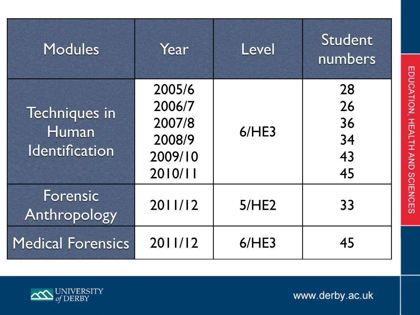
Modules Year Level Student numbers
Techniques in Human
Identification
2005/62006/72007/82008/92009/102010/11
6/HE3
282636344345
Forensic Anthropology
2011/12 5/HE2 33
Medical Forensics 2011/12 6/HE3 45

Working with skeletal materials
It takes time with ‘whole bones’ to develop requisite skills;
Recognition - which bone is which
Siding - right from left
Introducing incomplete bones too early can be counterproductive.
Students need to able to get a feel for the weight and appearance of bones.

Biological Forensic Anthropology Physical
Key skills
Measurement Observation
Metric analysis Morphological
Not black and white
• Often dealing with probably male or female in physical anthropology.
• “... W.M Krogman, who reports that the skull requires the most frequent sexing in medicolegal work, found himself 82-87% correct in sexing 750 specimens. T.D.Stewart determined he could sex 77% accurately by inspection.” (Giles E 1962)

Human variability
• We are all different
• Most books show only one skeleton. Anatomy texts tend to illustrate one typical skeleton rather than variations.
• Seeing a range of bones helps students understand individual variation.
• Need enough bones to be able to see

Disadvantages real or imagined?
“For comparison purposes with modern man, well preserved human skeletons, ............. generally show more detail and are, therefore, preferable for use in the elementary course to broken bones from archaeological collections.” (Lasker 1963 p.108)

Sex Age Stature Race Ancestry
Unique features
4 PILLARS OF BIOANTHROPOLOGY 5th
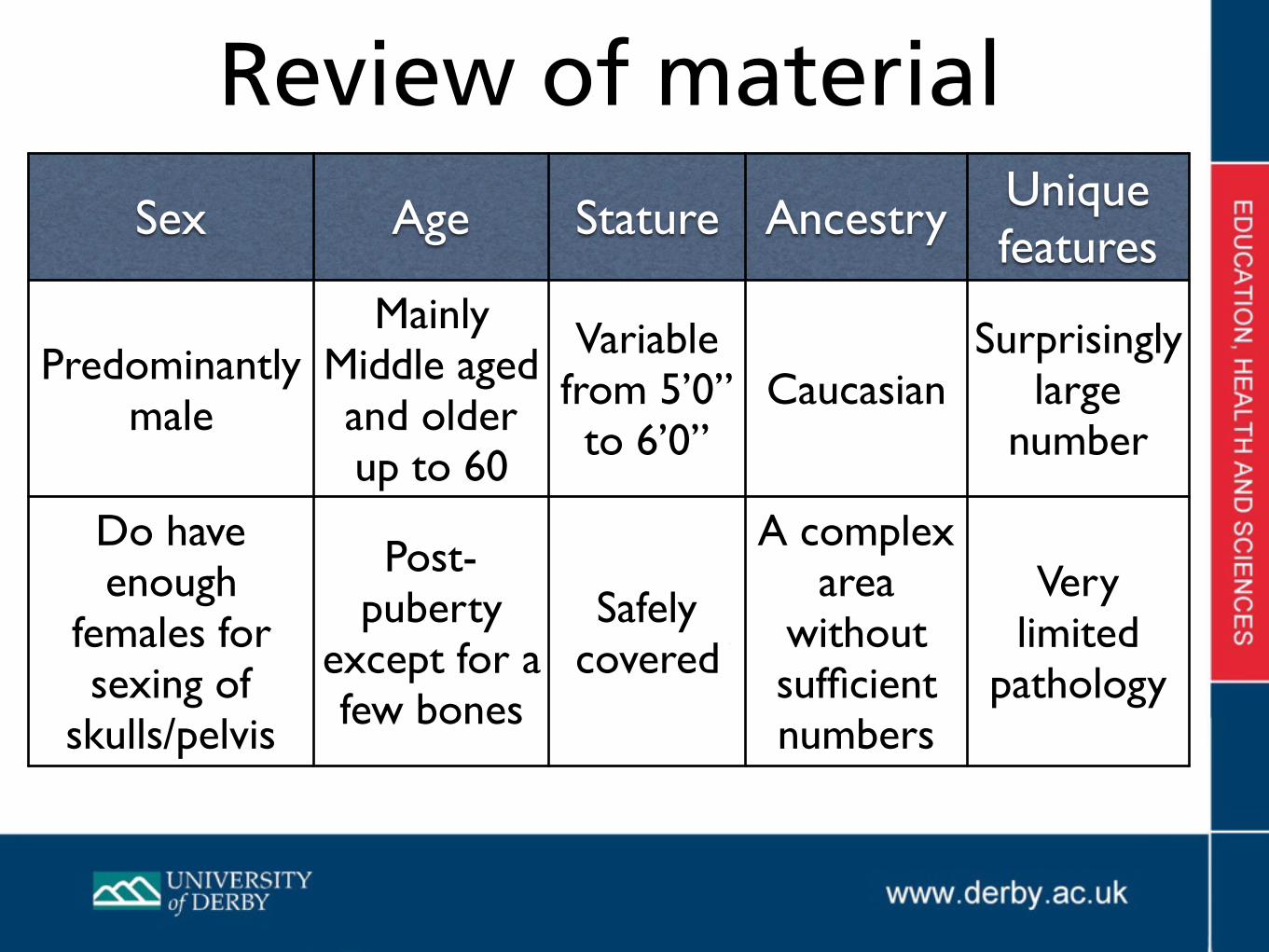
Sex Age Stature Ancestry Unique features
Predominantly male
Mainly Middle aged and older up to 60
Variable from 5’0” to 6’0”
CaucasianSurprisingly
large number
Do have enough
females for sexing of
skulls/pelvis
Post-puberty
except for a few bones
Safely covered
A complex area
without sufficient numbers
Very limited
pathology
Review of material

Making up for a limited collection
• Strong book collection - Student’s were using so we had an extra £3,500 just for this area and have added more.
• Availability of e-journals and e-books
• Use of learning materials and problem based learning
• Use of photographs of the collection and photography by students.

E-Book options in library

Practical exam
• 5 Benches are set up with 9 stations each covering an aspect of Human Identification, total 45 students.
• Student are given 10 minutes at each station.

Examples of questions
Identify the bones in this collection as accurately as possible

Examples of questions
What can these bones tell you about the stature? (Assume Male Caucasian)

Individual variation within our collection

Variation between same bone from different individuals
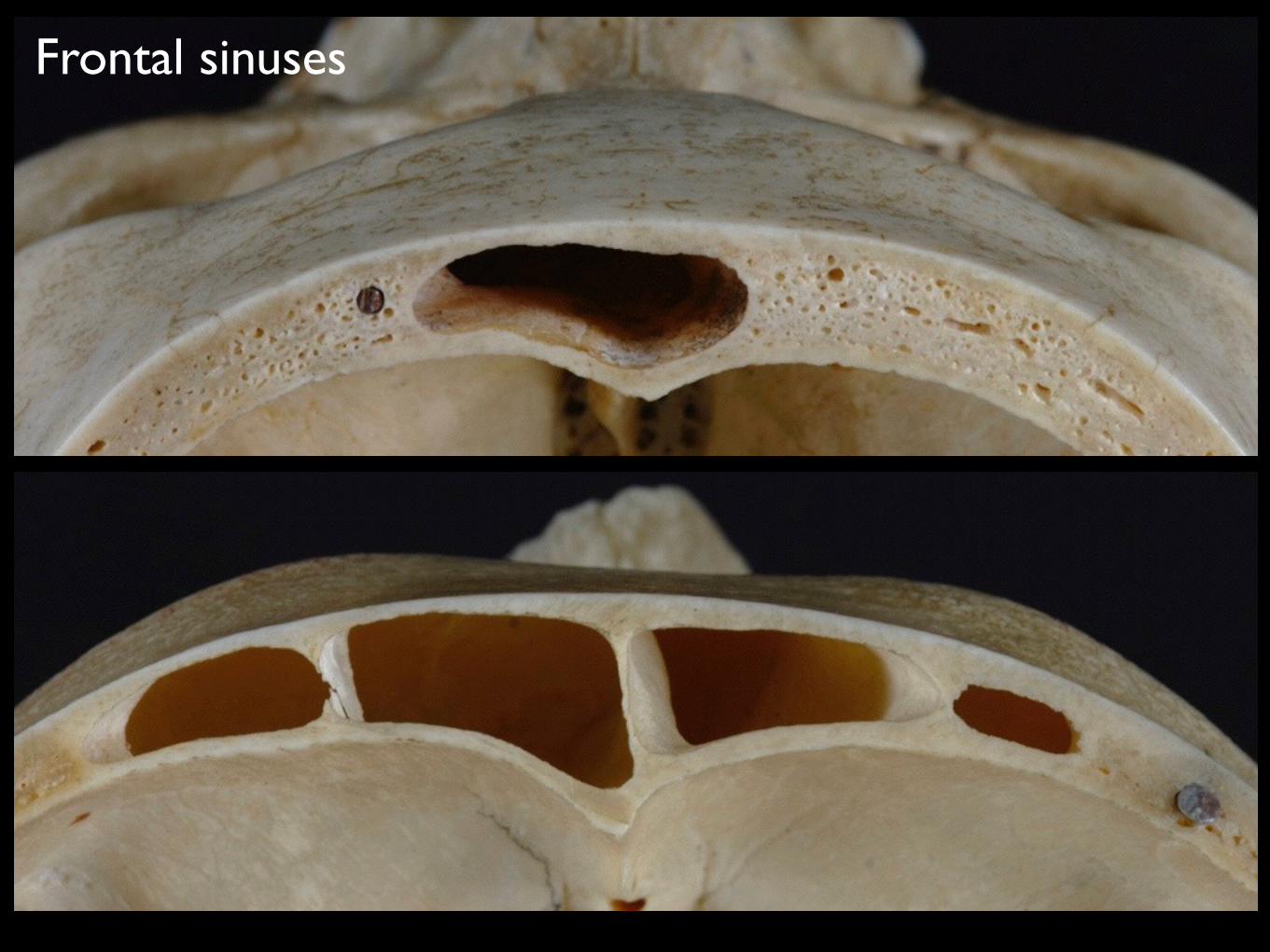
Frontal sinuses

Supraorbital foramen

Supraorbital notch
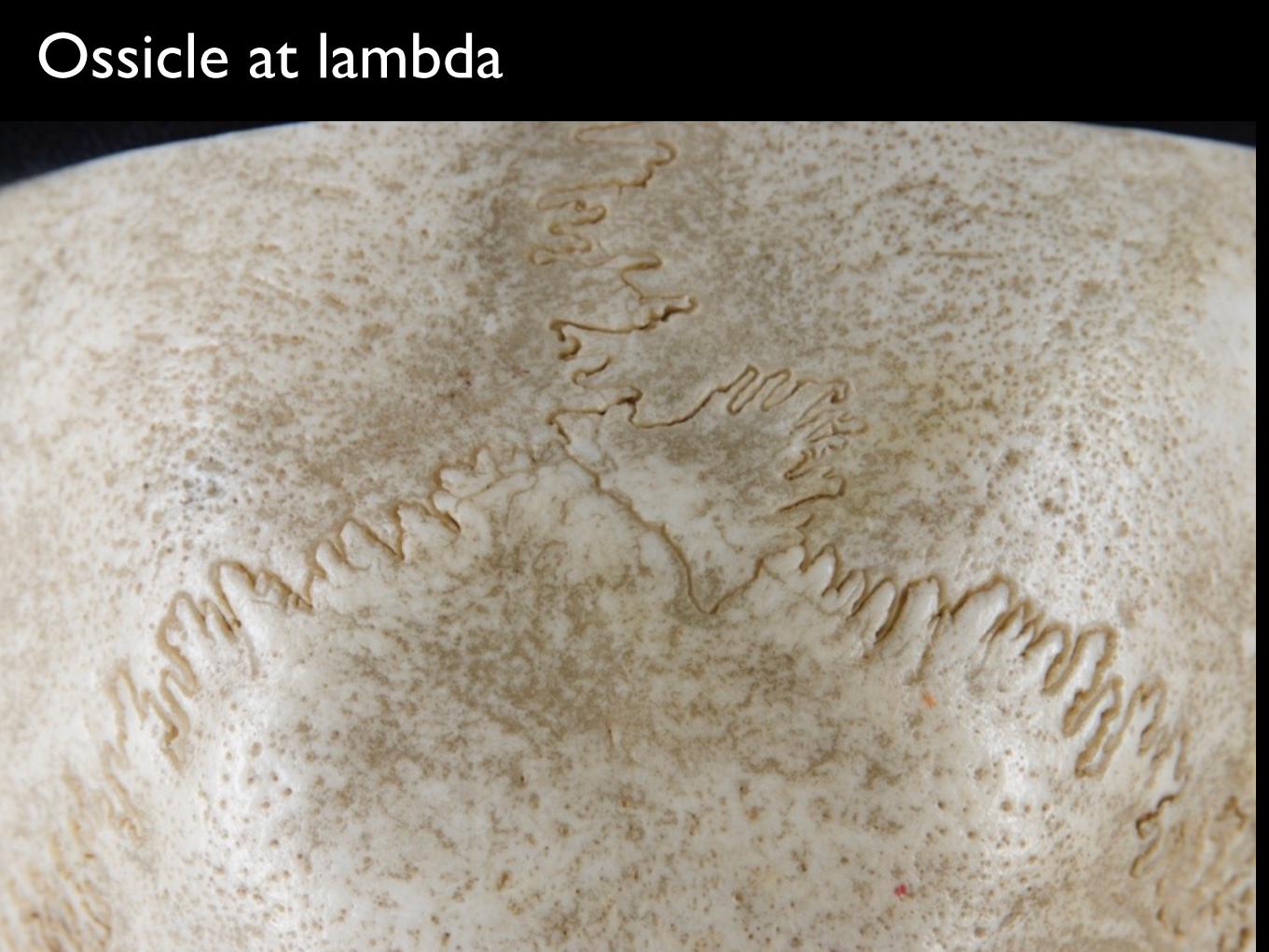
Ossicle at lambda
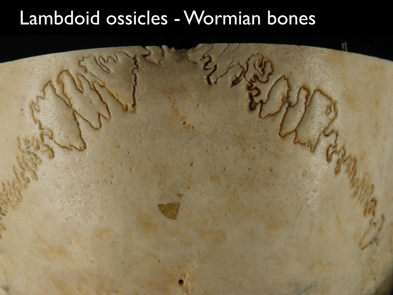
Lambdoid ossicles - Wormian bones
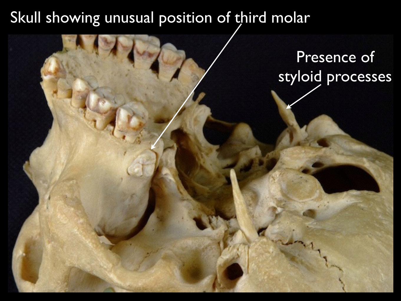
Skull showing unusual position of third molar
Presence of styloid processes
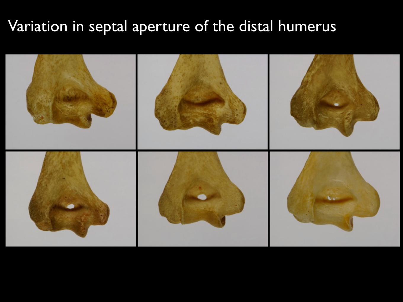
Variation in septal aperture of the distal humerus
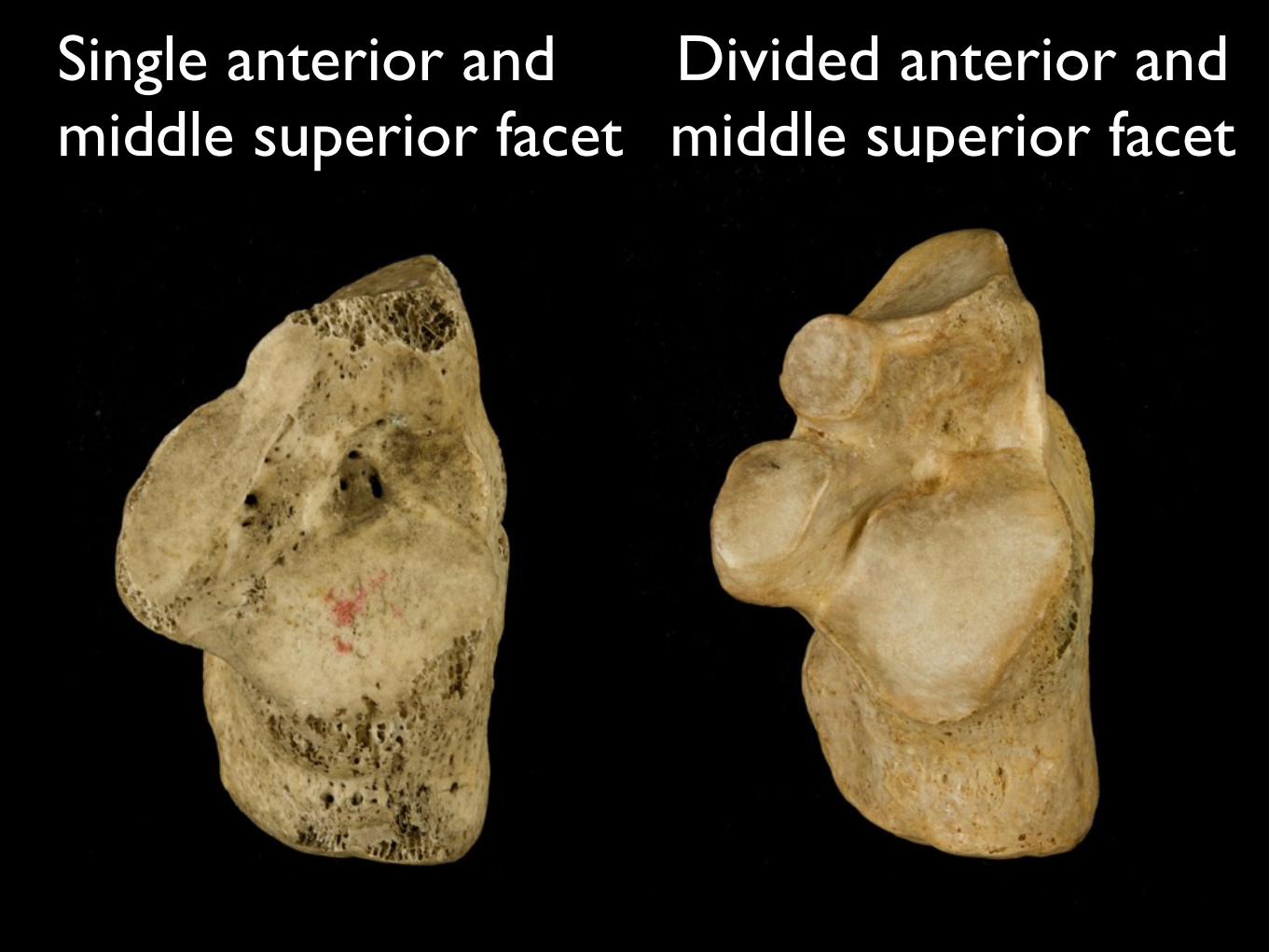
Divided anterior and middle superior facet
Single anterior and middle superior facet

Suprascapular notch
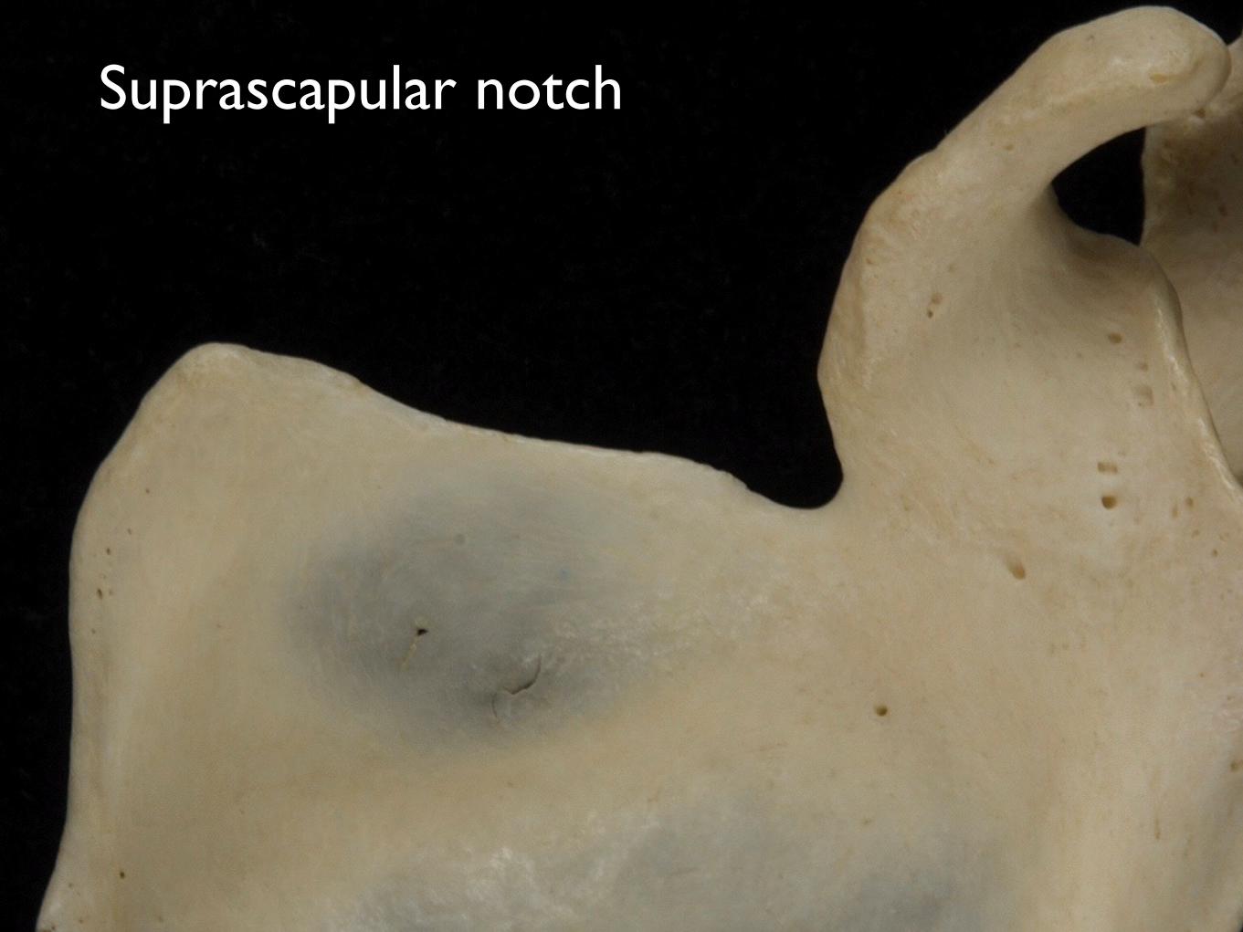
Suprascapular notch

No feature
59
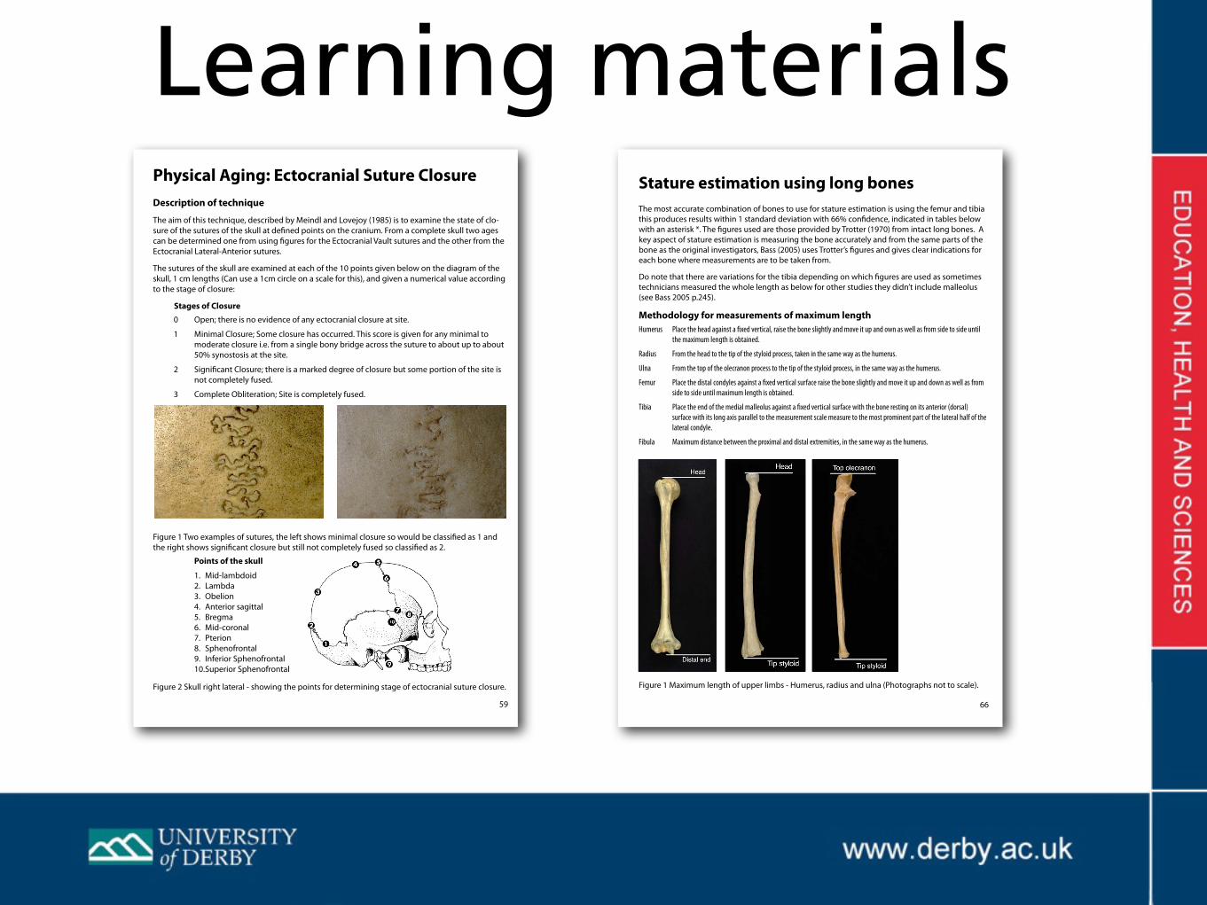
Physical Aging: Ectocranial Suture ClosureDescription of technique
The aim of this technique, described by Meindl and Lovejoy (1985) is to examine the state of clo-sure of the sutures of the skull at defined points on the cranium. From a complete skull two ages can be determined one from using figures for the Ectocranial Vault sutures and the other from the Ectocranial Lateral-Anterior sutures.
The sutures of the skull are examined at each of the 10 points given below on the diagram of the skull, 1 cm lengths (Can use a 1cm circle on a scale for this), and given a numerical value according to the stage of closure:
Stages of Closure0 Open; there is no evidence of any ectocranial closure at site.
1 Minimal Closure; Some closure has occurred. This score is given for any minimal to moderate closure i.e. from a single bony bridge across the suture to about up to about 50% synostosis at the site.
2 Significant Closure; there is a marked degree of closure but some portion of the site is not completely fused.
3 Complete Obliteration; Site is completely fused.
Figure 1 Two examples of sutures, the left shows minimal closure so would be classified as 1 and the right shows significant closure but still not completely fused so classified as 2.
Points of the skull
1. Mid-lambdoid 2. Lambda 3. Obelion 4. Anterior sagittal 5. Bregma 6. Mid-coronal 7. Pterion 8. Sphenofrontal 9. Inferior Sphenofrontal 10. Superior Sphenofrontal
Figure 2 Skull right lateral - showing the points for determining stage of ectocranial suture closure.
Learning materials
66
Stature estimation using long bonesThe most accurate combination of bones to use for stature estimation is using the femur and tibia this produces results within 1 standard deviation with 66% confidence, indicated in tables below with an asterisk *. The figures used are those provided by Trotter (1970) from intact long bones. A key aspect of stature estimation is measuring the bone accurately and from the same parts of the bone as the original investigators, Bass (2005) uses Trotter’s figures and gives clear indications for each bone where measurements are to be taken from.
Do note that there are variations for the tibia depending on which figures are used as sometimes technicians measured the whole length as below for other studies they didn’t include malleolus (see Bass 2005 p.245).
Methodology for measurements of maximum lengthHumerus Place the head against a fixed vertical, raise the bone slightly and move it up and own as well as from side to side until
the maximum length is obtained.
Radius From the head to the tip of the styloid process, taken in the same way as the humerus.
Ulna From the top of the olecranon process to the tip of the styloid process, in the same way as the humerus.
Femur Place the distal condyles against a fixed vertical surface raise the bone slightly and move it up and down as well as from side to side until maximum length is obtained.
Tibia Place the end of the medial malleolus against a fixed vertical surface with the bone resting on its anterior (dorsal) surface with its long axis parallel to the measurement scale measure to the most prominent part of the lateral half of the lateral condyle.
Fibula Maximum distance between the proximal and distal extremities, in the same way as the humerus.
Figure 1 Maximum length of upper limbs - Humerus, radius and ulna (Photographs not to scale).

Faculty of Education, Health and Science
PRACTICAL GUIDE TO TECHNIQUES IN HUMAN IDENTIFICATION David Bryson - April 2011
Ongoing development of practical guide with learning activities, videos, slideshows and interactive materials as an interactive e-book/pdf
ReferencesGiles, E & Elliot, O. (1962) Race identification from cranial measurements. Journal of Forensic Sciences 7 (2): 147-157.
Lasker, G.W. (1963) The introductory course. In: Mandelbaum, D.G., Lasker, G.W. & Albert, E.M. The teaching of physical anthropology. Berkeley: University of California Press.
The Bone Room, http://www.boneroom.com
Bone trafficking http://www.wired.com/medtech/health/magazine/15-12/ff_bones?currentPage=all