Embed Size (px)
Citation preview

179Regen. Med. (2014) 9(2), 179–190 ISSN 1746-0751
The purpose of this report is to describe regenerative medicine applications in the management of complex injuries sustained by service members injured in support of the wars in Afghanistan and Iraq. Improvements in body armor, resuscitative techniques and faster transport have translated into increased patient survivability and more complex wounds. Combat-related blast injuries have resulted in multiple extremity injuries, significant tissue loss and amputations. Due to the limited availability and morbidity associated with autologous tissue donor sites, the introduction of regenerative medicine has been critical in managing war extremity injuries with composite massive tissue loss. Through case reports and clinical images, this report reviews the application of regenerative medicine modalities employed to manage combat-related injuries. It illustrates that the novel use of hybrid reconstructions combining traditional and regenerative medicine approaches are an effective tool in managing wounds. Lessons learned can be adapted to civilian care.
Keywords: amputation • combat casualty care • extremity reconstruction • limb salvage • regenerative medicine
Combat operations expose military service members and civilians to the devastating effects of high-energy munitions that often lead to a complex pattern of injury (Figure 1). For US service members, improvements in personal protective gear and body armor, use of tourniquets [1], rapid transport from the battlefield to surgical stabilization [2,3], and improvements in resuscitation [4] and hem-orrhage control measures [5] have led to increased patient survivability. The survival rate of US service members injured in the current conflict exceeds any other period of conflict in history. These factors have con-tributed to an increased number of service members presenting to military treatment facilities with complex wounds including multiple extremity injuries and amputations [6,7]. Through the early years of the current conflicts in Iraq and Afghanistan, the multi-ple extremity amputation rate (18%) mir-rored that of the Vietnam Conflict [8,9]. More recently the rate of multiple extremity ampu-
tations sustained in combat has more than doubled, likely secondary to changes in the way operations are being conducted such as increased foot patrols.
Concurrent with the increased multiple extremity amputation rate has been a cor-responding increase in extremity injuries with composite tissue loss. Segmental bone loss coupled with massive skin and soft tis-sue deficits are commonplace in the blast-injured service member [10]. Such injuries can lead to severely compromised limb function and adversely affect the quality of life. These patients utilize a tremendous amount of med-ical resources [11], and their resulting bone and soft tissue losses lead to a major source of disability [12]. Moreover, these injuries present difficult clinical challenges not typically seen in civilian trauma [13].
This complex injury pattern has initiated efforts to create new and innovative tech-niques in tissue regeneration. At our cen-ter a multidisciplinary team has effectively
Mark E Fleming*,1,2, Husain Bharmal1,2 & Ian Valerio2,3
1Department of Orthopaedics, Walter
Reed National Military Medical Center,
8901 Wisconsin Ave, Bethesda,
MD 20889, USA 2Uniformed Services University of Health
Sciences, 4301 Jones Bridge Road,
Bethesda, MD 20814, USA 3Plastic & Reconstructive Surgery Service,
Department of Surgery, Walter Reed
National Military Medical Center, 8901
Wisconsin Ave, Bethesda, MD 20889,
USA
*Author for correspondence:
Tel.: +1 301 295 2441
Regenerative medicine applications in combat casualty care
part of
Special Report
10.2217/RME.13.96
Regen. Med.
Special Report9
2
2014
For reprint orders, please contact: [email protected]

180 Regen. Med. (2014) 9(2)
adapted advanced reconstructive techniques merged with regenerative medicine modalities to improve out-comes in our combat casualties. These treatments com-bine traditional reconstruction measures with regenera-tive medicine applications and has been termed ‘hybrid reconstructions’. The hybrid reconstruction model aids in maximizing the function while minimizing the disability and morbidity associated with traditional reconstruction.
This article describes a single medical facilities appli-cation of regenerative medicine modalities to manage complex combat-related injuries. It is descriptive in nature and does not attempt to define all of the out-comes related to introducing regenerative medicine modalities. The techniques employed by the authors have been used in the civilian market with commer-cially available products and are US FDA approved for trauma indications. With regard to a comparative analysis of methods and data on outcomes, that ques-tion is beyond the scope of this current article, which is descriptive in nature and not an outcomes article. Follow on retrospective and prospective reviews will analyze the outcomes utilized in these techniques.
Early management & evacuationEarly treatment of combat casualties includes the battle field (Level I) placement of tourniquets, gaining of intravenous or intraosseus access, and the estab-lishment or stabilization of a patent airway. Patients are then transferred to a Level II or III facility, which may be a mobile Army forward surgical team (FST),
US Marine Corps forward resuscitative surgical system (FRSS) or a fixed medical treatment facility such as a combat support hospital (CSH). Upon arrival to a Level II or III facility, medical and surgical resuscita-tion and stabilization is commenced. These facilities have surgical and support capabilities to facilitate stabi-lization of the wounded warriors who frequently arrive in extremis (Figure 2).
Following the initial injury treatment and stabiliza-tion at a Level III facility, patients are transported by the aeromedical evacuation system for further care in Landsthul (Germany; Level IV). There, patients are optimized for uneventful evacuation to stateside Level V facilities.
Level V facilities are stateside tertiary military medi-cal centers that are designed to provide multidisci-plinary definitive care including definitive stabilization, reconstruction or amputation revisions of extremity injuries [14]. Patients typically arrive at the Level V facil-ities between 3 and 5 days after their initial battlefield injury. Management of these patients requires a mul-tidisciplinary approach including consultations from general, orthopedic, plastic, neurology, ophthalmic and urologic surgical services [15]. In addition, input from the Anesthesia Acute Pain and Chronic Pain services, physical therapy, occupational therapy, traumatic brain injury (TBI), social work, case management, physiatrist and other services is obtained for each wounded warrior upon admission to the Level V facility.
Walter Reed National Military Medical Center (WRNMMC) is one of the two major tertiary refer-
future science group
Special Report Fleming, Bharmal & Valerio
Figure 1. Complex battle injury with bilateral lower extremity amputations and significant composite soft tissue loss.

www.futuremedicine.com 181
ral centers where the most severely injured patients have been triaged. Over the past 12 years of war more than 50% of all wounded warriors have been triaged to WRNMMC to receive their definitive care. Defini-tive management of these injuries typically requires a prolonged hospital length of stay for extensive medi-cal and surgical management. Approximately 25% of wounded warriors with traumatic wounds receive sur-gical management augmented with regenerative medi-cine modalities or hybrid reconstructions. The resources required to perform these reconstructions are extensive and include significant personnel, long hospital length of stay, extended periods of rehabilitation and expensive materials that may be cost prohibitive in certain systems or centers.
Challenges in the definitive management of combat casualtiesProfound shock should be expected upon presentation in the blast-injured patient due to the complex nature of the injuries and associated massive hemorrhage. Massive transfusion protocols may be required to mitigate the lethal triad of acidosis, hypothermia and coagulopathy (Figure 3). A 1:1:1 ratio of packed red blood cells, fresh
frozen plasma and platelets [16] or fresh whole blood may be beneficial to those requiring massive transfusions [17].
The initial goals of surgical management include hemorrhage control, resuscitation, decontamination and provisional fracture stabilization. A concomitant goal is preservation of as much viable tissue as possible to maximize the potential for limb salvage and soft tissue reconstruction [18].
The standard treatments for extremity injuries with massive composite tissue loss (bone, skin, soft tissue, nerves) require a spectrum of therapies. These therapies include extremity amputation, limb-shortening to assist
future science group
Regenerative medicine applications in combat casualty care Special Report
Figure 2. Five levels (echelons) of care. (A) Buddy aid immediate first aid/life-saving measures at point of injury. (B) US Marine Corps forward resuscitative surgical system (USMC FRSS)/US Army forward surgical team (USA FST) – far forward surgical teams provide resuscitation. (C) Fleet hospital/combat support hospital (CSH) – increased surgical capacity. (D) Landsthul Regional Medical Center (LRMC) – first facility outside theater to provide further stabilization. (E) Stateside tertiary referral center provides definitive care and rehabilitation. Casevac: Emergency evacuation by ground or air assets; evac: Evacuation.
Echelons of medical care
Tactical evac
Strategic evac
Casevac
Evac route
Surgical capability
Figure 3. Triad of death. Hypothermia, coagulopathy and acidosis.
Triad of death
HYPOTHERMIA
COAGULOPATHY ACIDOSIS

182 Regen. Med. (2014) 9(2)
in residual limb soft tissue coverage, free tissue trans-fers, pedicle flaps, local flaps, skin grafting, bone recon-struction, nerve repair or reconstruction and vascular repair. The traditional therapies may subtract from an already decreased functional capacity and may result in significant donor site morbidity. Revised amputations may have nonpliable and/or nondurable surface areas prone to erosive wear with prosthetic use. Furthermore, the multiple limb injuries and amputations seen in combat casualties typically involve expanded zones of
injury that extend beyond the directly affected extremi-ties that can complicate reconstructive efforts. Further-more, in the multiple extremity-injured service mem-ber, the common accepted donor sites for autologous tissues become increasingly limited (Figure 4). Conse-quently, this has led to our adoption and increased use of regenerative medicine modalities to enhance tissue regeneration and improve reconstructive outcomes at our center. The regenerative medicine products we have employed have been used in the civilian market to varying degrees and are FDA approved for trauma indications. No compassionate use or emergency use of products described in this paper has been performed, but there has been consideration of such use in future compassionate care cases.
Management of segmental bone defectsHistorically, segmental bone defects were difficult to manage and had unfavorably high rates of nonunion, infection and poor outcomes that contributed to the support for amputation of affected extremities. Several methods have been described in the literature to address many of the aforementioned issues in an attempt to improve the outcomes of extremities with segmen-tal bone defects. Techniques that have contributed to improved outcomes to treat these bone defects include bone transport (Figure 5) [19] or distraction osteogenesis, autologous bone grafting, vascularized bone grafting, bulk allograft and/or a combination of modalities [20]. While these techniques have proven useful in civilian trauma, they may not be applicable in our patient pop-ulation secondary to extended zones of injury, donor site morbidity [21], as well as lack of tissue availability as previously described (e.g., the bilateral lower limb amputee without associated vascularized fibula bone graft donor sites). A limitation of utilizing allografts for segmental bone defects includes the size of the critical defect. For defects greater than 5 cm in size, the chance for healing is significantly diminished due to the inad-equacy of blood vessel penetration. Clinical dilemmas such as limited vascularized bone graft donor sites have further necessitated the need for and use of regenerative tissue options.
Recently, our reconstructive teams have adopted the use of allograft mesenchymal stem cells (MSCs) to address segmental bone gaps, both in combina-tion with autologous bone grafts and in isolation (Figure 6). MSCs, derived from adult bone marrow, are multipotent stem cells capable of differentiating along several lineage pathways [22]. MSCs are alloge-neic, cadaveric and not cultured. The allogeneic MSC product approved in the USA consists of a nonpuri-fied population of allograft MSCs and demineralized bone graft. The MSC product used is prepared as a
future science group
Special Report Fleming, Bharmal & Valerio
Figure 4. Multiple extremity injured patient from an improvised explosive device with significant composite tissue loss. Representative injuries: (A) mangled right upper extremity with massive soft tissue loss and segmental nerve loss; (B) unstable pelvis fracture; (C) mangled left hand with soft tissue loss; (D) right below knee amputation with full-thickness soft tissue loss; and (E) left lower extremity >1000-cm2 full-thickness skin loss, volumetric muscle loss and greater than 10-cm segmental tibial nerve loss.

www.futuremedicine.com 183
cancellous bone allograft matrix. MSC do not express MHC/HLA class II antigens and costimulatory mol-ecules required to cause T-cell proliferation, and there-fore do not induce a response [23]. Cell health and the safety of Trinity Evolution™ (Orthofix, TX, USA) are ensured by rigorous donor screening, meticulous processing and microbial treatment of tissue, and pre-clinical immunogenicity and biocompatibility testing. In vitro studies have demonstrated that cells contained within the matrix of Trinity Evolution are positive for CD166, a marker for immunoprivileged MSCs and osteoprogenitor cells (OPCs). They also stain positive for osteocalcin, which verifies the presence of OPCs.
Theoretical advantages of MSC-based allografts are that they contain the three essential components for bone formation, including: the ability to serve as an osteoconductive scaffold; they possess an osteo-inductive potential; and contain oteogenic cells [24]. An additional advantage is the lack of donor site morbidity.
Several animal studies have demonstrated the feasi-bility of the use of MSCs to enhance bone formation. In addition, several human clinical studies have shown efficacy in the use of culture-expanded, nongenetically modified MSCs for bone formation [25].
The authors have not adopted the use of BMP-2 or similar products because of the documented con-cerns related to these growth factor-containing grafts including complications such as heterotopic ossifica-tion, the potential for tumorgenicity, interaction with exposed dura, systemic toxicity, reproductive toxicity, immunogenicity, local toxicity, osteoclastic activation, effects on distal organs, osteolysis, infection, arach-noiditis, increased neurological deficits and retrograde ejaculation [26–28].
While early results are promising, our reconstructive team believes that further advancements in allograft and autograft MSCs will continue to improve bony reconstruction in certain combat casualties as well as civilian trauma cases.
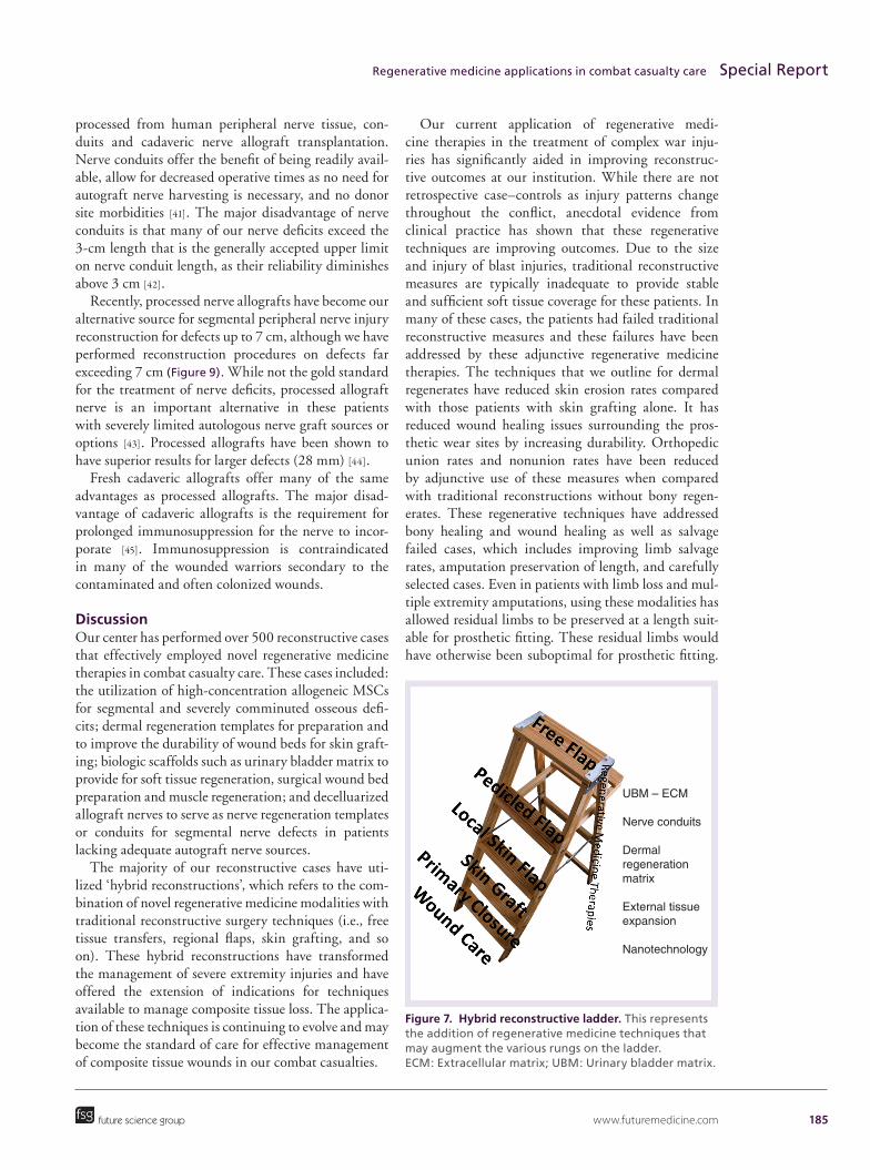
Management of soft tissue defectThe reconstructive ladder was a term coined by plastic and reconstructive surgeons to describe levels of increas-ingly complex management of soft tissue wounds [29]. Theoretically, the surgeon would utilize the lowest rung of the ladder – that is, the simplest reconstruc-tion technique – to address a clinical reconstructive problem. The reconstructive surgeon would move up the ladder as a more complex or suitable method was required for a given reconstruction problem. We propose a hybrid reconstructive ladder (Figure 7) that augments the traditional reconstructive ladder with regenerative medicine modalities. We theorize that there may be improved outcomes at each rung on the reconstruction ladder and these modalities may allow for the expansion of indications for each rung on the reconstruction ladder.
We have effectively employed dermal regenerates, soft tissue regeneration techniques, biologic scaffolds, fat grafting techniques and adipose-derived stem cells in a number of reconstructions for combat casualties in our center and elsewhere with promising results and clinical outcomes.
An example of a recent reconstruction case is depicted in Figure 8. This case represents the application of the hybrid ladder to the surgical management of a degloved tibia. Microvascular surgical techniques in the form of a latissimus free muscle flap transfer were employed for partial coverage of the proximal two-thirds of the
future science group
Regenerative medicine applications in combat casualty care Special Report
Figure 5. Management of an open tibia fracture with segmental bone loss. (A) Anteroposterior x-ray of an open tibia fracture in a soldier injured by a high-powered rifle. After initial irrigation, debridement, antibiotic bead placement and stabilization with a monolateral external fixator, he had normal neurovascular function to the foot and 12 cm of segmental bone loss. (B) Management via bone transport accomplished with a proximal corticotomy and circular frame application. (C) Complete healing at 1 year postinjury.

184 Regen. Med. (2014) 9(2)
exposed bone and wound. The lower third of exposed bone was effectively covered with the combination of regenerative medicine products including porcine-derived urinary bladder matrix (UBM) and a dermal regenerative matrix. After 5 days, a suitable, viable and durable tissue regenerate completely covered the soft tissue-deficient bone, allowing for split-thickness skin grafting and definitive wound closure. Such innovative techniques in soft tissue coverage may provide a means towards limb salvage in an injury that would have oth-erwise been treated with amputation or multiple flaps to facilitate soft tissue coverage.
For hypovascular or avascular surfaces that require soft tissue coverage, we frequently employ UBM to stimulate granulation tissue formation. UBM is a state-of-the-art modality that has multiple applications in wound care. By stimulating blood vessel formation and chemotaxis of progenitor cells to the site of injury, it promotes a unique healing environment favoring regen-eration of native tissue over scar formation [30]. UBM is an extracellular matrix (ECM) that consists of a com-plex mixture of structural and functional proteins and serves an important role in tissue and organ morpho-genesis, maintenance of cell and tissue structure and function, and in the host response to injury [31–33].
Dermal regenerative matrices are another modality that is frequently employed to manage soft tissue loss [34,35]. To manage complex combat-related soft tissue
wounds we regularly utilize bioartificial dermal sub-stitutes combined with subatmospheric pressure (vac-uum-assisted closure) dressings for wound preparation prior to delayed split-thickness skin grafting [36]. The dermal regenerative matrix we utilize is a bilaminar membrane system consisting of porous bovine tendon collagen and shark glycosaminoglycan (chondro itin-6-sulfate) covered by a temporary epidermal substitute made of silicone [37]. The collagen layer acts as a matrix for the ingrowth of fibroblasts and endothelial cells, and it is gradually replaced by the host collagen. Once neovascularization is accomplished, split-thickness skin grafts are employed [38].
Management of segmental nerve nefectsThe standard treatment for large segmental peripheral nerve injuries is the delayed grafting of autograft nerves harvested from a less critical location, such as the sural nerve [39], lateral antebrachial cutaneous nerve, anterior division of the medial antebrachial cutaneous nerve, dorsal cutaneous branch of the ulnar nerve, and super-ficial sensory branch of the radial nerve [40]. Unfortu-nately, given the nature of current injury patterns and the high rates of multiple-extremity amputations and injuries, the availability of suitable and sufficient nerve autograft donor sites is severely limited.
Nerve regeneration repair techniques include decel-lularized and sterile extracellular matrix, nerve grafts
future science group
Special Report Fleming, Bharmal & Valerio
Figure 6. Management of an open tibia fracture with mesenchymal stem cells. (A) Anteroposterior x-ray of an open tibia fracture sustained as a result of a blast injury after irrigation, debridement, antibiotic bead placement and stabilization with a ring fixator. (B) At 6 weeks postinjury, an allograft matrix that contains high concentrations of allogeneic adult mesenchymal stem cells was grafted to the site. The allogeneic mesenchymal stem cell product, approved in the USA, consists of a nonpurified population of allograft mesenchymal stem cells and demineralized bone graft. The advantage of the matrix is that it contains all of the essential properties of an ideal graft: osteoinductivity, osteoconductivity and high levels of osteogenic mesenchymal stem cells essential for bone formation. (C) At 8 months postinjury, copious new bone formation can be seen.
www.futuremedicine.com 185
processed from human peripheral nerve tissue, con-duits and cadaveric nerve allograft transplantation. Nerve conduits offer the benefit of being readily avail-able, allow for decreased operative times as no need for autograft nerve harvesting is necessary, and no donor site morbidities [41]. The major disadvantage of nerve conduits is that many of our nerve deficits exceed the 3-cm length that is the generally accepted upper limit on nerve conduit length, as their reliability diminishes above 3 cm [42].
Recently, processed nerve allografts have become our alternative source for segmental peripheral nerve injury reconstruction for defects up to 7 cm, although we have performed reconstruction procedures on defects far exceeding 7 cm (Figure 9). While not the gold standard for the treatment of nerve deficits, processed allograft nerve is an important alternative in these patients with severely limited autologous nerve graft sources or options [43]. Processed allografts have been shown to have superior results for larger defects (28 mm) [44].
Fresh cadaveric allografts offer many of the same advantages as processed allografts. The major disad-vantage of cadaveric allografts is the requirement for prolonged immunosuppression for the nerve to incor-porate [45]. Immunosuppression is contraindicated in many of the wounded warriors secondary to the contaminated and often colonized wounds.
DiscussionOur center has performed over 500 reconstructive cases that effectively employed novel regenerative medicine therapies in combat casualty care. These cases included: the utilization of high-concentration allogeneic MSCs for segmental and severely comminuted osseous defi-cits; dermal regeneration templates for preparation and to improve the durability of wound beds for skin graft-ing; biologic scaffolds such as urinary bladder matrix to provide for soft tissue regeneration, surgical wound bed preparation and muscle regeneration; and decelluarized allograft nerves to serve as nerve regeneration templates or conduits for segmental nerve defects in patients lacking adequate autograft nerve sources.
The majority of our reconstructive cases have uti-lized ‘hybrid reconstructions’, which refers to the com-bination of novel regenerative medicine modalities with traditional reconstructive surgery techniques (i.e., free tissue transfers, regional flaps, skin grafting, and so on). These hybrid reconstructions have transformed the management of severe extremity injuries and have offered the extension of indications for techniques available to manage composite tissue loss. The applica-tion of these techniques is continuing to evolve and may become the standard of care for effective management of composite tissue wounds in our combat casualties.
Our current application of regenerative medi-cine therapies in the treatment of complex war inju-ries has significantly aided in improving reconstruc-tive outcomes at our institution. While there are not retro spective case–controls as injury patterns change throughout the conflict, anecdotal evidence from clinical practice has shown that these regenerative techniques are improving outcomes. Due to the size and injury of blast injuries, traditional reconstructive measures are typically inadequate to provide stable and sufficient soft tissue coverage for these patients. In many of these cases, the patients had failed traditional reconstructive measures and these failures have been addressed by these adjunctive regenerative medicine therapies. The techniques that we outline for dermal regenerates have reduced skin erosion rates compared with those patients with skin grafting alone. It has reduced wound healing issues surrounding the pros-thetic wear sites by increasing durability. Orthopedic union rates and nonunion rates have been reduced by adjunctive use of these measures when compared with traditional reconstructions without bony regen-erates. These regenerative techniques have addressed bony healing and wound healing as well as salvage failed cases, which includes improving limb salvage rates, amputation preservation of length, and carefully selected cases. Even in patients with limb loss and mul-tiple extremity amputations, using these modalities has allowed residual limbs to be preserved at a length suit-able for prosthetic fitting. These residual limbs would have otherwise been suboptimal for prosthetic fitting.
future science group
Regenerative medicine applications in combat casualty care Special Report
Figure 7. Hybrid reconstructive ladder. This represents the addition of regenerative medicine techniques that may augment the various rungs on the ladder. ECM: Extracellular matrix; UBM: Urinary bladder matrix.
UBM – ECM
Nerve conduits
Dermal regeneration matrix
External tissue expansion
Nanotechnology

186 Regen. Med. (2014) 9(2)
Follow-on retrospective and prospective reviews will analyze the outcomes utilized in these techniques.
The lessons learned from these challenging cases may be adapted to certain civilian trauma and inju-
future science group
Special Report Fleming, Bharmal & Valerio
Figure 8. Management of a degloved tibia (including loss of periosteum) treated with a latissimus free flap for the proximal half of the wound and the lower half with the combination of therapies including urinary bladder matrix, dermal regenerative matrix and nanocrystalline silver dressings. (A) Degloved open tibia fracture with loss of soft tissue including periosteum. (B) Harvesting of latissimus free flap. (C) Anastomosis and in-setting of latissimus free flap. (D) Post split-thickness skin grafting following dermal regenerative matrix and extracellular matrix applications. (E) 1 year postreconstruction.
www.futuremedicine.com 187
ries in an effort to provide better function and defini-tive wound closure. Because of the severity of these war-wounded injuries and the limitations with tradi-tional reconstructive measures due to the massive size and zones of blast injury, it may be extracted to treat lesser severe injuries from trauma, burn or oncologic cases using hybrid reconstructions. However, all the mechanisms of injury are slightly different and must be considered and extracted from our blast patients. Our experiences will hopefully lead to further inno-vations in the field as well as lay the groundwork for future directions in novel breakthroughs to treat our combat casualties as well as our civilian trauma counterparts.
Soft tissue regeneration templates for skin and muscle regeneration are of high interest and need for the resulting massive soft tissue injury patient – both in military and civilian trauma [46]. Large soft tis-sue losses due to avulsion, burn or ballistic injuries frequently present with volumetric or 3D defects, and often leads to severely compromised extremity function. The reconstruction options that currently
exist to treat such defects frequently fail to satisfac-torily address the aesthetic and functional require-ments of the resulting soft tissue defects. Continued research and the development of strategies to address volumetric muscle loss are of continued interest. The utilization of biologic scaffolds may enhance the musculotendinous repair process [47].
Given the limited autologous or donor source for tissues and flaps in the polyextremity amputee or injured service member, coupled with the compro-mised tissues from extensive zones of injury, better options for tissue regeneration and/or substitution are important for the ultimate reconstruction and recovery of combat-injured patients. Most traditional reconstruction measures are very effective in the man-agement of single-tissue soft tissue loss or address-ing minor bone loss. However, composite tissue loss typically results in severely compromised extremity function. While a few functional muscle transfers are available (e.g., functional free gracilis transfers), many of our wound warriors lack appropriate donor tissues and/or necessary nerves to be successful. As previ-
future science group
Regenerative medicine applications in combat casualty care Special Report
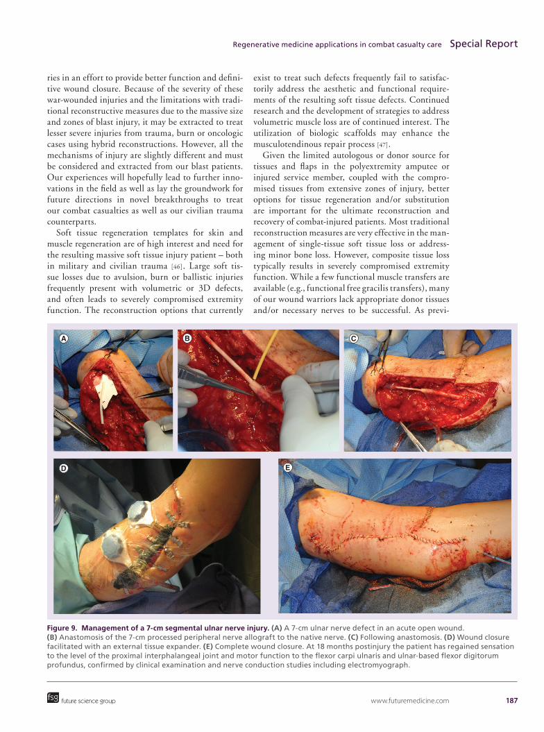
Figure 9. Management of a 7-cm segmental ulnar nerve injury. (A) A 7-cm ulnar nerve defect in an acute open wound. (B) Anastomosis of the 7-cm processed peripheral nerve allograft to the native nerve. (C) Following anastomosis. (D) Wound closure facilitated with an external tissue expander. (E) Complete wound closure. At 18 months postinjury the patient has regained sensation to the level of the proximal interphalangeal joint and motor function to the flexor carpi ulnaris and ulnar-based flexor digitorum profundus, confirmed by clinical examination and nerve conduction studies including electromyograph.

188 Regen. Med. (2014) 9(2)
ously discussed, in addition to the actual aesthetic and functional reconstructive needs of the complex wounds themselves, many complicated polyextremity amputees have limited donor sites. Thus, regenerative medicine therapies that could augment such deficits would and are of great interest, utility, and need for improving the aesthetic and functional outcomes of the complex trauma reconstructive patient.
Future perspectiveFuture directions in each of the areas discussed are promising in enhancing the management of complex wounds. The future for large peripheral nerve defect reconstruction include tolerance induction and mini-mal immunosuppression for nerve allografting, cell-based supportive therapies and bioengineering of nerve conduits [48].
The future of dermal regenerates includes enhanc-ing existing technologies and adapting them to sin-gle-stage procedures to manage soft tissue avulsion injuries. Spray skin technologies are being investi-gated as a therapeutic measure to address significant skin loss [49]. Spray skin technologies include the use of nonculture autologous cells to promote wound healing and reconstructive procedures. Other dermal regenerate technologies that may have applications in combat casualty care include allogeneic matrices that are embedded with immortal keratinocytes, which is a continuous cell line that is virus free and nontumorigenic.
Future strategies that may enhance bone regenera-tion include seeded matrices that will allow imme-diate weight bearing, vascularized bioartificial bone grafts [50,51] and gene therapy.
Vascularized composite tissue allograft transplan-tation is another possible therapy that combat casu-alties may benefit from [52]. Currently, the major
disadvantage of this modality is the life-long immu-nosuppressant agents required. Finally, the use of prefabricated composite tissue flaps in combination with regenerative medicine applications is an option that has yet to be fully tapped as a source for complex soft tissue management [53].
In conclusion, the goal is to provide the best pos-sible care to our combat casualties while developing better techniques at tissue salvage for the benefit of all patients, both active-duty and civilian.
AcknowledgementsThe authors thank Nancy Lee (Walter Reed National Mili-
tary Medical Center Department of Orthopaedics Research
Coordinator) for her expertise and support in the editing of
this manuscript.
DisclaimerThe views expressed in this article are those of the authors
and do not reflect the official policy of the Department of
Defense, or US Government.
Financial & competing interests disclosureThe authors have no relevant affiliations or financial in-
volvement with any organization or entity with a financial
interest in or financial conflict with the subject matter or
materials discussed in the manuscript. This includes employ-
ment, consultancies, honoraria, stock ownership or options,
expert testimony, grants or patents received or pending, or
royalties.
No writing assistance was utilized in the production of
this manuscript.
Informed consent disclosureWritten informed consent has been obtained from the par-
ticipants involved in the study. Appropriate Institutional
Review Board clearance obtained.
future science group
Special Report Fleming, Bharmal & Valerio
Executive summary
Early management & evacuation• Combat-related injuries are often devastating and require multiple resources to manage.Challenges in the definitive management of combat casualties• Resuscitation, decontamination and stabilization are the hallmarks of the initial management of
combat-related injuries.Management of soft tissue defect• Dermal regenerative matrices have proven beneficial in the management of burn patients and are proving
promising in the management of traumatic full-thickness wounds.Management of segmental bone defects• Mesenchymal stem cell-based therapy to replace segmental bone loss is a promising therapy for patients with
segmental bone loss.Management of segmental nerve defects• Expansion of indications for existing nerve conduits is needed for large segmental nerve defects.Future perspective• Cell-based therapies and allotransplantation will enable new pathways in reconstruction.
www.futuremedicine.com 189
ReferencesPapers of special note have been highlighted as: • of interest; •• of considerable interest
1 Beekley AC, Sebesta JA, Blackbourne LH et al.; 31st Combat Support Hospital Research Group. Prehospital tourniquet use in Operation Iraqi Freedom: effect on hemorrhage control and outcomes. J. Trauma 64(2 Suppl.), S28–S37 (2008).
2 Beninati W, Meyer MT, Carter TE. The critical care air trans port program. Crit. Care Med. 36(7 Suppl.), S370–S376 (2008).
3 Morrison JJ, Oh J, Dubose JJ et al. En-route care capability from point of injury impacts mortality after severe wartime injury. Ann. Surg. 257(2), 330–334 (2013).
4 Cap AP, Spinella PC, Borgman MA, Blackbourne LH, Perkins JG. Timing and location of blood product transfusion and outcomes in massively transfused combat casualties. J. Trauma Acute Care Surg. 73(2 Suppl. 1), S89–S94 (2012).
5 Fox CJ, Starnes BW. Vascular surgery on the modern battlefield. Surg. Clin. North Am. 87(5), 1193–1211, xi (2007).
6 Andersen RC, Fleming M, Forsberg JA et al. Dismounted complex blast injury. J. Surg. Orthop. Adv. 21(1), 2–7 (2012).
7 Belmont PJ Jr, McCriskin BJ, Sieg RN, Burks R, Schoenfeld AJ. Combat wounds in Iraq and Afghanistan from 2005 to 2009. J. Trauma Acute Care Surg. 73(1), 3–12 (2012).
8 Potter BK, Scoville CR. Amputation is not isolated: an overview of the US Army Amputee Patient Care Program and associated amputee injuries. J. Am. Acad. Orthop. Surg. 14(10 Spec. No.), S188–S190 (2006).
9 Stansbury LG, Lalliss SJ, Branstetter JG, Bagg MR, Holcomb JB. Amputations in U. S. military personnel in the current conflicts in Afghanistan and Iraq. J. Orthop. Trauma 22(1), 43–46 (2008).
10 Owens BD, Kragh JF Jr, Macaitis J, Svoboda SJ, Wenke JC. Characterization of extremity wounds in Operation Iraqi Freedom and Operation Enduring Freedom. J. Orthop. Trauma 21(4), 254–257 (2007).
11 Fleming M, Waterman S, Dunne J, D’Alleyrand JC, Andersen RC. Dismounted complex blast injuries: patterns of injuries and resource utilization associated with the multiple extremity amputee. J. Surg. Orthop. Adv. 21(1), 32–37 (2012).
•• Describes a complex injury pattern sustained in combat and reveals the extraordinary resources required to manage these complex injuries.
12 Masini BD, Waterman SM, Wenke JC, Owens BD, Hsu JR, Ficke JR. Resource utilization and disability outcome assessment of combat casualties from Operation Iraqi Freedom and Operation Enduring Freedom. J. Orthop. Trauma 23(4), 261–266 (2009).
13 Belmont PJ Jr, Thomas D, Goodman GP et al. Combat musculoskeletal wounds in a US Army Brigade Combat Team during operation Iraqi Freedom. J. Trauma 71(1), e1–e7 (2011).
14 Fang R, Pruitt VM, Dorlac GR et al. Critical care at Landstuhl Regional Medical Center. Crit. Care Med. 36(7 Suppl.), S383–S387 (2008).
15 Liston WA, Dunne JR. Transforming an academic military treatment facility into a trauma center: lessons learned from Operation Iraqi Freedom. Eplasty 9, e31 (2009).
16 Bhananker SM, Ramaiah R. Trends in trauma transfusion. Int. J. Crit. Illn. Inj. Sci. 1(1), 51–56 (2011).
17 Spinella PC, Perkins JG, Grathwohl KW et al. 31st CSH Research Working Group. Fresh whole blood transfusions in coalition military, foreign national, and enemy combatant patients during Operation Iraqi Freedom at a U.S. combat support hospital. World J. Surg. 32(1), 2–6 (2008).
18 Mamczak CN, Elster EA. Complex dismounted IED blast injuries: the initial management of bilateral lower extremity amputations with and without pelvic and perineal involvement. J. Surg. Orthop. Adv. 21(1), 8–14 (2012).
19 Chaddha M, Gulati D, Singh AP, Singh AP, Maini L. Management of massive posttraumatic bone defects in the lower limb with the Ilizarov technique. Acta Orthop. Belg. 76(6), 811–820 (2010).
20 Gessmann J, Köller M, Godry H, Schildhauer TA, Seybold D. Regenerate augmentation with bone marrow concentrate after traumatic bone loss. Orthop. Rev. (Pavia) 4(1), e14 (2012).
21 Younger EM, Chapman M. Morbidity at bone graft donor sites. J. Orthop. Trauma 3, 192–195 (1989).
22 Arinzeh TL. Mesenchymal stem cells for bone repair: preclinical studies and potential orthopedic applications. Foot Ankle Clin. 10(4), 651–665, viii (2005).
23 Ryan JM, Barry FP, Murphy JM, Mahon BP. Mesenchymal stem cells avoid allogeneic rejection. J. Inflamm. (Lond.) 2, 8 (2005).
•• Allogeneic mesenchymal stem cells avoid allogeneic rejection in humans and in animal models. These finding are supported by in vitro co-culture studies.
24 Rush SM. Trinity evolution: mesenchymal stem cell allografting in foot and ankle surgery. Foot Ankle Spec. 3(3), 140–143 (2010).
25 Chatterjea A, Meijer G, van Blitterswijk C, de Boer J. Clinical application of human mesenchymal stromal cells for bone tissue engineering. Stem Cells Int. 11, 215625 (2010).
26 Poynton AR, Lane JM. Safety profile for the clinical use of bone morphogenetic proteins in the spine. Spine (Phila. PA 1976) 27(16 Suppl. 1), S40–S48 (2002).
27 Epstein NE. Complications due to the use of BMP/INFUSE in spine surgery: the evidence continues to mount. Surg. Neurol. Int. 4(Suppl. 5), S343–S352 (2013).
28 Carragee EJ, Hurwitz EL, Weiner BK. A critical review of recombinant human bone morphogenetic protein-2 trials in spinal surgery: emerging safety concerns and lessons learned. Spine J. 11(6), 471–491 (2011).
29 Levin LS. The reconstructive ladder. An orthoplastic approach. Orthop. Clin. North Am. 24(3), 393–409 (1993).
•• Describes the reconstructive ladder used in complex injury reconstruction. Serves as the foundation for the hybrid reconstruction model described in this report.
future science group
Regenerative medicine applications in combat casualty care Special Report
190 Regen. Med. (2014) 9(2)
30 Mitchell KB, Gallagher JJ. Porcine bladder extracellular matrix for closure of a large defect in a burn contracture release. J. Wound Care 21(9), 454–456 (2012).
31 Badylak SF. The extracellular matrix as a scaffold for tissue reconstruction. Semin. Cell Dev. Biol. 13(5), 377–383 (2002).
•• Summarizes the concept of extracellular matrices utilized as a scaffold for tissue reconstruction.
32 Kruper GJ, Vandegriend ZP, Lin HS, Zuliani GF. Salvage of failed local and regional flaps with porcine urinary bladder extracellular matrix aided tissue regeneration. Case Rep. Otolaryngol. 2013, 917183 (2013).
33 Remlinger NT, Gilbert TW, Yoshida M et al. Urinary bladder matrix promotes site appropriate tissue formation following right ventricle outflow tract repair. Organogenesis 9(3), 149–160 (2013).
34 Demiri E, Papaconstantinou A, Dionyssiou D, Dionyssopoulos A, Kaidoglou K, Efstratiou I. Reconstruction of skin avulsion injuries of the upper extremity with Integra(®) Dermal Regeneration Template and skin grafts in a single-stage procedure. Arch. Orthop. Trauma Surg. 133(11), 1521–1526 (2013).
35 Graham GP, Helmer SD, Haan JM, Khandelwal A. The use of Integra® Dermal Regeneration Template in the reconstruction of traumatic degloving injuries. J. Burn Care Res. 34(2), 261–266 (2013).
36 Helgeson MD, Potter BK, Evans KN, Shawen SB. Bioartificial dermal substitute: a preliminary report on its use for the management of complex combat-related soft tissue wounds. J. Orthop. Trauma 21(6), 394–399 (2007).
• Demonstrates the successful application of bioartificial dermal substitutes for the management of complex combat-related wounds.
37 Moiemen NS, Staiano JJ, Ojeh NO et al. Reconstructive surgery with a dermal regeneration template: clinical and histologic study. Plast. Reconstr. Surg. 108, 93–103 (2001).
38 Stern R, McPherson M, Longaker MT. Histologic study of artificial skin used in the treatment of full-thickness thermal injury. J. Burn Care Rehabil. 11, 7–13 (1990).
39 Whitlock EL, Tuffaha SH, Luciano JP et al. Processed allografts and type I collagen conduits for repair of peripheral nerve gaps. Muscle Nerve 39(6), 787–799 (2009).
40 Matsuyama T, Mackay M, Midha R. Peripheral nerve repair and grafting techniques: a review. Neurol. Med. Chir. (Tokyo) 40(4), 187–199 (2000).
41 Rivlin M, Sheikh E, Isaac R, Beredjiklian PK. The role of nerve allografts and conduits for nerve injuries. Hand Clin. 26(3), 435–446 (2010).
42 Deal DN, Griffin JW, Hogan MV. Nerve conduits for nerve repair or reconstruction. J. Am. Acad. Orthop. Surg. 20(2), 63–68 (2012).
43 Hudson TW, Zawko S, Deister C et al. Optimized acellular nerve graft is immunologically tolerated and supports regeneration. Tissue Eng. 10(11–12), 1641–1651 (2004).
44 Whitlock EL, Tuffaha SH, Luciano JP et al. Processed allografts and type I collagen conduits for repair of peripheral nerve gaps. Muscle Nerve 39(6), 787–799 (2009).
45 Mackinnon SE, Doolabh VB, Novak CB, Trulock EP. Clinical outcome following nerve allograft transplantation. Plast. Reconstr. Surg. 107(6), 1419–1429 (2001).
46 Ficke JR, Obremskey WT, Gaines RJ et al. Reprioritization of research for combat casualty care. J. Am. Acad. Orthop. Surg. 20(Suppl. 1), S99–S102 (2012).
• Discusses the research gaps and the need for reprioritization of research in combat casualty care.
47 Turner NJ, Badylak SF. Biologic scaffolds for musculotendinous tissue repair. Eur. Cell Mater. 25, 130–143 (2013).
48 Siemionow M, Uygur S, Ozturk C, Siemionow K. Techniques and materials for enhancement of peripheral nerve regeneration: a literature review. Microsurgery 33(4), 318–328 (2013).
•• Summarizes the regenerative medicine options for peripheral nerve regeneration.
49 Tenenhaus M, Rennekampff HO. Surgical advances in burn and reconstructive plastic surgery: new and emerging technologies. Clin. Plast. Surg. 39(4), 435–443 (2012).
50 Kokemueller H, Spalthoff S, Nolff M et al. Prefabrication of vascularized bioartificial bone grafts in vivo for segmental mandibular reconstruction: experimental pilot study in sheep and first clinical application. Int. J. Oral Maxillofac. Surg. 39(4), 379–387 (2010).
51 Kneser U, Polykandriotis E, Ohnolz J et al. Engineering of vascularized transplantable bone tissues: induction of axial vascularization in an osteoconductive matrix using an arteriovenous loop. Tissue Eng. 12(7), 1721–1731 (2006).
52 Murphy BD, Zuker RM, Borschel GH. Vascularized composite allotransplantation: an update on medical and surgical progress and remaining challenges. J. Plast. Reconstr. Aesthet. Surg. 66(11), 1449–1455 (2013).
53 Erkin UR, Kerem M, Tug M, Orbay H, Sensöz O. Prefabrication of a conjoint flap containing xenogenic tissues: a preliminary report on an experimental model. J. Craniofac. Surg. 18(6), 1451–1456 (2007).
future science group
Special Report Fleming, Bharmal & Valerio