Embed Size (px)
Citation preview
Presented by
Dr. Md. Saroat Hossain
DEPT. OF ANIMAL HUSBANDRY & VETERINARY
SCIENCE
UNIVERSITY OF RAJSHAHI
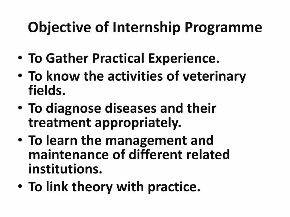
Objective of Internship Programme
• To Gather Practical Experience.• To know the activities of veterinary
fields.• To diagnose diseases and their
treatment appropriately. • To learn the management and
maintenance of different related institutions.
• To link theory with practice.

Name of the
institution
Address Name of the institution Address
01 Veterinary Clinic & AI
Centre
Narkelbaria,
Rajshahi
07 Bangladesh Livestock
Research Institute (BLRI)
Savar,
Dhaka
02 Rajshahi Dairy &
Cattle Breeding Farm
Rajabarihat,
Rajshahi
08 Bangladesh National Zoo Mirpur,
Dhaka
03 Goat Development
Farm
Rajabarihat,
Rajshahi
09 Upazilla livestock office Poba,
Rajshahi
04 Shahid A.H.M
Kamruzzaman Central
Park & Zoo
Rajshahi 10 Upazilla livestock office Durgapur,
Rajshahi
05 District artificial
insemination center
Rajshahi 11 Upazilla livestock office Pithia,Rajsh
ahi
06 CCBDF Savar,
Dhaka
12 Thana livestock
office(Metro)
Boalia,
Rajshahi
Hospital practices

• Cause:
• Biting of Rabid Dog.
• No post exposure vaccine given.
• Signs:
• Complete paralysis
• Complete anorexia
• Excessive salivation
• Treatment:
• No treatment given
• Advice:• Performing Euthanasia and
then Buried was suggested.
• Keep Animal Safe from Rabid animal.
Fig: Rabies of calf
1.Complete paralysis
2.Excessive salivation
1
2

Fig 1: Removal of aborted fetus Fig 2: Washing uterus with PPM solution
Diagnosis: Abortion of Doe
Cause: Complication of PPR
Management of Dystocia:
•Removal of dead fetus
•Washing the Uterus with PPM solution
•Bolus Renartim inserted into the uterus
Advice: Provide balanced Diet, Provide Stress free Condition

Diagnosis: Myiasis
Causal agent: Diptera larvae
Management:•Removal of Maggot by a forceps.
•Washed thoroughly with gauze soaked with Tinc. Iodine.
•Then ivermectin was administered.
Advice: Turpin oil applied tropically.
Fig1:Myiasis of calf Fig 2:Myiasis of doe
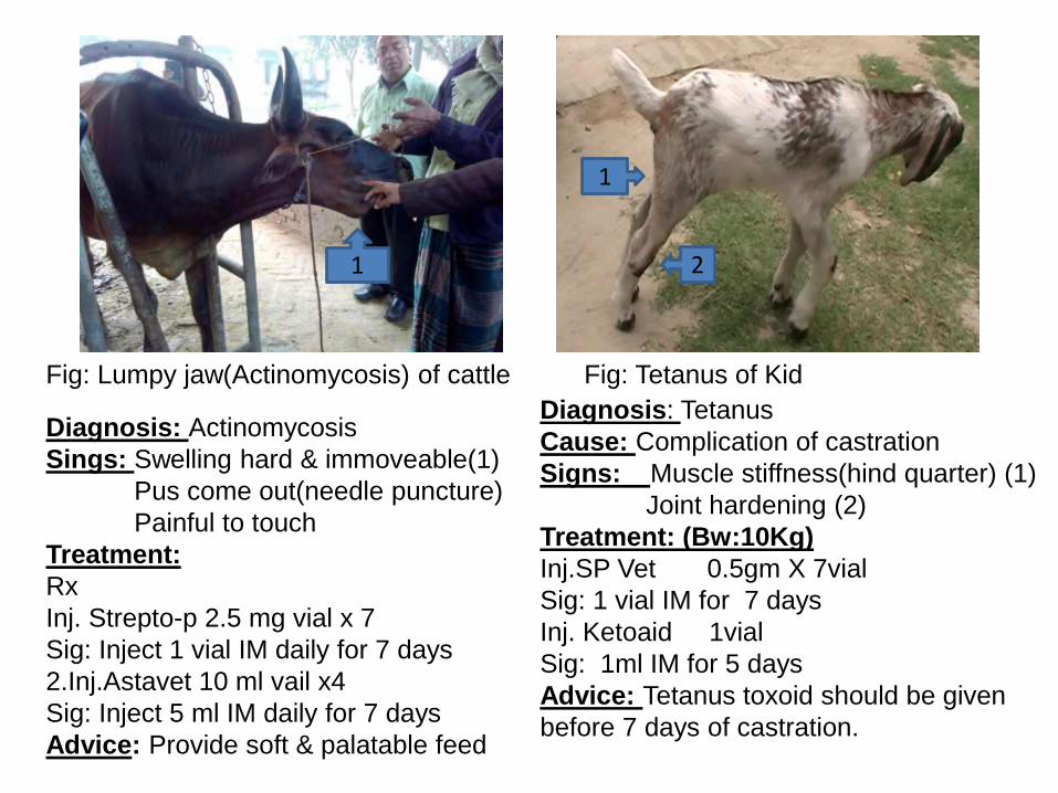
Diagnosis: Tetanus
Cause: Complication of castration
Signs: Muscle stiffness(hind quarter) (1)
Joint hardening (2)
Treatment: (Bw:10Kg)
Inj.SP Vet 0.5gm X 7vial
Sig: 1 vial IM for 7 days
Inj. Ketoaid 1vial
Sig: 1ml IM for 5 days
Advice: Tetanus toxoid should be given
before 7 days of castration.
Fig: Tetanus of Kid
1
2
Fig: Lumpy jaw(Actinomycosis) of cattle
Diagnosis: Actinomycosis
Sings: Swelling hard & immoveable(1)
Pus come out(needle puncture)
Painful to touch
Treatment:
Rx
Inj. Strepto-p 2.5 mg vial x 7
Sig: Inject 1 vial IM daily for 7 days
2.Inj.Astavet 10 ml vail x4
Sig: Inject 5 ml IM daily for 7 days
Advice: Provide soft & palatable feed
1
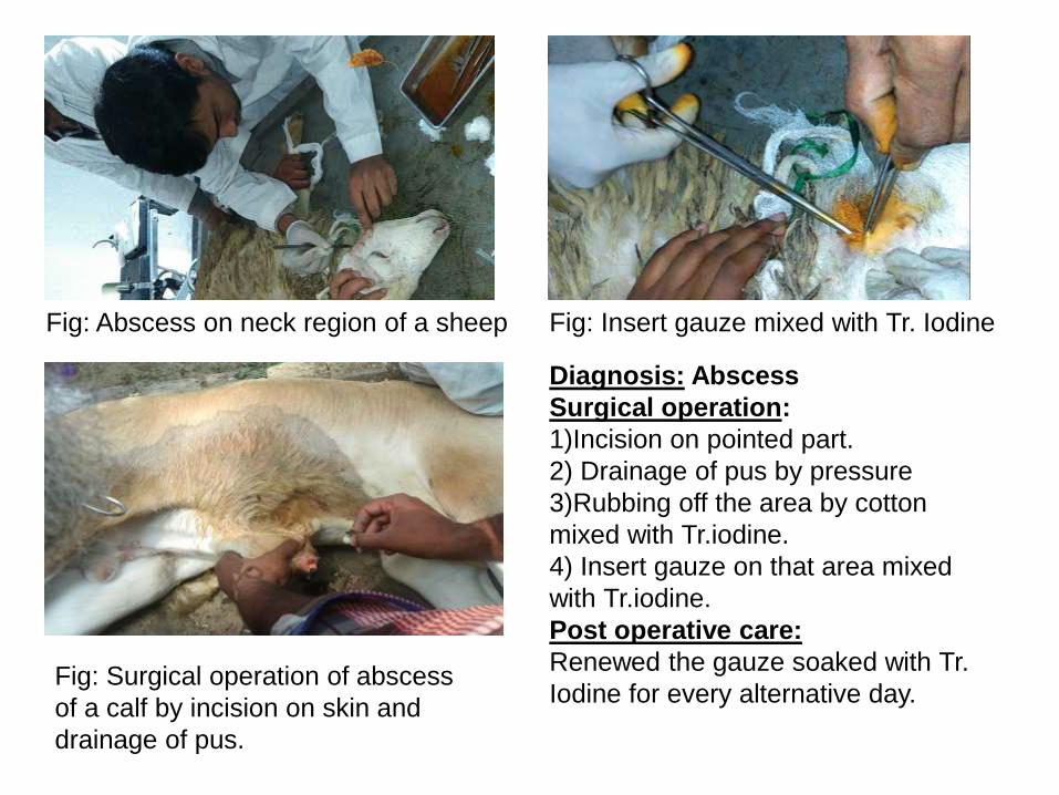
Fig: Surgical operation of abscess
of a calf by incision on skin and
drainage of pus.
Diagnosis: Abscess
Surgical operation:
1)Incision on pointed part.
2) Drainage of pus by pressure
3)Rubbing off the area by cotton
mixed with Tr.iodine.
4) Insert gauze on that area mixed
with Tr.iodine.
Post operative care:
Renewed the gauze soaked with Tr.
Iodine for every alternative day.
Fig: Abscess on neck region of a sheep Fig: Insert gauze mixed with Tr. Iodine

Diagnosis: Compaction of feces
Management:
•Removal of feces manually from
rectum.
•Then washed with Potassium
permanganate
•linseed oil was applied.
Advice:Provide soft feed.
Fig: Compaction of feces of duck Fig: Removal of feces manually
Fig: Wash with PPM solution

Fig: The photograph of the bird on the left has Canker(1), the photograph
on the right is another bird without Canker(2)
2
1
Diagnosis: Canker of pigeon
Causal agent: Trichomonas gallinae or Trichomonas columbae
Clinical findings:
•Caseous yellowish depositions are observed
•Reduce feed and water intake
Treatment:
Treated with Metronidazole(eg. Flagyl ) @30mg/kg orally daily for 5 days.

Farm practices
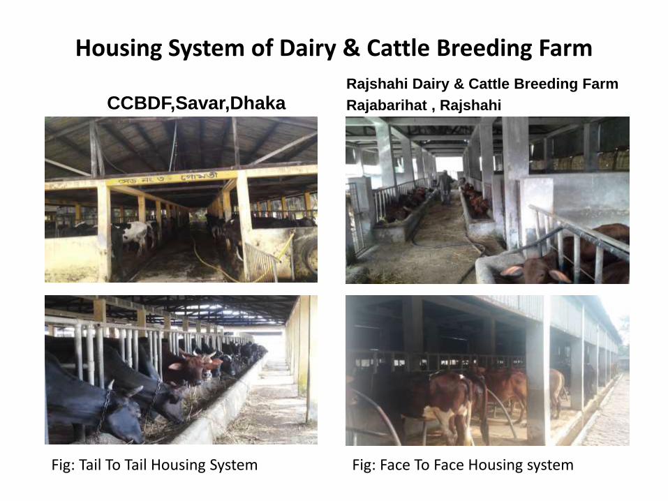
Housing System of Dairy & Cattle Breeding Farm
CCBDF,Savar,DhakaRajshahi Dairy & Cattle Breeding Farm
Rajabarihat , Rajshahi
Fig: Face To Face Housing systemFig: Tail To Tail Housing System

Fig: Ear marking pilers(1) & Ear tag (2) Fig: Weight machine
Fig: Ear tag Fig: Calf pen
1
2

Fig: Artificial insemination by using AI gun(1)
Fig: Semen collection, Dummy(1), Artificial Vagina(2), Bull (3)
13
2
Fig. Pure Friesian bull(100%, BW:1130 Kg)
1
Observation of semen
collection: •A dummy bull is prepared in
travis and a breeding bull is
allowed to jump on the dummy.
• When bull is attempt to
ejaculate, semen is collected by
artificial vagina.

Fig: Milk cooling tank(2000L) Fig: Lactoscan
Fig: milk packaging & sealing Fig: Milk weight machine

Disease Diagnosis & Investigation
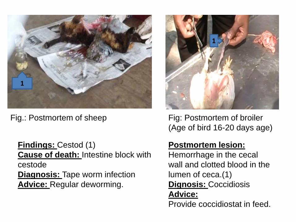
Postmortem lesion:
Hemorrhage in the cecal
wall and clotted blood in the
lumen of ceca.(1)
Dignosis: Coccidiosis
Advice:
Provide coccidiostat in feed.
Fig: Postmortem of broiler
(Age of bird 16-20 days age)
Fig.: Postmortem of sheep
Findings: Cestod (1)
Cause of death: Intestine block with
cestode
Diagnosis: Tape worm infection
Advice: Regular deworming.
1
1

Rapid kit test for avian influenza virus
• Feces collected from vent or swab from trachea with cotton bar.
• Insert cotton bar into Dilute.
• 4-5 drops taken into Rapid test kit by the use of dropper.
• Wait for 10min.
• Two RED band (+Ve)
• One RED band (-Ve)
Dilute & dropper
Fig: Rapid test kit
Obstacles :
During the internship, we faced few
difficulties-
• Schedule problem
• Accommodation problem
• Communication problem
• Shorter time period distribution in
some placement etc.
An overall learning during
my internship
• Diseases diagnosis
• Writing prescription after disease diagnosed
• Providing treatment to the patient
• Attending surgical operation
• Medication
• Vaccination
• Feces examination
• Farm management

Thank you