Embed Size (px)
Citation preview

Investigations of Breast Cancer -by preetam goswami 8th semester,unit-1
Contents
• INVESTIGATIONS FOR DETECTION.
• INVESTIGATIONS FOR STAGING.
• INVESTIGATIONS FOR TREATMENT.
INVESTIGATIONS FOR DETECTION
MODALITIES: Mammography. Ultrasound of Breast MRI of BreastFNAC (Fine Needle Aspiration Cytology) Biopsy Other Investigations

Mammography
• It is a non invasive procedure for detection of breast cancer by using low energy x-rays.
• Principle : It identifies the areas of microcalcifications and tissue densities.
• Procedure: Compression X-rays of superior and medial aspects.
• Detection: Malignant Lesions show irregular densities and
intraductal calcifications Benign Lesions show well defined borders and
peripheral calcifications.
• It is a part of triple assessment therapy which also includes clinical assessment and cytological diagnosis by FNAC

NORMAL BREAST BENIGN LESION MALIGNANT LESION
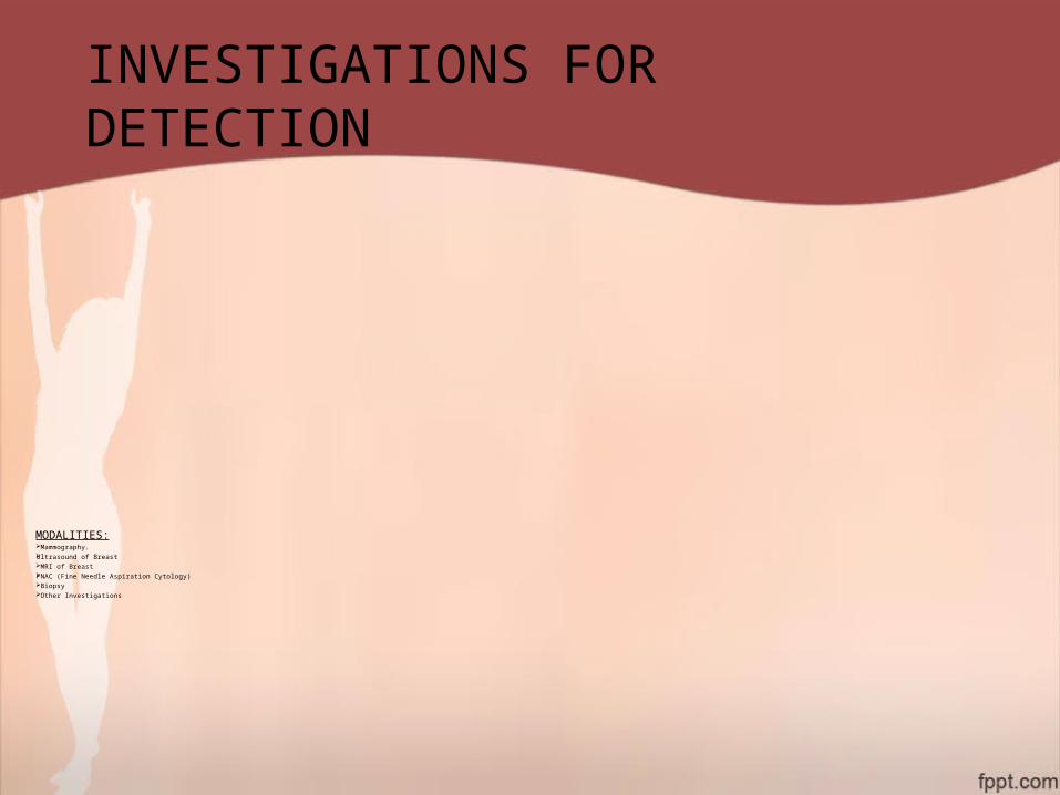
USG of Breasts
Benign Cyst It is a Non Invasive technique that uses sonic energy in the frequency range of 1 – 10 MHz.
Appearance
1.Cysts : Fluid Filled Lesions – No internal echo. 2. Benign : Solid Lesions – Smooth and Well
defined border 3. Malignant : Jagged Borders.
Malignant Cyst
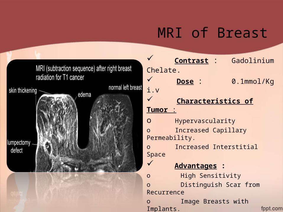
MRI of Breast
Contrast : Gadolinium Chelate. Dose : 0.1mmol/Kg i.v
Characteristics of Tumor :
o Hypervascularityo Increased Capillary Permeability.o Increased Interstitial Space
Advantages :o High Sensitivityo Distinguish Scar from Recurrenceo Image Breasts with Implants.o Choice of Imaging in Pregnancyo Management of Axillary InfiltrationDisadvantage:
Costly, Non Available, Not Sensitive to Premalignant lesions

FNAC
Done in case of cystic lumps of
breast. Criteria for malignancy 1. Blood stained aspirate.
2. Mass does not completely. disappear after aspiration.
FNAC Scoring Co : No epithelial cells.
C1 : Scanty epithelial cells,
Benign.
C2 : Benign Cells.
C3 : Atypical Cells.
C4 : Suspicious Cells.
C5 : Malignant Cells.
BIOPSY
TYPES
Frozen Section Biopsy. Corecut/Trucut Biopsy.
Excision Biopsy. Edge Biopsy.
It is used for definitive diagnosis of malignancy.
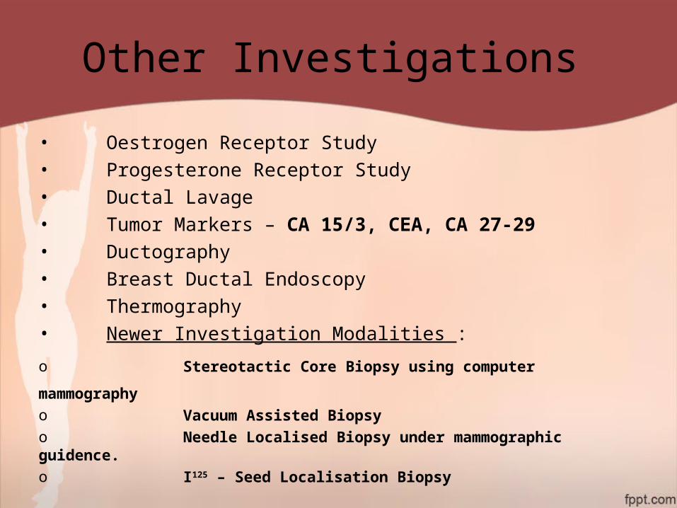
Other Investigations
• Oestrogen Receptor Study• Progesterone Receptor Study• Ductal Lavage• Tumor Markers – CA 15/3, CEA, CA 27-29• Ductography• Breast Ductal Endoscopy• Thermography• Newer Investigation Modalities :
o Stereotactic Core Biopsy using computer mammographyo Vacuum Assisted Biopsyo Needle Localised Biopsy under mammographic guidence.o I125 – Seed Localisation Biopsy

INVESTIGATIONS FOR STAGING
MODALITIES For Tumor Size - MRI Scan
For Nodal involvement – Lympho Scintigraphy - CT Scan
For Metastatic Involvement – Bone X-Ray
- Bone Scan (PET)
- Chest X-Ray
- USG/CT Abdomen
- X-Ray/CT Spine
- Biochemical Studies :- ALP (Bone and Liver)
:- GGT (Liver)
:- Urinary Steroids
:- Urinary Hydroxy Proline

Bone Scan showing metastasis due to advance Carcinoma
Breast.
Liver Scan showing
Metastatic mass.
CT Scan of Chest and Abdomen showing mediastinal and retroperitoneal lymphadenopathy
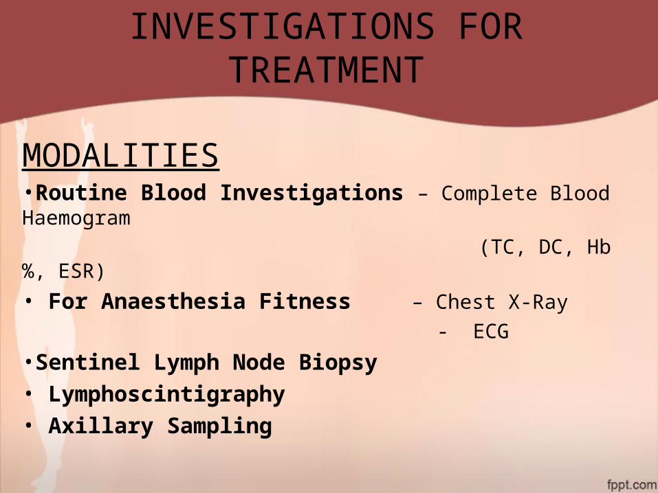
INVESTIGATIONS FOR TREATMENT
MODALITIES•Routine Blood Investigations – Complete Blood Haemogram
(TC, DC, Hb%, ESR)
• For Anaesthesia Fitness – Chest X-Ray
- ECG
•Sentinel Lymph Node Biopsy• Lymphoscintigraphy• Axillary Sampling

Sentinel Lymph Node Biopsy
Sentinel Node indicates first node encountered by the tumor cells and its histological status predicts the status of distant Lymph Nodes.Merits: It is not done in clinically palpable axillary node as there is already distortion of lymphatic flow due to tumor.If there is no involvement of sentinel node further axillary dissection is not required.Demerits:There is high chance of false negative results.Contraindications – Allergy to vital blue dye or radio colloid, pregnancy, inflammatory carcinoma of breast.Complications – Blue Tattooing of skin, Bluish green urine and stool, anaphylaxis, seroma-formation.


To conclude with…Mammography is highly reliable for evaluation of breast cancer as it has a sensitivity of over 90%.Core needle biopsy has to be done wherever the FNAC is inconclusive.USG may not detect lesions less than 1cm size.USG is the Investigation of choice in young women less than 30 years of age.ER and PR status are important for treatment by hormonal therapy.Senitnel Lymph node Biopsy has to be done all in cases of node negative patients in clinical grounds.Most of the deaths due to breast cancer is due to distant metastasis,hence early diagnosis of metastasis has to be done whenever suspected.