Embed Size (px)
Citation preview

Hormonal Causes of Infertility
By
Dr. Atef Abdel-Hai Khalil SelmiProfessor of Obstetrics,
Gynecology, & A.I.Faculty of Veterinary Medicine
Zagazig University( 2 )

Hormonal Causes of Infertility 1-Ovarian Atrophy
The condition was manifested clinically by a history of prolonged period of anestrum in the affected females. It is usually diagnosed in association with chronic debilitating diseases or in aged cows over 15 -20 years of age.The condition was found affecting both ovaries (small, smooth, and firm in consistency), and the genital tract was found reduced in size.The prognosis is poor and the affected female should be discard from breeding or replaced.

2- Follicular AtresiaThe condition means regression of the growing follicles caused by insufficiency of proper gonadotropin stimulation or due to hormonal imbalance. The atretic follicles fail to grow beyond its size, looses its turgidity, and never ovulate. The condition was manifested clinically by a history of anestrum in the affected females.Histopathological examination of atretic follicles revealed that the granulosa cell layer was found reduced in thickness and appeared degenerated or detached. The antrum appeared collapsed and the oocyte with the surrounded cumulus oophorus appeared degenerated and loosely detached in the antrum . Degenerated tissues are further replaced by fibrous formation with complete antrum obliteration in advanced follicular atresia. The condition could be controlled by feeding (concentrate ration supplemented with vitamins and minerals) together with GnRH administration.
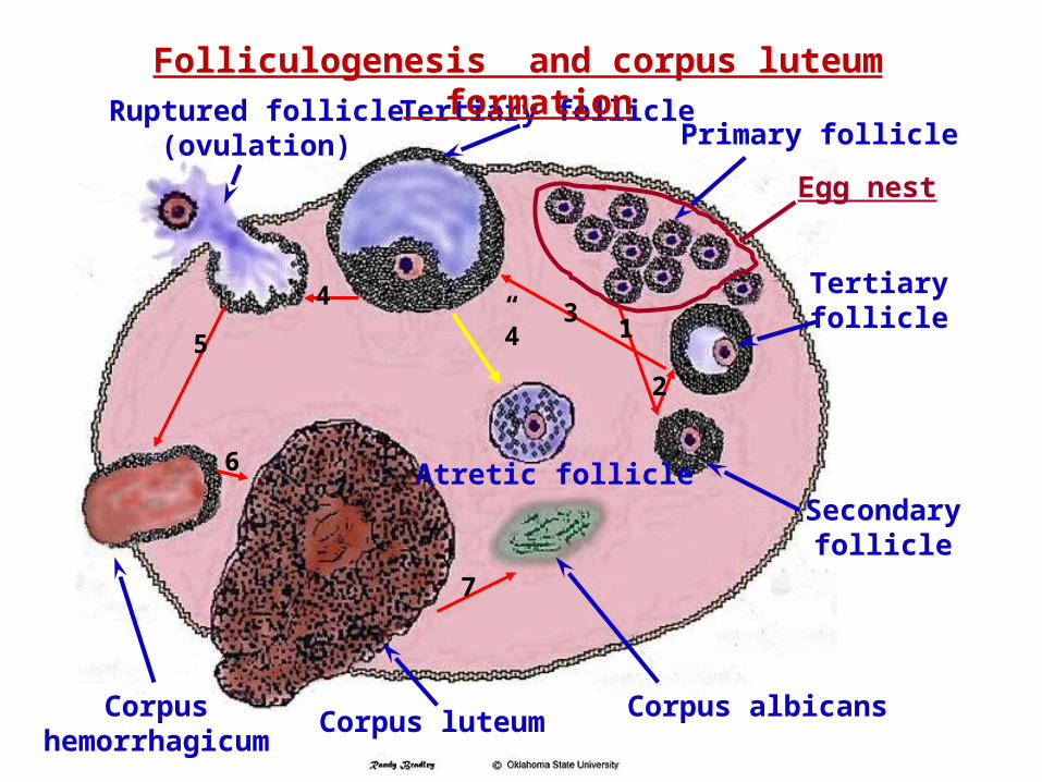
Tertiary folliclePrimary follicle
Tertiaryfollicle
Egg nest
Secondary follicle
Atretic follicle
Corpus albicansCorpus luteumCorpushemorrhagicum
Ruptured follicle)ovulation(
1
2
34
5
6
7
4”
Folliculogenesis and corpus luteum formation

3- Silent heat 4- Subestrum
5- weak estrum 6- Short Unobserved heat
These conditions are characterized clinically by that the affected females fail to express the behavioral signs of estrum or exhibited a marked reduction in the heat period despite the presence of cyclic estral changes in their genital organs. Signs of short unobserved heat could not be clinically differentiated from those of silent heat, subestrum, or weak estrum. Silent heat can be classified as functional anestrum and constitute 90% of all anestrus cases especially in hot climate. Functional anestrum increases open days and causes severe economic losses that could be calculated as 100 pound per open day.
The possible etiological factors:
1- These conditions occur more frequently during the next 60 days postpartum because incidence of silent heat was found high in the first postpartum estrus (77%), then decline to 55% and 35% by the second and third postpartum estrus.
2- These conditions are observed more frequently in foreign breeds especially when lived under hot stressful humid environment.
3- These conditions occur more frequently in nursed than in milked cows. Moreover, the interval from calving to first postpartum estrus in nursed cows was found 30 days longer than in milked cows.
4- The physiologic bases of these condition is not clearly defined, but the central nervous system may be less sensitive in those cases or requires a higher concentrations of estrogen to produce the behavioral signs of heat and consequent acceptance of the male.
5- Hereditary predisposition: High incidence of silent heat was observed in a certain sire line in a herd of Holstein breed, where daughters of silent dams may developed silent heat.6- Cows in advanced age, arthritic cows, cows with foot rot or untrimmed feet, or affected with painful diseases may fail to express signs of heat.7- High lactating cows or fatty cows may also acquire silent heat or quite ovulation.

Diagnoses and treatment: These conditions can be diagnosed by the following application:1-Close observations to the herd to detect estrus cows.2-Application of heat detecting device or aids…………………3-Careful clinical examination for prediction of heat.4-Good keeping records. Treatment of those condition can be practiced more easily by:1-Improve managemental practices(feeding, housing, medical care, and education of laymen). 2-Application of estrus synchronization in the affected herd.3-Regular watching of cows during feeding or milking.4-Selection against those conditions and replacement of chronically recurred cases.

7 -Delayed OvulationThe condition means that the process of ovulation occurs in cows after the proper time (beyond 12 hour after the end of heat signs). Delayed ovulation is usually associated with aging of both gametes (sperm and ova) and results in reduction of their fertilizing capacity. Therefore, delayed ovulation is usually associated with failure of fertilization. The affected females exhibiting prolonged estrus phase and developed cyclic non-breeding syndrome ( regular repeat breeder) despite mating in the proper time ( meddle of estrum).The condition can be diagnosed by the history and clinical examination (persistence of mature Graffian follicle on surface of the ovary for 1 or 2 days later).The condition can be controlled either by: 1-Application of second insemination by the next day. 2-Administration of GnRH or LH at the time of breeding.

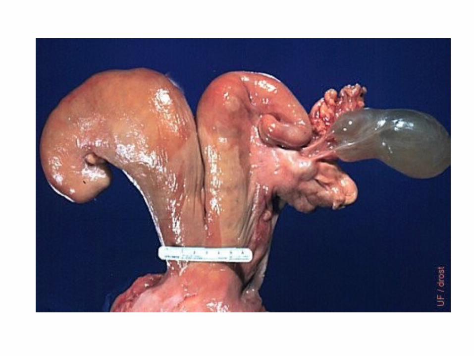
8- Ovarian Cysts (Cystic Ovary)Ovarian cysts can be defined as follicular structures of 2.5 cm in diameter or more that persist on the ovary for at least 10 days up to several months.This syndrome affects all cows ages especially after their 2nd to 5th parturition, and develops most commonly during the second to the seventh week after calving.Four types of cysts can be detected on the ovary(ies) of the affected cases ( follicle theca cyst, or follicle lutein cyst, or corpus luteum cyst, or small cystic ovarian degeneration ).Behavioral signs of nymphomania ( follicle theca cyst ) or anestrum ( other types of cyst ) were found associating cystic ovarian degeneration in the affected cows.

The possible causes of cystic ovarian degeneration:
1- Hereditary causes: A- Incidence of cystic ovarian degeneration was found to be higher in dairy breeds (5.6-18.8%) than in beef breeds. B- It was found to be closely related to the rate of milk production ( commonly in high producing cows ). C- It was found to be closely related to the amount of fat contents in the milk (commonly associating high contents). D- It is also higher in daughters of caws that had ovarian cyst (26.8 %) than in daughters of caws that had no history of ovarian cyst (9.2%). E- Incidence of cystic ovary was reduced from 10.8 to 3 % following selection of cows against cystic ovarian degeneration (culling of cows developed cystic ovary and replacement of their daughters).
2 -Nutritional and Managemental causes: A- Feeding high concentrates ration necessary for milk production (urea concentration was found increased in the follicular fluid of cystic follicle). B- Reduced amount of green feeds in the ration (vitamin A was found reduced in the follicular fluid of cystic follicle). C- Incidence of cystic ovary was high in stabled cows (lack of exercise, lameness, stress of lactation, reduced exposure to sun light, reduced aeration, crowdedness and heat stress together with postpartum uterine infection).

3- Hormonal causes:
A- Increased secretion of prolactin and growth hormone necessary for high lactation:prolactin causes direct inhibition to granulosa cells to secrete progesterone with a consequent increase in estrogen secreation and development of follicular atresia that followed by development of follicle lutein cyst or development of cystic CL (with a consequent development of anestrum). B- An endocrine imbalance: 1-Excessive amounts of FSH that over stimulating follicular growth.
2-Subnormal availability of LH to induce ovulation (anti-LH).3-Failure in the mechanism controlling LH secretion (GnRH administration).4-Deficiency in synthesis or release of GnRH was also suggested (GnRH administration).5-Reduced pituitary-hypothalamus responsiveness to estrogen during the immediate postpartum period (progesterone dominance during pregnancy, supraphysiological concentration of estrogen shortly before calving, most cases develops cystic ovary within 45 days after calving, spontaneous recovery of the cyst, and responsiveness of pituitary and hypothalamus was found to be returned by 4-6 week postpartum).

Classification of ovarian cysts:1- Location of the cyst on the ovary: A- Central cyst. B- Peripheral cyst.2- Number of the cyst on the ovary: A- Single cyst. B- Multiple cysts.3- Origen of the cyst: A- Unovulated follicle: a- Follicle theca cyst ( nymphomania ). b- Follicle lutein cyst ( anestrum ). B- Corpus luteum: Cystic CL ( anestrum ).4- Clinical manifestation: A- Nymphomania. B- Anestrum.

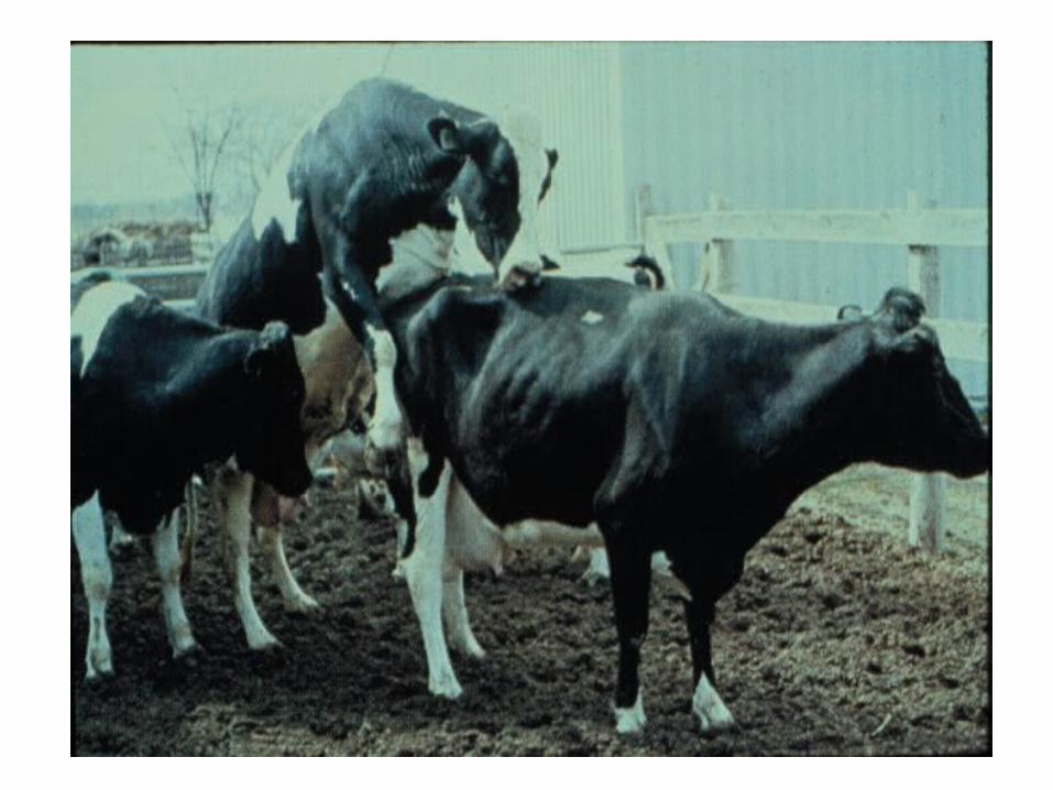
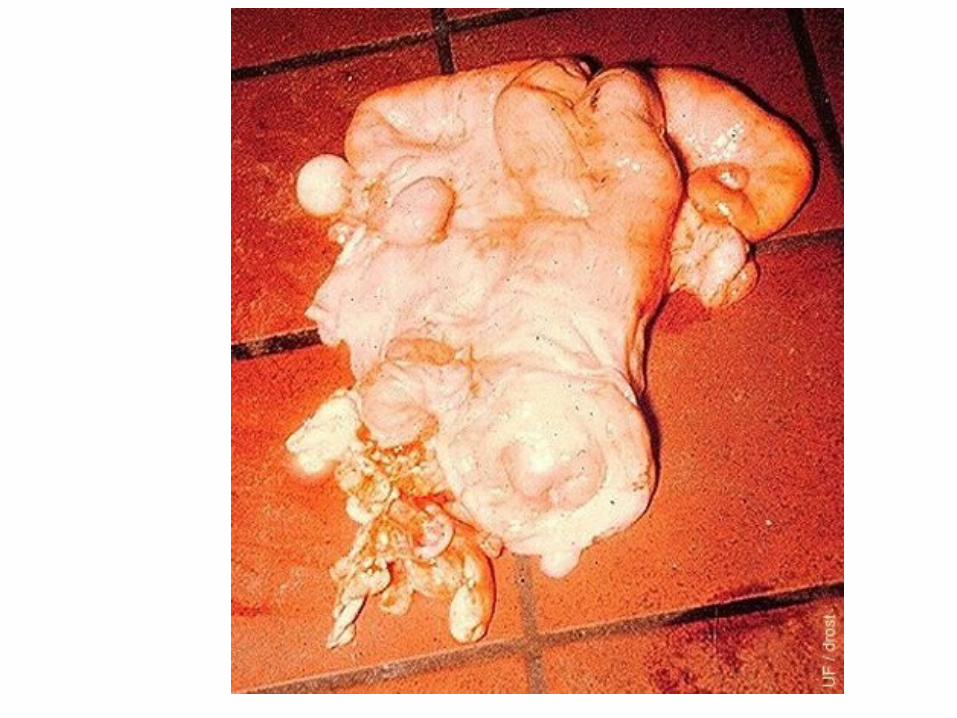
Symptoms and clinical findings:1- Follicle theca cyst (75 %): A-Behavioral and phenotypic changes: a-Frequent, irregular or continuous heat and bellow frequently b-Nervous, restless, sexually aggressive, acquires masculine behavior, and mount other cows ( Nymphomania). c-Lose weight and the voice changed to masculine pitch. d-Relaxation of the pelvic ligaments with the consequent development of sterility hump (elevation of the tail rote). e-Edema in the vulva and tail folds together with signs of pneumovagina and presence of grayish white secretion. f-Sharp decrease in milk production rate due to elevated concentration of estrogen together with reduced appetite.
B- Rectal and vaginal examination:
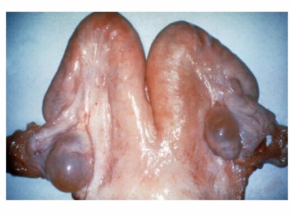
a-Presence of 1 - 4 thin walled cyst on one or both ovaries( 2.5 - 10 cm in diameter).b-Fallopian tubes are enlarged an relaxed or flaccid.c-The uterus is enlarged, edematous and flaccid.d-In longstanding cases, the uterine wall became thin and the lumen became distended with mucoid fluid (hydrometra or mucometra).e- Endometrial glands exhibite cystic degeneration. f-Bartholin glands and Gartner ducts became cystic too.g-Portio vaginal is enlarged, dilated and relaxed together with presence of grayish white mucous.

C-Histopathological changes and fate:
a-Degenerative changes together with partial luteinization occurs in the granulosa cell layer of follicle theca cyst.
b-Progesterone production is increased and estrogen production is reduced. c-The affected cow went anestrum and the condition might spontaneously recovered .


2- Follicle lutein cysts (22%):
It comprises about 23% of the cows affected with cystic ovarian degeneration, but the percentage would be higher than this value because the condition is not associated with abnormal sexual behavior as in follicle theca cyst (nymphomania).Affected cases are anestrum, but spontaneous recovery may occurs.The cyst is firm, less fluctuating than follicle theca cyst, and persists on the ovary without any cyclic changes.
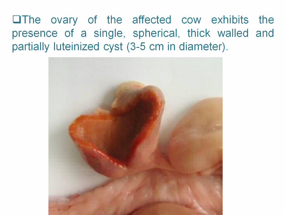

3- Corpus luteom cyst ( 2.5%):It develops from an ovulated follicle, but luteinization is not complete that leading to formation of central lacuna filled with serous fluid.It is associated with absence of cyclic ovarian changes.The affected cow exhibits anestrum.
4-Small cystic ovarian degeneration(0.5%):
The affected cases exhibited the presence of small multiple growing follicles on the ovarian surface ( 0.5 – 1 cm in diameter), that makes the ovary likes mulberry.The condition may be due to arrested follicular growth owing to deficiency in FSH secretion or improper gonadotropin stimulation.Follicular degeneration and atresia can be detected in those cysts.

Diagnosis: A- History. B-Symptoms. C-Clinical examination.
Prognoses:The prognosis is favorable when the affected cases were early diagnosed and treated.
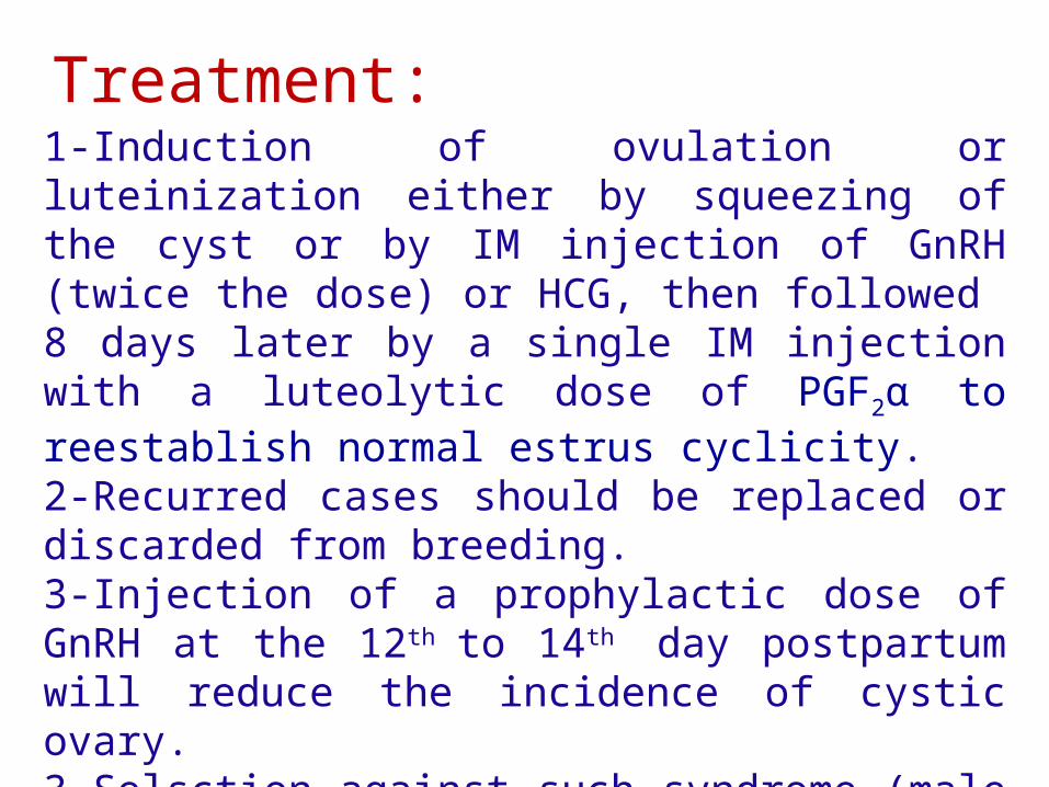
Treatment:1-Induction of ovulation or luteinization either by squeezing of the cyst or by IM injection of GnRH (twice the dose) or HCG, then followed 8 days later by a single IM injection with a luteolytic dose of PGF2α to reestablish normal estrus cyclicity.2-Recurred cases should be replaced or discarded from breeding.3-Injection of a prophylactic dose of GnRH at the 12th
to 14th day postpartum will reduce the incidence of cystic ovary. 3-Selsction against such syndrome (male and female).

Economic importance:Increased incidence of cystic ovarian degeneration (especially those associated with signs of nymphomania) in cows causes severe economic loss through the following: 1-Increases open days.2-Causes sharp decrease in milk yield.3-Reduces body condition score.4-Increases the possibilities of infectious illness.5-Increases coast of medical treatment.6-Increases the possibilities of dissemination.

9-Persistent corpus luteumTrue persistent corpus luteum means persistence of CL on the ovary beyond its normal life span without any detectable changes in the genital tract as that recorded in the following conditions:1-High lactating cows.2-Hereditary deficiency of endometrial glands.3-Marked decrease in the endometrial caruncles (hereditary or pathological).4-Marked reduction in endometrial glands due to repeated uterine infection or chronic degenerative changes in the endometrium.

False persistent CL is the corpus luteum observed most commonly after breeding and conception. It is associated with uterine changes as that detected in the following condition: 1-Normal pregnancy (exhibits progressive uterine changes according to the stage of pregnancy).2-Following embryonic or fetal death due to trauma or genital infections (develops fetal resorption, abortion, closed pyometra, macerated fetus, or mummified).3-Uterine distention (hydrometra, mucometra & pyometra).4-White heifer disease(segmental aplasia of the Mullarian ducts, uterus unicorns, or persistent hymen).

Symptoms and diagnosis:Retained corpus luteum on the ovary is always associated with anestrum and the affected cases never cycles.Its diagnosis requires two successive examinations at two weeks intervals and depends upon the detection of that CL on the same ovary, with the same size, without any detectable changes in the genital tract.Retained CL becomes deeply embedded, more centrally located, and could not be palpated easily, especially in longstanding cases.
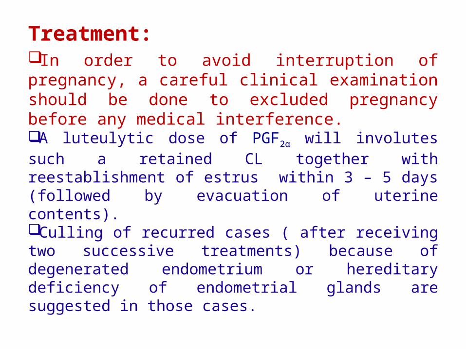
Treatment:In order to avoid interruption of pregnancy, a careful clinical examination should be done to excluded pregnancy before any medical interference. A luteulytic dose of PGF2α will involutes such a retained CL together with reestablishment of estrus within 3 – 5 days (followed by evacuation of uterine contents).Culling of recurred cases ( after receiving two successive treatments) because of degenerated endometrium or hereditary deficiency of endometrial glands are suggested in those cases.

Heifers exhibit persistent CL due to hereditary causes are culled too.
Avoid enucleating deeply embedded retained CL detected in association with pyometra.


![Untitled-1 [imadombivli.com] · Infertility counselling and consulting General women's health screening Hormonal disorders, PCOD clinic Menopause counselling & consulting Eva General](https://img.dokumen.tips/doc/110x75/5f91bca9965f5833580b5e00/untitled-1-infertility-counselling-and-consulting-general-womens-health-screening.jpg)