Embed Size (px)
Citation preview

Department of General HistologyDepartment of General Histology
Practical lessonPractical lesson
TopicTopic: : Nervous system

General informationThe nervous system provides for the control and coordination of all the body's activities.The interaction of the organism with the environment, metabolism, mental activity are under the control of the nervous system.Anatomically, the nervous system is divided into central and peripheral. Physiologically - into somatic and autonomic (vegetative). The autonomic nervous system is the sympathetic and parasympathetic divisions.

Central nervous systemCentral nervous system Central nervous system is represented spinal cord Central nervous system is represented spinal cord
and brain.and brain. Brain and spinal cord consist of gray and white Brain and spinal cord consist of gray and white
matter.matter. The neurons, nerve fibers and glia are the main The neurons, nerve fibers and glia are the main
structural components of the gray matter. The structural components of the gray matter. The white matter is mainly formed by nerve fibers.white matter is mainly formed by nerve fibers.


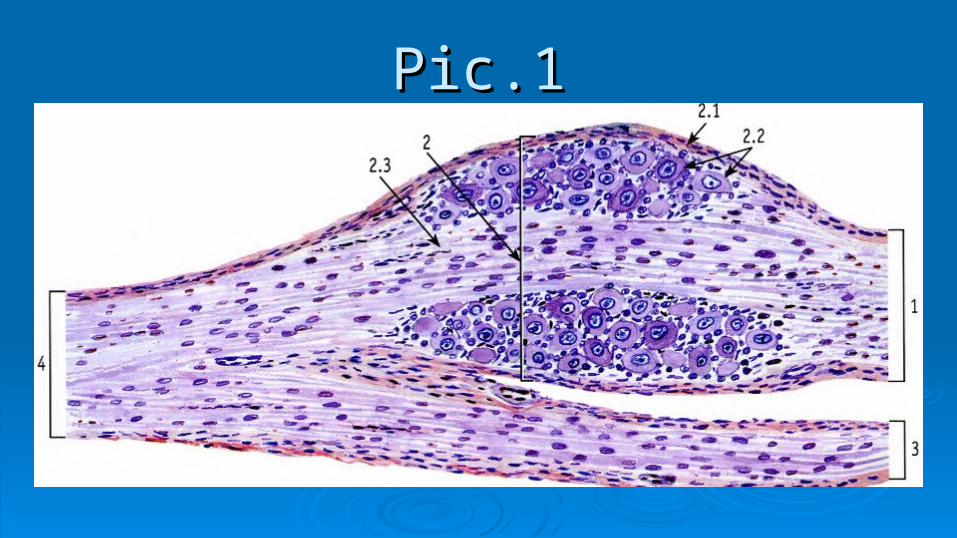
Spinal cordSpinal cord The sensory, motor, and interneurons are found in
specific parts of the spinal cord and nearby structures. Sensory neurons have their cell bodies in the spinal (dorsal root) ganglion. Their axons travel through the dorsal root into the gray matter of the cord. Within the gray matter are interneurons with which the sensory neurons may connect. Also located in the gray matter are the motor neurons whose axons travel out of the cord through the ventral root.

Spinal cordSpinal cord The white matter surrounds the gray matter. It
contains the spinal tracts which ascend and descend the spinal cord. Surrounding both the spinal cord and the brain are the meninges, a three layered covering of connective tissue.

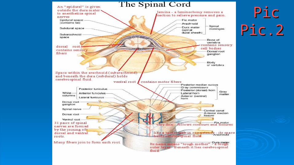
Spinal cordSpinal cord The dura mater is the tough outer layer. Beneath the dura is the
arachnoid which is like a spider web in consistency. The arachnoid has abundant space within and beneath it (the subarachnoid space) which contains cerebrospinal fluid, as does the space beneath the dura mater (subdural space). This cerebrospinal fluid supplies buoyancy for the spinal cord and brain to help provide shock absorption. The pia mater is a very thin layer which adheres tightly to the surface of the brain and spinal cord. It follows all contours and fissures (sulci) of the brain and cord.
Pic.1Pic.1
Pic P Pic.2Pic P Pic.2


Reflex arcReflex arc The basic form of the nervous system is a reflex. Reflex The basic form of the nervous system is a reflex. Reflex
reaction - is answer of the organism to the action of external reaction - is answer of the organism to the action of external irritants or internal environment, which is performed with CNS.irritants or internal environment, which is performed with CNS.
In the nervous tissue of nerve cells in contact with each other, In the nervous tissue of nerve cells in contact with each other, forming a chain of neurons. The chain of neurons forming a chain of neurons. The chain of neurons interconnected by synapses, which provide conducting nerve interconnected by synapses, which provide conducting nerve impulses from sensory receptor neuron to effector end of the impulses from sensory receptor neuron to effector end of the working body. The reflex arc – is way of nerve impulses from working body. The reflex arc – is way of nerve impulses from receptor to effector.receptor to effector.

Reflex arcReflex arc

Brainstem:Brainstem: The brainstem is the region of the brain that connects the cerebrum with the spinal cord. It The brainstem is the region of the brain that connects the cerebrum with the spinal cord. It
consists of the midbrain, medulla oblongata, and the pons. Motor and sensory neurons consists of the midbrain, medulla oblongata, and the pons. Motor and sensory neurons travel through the brainstem allowing for the relay of signals between the brain and the travel through the brainstem allowing for the relay of signals between the brain and the spinal cord. The brainstem coordinates motor control signals sent from the brain to the spinal cord. The brainstem coordinates motor control signals sent from the brain to the body. The brainstem also controls life supporting autonomic functions of the peripheral body. The brainstem also controls life supporting autonomic functions of the peripheral nervous system.nervous system.
Function:Function: The brainstem controls several important functions of the body including: The brainstem controls several important functions of the body including: Alertness, Arousal, Breathing, Blood Pressure, Digestion, Heart RateAlertness, Arousal, Breathing, Blood Pressure, Digestion, Heart Rate
Other Autonomic FunctionsOther Autonomic Functions Relays Information Between the Peripheral Nerves and Spinal Cord to the Upper Parts of Relays Information Between the Peripheral Nerves and Spinal Cord to the Upper Parts of
the Brainthe Brain Location:Location: Directionally, the brainstem is located at the juncture of the cerebrum and the spinal Directionally, the brainstem is located at the juncture of the cerebrum and the spinal
column. It is anterior to the cerebellum.column. It is anterior to the cerebellum.

Brainstem:Brainstem:

The cerebellumThe cerebellum
The cerebellum (Latin for little brain) is a region of the brain The cerebellum (Latin for little brain) is a region of the brain that plays an important role in motor control. The cerebellum that plays an important role in motor control. The cerebellum does not initiate movement, but it contributes to coordination, does not initiate movement, but it contributes to coordination, precision, and accurate timing. It receives input from sensory precision, and accurate timing. It receives input from sensory systems of the spinal cord and from other parts of the brain, systems of the spinal cord and from other parts of the brain, and integrates these inputs to fine tune motor activity. and integrates these inputs to fine tune motor activity. Cerebellar damage does not cause paralysis, but instead Cerebellar damage does not cause paralysis, but instead produces disorders in fine movement, equilibrium, posture, produces disorders in fine movement, equilibrium, posture, and motor learning.and motor learning.

The cerebellumThe cerebellumPic.3 мозжечок1. - молекулярный слой (molecular layer)1.1 - дендриты клеток Пуркинье (dendrites of Purkinje cells)1.2. - афферентные (лазящие) волокна (afferents (scansorial) fibers)1.3. - нейроны молекулярного слоя (ganglionic layer)2. - ганглионарный слой (ganglionic layer)2.1. - тела клеток Пуркинье (the body of Purkinje cells )2.2. - корзинки, образованные коллатералями аксонов корзинчатых клеток (baskets formed axon collaterals basket cells)3. - зернистый слой (granular layer)3.1. - тела клеток зерен ( bodies of cell grains)3.2. - аксоны клеток Пуркинье (Purkinje cell axons)4. - белое вещество ( white matter)

The cerebrum The cerebrum, also known as the telencephalon, is the largest and
most highly developed part of the human brain. It encompasses about two-thirds of the brain mass and lies over and around most of the structures of the brain. The outer portion (1.5mm to 5mm) of the cerebrum is covered by a thin layer of gray tissue called the cerebral cortex. The cerebrum is divided into right and left hemispheres that are connected by the corpus callosum. Each hemisphere is in turn divided into four lobes. The cerebrum or telencephalon, along with the diencephalon comprise the two major divisions of prosencephalon (forebrain).

Function: The cerebrum is involved in several functions of the body including: Determining Intelligence Determining Personality Thinking Perceiving Producing and Understanding Language Interpretation of Sensory Impulses Motor Function Planning and Organization Touch Sensation
The cerebrum

The cerebrum
Location: Directionally, the cerebrum and the cortex that covers it
is the uppermost part of the brain. It is the anterior portion of the forebrain and is superior to other brain structures such as the pons, cerebellum and medulla oblongata.

Pic.4 Pic.4 полушарие большого мозга. Кораполушарие большого мозга. Кора 1 – 1 – pia materpia mater I - I - молекулярный слой (молекулярный слой (molecular layer)molecular layer) II - II - наружный зернистый слой (наружный зернистый слой (external granular layer)external granular layer) III - III - наружный пирамидный слой (наружный пирамидный слой (outer layer of the pyramid)outer layer of the pyramid) IV - IV - внутренний зернистый слой (внутренний зернистый слой (inner granular layer)inner granular layer) V - V - внутренний пирамидный (ганглионарный) слой внутренний пирамидный (ганглионарный) слой
((inner pyramidal (ganglionic) layer)inner pyramidal (ganglionic) layer) VI - VI - слой полиморфных клеток (слой полиморфных клеток (layer of polymorphic cells)layer of polymorphic cells) 2 – gray matter2 – gray matter 3 – white matter3 – white matter