Embed Size (px)
Citation preview

• In cnidarians and flatworms, the gastrovascular cavity functions in both
– digestion
– internal transport
Several types of internal transport have evolved in animals
MECHANISMS OF INTERNAL TRANSPORT
Figure 23.2A
Mouth
Circularcanal
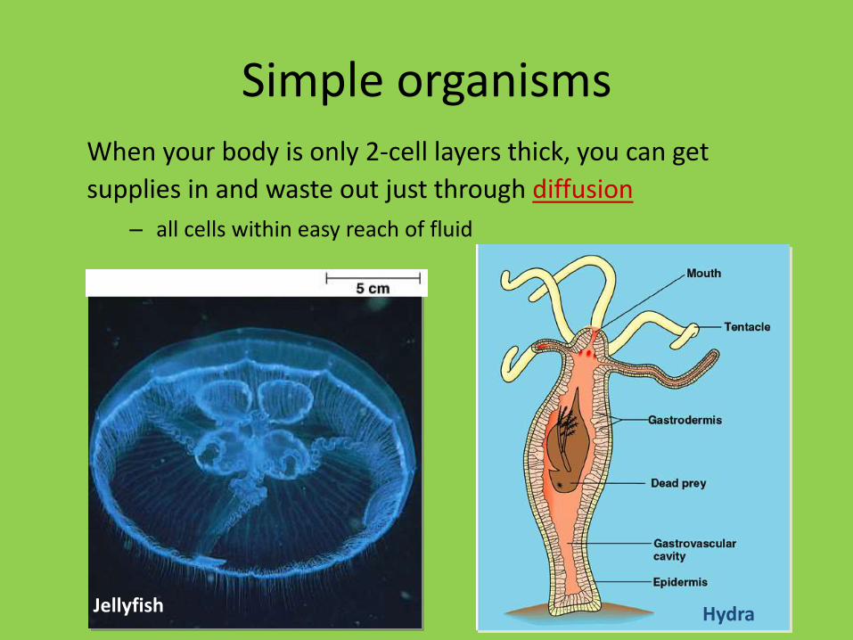
Simple organismsWhen your body is only 2-cell layers thick, you can get
supplies in and waste out just through diffusion
– all cells within easy reach of fluid
HydraJellyfish

Circulatory systems
• All animals have:– muscular pump = heart
– tubes = blood vessels
– circulatory fluid = “blood”
open closed
hemolymph blood

• Most animals have a separate circulatory system, either open or closed
• Open systems
–A heart pumps blood through open-ended vessels into spaces between cells
Figure 23.2B
Pores
Tubular heart
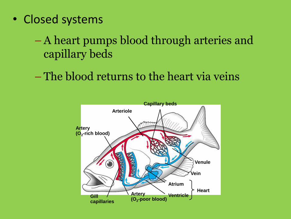
• Closed systems
–A heart pumps blood through arteries and capillary beds
–The blood returns to the heart via veins
Artery(O2-rich blood)
Arteriole
Capillary beds
Venule
Vein
Atrium
VentricleHeart
Artery(O2-poor blood)
Gillcapillaries

Two-chambered heart
• The simplest vertebrate heart is the two-chambered heart, seen in fishes.
• A single atrium receives blood from the body cells. A ventricle sends blood to the gills to collect oxygen.

Three-chambered heart
• Separate atria allow some separation of oxygenated and deoxygenated blood, which was an advantage for land organisms (reptiles, amphibians).
• Though blood can mix in the ventricle, mixing is minimal. Some reptiles have partial separation of the ventricle.

Four-chambered heart
• The four-chambered heart, seen in birds and mammals, allows complete separation of oxygenated and deoxygenated blood.
• Complete separation is necessary to support a fast metabolism found in homeotherms.

Evolution of circulatory system
fish amphibian reptiles birds & mammals
A A
V
V V VV
A AAAA
V
2 chamber 3 chamber 3 chamber 4 chamber
Not everyone has a 4-chambered heart

• The cardiovascular system of land vertebrates has two circuits
• The pulmonary circuit
– conveys blood between the heart and gas-exchange tissues
• The systemic circuit
– carries blood between the heart and the rest of the body
PULMONARYCIRCUIT
A
Systemic capillaries
Lung capillaries
V
Right
SYSTEMICCIRCUIT
A
V
Left

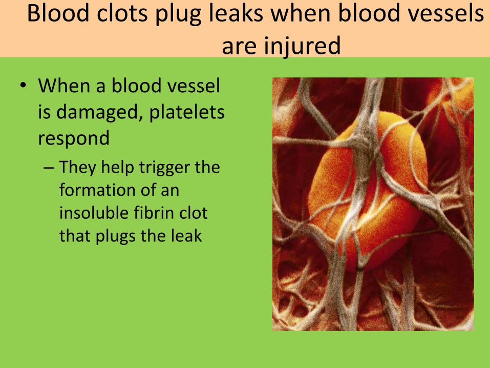
Blood clots plug leaks when blood vessels are injured
Figure 23.16B
• When a blood vessel is damaged, platelets respond
– They help trigger the formation of an insoluble fibrin clot that plugs the leak

Figure 23.16A
Platelet releases chemicalsthat make nearby platelets sticky
Injury to lining of blood
vessel exposes connective
tissue; platelets adhere
1 2 3Platelet plug forms Fibrin clot traps
blood cells
Connectivetissue
Plateletplug
Clotting factors from:
Platelets
Damaged cells
Calcium andother factorsin blood plasma
Prothrombin Thrombin
Fibrinogen Fibrin

Connection: Stem cells offer a potential cure for leukemia and other blood cell diseases
Figure 23.17
• All blood cells develop from stem cells in bone marrow
– Such cells may prove valuable for treating certain blood disorders

BLOOD GROUP SYSTEM
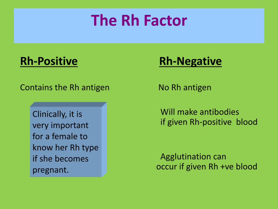
The Rh Factor
Rh-Positive Rh-Negative
Contains the Rh antigen No Rh antigen
Will make antibodies if given Rh-positive blood
Agglutination can occur if given Rh +ve blood
Clinically, it is very important for a female to know her Rh type if she becomes pregnant.

Circulatory SystemBlood Vessels
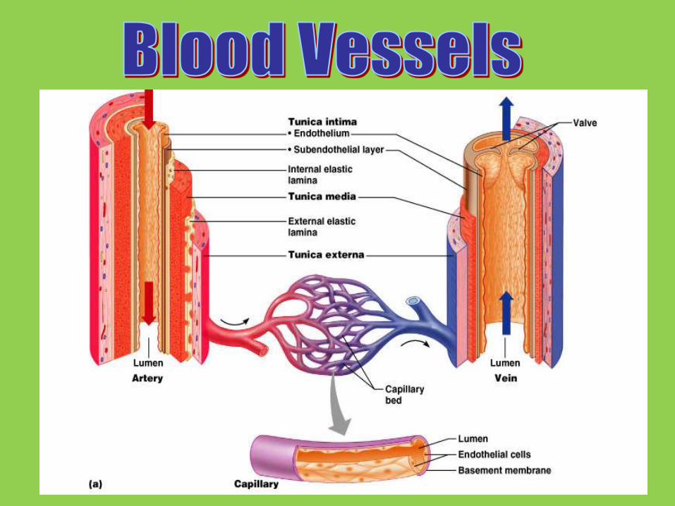

Blood vesselsarteries
arterioles
capillaries
venules
veins
artery
arteriolesvenules
veins

Arteries: Built for their job• Arteries
– blood flows away from heart
– thicker walls
• provide strength for high pressure pumping of blood
– elastic & stretchable
• maintains blood pressure even when heart relaxes

Major arteries
pulmonaryartery
pulmonaryartery =to lungs
aorta carotid = to headto brain & left arm to right arm
coronary arteries
to body

Coronary artery bypass
bypass surgery

Veins: Built for their job• Veins
– blood returns back to heart– thinner-walled
• blood travels back to heart at low speed & pressure
• why low pressure?– far from heart
• blood flows because muscles contract when we move – squeeze blood through veins
– valves in large veins• in larger veins one-way valves
allow blood to flow only toward heart• Presence of deoxygenated blood imparts bluish black
color to veins
Open valve
Blood flowstoward heart
Closed valve

Major Veins
pulmonaryvein =
from lung
superiorvena cava = from upper body
pulmonaryvein = from lung
inferiorvena cava = from lower body

Structure-function relationship
• Capillaries
– very thin walls
– allows diffusion of materials across capillary
• O2, CO2, H2O, food, waste
• Very slow movement
• Artery—arterioles—capillaries--veins--venules
body cell
O2
food
waste
CO2

Location of Heart in Thorax

Layers of Heart
• Epicardium (most superficial)
– Visceral pleura
• Myocardium (middle layer)– Cardiac muscle
– Contracts
• Endocardium (inner)– Endothelium on CT
– Lines the heart
– Creates the valves
Pulmonaryartery
Superiorvena cava
RIGHTATRIUM
Pulmonaryveins
Semilunarvalve
Atrioventricularvalve
Inferiorvena cava
Aorta
Pulmonaryartery
LEFTATRIUM
Pulmonaryveins
Semilunarvalve
Atrioventricularvalve
RIGHTVENTRICLE
LEFTVENTRICLE

RIGHT VENTRICLE
1
23
Capillaries
of right lung
3
Capillaries
of left lung
4
LEFT ATRIUM5
LEFT VENTRICLE
6
Aorta
7Capillaries of
Head and arms
8
Capillaries of
abdominal organs
and legs
9
Superior
vena cava
10
Inferior
vena cava
11
RIGHT ATRIUM
Pulmonary
vein
Aorta
Pulmonary
vein
Pulmonary
artery
Pulmonary
artery


• Diastole
– Blood flows from the veins into the heart chambers
The heart contracts and relaxes rhythmically
Figure 23.6
Heart is
relaxed.
AV valves
are open.
1 2
3
Atria
contract.
Ventricles
contract.
Semilunar
valves
are open.
SYSTOLE
DIASTOLE
0.4 sec
0.1 sec
0.3 sec
• Systole
–The atria briefly contract and fill the ventricles with blood
–Then the ventricles contract and propel blood out
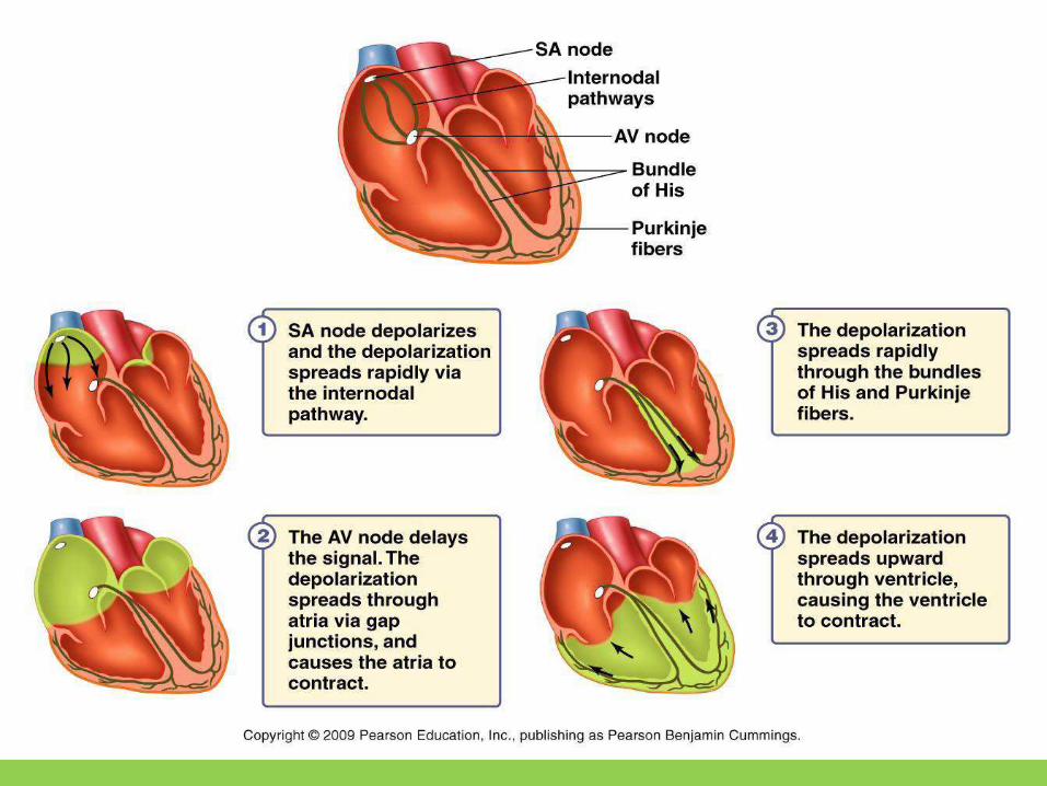
Mammalian Conducting Pathways
SA node initiates the action potential
◦Depolarization spreads rapidly via internodalpathway through the walls of the atria.
• Depolarization reaches atrioventricular (AV) node which communicates signal to the ventricle.
• AV node causes signal delay
◦allows atrium to finish contracting before ventricles contract.

Cardiac Output
Cardiac Output (CO) = the amount of blood that the heart pumps per unit time.
• CO = HR x SV
• Heart rate (HR) =(72) beats per minute
• Stroke volume (SV) = (80cm3)amount of blood pumped per beat

• 2 part system
– Circulation to lungs
• blood gets O2 from lungs
• drops off CO2 to lungs
• brings O2-rich blood from lungs to heart
– Circulation to body
• pumps O2-rich blood to body
• picks up nutrients from digestive system
• collects CO2 & cell wastes
Circulation of Blood
heart
lungs
body
Circulationto lungs
Circulationto body
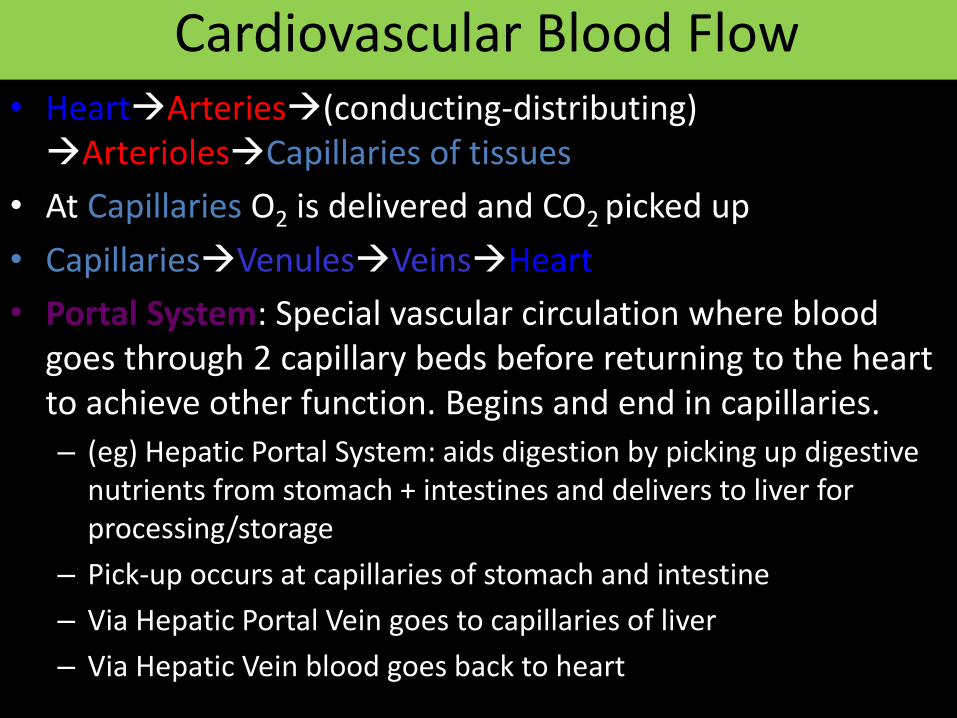
Cardiovascular Blood Flow• HeartArteries(conducting-distributing) ArteriolesCapillaries of tissues
• At Capillaries O2 is delivered and CO2 picked up
• CapillariesVenulesVeinsHeart
• Portal System: Special vascular circulation where blood goes through 2 capillary beds before returning to the heart to achieve other function. Begins and end in capillaries.
– (eg) Hepatic Portal System: aids digestion by picking up digestive nutrients from stomach + intestines and delivers to liver for processing/storage
– Pick-up occurs at capillaries of stomach and intestine
– Via Hepatic Portal Vein goes to capillaries of liver
– Via Hepatic Vein blood goes back to heart

Hepatic Portal System
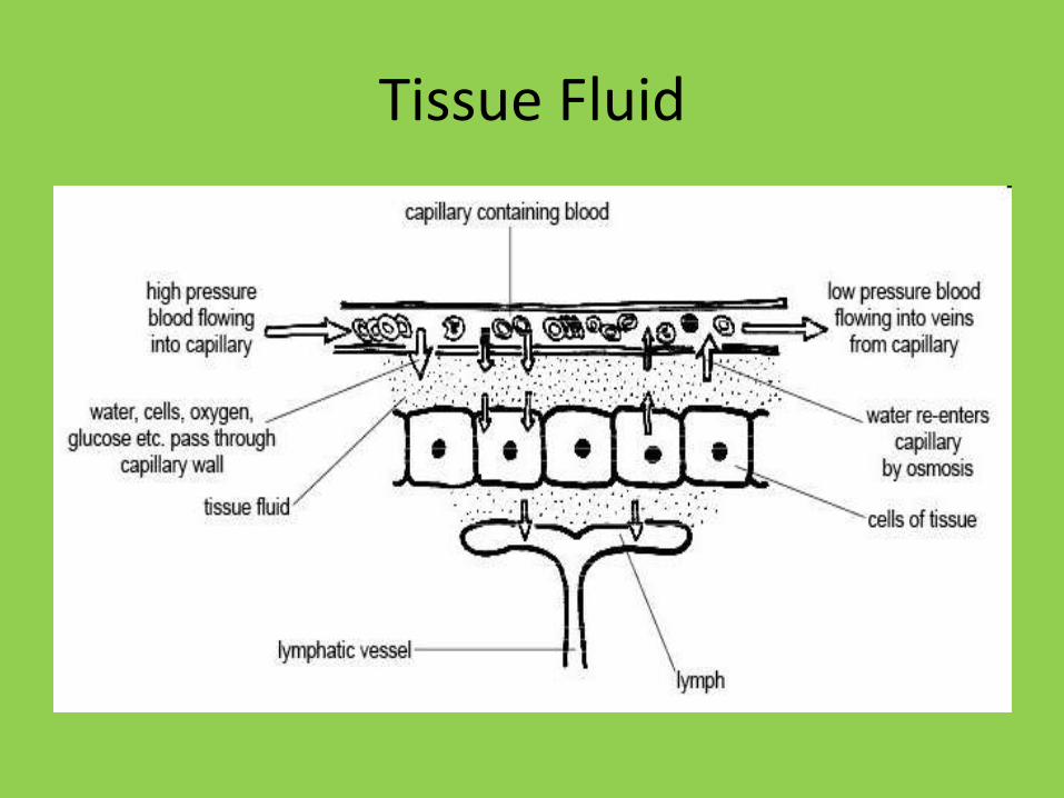
Tissue Fluid
Tissue Fluid
• What is the role of tissue fluid?
It is the fluid which allows the exchange of substances between the blood and cells
• What substances are found in tissue fluid?
glucose, amino acids, fatty acids, salts and oxygen = all delivered to the cells.
carbon dioxide and other waste substances = removed from the cells.

Return of tissue fluid
• Most tissue fluid is returned to the blood plasma via the capillaries. – Hydrostatic pressure at the venule end of the
capillary is higher outside the capillary and tissue fluid is forced back in.
– Osmotic forces (resulting from the proteins in the plasma) pull water back into capillaries.
• Remaining tissue fluid enters the lymph vessels – drain back into the veins close to the heart.

Lymph
• Lymph is moved by:
– Light yellow viscous fluid formed from tissue fluid by special lymph capillaries for passage into venous blood.
– No venous blood, no platelets, lymphocytes and WBC present.
– Contraction of body muscles (aided by valves in the lymph vessels)

42
Lymphatic System
• One way system: to the heart
• Return of collected excess tissue fluid
• Return of leaked protein
• “Lymph” is this fluid
• Edema results if system blocked or surgically removed

Lymph System

44
Lymphoid Organs
• Lymph nodes
• Spleen
• Thymus
• Tonsils
• Small intestine & appendix aggregated lymphoid nodules

45
• Lymph capillaries
– Have one way minivalves allowing excess fluid to enter but not leave
– Picks up bacteria and viruses as well as proteins, electrolytes and fluid
(lymph nodes destroy most pathogens)

46

47
• Lymph capillaries
– Absent from bone, bone marrow, teeth, CNS
– Enter lymphatic collecting vessels
• Lymphatic collecting vessels
– Similar to blood vessels (3 layers), but thin & delicate
– Superficial ones in skin travel with superficial veins
– Deep ones of trunk and digestive viscera travel with deep arteries
– Very low pressure
– Distinctive appearance on lymphangiography
– Drain into lymph nodes

48
• Lymph nodes: bean shaped organs along lymphatic collecting vessels
• Up to 1 inch in size
• Clusters of both deep and superficial LNs


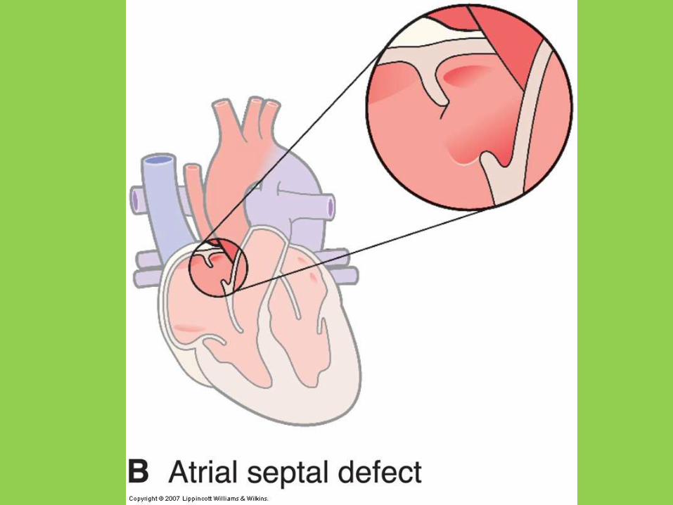
Congenital Heart Disease
• One of most common congenital abnormalities
– 8 in 1000 live births
• Cause usually unknown
• Defects develop in 1st 10 weeks
• Malrotation defects
• Expansion defects
• Septal defects


Malformations with Obstruction to Flow
• Embryonic vessels fail to expand properly
• Coarctation of the aorta
– high BP in arms but low BP in legs
– low blood flow to kidneys
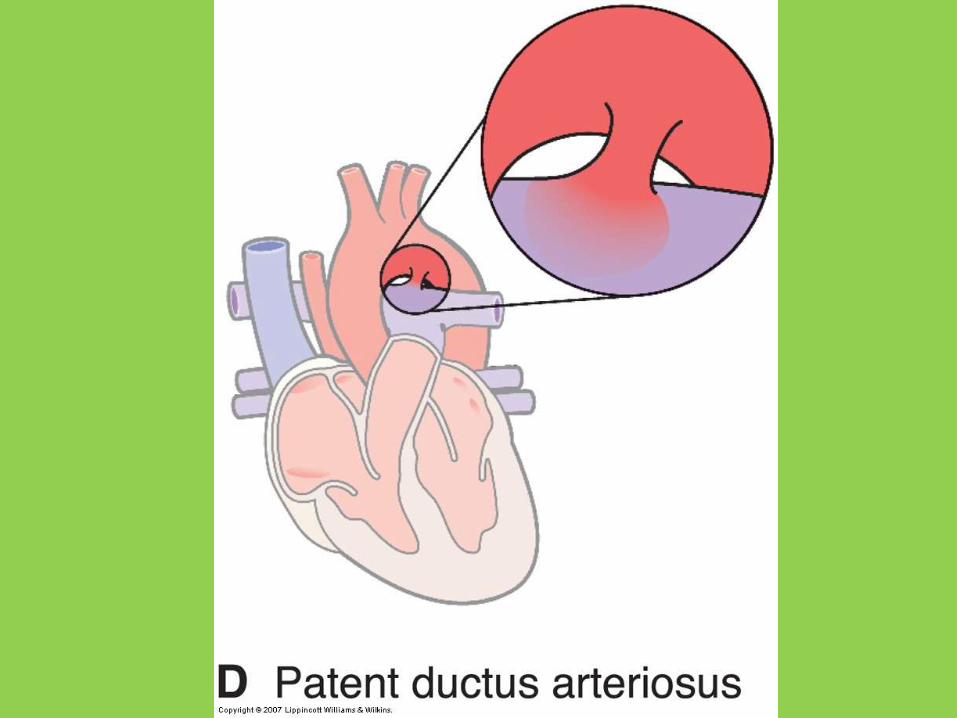
– 50% of cases also have PDA

Pericardial Disease
• Pericarditis
– usually viral infection
– atypical chest pain
– friction rub
• Pericardial effusion
– may occur in noninflammatory conditions
– hemopericardium


Complex Permanent Tissue
A. XYLEM or WOOD
Vascular Tissues -:
Specialized for long-distance transport of water and dissolved
substances.
Contain transfer ceIIs, fibers in addition to parenchyma and
conducting ceIIs
Location- the veins in Ieaves
GW xyIos w/c means “wood”
transports water and dissolved nutrients from the roots to aII parts of a plant.
direction of transport is upward.
Tracheids
Vessels
Xylem fibres or collenchyma
Xylem parenchyma
Living
Dead

• Tracheids
– Characteristics
tapered elongated cells
dead at functional maturity
connect to each other through pits
secondary cell walls strengthened with lignin
– Functions
transport of water & dissolved minerals(?) from cell to cell via pits.
Support
• Vessel Elements– Characteristics
Long tube like and wider than tracheidspossess thinner cell walls than tracheidsAligned end-to-end to form long water-pipesdead at functional maturity
– Functions transport of water plus dissolved mineralssupport

• Xylem fibre: Dead, lignified sclerenchymatous cells supportive in funtion.
• Xylem Parenchyma: Living parenchymatous cells – helps in storage of food
Lateral conduction of sap.
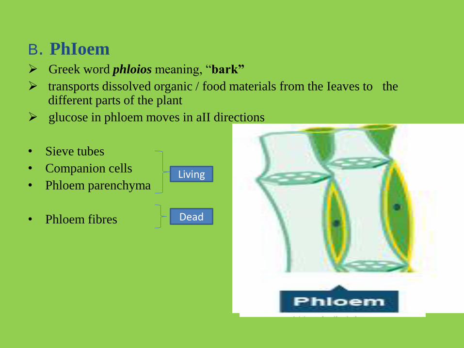
B. PhIoem Greek word phloios meaning, “bark”
transports dissolved organic / food materials from the Ieaves to the different parts of the plant
glucose in phloem moves in aII directions
• Sieve tubes
• Companion cells
• Phloem parenchyma
• Phloem fibres
Living
Dead
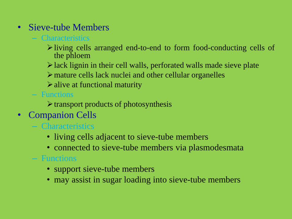
• Sieve-tube Members– Characteristics
living cells arranged end-to-end to form food-conducting cells ofthe phloem
lack lignin in their cell walls, perforated walls made sieve plate
mature cells lack nuclei and other cellular organelles
alive at functional maturity
– Functions
transport products of photosynthesis
• Companion Cells– Characteristics
• living cells adjacent to sieve-tube members
• connected to sieve-tube members via plasmodesmata
– Functions
• support sieve-tube members
• may assist in sugar loading into sieve-tube members


Transport in plants
• Water and mineral nutrients must be absorbed by the roots and transported throughout the plant
• Sugars must be transported from site of production, throughout the plant, and stored

Osmotic potential, solutes, and water movement

Low ΨpHigh Ψp


low ψ
Transpiration
creates tension
higher ψ
cohesion
higher ψ
lower ψhigher ψ
lower ψ higher ψ highest ψ
lower ψ

Cells flaccid/Stoma closedCells turgid/Stoma open
Radially oriented cellulose microfibrils
Cellwall
Vacuole
Guard cell
Changes in guard cell shape and stomatal opening and closing (surface view). Guard cells of a typical angiosperm are illustrated in their turgid (stoma open)and flaccid (stoma closed) states. The pair of guard cells buckle outward when turgid. Cellulose microfibrilsin the walls resist stretching and compression in the direction parallel to the microfibrils. Thus, the radial orientation of the microfibrils causes the cells to increasein length more than width when turgor increases. The two guard cells are attached at their tips, so the increase in length causes buckling.
(a)
H2O
H2O
H2OH2O
H2O
K+
Role of potassium in stomatal opening and closing.The transport of K+ (potassium ions, symbolized here as red dots) across the plasma membrane andvacuolar membrane causes the turgor changes of guard cells.
(b)H2O H2O
H2O
H2O
H2O
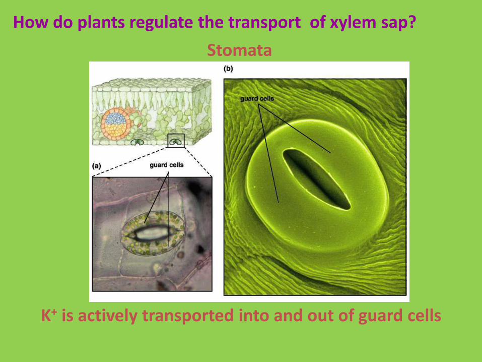
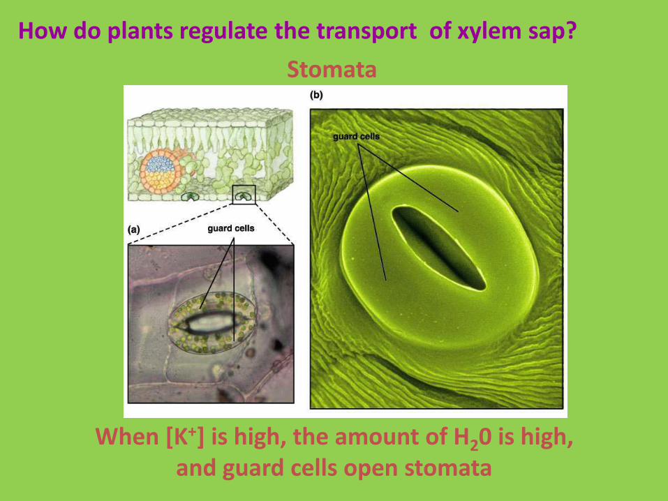
How do plants regulate the transport of xylem sap?
Stomata
K+ is actively transported into and out of guard cells
How do plants regulate the transport of xylem sap?
Stomata
When [K+] is high, the amount of H20 is high, and guard cells open stomata

How do plants regulate the transport of xylem sap?
Stomata
When [K+] is low, the amount of H20 is low, and guard cells close stomata

How do plants regulate the transport of xylem sap?
Stomata
Light stimulates the uptake of K+ by guard cells, opening stomata

How do plants regulate the transport of xylem sap?
Stomata
Low [CO2] stimulates the uptake of K+ by guard cells, opening stomata

How do plants regulate the transport of xylem sap?
Stomata
Low H2O availability inhibits the uptake of K+ by guard cells, closing stomata

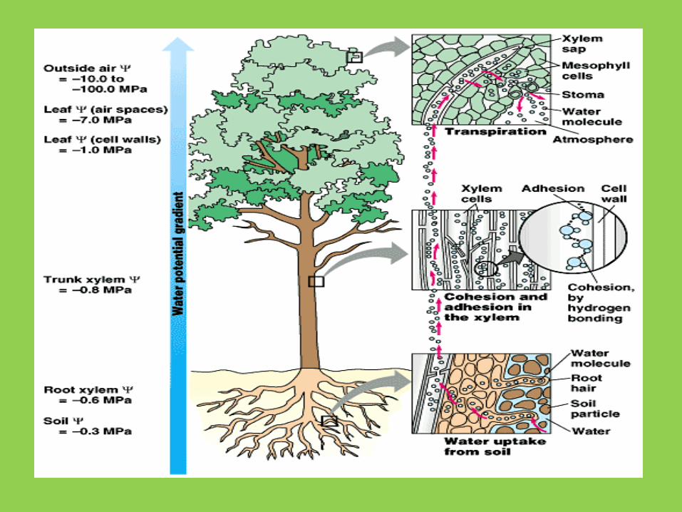
Adhesion and Cohesion Theory
• Cohesion: polar water molecules tend to stick together with hydrogen bonds.
• Adhesion: water molecules tend to stick to polar surfaces.

• Cohesion and adhesion cause water to “crawl” up narrow tubes. The narrower the tube the higher the same mass of water can climb.
• Maximum height: 32 feet.

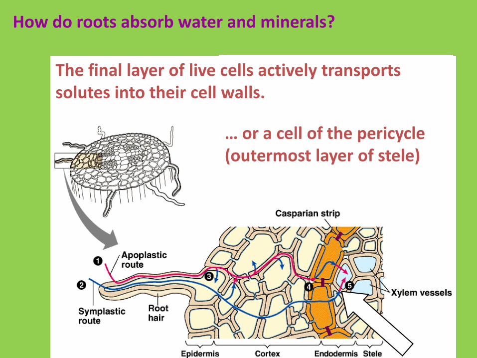
How do roots absorb water and minerals?
• Roots absorb water and minerals in a 4-step process:
– Active transport of minerals into root hairs.
– Diffusion to the pericycle.
– Active transport into the vascular cylinder.
– Diffusion into the xylem.


How do roots absorb water and minerals?
Symplastic route: Active transport occurs through proton pumps, that set up membrane potentials, that drive the uptake of mineral ions

How do roots absorb water and minerals?
Apoplastic route: Some water and dissolved minerals passively diffuse into cell walls

How do roots absorb water and minerals?
Solutes diffuse through the cells (or cell walls) of the epidermis and cortex (the
innermost layer of which is the endodermis)

How do roots absorb water and minerals?
The final layer of live cells actively transports solutes into their cell walls
Solutes then diffuse into xylem vessels to be transported upward

How do roots absorb water and minerals?
The final layer of live cells actively transports solutes into their cell walls
The final layer may be an endodermal cell…
How do roots absorb water and minerals?
The final layer of live cells actively transports solutes into their cell walls.
… or a cell of the pericycle(outermost layer of stele)

Water transport
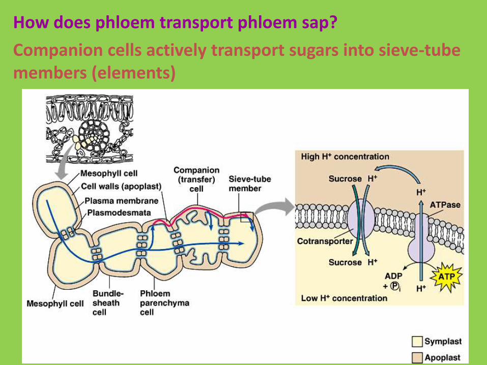
How does phloem transport phloem sap?
Companion cells actively transport sugars into sieve-tube members (elements)

How does phloem transport phloem sap?
Food (sugars) are then translocated from sources to sinks according to the Pressure-Flow Theory:
1. At sources, sugars are actively transported into phloem

How does phloem transport phloem sap?
Food (sugars) are then translocated from sources to sinks according to the Pressure-Flow Theory:
2. Water follows by osmosis from source cells and xylem;
this creates high pressure

How does phloem transport phloem sap?
Food (sugars) are then translocated from sources to sinks according to the Pressure-Flow Theory:
3. At the sink, sugars diffuse out of the phloem and water follows by osmosis;
this creates low pressure
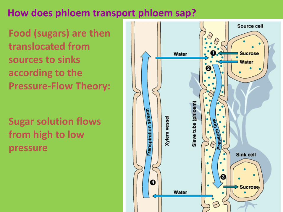
How does phloem transport phloem sap?
Food (sugars) are then translocated from sources to sinks according to the Pressure-Flow Theory:
Sugar solution flows from high to low pressure
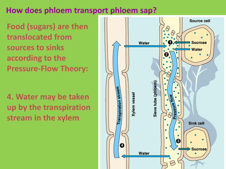
How does phloem transport phloem sap?
Food (sugars) are then translocated from sources to sinks according to the Pressure-Flow Theory:
4. Water may be taken up by the transpiration stream in the xylem

How does phloem transport phloem sap?Pressure-Flow Theory