Embed Size (px)
Citation preview

Ca2+ OMEOSTASIS and BONE TISSUE METABOLISM
CALCIUM
• Present in large amount in the body (1-2 kg in adults)
• More than 99% is contained in the skeleton where it is complexed with phosphate to form hydroxyapatite which has a structural role and confers mechanical stability to the bone
• 1% of the Ca2+ present in the bone tissue is a reserve to maintain a stable concentration of Ca2+ in the extracellular fluids (ECF). There is a continuous dynamic exchange related to chemical equilibrium between bone and ECF.
• It plays important roles of mediation and control of neuromuscular cellular secretion and signal transduction
• Approximately 50% of Ca2+ in the blood is bound to proteins (mainly albumin), the active form is the free one (ionized calcium)
• Its concentration need to be stable to ensure its important functions.

Ca2+ OMEOSTASIS
Determined by several factors:
• Entries
• Intestinal absorption (variable between 0.5 and 1.5 g per day)
• Renal reabsorption (variable between 7.9 and 9.7 g per day, equal to about 98% of the Ca2+ filtered)
• Outputs
• Intestinal excretion (variable between 0.3 and 1 g per day)
• Renal excretion (ranging from 0.15 to 0.3 g per day)
• Exchange between ECF BONE (0.25 - 0.5 g / day)

Copyright © 2015 McGraw-Hill Education. All rights reserved.
From: Bone and Mineral Metabolism in Health and DiseaseHarrison's Principles of Internal Medicine, 19e, 2015
From: Bone and Mineral Metabolism in Health and DiseaseHarrison's Principles of Internal Medicine, 19e, 2015

Skeletal calcium accretion first becomes significant during the third trimester of fetal life, accelerates throughout childhood and adolescence, reaches a peak in early adulthood, and gradually declines thereafter at rates that rarely exceed 1–2% per year.
These slow changes in total skeletal calcium content contrast with relatively high daily rates of closely matched fluxes of calcium into and out of bone (~250–500 mg each), a process mediated by coupled osteoblastic and osteoclastic activity. Another 0.5–1% of skeletal calcium is freely exchangeable (e.g., in chemical equilibrium) with that in the ECF.

The concentration of ionized calcium in the ECF must be maintained within a narrow range because of the critical role calcium plays in a wide array of cellular functions, especially those involved in neuromuscular activity, secretion, and signal transduction. Intracellular cytosolic free calcium levels are ~100 nmol/L and are 10,000-fold lower than ionized calcium concentrations in the blood and ECF (1.1–1.3 mmol/L). Cytosolic calcium does not play the structural role played by extracellular calcium; instead, it serves a signaling function. The steep chemical gradient of calcium from outside to inside the cell promotes rapid calcium influx through various membrane calcium channels that can be activated by hormones, metabolites, or neurotransmitters, swiftly changing cellular function. In blood, total calcium concentration is normally 2.2–2.6 mM (8.5–10.5 mg/dL), of which ~50% is ionized. The remainder is bound ionically to negatively charged proteins (predominantly albumin and immunoglobulins) or loosely complexed with phosphate, citrate, sulfate, or other anions.
Ca2+ REGULATION
CONTROL of Ca2+ OMEOSTASIS
• Parathormone
Hormone produced by parathyroids
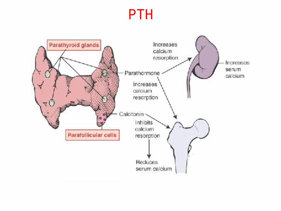
PTH causes the increase of plasma calcium concentration by stimulating both osteoclasts activity and tubular reabsorption of Ca2+
• Calcitonin
Produced by the thyroid C cells
It seems to have a negligible physiological significance
• 1,25-dihydroxycholeclaciferol (Vitamin D)
It is an hormone but it is also present in some foods
It increases intestinal absorption of calcium and its deposition in the bone tissue

PARATHORMONE (PTH)
• PTH is produced by the parathyroid glands (four small glands located posterior to the thyroid)
• Synthesized as pre-proparathormone (115 aa), which is subsequently subjected to enzymatic cuts with the f
ormation of proparathormone (90 aa) and, finally, parathormone (84 aa)
• The active hormone consists of 84 aa, the sequence 1-34 is highly conserved and appears to be involved in receptor
binding• Its secretion is stimulated by the reduction of calcium
concentration in the extracellular fluid and is inhibited by its increase and by 1,25- (OH) 2D
• Circulating hormone is cleared by the liver and the kidney

REGULATION OF PTH SECRETION

• Parathyroid cells exhibit a receptor able to bind Ca2+ (GPCR - G protein coupled receptor)
• The GPCR has an extensive extracellular domain that binds Ca2+, the ion binding activates the receptor that has inhibitory effect on the secretion of PTH; as the amount of Ca2+ bound to the receptor decreases, the inhibition falls causing a ready secretory response of PTH
REGULATION OF PTH SECRETION: THE ROLE OF Ca2+

PTH EFFECTS
PTH actions are mediated by the interaction with the type I receptor (PTH1R); this receptor is able to activate the different mechanisms of signal transduction with different biological effects.
At the level of the bone it determines stimulation of osteoclasts with resorption of the bone matrix with release of Ca2+, proteins and growth factors.
In the kidneys it increases the tubular reabsorption of Ca2+ due to the translocation of Ca2+ channel to the luminal membrane in distal convoluted tubule (DCT).
It increases the activity of the 1-hydroxylase resulting in increased synthesis of 1,25 (OH)2D
PTH

PTH RELATED PEPTIDE (PTHrP)
• PTHrP is produced by different cell types (myocardial cells, smooth muscle, brain, pancreas, lung, breast, endothelium, placenta)Although it’s the product of a different gene PTHrP has several structural and functional homologies with PTH
• It is produced in different isoforms for alternative splicing (139, 141 and 173 aa)
• The portion of the molecule analogous to PTH resides in the first 30 aa
• Its functions are not entirely clear, but appears to have an indispensable role in fetal development, its plasma concentration in healthy adults seems negligible
• Many solid tumors (particularly squamous cell cancers) produce PTHrP
• This protein causes severe clinical manifestations related to cancer (hypercalcemia)

1,25-DIHYDROXYCHOLECALCIFEROL (Vitamin D)
• It is defined vitamin as it is present in some foods but, since it can be synthesized, it is an hormone
• Vitamin D requires activation by double hydroxylation (the first, in position 25, is implemented by the liver and is not subject to any control, the second, in position 1, is operated by the kidney and is controlled by PTH)
• The activity of the 1-hydroxylase is increased by PTH and by the reduction of Ca2+ concentration in ECF while it is inhibited by the product of the reaction (1,25-dihydroxycholecalciferol)
• It is present in the circulation bound to an a-globulin synthesized by the liver and to albumin
• It is inactivated by hydroxylation in position 24 and eliminated in the gall

EFFECTS OF VITAMIN D
It acts by binding its specific receptor (VDR) belonging to the nuclear receptor superfamily
VDR activation is followed by nuclear translocation and induction of the expression of target genes or, depending on the case, repression of the expression of certain genes by blocking of transcription factors
Target organs: Intestinal epithelium: increased expression of calbindin-D9K, increased
expression of membrane Ca2+ channels Bone: it stimulates osteoblasts to express certain genes (osteopontin and
osteocalcin), it promotes the differentiation and activation of osteoclasts Parathyroid: it determines arrest of cell proliferation and arrest of the
transcription of the PTH gene.

Copyright © 2015 McGraw-Hill Education. All rights reserved.
Vitamin D synthesis and activation. Vitamin D is synthesized in the skin in response to ultraviolet radiation and also is absorbed from the diet. It is then transported to the liver, where it undergoes 25-hydroxylation. This metabolite is the major circulating form of vitamin D. The final step in hormone activation, 1α-hydroxylation, occurs in the kidney.
Legend:

Copyright © 2015 McGraw-Hill Education. All rights reserved.
Schematic representation of the hormonal control loop for vitamin D metabolism and function. A reduction in the serum calcium below ~2.2 mmol/L (8.8 mg/dL) prompts a proportional increase in the secretion of parathyroid hormone (PTH) and so mobilizes additional calcium from the bone. PTH promotes the synthesis of 1,25(OH)2D in the kidney, which in turn stimulates the mobilization of calcium from bone and intestine and regulates the synthesis of PTH by negative feedback.
Legend:
• Bone is a dynamic tissue that is remodeled constantly. The arrangement of compact and cancellous (or spongy) bone provides strength and density suitable for both mobility and protection.
• Bone provides a reservoir for calcium, magnesium, phosphorus, sodium, and other ions necessary for homeostatic functions.
• Bone also hosts and regulates hematopoiesis by providing niches for hematopoietic cell proliferation and differentiation.
• The skeleton is highly vascular and receives about 10% of the cardiac output.
• Remodeling of bone is accomplished by two distinct cell types: osteoblasts produce bone matrix, and osteoclasts resorb the matrix
BONE TISSUE

CLASSIFICATION OF BONE BY SHAPE

LONG BONE
Flat bones are bones whose principle function is either extensive protection or the provision of broad surfaces for muscular attachment. These bones are expanded into broad, flat plates, as in the cranium (skull), the ilium (pelvis), the sternum and the rib cage.
FLAT BONE

• The extracellular components of bone consist of a solid mineral phase in close association with an organic matrix, of which 90–95% is type I collagen.
• The noncollagenous portion of the organic matrix is heterogeneous and contains serum proteins (as albumin) and many locally produced proteins, (cell attachment/signaling proteins such as thrombospondin, osteopontin, and fibronectin; calcium-binding proteins such as matrix gla protein and osteocalcin; and proteoglycans such as biglycan and decorin).
BONE PROTEINS

• The mineral phase is made up of calcium and phosphate and is best characterized as a poorly crystalline hydroxyapatite.
• The mineral phase of bone is deposited initially in intimate relation to the collagen fibrils and is found in specific locations in the “holes” between the collagen fibrils. This architectural arrangement of mineral and matrix results in a two-phase material well suited to withstand mechanical stresses.
• The organization of collagen influences the amount and type of mineral phase formed in bone. Although the primary structures of type I collagen in skin and bone tissues are similar, there are differences in posttranslational modifications and distribution of intermolecular cross-links.
BONE STRUCTURE

• Osteoblasts synthesize and secrete the organic matrix and regulate its mineralization.
• They are derived from cells of mesenchymal origin • Active osteoblasts are found on the surface of newly forming bone.
As an osteoblast secretes matrix, which then is mineralized, the cell becomes an osteocyte, still connected with its blood supply through a series of canaliculi.
• Osteocytes account for the vast majority of the cells in bone. They are thought to be the mechanosensors in bone that communicate signals to surface osteoblasts and their progenitors through the canalicular network and thereby serve as master regulators of bone formation and resorption.
BONE CELLS - OSTEOBLASTS

• Osteocytes secrete fibroblast growth factor 23 (FGF23), a major regulator of phosphate metabolism.
• Mineralization of the matrix, both in trabecular bone and in osteones of compact cortical bone (Haversian systems), begins soon after the matrix is secreted (primary mineralization) but is not completed for several weeks or even longer (secondary mineralization).
• Mineralization is a carefully regulated process that is dependent on the activity of osteoblast-derived alkaline phosphatase, which probably works by hydrolyzing inhibitors of mineralization.
OSTEOBLASTS (2)

• Osteoclasts are multinucleated cells that derive from the hematopoietic lineage
• They are formed by fusion of cells derived from the common precursor of macrophages and osteoclasts. Multiple factors that regulate osteoclast development have been identified: factors produced by osteoblasts or marrow stromal cells allow osteoblasts to control osteoclast development and activity. Macrophage colony-stimulating factor (M-CSF) leads to fusion of osteoclast progenitor cells to form multinucleated, active osteoclasts.
• RANK ligand, a member of the tumor necrosis factor (TNF) family, is expressed on the surface of osteoblast progenitors and stromal fibroblasts.
• RANK ligand binds to the RANK receptor on osteoclast progenitors, stimulating osteoclast differentiation and activation. Alternatively, a soluble decoy receptor, referred to as osteoprotegerin, can bind RANK ligand and inhibit osteoclast differentiation.
OSTEOCLASTS (1)

• Several growth factors and cytokines (IL1, IL6, and IL11; TNF; and interferon γ) modulate osteoclast differentiation and function.
• Most hormones that influence osteoclast function do not target these cells directly but instead act on cells of the osteoblast lineage to increase production of M-CSF and RANK.
• Both PTH and 1,25(OH)2D increase osteoclast number and activity by this indirect mechanism.
• Calcitonin directly inhibits osteoclast function. • Estradiol has multiple cellular targets in bone, including osteoclasts,
immune cells, and osteoblasts; actions on all these cells serve to decrease osteoclast number and decrease bone resorption.
OSTEOCLASTS (2)

Copyright © 2015 McGraw-Hill Education. All rights reserved.
Pathways regulating development of (A) osteoblasts and (B) osteoclasts. Hormones, cytokines, and growth factors that control cell proliferation and differentiation are shown above the arrows. Transcription factors and other markers specific for various stages of development are depicted below the arrows. BMPs, bone morphogenic proteins; IGFs, insulin-like growth factors; IL-1, interleukin 1; IL-6, interleukin 6; M-CSF, macrophage colony-stimulating factor; NFκB, nuclear factor κB; PTH, parathyroid hormone; PU-1, a monocyte- and B lymphocyte–specific ets family transcription factor; RANK ligand, receptor activator of NFκB ligand; Runx2, Runt-related transcription factor 2; TRAF, tumor necrosis factor receptor–associated factors; Vit D, vitamin D; wnts, wingless-type mouse mammary tumor virus integration site. (Modified from T Suda et al: Endocr Rev 20:345, 1999, with permission.)
Legend:
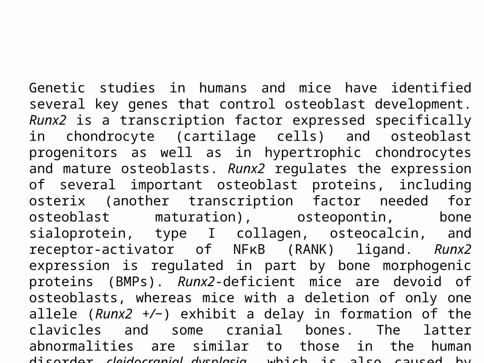
Genetic studies in humans and mice have identified several key genes that control osteoblast development. Runx2 is a transcription factor expressed specifically in chondrocyte (cartilage cells) and osteoblast progenitors as well as in hypertrophic chondrocytes and mature osteoblasts. Runx2 regulates the expression of several important osteoblast proteins, including osterix (another transcription factor needed for osteoblast maturation), osteopontin, bone sialoprotein, type I collagen, osteocalcin, and receptor-activator of NFκB (RANK) ligand. Runx2 expression is regulated in part by bone morphogenic proteins (BMPs). Runx2-deficient mice are devoid of osteoblasts, whereas mice with a deletion of only one allele (Runx2 +/−) exhibit a delay in formation of the clavicles and some cranial bones. The latter abnormalities are similar to those in the human disorder cleidocranial dysplasia, which is also caused by heterozygous inactivating mutations in Runx2.
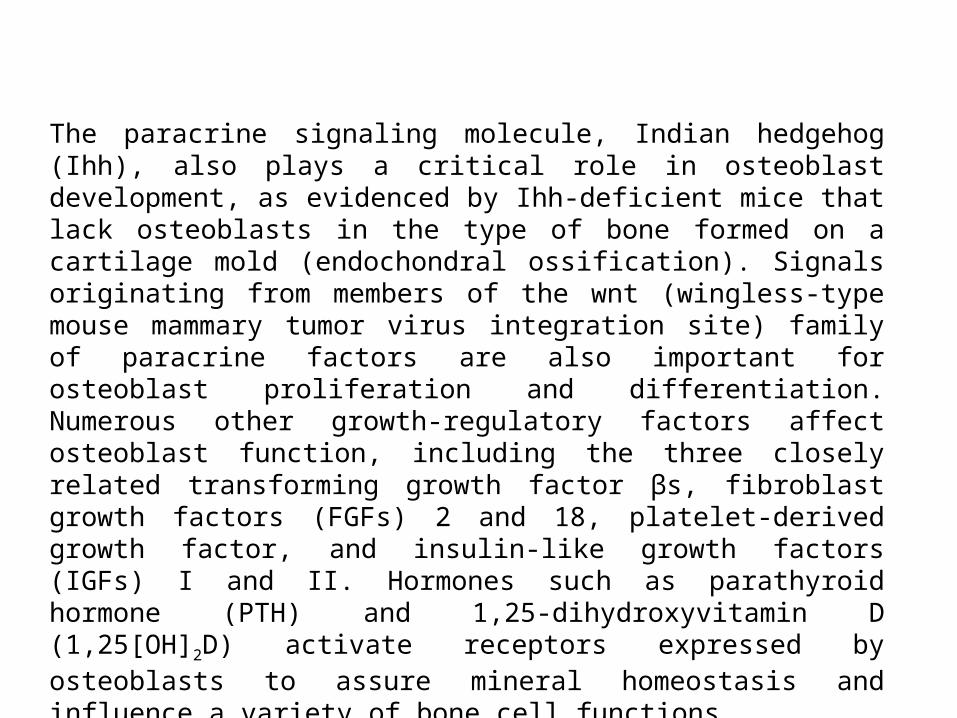
The paracrine signaling molecule, Indian hedgehog (Ihh), also plays a critical role in osteoblast development, as evidenced by Ihh-deficient mice that lack osteoblasts in the type of bone formed on a cartilage mold (endochondral ossification). Signals originating from members of the wnt (wingless-type mouse mammary tumor virus integration site) family of paracrine factors are also important for osteoblast proliferation and differentiation. Numerous other growth-regulatory factors affect osteoblast function, including the three closely related transforming growth factor βs, fibroblast growth factors (FGFs) 2 and 18, platelet-derived growth factor, and insulin-like growth factors (IGFs) I and II. Hormones such as parathyroid hormone (PTH) and 1,25-dihydroxyvitamin D (1,25[OH]2D) activate receptors expressed by osteoblasts to assure mineral homeostasis and influence a variety of bone cell functions

Osteoclast-mediated resorption of bone takes place in scalloped spaces (Howship’s lacunae) where the osteoclasts are attached through a specific αvβ3 integrin to components of the bone matrix such as osteopontin. The osteoclast forms a tight seal to the underlying matrix and secretes protons, chloride, and proteinases into a confined space that has been likened to an extracellular lysosome. The active osteoclast surface forms a ruffled border that contains a specialized proton pump ATPase that secretes acid and solubilizes the mineral phase. Carbonic anhydrase (type II isoenzyme) within the osteoclast generates the needed protons. The bone matrix is resorbed in the acid environment adjacent to the ruffled border by proteases, such as cathepsin K, that act at low pH.
BONE REMODELING (1)

In the embryo and the growing child, bone develops mostly by remodeling and replacing previously calcified cartilage (endochondral bone formation) or, in a few bones, is formed without a cartilage matrix (intramembranous bone formation). During endochondral bone formation, chondrocytes proliferate, secrete and mineralize a matrix, enlarge (hypertrophy), and then die, enlarging bone and providing the matrix and factors that stimulate endochondral bone formation. This program is regulated by both local factors, such as IGF-I and -II, Ihh, PTH-related peptide (PTHrP), and FGFs, and by systemic hormones, such as growth hormone, glucocorticoids, and estrogen.
BONE REMODELING (2)
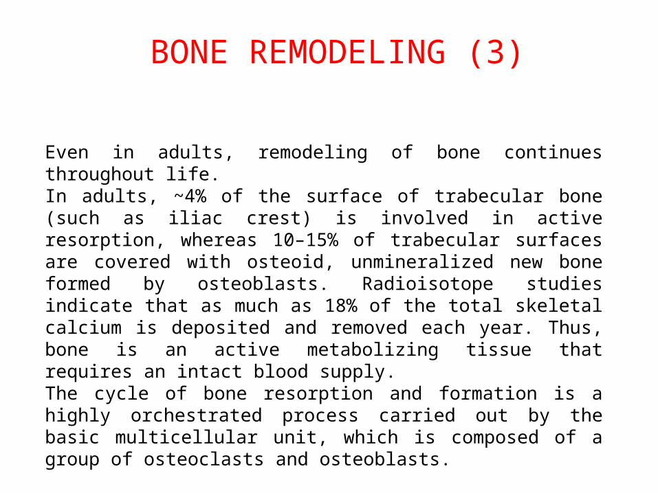
Even in adults, remodeling of bone continues throughout life. In adults, ~4% of the surface of trabecular bone (such as iliac crest) is involved in active resorption, whereas 10–15% of trabecular surfaces are covered with osteoid, unmineralized new bone formed by osteoblasts. Radioisotope studies indicate that as much as 18% of the total skeletal calcium is deposited and removed each year. Thus, bone is an active metabolizing tissue that requires an intact blood supply. The cycle of bone resorption and formation is a highly orchestrated process carried out by the basic multicellular unit, which is composed of a group of osteoclasts and osteoblasts.
BONE REMODELING (3)

Copyright © 2015 McGraw-Hill Education. All rights reserved.
Schematic representation of bone remodeling. The cycle of bone remodeling is carried out by the basic multicellular unit (BMU), which consists of a group of osteoclasts and osteoblasts. In cortical bone, the BMUs tunnel through the tissue, whereas in cancellous bone, they move across the trabecular surface. The process of bone remodeling is initiated by contraction of the lining cells and the recruitment of osteoclast precursors. These precursors fuse to form multinucleated, active osteoclasts that mediate bone resorption. Osteoclasts adhere to bone and subsequently remove it by acidification and proteolytic digestion. As the BMU advances, osteoclasts leave the resorption site and osteoblasts move in to cover the excavated area and begin the process of new bone formation by secreting osteoid, which eventually is mineralized into new bone. After osteoid mineralization, osteoblasts flatten and form a layer of lining cells over new bone.
Legend:
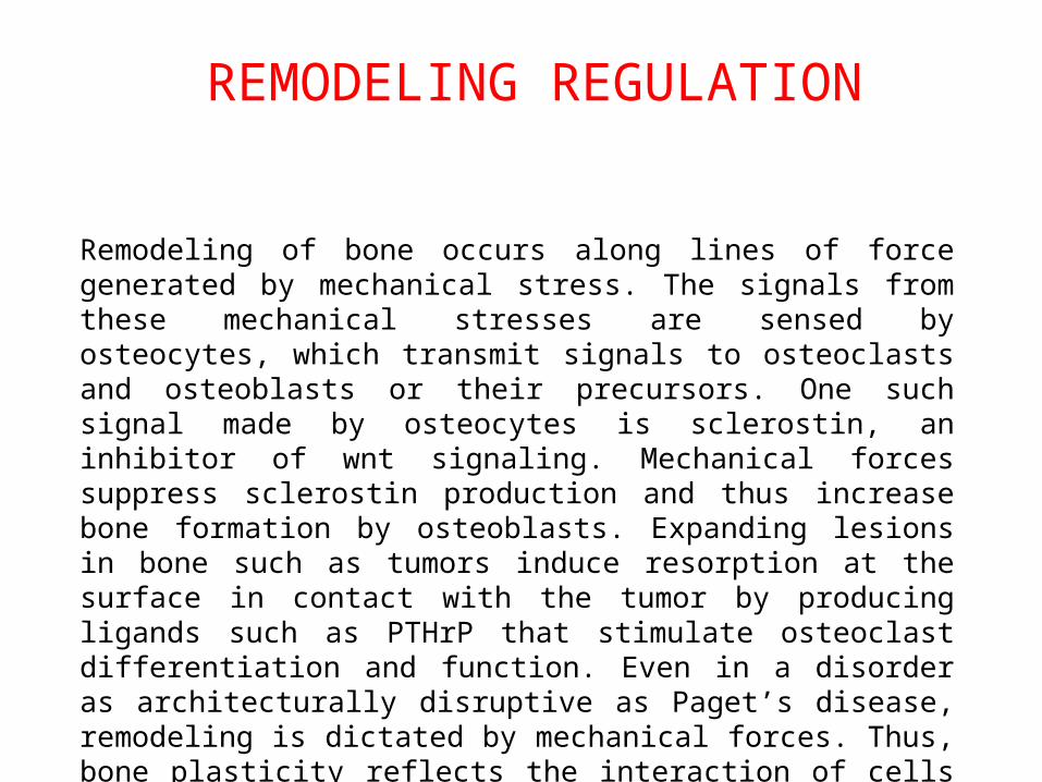
Remodeling of bone occurs along lines of force generated by mechanical stress. The signals from these mechanical stresses are sensed by osteocytes, which transmit signals to osteoclasts and osteoblasts or their precursors. One such signal made by osteocytes is sclerostin, an inhibitor of wnt signaling. Mechanical forces suppress sclerostin production and thus increase bone formation by osteoblasts. Expanding lesions in bone such as tumors induce resorption at the surface in contact with the tumor by producing ligands such as PTHrP that stimulate osteoclast differentiation and function. Even in a disorder as architecturally disruptive as Paget’s disease, remodeling is dictated by mechanical forces. Thus, bone plasticity reflects the interaction of cells with each other and with the environment.
REMODELING REGULATION

RANK/RANKL/OPG
An essential mediator of the formation, function and survival of osteoclasts and bone loss

RANK ligandRANK ligand
• RANK ligand is one of the major mediators of osteoclast activity: it is essential for their differentiation, activation and survival in both cortical and trabecular bone
• RANK ligand is produced by osteoblasts, it binds to and activates its receptor RANK expressed both on osteoclasts and their precursors, resulting in the formation and differentiation of pre-osteoclasts in mature osteoclasts, resulting in activation of bone resorption
• RANK Ligand is also necessary for the adhesion of osteoclasts to the bone surface and for the survival of mature osteoclasts, through the inhibition of apoptosis
• The RANK Ligand exerts a catabolic effect on bone trabecular and cortical bone, which reduces the volume of mineral density and bone strength

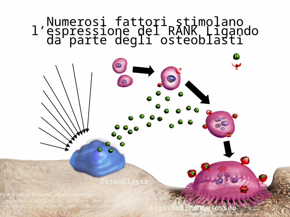
Osteoblasti
Osteoclastiattivati
CFU-M Pre-osteoclasti
Osteoclastimultinucleati
OrmoniFattori di crescitaCitochine
RANKL
RANK
Bone formation
Bone resorptionAdattato da Boyle WJ, et al. Nature. 2003;423:337-342.
In presenza di M-CSF
CFU-M= unità formante colonie macrofagicheM-CSF= fattore stimolante le colonie macrofagiche
Materiale educazionale. Non copiare o distribuire.© 2009 Amgen. Tutti i diritti riservati.
Osteoclast precursor
RANK receptor (red) and ligand (green)
OsteoblastsActivatedOsteoclast
Osteoblasti
TNF-
PTH
IL-1
PTHrP
Glucocorticoidi
Vitamina DPGE2
IL-11
RANKL
RANK
Adattato da Boyle WJ, et al. Nature. 2003;423:337-342.Hofbauer LC, Schoppet Ml. JAMA. 2004;292:490-495.
IL-6
Materiale educazionale. Non copiare o distribuire.
Pre-osteoclastiCFU-M
CFU-M = unità formante colonie macrofagiche
Numerosi fattori stimolano l’espressione del RANK Ligando da
parte degli osteoblastiNumerosi fattori stimolano
l’espressione del RANK Ligando da parte degli osteoblasti
Osteoclastimultinucleati
Osteoclastiattivati
Riassorbimento osseo

OrmoniFattori di crescitaCitochine
RANKL
RANK
OPG
Bone formation
Adattato da Boyle WJ, et al. Nature. 2003;423:337-342.
Inhibition of bone resorption
Inhibition of the formation, function and survival of osteoclasts
CFU-M Pre-osteoclasti
In presenza di M-CSF
Materiale educazionale. Non copiare o distribuire.
Osteoblasti
The osteoprotegerin (OPG) is a decoy receptor that blocks the binding between the RANK ligand and the RANK receptor;
OPG and inhibits the formation, function and survival of osteoclasts
CFU-M= unità formante colonie macrofagicheM-CSF= fattore stimolante le colonie macrofagiche
OPG

CANCER RELATED HYPERCALCEMIA• Increased plasma concentration of Ca2 + in excess of 10.5 mg / dL
(separate into mild, 10.5-11.9, moderate, 12-13.9, and severe> 14 mg / dL)
• It affects 20-30% of cancer patients and is a poor prognostic factor
• Clinical manifestations of hypercalcemia:

There are four known mechanisms that may determine cancer related hypercalcemia:
• Hypercalcemia due to bone metastasis with the development of osteolytic lesions (activation of osteoclasts operated by cytokines)
• Hypercalcemia due to PTHrP production by tumor cells, (called Humoral Hypercalcemia of Malignancy or HHM)
• Hypercalcemia due to production by the tumor of 1-hydroxylase
• Ectopic PTH secretion (rare form)
PATHOPHYSIOLOGY

CANCER RELATED HYPERCALCEMIA


CANCER
+PTHrP

LIMPHOMA