Embed Size (px)
Citation preview

31 Cystic Renal Masses on Computed Tomography
CLINICAL IMAGAGINGAN ATLAS OF DIFFERENTIAL DAIGNOSIS
EISENBERG
DR. Muhammad Bin Zulfiqar PGR-FCPS III SIMS/SHL

• Fig GU 31-1 Benign renal cyst. Nonenhancing left renal mass (C) with a sharply marginated border and a thin wall.

• Fig GU 31-2 Benign renal cyst. High attenuation in the cyst (C) represents hemorrhage.
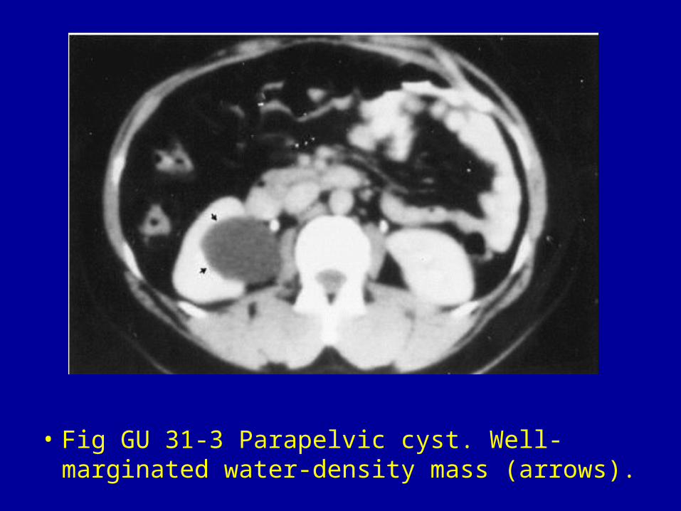
• Fig GU 31-3 Parapelvic cyst. Well-marginated water-density mass (arrows).

• Fig GU 31-4 Polycystic disease. (A) Rim of contrast enhancement in the severely thinned renal parenchyma about the innumerable large renal cysts. (B) Scan at a higher level also shows diffuse cystic involvement of the liver.
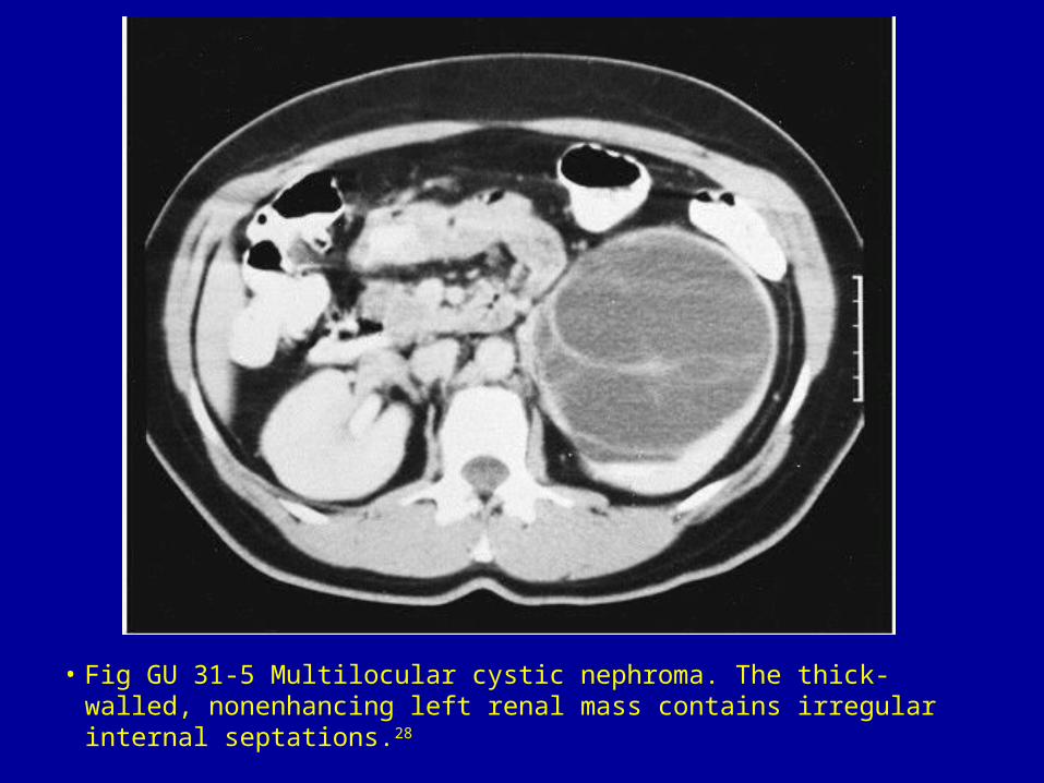
• Fig GU 31-5 Multilocular cystic nephroma. The thick-walled, nonenhancing left renal mass contains irregular internal septations.28
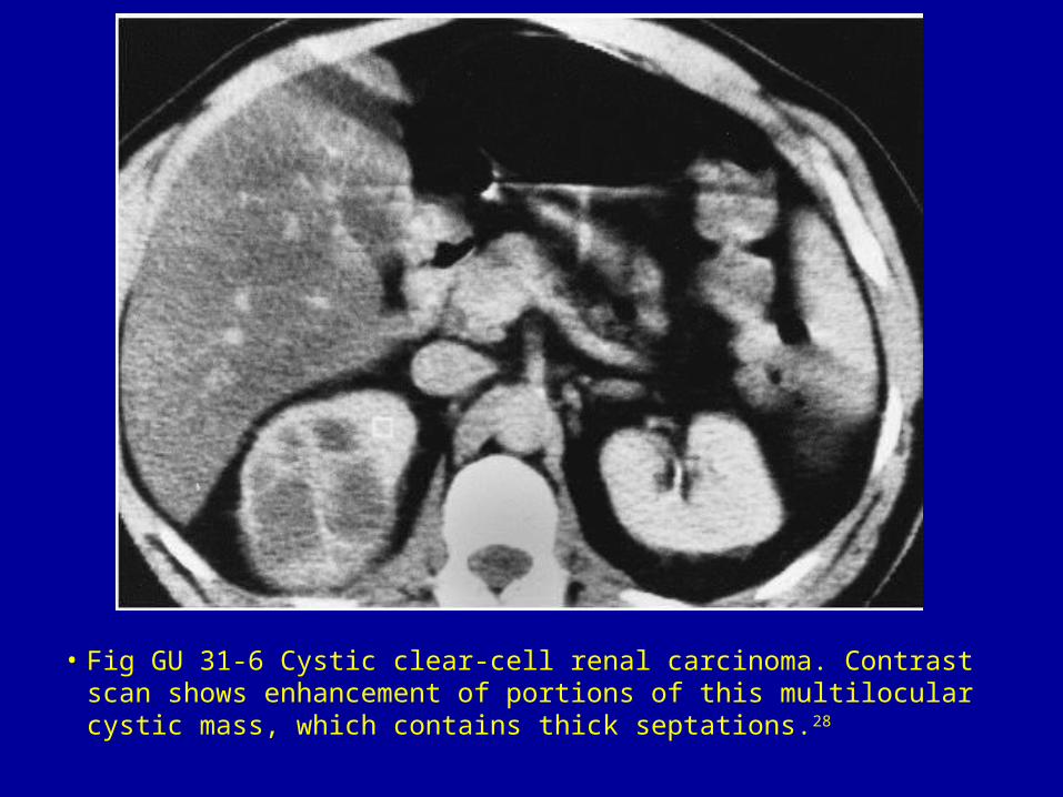
• Fig GU 31-6 Cystic clear-cell renal carcinoma. Contrast scan shows enhancement of portions of this multilocular cystic mass, which contains thick septations.28

• Fig GU 31-7 Necrotic renal cell carcinoma. The huge nonenhancing, cystlike mass (M) has irregular margins (especially on its medial and posterior aspects).

• Fig GU 31-8 Renal abscesses. Multiple nonenhancing lesions in the left kidney of an insulin-dependent diabetic woman with fever of unknown origin, leukocytosis, pyuria, and urine cultures positive for Escherichia coli.28