Embed Size (px)
Citation preview

2 Ill-Defined Breast Masses

CLINICAL IMAGAGINGAN ATLAS OF DIFFERENTIAL DAIGNOSIS
EISENBERG
DR. Muhammad Bin Zulfiqar PGR-FCPS III SIMS/SHL

• Fig MA 2-1 Breast cancer. Magnified coned view demonstrates an ill-defined, irregular mass with radiating spicules.

• Fig MA 2-2 Radial scar. Note the absence of a central mass in this lesion, which was pathologically benign.4

• Fig MA 2-3 Fat necrosis. Mediolateral oblique view obtained 3 months after biopsy shows a dense, spiculated mass associated with architectural distortion and skin retraction and thickening.1
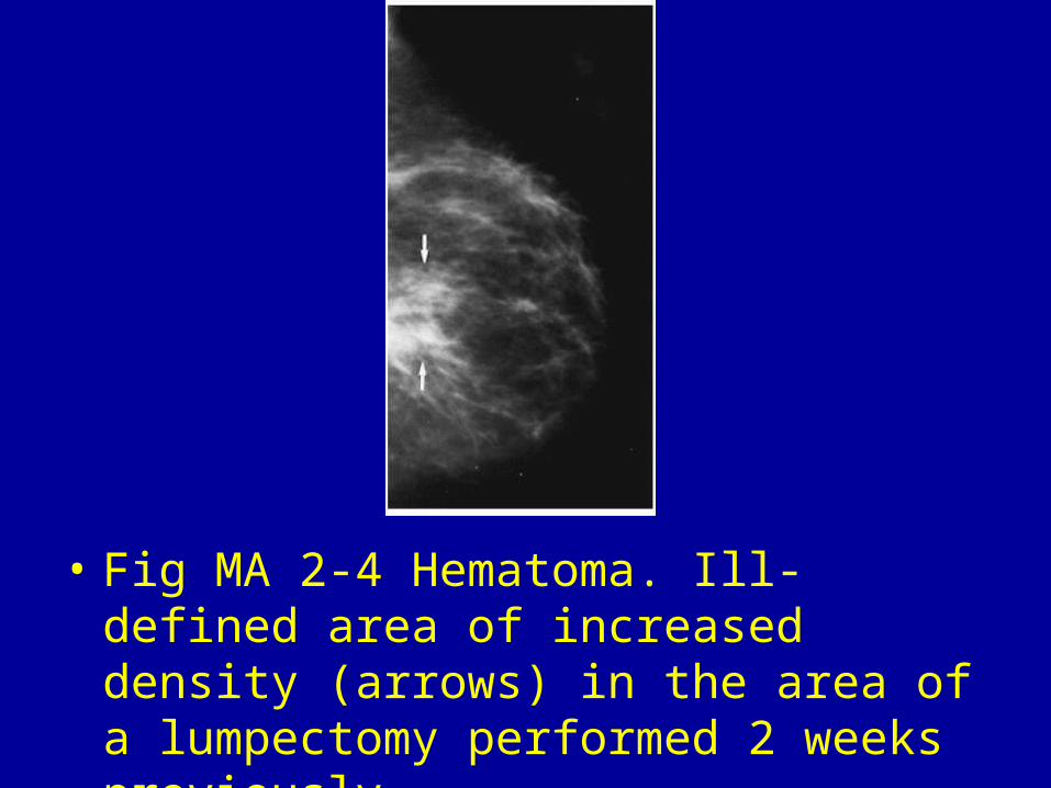
• Fig MA 2-4 Hematoma. Ill-defined area of increased density (arrows) in the area of a lumpectomy performed 2 weeks previously.

• Fig MA 2-5 Fibrocystic disease. Ill-defined density indistinguishable from malignancy. Needle localization and biopsy revealed benign focal fibrosis.3

• Fig MA 2-6 Abscess. Huge, dense, retroareolar mass with unsharp borders associated with nipple retraction and skin thickening over the areola.5
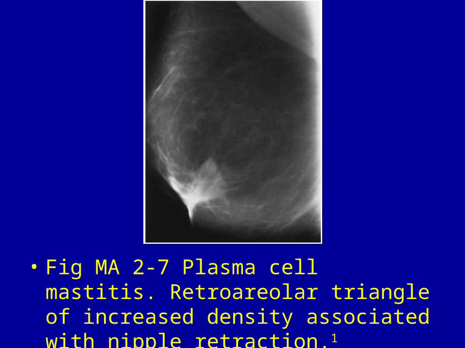
• Fig MA 2-7 Plasma cell mastitis. Retroareolar triangle of increased density associated with nipple retraction.1

• Fig MA 2-8 Granular cell myoblastoma. Ill-defined, low-density nodule (arrow) in an asymptomatic woman.2

• Fig MA 2-9 Fibromatosis. Craniocaudal view from a needle localization procedure shows an ill-defined, somewhat spiculated mass of medium density in the deep central portion of the left breast.2